Embed Size (px)
Citation preview

37
Volume: 7, JANUARY-DECEMBER 2021
INTERNATIONAL JOURNAL OF INNOVATIONS IN APPLIED SCIENCES AND ENGINEERING
Volume: 7, JANUARY-DECEMBER 2021
INTERNATIONAL JOURNAL OF INNOVATIONS IN APPLIED SCIENCES AND ENGINEERING
INTERNATIONAL JOURNAL OF
INNOVATIONS IN APPLIED SCIENCE
AND ENGINEERING
e-ISSN: 2454-9258; p-ISSN: 2454-809X
Using Images for Analysis of Tumors Grading and
Discrimination (Quantitative Texture Analysis
Techniques)
Lubna Emad Kadhim, Nawar Banwan Hassan
Computer Engineering Techniques Department , Imam Al-Kadhum College
(IKC), Baghdad – Iraq
[email protected] , [email protected]
Paper Received: 14th February, 2021; Paper Accepted: 08th March, 2021;
Paper Published: 30th March, 2021
How to cite the article:
Lubna Emad Kadhim, Nawar
Banwan Hassan, Using Images
for Analysis of Tumors
Grading and Discrimination
(Quantitative Texture Analysis
Techniques), IJIASE, January-
December 2021, Vol 7; 37-48
38
Volume: 7, JANUARY-DECEMBER 2021
INTERNATIONAL JOURNAL OF INNOVATIONS IN APPLIED SCIENCES AND ENGINEERING
INTRODUCTION
Unusual growing of cells established within
frame was named as tumors. Brain tumors
were the intra-cranial blocked growth
happens at intervals of The central canals of
the spine or the brain Because the brain is
such a delicate component of the body, brain
tumors were assumed to be a serious and life-
threatening condition (1). Conversely,
Malignant (cancerous) and benign (non-
cancerous) brain tumors were both common.
Treatment for brain tumors is halted pending
proper diagnosis and is dependent on a
number of factors such as the kind of tumor,
its location, size, and development stage.
Magnetic resonance imagines were technique
accustomed mensuration density of gauge
bosons in tissue. It is supported by the
photon's basic feature of rotation and
attractive movement. It is carried out in order
to determine the interior structure of the
frame and to provide better image quality.
(2).
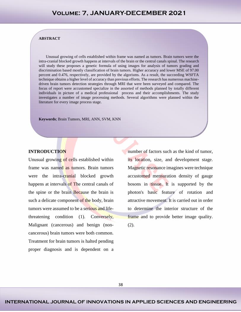
ABSTRACT
Unusual growing of cells established within frame was named as tumors. Brain tumors were the
intra-cranial blocked growth happens at intervals of the brain or the central canals spinal. The research
will study these proposes a genetic formula of using images for analysis of tumors grading and
discrimination based mostly classification of brain tumors. Higher accuracy and lower MSE of 97.00
percent and 0.476, respectively, are provided by the algorisms. As a result, the succeeding WSFTA
technique obtains a higher level of accuracy than previous efforts. The research has numerous machine-
driven brain tumors detection strategies through MRI that were been surveyed and compared. The
focus of report were accustomed specialize in the assorted of methods planned by totally different
individuals in picture of a medical professional process and their accomplishments. The study
investigates a number of image processing methods. Several algorithms were planned within the
literature for every image process stage.
Keywords; Brain Tumors, MRI, ANN, SVM, KNN

39
Volume: 7, JANUARY-DECEMBER 2021
INTERNATIONAL JOURNAL OF INNOVATIONS IN APPLIED SCIENCES AND ENGINEERING
Figure 1 Normal Brain Image and Brain Tumor
Figure 1 show a standard brain picture with
MRI and a tumor image. The key to the
appropriate therapy was early and accurate
tumor identification. Previously, stages of
cancers were identified physically with the
use of image monitoring by doctors;
however, this took longer and the findings
were sometimes incorrect. (3).
There are several forms of brain tumors, and
only a fully competent and knowledgeable
physician can provide an accurate diagnosis.
As a result, researchers are more likely to
require a proper identification a therapeutic
instrument. Detection entails determining the
presence of cancers; segmentation entails
determining the size, mass, and location of
tumors; and classification entails determining
the phase of tumors. In today's world, several
laptop additional tools is employed in
medical area. These tools have chattels of fast
and correct result. The illustrious magnetic
resonance imaging pictures were known as
MRI the be the first managed through
numerous image process steps like histogram
leveling, sharpening filter and so options
were extracted via wavelet or quad tree
rework within the particular feature is Gray
Level Cooccurrence Matrix (GLCM) (4).
The options extracted were utilized in the
content that helps in information cataloging
of unknown pictures. These choices were
regularized within the range of -1 to 1 and
provided as a keyed provision vector
machine classifier.

40
Volume: 7, JANUARY-DECEMBER 2021
INTERNATIONAL JOURNAL OF INNOVATIONS IN APPLIED SCIENCES AND ENGINEERING
The research will study these proposes a
genetic formula of using images for analysis
of tumors grading and discrimination based
mostly classification of brain tumors.
LITERATURE REVIEW
Chaddad A was given (MRI) magnetic
resonance imaging brain tumors cataloging
using support vector machines technique.
Chaddad was developed a replacement
approach for machine-driven designation,
supported classification of resonance (MRI)
human brain pictures. The 2 dimensions
rework and spatial grey level dependence
matrix was employed to extracting features.
Simulated annealing was used to reduce the
amount of alternatives for feature selection.
The next stage To prevent going overboard,
we stratified our method using k-fold cross
corroboration. To enhance support vector
machine (SVM) limitations which the
research would have a tendency to use
genetic formula and support vector machine
simulation. SVM was functional to concept
the classifier. The intelligent arrangement
rate of 89,85 % that be accomplished
operating the support vector machine (SVM)
(5).
Sachdeva J planned brain tumors discovery
utilizing integration based mostly object
labeling formula. Using the K-means method
and the object labeling algorithm, this
approach extracts the tumors.
Correspondingly, The idea of tumor
identification was based on several pre-
management phases (median filtration and
morphological procedure). (6).
It has been established that the proposed
technique's investigative outcomes are
superior to those of other procedures.
Demirhan A had given the economical tactic
for neoplasm detection supported changed a
growing area and (ANN) Neural Network in
magnetic resonance imaging pictures. Pre-
processing, altered region increasing,
characteristic removal of the region, and final
cataloging comprise the technique. The
magnetic resonance imaging image dataset,
which was created from publicly available
data, comprises 39 brain magnetic resonance
imaging images, 19 of which have tumors
and the other twenty of which do not. (7).
The functioning of planned technique was
assessed by bearing in mind the region
increasing formula and therefore the changed
region increasing formula in terms of the
standard rate. The tumors discovery was
appraised through enactment metrics
particularly, sympathy, specificity and
correctness. Utilizing the Feed Forward

41
Volume: 7, JANUARY-DECEMBER 2021
INTERNATIONAL JOURNAL OF INNOVATIONS IN APPLIED SCIENCES AND ENGINEERING
Neural Network (FFNN), Radial Basis
Function (RBF), and neural network,
proportional analyses were performed using
the conventional and therefore altered region
expanding (NN). Beginning with the metrics
obtained, it is discovered that the intended
approach provides superior results in terms of
sympathy, specificity, and precision,
demonstrating its efficacy. Dubey RB was
given an automatic designation system using
wavelet based mostly SFTA Texture options
(8).
The 2 stratified feed advancing neural
networks (NN) were employed for the
cataloging of MRI brain pictures into
traditional or unusual phases. Enactments of
these planned methods were associated with
texture characteristic relating to MSE and
cataloging precision. There is also a greater
accuracy and a lower MSE of 97.00 percent
and 0.476, respectively. As a result, the
succeeding WSFTA technique achieves a far
higher level of accuracy than the prior
studies.
STRUCTURAL DESIGN
Suitable image distinction will offer higher
impact of concept; it's particularly vital for
medical image wherever the slight anatomic
or physical characteristic might cause totally
different diagnostic result. The gray level
values utilized by the pixels inside the bar
graph are frequently used to calculate image
distinctions. It’s typically difficult to
accumulate a numerical image with full-
range gray level utilizing business product
presently offered. The overall method to
provide assistance the aim in numerical
image process is that the histogram-
modification method. Conversely, like as the
tactic of typically ends up in image
deformation (9).
Stages of Digital Image Managing
The following are the expected fundamental
processes in digital image management:
1. Image Acquisition
The MRI scan photos of a specific patient
were first thought-about which were each
color, Gray-image or intensity pictures in this
were shown with a defaulting dimension of
210*210. If a color image was used, a Gray-
scale image was created by employing a big
matrix with a large number of entries were
numerical rates between 0 and 255 depths,
with zero corresponding to black and 255
corresponding to white, for example. The
identification of brain tumors in suspected
patients is divided into two phases: image
division and border finding.

42
Volume: 7, JANUARY-DECEMBER 2021
INTERNATIONAL JOURNAL OF INNOVATIONS IN APPLIED SCIENCES AND ENGINEERING
2. Image Preprocessing
The preprocessing phase changes the image
according to the main level's requirements. It
accomplishes noise filtering inside the
picture. Converting RGB to grayscale and
reshaping are both done here. It features a
noise-reduction average filter. The chances
of noise entering a modern magnetic
resonance imaging scan were quite low.
Because of the heat impact, it will come.
Image Smoothing: it's the action of
simplifying a picture whereas
conserving crucial information. The
objective is to reduce noise and
unnecessary information while
avoiding introducing an excessive
amount of distortion therefore on
change ulterior analysis.
Image Registration: The technique of
aligning two or more pictures
spatially was called image
registration (aligning them). Image
registration in medical imaging
allows for the simultaneous use of
images captured with completely
distinct modalities, at various times,
and with different patient locations.
Pictures were not inheritable before
(pre-operative) or during (intra-
operative) surgery, for example.
Because of time restrictions, the
accuracy of intraoperative imaging is
lower. than preoperative photographs
taken before to operation.
Furthermore, the deformations
occurred spontaneously throughout
the procedure, making it challenging..
3. Image Segmentation
The segmentation stage is crucial for
accurately assessing a picture since it
influences the precision of the subsequent
phases. Correct segmentation, on the other
hand, is difficult due to the fine principles of
lesion forms, dimensions, and colors, as well
as the various skin textures and kinds.
Furthermore, some cancerous tumors have
uneven borders, and there is a swish change
between the lesion and the skin in some
situations. Many strategies were devised to
address this drawback. They will be
classified using edge-based or region-based
techniques, as well as well as monitored and
unattended classification methods:
Threshold segmentation
Water shed segmentation
Gradient Vector Flow (GVF)
K-mean Clustering
Fuzzy C-means Clustering

43
Volume: 7, JANUARY-DECEMBER 2021
INTERNATIONAL JOURNAL OF INNOVATIONS IN APPLIED SCIENCES AND ENGINEERING
Figure 2 Imaging Flow Chart
Start
Read Image
Is Image
Gray
Add Noise
Enhance Use LoG
& Gabar
Detect Edge
Convert to Gray
YES NO
Classification
Decision Normal
Abnormal

44
Volume: 7, JANUARY-DECEMBER 2021
INTERNATIONAL JOURNAL OF INNOVATIONS IN APPLIED SCIENCES AND ENGINEERING
4. Feature Extraction
Characteristics, or the physiognomies of the
objects of interest, if meticulously labeled,
were indicative of the most critical
information that the picture must provide for
a comprehensive description of a blemish.
Feature removal techniques examine and
evaluate things and pictures in order to
remove the most notable alternatives that
were indicative of many kinds object
categories. Options were used as inputs to
classifiers, which assigned them to the
appropriate category. Feature elimination
aims to minimize basic knowledge by
assessing specific assets, or choices, that
distinguish one inputted pattern from
another. By reflecting the contour of the
image's relevant possessions into distinctive
vectors, the extracted feature should provide
the input sort's features to the classifier. We
have a propensity to extract the following
alternatives throughout our intended
procedure.
The process of deciding a collection of
important alternatives for developing By
eliminating the majority of orthogonal and
redundant alternatives from the input, robust
learning models may be created, the
enactment of learning patterns is aided by
feature selection because:
Reducing the effect of the
Dimensional Curse.
Increasing the capacity to generalize.
a learning method that moves at a
rapid pace.
Successful standard interpretability.
CLASSIFICATION METHODS
There were various categorization systems
used to classify the brain as conventional or
aberrant. These categorization techniques
were explained as follows:
1. Artificial Neural Network (ANN)
The picture is plotted into a Neural Network
using the ANN technique. The neural
network process is divided into two parts:
coaching and testing. First and foremost, the
neural network was trained using coaching
examples from the coaching section.
Following training, the neural network is
evaluated on unidentified occurrences. The
feature elimination stage in the neural
network approach is critical. Characteristics
removal was critical since the neural network
relied on the options derived from the input
(ANN). The ANN was split into two classes:

45
Volume: 7, JANUARY-DECEMBER 2021
INTERNATIONAL JOURNAL OF INNOVATIONS IN APPLIED SCIENCES AND ENGINEERING
1. Feed-Forward Neural Network (FFNN).
2. Recurrent Network or Feed-Backward
Network (RN/FBN).
The neurons in (FFNN) Layers were
structured and illegal connections were made
between them, they only generate a single set
of output values. These were characterized as
static networks since the output values were
only supported by the present input. The
output value does not rely on prior input
values. They were sometimes referred to as
memory-less networks.
Bifacial connections exist among the neurons
of the (FBN). Based on the previous input
values, feedback or continuous networks
create a set of values. Because the output
values are always dependent on the prior
input values, A dynamic network is another
term for a feedback network. In a feed-
forward neural network, the back
propagation formula is used. The neurons in
this network were arranged into layers and
sent their output in a forward manner. The
produced errors were returned to the input
layer in a backward way. The network gets
input from neurons in the neural network's
input layer, and the network's output is
provided by the neurons in the neural
network's output layer. A neural network is
made up of one or more intermediate hidden
layers. The supervised learning was used in
the back propagation formula.
The error between the inputs was examined
and back-propagated. With random weights,
the network was trained., which were then
modified via back propagation to get the
lowest error. If the error rate was low, the
network was perfect. The weights were
changed in back propagation whenever the
mistake decreased bit by bit. This happens
again and again till the mistake doesn't
change.
2. Fuzzy C-Means
It's a clumping strategy. One element can
belong to two or more clusters using this
approach, each of which represents a cluster.
During this method, the finite collections of
pixels were split into a bunch of "c" fuzzy
clusters based on a given criterion. The sum
of distances between cluster centers and
patterns is the target function of this formula.
To establish groups based on the data and, as
a result, the application in which it was to be
used, many types of similarity measures were
used. There were several examples of
intensity distance and attribute that might be
used as similarity measures. Following are
the phases of the formula:

46
Volume: 7, JANUARY-DECEMBER 2021
INTERNATIONAL JOURNAL OF INNOVATIONS IN APPLIED SCIENCES AND ENGINEERING
Set the M matrix to zero.
The vectors for the centers are
computed.
Count up to K steps till you reach the
end value.
3. Support Vector Machines (SVM)
The SVM is a supervised classifier with a
learning formula attached to it. The coaching
samples were assisted by SVM. It tries to
minimize the generalization error's certainty.
The generalization error was caused by a
machine error on the check knowledge that
was not employed during the training. As a
result, once the SVM is exposed to
knowledge beyond the coaching set, it
continues to perform effectively. SVM takes
advantage of this and focuses on coaching
instances that are challenging to categorize.
These "borderline" coaching instances,
which were difficult to categorize, are
referred to as vectors of assistance. By
including a statistical process element in the
price operate, the SVM formulation is
somewhat altered. By needing just the
solution of a set of linear equations, it
removes the requirement to tackle a more
complex quadratic programming issue. This
method greatly reduces the time and effort
required to locate the classification matter. It
is supported by the hyper plane, which
optimizes the separation margin between the
two groups. Support Vector Machines
(SVM) is a type of machine that operates in
two stages: coaching and testing. SVM learns
alternatives that are provided to it as inputs to
its learning formula. SVM chooses the proper
margins between its two categories
throughout the coaching phase. For each
disadvantage, an artificial neural network
must solve a set of challenges, such as finding
native minima and selecting a range of
neurons. As a result, there are no native
minima in the SVM classifier. For two-
category problems, SVM might be a
methodical and successful technique. The
SVM classifier was used to divide the
magnetic resonance imaging brain images
into two distinct categories: conventional and
aberrant. The SVM classifier approach
outperforms rule-based methods.
4. K-Nearest Neighbor (KNN)
The k-Nearest Neighbors are
calculated using a KNN formula
based on a Euclidean Distance
function and a decision function.The
geometer distance is the gap metric
employed, and it has more precision
and consistency for magnetic
resonance imaging images than other
classifiers. A sluggish period is

47
Volume: 7, JANUARY-DECEMBER 2021
INTERNATIONAL JOURNAL OF INNOVATIONS IN APPLIED SCIENCES AND ENGINEERING
included in the KNN formula. The
KNN formula's /segmentation phases
were as follows:
The number of nearest neighbors is
determined by the k rate of K.
All of the training examples and the
query case's space were
approximated.
The source of KTH lowest space, the
space was arranged.
The popular period is allocated.
The class was resolute.
The brain abnormalities were
segmented.
CONCLUSION
Several machine-driven brain tumor
detection techniques using MRI were
evaluated and compared in this study. The
focus of this paper was on the many
approaches devised by various persons in the
medical image processing process, as well as
their results. The study focuses on a variety
of image processing methods. For each stage
of the image processing process, several
methods were planned in the literature.
Appendix A details the advantages and
drawbacks of several categorization systems.
REFERENCES
1. Brain tumor classification using neural network
based methods. . Kharat KD, Kulkarni PP. 4, s.l. :
Int J Comput Sci Inform , 2014, Vol. 1.
2. Classification of brain tumor using discrete
wavelet transform, principal component analysis and
probabilistic neural network. Sawakare S,
Chaudhari D. 3, s.l. : Int J Res Emerg Sci Technol,
2015, Vol. 5. 2349-7610.
3. Study of different brain tumor MRI image
segmentation techniques. C, Jadhav. 4, s.l. : Int J
Comput Sci Eng Technol (IJCSET), 2014, Vol. 4.
133–136..
4. Textural features for image classification.
Haralick RM, Shanmugam K, Dinstein I. 2, s.l. :
IEEE Trans Syst Man Cybern, 2011, Vol. 3.
5. Automated feature extraction in brain tumor by
magnetic resonance imaging using Gaussian mixture
models. A, Chaddad. s.l. : Int J Biomed Imaging,
2015, Vol. 11.
6. Segmentation, feature extraction, and multi class
brain tumor classification. Sachdeva J, Kumar V,
Gupta I, Khandelwal N, Ahuja CK. 5, s.l. : J Digit
Imaging, 2013, Vol. 26. 1141–1150.
7. Segmentation of tumor and edema along with
healthy tissues of brain using wavelets and neural
networks. A, Demirhan. 5, s.l. : IEEE J Biomed
Health Inform, 2015, Vol. 32.
8. Region growing for MRI brain tumor volume
analysis. RB, Dubey. 9, s.l. : Indian J Sci Technol ,
2016, Vol. 2.
9. Brain tumor identification using MRI images. MV,
Shinde. 10, s.l. : Int J Recent Innov Trends Comput
Commun, 2016, Vol. 2. 2321-8169.

48
Volume: 7, JANUARY-DECEMBER 2021
INTERNATIONAL JOURNAL OF INNOVATIONS IN APPLIED SCIENCES AND ENGINEERING
Appendix A
ANN Fuzzy SVM KNN algorithm
Advantages The neural
networks have
high parallel
ability and fast
computing.
Expert
intervention is
reduced during
the whole process
It is very simple
and fast algorithm.
This algorithm is
more robust to
noise and
Provides better
segmentation
quality.
This algorithm has
high generalization
performance.
It works well in
case of high
dimensional feature
space.
This algorithm
works independent
of the
dimensionality of
the feature space.
The results given
by support vector
machines are very
accurate.
KNN algorithm is
fairly simple to
implement.
Real time image
segmentation is
done using KNN
algorithm as it runs
more quickly
Disadvantages Some of the
information
should be known
beforehand.
They should be
first trained using
learning process
beforehand.
Period of training
neural networks
may be very long
It considers only
image intensity
values
The training time is
very long.
This algorithm is
highly dependent
on the size of data
There is some
possibility of
yielding an
erroneous decision
if the obtained
single neighbor is
an outlier of some
other class