Embed Size (px)
Citation preview
Contents lists available at ScienceDirect
International Journal of Food Microbiology
journal homepage: www.elsevier.com/locate/ijfoodmicro
Investigating the biocontrol and anti-biofilm potential of a three phagecocktail against Cronobacter sakazakii in different brands of infant formula
Lorraine Endersena, Colin Buttimera, Eoghan Nevina, Aidan Coffeya, Horst Neveb, Hugo Oliveirac,Rob Lavigned, Jim O'Mahonya,⁎
a Department of Biological Sciences, Cork Institute of Technology, Cork, Irelandb Department of Microbiology and Biotechnology, Max Rubner-Institute, Federal Research Institute of Nutrition and Food, 1, Kiel, Germanyc CEB-Centre of Biological Engineering, LIBRO – Laboratório de Investigação em Biofilmes Rosário Oliveira, University of Minho, 4710-057 Braga, Portugald Laboratory of Gene Technology, KU-Leuven, Leuven, Belgium
A R T I C L E I N F O
Keywords:BacteriophagesBiocontrolBiofilmCronobacter sakazakiiInfant formula
A B S T R A C T
In recent years, the microbiological safety of powdered infant formula has gained increasing attention due to theidentification of contaminating C. sakazakii and its epidemiological link with life-threatening neonatalinfections. Current intervention strategies have fallen short of ensuring the production of infant formula thatis free from C. sakazakii. In this study, we describe the isolation and characterisation of three bacteriophages(phages) and their application as a phage cocktail to inhibit the growth of C. sakazakii in different brands ofinfant formula, while also assessing the phages ability to prevent biofilm formation. All three phages, isolatedfrom slurry, possess a relatively broad host range, verified by their ability to infect across genera and species.When all three phages were combined and used as part of a phage cocktail, 73% coverage was obtained across allCronobacter strains tested. Optimum thermo-tolerance and pH stability were determined between 4 °C–37 °C,and pH 6–8, respectively, well within the normal range of application of infant formula. Genome sequencing andanalysis revealed all the phages to be free from lysogenic properties, a trait which renders each favourable forphage therapy applications. As such, the combined-phage preparation (3 × 10⁸ pfu/mL) was found to possess astrong bactericidal effect on C. sakazakii/C. sakazakii LUX cells (≤104 cfu/mL), resulting in a significantreduction in cell numbers, to below the limit of detection (< 10 cfu/mL). This was observed following a 20 hchallenge in different brands of infant formula, where samples in the absence of the phage cocktail reachedconcentrations of ~109 cfu/mL. The phage cocktail also demonstrated promise in preventing the establishmentof biofilm, as biofilm formation could not be detected for up to 48 h post treatment. These results highlight thepotential application of this phage preparation for biocontrol of C. sakazakii contamination in reconstitutedinfant formula and also as a preventative agent against biofilm formation.
1. Introduction
Cronobacter spp. (formally Enterobacter sakazakii) consists of adiverse group of Gram-negative, facultatively anaerobic, motile bacillibelonging to the Enterobacteriaceae family (Iversen et al., 2007). Thegenus comprises seven species: Cronobacter sakazakii, Cronobactermalonaticus, Cronobacter turicensis, Cronobacter universalis, Cronobactermuytjensi, Cronobacter dublinensis and Cronobacter condiment (Bradyet al., 2013; Joseph et al., 2012). In recent years, C. sakazakii hasgained significant attention as an emerging food-borne pathogen due tothe associated link between infectious disease and the consumption ofcontaminated foods, in particular, reconstituted infant milk formula.
While C. sakazakii is responsible for causing severe clinical infections inimmunocompromised individuals of all ages, it is pre-term, low-birthweight infants who are most at risk (FAO/WHO 2008; Healy et al.,2010). Clinical symptoms of infection in infants include meningitis,bacteraemia and severe forms of necrotising enterocolitis, with casefatality rates ranging between 40 and 80% (Friedemann, 2009). Thesehigh mortality rates and the fact that many survivors are very often leftwith chronic neurological and developmental disorders, highlights thedamaging effect this organism has on infant health (Forsythe, 2005; Lai,2001). Accordingly, The International Commission for MicrobiologicalSpecifications for Foods has ranked C. sakazakii as a “Severe hazard forrestricted populations, life threatening or substantial chronic sequelae
http://dx.doi.org/10.1016/j.ijfoodmicro.2017.04.009Received 6 January 2017; Received in revised form 13 April 2017; Accepted 18 April 2017
⁎ Corresponding author at: Department of Biological Sciences, Cork Institute of Technology, Cork, Ireland.E-mail addresses: [email protected] (L. Endersen), [email protected] (C. Buttimer), [email protected] (E. Nevin), [email protected] (A. Coffey),
[email protected] (H. Neve), [email protected] (H. Oliveira), [email protected] (R. Lavigne), [email protected] (J. O'Mahony).
International Journal of Food Microbiology 253 (2017) 1–11
Available online 21 April 20170168-1605/ © 2017 Elsevier B.V. All rights reserved.
MARK
of long duration”, placing the organism in the same category asClostridium botulinum, Cryptosporidium parvum and Listeria monocyto-genes (types A and B) (ICMSF, 2002).
C. sakazakii is ubiquitous in nature with many studies indicatingthat plant material is its primary niche (in particular vegetables, fruits,cereals, wheat, rice, herbs and spices). However, other more pertinentsources also found to harbour this pathogen include powdered infantformula (PIF) and milk powder manufacturing environments(Friedemann, 2007; Kandhai et al., 2004).
C. sakazakii possess physiological traits which affords its ability tosurvive in such environments and thus permit PIF to serve as a primevehicle for transmission to the immunocompromised infant. These traitsinclude, (1) resistance to desiccation and osmotic stress (Breeuweret al., 2003), (2) an extended temperature growth range (Iversen andForsythe, 2004), (3) thermo-tolerance compared to other Enterobacter-iaceae found in PIF (Nazarowec-White and Farber, 1997a, 1997b), and(4) the ability to form biofilms on a range of different materialsincluding polycarbonate which is often used to make babies bottles(Iversen and Forsythe, 2004).
PIF is not manufactured as a sterile preparation and hence canbecome contaminated with C. sakazakii during production. Althoughthe organism is effectively inactivated during pasteurisation(Nazarowec-White and Farber, 1997a, 1997b), contamination is likelyto occur from the addition of non-sterile ingredients during manufac-ture. Indeed, it has been suggested that PIF ingredients originating fromplant material, which have not been heat treated are a potential sourceof C. sakazakii contamination (Healy et al., 2010). Other possiblesources of contamination include the use of non-sterile equipmentduring processing or reconstitution, and from temperature abuse of thereconstituted formula itself (Al-Nabulsi et al., 2009).
Capsular polysaccharides on the outer surface of C. sakazakii cellsplay a central role in biofilm formation, giving the organism the abilityto attach and colonise a variety of surfaces including stainless steel,glass, latex, polycarbonate, silicon and polyvinyl chloride (PVC)(Iversen et al., 2004). The bacterial cells embedded in a matrix ofexopolymeric substances, are physiologically distinct from their plank-tonic counterparts. These cells demonstrate changes in growth rate andgene transcription and often exhibit a significantly higher tolerance toantibiotics (≤1000 times higher) and other sanitising agents. Thepresence of persister cells, the reduced metabolic activity present in theinner layers of the biofilm and the decreased penetration of antibioticsthrough the exopolymeric matrix, all contribute to this increasedresistance (Donlan and Costerton, 2002; Keren et al., 2004). As aresult, complete elimination is very often compromised and re-infectioncommonly occurs.
Consequently, the microbiological safety of PIF is under continuousscrutiny as a result of contaminating C. sakazakii and its epidemiolo-gical link with life-threatening neonatal infections (Forsythe, 2005;Himelright, 2002; Hunter and Bean, 2013). The destructive economicimpact the pathogen has on healthcare systems and PIF productionfacilities due to contaminated product recalls is also apparent (Chenuand Cox, 2009). Indeed, there is a growing requirement for thedevelopment of new and effective mechanisms to further prevent C.sakazakii contamination in both food, and the food processing environ-ment.
Bacteriophages (phages) and their derivatives are well recognisedfor their antibacterial properties, demonstrating promise as natural,safe and effective alternatives for the prevention, treatment and/oreradication of foodborne pathogens in a range of different foods andfood processing environments. These include, decontamination of live-stock, sanitation of contact surfaces and equipment, in addition tobiocontrol of raw meats, fresh foods and vegetables (Endersen et al.,2014; Goodridge and Bisha, 2011), cheese (Carlton et al., 2005), ready-to-eat (RTE) foods (Bigot et al., 2011), skim milk (Ellis et al., 1973;Endersen et al., 2013), and reconstituted infant formula (Kim et al.,2007), all of which demonstrate their applicability for use at each stage
of the food production process. ListShield™, approved for use in the USin 2006 as a processing aid to control Listeria monocytogenes in meat andpoultry products, marked the arrival of the first phage-based product tothe commercial marketplace in the Western world (Bren, 2006).Following this significant development, several phage related productshave been approved for use in the US, including preparations activeagainst the prominent foodborne pathogens, E. coli O157:H7, Salmo-nella, and additional preparations against L. monocytogenes (Endersenet al., 2014; Goodridge and Bisha, 2011). The pioneering anti-listeriaphage-based preparation is now registered in Europe as an organic foodadditive and has also been approved for use as a food processing aid bythe Food Standards Australia & New Zealand, highlighting the fact thatphage-based preparations are continuing to gain global acceptance assafe and effective alternatives for the biocontrol of harmful foodbornepathogens (Fsanz, 2012; Hodgson, 2013).
Here we report the isolation and characterisation of three phagesagainst the opportunistic, foodborne, infant formula pathogen, C.sakazakii. In addition, we demonstrate the efficacy of the phages, usedas part of a phage cocktail, at inhibiting the growth of C. sakazakii infour different brands of reconstituted infant milk formula while alsoassessing the phages ability to prevent biofilm formation.
2. Materials and methods
2.1. Bacterial strains and growth media
The following strains were used in this study: C. sakazakii ATCCBAA 894, C. sakazakii ATCC BAA 894 LUX, C. sakazakii DPC 6258, C.muytjensi ATCC 51329, C. sakazakii ATCC 29004, and C. sakazakii DPC8155. These were sourced from the Dairy Products Research Centre,DPC, Moorepark, Fermoy, Co. Cork, Ireland. Strains were stored at−80 °C and routinely grown on Luria-Bertani (LB) agar, in LB brothand in some cases supplemented with 1% or 2.5% D-(+)-glucose at37 °C. HiChrome™ Cronobacter spp. Agar, Modified (14,763, SigmaAldrich) was used for selective growth of C. sakazakii when necessary.Selective growth of C. sakazakii ATCC BAA 894 LUX was achieved bythe addition of 500 μg/mL erythromycin to LB broth/agar.
2.2. Isolation of phages
Phages were isolated from slurry, sourced from a cattle farmer inClonakilty, West Cork, Ireland, using methods described previously(Carlson, 2005). Briefly, each sample (5 mL) was added to equalvolumes of LB broth (Sigma Aldrich, UK), supplemented with 10 mMCaCl2 (Sigma Aldrich) and inoculated with a mid-log phase C. sakazakiiATCC BAA 894 culture. Samples were incubated overnight at 37 °C withshaking. Samples were centrifuged at 4000g for 15 min to pellet cellsand debris. The supernatant was filtered through a 0.45 μM filter andthe filtrate was re-enriched with mid-log phage C. sakazakii culture twomore times. The supernatant obtained from the final enrichment stepwas filter-sterilised and tested for the presence of viable infectivephages by adding 100 μL of the filtrate to 100 μL of early log phase C.sakazakii cells in a 5 mL LB 0.4% w/v overlay agar tube tempered to50 °C and subsequently poured onto the surface of a LB 1.5% w/v agarplate. Plates were incubated at 37 °C overnight and then examined forphage plaques.
2.3. Phage purification and amplification
Presumptive phages were purified by successive single plaqueisolation and were routinely propagated on C. sakazakii ATCC BAA894 as previously described (O'Flaherty et al., 2005). Briefly, a singleisolated plaque was aseptically picked from a lawn of C. sakazakii ATCCBAA 894 on LB agar using a sterile capillary tube and added to 100 μLof early-log phase culture. The sample was incubated at 37 °C over-night. The resulting lysate was centrifuged, filter sterilised and serially
L. Endersen et al. International Journal of Food Microbiology 253 (2017) 1–11
2
diluted followed by standard plaque assay on C. sakazakii, as describedpreviously. Single plaque purification was repeated twice, after whichpurified phage was recovered. The phage titre was assessed using a spotplaque assay method, where 10 μL of serial dilutions of each phagesuspension were spotted onto C. sakazakii seeded indicator plates.Plates were incubated at 37 °C overnight and spot dilutions weresubsequently examined for the presence of plaques. High titre phagesuspensions were obtained by adding 5 mL of phage buffer (50 mM TrispH 8, 150 mM NaCl, 10 mM MgCl2, 2 mM CaCl2) to a standard plaqueassay plate revealing confluent lysis. Phages were recovered byaseptically running sterile hockey sticks across the surface of the agarplate to physically recover the phages, incubating for 2 h at 37 °C withshaking. The buffer was then recovered from the plate, centrifuged topellet debris and filter sterilised using a 0.45 μm filter. Again, phagetitres were assessed using a spot plaque assay technique as describedpreviously. The purified high-titre phage solutions were stored at 4 °C.
2.4. Phage DNA preparation
Phage DNA isolation and restriction analysis were carried out aspreviously described (Kropinski and Clokie, 2009). Briefly, 18 μL of1 mg/mL DNAse I (Fischer Scientific, 11873795) and 8 μL of 12.5 mg/mL RNase A (Sigma-Aldrich) were added to 1.8 mL aliquots of phagelysate. Samples were mixed and incubated at 37 °C for 30 min in orderto remove host genomic contaminants. Subsequently, 92 μL of 10% SDSand 18 μL of 10 mg/mL proteinase K (Roche) were added. Followingfurther 30 min incubation at 65 °C, proteins were removed by twochloroform steps. Equal volumes of chloroform: isoamyl alcohol: phenol(24:24:1) (Sigma-Aldrich, Dublin, Ireland) was added, mixed thor-oughly and centrifuged at 1500g for 5 min (repeated twice). The upperlayer was carefully removed and transferred to a new sterile tube. Thisstep was repeated using equal volumes of chloroform: isoamyl alcohol(24:1), centrifuged for 5 min at 6000g. DNA was precipitated from thesample using 0.3 M Sodium Acetate solution pH 5.2 and 100% ethanol(2/3 volume of extract) following incubation at room temperature for20 min. Samples were centrifuged for 20 min at 14000g. The DNA waswashed twice with 70% ethanol. DNA pellets were dried in a Rotovap(DNA speed Vac, DNA 110, Savant, Instruments. Inc. Holbrook, NY) onlow power for 2–3 min to remove existing ethanol. DNA was re-suspended in TE buffer pH 7.4 and dissolved at 55 °C for 1–2 h. Thequality and quantity of DNA was assessed by use of Nanodrop spectro-photometer (Nanodrop ND 1000) and running DNA on an agarose gelby electrophoresis followed by visualisation.
2.5. Genome sequencing and annotation
Phage DNA was sequenced with the Illumina MiSeq system at VIBNucleomics Core, Belgium for phages vB_CsaM_leB and vB_CsaM_leNand at the University of Liverpool, Centre for Genomic Research, UK forphage vB_CsaM_leE (herein referred to as leB, leN, and leE, respec-tively). Libraries for phage leB and leN were created with a customNEBNext® Ultra™ DNA Kit and for phage leE the TruSeq DNA Nano LTlibrary Sample Preparation Kit was used. The quality of each librarypreparation was assessed using the Agilent Bioanalyzer and Qubitmeasurements before being sequenced with paired end reads of2 × 250 bp for phage leE and 2 × 150 bp for phage leB and leN.Reads were assembled for phage leB and leN with the CLC BioGenomics Workbench v7.0 (Aarhus, Denmark) and for phage leE withthe Spades genome assembler v.3.10 (St. Petersburg, Russia). Contigswere resolved with average coverage of 964×, 2,182× and 2030× forphages leB, leN and leE, respectively. Open reading frames (ORFs)encoding potential proteins were predicted with Glimmer (http://www.ncbi.nlm.nih.gov/genomes/MICROBES/glimmer_3.cgi) (Delcher et al.,1999) and GenemarkS (http://exon.gatech.edu/genemark/genemarks.cgi) (Besemer et al., 2001). Possible functions of these proteins werepredicted with BLASTP (http://blast.ncbi.nlm.nih.gov/Blast.
cgi?PAGE=Proteins) with tRNA genes being predicted with ARAGORN(http://130.235.46.10/ARAGORN/) (Laslett and Canback, 2004). Lin-ear genome map comparison of the phages was created using Easyfig(Sullivan et al., 2011) with comparison of genome sequences utilisingTBLASTX. Coregenes (Turner et al., 2013) was used for total proteomecomparisons between phages leB, leE, and leN to Enterobacteria phageT4 (BLASTP threshold was set at 75%) with PHACTs being used topredict the lifestyles of phages (http://www.phantome.org/PHACTS/index.php) (McNair et al., 2012).
2.6. Accession numbers
The genomes of all three phages were submitted to Genbank underaccession numbers KX443552, KX431559 B and KX431560 for phageleE, leB and leN, respectively.
2.7. Electron microscopy
Transmission electron microscopy (TEM) was performed as de-scribed previously (Kelly et al., 2012b, Sambrook and Russell, 2006).Briefly, a purified phage stock was prepared using CsCl2 gradient toachieve a titre in excess of 108 pfu/mL. The sample was negativelystained with 2% (w/v) uranyl acetate on freshly prepared carbon films.Grids were analysed using a Tecnai 10 transmission electron micro-scope (FEI Company, Eindhoven, the Netherlands) at an accelerationvoltage of 80 kV. Micrographs were taken with a MegaView G2 CCD-camera (EMSIS, Münster, Germany).
2.8. Host range profiles of phages leB, leE, and leN
The host range of each phage was examined against 21 differentCronobacter/Enterobacter strains, many of clinical origin, which weresourced from the Dairy Products Research Centre, DPC, Moorepark,Fermoy, Co. Cork, Ireland. Host range was determined by standardplaque assay technique as described previously (O'Flaherty et al.,2005).
2.9. Effect of thermal treatment on phage infectivity
For each phage, 3 × 250 μL phage suspensions (~1.0 × 10⁸ pfu/mL) were each placed either in the fridge at 4 °C or in water baths set to37 °C, 45 °C, 55 °C, 60 °C, 72 °C and 90 °C respectively. Following 1 hincubation, 10 μL volumes of serially diluted samples were spotted onC. sakazakii ATCC BAA 894 seeded agar. Plates were left to dry andplaced in the incubator at 37 °C overnight. A thermocouple (Kane-MayKM330, Inlec, UK) was placed in a control tube containing 250 μL ofphage buffer to ensure the desired temperature of the phage suspensionwas achieved for each experiment.
2.10. Effect of pH on phage infectivity
For each phage, 3 × 250 μL aliquots of each phage suspension(~1.0 × 10⁸ pfu/mL) were exposed to different pH values rangingfrom pH 2.0 to pH 10.0. Phages were suspended in MP buffer [50 mMTris pH 8, 150 mM NaCl, 10 mM MgCl2, 2 mM CaCl2], which had beenadjusted to desired pH values of 2.0, 4.0, 6.0, 7.0, 8.0, and 10.0,respectively. The samples was incubated at 37 °C for 1 h, seriallydiluted and spotted (10 μL) onto C. sakazakii ATCC BAA 894 seededagar. Plates were left to dry and the plates were incubated at 37 °Covernight.
2.11. Preparation of phage cocktail
The phage cocktail was prepared by combining equal volumes ofeach individual purified phage solution at ~109 pfu/mL. The high titrephage preparation was stored at 4 °C until needed.
L. Endersen et al. International Journal of Food Microbiology 253 (2017) 1–11
3
2.12. Investigation of the antibacterial potential of the three phage cocktailagainst C. Sakazakii ATCC BAA 894 in four different brands ofreconstituted infant milk formula
The three phage cocktail (~3 × 10⁸ pfu/mL) was inoculated intoeach of the four reconstituted infant milk formulae containing~1 × 10⁴ cfu/mL of C. sakazakii ATCC BAA 894 to a final volume of10 mL, and incubated with shaking at 37 °C for 20 h. Standard platecounts were performed on HiChrome™ Cronobacter spp. agar every 2 hover the 20 h period to quantify surviving cells. This was achieved bysetting up a staggered experiment 12 h apart to account for the hoursthat could not be monitored during the night. PIF was preparedaseptically using sterile water (10% w/v) and mixed to ensure homo-geneity. Positive controls were represented as samples containing theinoculated culture at ~1 × 10⁴ cfu/mL +MP buffer only. Negativecontrols were represented as samples containing reconstituted infantformula only. All experiments were performed in triplicate andbacterial concentrations were expressed as mean cfu/mL counts andstandard deviation. In addition, parallel experiments were performedusing a LUX labelled C. sakazakii ATCC BAA 894 strain supplementedwith 500 μg/mL erythromycin in a microtitre plate assay and lumines-cence was monitored using the in vivo IVIS Imaging System (XenogenInc).
2.13. Biofilm formation
Static microtitre plate assays based on previous studies (Cerca et al.,2007) were used to investigate biofilm formation but with modifica-tions. Briefly, an overnight pure culture of the following strains (C.sakazakii ATCC BAA 894, C. sakazakii ATCC 29004, C. sakazakii DPC6528, C. sakazakii DPC 8155 and C. muytjensi ATCC 51329) were eachsuspended in LB broth to an optical density equivalent of 0.5 McFarlandunits. A selection of different broths (TSB, BHI and LB) supplementedwith 1% and 2.5% D-(+)-glucose and four different brands of infantformula were tested for their ability to aid in biofilm formation. Cellswere subsequently diluted into the appropriate solution to give astarting inoculum of ~1 × 10⁴ cfu/mL. 200 μL of the test sample(BHIglucose(g), LBg, TSBg or PIF) containing the culture was addedto a sterile 96 well plate. 200 μL of test media alone was added as anegative control. All wells were seeded in triplicate. Microtitre plateswere incubated at 37 °C for 48 h to allow biofilm formation. Followingincubation, media was removed and wells were washed three timeswith 200 μL of saline (Ringer's) solution using a multichannel pipette(Gilson) to remove media and planktonic cells. Microtitre plates wereplaced in a 50 °C incubator in an inverted position and allowed to dryfor 1 h. Formed biofilms were stained with 200 μL of 1% crystal violetsolution (Sigma Aldrich, UK) for 10 min at room temperature. The wellswere washed to remove excess dye and left to dry before visuallycomparing the intensity of staining in the wells.
2.14. Biofilm prevention with phage cocktail
Following the identification of two C. sakazakii strains (C. sakazakiiATCC BAA 894, and C. sakazakii DPC 6528) that were capable offorming strong biofilms when grown in different types of infant formula(outlined above, 2.13.), the ability of the phage cocktail to preventbiofilm formation by these strains was investigated using a platestaining and live cell count assay.
2.14.1. Plate staining assayThe plate staining assay was performed as per the biofilm formation
method described above with the following adjustments. At the start ofthe assay, 50 μL of the phage cocktail (~3 × 10⁸ pfu/mL) was added to200 μL of C. sakazakii culture in one set of wells in the microtitre plate,while 50 μL of MP buffer was added to a second set of wells alsocontaining 200 μL of C. sakazakii culture. PIF alone was used as anegative control. The plates were incubated at 37 °C for 48 h, afterwhich the media was poured off and biofilms were stained as describedabove. The stain was removed and the wells were washed gently. Theplates were left to dry, after which 30% glacial acetic acid was added tosolubilise the stain. The biofilm prevention ability of the phage cocktailwas assessed by examining the optical density of the wells spectro-photometrically at OD590nm.
2.14.2. Live cell count assayThe live cell count assay was performed as per the plate staining
method with the following modifications. At the end of the 48 hincubation period, the media was removed and biofilms were washedthree times with ringers to remove media and planktonic cells. Biofilmswere resuspended by mechanical scraping in 100 μL of saline solution(Ringer's) and immediately diluted and plated on to HiChrome™Cronobacter spp. agar plates to determine cfu/mL counts.
3. Results
3.1. Phage isolation and morphology
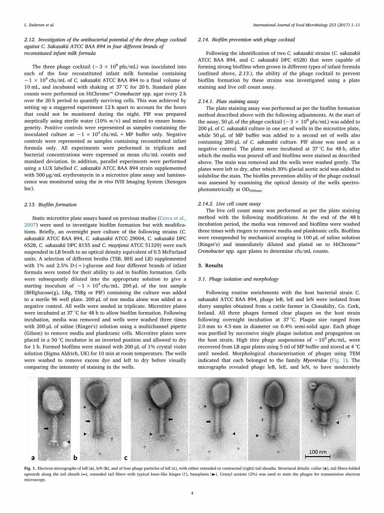
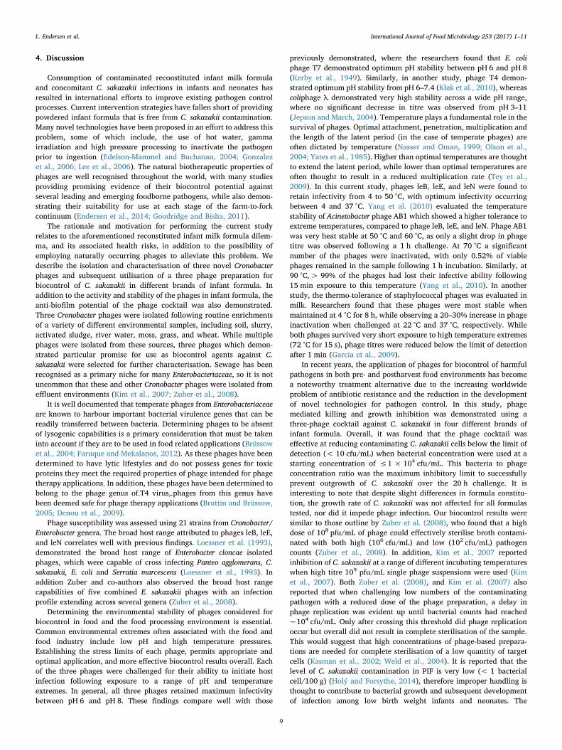
Following routine enrichments with the host bacterial strain C.sakazakii ATCC BAA 894, phage leB, leE and leN were isolated fromslurry samples obtained from a cattle farmer in Clonakilty, Co. Cork,Ireland. All three phages formed clear plaques on the host strainfollowing overnight incubation at 37 °C. Plaque size ranged from2.0 mm to 4.5 mm in diameter on 0.4% semi-solid agar. Each phagewas purified by successive single plaque isolation and propagation onthe host strain. High titre phage suspensions of ~109 pfu/mL, wererecovered from LB agar plates using 5 ml of MP buffer and stored at 4 °Cuntil needed. Morphological characterisation of phages using TEMindicated that each belonged to the family Myoviridae (Fig. 1). Themicrographs revealed phage leB, leE, and leN, to have moderately
Fig. 1. Electron micrographs of leB (a), leN (b), and of four phage particles of leE (c), with either extended or contracted (right) tail sheaths. Structural details: collar ( ), tail fibers foldedupwards along the tail sheath ( ), extended tail fibers with typical knee-like hinges (↑), baseplates (►). Uranyl acetate (2%) was used to stain the phages for transmission electronmicroscopy.
L. Endersen et al. International Journal of Food Microbiology 253 (2017) 1–11
4
elongated heads accompanied by contractile tails, indicating that eachbelongs to the family Myoviridae, subtype A2 (Ackermann, 2001).Phage dimensions are represented in Table 1.
3.2. Results from genome sequencing and annotation
Through sequencing, genomes with sizes of 177,907 bp, 181,570 bpand 179,516 bp were obtained for phages leB, leE and leN, respectively,with a G + C% content of 45%. Following bioinformatic annotation atotal number of 281 ORFs, 284 ORFs and 286 ORFs were predicted forphages leB, leE and leN, respectively, with it being possible to annotateabout 40% (107 ORFs, 111 ORFs and 109 ORFs for leB, leE and leN,respectively) of their ORFs with a possible function (SupplementaryTable 1). Additionally, one tRNA gene for glycine was predicted to bepresent in the genomes of all three phages (see supplemental data).
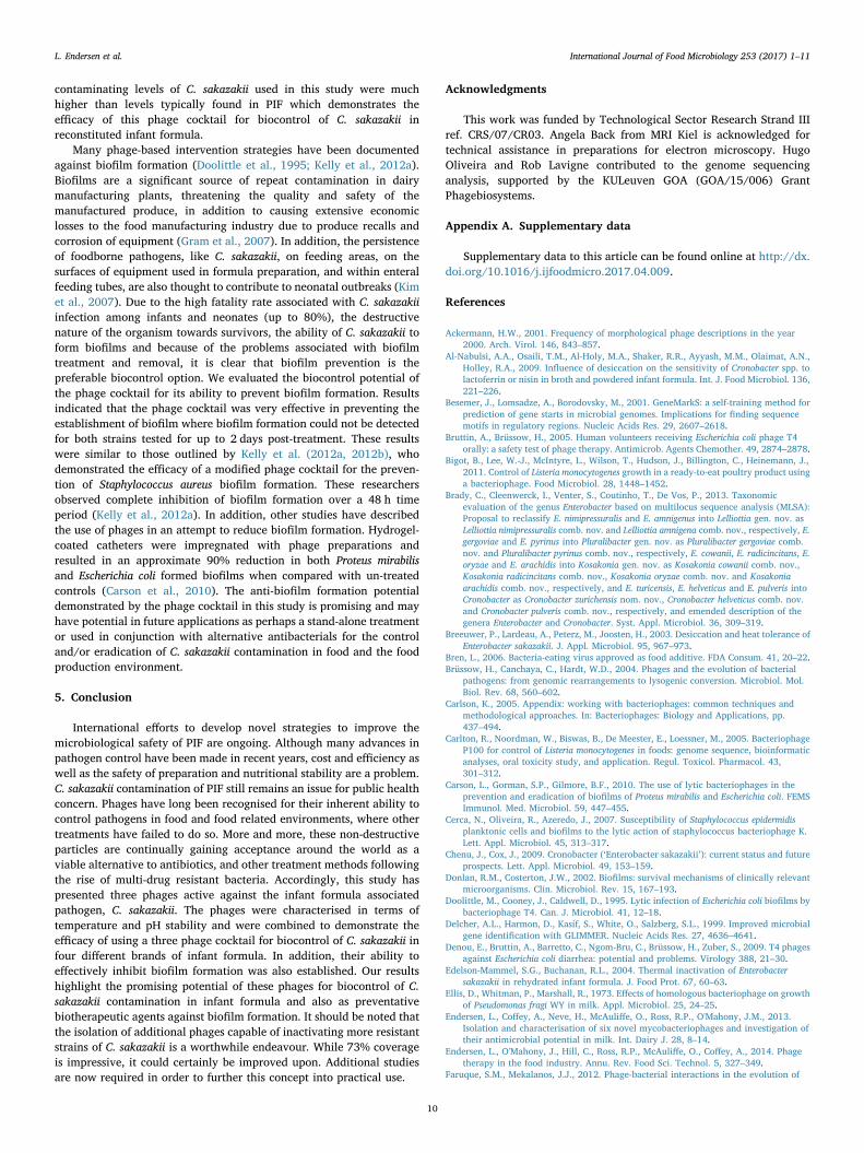
The genomes of these phage were found to be highly similar to eachother (99% identity with 94–99% coverage) with their closest knownrelatives at the nucleotide level being found to be T4 like phagesCronobacter phage GAP161 [JN882287.1] and Citrobacter phageMargaery [KT381880.1] (98–99% identity with 92–97% coverage).
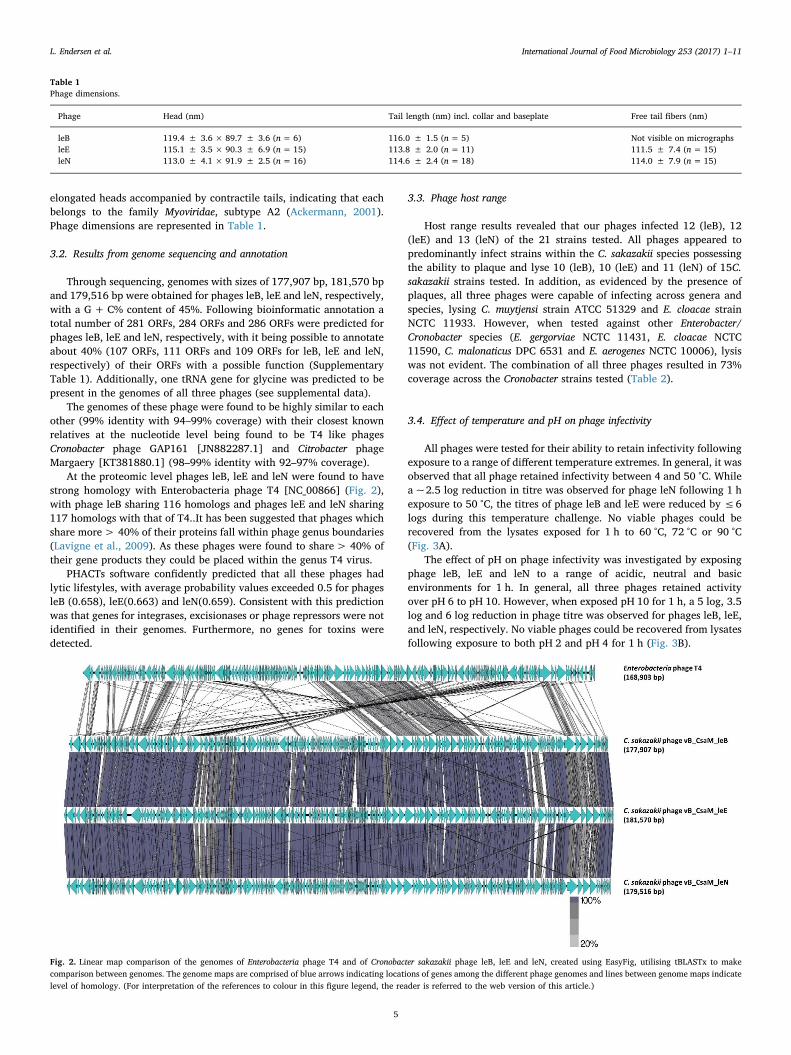
At the proteomic level phages leB, leE and leN were found to havestrong homology with Enterobacteria phage T4 [NC_00866] (Fig. 2),with phage leB sharing 116 homologs and phages leE and leN sharing117 homologs with that of T4..It has been suggested that phages whichshare more> 40% of their proteins fall within phage genus boundaries(Lavigne et al., 2009). As these phages were found to share> 40% oftheir gene products they could be placed within the genus T4 virus.
PHACTs software confidently predicted that all these phages hadlytic lifestyles, with average probability values exceeded 0.5 for phagesleB (0.658), leE(0.663) and leN(0.659). Consistent with this predictionwas that genes for integrases, excisionases or phage repressors were notidentified in their genomes. Furthermore, no genes for toxins weredetected.
3.3. Phage host range
Host range results revealed that our phages infected 12 (leB), 12(leE) and 13 (leN) of the 21 strains tested. All phages appeared topredominantly infect strains within the C. sakazakii species possessingthe ability to plaque and lyse 10 (leB), 10 (leE) and 11 (leN) of 15C.sakazakii strains tested. In addition, as evidenced by the presence ofplaques, all three phages were capable of infecting across genera andspecies, lysing C. muytjensi strain ATCC 51329 and E. cloacae strainNCTC 11933. However, when tested against other Enterobacter/Cronobacter species (E. gergorviae NCTC 11431, E. cloacae NCTC11590, C. malonaticus DPC 6531 and E. aerogenes NCTC 10006), lysiswas not evident. The combination of all three phages resulted in 73%coverage across the Cronobacter strains tested (Table 2).
3.4. Effect of temperature and pH on phage infectivity
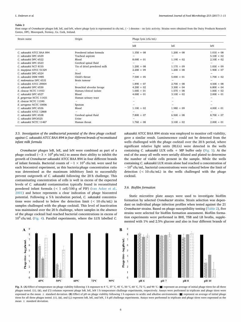
All phages were tested for their ability to retain infectivity followingexposure to a range of different temperature extremes. In general, it wasobserved that all phage retained infectivity between 4 and 50 °C. Whilea ~2.5 log reduction in titre was observed for phage leN following 1 hexposure to 50 °C, the titres of phage leB and leE were reduced by ≤6logs during this temperature challenge. No viable phages could berecovered from the lysates exposed for 1 h to 60 °C, 72 °C or 90 °C(Fig. 3A).
The effect of pH on phage infectivity was investigated by exposingphage leB, leE and leN to a range of acidic, neutral and basicenvironments for 1 h. In general, all three phages retained activityover pH 6 to pH 10. However, when exposed pH 10 for 1 h, a 5 log, 3.5log and 6 log reduction in phage titre was observed for phages leB, leE,and leN, respectively. No viable phages could be recovered from lysatesfollowing exposure to both pH 2 and pH 4 for 1 h (Fig. 3B).
Table 1Phage dimensions.
Phage Head (nm) Tail length (nm) incl. collar and baseplate Free tail fibers (nm)
leB 119.4 ± 3.6 × 89.7 ± 3.6 (n= 6) 116.0 ± 1.5 (n = 5) Not visible on micrographsleE 115.1 ± 3.5 × 90.3 ± 6.9 (n= 15) 113.8 ± 2.0 (n = 11) 111.5 ± 7.4 (n = 15)leN 113.0 ± 4.1 × 91.9 ± 2.5 (n= 16) 114.6 ± 2.4 (n = 18) 114.0 ± 7.9 (n = 15)
Fig. 2. Linear map comparison of the genomes of Enterobacteria phage T4 and of Cronobacter sakazakii phage leB, leE and leN, created using EasyFig, utilising tBLASTx to makecomparison between genomes. The genome maps are comprised of blue arrows indicating locations of genes among the different phage genomes and lines between genome maps indicatelevel of homology. (For interpretation of the references to colour in this figure legend, the reader is referred to the web version of this article.)
L. Endersen et al. International Journal of Food Microbiology 253 (2017) 1–11
5
3.5. Investigation of the antibacterial potential of the three phage cocktailagainst C. sakazakii ATCC BAA 894 in four different brands of reconstitutedinfant milk formula
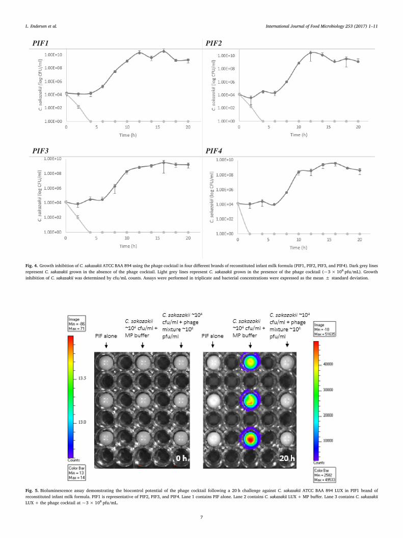
Cronobacter phages leB, leE, and leN were combined as part of aphage cocktail (~3 × 10⁸ pfu/mL) to assess their ability to inhibit thegrowth of Cronobacter sakazakii ATCC BAA 894 in four different brandsof infant formula. Bacterial counts of ~1 × 10⁴ cfu/mL were used foreach biocontrol experiment, as this bacteria:phage concentration ratiowas determined as the maximum inhibitory limit to successfullyprevent outgrowth of C. sakazakii following the 20 h challenge. Thiscontaminating concentration of cells is well in excess of the expectedlevels of C. sakazakii contamination typically found in reconstitutedpowdered infant formula (< 1 cell/100 g of PIF) (van Acker et al.,2001) and hence represents a clear indication of phage biocontrolpotential. Following a 5 h incubation period, C. sakazakii concentra-tions were reduced to below the detection limit (< 10 cfu/mL) insamples challenged with the phage cocktail. This level of inactivationwas maintained over the 20 h challenge, where samples in the absenceof the phage cocktail had reached bacterial concentrations in excess of109 cfu/mL (Fig. 4). Parallel experiments, where the LUX labelled C.
sakazakii ATCC BAA 894 strain was employed to monitor cell viability,gave a similar result. Luminescence could not be detected from thewells challenged with the phage cocktail over the 20 h period, wheresignificant relative light units (RLUs) were detected in the wellscontaining C. sakazakii LUX cells + MP buffer only (Fig. 5). At theend of the assay all wells were serially diluted and plated to determinethe number of viable cells present in the sample. While the wellscontaining C. sakazakii LUX strain alone had reached a concentration of~109 cfu/mL, bacterial concentrations were reduced below the limit ofdetection (< 10 cfu/mL) in the wells challenged with the phagecocktail.
3.6. Biofilm formation
Static microtitre plate assays were used to investigate biofilmformation by selected Cronobacter strains. Strain selection was depen-dant on individual phage infection profiles when tested against the 21Cronobacter strains. Based on phage susceptibility testing (Table 2), fivestrains were selected for biofilm formation assessment. Biofilm forma-tion experiments were performed in BHI, TSB and LB broths, supple-mented with 1% and 2.5% glucose and also in four different brands of
Table 2Host range of Cronobacter phages leB, leE, and leN, where phage lysis is represented in cfu/mL. (−) denotes – no lytic activity. Strains were obtained from the Dairy Products ResearchCentre, DPC, Moorepark, Fermoy, Co. Cork, Ireland.
Strain name Origin Phage lysis (cfu/mL)
leB leE leN
C. sakazakii ATCC BAA 894 Powdered infant formula 1.35E + 08 1.20E + 08 1.01E + 08C. sakazakii DPC 6529 Tracheal aspirate − − 5.10E + 02C. sakazakii DPC 6522 Blood 8.00E + 01 1.19E + 02 2.10E + 02C. sakazakii DPC 6523 Cerebral spinal fluid − − −C. sakazakii NCT 8155 Tin of dried powdered milk 1.20E + 08 1.17E + 09 1.05E + 09C. muytjensi ATCC 51329 − 2.20E + 09 1.20E + 08 1.90E + 07C. sakazakii DPC 6524 Stool − − −C. sakazakii DSM 4485 Child's throat 7.50E + 05 5.00E + 01 1.70E + 02C. malonaticus DPC 6531 Brain tumour − − −C. sakazakii ATCC 29004 − 1.89E + 07 2.70E + 08 4.50E + 08C. sakazakii DPC 6530 Bronchial alveolar lavage 4.20E + 02 3.30E + 04 6.80E + 04E. cloacae NCTC 11933 Human/clinical isolate 1.00E + 01 1.57E + 08 1.00E + 02C. sakazakii DPC 6527 Blood 4.00E + 01 3.10E + 02 2.00E + 01E. gergorviae NCTC 11431 Human urinary tract − − −E. cloacae NCTC 11590 − − − −E. aerogenes NCTC 10006 Sputum − − −C. sakazakii DPC 6526 Blood 1.10E + 02 1.98E + 09 4.00E + 01C. sakazakii ATCC 12868 − − − −C. sakazakii DPC 6528 Cerebral spinal fluid 7.80E + 07 6.50E + 08 8.70E + 07C. sakazakii DPC6525 Urine − − −C. sakazakii NCTC 11467 Human throat 1.76E + 08 3.10E + 02 2.00E + 01
Fig. 3. (A) Effect of temperature on phage viability following 1 h exposure to 4 °C, 37 °C, 45 °C, 50 °C, 60 °C, 72 °C, and 90 °C. (■) represent an average of initial phage titres for all threephages tested. ( ), ( ), and ( ) columns represent phage leB, leE, leN 1 h temperature challenge experiments, respectively. Assays were performed in triplicate and phage titres wereexpressed as the mean ± standard deviation. (B) Effect of pH on phage viability following 1 h exposure to acidic and alkaline environments. (■) represent an average of initial phagetitres for all three phages tested. ( ), ( ), and ( ) represent leB, leE, and leN, 1 h pH challenge experiments. Assays were performed in triplicate and phage titres were expressed as themean ± standard deviation.
L. Endersen et al. International Journal of Food Microbiology 253 (2017) 1–11
6
2FIP1FIP
4FIP3FIP
Fig. 4. Growth inhibition of C. sakazakii ATCC BAA 894 using the phage cocktail in four different brands of reconstituted infant milk formula (PIF1, PIF2, PIF3, and PIF4). Dark grey linesrepresent C. sakazakii grown in the absence of the phage cocktail. Light grey lines represent C. sakazakii grown in the presence of the phage cocktail (~3 × 10⁸ pfu/mL). Growthinhibition of C. sakazakii was determined by cfu/mL counts. Assays were performed in triplicate and bacterial concentrations were expressed as the mean ± standard deviation.
Fig. 5. Bioluminescence assay demonstrating the biocontrol potential of the phage cocktail following a 20 h challenge against C. sakazakii ATCC BAA 894 LUX in PIF1 brand ofreconstituted infant milk formula. PIF1 is representative of PIF2, PIF3, and PIF4. Lane 1 contains PIF alone. Lane 2 contains C. sakazakii LUX +MP buffer. Lane 3 contains C. sakazakiiLUX + the phage cocktail at ~3 × 10⁸ pfu/mL.
L. Endersen et al. International Journal of Food Microbiology 253 (2017) 1–11
7
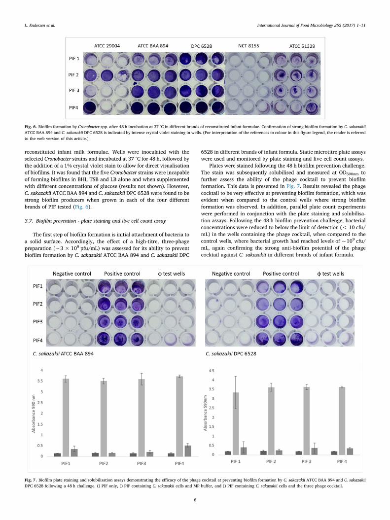
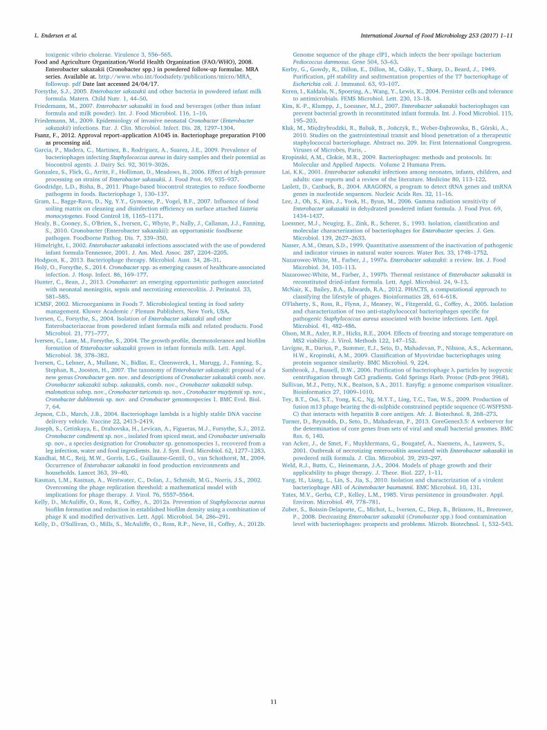
reconstituted infant milk formulae. Wells were inoculated with theselected Cronobacter strains and incubated at 37 °C for 48 h, followed bythe addition of a 1% crystal violet stain to allow for direct visualisationof biofilms. It was found that the five Cronobacter strains were incapableof forming biofilms in BHI, TSB and LB alone and when supplementedwith different concentrations of glucose (results not shown). However,C. sakazakii ATCC BAA 894 and C. sakazakii DPC 6528 were found to bestrong biofilm producers when grown in each of the four differentbrands of PIF tested (Fig. 6).
3.7. Biofilm prevention - plate staining and live cell count assay
The first step of biofilm formation is initial attachment of bacteria toa solid surface. Accordingly, the effect of a high-titre, three-phagepreparation (~3 × 10⁸ pfu/mL) was assessed for its ability to preventbiofilm formation by C. sakazakii ATCC BAA 894 and C. sakazakii DPC
6528 in different brands of infant formula. Static microtitre plate assayswere used and monitored by plate staining and live cell count assays.
Plates were stained following the 48 h biofilm prevention challenge.The stain was subsequently solubilised and measured at OD590nm tofurther assess the ability of the phage cocktail to prevent biofilmformation. This data is presented in Fig. 7. Results revealed the phagecocktail to be very effective at preventing biofilm formation, which wasevident when compared to the control wells where strong biofilmformation was observed. In addition, parallel plate count experimentswere performed in conjunction with the plate staining and solubilisa-tion assays. Following the 48 h biofilm prevention challenge, bacterialconcentrations were reduced to below the limit of detection (< 10 cfu/mL) in the wells containing the phage cocktail, when compared to thecontrol wells, where bacterial growth had reached levels of ~109 cfu/mL, again confirming the strong anti-biofilm potential of the phagecocktail against C. sakazakii in different brands of infant formula.
Fig. 6. Biofilm formation by Cronobacter spp. after 48 h incubation at 37 °C in different brands of reconstituted infant formulae. Confirmation of strong biofilm formation by C. sakazakiiATCC BAA 894 and C. sakazakii DPC 6528 is indicated by intense crystal violet staining in wells. (For interpretation of the references to colour in this figure legend, the reader is referredto the web version of this article.)
Fig. 7. Biofilm plate staining and solubilisation assays demonstrating the efficacy of the phage cocktail at preventing biofilm formation by C. sakazakii ATCC BAA 894 and C. sakazakiiDPC 6528 following a 48 h challenge. () PIF only, () PIF containing C. sakazakii cells and MP buffer, and () PIF containing C. sakazakii cells and the three phage cocktail.
L. Endersen et al. International Journal of Food Microbiology 253 (2017) 1–11
8
4. Discussion
Consumption of contaminated reconstituted infant milk formulaand concomitant C. sakazakii infections in infants and neonates hasresulted in international efforts to improve existing pathogen controlprocesses. Current intervention strategies have fallen short of providingpowdered infant formula that is free from C. sakazakii contamination.Many novel technologies have been proposed in an effort to address thisproblem, some of which include, the use of hot water, gammairradiation and high pressure processing to inactivate the pathogenprior to ingestion (Edelson-Mammel and Buchanan, 2004; Gonzalezet al., 2006; Lee et al., 2006). The natural biotherapeutic properties ofphages are well recognised throughout the world, with many studiesproviding promising evidence of their biocontrol potential againstseveral leading and emerging foodborne pathogens, while also demon-strating their suitability for use at each stage of the farm-to-forkcontinuum (Endersen et al., 2014; Goodridge and Bisha, 2011).
The rationale and motivation for performing the current studyrelates to the aforementioned reconstituted infant milk formula dilem-ma, and its associated health risks, in addition to the possibility ofemploying naturally occurring phages to alleviate this problem. Wedescribe the isolation and characterisation of three novel Cronobacterphages and subsequent utilisation of a three phage preparation forbiocontrol of C. sakazakii in different brands of infant formula. Inaddition to the activity and stability of the phages in infant formula, theanti-biofilm potential of the phage cocktail was also demonstrated.Three Cronobacter phages were isolated following routine enrichmentsof a variety of different environmental samples, including soil, slurry,activated sludge, river water, moss, grass, and wheat. While multiplephages were isolated from these sources, three phages which demon-strated particular promise for use as biocontrol agents against C.sakazakii were selected for further characterisation. Sewage has beenrecognised as a primary niche for many Enterobacteriaceae, so it is notuncommon that these and other Cronobacter phages were isolated fromeffluent environments (Kim et al., 2007; Zuber et al., 2008).
It is well documented that temperate phages from Enterobacteriaceaeare known to harbour important bacterial virulence genes that can bereadily transferred between bacteria. Determining phages to be absentof lysogenic capabilities is a primary consideration that must be takeninto account if they are to be used in food related applications (Brüssowet al., 2004; Faruque and Mekalanos, 2012). As these phages have beendetermined to have lytic lifestyles and do not possess genes for toxicproteins they meet the required properties of phage intended for phagetherapy applications. In addition, these phages have been determined tobelong to the phage genus of.T4 virus,.phages from this genus havebeen deemed safe for phage therapy applications (Bruttin and Brüssow,2005; Denou et al., 2009).
Phage susceptibility was assessed using 21 strains from Cronobacter/Enterobacter genera. The broad host range attributed to phages leB, leE,and leN correlates well with previous findings. Loessner et al. (1993),demonstrated the broad host range of Enterobacter cloncae isolatedphages, which were capable of cross infecting Panteo agglomerans, C.sakazakii, E. coli and Serratia marcescens (Loessner et al., 1993). Inaddition Zuber and co-authors also observed the broad host rangecapabilities of five combined E. sakazakii phages with an infectionprofile extending across several genera (Zuber et al., 2008).
Determining the environmental stability of phages considered forbiocontrol in food and the food processing environment is essential.Common environmental extremes often associated with the food andfood industry include low pH and high temperature pressures.Establishing the stress limits of each phage, permits appropriate andoptimal application, and more effective biocontrol results overall. Eachof the three phages were challenged for their ability to initiate hostinfection following exposure to a range of pH and temperatureextremes. In general, all three phages retained maximum infectivitybetween pH 6 and pH 8. These findings compare well with those
previously demonstrated, where the researchers found that E. coliphage T7 demonstrated optimum pH stability between pH 6 and pH 8(Kerby et al., 1949). Similarly, in another study, phage T4 demon-strated optimum pH stability from pH 6–7.4 (Kłak et al., 2010), whereascoliphage λ demonstrated very high stability across a wide pH range,where no significant decrease in titre was observed from pH 3–11(Jepson and March, 2004). Temperature plays a fundamental role in thesurvival of phages. Optimal attachment, penetration, multiplication andthe length of the latent period (in the case of temperate phages) areoften dictated by temperature (Nasser and Oman, 1999; Olson et al.,2004; Yates et al., 1985). Higher than optimal temperatures are thoughtto extend the latent period, while lower than optimal temperatures areoften thought to result in a reduced multiplication rate (Tey et al.,2009). In this current study, phages leB, leE, and leN were found toretain infectivity from 4 to 50 °C, with optimum infectivity occurringbetween 4 and 37 °C. Yang et al. (2010) evaluated the temperaturestability of Acinetobacter phage AB1 which showed a higher tolerance toextreme temperatures, compared to phage leB, leE, and leN. Phage AB1was very heat stable at 50 °C and 60 °C, as only a slight drop in phagetitre was observed following a 1 h challenge. At 70 °C a significantnumber of the phages were inactivated, with only 0.52% of viablephages remained in the sample following 1 h incubation. Similarly, at90 °C,> 99% of the phages had lost their infective ability following15 min exposure to this temperature (Yang et al., 2010). In anotherstudy, the thermo-tolerance of staphylococcal phages was evaluated inmilk. Researchers found that these phages were most stable whenmaintained at 4 °C for 8 h, while observing a 20–30% increase in phageinactivation when challenged at 22 °C and 37 °C, respectively. Whileboth phages survived very short exposure to high temperature extremes(72 °C for 15 s), phage titres were reduced below the limit of detectionafter 1 min (Garcia et al., 2009).
In recent years, the application of phages for biocontrol of harmfulpathogens in both pre- and postharvest food environments has becomea noteworthy treatment alternative due to the increasing worldwideproblem of antibiotic resistance and the reduction in the developmentof novel technologies for pathogen control. In this study, phagemediated killing and growth inhibition was demonstrated using athree-phage cocktail against C. sakazakii in four different brands ofinfant formula. Overall, it was found that the phage cocktail waseffective at reducing contaminating C. sakazakii cells below the limit ofdetection (< 10 cfu/mL) when bacterial concentration were used at astarting concentration of ≤1 × 10⁴ cfu/mL. This bacteria to phageconcentration ratio was the maximum inhibitory limit to successfullyprevent outgrowth of C. sakazakii over the 20 h challenge. It isinteresting to note that despite slight differences in formula constitu-tion, the growth rate of C. sakazakii was not affected for all formulastested, nor did it impede phage infection. Our biocontrol results weresimilar to those outline by Zuber et al. (2008), who found that a highdose of 108 pfu/mL of phage could effectively sterilise broth contami-nated with both high (106 cfu/mL) and low (102 cfu/mL) pathogencounts (Zuber et al., 2008). In addition, Kim et al., 2007 reportedinhibition of C. sakazakii at a range of different incubating temperatureswhen high titre 109 pfu/mL single phage suspensions were used (Kimet al., 2007). Both Zuber et al. (2008), and Kim et al. (2007) alsoreported that when challenging low numbers of the contaminatingpathogen with a reduced dose of the phage preparation, a delay inphage replication was evident up until bacterial counts had reached~104 cfu/mL. Only after crossing this threshold did phage replicationoccur but overall did not result in complete sterilisation of the sample.This would suggest that high concentrations of phage-based prepara-tions are needed for complete sterilisation of a low quantity of targetcells (Kasman et al., 2002; Weld et al., 2004). It is reported that thelevel of C. sakazakii contamination in PIF is very low (< 1 bacterialcell/100 g) (Holý and Forsythe, 2014), therefore improper handling isthought to contribute to bacterial growth and subsequent developmentof infection among low birth weight infants and neonates. The
L. Endersen et al. International Journal of Food Microbiology 253 (2017) 1–11
9
contaminating levels of C. sakazakii used in this study were muchhigher than levels typically found in PIF which demonstrates theefficacy of this phage cocktail for biocontrol of C. sakazakii inreconstituted infant formula.
Many phage-based intervention strategies have been documentedagainst biofilm formation (Doolittle et al., 1995; Kelly et al., 2012a).Biofilms are a significant source of repeat contamination in dairymanufacturing plants, threatening the quality and safety of themanufactured produce, in addition to causing extensive economiclosses to the food manufacturing industry due to produce recalls andcorrosion of equipment (Gram et al., 2007). In addition, the persistenceof foodborne pathogens, like C. sakazakii, on feeding areas, on thesurfaces of equipment used in formula preparation, and within enteralfeeding tubes, are also thought to contribute to neonatal outbreaks (Kimet al., 2007). Due to the high fatality rate associated with C. sakazakiiinfection among infants and neonates (up to 80%), the destructivenature of the organism towards survivors, the ability of C. sakazakii toform biofilms and because of the problems associated with biofilmtreatment and removal, it is clear that biofilm prevention is thepreferable biocontrol option. We evaluated the biocontrol potential ofthe phage cocktail for its ability to prevent biofilm formation. Resultsindicated that the phage cocktail was very effective in preventing theestablishment of biofilm where biofilm formation could not be detectedfor both strains tested for up to 2 days post-treatment. These resultswere similar to those outlined by Kelly et al. (2012a, 2012b), whodemonstrated the efficacy of a modified phage cocktail for the preven-tion of Staphylococcus aureus biofilm formation. These researchersobserved complete inhibition of biofilm formation over a 48 h timeperiod (Kelly et al., 2012a). In addition, other studies have describedthe use of phages in an attempt to reduce biofilm formation. Hydrogel-coated catheters were impregnated with phage preparations andresulted in an approximate 90% reduction in both Proteus mirabilisand Escherichia coli formed biofilms when compared with un-treatedcontrols (Carson et al., 2010). The anti-biofilm formation potentialdemonstrated by the phage cocktail in this study is promising and mayhave potential in future applications as perhaps a stand-alone treatmentor used in conjunction with alternative antibacterials for the controland/or eradication of C. sakazakii contamination in food and the foodproduction environment.
5. Conclusion
International efforts to develop novel strategies to improve themicrobiological safety of PIF are ongoing. Although many advances inpathogen control have been made in recent years, cost and efficiency aswell as the safety of preparation and nutritional stability are a problem.C. sakazakii contamination of PIF still remains an issue for public healthconcern. Phages have long been recognised for their inherent ability tocontrol pathogens in food and food related environments, where othertreatments have failed to do so. More and more, these non-destructiveparticles are continually gaining acceptance around the world as aviable alternative to antibiotics, and other treatment methods followingthe rise of multi-drug resistant bacteria. Accordingly, this study haspresented three phages active against the infant formula associatedpathogen, C. sakazakii. The phages were characterised in terms oftemperature and pH stability and were combined to demonstrate theefficacy of using a three phage cocktail for biocontrol of C. sakazakii infour different brands of infant formula. In addition, their ability toeffectively inhibit biofilm formation was also established. Our resultshighlight the promising potential of these phages for biocontrol of C.sakazakii contamination in infant formula and also as preventativebiotherapeutic agents against biofilm formation. It should be noted thatthe isolation of additional phages capable of inactivating more resistantstrains of C. sakazakii is a worthwhile endeavour. While 73% coverageis impressive, it could certainly be improved upon. Additional studiesare now required in order to further this concept into practical use.
Acknowledgments
This work was funded by Technological Sector Research Strand IIIref. CRS/07/CR03. Angela Back from MRI Kiel is acknowledged fortechnical assistance in preparations for electron microscopy. HugoOliveira and Rob Lavigne contributed to the genome sequencinganalysis, supported by the KULeuven GOA (GOA/15/006) GrantPhagebiosystems.
Appendix A. Supplementary data
Supplementary data to this article can be found online at http://dx.doi.org/10.1016/j.ijfoodmicro.2017.04.009.
References
Ackermann, H.W., 2001. Frequency of morphological phage descriptions in the year2000. Arch. Virol. 146, 843–857.
Al-Nabulsi, A.A., Osaili, T.M., Al-Holy, M.A., Shaker, R.R., Ayyash, M.M., Olaimat, A.N.,Holley, R.A., 2009. Influence of desiccation on the sensitivity of Cronobacter spp. tolactoferrin or nisin in broth and powdered infant formula. Int. J. Food Microbiol. 136,221–226.
Besemer, J., Lomsadze, A., Borodovsky, M., 2001. GeneMarkS: a self-training method forprediction of gene starts in microbial genomes. Implications for finding sequencemotifs in regulatory regions. Nucleic Acids Res. 29, 2607–2618.
Bruttin, A., Brüssow, H., 2005. Human volunteers receiving Escherichia coli phage T4orally: a safety test of phage therapy. Antimicrob. Agents Chemother. 49, 2874–2878.
Bigot, B., Lee, W.-J., McIntyre, L., Wilson, T., Hudson, J., Billington, C., Heinemann, J.,2011. Control of Listeria monocytogenes growth in a ready-to-eat poultry product usinga bacteriophage. Food Microbiol. 28, 1448–1452.
Brady, C., Cleenwerck, I., Venter, S., Coutinho, T., De Vos, P., 2013. Taxonomicevaluation of the genus Enterobacter based on multilocus sequence analysis (MLSA):Proposal to reclassify E. nimipressuralis and E. amnigenus into Lelliottia gen. nov. asLelliottia nimipressuralis comb. nov. and Lelliottia amnigena comb. nov., respectively, E.gergoviae and E. pyrinus into Pluralibacter gen. nov. as Pluralibacter gergoviae comb.nov. and Pluralibacter pyrinus comb. nov., respectively, E. cowanii, E. radicincitans, E.oryzae and E. arachidis into Kosakonia gen. nov. as Kosakonia cowanii comb. nov.,Kosakonia radicincitans comb. nov., Kosakonia oryzae comb. nov. and Kosakoniaarachidis comb. nov., respectively, and E. turicensis, E. helveticus and E. pulveris intoCronobacter as Cronobacter zurichensis nom. nov., Cronobacter helveticus comb. nov.and Cronobacter pulveris comb. nov., respectively, and emended description of thegenera Enterobacter and Cronobacter. Syst. Appl. Microbiol. 36, 309–319.
Breeuwer, P., Lardeau, A., Peterz, M., Joosten, H., 2003. Desiccation and heat tolerance ofEnterobacter sakazakii. J. Appl. Microbiol. 95, 967–973.
Bren, L., 2006. Bacteria-eating virus approved as food additive. FDA Consum. 41, 20–22.Brüssow, H., Canchaya, C., Hardt, W.D., 2004. Phages and the evolution of bacterial
pathogens: from genomic rearrangements to lysogenic conversion. Microbiol. Mol.Biol. Rev. 68, 560–602.
Carlson, K., 2005. Appendix: working with bacteriophages: common techniques andmethodological approaches. In: Bacteriophages: Biology and Applications, pp.437–494.
Carlton, R., Noordman, W., Biswas, B., De Meester, E., Loessner, M., 2005. BacteriophageP100 for control of Listeria monocytogenes in foods: genome sequence, bioinformaticanalyses, oral toxicity study, and application. Regul. Toxicol. Pharmacol. 43,301–312.
Carson, L., Gorman, S.P., Gilmore, B.F., 2010. The use of lytic bacteriophages in theprevention and eradication of biofilms of Proteus mirabilis and Escherichia coli. FEMSImmunol. Med. Microbiol. 59, 447–455.
Cerca, N., Oliveira, R., Azeredo, J., 2007. Susceptibility of Staphylococcus epidermidisplanktonic cells and biofilms to the lytic action of staphylococcus bacteriophage K.Lett. Appl. Microbiol. 45, 313–317.
Chenu, J., Cox, J., 2009. Cronobacter (‘Enterobacter sakazakii’): current status and futureprospects. Lett. Appl. Microbiol. 49, 153–159.
Donlan, R.M., Costerton, J.W., 2002. Biofilms: survival mechanisms of clinically relevantmicroorganisms. Clin. Microbiol. Rev. 15, 167–193.
Doolittle, M., Cooney, J., Caldwell, D., 1995. Lytic infection of Escherichia coli biofilms bybacteriophage T4. Can. J. Microbiol. 41, 12–18.
Delcher, A.L., Harmon, D., Kasif, S., White, O., Salzberg, S.L., 1999. Improved microbialgene identification with GLIMMER. Nucleic Acids Res. 27, 4636–4641.
Denou, E., Bruttin, A., Barretto, C., Ngom-Bru, C., Brüssow, H., Zuber, S., 2009. T4 phagesagainst Escherichia coli diarrhea: potential and problems. Virology 388, 21–30.
Edelson-Mammel, S.G., Buchanan, R.L., 2004. Thermal inactivation of Enterobactersakazakii in rehydrated infant formula. J. Food Prot. 67, 60–63.
Ellis, D., Whitman, P., Marshall, R., 1973. Effects of homologous bacteriophage on growthof Pseudomonas fragi WY in milk. Appl. Microbiol. 25, 24–25.
Endersen, L., Coffey, A., Neve, H., McAuliffe, O., Ross, R.P., O'Mahony, J.M., 2013.Isolation and characterisation of six novel mycobacteriophages and investigation oftheir antimicrobial potential in milk. Int. Dairy J. 28, 8–14.
Endersen, L., O'Mahony, J., Hill, C., Ross, R.P., McAuliffe, O., Coffey, A., 2014. Phagetherapy in the food industry. Annu. Rev. Food Sci. Technol. 5, 327–349.
Faruque, S.M., Mekalanos, J.J., 2012. Phage-bacterial interactions in the evolution of
L. Endersen et al. International Journal of Food Microbiology 253 (2017) 1–11
10
toxigenic vibrio cholerae. Virulence 3, 556–565.Food and Agriculture Organization/World Health Organization (FAO/WHO), 2008.
Enterobacter sakazakii (Cronobacter spp.) in powdered follow-up formulae. MRAseries. Available at. http://www.who.int/foodsafety/publications/micro/MRA_followup. pdf Date last accessed 24/04/17.
Forsythe, S.J., 2005. Enterobacter sakazakii and other bacteria in powdered infant milkformula. Matern. Child Nutr. 1, 44–50.
Friedemann, M., 2007. Enterobacter sakazakii in food and beverages (other than infantformula and milk powder). Int. J. Food Microbiol. 116, 1–10.
Friedemann, M., 2009. Epidemiology of invasive neonatal Cronobacter (Enterobactersakazakii) infections. Eur. J. Clin. Microbiol. Infect. Dis. 28, 1297–1304.
Fsanz, F., 2012. Approval report–application A1045 in. Bacteriophage preparation P100as processing aid.
Garcia, P., Madera, C., Martinez, B., Rodriguez, A., Suarez, J.E., 2009. Prevalence ofbacteriophages infecting Staphylococcus aureus in dairy samples and their potential asbiocontrol agents. J. Dairy Sci. 92, 3019–3026.
Gonzalez, S., Flick, G., Arritt, F., Holliman, D., Meadows, B., 2006. Effect of high-pressureprocessing on strains of Enterobacter sakazakii. J. Food Prot. 69, 935–937.
Goodridge, L.D., Bisha, B., 2011. Phage-based biocontrol strategies to reduce foodbornepathogens in foods. Bacteriophage 1, 130–137.
Gram, L., Bagge-Ravn, D., Ng, Y.Y., Gymoese, P., Vogel, B.F., 2007. Influence of foodsoiling matrix on cleaning and disinfection efficiency on surface attached Listeriamonocytogenes. Food Control 18, 1165–1171.
Healy, B., Cooney, S., O'Brien, S., Iversen, C., Whyte, P., Nally, J., Callanan, J.J., Fanning,S., 2010. Cronobacter (Enterobacter sakazakii): an opportunistic foodbornepathogen. Foodborne Pathog. Dis. 7, 339–350.
Himelright, I., 2002. Enterobacter sakazakii infections associated with the use of powderedinfant formula-Tennessee, 2001. J. Am. Med. Assoc. 287, 2204–2205.
Hodgson, K., 2013. Bacteriophage therapy. Microbiol. Aust. 34, 28–31.Holý, O., Forsythe, S., 2014. Cronobacter spp. as emerging causes of healthcare-associated
infection. J. Hosp. Infect. 86, 169–177.Hunter, C., Bean, J., 2013. Cronobacter: an emerging opportunistic pathogen associated
with neonatal meningitis, sepsis and necrotizing enterocolitis. J. Perinatol. 33,581–585.
ICMSF, 2002. Microorganisms in Foods 7. Microbiological testing in food safetymanagement. Kluwer Academic / Plenum Publishers, New York, USA.
Iversen, C., Forsythe, S., 2004. Isolation of Enterobacter sakazakii and otherEnterobacteriaceae from powdered infant formula milk and related products. FoodMicrobiol. 21, 771–777.
Iversen, C., Lane, M., Forsythe, S., 2004. The growth profile, thermotolerance and biofilmformation of Enterobacter sakazakii grown in infant formula milk. Lett. Appl.Microbiol. 38, 378–382.
Iversen, C., Lehner, A., Mullane, N., Bidlas, E., Cleenwerck, I., Marugg, J., Fanning, S.,Stephan, R., Joosten, H., 2007. The taxonomy of Enterobacter sakazakii: proposal of anew genus Cronobacter gen. nov. and descriptions of Cronobacter sakazakii comb. nov.Cronobacter sakazakii subsp. sakazakii, comb. nov., Cronobacter sakazakii subsp.malonaticus subsp. nov., Cronobacter turicensis sp. nov., Cronobacter muytjensii sp. nov.,Cronobacter dublinensis sp. nov. and Cronobacter genomospecies 1. BMC Evol. Biol.7, 64.
Jepson, C.D., March, J.B., 2004. Bacteriophage lambda is a highly stable DNA vaccinedelivery vehicle. Vaccine 22, 2413–2419.
Joseph, S., Cetinkaya, E., Drahovska, H., Levican, A., Figueras, M.J., Forsythe, S.J., 2012.Cronobacter condimenti sp. nov., isolated from spiced meat, and Cronobacter universalissp. nov., a species designation for Cronobacter sp. genomospecies 1, recovered from aleg infection, water and food ingredients. Int. J. Syst. Evol. Microbiol. 62, 1277–1283.
Kandhai, M.C., Reij, M.W., Gorris, L.G., Guillaume-Gentil, O., van Schothorst, M., 2004.Occurrence of Enterobacter sakazakii in food production environments andhouseholds. Lancet 363, 39–40.
Kasman, L.M., Kasman, A., Westwater, C., Dolan, J., Schmidt, M.G., Norris, J.S., 2002.Overcoming the phage replication threshold: a mathematical model withimplications for phage therapy. J. Virol. 76, 5557–5564.
Kelly, D., McAuliffe, O., Ross, R., Coffey, A., 2012a. Prevention of Staphylococcus aureusbiofilm formation and reduction in established biofilm density using a combination ofphage K and modified derivatives. Lett. Appl. Microbiol. 54, 286–291.
Kelly, D., O'Sullivan, O., Mills, S., McAuliffe, O., Ross, R.P., Neve, H., Coffey, A., 2012b.
Genome sequence of the phage clP1, which infects the beer spoilage bacteriumPediococcus damnosus. Gene 504, 53–63.
Kerby, G., Gowdy, R., Dillon, E., Dillon, M., Csâky, T., Sharp, D., Beard, J., 1949.Purification, pH stability and sedimentation properties of the T7 bacteriophage ofEscherichia coli. J. Immunol. 63, 93–107.
Keren, I., Kaldalu, N., Spoering, A., Wang, Y., Lewis, K., 2004. Persister cells and toleranceto antimicrobials. FEMS Microbiol. Lett. 230, 13–18.
Kim, K.-P., Klumpp, J., Loessner, M.J., 2007. Enterobacter sakazakii bacteriophages canprevent bacterial growth in reconstituted infant formula. Int. J. Food Microbiol. 115,195–203.
Kłak, M., Międzybrodzki, R., Bubak, B., Jończyk, E., Weber-Dąbrowska, B., Górski, A.,2010. Studies on the gastrointestinal transit and blood penetration of a therapeuticstaphylococcal bacteriophage. Abstract no. 209. In: First International Congrogress.Viruses of Microbes, Paris, .
Kropinski, A.M., Clokie, M.R., 2009. Bacteriophages: methods and protocols. In:Molecular and Applied Aspects. Volume 2 Humana Press.
Lai, K.K., 2001. Enterobacter sakazakii infections among neonates, infants, children, andadults: case reports and a review of the literature. Medicine 80, 113–122.
Laslett, D., Canback, B., 2004. ARAGORN, a program to detect tRNA genes and tmRNAgenes in nucleotide sequences. Nucleic Acids Res. 32, 11–16.
Lee, J., Oh, S., Kim, J., Yook, H., Byun, M., 2006. Gamma radiation sensitivity ofEnterobacter sakazakii in dehydrated powdered infant formula. J. Food Prot. 69,1434–1437.
Loessner, M.J., Neugirg, E., Zink, R., Scherer, S., 1993. Isolation, classification andmolecular characterization of bacteriophages for Enterobacter species. J. Gen.Microbiol. 139, 2627–2633.
Nasser, A.M., Oman, S.D., 1999. Quantitative assessment of the inactivation of pathogenicand indicator viruses in natural water sources. Water Res. 33, 1748–1752.
Nazarowec-White, M., Farber, J., 1997a. Enterobacter sakazakii: a review. Int. J. FoodMicrobiol. 34, 103–113.
Nazarowec-White, M., Farber, J., 1997b. Thermal resistance of Enterobacter sakazakii inreconstituted dried-infant formula. Lett. Appl. Microbiol. 24, 9–13.
McNair, K., Bailey, B.A., Edwards, R.A., 2012. PHACTS, a computational approach toclassifying the lifestyle of phages. Bioinformatics 28, 614–618.
O'Flaherty, S., Ross, R., Flynn, J., Meaney, W., Fitzgerald, G., Coffey, A., 2005. Isolationand characterization of two anti-staphylococcal bacteriophages specific forpathogenic Staphylococcus aureus associated with bovine infections. Lett. Appl.Microbiol. 41, 482–486.
Olson, M.R., Axler, R.P., Hicks, R.E., 2004. Effects of freezing and storage temperature onMS2 viability. J. Virol. Methods 122, 147–152.
Lavigne, R., Darius, P., Summer, E.J., Seto, D., Mahadevan, P., Nilsson, A.S., Ackermann,H.W., Kropinski, A.M., 2009. Classification of Myoviridae bacteriophages usingprotein sequence similarity. BMC Microbiol. 9, 224.
Sambrook, J., Russell, D.W., 2006. Purification of bacteriophage λ particles by isopycniccentrifugation through CsCl gradients. Cold Springs Harb. Protoc (Pdb-prot 3968).
Sullivan, M.J., Petty, N.K., Beatson, S.A., 2011. Easyfig: a genome comparison visualizer.Bioinformatics 27, 1009–1010.
Tey, B.T., Ooi, S.T., Yong, K.C., Ng, M.Y.T., Ling, T.C., Tan, W.S., 2009. Production offusion m13 phage bearing the di-sulphide constrained peptide sequence (C-WSFFSNI-C) that interacts with hepatitis B core antigen. Afr. J. Biotechnol. 8, 268–273.
Turner, D., Reynolds, D., Seto, D., Mahadevan, P., 2013. CoreGenes3.5: A webserver forthe determination of core genes from sets of viral and small bacterial genomes. BMCRes. 6, 140.
van Acker, J., de Smet, F., Muyldermans, G., Bougatef, A., Naessens, A., Lauwers, S.,2001. Outbreak of necrotizing enterocolitis associated with Enterobacter sakazakii inpowdered milk formula. J. Clin. Microbiol. 39, 293–297.
Weld, R.J., Butts, C., Heinemann, J.A., 2004. Models of phage growth and theirapplicability to phage therapy. J. Theor. Biol. 227, 1–11.
Yang, H., Liang, L., Lin, S., Jia, S., 2010. Isolation and characterization of a virulentbacteriophage AB1 of Acinetobacter baumannii. BMC Microbiol. 10, 131.
Yates, M.V., Gerba, C.P., Kelley, L.M., 1985. Virus persistence in groundwater. Appl.Environ. Microbiol. 49, 778–781.
Zuber, S., Boissin-Delaporte, C., Michot, L., Iversen, C., Diep, B., Brüssow, H., Breeuwer,P., 2008. Decreasing Enterobacter sakazakii (Cronobacter spp.) food contaminationlevel with bacteriophages: prospects and problems. Microb. Biotechnol. 1, 532–543.
L. Endersen et al. International Journal of Food Microbiology 253 (2017) 1–11
11