Embed Size (px)
Citation preview
International Journal of Science and Research (IJSR) ISSN (Online): 2319-7064
Impact Factor (2012): 3.358
Volume 3 Issue 11, November 2014 www.ijsr.net
Licensed Under Creative Commons Attribution CC BY
Interleukin-17 Role on the Occurrence of Plasma Leakage Dengue Hemorrhagic Fever Through
Cytokine Proinflamasi Activation: (Study of in-vivo and in-vitro macrophage culture and cell endothelia)
Edi Hartoyo1,Rasjad Indra2, Edi Widajanto3, Sri Rezeki Hadinegoro4
1Children Health departement, Medical Faculty of Lambung Mangkurat University Banjarmasin
2 Laboratory of Molecular physiologies, Medical Faculty of Brawijaya University Malang
3Laboratory of clinic pathology, RSUD Saiful Anwar, Medical Faculty of Brawijaya Malang University
4 Children Health study, Medical Faculty of Indonesia University, Jakarta
Abstracts: Background: The role of IL-17 in virus infection has been evaluated, but there isn’t data about its role in dengue hemorrhagic fever. The research purpose step 1 was to measure Th17 and IL-17 level on Dengue Haemorrhagic Fever shock and non shock. The second was to know the exposure effect of IL-17 recombinant on macrophage culture so it secreted TNF-α, MMP-2 and IL-8. The third was to know the plasma leakage on cell endothelial culture which was exposed by using activated macrophage. Methods: The measurement of interleukin 17 was done by using ELISA method, Th17 intracell by flowcytrometry, while TNFα, MMP-2 and IL-8 was measured by ELISA method. Albumin concentration was evaluated by spectroscopy. Results: There’s significant difference on Th17 and IL-17 level on DHF non shock case compared by DHF shock with p<0.000, There’s significant increase on TNF α among groups. There’s significance difference on the increase of MMP-2 and IL-8 level (p<0.000) on the shelf with IL-17 recombinant on macrophage culture. There’ s significant difference for IL-8 between time exposure 0 hour with 24 hours, 0 hour with 48 hours (p<0.000) and there’s significant difference for MMP-2 between time exposure 0 hour with 16 hours, 0 hour with 24 hours and 0 hour with 48 hours(p<0.000). There’s significant difference between IL-17 recombinant with albumin concentration on time variation (p<0.000). Conclusion:
Interleukin 17 has role on pathogenesis plasma DHF leakage through cytokine proinflamasi activation. Keywords: Plasma leakage, Dengue Hemorrhagic Fever (DHF), IL-17, cytokine proinflamasi
1. Background Dengue hemorrhagic fever is acute virus infection which is infected by Aedes aegypty mosquitoes bitten. World Health Organization predicted that 2,5 billion peoples live in dengue virus endemic areas (WHO, 2009). In south-east Asia, 2007, it had happened the increasing of dengue hemorrhagic fever reached 18 % and about 15% of death case compared by 2006 (Sapir and Schimmer, 2005). In Indonesia, this disease was firstly found in Surabaya, 1968. At that time, the number of suffered reached 58 peoples and the death number was 24 peoples (41, 3 %). Next, this finding had expanded over the country and reached the top in 1998, the number of case was 13,45% by 100.000 citizens. In 2008, the number of occurrence was 31, 5% and the number of death was 0,86%. In south Kalimantan, the number of occurrence was 30, 4%, while the death number was 5 – 8 % (Dinkes, 2009). The spread of Aedes aegypty vector causes the existence of DHF and dengue fever epidemic and result on hyperendemicity mainly in tropic area. The main of Dengue Haemorhagic fever (DHF) pathogenesis is the increasing of capillary partition permeability which is by the dynamics of opening and closing connection between endotheliacell (adherens junction), it is consisted of transmembran protein group, that is vascular endothelia cadherin (VE caderin) which is connected by cytoplasm, that’s p120, β catenin and plakoglobin (Elisabetta et al , 2008). Some mediator that
cause the capillary block permeability are vascular endothelial growth factor (VEGF), TNF α, matricmetaloprotein (MMP), IL -8 and nitric oxide (NO). Mechanism of TNF (Tumor Necrotic Factor) alfa and IL -8 is through fosphorilation tyrosin of VE Caderin (Elisabetta et al 2008). In dengue virus infection also will happen over production of matricmetalaprotein prominently MMP2 that’s easy to fused and gelatinolitic character, can cause lysis endothelial cell, next it can increase capillary permeability and related by disappear of endothelial thrombosite adhesion molecule expression1 (PECAM – 1) and adhesion molecule from VE Caderin will cause F – actin redistribution . This is proved by invitro observation which is confirmed through invivo by plasma leakage on mouse model. The production of matricmetaloproteinase will have effect on endothelia cell integrity (Asahi et al, 2001), meanwhile, NO mechanism in influencing capillary permeability through oxides with plasma membrane. Nitritoxide will cause permeability increasing and induct lipid peroxides , break DNA chain, endothelial cell contraction through RhoA and MLCK so that increase Ca²+ intracellular ion concentration, phospolifase A₂ enzim activation, phosfolifase D, PLC , PKC, MAP and Src kinase (Jovanovic D. et al , 1998). In DHF will occur “cytokine storm (badai sitokin)”. Along this time, paradigm which is still used is T helper 1 role (Th1) and T helper 2 (Th2) toward DHF pathogenesis. Cytokine that is produced by Th1 cell, in general showing
Paper ID: OCT141263 1711
International Journal of Science and Research (IJSR) ISSN (Online): 2319-7064
Impact Factor (2012): 3.358
Volume 3 Issue 11, November 2014 www.ijsr.net
Licensed Under Creative Commons Attribution CC BY
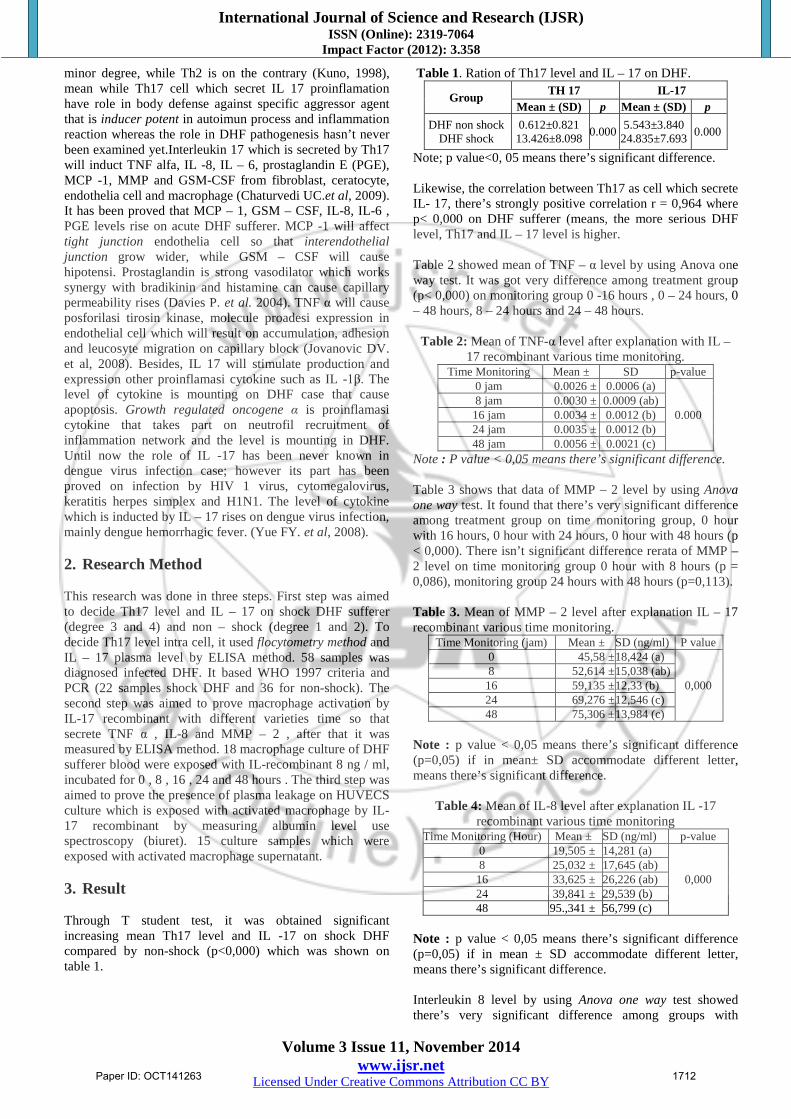
minor degree, while Th2 is on the contrary (Kuno, 1998), mean while Th17 cell which secret IL 17 proinflamation have role in body defense against specific aggressor agent that is inducer potent in autoimun process and inflammation reaction whereas the role in DHF pathogenesis hasn’t never been examined yet.Interleukin 17 which is secreted by Th17 will induct TNF alfa, IL -8, IL – 6, prostaglandin E (PGE), MCP -1, MMP and GSM-CSF from fibroblast, ceratocyte, endothelia cell and macrophage (Chaturvedi UC.et al, 2009). It has been proved that MCP – 1, GSM – CSF, IL-8, IL-6 , PGE levels rise on acute DHF sufferer. MCP -1 will affect tight junction endothelia cell so that interendothelial junction grow wider, while GSM – CSF will cause hipotensi. Prostaglandin is strong vasodilator which works synergy with bradikinin and histamine can cause capillary permeability rises (Davies P. et al. 2004). TNF α will cause posforilasi tirosin kinase, molecule proadesi expression in endothelial cell which will result on accumulation, adhesion and leucosyte migration on capillary block (Jovanovic DV. et al, 2008). Besides, IL 17 will stimulate production and expression other proinflamasi cytokine such as IL -1β. The level of cytokine is mounting on DHF case that cause apoptosis. Growth regulated oncogene α is proinflamasi cytokine that takes part on neutrofil recruitment of inflammation network and the level is mounting in DHF. Until now the role of IL -17 has been never known in dengue virus infection case; however its part has been proved on infection by HIV 1 virus, cytomegalovirus, keratitis herpes simplex and H1N1. The level of cytokine which is inducted by IL – 17 rises on dengue virus infection, mainly dengue hemorrhagic fever. (Yue FY. et al, 2008). 2. Research Method This research was done in three steps. First step was aimed to decide Th17 level and IL – 17 on shock DHF sufferer (degree 3 and 4) and non – shock (degree 1 and 2). To decide Th17 level intra cell, it used flocytometry method and IL – 17 plasma level by ELISA method. 58 samples was diagnosed infected DHF. It based WHO 1997 criteria and PCR (22 samples shock DHF and 36 for non-shock). The second step was aimed to prove macrophage activation by IL-17 recombinant with different varieties time so that secrete TNF α , IL-8 and MMP – 2 , after that it was measured by ELISA method. 18 macrophage culture of DHF sufferer blood were exposed with IL-recombinant 8 ng / ml, incubated for 0 , 8 , 16 , 24 and 48 hours . The third step was aimed to prove the presence of plasma leakage on HUVECS culture which is exposed with activated macrophage by IL-17 recombinant by measuring albumin level use spectroscopy (biuret). 15 culture samples which were exposed with activated macrophage supernatant. 3. Result Through T student test, it was obtained significant increasing mean Th17 level and IL -17 on shock DHF compared by non-shock (p<0,000) which was shown on table 1.
Table 1. Ration of Th17 level and IL – 17 on DHF.
Group TH 17 IL-17 Mean ± (SD) p Mean ± (SD) p
DHF non shock DHF shock
0.612±0.821 13.426±8.098 0.000 5.543±3.840
24.835±7.693 0.000
Note; p value<0, 05 means there’s significant difference. Likewise, the correlation between Th17 as cell which secrete IL- 17, there’s strongly positive correlation r = 0,964 where p< 0,000 on DHF sufferer (means, the more serious DHF level, Th17 and IL – 17 level is higher. Table 2 showed mean of TNF – α level by using Anova one way test. It was got very difference among treatment group (p< 0,000) on monitoring group 0 -16 hours , 0 – 24 hours, 0 – 48 hours, 8 – 24 hours and 24 – 48 hours.
Table 2: Mean of TNF-α level after explanation with IL – 17 recombinant various time monitoring.
Time Monitoring Mean ± SD p-value 0 jam 0.0026 ± 0.0006 (a)
0.000 8 jam 0.0030 ± 0.0009 (ab) 16 jam 0.0034 ± 0.0012 (b) 24 jam 0.0035 ± 0.0012 (b) 48 jam 0.0056 ± 0.0021 (c)
Note : P value < 0,05 means there’s significant difference. Table 3 shows that data of MMP – 2 level by using Anova one way test. It found that there’s very significant difference among treatment group on time monitoring group, 0 hour with 16 hours, 0 hour with 24 hours, 0 hour with 48 hours (p < 0,000). There isn’t significant difference rerata of MMP – 2 level on time monitoring group 0 hour with 8 hours (p = 0,086), monitoring group 24 hours with 48 hours (p=0,113). Table 3. Mean of MMP – 2 level after explanation IL – 17 recombinant various time monitoring.
Time Monitoring (jam) Mean ± SD (ng/ml) P value 0 45,58 ± 18,424 (a)
0,000 8 52,614 ± 15,038 (ab)
16 59,135 ± 12,33 (b) 24 69,276 ± 12,546 (c) 48 75,306 ± 13,984 (c)
Note : p value < 0,05 means there’s significant difference (p=0,05) if in mean± SD accommodate different letter, means there’s significant difference.
Table 4: Mean of IL-8 level after explanation IL -17 recombinant various time monitoring
Time Monitoring (Hour) Mean ± SD (ng/ml) p-value 0 19,505 ± 14,281 (a)
0,000 8 25,032 ± 17,645 (ab)
16 33,625 ± 26,226 (ab) 24 39,841 ± 29,539 (b) 48 95.,341 ± 56,799 (c)
Note : p value < 0,05 means there’s significant difference (p=0,05) if in mean ± SD accommodate different letter, means there’s significant difference. Interleukin 8 level by using Anova one way test showed there’s very significant difference among groups with
Paper ID: OCT141263 1712

International Journal of Science and Research (IJSR) ISSN (Online): 2319-7064
Impact Factor (2012): 3.358
Volume 3 Issue 11, November 2014 www.ijsr.net
Licensed Under Creative Commons Attribution CC BY
different time monitoring (p=0,000). There’s significant difference IL–8 level on time monitoring 0 with 24 hours group, 0 with 48 hours. There isn’t significant difference IL-8 level on time monitoring 0 with 8 hours group (p=0,512), time monitoring 0 with 16 hours group (p=0,095), time monitoring 8 with 16 hours (p=0,308), time monitoring 8
and 16 hours(p=0,080) on ratio test of concentration IL-17 rerata recombinant among and incubation time by using Anova two way test. It is showed in detail on table 5. 3.1 Concentration ratio on different incubation time
Picture 1: Ratio of mean concentration among group with concentration IL-17 treatment and different incubation time.
Note: It’s clearly showed on histogram, rising had happened on IL-17 concentration group. Picture 1, shows that that the value of albumin mean level accommodate significant difference on 0 ng/ml concentration (control) by using treatment (2 , 4 , 8 ng / ml). Next, based on double ratio test by LSD test, it’s explained shortly on table 5.
Table 5: Result of double ratio test by LSD Concentration IL-17 (ng/ml)
p-value Incubation Time (Hour)
p-value
0 – 2 0,000 0 – 8 0,920 0 – 4 0,000 0 – 24 0,596 0 – 8 0,000 0 – 48 0,000 2 – 4 0,066 8 – 24 0,529 2 – 8 0,034 8 – 48 0,000 4 – 8 0,769 24 – 48 0,000
Note : p value < 0,05 , there’s significant difference. On table 5, IL – 17 concentration recombinant 0 with 2ng/ml, 0 with 4 ng/ml and 0 with 8 ng/ml showed
significant difference (p < 0,000). This means the higher IL-17 concentration correlate with Albumin level, while time variation showed the highest correlation in 48 hours.
Table 6: Correlation coefficient among variable. Variable Conection Coefficient
correlation (r) p-value Deduction
TH-17 IL-17 0.964 0.000 significant
IL-17 MPP2 0.612 0.000 significant TNF-∝ 0.847 0.000 significant
IL-8 0.865 0.000 significant MPP2 Degree of
DHF
0.608 0.000 significant TNF-∝ 0.739 0.000 significant
IL-8 0.860 0.000 significant On table 6 explained tightness relationship degree among variable or showed correlation coefficient (r) with degree value / probability empiric error (p). Correlation coefficient between DHF degree and TH – 17 level is 0,936 (p– 0,000). Completely, it can be seen on picture 2.
Picture 2: correlation coefficient among TH – 17 (X₁), IL – 17 (Y₁), MPP2 (Y₂), TNF –α (Y₃), IL – 8 (Y₄ ) and DHF degree
Paper ID: OCT141263 1713

International Journal of Science and Research (IJSR) ISSN (Online): 2319-7064
Impact Factor (2012): 3.358
Volume 3 Issue 11, November 2014 www.ijsr.net
Licensed Under Creative Commons Attribution CC BY
(Y₅).
Picture 3: track coefficient among variable
4. Discussion Dengue hemorrhagic fever is acute virus infection disease which is infected through Aedes mosquitos bitten. Until now, DHF Pathogenesis hasn’t been known completely. Research is still doing to know the theory of pathogenesis and influenced factors including cytokine role on DHF (chaturvedi, 2000). Dengue hemorrhagic fever (DHF) is mosquito born infection which is its spread all over the world, especially in tropic, sub tropic and urban areas. It’s about 2,5 billion peoples who live in the countries of DHF or Dengue fever (Dengue Fever) endemic and 50 million of them has been infected by dengue virus every year (WHO, 2009). From the data above, the mostly sufferer is between 5 – 10 years old. It based the result of Kan et al. research (5 – 9 years old), while, Rampengan got the younger (4 – 6 years old) ¹. In Thailand, the mostly sufferer was on 5 – 9 years old, the second rank was on 10 – 14 years old (Nimmanitya S, 2004). Based on Depkes (2009), the mostly number was on 5 – 14 years old (32%). From the distribution of DHF case based on gender, there isn’t difference between male and female. Depkes (2009) showed the occurrence number of DHF on male was 53, 78%, while female was 46, 23%. It can be concluded that potency to be infected by DHF between male and female were almost same. Children with obesities have heavier risk than other nutrient status. While, children who have bad nutrient status rarely become heavy since their immune responses decreased (Halstead, 1997). On the table above, stereotype den 2 and den 3 often cause heavy level of DHF compared by other serotypes. Research in Thailand from 105 cases of den 1 (32, 4%), den 2 (54, 3%), den 3 (7, 6%) and den 4(5, 7%)
(Nimanitya et al, 1988). Depkes’ report 2005, den 2 often cause seriously DHF, next followed by den 3 and den 1. Secondary infection more often cause seriously infection. It based on secondary infection theories (Halstead, 1997). 4.1 T helper 17 and Interleukin 17 Humam interleukin 17 is a homodimeric with molecule 20 – 30 kDa which contains gylcosylate polypeptide secreted by T CD41 activated memory (CD45IRO1) Th 17. In this research, there’s significant difference of Th17 rerata and IL – 17 DBD sufferer shock and non – shock 0,612 ± 0,821 vs 13, 426 ± 8,098 (P < 0,000) and 5,543 ± 3,840 vs 24,835 vs 7,693 (P < 0,000). It indicates that dengue virus infection will induct CD4+ and produce Th17, as the result of simulation from TGF - 1β, IL – 23 and RORyt (Yoichiro, 2004). Next, IL – 17 regulation mechanism on macrophage by controlling gene expression such as AP-1 , CREB, and NF –ƘB that will bind sequence proinflamasi cytokine promotor such ans ILte – 8 ,TNFα, IL - 1β, MMP – 2 and IL -6 so that the cytokine production rises (Fujiwara N. et al, 2005). On DHF, there’s IFN-ɣ, c, IL- 8 cytokine increase, MMP-2 and TNF-β which related with pathogenesis on secondary infection (Mongkolsapaya .et al. 2006). The main function IL-17 which is secreted by T cell is stimulate proinflamasi cytokine production such as TNF-α, IL-1β, IL-6 and inflammation khemokin such as CXCL – 6, CXCL – 7, CXCL – 8, IL – 8, monocyte chemoatractant protein-1 (MCP – 1) and metalloproteinase matrix that will pull neutrofil and macrophage so that cause inflammation reaction on network. Th 17 role has ever been evaluated on virus infection research, like on cornea infection by virus H1V1, cytomegalovirus, hepatitis, virus C and autoimmune disease (Fujiwara et al, 2005). Many research involved autoimmune mechanism in DHF pathogenesis, it had been proved that antibody (Abs) nonstructural protein 1 (NS1) will react crosswise toward trombosyte and endothelia cell
Paper ID: OCT141263 1714

International Journal of Science and Research (IJSR) ISSN (Online): 2319-7064
Impact Factor (2012): 3.358
Volume 3 Issue 11, November 2014 www.ijsr.net
Licensed Under Creative Commons Attribution CC BY
will cause immunologic reaction that has role on auto antibodies production (autoAb). Autoantibody toward endothelia cell was found on dengue virus infection with different serotype, so it will cause apoptosis endothelia cell process (Zhou, 2002). On DHF/ DSS (Dengue Shock Syndrome) sufferer, the level of Abs anti trombosit and Abs endothelial is higher than dengue fever sufferer (Lien CF, 2003). 4.2 TNF-α Macrophage is fagocitic cell which has important role in body defense system. Macrophage is Antigen Presenting Cell (APCs) that process antigen and provide to cell T for starting immune response. Dengue virus comes into macrophage cell through receptor Fc receptor (Fc ᵧRI) immune complex mechanism (Reyes et al, 2005). Dengue virus simulation on macrophage cell will secrete some TNF α, IL -8, IL-6 and MMP 2. TNF α is proinflamasi cytokine which takes part on seriousness of DHF sufferer (Medin Lc, 2005). On this research, macrophage culture is explained with interleukin 17 recombinant (rh IL17) by using dosis (8 ng/ml) and time variation. There’s increasing of TNF α level based time variation although statistically, there isn’t significance difference (P > 0, 005). The highest increasing of TNF α level is 48 hours after explanation. In another research, TNF α will start to raise up on first day after infection and will reach the peak on the second day ( Hober D, 1993). Research with dengue virus explanation on secretion macrophage TNF α reach the peak less than 2 – 4 days after infection (Zhou et al, 2002). Secretion proinflamasi cytokine on macrophage culture depend on dose and time explanation (Murphy PM, 1994). IL-8 Cytokine IL-8 is pro inflammation cytokine which is its level do rise up on serious DBD sufferer. This cytokine has pro inflammatory effect, chemotraktanactivation which is secreted by variety of cell type and takes part in inflammation process, wound healing, angiogenesis, metastasis and lymphoid trafficking. In this research, IL-8 increasing is different in meaning among time monitoring group (p < 0,000) with the highest level is on 48 hours. It’s appropriate with the previous research that IL- 8 level reaches the peak on the two days after infection (Hober D et al, 1993). In another research, 61% of this cytokine was detected on DHF level IV and only 14% was detected on dengue fever sufferer (Chaturvedi et al , 2000).This shows the level of DHF shock is higher than non shock. Some researchers found that IL-8 started to be detected in some hours after infection, next it will disappear until the ninth days (Medin Lc, dkk.2005). Detection of IL-8 on DHF sufferer shows the situation into the serious situation Dengue Shock Syndrome (DSS). On infection caused by DEN 2 virus, ekspresin, IL-8 gene rises up maximally in six hours-after infection, next it will decrease after 24 hours (Ragano et al , 2001), IL-8 secretion will stimulate platelet activating factor, leukotriene so cause inflammation reaction, beside that it will activate activator protein -1 (AP-1), NF –kβ which will cause the increase of vascular permeability (Hober D, 1993). Inh general, thisa research appropriate with hypothesis of IL-8 cytokine profile.
On microstructure analysis, there are 375 genes which related with IL-8 cytokine that have connection with virus dengue infection on macrophage (Murphy PM, 1994). The increase of IL-8e also happens on infection causedby virus HSV1 and RSV in 6 hours after infection, next it will decrease step by step until 72 hours after infection. This interleukin 8 will stimulate endothelia cell to product prostaglandin E2 and platelet activating factor (PAF), as strong vasodilator, this cause the presence of blood vessels dam, infiltration of inflamed cell and endothelia cell leakage (Chen Y, 2005). 4.3 MMP-2 Besides stimulate cytokine proinflammation, virus dengue infection also stimulate matrix metaloproteinase (MMPs) production especially MMP-2 which has gelatinolitic character and can increase cell endothelia permeability. In this research, MMP-2 level was measured based on time variation, there’s significant increase between control and treatment group (p<0,000). In the research, it can be seen that the highest level is on time period 48 hours. In research by using cell dentdritic cell, MMP -2 started to be detected in three hours after virus infection and reach the top in 24 hours, next it will disappear after five days (Marrovich, 2001). This result is different with the above research. This is caused by different culture media. Dengue virus infection will stimulate cell macrophage and cell dendritic to secrete gelatinosa matrix such as MMP-2BD, MMP-9 and MMP-13 (Lei et al, 2001). MMP-2 mechanism in vascular permeability change by disturbing remodeling matrix extracellular process needs metalloproteinase (MMPs) matrix. This permeability relates with the vanish of adhesion molecule endothelia 1 trombosit (PECAM -1) and adhesion molecule vascular endothelium cadherin (VE – cadherin) cell and redistribution from F-aktin fiber. This becomes molecular basic on plasma leakage process on DHF caused by virus infection and matrix metalloproteinase gelatinolitic (Asahi et al, 2001). In another research, it was got virus den 2 infection will increase MMPs level especially for MMP2 which its activity is in adhesion and cell inflamed migration. Onvirus HIV infection, the level of MMP-2 and MMP-9 rise up that will cause the change of blood vessel permeability. The characteristic of DHF is the occurrence of plasma leakage, it happens from low until high level so it can cause shock. Complex interaction among virus, immune host response and cell endothelia will cause interference of barrier integrity and cell endothelia function so that it causes plasma leakage. The third step of research was aimed to prove the occurrence of plasma leakage and endothelia culture (HUVECS) with pored media and added with albumin 2%, next it’s flatted by activated macrophage (supernatant) which has been flatted by IL-17 recombinant. There’s significance difference (p<0.000) among treatment groups. Activated macrophage (secrete cytokine TNF – α, IL-8 and MMP 2) can cause interendothelia junction widen, so that plasma leakage happened. Blood vessel permeability is controlled by the connection among cell endothelia which is mediated by vascular endothelium cadherin ( VE- cadherin) transmembran protein (Dejana E, 2008) TNF – α
Paper ID: OCT141263 1715
International Journal of Science and Research (IJSR) ISSN (Online): 2319-7064
Impact Factor (2012): 3.358
Volume 3 Issue 11, November 2014 www.ijsr.net
Licensed Under Creative Commons Attribution CC BY
can cause blood vessel permeability widen through fosporilasi tyrosin , disturb protein VE-cadherin transcription that cause down regulation of VE- cadherin expression and apoptosis ( Hofmann S, 2002). Cell endothelia produces adhesion molecule such as cadherin, PECAM -1 that is placed on antar cell relation, consisted by F- actin fiber. Metalloproteinase (MMP-2) matrix is protein gelatinase which its level is high on DBD with shock. Dengue virus infection on monosit imatur will induct cytokine proinflammation production of IL -8, TNF-α, MMP – 9, MMP-13, MMP-2. The increase of MMP-2 on DHF cases with shock compared by DHF non shock shows that there’s connection between plasma leakage and pathogenesis on DHF shock (Rothman AL, 2004). MMP-2 secretion will activate MAPK track so that fosporilasi ERK happened on protein p38 cause destruction on endothelia cell (Holvoet et al, 2003). Interleukin 8 on DHF sufferer has kemotraktan activity, neutrophil degranulation, activate receptor C5a and C3a through protein p38 and p44 so that inflammation reaction and infiltration cell happened (Irene B, et al, 2002). 5. Conclusion and Suggestion From the research, it can be concluded that: 1. It occurs the increase of Th17 and IL-17 level on DHF
shock sufferer. 2. Interleukin 17 increase the secretion of IL-8, MMP-2 and
TNF-α by macrophage on DHF 3. TNF- α, Il-8 and MMP-2 cause plasma leakage on
endothelia culture. 4. There’s correlation between IL-17 and plasma leakage
level on cell endothelia culture. 5. Plasma leakage on DHF shock happens as the result of
IL-8 secretion, MMP-2 and TNF-α by macrophage which is inducted by IL-17.
5.1 Suggestion The result of research shows there’s IL-17 role on DHF pathogenesis through macrophage activation so that secrete cytokine TNF-α proinflamasi , IL-8 and MMP-2. This shows that not only Th1 and Th2 that have roles on DHF pathogenesis, but also Th17 which is as producer cell IL-17, it has been proved to have role on the occurrence of plasma leakage on DHF. On the next research, it’s expected to do better as following: 1. The measurement of cytokine IL-17 plasma on DHF
sufferer is done by serial based on fever day, to know variation and the highest level of IL-17 in the blood.
2. Shelf time variation needs to be reconsidered whether it needs longer duration (72 hours) or only 48 hours.
3. It needs to do measurement of other cytokines which is stimulated by IL-17, but not produced by macrophage to know how much its role on plasma leakage.
4. The research need to be done to make antiinterleukin 17 antibody to block IL-17 so it’s expected to be able to avoid the more serious of plasma leakage
References
[1] Ashahi M, Wang X, Mori T, Summi T, Jung JT, Moskowitz MA et al. (2001).Effect of matrix metalloproteinase-9 gene knock out on the proteolysis of blood brain barrier and white matter component after cerebral ischemia. J Neurosci. 21;7724-7732
[2] Chen Y, Maguire R, Hileman JR, Esko RJ. 2005. Dengue virus infectivity depend on envelope protein binding to target cell heparan sulfate. Nad Med 3:866-871
[3] Chaturvedi UC, Agrawal R, Elbishbishi, Raghupathy, Nagar R, Tandon R et al. 2000. Sequential production of cytokine by dengue virus infected human peripheral blood leukocyte culture. J. Med.Virol.59;335:340.
[4] Departemen Kesehatan Republik Indonesia. 2009. Data kasus demam berdarah/ demam dengue di Indonesia.
[5] Dejana E. 2008. Endothelial cell-cell junctions happy together. Nat Rev Mol Cell Biol;5: 263-270
[6] Fujiwara N, Kobayashi K. 2005. Macrophages in inflamation. Curr Drug Targ;4:281-6.
[7] Holvoet P, Collen D. 1997. Thrombosis and artherosclerosis. Curr opin lipidol; 8:320-328.
[8] Halstead SB, 1989. Antibody, macrophages dengue virus infection, shock and hemorrhage: Pathogenic cascade. Review of infection disease 11;830-839
[9] Hober D, Poli L, Robin B, Gestas P, Chungue P, Granic G.1993. Serum level of TNFα, IL-6 and IL1β in dengue infected patients. Am.J.Trop. Hyg.48;324-331
[10] Huang YH, Lei HY, Liu HS, et al. 2003. Tissue plasminogen activator induced by dengue virus infection of human endothelial cells. J Med Virol; 70-610-616
[11] Hofmann S, Jung P, Janssen OE, Bldlingmanger M. 2002. Tumor necrosis factor alfa induced vascular permeability is associated with a reduction of VE-cadherin expression. Eur Med Res; 30:174-76
[12] Ley HY, Yeh TM, Liu HS, Lin YS, Chen SH. 2001. Immunophatogenesis of dengue infection. J. Biomed Sci 8:377-88
[13] Lin CF, Lei Hy, Shiau AL, et al. 2002. Endothelial apoptosis induced by antibody against dengue virus nonstructural protein 1 via production nitric oxide. The Journal of Immunology;169:657-64
[14] Lin CF, Lei Hy, Shiau AL, et al. 2003. Endothelial apoptosis induced by antibody against dengue virus nonstructural protein 1 via production nitric oxide. The Journal of Immunology;169:657-64
[15] Medin CL, Fiztgeral KA, Rothman AL. 2005. Dengue virus nonstructural protein NS5 induces interleukin 8 transcriptions and secretion. J. Virol.79:11053-61
[16] Marovich M, Grouand V, Vogel G, Louder L.et al. 2001. Human dendritics cell a target of dengue virus infection. J. Investig Dermatol Symp Procc 6:219-224.
[17] Murphy PM. 1994. The molecular biology of leukocyte chemotattractant receptors. Annu Rev Immunol; 12:593-633
[18] Mongkolsapaya J, Duangchinda T, Dejnitisai W. et al. 2006. T cell responses in dengue haemorrhagic fever, are cross reactive T cell suboptimal. J Immunol;176:3821-3829.
Paper ID: OCT141263 1716

International Journal of Science and Research (IJSR) ISSN (Online): 2319-7064
Impact Factor (2012): 3.358
Volume 3 Issue 11, November 2014 www.ijsr.net
Licensed Under Creative Commons Attribution CC BY
[19] Nimanitya S, Burke DS, Nisalak A. Et al. 1988. A prospective study of dengue infection in Bangkok. J Trop Med Hygg; 38:172-80.
[20] Reyes D, Chaves S, Medina F. 2005. Heat shock protein 90 and heat shock protein 70 are component of dengue virus receptors complex in humman cell. J Virol ; 79:4557-67.
[21] Rothman AL. 2004. Dengue, defining protective versus pathologic immunity. J Clin Invest; 113:946-951
[22] WHO. 1999. Dengue hemorrhagic fever : Diagnosis, treatment and control. Geneva.
[23] Zhou J, Stholman SA, Atkinson R et al. 2002. Matrix metalloproteinase expression correlates with virulence following of neurotropics hepatitis virus infection. J virol; 76:7374-84
[24] Srikiatkhachorn A, Ajariyakhajor Endy TP, et al. 2007. Virus induced decline in soluble vascular growth reseptor 2 is associate with plasma leakage in dengue haemorrhagic fever. J. Virol 81:1592-1600.
Paper ID: OCT141263 1717