Embed Size (px)
Citation preview

Interactive Case Presentation
Doug Kutz MD
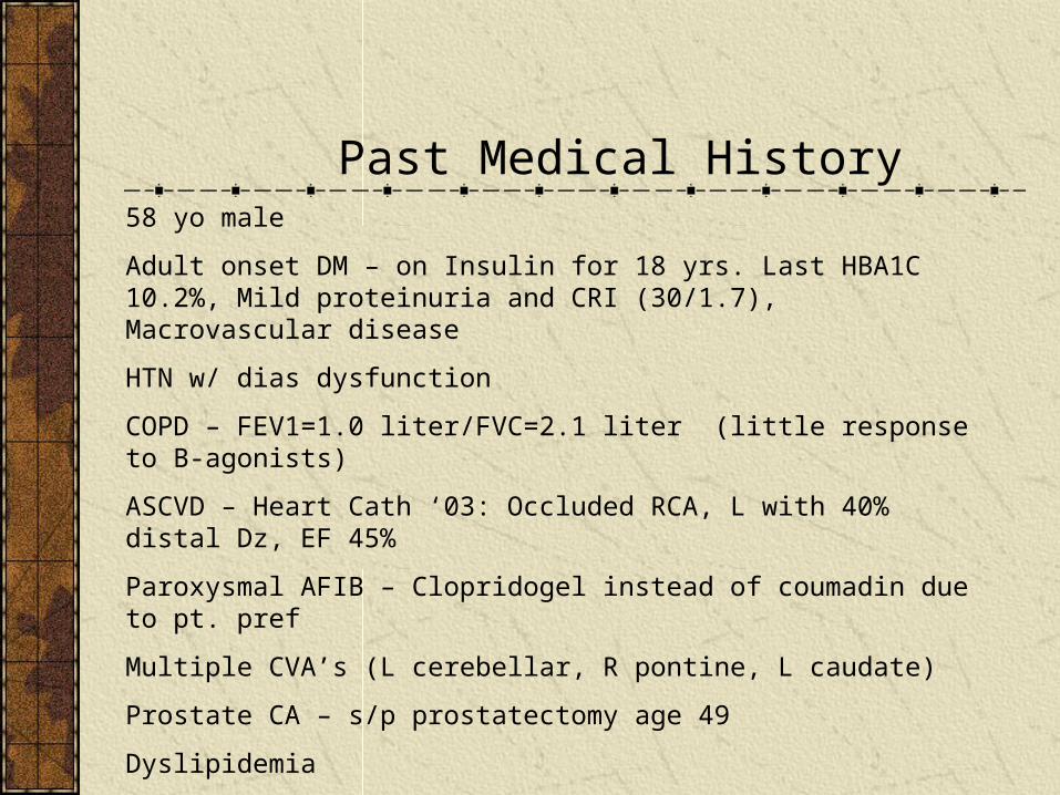
Past Medical History58 yo male
Adult onset DM – on Insulin for 18 yrs. Last HBA1C 10.2%, Mild proteinuria and CRI (30/1.7), Macrovascular disease
HTN w/ dias dysfunction
COPD – FEV1=1.0 liter/FVC=2.1 liter (little response to B-agonists)
ASCVD – Heart Cath ‘03: Occluded RCA, L with 40% distal Dz, EF 45%
Paroxysmal AFIB – Clopridogel instead of coumadin due to pt. pref
Multiple CVA’s (L cerebellar, R pontine, L caudate)
Prostate CA – s/p prostatectomy age 49
Dyslipidemia
80+ pack year Tobacco Abuse (Ongoing)
Depression/PTSD – intolerant of anything but MAOI Rx and Clonazepam
“Mononucleolis” with hepatitis while serving in Vietnam

Albuterol 2.5mg unit dose via nebulizer QID
Clopidogrel 75mg QD
Clonazepam 1mg TID
Furosemide 120mg po BID
NPH and Lispro Insulin
Metoprolol 25mg po bid
Pantorazole 40mg QD
Spironolactone 25mg QD
KCL 40meq po BID
Prednisone 10mg po QD
Phenelzine 30mg po BID
Medications

Family History
Mother died age 45 of Uterine CA
Father died age 76 sudden death
Brother died 67 lung CA and COPD
3 Healthy children ages 24 - 36

Admission 12/04CC: Lightheaded and weakHPI: Progressive nausea, some emesis, weakness, and chills. Not using his insulin or taking his meds for 5 days
Exam:Vitals Afeb, 148/82 supine, 108 irreg, 22, P.O. 96% (ra)
HEENT anicteric slcera, dry mm, neck “thick” no obvious jvd
Lungs diffusely diminished breath sounds
CV distant, irreg irreg, no murmur, no rubs
Abdm soft, nontender, nabs
Ext trace edema both ankles
Skin no jaundice or rashes
CNS nonfocal but slightly confused
Labs 12/04
WBC 15.2k, H/H 9.0/26.9, Plt 293kBun/cr 2.9/63 Nml lytesGlucose 390, Slight pos serum ketonesAst 6098, Alt 1601, Alb 2.8, Alk 386, Bili 0.9, Nh3 51Coags nmlTroponin I 1.94ECG: AFIB w/RVR, LVH, nonspecific ST

Imaging/Other Studies 12/04
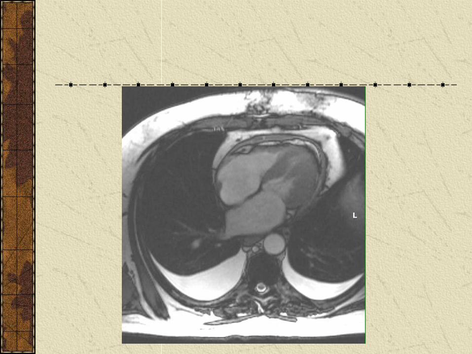
CT chest: COPD and pericardial effusion
U/S Abdm: nml liver and GB, no masses
Echocardiogram: Large pericardial effusion without tamponade, LVH with diastolic relaxation abnormality
RN: “He is becoming hypotensive”
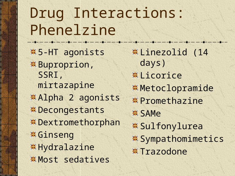
Drug Interactions: Phenelzine
5-HT agonists
Buproprion, SSRI, mirtazapine
Alpha 2 agonists
Decongestants
Dextromethorphan
Ginseng
Hydralazine
Most sedatives
Linezolid (14 days)
Licorice
Metoclopramide
Promethazine
SAMe
Sulfonylurea
Sympathomimetics
Trazodone
Hospital Course
Aggressively rehydrated
Oliguria and Azotemia resolved after 3 days
Liver function normalized over 3-4 days
Hepatitis serology negative
AFIB did not recur, not a candidate for anticoagulation
Discharge Diagnoses
Severe dehydration due to severe hyperglycemia/medication noncompliance and possible viral GEAcute Tubular NecrosisIschemic HepatitisCardiac “Enzyme Leak” Pericardial Effusion, Incidental/? viralParoxysmal AFIB

Heart disease and Hepatic dysfunction
Hepatic congestionTypically due to exacerbation of chronic CHFLiver enlarged and firm on examModest elevations in ALT, AST, LDH, GGT and sometimes alk phos, total bili, and slight decrease in albuminMild transient jaundice can occurChronic congestion can lead to “cardiac cirrhosis” with fibrosis of liver on biopsy
Cardiogenic Ischemic HepatitisMore acute and severe fall in cardiac output
(such as with an acute MI or Severe CHF)
Enzyme levels often >10x normal
Coagulopathy and Functional renal impairment can be associated
No specific marker for Dx, but typically the transaminases drop >50% in first 72hrs of onset
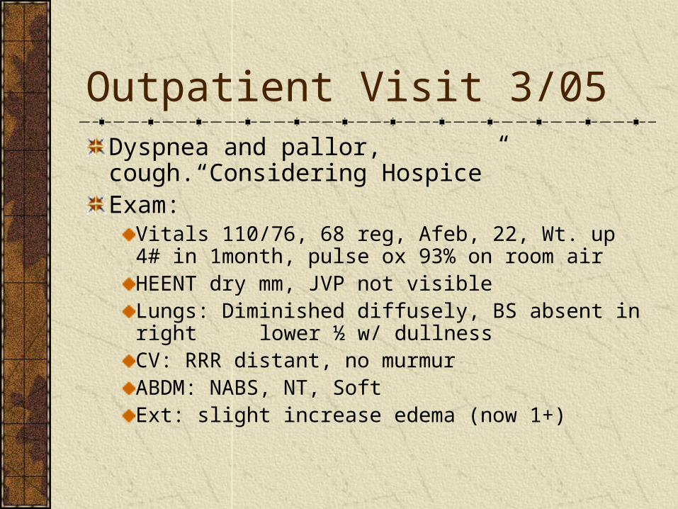
Outpatient Visit 3/05Dyspnea and pallor, cough.“Considering Hospice”Exam:
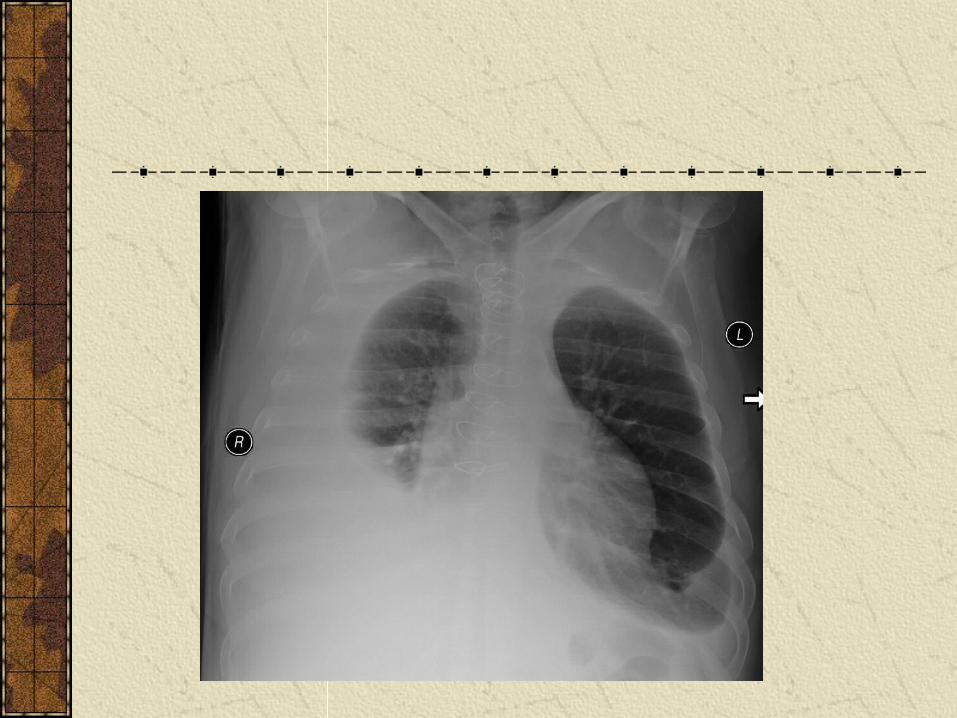
Vitals 110/76, 68 reg, Afeb, 22, Wt. up 4# in 1month, pulse ox 93% on room airHEENT dry mm, JVP not visibleLungs: Diminished diffusely, BS absent in right lower ½ w/ dullnessCV: RRR distant, no murmurABDM: NABS, NT, SoftExt: slight increase edema (now 1+)

Outpatient Labs 3/05
WBC 9.3k, H/H 10/34.3, Plt 220
BS 248, Bun/Cr 27/1.3, Nml lytes
Lfts nml except alk 346
TSH 1.70
BNP 467 (nml)
EKG unchanged
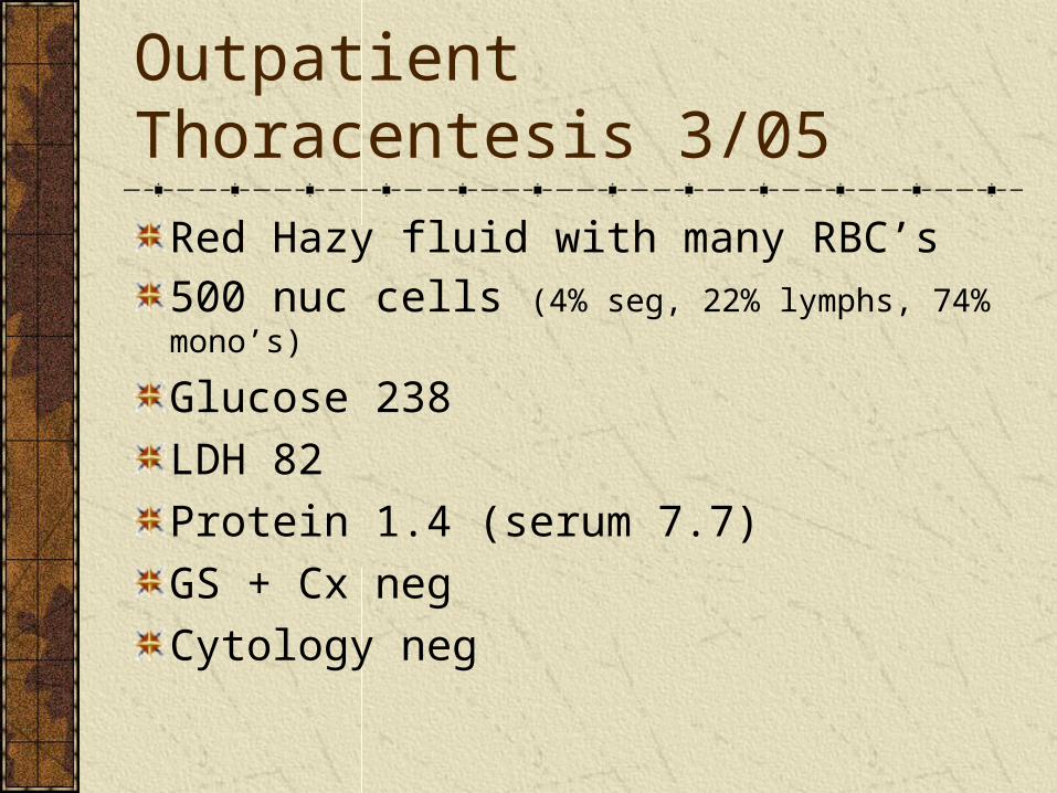
Outpatient Thoracentesis 3/05
Red Hazy fluid with many RBC’s
500 nuc cells (4% seg, 22% lymphs, 74% mono’s)
Glucose 238
LDH 82
Protein 1.4 (serum 7.7)
GS + Cx neg
Cytology neg

Outpatient Imaging 3/05
Echocardiogram LVH with no wall motion abnormalities, nearly resolved pericardial effusion.

Admission 4/4/05
CC:Worsening edema, dyspnea and falls
HPI: Despite increasing doses of furosemide, fluid build-up in legs has extended up to chest wall, now distended and bloated abdomen, weight is up 30#. Positive orthop and PND.
Dyspnea continues and is now associated with a cough. Cough is associated with dizziness and lightheadedness. Cough produces yellow sputum 1-2 tbsp per day.
Fell yesterday after a coughing spell and hit his R orbit; now has a “black eye”.
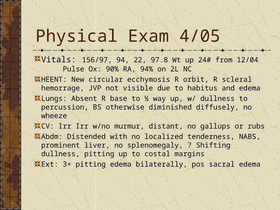
Physical Exam 4/05Vitals: 156/97, 94, 22, 97.8 Wt up 24# from 12/04 Pulse Ox: 90% RA, 94% on 2L NC
HEENT: New circular ecchymosis R orbit, R scleral hemorrage, JVP not visible due to habitus and edema
Lungs: Absent R base to ½ way up, w/ dullness to percussion, BS otherwise diminished diffusely, no wheeze
CV: Irr Irr w/no murmur, distant, no gallups or rubs
Abdm: Distended with no localized tenderness, NABS, prominent liver, no splenomegaly, ? Shifting dullness, pitting up to costal margins
Ext: 3+ pitting edema bilaterally, pos sacral edema
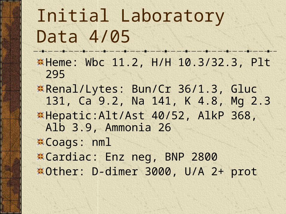
Initial Laboratory Data 4/05
Heme: Wbc 11.2, H/H 10.3/32.3, Plt 295Renal/Lytes: Bun/Cr 36/1.3, Gluc 131, Ca 9.2, Na 141, K 4.8, Mg 2.3Hepatic:Alt/Ast 40/52, AlkP 368, Alb 3.9, Ammonia 26Coags: nmlCardiac: Enz neg, BNP 2800Other: D-dimer 3000, U/A 2+ prot
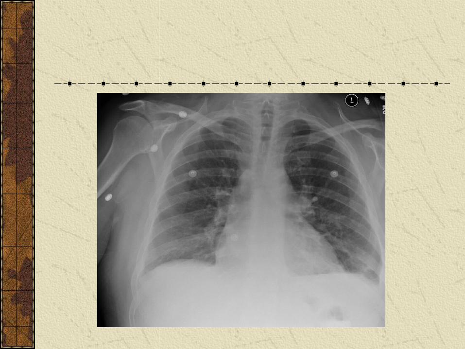

Imaging 4/05
CXR: R effusion, mild PVC
CT chest: No PE, R pleural eff, some obstructive changes
Head CT: no change
U/S abdm: normal except ascites
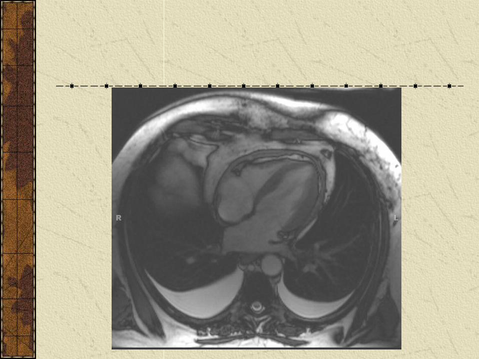
Echo: Nml wall motion, LVH w/ dias dysfunction, trace effusion
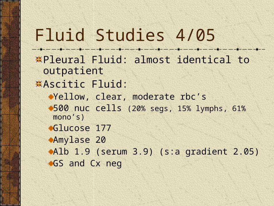
Fluid Studies 4/05
Pleural Fluid: almost identical to outpatientAscitic Fluid:
Yellow, clear, moderate rbc’s500 nuc cells (20% segs, 15% lymphs, 61% mono’s)
Glucose 177Amylase 20Alb 1.9 (serum 3.9) (s:a gradient 2.05)GS and Cx neg

Diuresed 30#
JVP now visible to 10cm
“A Diagnostic Study was Obtained”
“Doctor I have to get out of here !”
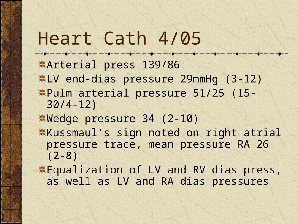
Heart Cath 4/05
Arterial press 139/86LV end-dias pressure 29mmHg (3-12)Pulm arterial pressure 51/25 (15-30/4-12) Wedge pressure 34 (2-10)Kussmaul’s sign noted on right atrial pressure trace, mean pressure RA 26 (2-8)Equalization of LV and RV dias press, as well as LV and RA dias pressures
Tissue Diagnosis:
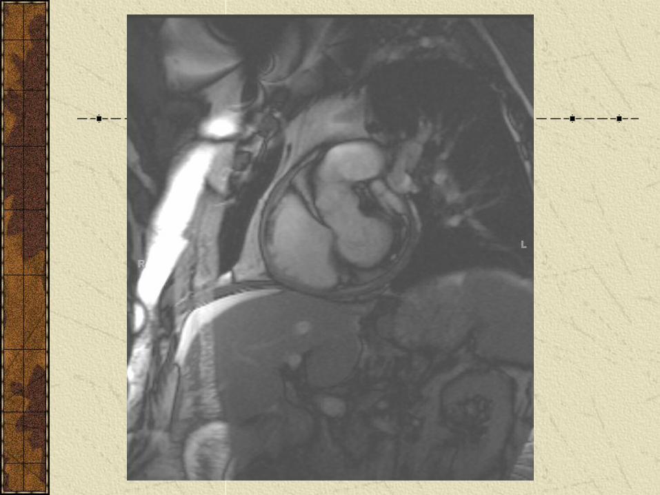
Fibrotic Pericardium, up to 5mm thick.
Pericarditis
Can present in 4 ways:Acute pericarditis
Incidental effusion
Tamponade
Constriction

Acute Pericarditis85-90% idiopathic, 1-4% viralRemainder of cases are post MI, other infx, AAA, trauma, neoplastic, post surgical or XRT, uremic, connective tissue disease or drug inducedClassic ECG changes: diffuse ST elevationPericardial rub pathognomonic (85% develop)Pericardiocentesis indicated for tamponade, or if strong suspicion of bacterial infx or neoplasmSerologic studies not very helpful (<10% dx)“Troponin Leak” occurs in 35-50%
Tamponade
Occurs in 15% idiopathic, but up to 60% with Tb, bacterial or neoplastic etiology
Presents with “Beck’s triad”• Hypotension
• Quiet heart sounds
• Increased Jugular venous pressure
Can also note compensatory tachycardia and pulsus paradoxus (fall in SBP >10 during insp)

Constrictive Pericarditis
Chronic fibrous and/or calcific thickening of the pericardium that leads to abnormaly elevated diastolic filling pressures
Most commonly idiopathic after acute or sub acute pericarditis (Tb still most common in undeveloped countries)
Post cardiac surgery and radiation therapy becoming more common
Constrictive Pericarditis…..
Clinical findings:Pulsatile hepatomegaly
Pericardial knock (early diastole)
Kussmaul’s Sign: JVP rises (or at least fails to fall) during inspiration, due to separation of the cardiac pressures from the thoracic pressure changes in respiration

Constrictive Pericarditis…..Differential Diagnosis
Other causes of right heart failure• Restrictive Cardiomyopathy• PE or Pulm HTN• Right ventricular infarction• Mitral stenosis or Tricuspid Disease
Cirrhosis or Hepatic Vein ThrombosisAcute Renal Failure or Nephrotic syndromeSVC obstruction or Lymph obstructionMyxedemaDrug Induced (Ca channel, minoxidil, steroids, “glitazones”, NSAIDs,)

Constrictive Pericarditis…..Diagnosis
Unfortunately clinical findings not very specific
Key echo findings are that of a thickened pericardium, a septal “bounce”, inspiratory decrease in pulmonary venous flow, and normal relaxation indices.
MRI is 88% sens, 100% specific using same criteria above
Cath findings that are most specific are equalization of RV and LV end dias pressures.
No widely accepted “gold standard”
Constrictive Pericarditis….
Treatment: PericardectomyUse caution with diureses pre-op
1 month follow up