Embed Size (px)
Citation preview

[CANCER RESEARCH 48, 14-18, January 1, 1988)
Interactions of Putative Estrogens with the Intracellular Receptor Complex inMouse Leydig Cells: Relationship to Preneoplastic HyperplasiaR. Lloyd Juriansz,1 Robert A. Huseby, and R. Bruce Wilcox2
Department of Biochemistry, Loma Linda University, Loma Linda, California 92350
ABSTRACT
The interaction of 14 steroidal and nonsteroidal estrogen agonists andantagonists with the intracellular estrogen receptor system was examinedin cell suspensions prepared from the testes of mice that develop malignant Leydig cell tumors after prolonged estrogen administration. Theability of these substances to stimulate DNA synthesis in short-term (3-day) studies and to provoke Leydig cell hyperplasia with prolonged (3-mo) administration was also measured. Our data were consistent withthe proposal that, in Leydig cells, the carcinogenic effects of estrogensare mediated through the intracellular receptor complex that results in alocalization of hormone bound to chromâtin and nuclear matrix.
All tested compounds displaced 170-[3H]estradiol from the cytosolic
estrogen receptor, but to varying degrees; and there was no discerniblerelationship between their ability to compete for this receptor and theirefficacy in stimulating DNA synthesis. The effect of the test compoundson the levels of estrogen receptor in cytosol and in nuclei was measuredby |'II|ostradio! exchange. 17/8-Estradiol, equilin, 17a-ethinylestradiol,
diethylstilbestrol, hexestrol, dienestrol, coumestrol, and nafoxidine provoked a complete estrogen receptor response: acutely a decrease in thelevel of cytosolic estrogen receptor and an increase in the nuclear estrogenreceptor. All of these substances acutely stimulated DNA synthesis.Tamoxifen, clomiphene, and nitromifene provoked a decrease in cytosolicreceptor but no increase in demonstrable nuclear estrogen receptor. 17a-EstradioL, mestranol, and estriol did not significantly alter the levels ofestrogen receptor in cytosol or nuclei. Only those substances that increased measurable nuclear estrogen receptor also acutely stimulatedDNA synthesis.
Chronic (3-mo) treatment of 2-mo-oId male BALB/c mice with diethylstilbestrol, 17/3-estradiol, ethinylestradiol, and nafoxidine led to Leydigcell hyperplasia. Chronic mestranol treatment also provoked Leydig cellhyperplasia; this is most probably due to induction of liver metabolismof mestranol to ethinylestradiol. Chronic treatment with 17a-estradiol,tamoxifen, and clomiphene failed to produce significant histologie alchanges in the testes. Only chronic administration of those substancesthat exhibited a complete estrogen receptor response and acutely stimulated DNA synthesis produced Leydig cell hyperplasia.
INTRODUCTION
Malignant neoplasms arising from mature testicular Leydigcells can be induced readily in genetically susceptible mice bychronically administering estrogenically active compounds,either steroidal or nonsteroidal in chemical structure (1, 2).Twenty-four h after the initiation of treatment of such susceptible animals, but not of nonsusceptible ones, an increasedreplicative synthesis of DNA can be demonstrated in the Leydigcells (3-5). This new DNA synthesis increases rapidly to peakduring the third day of treatment but is not associated with acorresponding increase in mitotic activity. It then declinessignificantly until several mo later when neoplasms form andincreases in both DNA synthesis and mitotic activity becomeevident. Although a functional hypophysis is required for theproduction of Leydig cell hyperplasia and subsequent neoplasia
Received 12/29/86; revised 6/30/87; accepted 9/28/87.The costs of publication of this article were defrayed in part by the payment
of page charges. This article must therefore be hereby marked advertisement inaccordance with 18 U.S.C. Section 1734 solely to indicate this fact.
1Portions of these investigations were submitted as a dissertation in partial
fulfillment of the requirements for the degree of Doctor of Philosophy.1To whom requests for reprints should be sent.
(3), the initial spurt of DNA synthesis occurs independent ofpituitary function (6). Actual tumor induction, however, resultsfrom action of unmetabolized estrogen directly upon the Leydigcells (7). These cells in both a susceptible and a nonsusceptiblestrain of mouse contain an ER3 complex. The cells from the
two strains differ significantly in that nuclei from susceptibleanimals bind more estrogen, and the proportion of the nuclearER that remains fixed to the nuclear matrix in solutions ofhigh salt concentration is greater in the tumor-susceptible animals (8). Furthermore this fraction of the ER increases inamount per Leydig cell as estrogen treatment of susceptiblemice continues over a 3-mo period and hyperplasia is initiated(9).
The present study was undertaken to investigate the reactionof a number of putative estrogen agonists and antagonists withthe ER system in the Leydig cells of the tumor-susceptibleBALB/c strain. Our specific objective was to correlate thecytosolic ER to nuclear acceptor interaction with the earlyinitiation of DNA synthesis and with the subsequent development of Leydig cell hyperplasia.
MATERIALS AND METHODS
Animals. Young adult BALB/c mice from the colony in the Biochemistry Department of Loma Linda University were used in most of theexperiments reported here. These animals are descended from breedingstock obtained from the colony at the AMC Cancer Research Center,Lakewood, CO. Occasionally BALB/c BYJ mice were purchased fromthe Jackson Laboratories (Bar Harbor, ME).
Chemicals. DES, dienestrol, hexestrol, equilin, 17/3-estradiol, 17«-estradiol, estriol, 17a-ethinylestradiol, mestranol, tamoxifen, clomiphene, and nafoxidine were purchased from Sigma Chemical Co., St.Louis, MO. Nitromifene was purchased from Warner Lambert Co.,Ann Arbor, MI. Leibovitz' L-15 medium and Dulbecco's phosphate-
buffered saline (0.101 g/liter MgCl-6H2O, 0.2 g/liter KCL, 0.2 g/literKH2PO4, 8 g/liter NaCl, 1.5 g/liter K2HPO4, pH 7.0) were purchasedfrom Sigma. Sephadex G-50 and blue dextran were from PharmaciaInc., Uppsala, Sweden. Hydroxylapatite was from Bio-Rad Laboratories, Richmond, CA. [me/A.y/-:'H]Thymidine was purchased from ICN
Chemical and Radioisotope Division, Irvine, CA. 17/3-[2,3,6,7,16,17-3H]Estradiol, 140-170 Ci/mmol, was purchased from Amersham Cor
poration, Arlington Heights, IL, and purified by chromatography onLipidex 5000 (Packard Instrument Company, Downers Grove, IL) inchloroform/hexane. Liquid scintillation solutions were Betaphase(Westchem, San Diego, CA) and a cocktail composed of 4 liters toluene,21 ml ethanol, and 16 g Omnifluor (New England Nuclear, Boston,MA). Beckman tissue solubilizer BTS-450 was obtained from BeckmanInstruments Inc., Fullerton, CA. All other chemicals were analyticalreagent grade.
Administration of Test Substances. In the experiments measuring theeffect on DNA synthesis, estrogens and antiestrogens were administeredas 10-mg pellets of 10% test substance in cholesterol implanted s.c.(10). In most cases, the test substance fused with the cholesterol whenthe two compounds were mixed and heated to 200°C.Coumestrol wasan exception: it formed a slurry with cholesterol at 200°C.In experi-
3The abbreviations used are: ER, estrogen receptor; DES, diethylstilbestrol;E2, 17/3-estradiol; EE, 17a-ethinylestradiol; TEM, 1.0 min Tris, 1.5 mm EDTA,2.0 HIMmercaptoethanol, pH 7.4; ](„„concentration of unlabeled substance thatdisplaces 50% of [3H)E2;SV-CGC, seminal vesicle-coagulating gland complex.
14
on April 10, 2019. © 1988 American Association for Cancer Research. cancerres.aacrjournals.org Downloaded from
ER IN LEYDIG CELL PRENEOPLASIA
ments measuring receptor translocation, the test substances were dissolved in 70% ethanol in 0.167 M NaCl at a concentration of 2.5 mg/ml and 100 M'(250 Mg)were injected s.c.
Preparation of Cell Suspensions. Testis cell suspensions were prepared by a modification of the mechanical dispersion procedure described by Schumacher et ai. (11). Decapsulated testes, six to a flask,were placed in 10 ml L-15 medium, pH 7.4, in 50-ml Erlenmeyer flasksand dispersed by gently drawing them into and expelling them 50 timesfrom a 5 x 400-mm Teflon tube attached to a 50-ml syringe. The flaskswere then placed in a Dubnoff metabolic shaking incubator (PrecisionScientific, Chicago, IL) at 24°Cfor 15 min with moderate agitation.
The flask contents were transferred to 16 x 125-mm Falcon (Becton,Dickinson and Co., Oxnard, CA) screw-capped plastic test tubes andthe tubes placed on an Ames (Miles Laboratory Inc., Elkhart, IN)aliquot mixer for 1 min. After mixing, the tubes were placed upright incrushed ice. After 30 sec the supernatant portions were transferred withPasteur pipets of 50-ml polypropylene centrifuge tubes through 62-/imnylon mesh (Small Parts, Inc., Miami, FL). L-15 medium (5 ml) wasadded to the tubes and the mixing-decanting repeated until 50 ml ofsupernatant had been collected. The cells were collected by centrifuga-tion at 500 x g for 5 min at 4"C. The pelleted cells were suspended in
L-15 for thy inidino incorporation studies and in TEM buffer for preparation of cytosolic and nuclear fractions.
Preparation of Cytosolic and Nuclear Fractions. Cell suspensions werehomogenized in conical Teflon-glass tissue grinders (Kontes Glass Co.,Vineland, NJ) at 0-4°C.The homogenates were centrifugea at 800 xg for 10 min at 4°Cin the type SW 50.1 rotor of a Beckman L5-75B
Ultracentrifuge (Beckman Instruments Co. Inc., Fullerton, CA). Super-natants were recentrifuged at 105,000 x g for l h at 4°C,and the
supernatant from this step was collected without including the superfici;il lipid layer. A low-molecular-weight inhibitor of estrogen bindingto cytosolic ER was removed by Sephadex G-50 chromatography asdescribed by Sato et al. (12). The Sephadex eluates were treated withdextran-coated charcoal to remove any endogenous estrogens (13) andused for studies measuring cytosolic ER binding and exchange.
Pellets from the first homogenate centrifugation were resuspendedin 2 ml 0.2% Tween 80 and centrifuged at 1000 x g for 10 min at 4°C.
The supernatants were discarded and the pellets washed twice byresuspending in 2 ml TEM buffer and centrifuging. The final pelletswere resuspended in 5.5 ml TEM buffer and used as the nuclear fraction.
Binding to Cytosolic ER. The binding of E2 to cytosolic ER wasmeasured by a modification of the exchange assay described by Katze-nellenbogen et ai. (14). [3H]E2was dissolved in ethanol and then dilutedwith TEM buffer to the desired concentrations. [3H]E2 (30 /»I;1.29 xIO"8 M to 1.55 x 10~7 M was added to 1.5-ml Eppendorf centrifuge
tubes (Brinkman Instruments Inc., Orange, CA). Nonspecific bindingwas measured in replicate samples that contained a 300-fold excess ofunlabeled I...dissolved in dimethylformamide and carried through serialdilutions with dimethylformamide and TEM buffer to the desiredconcentrations. The tubes were capped and placed in a 4°Ccold room
for 2 h. All subsequent operations were carried out in the cold room.Cytosol (125 /»I;0.125-0.15 mg protein) was added to each of the tubeswhich were then capped and incubated for 18 h with continuousagitation in an Eberbach shaker (Eberbach Instruments Inc., AnnArbor, MI). Hydroxylapatite (250 /il; 1 mg/ml in TEM buffer) wasadded to each tube. The tubes were incubated at 4°Cfor 15 min, with
vortexing every 3 min, and centrifuged at 8800 x g for 5 min. Thesupernatants were removed by aspiration and the pellets washed 3 timesby resuspending in 1 ml TEM and centrifuging. The washed hydrox-ylapatite pellets were suspended in 0.5 ml ethanol and the tubesincubated at 45°Cfor 2 h with occasional vortexing. The tubes werecooled to 4°Cand centrifuged. Duplicate 100-fil portions of the super
natants were transferred to 7-nil glass scintillation vials, 7 ml Betaphasewere added to each, and the samples were counted in a Beckman LS-7500 liquid scintillation counter (Beckman Instruments, Fullerton,CA).
The ability of several putative estrogen agonists and antagonists tocomplete with [3H]E2 for binding to cytosols was also measured. The
test substances were dissolved in ethanol or dimethylformamide andcarried through serial dilutions with ethanol, dimethylformamide, and
TEM buffer to the desired concentrations. [3H]E2(25 /J; 6.2 x 10~9M)and the competing substances (5 /il; 3.1 x 10~4 M to 3.1 x 10~'°M)
were added to appropriate tubes, followed by cytosol (125 /il)- Incubation of the tubes and quantification of the bound [3H]E2 were carried
out as described above.Binding to Nuclear ER. The binding of [3H]E2to nuclei was measured
by an adaptation of the exchange assay of Anderson et al. (15) underconditions that measure total available receptor sites. [3H]E2 (30 /il;1.29 x IO"8 M to 1.55 x IO"7 M) was added to 1.5-ml Eppendorf
centrifuge tubes, followed by nuclear suspension (125 /il; 0.2-0.3 mgprotein). Nonspecific binding was measured in a series of replicatescontaining a 300-fold excess of unlabeled E2. The tubes were cappedand incubated at 37°Cfor l h with moderate agitation in a New
Brunswick Controlled Environment Incubator Shaker (New BrunswickScientific Co. Inc., Edison, NJ), then cooled to 4°C.The tubes werecentrifuged at 1000 x g for 10 min at 4°Cand the supernatants removed
by aspiration. The pellets were washed twice by resuspending in 0.5 mlTEM buffer and centrifuging. The washed pellets were suspended in0.5 ml of BTS-450 and incubated at 45°Cfor 2 h with occasional
mixing. After cooling, duplicate 100-fil portions were taken for liquidscintillation counting in 10 ml of the Omnifluor cocktail.
Cytosolic and Nuclear ER Levels. In experiments measuring thetranslocation of ER from cytoplasm to nucleus, the cytosolic andnuclear ER were quantified by [3H]E2 exchange. Eight-wk-old mice
were injected s.c. with 250 ng test substance dissolved in 100 /¿I70%ethanol-30% 0.167 M NaCl. Control animals received vehicle only. Atselected time intervals the animals were killed and testis cell fractionsprepared as described above. Total binding was measured by incubatingcytosols with 15 nM [3H]E2at 4°Cfor 18 h; nuclei were incubated with15 nM [3H]E2 for 30 min at 30°C.Nonspecific binding was estimatedby similar incubations with the addition of 4.5 MME2. [3H]E2bound to
cytosols or nuclei was quantified by the procedures previously described.The amount of exchangeable ER was calculated from the specificbinding knowing the specific activity of the labeled E2 and expressed asfmol/mg protein. Protein was measured by the method of Lowry et al.(16).
l'I l|Tliymiiliiif Incorporation in Vitro. The procedure used here was
similar to that used by Spruance et al. (5). One ml cell suspension inL-15 was incubated in polyethylene scintillation vials with [methyI-3H]-thymidine (10 /iCi; .16 nmol) for 1 h at 37°Cin a Dubnoff Metabolic
Shaking Incubator. The reaction was stopped by adding an equalvolume of 8% formaldehyde and homogenizing the cells in a conicalTeflon-glass tissue grinder. The homogenization and subsequent stepswere carried out at 4°C.Samples of the homogenate were diluted inDulbecco's phosphate-buffered saline for protein analysis, and 2 portions of 50 n\ were digested with BTS-450 and counted to determinethe total radioactivity in each incubation. The remaining homogenateswere transferred to graduated conical centrifuge tubes, and the acid-insoluble material was precipitated by the addition of 3 times theirvolume of 0.5 M perchloric acid. The precipitates were fractionatedinto RNA, DNA, and protein, and the radioactivity in each fractionwas quantified by the procedure described and validated in an earlierpublication (17) modified slightly to accommodate the present tissues.
RESULTS
Cytosolic and Nuclear ER. The presence of ER in Leydig cellcytosolic and nuclear fractions as previously reported (8,9) wasconfirmed in our preparations and characterized by carryingout the estrogen-binding assay with concentrations of [3H]E2
ranging from 2.5 to 30 nM. Both fractions exhibited saturable,specific binding. The Kas were estimated from double-reciprocalplots. The cytosolic receptor had a Ka of 6.5 x 10~8M, and thenuclear receptor had a Kt of 1.25 x 10~8M.
Competition of Test Substances for Cytosolic ER. Most of thesubstances studied were not readily available in radiolabeledform. Under the proper experimental conditions, that is, if theconcentrations of unlabeled ligand and of binding sites aremuch lower than the Ka, the concentration of unlabeled sub-
15
on April 10, 2019. © 1988 American Association for Cancer Research. cancerres.aacrjournals.org Downloaded from

ER IN LEYDIG CELL PRENEOPLASIA
stance that will displace 50% of the radiolabeled estradici fromthe ER (the K\„)provides an estimate of the ability of thatsubstance to compete with estradici for the ER (18).
To test the validity of this method the displacement ofradiolabeled E2 (1 x 10~9 M) from cytosolic ER by non labeledE2 was measured. The IC50 for E2 was 8.75 x 10~" M. Since
this was very close to the KAfrom the double-reciprocal plot ofthe saturation curve, we concluded that displacement of [3H]E2
would provide a valid estimate of relative affinity of the testsubstances for the cytosolic ER in this system. Competitioncurves for DES, E2, and EE are shown in Fig. 1.
The ability of a number of steroidal and nonsteroidal estrogens and of antiestrogens to complete for the cytosolic ER wasmeasured, and the results are shown in Table 1.
ER Redistribution. The effect of test substances injected invivo on the distribution of ER between cytosolic and nuclear
^ 120
Log Competitor Cone. (M)Fig. l. Displacement of 17/3-[3H]estradiol (1.0 nM) from the cytosolic estrogen
receptor by three compounds that produce Leydig cell hyperplasia: (O) DES, (C)E2, and (•)EE. Cytosol was incubated with label and competitor for 18 h andbound ligand separated by adsorption to hydroxylapatite. Each point, average of5 determinations; bars, standard deviation.
Table 1 Competition of estrogens and antiestrogens for the cytosolic estrogenreceptor
Concentrations of test substances ranging from 10 " to 10s M were incubatedwith cytosol and 1 nM 170-[3H]estradiol for 18 h at 4V and the bound ligandseparated by adsorption to hydroxylapatite as described in "Materials and Methods."
Substance17/3-Estradiol
EquilinEthinylestradiolMestranol17a-EstradiolEstriolDiethylstilbestrol
DienestrolHexestrolICM,
MSteroids8.8
x 10""3.6 x IO"71.7x 10"*7.9 X 10""9.9 x IO"74.8 xIO"6Stilbene
derivatives3.4x 10"'
2.3 x IO"71.2 x 10"'Affinity
relativeto 17/3-estradioI1.0
0.240.050.010.09
0.0226.0
0.390.74
Nonsteroidal plant estrogen(•(millesimi 4.5 X IO"7 0.19
Triphenylethylene derivativesTamoxifen 1.3 x 10"* 0.01Clomiphene 3.3 x 10"* 0.03Nitromifene 1.2 x 10"* 0.08
Diphenyldihydronaphthalene derivativeNafoxidine 3.1 x 10"* 0.03
fractions was measured. The time course of the change in ERlevels in cytosolic fractions is shown in Fig. 2 for the steroidsli: and 17a-estradiol, and for DES, tamoxifen, and nafoxidine.The time course of the change in ER levels in the nuclearfractions for the same substances is shown in Fig. 3. Themaximum change in ER in both cytosolic and nuclear fractionsfor all of the compounds tested are summarized in Table 2.
Stimulation of DNA Synthesis. The next set of experimentstested the ability of these compounds administered in vivo tostimulate DNA synthesis. This was done by quantifying [3H]-
thymidine incorporation in vitro into the DNA of testis cellsuspensions obtained 3 days after initiation of treatment. Fourgroups of animals were used in each experiment: a controlgroup that received cholesterol pellets, a "positive control"
group that received 10% DES 90% cholesterol pellets: and twogroups that received 10% "test" substance: 90% cholesterol
pellets. Differential cell counts revealed that suspensions prepared as outlined in "Materials and Methods" contained vary
ing proportions of germinal cells, some of which were undergo-
(0
O 120I0 100
£ 80
-O 60
1 40
LU~-, 0I
Hours Post InjectionFig. 2. Changes in cytosolic ER with time after in vivo injection of several
putative estrogens: (O) DES, (C) E2, (•)17a-estradiol, (A) nafoxidine, (A)tamoxifen. Mice were injected with 250 »igtest substance at 0 h and killed at 1,3, and 6 h. Testis cell cytosols were prepared and the estrogen receptor wasmeasured by [3H]estradiol exchange. Control animals were injected with saline
and killed at 0 h. Each point, average of 5 replicates; bars, standard deviation.
°
•aC3OCD
M
LU
300
250
200
150
100
50
0
0123456
Hours Post InjectionFig. 3. Changes in nuclear ER with time following in vivo injection of several
putative estrogens: (O) DES, (C) E2, (•)17a-estradiol, (A) nafoxidine, (A)tamoxifen. Animals were injected with 250 ^g test substance at O h and killed at1, 3, and 6 h. Testis cell nuclei were prepared and the estrogen receptor wasmeasured by |'H]estradiol exchange. Control animals were injected with saline
and killed at 0 h. Each pnint, average of 5 replicates; bars, standard deviation.
16
on April 10, 2019. © 1988 American Association for Cancer Research. cancerres.aacrjournals.org Downloaded from
ER IN LEYDIG CELL PRENEOPLASIA
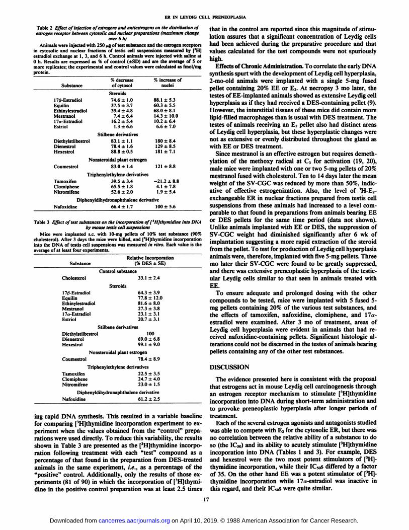
Table 2 Effect of injection of estrogens and antiestrogens on the distribution ofestrogen receptor between cytosolic and nuclear preparations (maximum change
over 6 h)Animals were injected with 250 fig of test substance and the estrogen receptors
in cytosolic and nuclear fractions of testis cell suspensions measured by | 'I l|
estradici exchange at 1, 3, and 6 h. Control animals were injected with saline at0 h. Results are expressed as % of control (±SD)and are the average of 5 ormore replicates; the experimental and control values were calculated as fmol/mgprotein.
Substance17/3-Estradiol
EquilinEthinylestradiolMestranol17,.-
FstradiniEstriolDiethylstilbestrol
DienestrolHexestrol%
decreaseofcytosolSteroids74.6
±1.037.5 ±3.739.4 ±4.8
7.4 ±6.416.2 ±5.4
1.3±6.6Stilbene
derivatives83.1±1.178.4±1.6
88.8 ±0.5%
increase ofnuclei88.1
±5.360.3 ±5.568.0 ±8.114.3 ±10.010.2 ±6.46.6 ±7.0180
±8.4129 ±8.5181 ±7.1
Coumestrol
TamoxifenClomipheneNitromifene
Nonsteroidal plant estrogen83.0 ±1.4 121 ±8.8
Triphenylethylene derivatives39.5 ±3.4 -21.2 ±8.865.5 ±1.8 4.1 ±7.852.6 ±2.0 1.9 ±5.4
Diphenyldihydronaphthalene derivativeNafoxidine 66.4 ±1.7 100 ±5.6
Table 3 Effect of test substances on the incorporation off'HJthymidine into DNA
by mouse testis cell suspensionsMice were implanted s.c. with 10-mg pellets of 10% test substance (90%
cholesterol). After 3 days the mice were killed, and | 'I Ijiliyniiilinc incorporation
into the DNA of testis cell suspensions was measured in vitro. Each value is theaverage of at least four experiments.
SubstanceRelative Incorporation
(% DES ±SE)
Control substanceCholesterol
Steroids17/3-EstradiolEquilinEthinylestradiolMestranol17a-EstradiolEstriol
33.1 ±2.4
64.3 ±3.977.8 ±12.081.6 ±8.027.3 ±3.823.1 ±3.120.7 ±3.1
Stilbene derivativesDiethylstilbestrolDienestrolHexestrol
10069.0 ±6.899.1 ±9.0
Nonsteroidal plant estrogenCoumestrol 78.4 ±8.9
Triphenylethylene derivativesTamoxifen 22.5 ±3.5Clomiphene 24.7 ±4.0Nitromifene 23.0+1.5
Diphenyldihydronaphthalene derivativeNafoxidine 61.2 ±2.5
ing rapid DNA synthesis. This resulted in a variable baselinefor comparing [3H]thymidine incorporation experiment to experiment when the values obtained from the "control" prepa
rations were used directly. To reduce this variability, the resultsshown in Table 3 are presented as the ['HJthymidine incorporation following treatment with each "test" compound as apercentage of that found in the preparation from DES-treatedanimals in the same experiment, i.e., as a percentage of the"positive" control. Additionally, only the results of those experiments (81 of 90) in which the incorporation of [3H]thymi-
dine in the positive control preparation was at least 2.5 times
that in the control are reported since this magnitude of stimulation assures that a significant concentration of Leydig cellshad been achieved during the preparative procedure and thatvalues calculated for the test compounds were not spuriouslyhigh.
Effects of Chronic Administration. To correlate the early DNAsynthesis spurt with the development of Leydig cell hyperplasia,2-iiKi old animals were implanted with a single 5-mg fusedpellet containing 20% EE or E2. At necropsy 3 mo later, thetestes of EE-implanted animals showed as extensive Leydig cellhyperplasia as if they had received a DES-containing pellet (9).However, the interstitial tissues of these mice did contain morelipid-filled macrophages than is usual with DES treatment. Thetestes of animals receiving an E2 pellet also had distinct areasof Leydig cell hyperplasia, but these hyperplastic changes werenot as extensive or evenly distributed throughout the gland aswith EE or DES treatment.
Since mestranol is an effective estrogen but requires demeth-ylation of the methoxy radical at C3 for activation (19, 20),male mice were implanted with one or two 5-mg pellets of 20%mestranol fused with cholesterol. Ten to 14 days later the meanweight of the SV-CGC was reduced by more than 50%, indicative of effective estrogenization. Also, the level of 3H-E2-
exchangeable ER in nuclear fractions prepared from testis cellsuspensions from these animals had increased to a level comparable to that found in preparations from animals bearing EEor DES pellets for the same time period (data not shown).Unlike animals implanted with EE or DES, the suppression ofSV-CGC weight had diminished significantly after 6 wk ofimplantation suggesting a more rapid extraction of the steroidfrom the pellet. To test for production of Leydig cell hyperplasiaanimals were, therefore, implanted with five 5-mg pellets. Threemo later their SV-CGC were found to be greatly suppressed,and there was extensive preneoplastic hyperplasia of the testic-ular Leydig cells similar to that seen in animals treated withEE.
To ensure adequate and prolonged dosing with the othercompounds to be tested, mice were implanted with 5 fused 5-mg pellets containing 20% of the various test substances, andthe effects of tamoxifen, nafoxidine, clomiphene, and 17a-estradiol were examined. After 3 mo of treatment, areas ofLeydig cell hyperplasia were evident in animals that had received nafoxidine-containing pellets. Significant histologie alterations could not be discerned in the testes of animals bearingpellets containing any of the other test substances.
DISCUSSION
The evidence presented here is consistent with the proposalthat estrogens act in mouse Leydig cell carcinogenesis throughan estrogen receptor mechanism to stimulate [3H]thymidineincorporation into DNA during short-term administration andto provoke preneoplastic hyperplasia after longer periods oftreatment.
Each of the several estrogen agonists and antagonists studiedwas able to compete with E2 for the cytosolic ER, but there wasno correlation between the relative ability of a substance to doso (the IC50) and its ability to acutely stimulate [3H]thymidine
incoporation into DNA (Tables 1 and 3). For example, DESand hexestrol were the two most potent stimulators of [3H]-
thymidine incorporation, while their IC5os differed by a factorof 35. On the other hand EE was a potent stimulator of [3H]-thymidine incorporation while 17a-estradiol was inactive inthis regard, and their IC50s were quite similar.
17
on April 10, 2019. © 1988 American Association for Cancer Research. cancerres.aacrjournals.org Downloaded from
ER IN LEYDIG CELL PRENEOPLASIA
When the same series of compounds was tested for theireffect on the levels of exchangeable cytosolic and nuclear ER,only those substances that acutely provoked a complete estrogen receptor response, that is, both a decrease in the cytosolicER and an increase in the nuclear ER, also stimulated | 'I I|-
thymidine incorporation into DNA (Tables 2 and 3). In thecase of the steroids, administration of E2, equilin, and EE wasfollowed by a marked increase in nuclear ER and [3H]thymidineincorporation, while mestranol, 17a-estradiol, and estriol provoked only a slight increase in nuclear ER and actually depressed [3H]thymidine incorporation below the control. The
stilbene derivatives diethylstilbestrol, hexestrol, and dienestrolwere all highly potent in stimulating increases in both nuclearER and [3H|thymidine incorporation. The triphenylethylene
antiestrogens tamoxifen, clomiphene, and nitromifene depressed the cytosolic ER but failed to elevate exchangeablenuclear ER or stimulate [3H]thymidine incorporation. The only
putative antiestrogen which provoked a complete estrogen receptor response was the diphenyldihydronaphthalene derivativenafoxidine, and it stimulated [3H]thymidine incorporation
acutely.The data presented here provide no explanation for the failure
of the triphenylethylene estrogen antagonists to provoke anincrease in the exchangeable nuclear ER even though theydepressed the cytosolic ER. One possibility is that when thesesubstances bind to the receptor they induce a conformationsuch that the [3H]E2 does not readily exchange with the bound
ligand and thus some of the receptor would not be detected bythis methodology, whether it is located in the cytosol or thenucleus. In this regard, tamoxifen, which has to be monohy-droxylated to be biologically effective, was the only compoundthat decreased the level of exchangeable nuclear ER. Rat uterinenuclear ER labeled with [3H]4-hydroxytamoxifen is much moreresistant to proteolytic digestion than ER labeled with [3H]-estradiol or [3H]diethylstilbesterol (21). This suggests a different conformation of the nuclear ER when it is bound to the 4-hydroxytamoxifen than when it is bound to its natural steroidligand or to the estrogen agonist DES.
When 2-mo-old male BALB/c mice were subjected to chronic(3-mo) treatment with several of these substances, Leydig cellhyperplasia occurred in animals treated with DES, E2, EE, andnafoxidine, all substances which provoked a complete estrogenreceptor response and acutely stimulated [3H]thymidine incorporation. 17a-Estradiol, tamoxifen, and clomiphene failed toproduce any significant histologie changes in the testes; theseare substances which neither increased the nuclear ER norstimulated [3H]thymidine incorporation.
The compound that did not fit this pattern was mestranol,which was inactive in provoking the short-term responses butwhen given continuously for 3 mo did lead to extensive Leydigcell hyperplasia. The estrogenic activity of mestranol in humansis attributed to the fact that it is demethylated to EE (19, 20),a reaction which also occurs in rat liver microsomes (22). Thedemethylase activity of these microsomes is inducible, since itis increased by pretreatment of the animals with phénobarbital.It may be that our mice lacked sufficient demethylase activityto convert mestranol to the active product during the first daysof administration but that the enzyme system was induced bylonger (10- to 14-day) treatment with mestranol, thus providingthe animals with physiologically active levels of EE.
A previous study demonstrated that the carcinogenic effectsof E2 on Leydig cells resulted from actions of the unmetabolizedsteroid directly on testicular tissues of genetically susceptible
mice (7). The present studies support the proposal that biologically active estrogens effect malignant transformation of Ley-dig cells through the intracellular receptor mechanism thatresults in binding of the active compound to chromatin and tothe nuclear matrix (8, 9). Within 3 days, this results in a surgeof replicative DNA synthesis and, upon chronic exposure, provokes a hyperplastic overgrowth of Leydig cells eventuallygiving rise to the evolution of malignant neoplasms.
ACKNOWLEDGMENTS
We thank Barbara Peterson for excellent technical assistance.
REFERENCES
1. Añilenoui. H. B., Shimkin, M. B., and Canter, H. Y. Susceptibility of seveninbred strains and the Fl hybrids to estrogen-induced testicular tumors andoccurrence of spontaneous testicular tumors in strain BALB/c mice. J. Nati.Cancer Inst., 25: 1069-1081, 1960.
2. Gardner, W. U., Pfeiffer, C. S., and Trentin, J. J. Hormonal factors inexperimental carcinogenesis. In: F. Homburger (ed.), Physiopathology ofCancer, p. 152. New York: Paul B. Hoeber, 1959.
3. Huseby. R. A. Estrogen-induced Leydig cell tumor in the mouse: a modelsystem for the study of carcinogenesis and hormone dependency. J. Toxicol.Environ. Health, (Suppl.) /: 177-192, 1976.
4. Uchikawa, T., Huseby, R. A., Zain-ul-Abedin, M., and Samuels, L. T.Changes in testicular DNA produced in BALB/c mice by diethylstilbestrol.J. Nati. Cancer Inst., 45: 525-533, 1970.
5. Spruance, S. L., Wilcox, B., Richards, O. C, Foster, D. N., Huseby, R. A.,and Samuels, L. T. DNA synthesis and DNA polymerase activity in Leydigcells of diethylstilbestrol-stimulated mouse testes. Cancer Res., .i.V:424-430,1978.
6. Huseby, R. A., and Samuels, L. T. Lack of influence of hypophysectomy onestrogen-induced DNA synthesis in Leydig cells of BALB/c mice. J. Nati.Cancer Inst., 58: 1047-1049, 1977.
7. Huseby, R. A. Demonstration of a direct carcinogenic effect of estradiol onLeydig cells of the mouse. Cancer Res., 40: 1006-1013, 1980.
8. Sato, B., Spomer, W., Huseby, R. A., and Samuels, L. T. The testicularestrogen receptor system in two strains of mice differing in susceptibility toestrogen-induced Leydig cell tumors. Endocrinology, 104:822-831, 1979.
9. Terakawa, N., Huseby, R. A., Fang, S., and Samuels, L. T. Quantitativechanges in estrogen receptor produced by chronic DES treatment of twomouse strains differing in susceptibility to Leydig cell tumor induction. J.Steroid Biochem., 16:643-652, 1982.
10. Wieder, R., and Shimkin, M. B. An improved method of producing hormone-cholesterol pellets. J. Nati. Cancer Inst., 32:957-958, 1964.
11. Schumacher, M., Schafer, G., Lichtenbert, V., and Hilz, H. Maximal ster-oidogenic capacity of mouse Leydig cells. FEBS Lett., 107: 398-402, 1979.
12. Sato, B., Huseby, R. A., and Samuels, L. T. Evidence of a small molecule inmouse Leydig cell tumors which inhibits the conversion of estrogen receptorfrom 4s to 5s. Endocrinology, 102: 545-555, 1978.
13. Sanborn, M. M., Rao, B. R., and Korenman, S. G. Interaction of estradiol-17/3 and its specific uterine receptor: evidence for complex kinetic andequilibrium behavior. Biochemistry, 10:4955-4960, 1971.
14. Katzenellenbogen, J. A., Johnson, H. J., and Carlson, K. E. Studies on theuterine, cytoplasmic estrogen receptor protein. Thermal stability and liganddissociation rate. An assay of empty and filled sites by exchange. Biochemistry, 12:4092-4099, 1973.
15. Anderson, J. N., Clark, J. H., and Peck, E. J., Jr. The relationship betweennuclear estrogen binding and uterotrophic responses. Biochem. Biophys. Res.Commun., 48: 1460-1468, 1972.
16. Lowry, O. H., Rosebrough, N. J., Fair, A. L., and Randall, R. J. Proteinmeasurement with the Polin phenol reagent. J. Biol. Chem., 193: 265-275,1951.
17. Huseby, R. A., Dormancy versus extinction of mouse Leydig cell tumorsfollowing endocrine-induced regression. Cancer Res., 43: 5365-5378, 1983.
18. Clark, J. H., Peck, E. J., Jr., and Markaverich, B. M. Steroid hormonereceptors: basic principles and measurement. In: W. T. Schrader and B. W.O'Malley (eds.). Laboratory Methods Manual for Hormone Action andMolecular Endocrinology, Ed. 8, pp. 1-70. Houston, Texas: Houston Biological Association, 1984.
19. Jensen, E. V., Jacobson, H. I., Flesher, J. W., Saha, N. N., Gupta, G. N.,Smith, S., Colucci, V., Shiplacoff, D., Neumann, H. G., DeSombre, E. R.,and Jungblut, P. W. Estrogen receptors in target tissues. In: G. Pincus, T.Nakao, and J. F. Tait (eds.), Steroid Dynamics, pp. 133-157. New York,NY: Academic Press, 1966.
20. Williams, I. H. The metabolism of radioactive 17a-ethynylestradiol 3-melhylether (mestranol) by women. Steroids, 13: 539-544, 1969.
21. Attardi, B., and Happe, H. K., Comparison of the physicochemical propertiesof uterine nuclear estrogen receptors bound to estradiol or 4-hydroxytamox-ifen. Endocrinology, 119:904-915, 1986.
22. Lee, S. (... and Chen, C. Liver microsomes demethylation of mestranol andsome if its effects on drug metabolism. Steroids, 18: 565-575, 1971.
18
on April 10, 2019. © 1988 American Association for Cancer Research. cancerres.aacrjournals.org Downloaded from
1988;48:14-18. Cancer Res R. Lloyd Juriansz, Robert A. Huseby and R. Bruce Wilcox Preneoplastic HyperplasiaReceptor Complex in Mouse Leydig Cells: Relationship to Interactions of Putative Estrogens with the Intracellular
Updated version
http://cancerres.aacrjournals.org/content/48/1/14
Access the most recent version of this article at:
E-mail alerts related to this article or journal.Sign up to receive free email-alerts
Subscriptions
Reprints and
To order reprints of this article or to subscribe to the journal, contact the AACR Publications
Permissions
Rightslink site. Click on "Request Permissions" which will take you to the Copyright Clearance Center's (CCC)
.http://cancerres.aacrjournals.org/content/48/1/14To request permission to re-use all or part of this article, use this link
on April 10, 2019. © 1988 American Association for Cancer Research. cancerres.aacrjournals.org Downloaded from