Embed Size (px)
Citation preview
Contents lists available at ScienceDirect
Journal of Steroid Biochemistry and Molecular Biology
journal homepage: www.elsevier.com/locate/jsbmb
Current strategies for quantification of estrogens in clinical researchNina Denvera,b,c, Shazia Khana,d, Natalie Z.M. Homera, Margaret R. MacLeanc, Ruth Andrewa,d,⁎
aMass Spectrometry Core, Edinburgh Clinical Research Facility, Queen’s Medical Research Institute, 47 Little France Crescent, Edinburgh, EH16 4TJ, United Kingdomb Institute of Cardiovascular and Medical Sciences, College of Medical, Veterinary and Life Sciences, University of Glasgow, University Avenue, Glasgow, G12 8QQ, UnitedKingdomc Strathclyde Institute of Pharmacy and Biomedical Sciences, University of Strathclyde, 161 Cathedral Street, Glasgow, G4 0RE, United KingdomdUniversity/BHF Centre for Cardiovascular Science, Queen's Medical Research Institute, University of Edinburgh, 47, Little France Crescent, Edinburgh, UK, EH16 4TJ
A R T I C L E I N F O
Keywords:EstrogenLiquid chromatography tandem massspectrometryGas chromatography tandem massspectrometryExtractionDerivatization
A B S T R A C T
Estrogens and their bioactive metabolites play key roles in regulating diverse processes in health and disease. Inparticular, estrogens and estrogenic metabolites have shown both protective and non-protective effects on diseasepathobiology, implicating the importance of this steroid pathway in disease diagnostics and monitoring. All estrogenscirculate in a wide range of concentrations, which in some patient cohorts can be extremely low. However, elevatedlevels of estradiol are reported in disease. For example, in pulmonary arterial hypertension (PAH) elevated levelshave been reported in men and postmenopausal women. Conventional immunoassay techniques have come underscrutiny, with their selectivity, accuracy and precision coming into question. Analytical methodologies such as gasand liquid chromatography coupled to single and tandem mass spectrometric approaches (GC–MS, GC–MS/MS,LC–MS and LC–MS/MS) have been developed to quantify endogenous estrogens and in some cases their bioactivemetabolites in biological fluids such as urine, serum, plasma and saliva. Liquid-liquid or solid-phase extraction ap-proaches are favoured with derivatization remaining a necessity for detection in lower volumes of sample. The limitsof quantitation of individual assays vary but are commonly in the range of 0.5–5 pg/mL for estrone and estradiol,with limits for their bioactive metabolites being higher. This review provides an overview of current approaches formeasurement of unconjugated estrogens in biological matrices by MS, highlighting the advances in this field and thechallenges remaining for routine use in the clinical and research environment.
1. Introduction
1.1. Estrogen biochemistry
Estrone (E1) and estradiol (E2) are the predominant circulating fe-male sex steroids with multiple functions throughout the body. The
third most common form in humans, estriol (E3 or 16OHE2), can beproduced from estradiol or from estrone, the latter via the 16-hydro-xyestrone (16OHE1) intermediate [1]. Estrogens can be synthesized ondemand in some tissues from the major circulating adrenal steroidsdehydroepiandrosterone (DHEA), andostenediol (A5), through andros-tenedione (A4) and testosterone (T) [2] via the enzyme aromatase,
https://doi.org/10.1016/j.jsbmb.2019.04.022Received 19 March 2019; Received in revised form 24 April 2019; Accepted 29 April 2019
Abbreviations: 17βHSD1 and 17βHSD2, 17beta-hydroxysteroid dehydrogenase type 1 & 2; PPZ, 1-(2,4-dinitro-5-fluorophenyl)-4-methylpiperazine; MPPZ, 1-(2, 4-dinitrophenyl)-4,4-di- methylpiperazinium; MIS, methylimidazole-2-sulfonyl chloride; DMIS, 1,2-dimethylimidazole-5-sulfonyl chloride; FMP, 2-fluoro-1-methyl-pyridinium p-toluene sulfonate; 2,4 or 16-OHE2, 2, 4 or 16-hydroxestradiol; 2,4 or 16-OHE1, 2, 4 or 16-hydroxestrone; DNBF, 2,4-dinitrofluorobenzene; 2 or 4-MeOE2, 2 or 4-methoxyestradiol; 2 or 4-MeOE1, 2 or 4-methoxyestrone; BMP, 3-bromomethyl-propyphenazone; APZ, 4-(4-methyl-1-piperazyl)-3-nitrobenzoyl azide;NBCOCL, 4-nitrobenzoyl chloride; 2OHE-3ME, 2-hydroxyestrone-3-methyl ether; 16epiOHE2, 16β-hydroxy-17β-estradiol; 16ketoOHE2, 16-oxo-17β-estradiol;17epiOHE2, 16α-hydroxy-17α-estradiol; APCI, atmospheric pressure chemical ionization; APPI, atmospheric pressure photoionization; COMT, catechol-O-methyl-transferase; CI, chemical ionization; CYP, cytochrome P450; DS, dansyl chloride; DT-IMS, drift tube-ion mobility mass spectrometry; E2, estradiol; E1, estrone; EOC,ethoxycarbonlyation; FA/D-IMS, field asymmetric/differential- ion mobility mass spectrometry; GC–MS/MS, gas chromatography tandem mass spectrometry; HFB,heptafluorobutyryl chloride; OHE, hydroxyestrogens; IMS, ion mobility mass spectrometry; IS, internal standard; LC–MS/MS, liquid chromatography tandem massspectrometry; LLE, Liquid Liquid Extraction; C1-NA-NHS, N-methyl-nicotinic acid N-hydroxysuccinimide ester; TMSI, N-(trimethylsilyl)imidazole; NMPS, N-methylpyridinium-3-sulfonyl chloride; MSTFA, N-methyl-N-(trimethylsilyl)-trifluoroacetamide; PED, N’-(5-fluoro-2,4-dinitrophenyl)-N,N-dimethyl-1,2- ethanediamine; NS,Not stated; PDFO, pentadecafluorooctanoyl chloride; PFBO, perfluorobenzoyl chloride; PFBHA, pentaflurobenzoyl hydroxylamine hydrochloride; P, picolinoylcarboxylate; PS, pyridine-3-sulfonyl chloride; SPE, solid phase extraction; TQ-S, tandem quadrupole mass spectrometry; TW-IMS, travelling wave-ion mobility massspectrometry; TFA, trifluoracetic acid; UFLC, ultraflow LC
⁎ Corresponding author.E-mail addresses: [email protected] (N. Denver), [email protected] (S. Khan), [email protected] (N.Z.M. Homer),
[email protected] (M.R. MacLean), [email protected] (R. Andrew).
Journal of Steroid Biochemistry and Molecular Biology 192 (2019) 105373
Available online 18 May 20190960-0760/ © 2019 The Authors. Published by Elsevier Ltd. This is an open access article under the CC BY license (http://creativecommons.org/licenses/BY/4.0/).
T
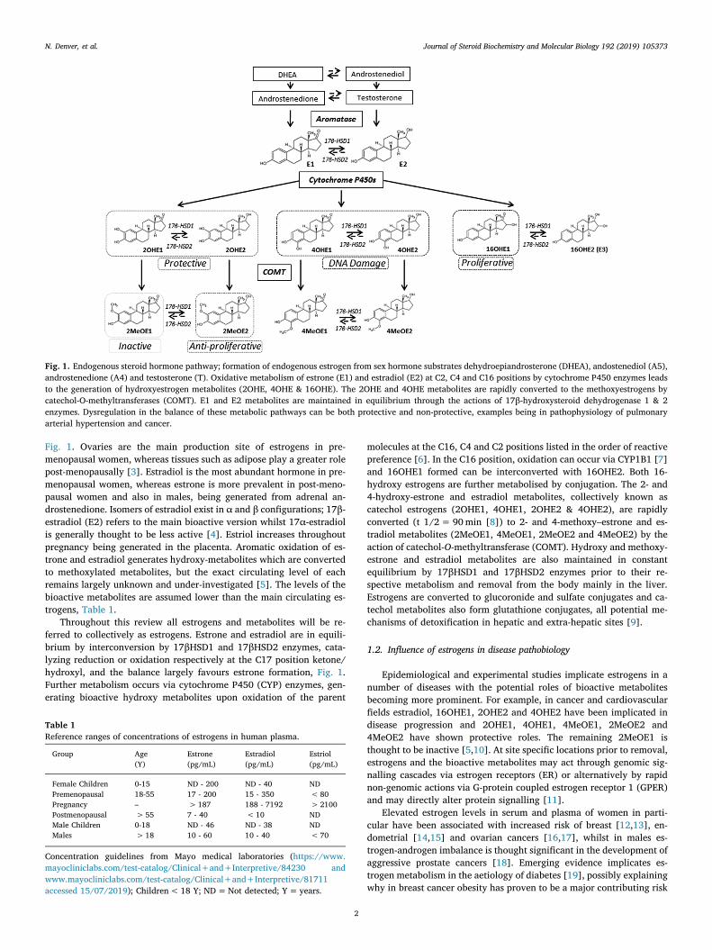
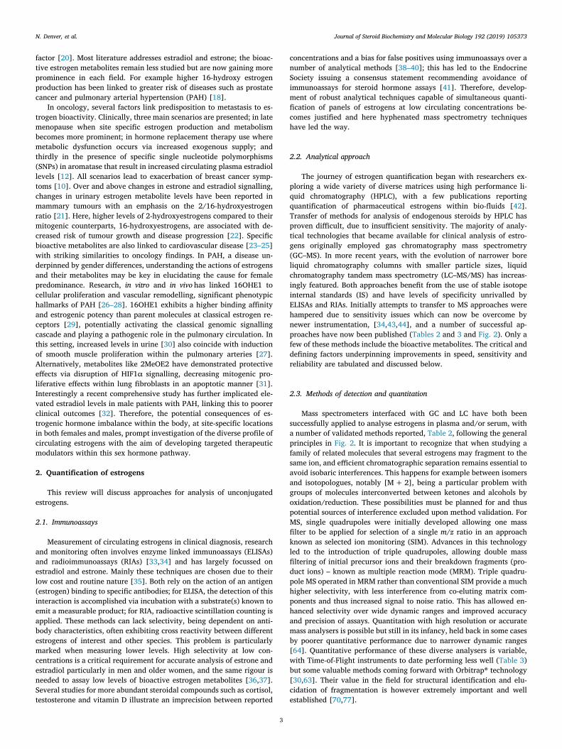
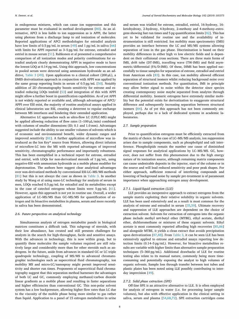
Fig. 1. Ovaries are the main production site of estrogens in pre-menopausal women, whereas tissues such as adipose play a greater rolepost-menopausally [3]. Estradiol is the most abundant hormone in pre-menopausal women, whereas estrone is more prevalent in post-meno-pausal women and also in males, being generated from adrenal an-drostenedione. Isomers of estradiol exist in α and β configurations; 17β-estradiol (E2) refers to the main bioactive version whilst 17α-estradiolis generally thought to be less active [4]. Estriol increases throughoutpregnancy being generated in the placenta. Aromatic oxidation of es-trone and estradiol generates hydroxy-metabolites which are convertedto methoxylated metabolites, but the exact circulating level of eachremains largely unknown and under-investigated [5]. The levels of thebioactive metabolites are assumed lower than the main circulating es-trogens, Table 1.
Throughout this review all estrogens and metabolites will be re-ferred to collectively as estrogens. Estrone and estradiol are in equili-brium by interconversion by 17βHSD1 and 17βHSD2 enzymes, cata-lyzing reduction or oxidation respectively at the C17 position ketone/hydroxyl, and the balance largely favours estrone formation, Fig. 1.Further metabolism occurs via cytochrome P450 (CYP) enzymes, gen-erating bioactive hydroxy metabolites upon oxidation of the parent
molecules at the C16, C4 and C2 positions listed in the order of reactivepreference [6]. In the C16 position, oxidation can occur via CYP1B1 [7]and 16OHE1 formed can be interconverted with 16OHE2. Both 16-hydroxy estrogens are further metabolised by conjugation. The 2- and4-hydroxy-estrone and estradiol metabolites, collectively known ascatechol estrogens (2OHE1, 4OHE1, 2OHE2 & 4OHE2), are rapidlyconverted (t 1/2= 90min [8]) to 2- and 4-methoxy–estrone and es-tradiol metabolites (2MeOE1, 4MeOE1, 2MeOE2 and 4MeOE2) by theaction of catechol-O-methyltransferase (COMT). Hydroxy and methoxy-estrone and estradiol metabolites are also maintained in constantequilibrium by 17βHSD1 and 17βHSD2 enzymes prior to their re-spective metabolism and removal from the body mainly in the liver.Estrogens are converted to glucoronide and sulfate conjugates and ca-techol metabolites also form glutathione conjugates, all potential me-chanisms of detoxification in hepatic and extra-hepatic sites [9].
1.2. Influence of estrogens in disease pathobiology
Epidemiological and experimental studies implicate estrogens in anumber of diseases with the potential roles of bioactive metabolitesbecoming more prominent. For example, in cancer and cardiovascularfields estradiol, 16OHE1, 2OHE2 and 4OHE2 have been implicated indisease progression and 2OHE1, 4OHE1, 4MeOE1, 2MeOE2 and4MeOE2 have shown protective roles. The remaining 2MeOE1 isthought to be inactive [5,10]. At site specific locations prior to removal,estrogens and the bioactive metabolites may act through genomic sig-nalling cascades via estrogen receptors (ER) or alternatively by rapidnon-genomic actions via G-protein coupled estrogen receptor 1 (GPER)and may directly alter protein signalling [11].
Elevated estrogen levels in serum and plasma of women in parti-cular have been associated with increased risk of breast [12,13], en-dometrial [14,15] and ovarian cancers [16,17], whilst in males es-trogen-androgen imbalance is thought significant in the development ofaggressive prostate cancers [18]. Emerging evidence implicates es-trogen metabolism in the aetiology of diabetes [19], possibly explainingwhy in breast cancer obesity has proven to be a major contributing risk
Fig. 1. Endogenous steroid hormone pathway; formation of endogenous estrogen from sex hormone substrates dehydroepiandrosterone (DHEA), andostenediol (A5),androstenedione (A4) and testosterone (T). Oxidative metabolism of estrone (E1) and estradiol (E2) at C2, C4 and C16 positions by cytochrome P450 enzymes leadsto the generation of hydroxyestrogen metabolites (2OHE, 4OHE & 16OHE). The 2OHE and 4OHE metabolites are rapidly converted to the methoxyestrogens bycatechol-O-methyltransferases (COMT). E1 and E2 metabolites are maintained in equilibrium through the actions of 17β-hydroxysteroid dehydrogenase 1 & 2enzymes. Dysregulation in the balance of these metabolic pathways can be both protective and non-protective, examples being in pathophysiology of pulmonaryarterial hypertension and cancer.
Table 1Reference ranges of concentrations of estrogens in human plasma.
Group Age Estrone Estradiol Estriol(Y) (pg/mL) (pg/mL) (pg/mL)
Female Children 0-15 ND - 200 ND - 40 NDPremenopausal 18-55 17 - 200 15 - 350 <80Pregnancy – > 187 188 - 7192 >2100Postmenopausal > 55 7 - 40 <10 NDMale Children 0-18 ND - 46 ND - 38 NDMales > 18 10 - 60 10 - 40 < 70
Concentration guidelines from Mayo medical laboratories (https://www.mayocliniclabs.com/test-catalog/Clinical+and+Interpretive/84230 andwww.mayocliniclabs.com/test-catalog/Clinical+and+Interpretive/81711accessed 15/07/2019); Children< 18 Y; ND=Not detected; Y= years.
N. Denver, et al. Journal of Steroid Biochemistry and Molecular Biology 192 (2019) 105373
2
factor [20]. Most literature addresses estradiol and estrone; the bioac-tive estrogen metabolites remain less studied but are now gaining moreprominence in each field. For example higher 16-hydroxy estrogenproduction has been linked to greater risk of diseases such as prostatecancer and pulmonary arterial hypertension (PAH) [18].
In oncology, several factors link predisposition to metastasis to es-trogen bioactivity. Clinically, three main scenarios are presented; in latemenopause when site specific estrogen production and metabolismbecomes more prominent; in hormone replacement therapy use wheremetabolic dysfunction occurs via increased exogenous supply; andthirdly in the presence of specific single nucleotide polymorphisms(SNPs) in aromatase that result in increased circulating plasma estradiollevels [12]. All scenarios lead to exacerbation of breast cancer symp-toms [10]. Over and above changes in estrone and estradiol signalling,changes in urinary estrogen metabolite levels have been reported inmammary tumours with an emphasis on the 2/16-hydroxyestrogenratio [21]. Here, higher levels of 2-hydroxyestrogens compared to theirmitogenic counterparts, 16-hydroxyestrogens, are associated with de-creased risk of tumour growth and disease progression [22]. Specificbioactive metabolites are also linked to cardiovascular disease [23–25]with striking similarities to oncology findings. In PAH, a disease un-derpinned by gender differences, understanding the actions of estrogensand their metabolites may be key in elucidating the cause for femalepredominance. Research, in vitro and in vivo has linked 16OHE1 tocellular proliferation and vascular remodelling, significant phenotypichallmarks of PAH [26–28]. 16OHE1 exhibits a higher binding affinityand estrogenic potency than parent molecules at classical estrogen re-ceptors [29], potentially activating the classical genomic signallingcascade and playing a pathogenic role in the pulmonary circulation. Inthis setting, increased levels in urine [30] also coincide with inductionof smooth muscle proliferation within the pulmonary arteries [27].Alternatively, metabolites like 2MeOE2 have demonstrated protectiveeffects via disruption of HIF1α signalling, decreasing mitogenic pro-liferative effects within lung fibroblasts in an apoptotic manner [31].Interestingly a recent comprehensive study has further implicated ele-vated estradiol levels in male patients with PAH, linking this to poorerclinical outcomes [32]. Therefore, the potential consequences of es-trogenic hormone imbalance within the body, at site-specific locationsin both females and males, prompt investigation of the diverse profile ofcirculating estrogens with the aim of developing targeted therapeuticmodulators within this sex hormone pathway.
2. Quantification of estrogens
This review will discuss approaches for analysis of unconjugatedestrogens.
2.1. Immunoassays
Measurement of circulating estrogens in clinical diagnosis, researchand monitoring often involves enzyme linked immunoassays (ELISAs)and radioimmunoassays (RIAs) [33,34] and has largely focussed onestradiol and estrone. Mainly these techniques are chosen due to theirlow cost and routine nature [35]. Both rely on the action of an antigen(estrogen) binding to specific antibodies; for ELISA, the detection of thisinteraction is accomplished via incubation with a substrate(s) known toemit a measurable product; for RIA, radioactive scintillation counting isapplied. These methods can lack selectivity, being dependent on anti-body characteristics, often exhibiting cross reactivity between differentestrogens of interest and other species. This problem is particularlymarked when measuring lower levels. High selectivity at low con-centrations is a critical requirement for accurate analysis of estrone andestradiol particularly in men and older women, and the same rigour isneeded to assay low levels of bioactive estrogen metabolites [36,37].Several studies for more abundant steroidal compounds such as cortisol,testosterone and vitamin D illustrate an imprecision between reported
concentrations and a bias for false positives using immunoassays over anumber of analytical methods [38–40]; this has led to the EndocrineSociety issuing a consensus statement recommending avoidance ofimmunoassays for steroid hormone assays [41]. Therefore, develop-ment of robust analytical techniques capable of simultaneous quanti-fication of panels of estrogens at low circulating concentrations be-comes justified and here hyphenated mass spectrometry techniqueshave led the way.
2.2. Analytical approach
The journey of estrogen quantification began with researchers ex-ploring a wide variety of diverse matrices using high performance li-quid chromatography (HPLC), with a few publications reportingquantification of pharmaceutical estrogens within bio-fluids [42].Transfer of methods for analysis of endogenous steroids by HPLC hasproven difficult, due to insufficient sensitivity. The majority of analy-tical technologies that became available for clinical analysis of estro-gens originally employed gas chromatography mass spectrometry(GC–MS). In more recent years, with the evolution of narrower boreliquid chromatography columns with smaller particle sizes, liquidchromatography tandem mass spectrometry (LC–MS/MS) has increas-ingly featured. Both approaches benefit from the use of stable isotopeinternal standards (IS) and have levels of specificity unrivalled byELISAs and RIAs. Initially attempts to transfer to MS approaches werehampered due to sensitivity issues which can now be overcome bynewer instrumentation, [34,43,44], and a number of successful ap-proaches have now been published (Tables 2 and 3 and Fig. 2). Only afew of these methods include the bioactive metabolites. The critical anddefining factors underpinning improvements in speed, sensitivity andreliability are tabulated and discussed below.
2.3. Methods of detection and quantitation
Mass spectrometers interfaced with GC and LC have both beensuccessfully applied to analyse estrogens in plasma and/or serum, witha number of validated methods reported, Table 2, following the generalprinciples in Fig. 2. It is important to recognize that when studying afamily of related molecules that several estrogens may fragment to thesame ion, and efficient chromatographic separation remains essential toavoid isobaric interferences. This happens for example between isomersand isotopologues, notably [M+2], being a particular problem withgroups of molecules interconverted between ketones and alcohols byoxidation/reduction. These possibilities must be planned for and thuspotential sources of interference excluded upon method validation. ForMS, single quadrupoles were initially developed allowing one massfilter to be applied for selection of a single m/z ratio in an approachknown as selected ion monitoring (SIM). Advances in this technologyled to the introduction of triple quadrupoles, allowing double massfiltering of initial precursor ions and their breakdown fragments (pro-duct ions) – known as multiple reaction mode (MRM). Triple quadru-pole MS operated in MRM rather than conventional SIM provide a muchhigher selectivity, with less interference from co-eluting matrix com-ponents and thus increased signal to noise ratio. This has allowed en-hanced selectivity over wide dynamic ranges and improved accuracyand precision of assays. Quantitation with high resolution or accuratemass analysers is possible but still in its infancy, held back in some casesby poorer quantitative performance due to narrower dynamic ranges[64]. Quantitative performance of these diverse analysers is variable,with Time-of-Flight instruments to date performing less well (Table 3)but some valuable methods coming forward with Orbitrap® technology[30,63]. Their value in the field for structural identification and elu-cidation of fragmentation is however extremely important and wellestablished [70,77].
N. Denver, et al. Journal of Steroid Biochemistry and Molecular Biology 192 (2019) 105373
3
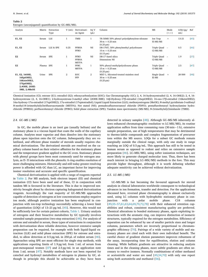
2.4. GC–MS (/MS)
In GC, the mobile phase is an inert gas (usually helium) and thestationary phase is a viscous liquid that coats the walls of the capillarycolumn. Analytes must vaporize and then dissolve into the stationaryphase upon injection onto the GC column. Subsequently they are vo-latilized, and efficient phase transfer of steroids usually requires che-mical derivatization. The derivatized steroids are resolved on the ca-pillary column based on their relative affinities for the stationary phaseand the temperature gradient applied to the GC oven. Stationary phaseswith phenyl groups have been most commonly used for estrogen ana-lysis, as П- П interactions with the phenolic A-ring enables resolution ofmore challenging mixtures. Historically and still today greater resolvingpower is afforded with GC than LC, an important factor for improvingisomer resolution and accurate and specific quantification.
Chemical derivatization is applied with a range of reagents reportedin Table 2. For MS analysis, both electron impact (EI) and chemicalionization (CI) have been used and of these, CI in conjunction withtandem MS is favoured in the literature. This is due to improved sen-sitivity brought about by electron capturing halogenated derivatizationreagents. Accordingly the vast majority of GC–MS/MS approachescapable of detection of estrogens at low concentrations employ negativeion mode, although positive ionization has been employed in con-junction with ion-trap technology successfully achieving a lower limitof quantitation (LOQ) of 13–21 pg/mL [45]. However, this would notbe sufficiently low for certain patient cohorts, Table 1. Urinary analysisof estrogens and their bioactive metabolites by GC typically involvesextended sample preparation (two-step extraction) [48]. For analysis ofestrone and estradiol in serum, these types of extractions in conjunctionwith derivatization show reliable detection, however extensive samplepreparation can be required, for example with both liquid-liquid ex-traction (LLE) and solid phase extraction (SPE) for estrone and estra-diol, to allow detection at 0.5 pg/mL in 250 μL of rodent serum [46].Approaches using SPE are most efficient for single step methods, withapplications reporting limits of 1.9 pg/mL from 1mL of serum frompost-menopausal women [37] and 2.5 pg/mL in an application notefrom 1mL serum [47]. Notably methods have not been reported forcatechol and hydroxyl metabolites of estrogens in plasma by GC, al-though in principle this should be achievable as they have been
detected in urinary samples [48]. Although GC–MS/MS inherently al-lows enhanced chromatographic resolution vs LC-MS(/MS), its routineapplication suffers from time consuming runs (30min – 1 h), extensivesample preparation, use of high temperatures that may be detrimentalto thermo-labile compounds and complex fragmentation of precursorions within the MS source. LOQs for a subset GC methods remainmarginally outside the clinical range, with only one study in rangereaching an LOQ of 0.5 pg/mL. This approach has still to be tested inhuman serum as opposed to rodent and relies on extensive samplepreparation [46]. LC–MS(/MS), using softer ionization techniques, aremore likely to generate charged molecular ions. Thus, there has beenmuch interest in bringing LC–MS(/MS) methods to the fore. This mayprovide higher throughput, although it is worth debating whetheradequate sensitivity can be achieved without derivatization.
2.5. LC–MS (/MS)
LC–MS/MS is fast becoming the favoured approach for steroidanalysis in clinical laboratories worldwide consequent to technologicaladvances in ion formation, transfer and detection. For the applicationsdiscussed here, reversed phase chromatography is almost exclusivelyused, using LC columns with hydrophobic stationary phase in con-junction with a polar mobile phase. C18 columns[49,55–57,61,63,64,69,70,72,78] with their enhanced retention cap-abilities and robust, consistent manufacturing quality are preferred.Chemical alterations to bonded stationary phases, again exploiting in-teractions with the aromatic ring, can improve distinction of isomericstructures, typically required for the estrogen metabolites. Efficiency ofseparation can be enhanced by use of smaller particle sizes and longercolumns, parameters which are inversely proportional to chromato-graphic efficiency [70]. Pairings of a wide variety of mobile and sta-tionary phases are cited each with their own individual benefit. Thecareful choice of gradient elution parameters improves robustness ofthe assay, incorporating time for equilibration, elution and columncleaning. While ballistic gradients are attractive in reducing analysistimes and in the cleaning phases, more subtle gradients are often ne-cessary to resolve isomers. Normally combinations of either methanolor acetonitrile and water are used [49,54,70] with only one reportusing both acetonitrile and methanol [56].
Table 2Estrogen (unconjugated) quantification by GC–MS(/MS).
Analyte Matrix ExtractionType
V (mL) Derivatizati-on Agent
Inj V(μL)
Column MS Mode(+/-)
LOQ (pg/mL)
Ref
E1, E2 Serum LLE 1 TMSI 1 TR-50MS 50% phenyl polysilphenylene-siloxane30m×0.25mm(0.25 μm)
Ion TrapEI-MS/MS
+ 13-21 [45]
E1, E2 Serum LLE & SPE 0.25 PFBHAPFBO
NS DB-17HT, 50% phenylmethyl polysiloxane15m×0.25mm(0.15 μm)
Triple QuadCI-MS/MS
– 0.5 [46]
E2 Serum SPE 1 PFBOPFBHAMSTFA
NS DB-17 fused silicaDimensions NS
Triple QuadCI-MS/MS
– 1.9 [37]
E2 Plasma SPE 1 PFBCMSTFA
1 50% phenyl-methylpolysiloxane phase15m×0.25mm(0.25 μm)
Triple QuadCI-MS/MS
– 2.5 [47]
E1, E2, 16OHE,16EpiOHE2,16KetoOHE2,17EpiOHE2,2,4OHE, 2OHE-3ME, 2,4MeOE
Urine SPE 2 EOCPFP
2 MXT-1, Silcosteel-treated stainless steel30m×0.25mm(0.25 μm)
Single QuadEI-MS
+ 20-500 [48]
Chemical Ionization (CI); estrone (E1); estradiol (E2); ethoxycarbonlyation (EOC); Gas Chromatography (GC); 2, 4, 16-hydroxyestradiol (2, 4, 16-OHE2); 2, 4, 16-hydroxyestrone (2, 4, 16-OHE1); 2-hydroxyestrone-3-methyl ether (2OHE-3ME); 16β-Hydroxy-17β-estradiol (16epiOHE2); 16-oxo-17β-estradiol (16ketoOHE2);16α-hydroxy-17α-estradiol (17epiOHE2); 17α-estradiol (17epiestradiol); Liquid Liquid Extraction (LLE); methoxyestrogens (MeOE); N-methyl pyridinium-3-sulfonylN-methyl-N-(trimethylsilyl)trifluoroacetamide (MSTFA); Not stated (NS); pentadecafluorooctanoyl chloride (PDFO); pentaflurobenzoyl hydroxylamine hydro-chloride (PFBHA); perfluorobenzoyl chloride (PFBO); Solid phase extraction (SPE); Tandem mass spectrometry (MS/MS); N-(trimethylsilyl)imidazole (TMSI).
N. Denver, et al. Journal of Steroid Biochemistry and Molecular Biology 192 (2019) 105373
4
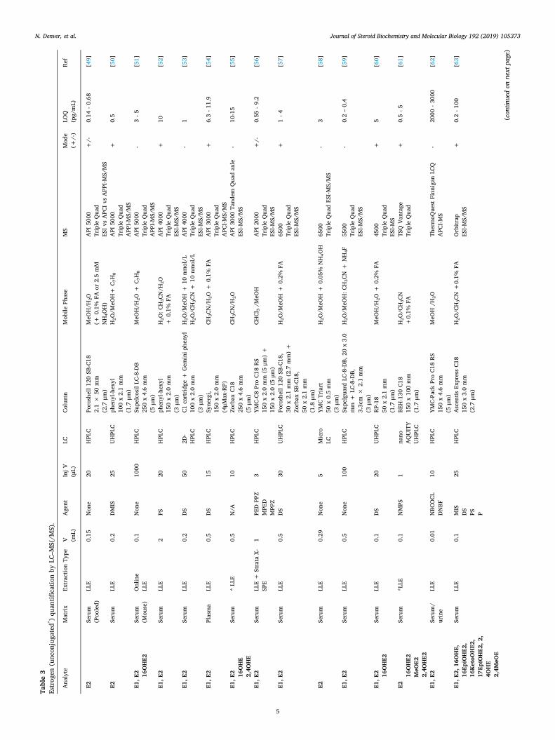
Table3
Estrogen
(unconjugated*)quantifi
catio
nby
LC–M
S(/M
S).
Analyte
Matrix
Extractio
nType
V (mL)
Agent
InjV
(μL)
LCCo
lumn
Mobile
Phase
MS
Mode
(+/-)
LOQ
(pg/mL)
Ref
E2Serum
(Pooled)
LLE
0.15
None
20HPLC
Poroshell1
20SB-C18
2.1×
50mm
(2.7
μm)
MeO
H/H
2O(+
0.1%
FAor
2.5mM
NH4O
H)
API
5000
TripleQuad
ESIv
sAPC
IvsAPP
I-MS/MS
+/-
0.14
-0.68
[49]
E2Serum
LLE
0.2
DMIS
25UHPLC
phenyl-hexyl
100x2.1mm
(1.7
μm)
H2O
/MeO
H+
C 7H8
API
5000
TripleQuad
APP
I-MS/MS
+0.5
[50]
E1,E
2 16OHE2
Serum
(Mouse)
Online
LLE
0.1
None
1000
HPLC
SupelcosilLC
-8-DB
250x4.6mm
(5μm
)
MeO
H/H
2O+
C 7H8
API
5000
TripleQuad
APP
I-MS/MS
-3-5
[51]
E1,E
2Serum
LLE
2PS
20HPLC
phenyl-hexyl
150x2.0mm
(3μm
)
H2O
:CH3CN/H
2O+
0.1%
FAAPI
4000
TripleQuad
ESI-M
S/MS
+10
[52]
E1,E
2Serum
LLE
0.2
DS
502D
-HPLC
C1cartridge+
Gem
iniphenyl
100x2.0mm
(3μm
)
H2O
/MeO
H+
10nm
ol/L
H2O
/CH3CN+
10nm
ol/L
API
4000
TripleQuad
ESI-M
S/MS
-1
[53]
E1,E
2Plasma
LLE
0.5
DS
15HPLC
Synergi,
150x2.0mm
(4μM
ax-RP)
CH3CN/H
2O+
0.1%
FAAPI
3000
TripleQuad
APC
I-MS/MS
+6.3-1
1.9
[54]
E1,E
2 16OHE
2,4O
HE
Serum
*LLE
0.5
N/A
10HPLC
Zorbax
C18
250x4.6mm
(5μm
)
CH3CN/H
2OAPI
3000
Tandem
Quadaxle
ESI-M
S/MS
-10-15
[55]
E1,E
2Serum
LLE+
StrataX-
SPE
1PE
DPP
ZMPE
DMPP
Z
3HPLC
YMC-C8
ProC1
8RS
150x2.0mm
(5μm
)+
150x2.0(5
μm)
CHCl
3/M
eOH
API
2000
TripleQuad
ESI-M
S/MS
+/-
0.55
-9.2
[56]
E1,E
2Serum
LLE
0.5
DS
30UHPLC
Poroshell1
20SB-C18,
30x2.1mm
(2.7
mm)+
Zorbax
SB-C18,
50x2.1mm
(1.8
μm)
H2O
/MeO
H+
0.2%
FA6500
TripleQuad
ESI-M
S/MS
+1-4
[57]
E2Serum
LLE
0.29
None
5Micro
LCYM
CTriart
50x0.5mm
(3μm
)
H2O
/MeO
H+
0.05%
NH4O
H6500
TripleQuadESI-M
S/MS
-3
[58]
E1,E
2Serum
LLE
0.5
None
100
HPLC
Supelguard
LC-8-DB,
20x3.0
mm
+LC
-8-DB,
3.3cm
×2.1mm
(3μm
)
H2O
/MeO
H:C
H3CN+
NH4F
5500
TripleQuad
ESI-M
S/MS
-0.2–0.4
[59]
E1,E
2 16OHE2
Serum
LLE
0.1
DS
20UHPLC
RP-18
50x2.1mm
(1.7
μm)
MeO
H/H
2O+
0.2%
FA4500
TripleQuad
ESI-M
S
+5
[60]
E216
OHE2
MeO
E22,4O
HE2
Serum
*LLE
0.1
NMPS
1nano
AQUITY
UHPLC
BEH-130
C18
150x100mm
(1.7
μm)
H2O
/CH3CN
+0.1%
FATSQVa
ntage
TripleQuad
+0.5-5
[61]
E1,E
2Serum/
urine
LLE
0.01
NBC
OCL
DNBF
10HPLC
YMC-Pack
ProC1
8RS
150x4.6mm
(5μm
)
MeO
H/H
2OTh
ermoQ
uestFinnigan
LCQ
APC
I-MS
-2000
-3000
[62]
E1,E
2,16
OHE,
16Ep
iOHE2
,16
KetoO
HE2
,17
EpiOHE2
,2,
4OHE
2,4M
eOE
Serum
LLE
0.1
MIS
DS
PS P
25HPLC
Ascentis
ExpressC1
8150x3.0mm
(2.7
μm)
H2O
/CH3CN+0.1%
FAOrbitrap
ESI-M
S/MS
+0.2-1
00[63]
(continuedonnextpage)
N. Denver, et al. Journal of Steroid Biochemistry and Molecular Biology 192 (2019) 105373
5
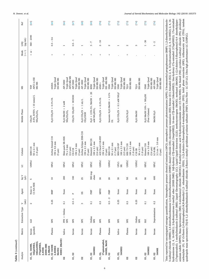
Table3(continued)
Analyte
Matrix
Extractio
nType
V (mL)
Agent
InjV
(μL)
LCCo
lumn
Mobile
Phase
MS
Mode
(+/-)
LOQ
(pg/mL)
Ref
E1,E
2,16
OHE,
16Ep
iOHE2
,16
KetoO
HE2
,17
EpiOHE2
,2,4O
HE
2OHE-3M
E2,4M
eOE
Serum
(pooled)
LLE
2Nonevs
C1-NA-NHS
5UHPLC
XDB-C1
850
x2.1mm
(1.8
μm)
CH3CN
A:5%,B
:95%
+10
mmol/L
NH4CH3CO2
TOF
APC
IvsESI
MS/MS
+or
-360-2
340
[64]
E1,E
216
OHE
2,4O
HE2
MeO
E24O
HE1
2MeO
E1
Plasma
SPE
0.25
BMP
10HPLC
Zorbax
Extend
C18
150x4.6mm
(5μm
)
H2O
/CH3CN+
0.1%
FA6420A
TripleQuad
ESI-M
S/MS
+0.3–3.6
[65]
E2Saliva
PPE+
Online
SPE
0.1
None
200
HPLC
Shim
-packXR
-ODS
75x3mm
(2.2
μm)
H2O
/MeO
H+
2mM
NH4CH3CO2
API
5000
TripleQuad
APC
I-MS/MS
+1
[66]
E1,E
2Serum
SPE
0.5-1
P100
HPLC
CD-C18
150x3mm
(3μm
)
CH3CN:C
H3OH+
HCO
OH
API
5000
TripleQuad
ESI-M
S/MS
+0.5-1
[67]
E2Serum
SPE
3DS
25HPLC
Zorbax
Eclip
seZD
B-C1
8150x2.1mm
(5μm
)
H2O
/CH3CN+
1mL/L
CH3COOH
API
4000
TripleQuad
ESI-M
S/MS
+1
[68]
E1,E
2 16OHE2
Serum
Online
SPE
0.1
FMP
300trap
elute
HPLC
Kinetex1
XB-C18
100x2.1mm
(2.6
μm)
H2O
+2.5%
FA/M
eOH+
20mM
NH4H
CO2
8050
TripleQuad
ESI-M
S/MS
+3-7
[69]
E1,E
2, 17epiestradiol,
16OHE,
MeO
E
Plasma
SPE
0.5
MPP
Z30
UHPLC
ACE
ExcelC
18-PFP
150x2.1mm
(2μm
)
H2O
/CH3CN+
0.1%
FA6500+
TripleQuad
ESI–
MS/MS
+2-1
0[70]
E1,E
2Plasma
SPE
0.5-2
FMP
20UHPLC
BEHC1
850
x2.1mm
(1.7μm
)
Isocratic
H2O
/MeO
H+
0.1%
FA5500
TripleQuad
ESI-M
S/MS
+2
[71]
E1,E
2, 16OHE2
Saliva
SPE
0.25
None
30UFLC-
XRBE
HC1
8-XP
100x2.1mm
(2.5
μm)
H2O
/CH3CN+
0.1mM
NH4F
5500
TripleQuad
ESI-M
S/MS
-1
[72]
E2Plasma
SPE
0.5
None
NS
UHPLC
HSS
T3C1
8100x2.1mm
(1.8
μm)
CH3CN/H
2OTQ
-SESI-M
S/MS
+2
[73]
E2Serum
Online
SPE
0.25
None
20UHPLC
C18SB
30x2.1mm
(1.8
μm)
H2O
/MeO
HTQ
-SESI-M
S/MS
-3
[74]
E1,E
2 16OHE2
Serum
SLE
0.1
None
90HPLC
KinetexC1
8100x3.0mm
(2.6
μm)
H2O
/MeO
H10%
+NH4OH
(PostCo
lumn)
5500
TripleQuad
ESI-M
S/MS
-1-3
0[75]
E1,E
2 16OHE
Serum
Deproteination
0.2
None
600
HPLC
LC-8-DB,
3.3cm
×3.0mm
(3μm
)
MeO
H/H
2OAPI
5000
TripleQuad
ESI-M
S/MS
-1-2
[76]
* Datareported
foru
nconjugatedestrogen
quantifi
catio
n,Atm
osphericpressurechem
icalionizatio
n(APC
I);atm
osphericpressurephotoionization(APP
I);3-bromom
ethyl-p
ropyphenazone(BMP);1,2-dim
ethylim
idazole-
5-sulfo
nylchloride(DMIS);2,4-dinitrofl
uorobenzene2,4-dinitrofl
uorobenzene(DNBF);dansylchloride
(DS);electrosprayionizatio
n(ESI);Estrone(E1);E
stradiol(E2);2,4,16-hydroxetradiol(2,4,16-OHE2);2,4,16-
hydroxetrone
(2,4,
16-OHE1);2-hydroxyestrone-3-m
ethylether(2OHE-3M
E);16β-hydroxy-17β-estradiol(16epiOHE2);16-oxo-17β-estradiol
(16ketoO
HE2);16α-hydroxy-17α-estradiol(17epiOHE2);17α-estradiol
(17epiestradiol);1-methylim
idazole-2-sulfo
nyl(M
IS);Liquid
Chromatography(LC);Liquid
Liquid
Extractio
n(LLE);methoxyestrogens(M
eOE);methanol(M
eOH);1-(2,4-dinitro-5-flu
orophenyl)-4,4-dimethylpiper-
azinium
iodide
(MPP
Z);M
assSpectrom
etry
(MS);4
-nitrobenzoyl
chloride
(NBC
OCL
);N-m
ethyl-n
icotinicacidN-hydroxysuccinim
ideester(C1-NA-NHS);N
otStated
(NS);p
yridine-3-sulfo
nylchloride(PS);p
icolinoyl
carboxylate(P);N’-(5-flu
oro-2,4-dinitrophenyl)-N,N-dim
ethyl-1
,2-ethanediam
ine(PED
);1-(2,4-dinitro-5-flu
orophenyl)-4-m
ethylpiperazine(PPZ
);Solid
phaseextractio
n(SPE
);trifluoracetic
acid
(TFA
);tandem
quadrupolemassspectrom
etry
(TQ-S)1,2-
dimethylim
idazole-5-sulfo
nylchloride;2-flu
oro-1-methyl-pyridinump-toluenesulfo
nate
(FMP);U
ltraFlow
–LC
(UF-LC
);Ultrahigh
performance–LC(UHPLC).
N. Denver, et al. Journal of Steroid Biochemistry and Molecular Biology 192 (2019) 105373
6
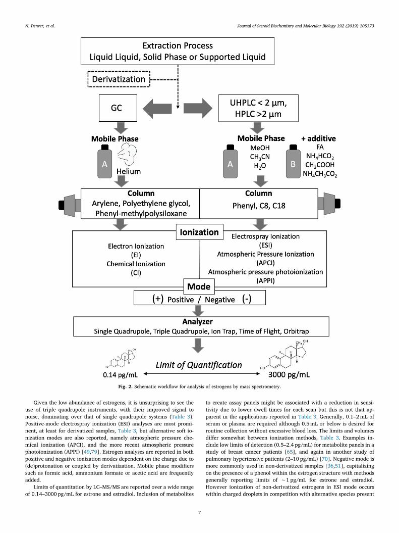
Given the low abundance of estrogens, it is unsurprising to see theuse of triple quadrupole instruments, with their improved signal tonoise, dominating over that of single quadrupole systems (Table 3).Positive-mode electrospray ionization (ESI) analyses are most promi-nent, at least for derivatized samples, Table 3, but alternative soft io-nization modes are also reported, namely atmospheric pressure che-mical ionization (APCI), and the more recent atmospheric pressurephotoionization (APPI) [49,79]. Estrogen analyses are reported in bothpositive and negative ionization modes dependent on the charge due to(de)protonation or coupled by derivatization. Mobile phase modifierssuch as formic acid, ammonium formate or acetic acid are frequentlyadded.
Limits of quantitation by LC–MS/MS are reported over a wide rangeof 0.14–3000 pg/mL for estrone and estradiol. Inclusion of metabolites
to create assay panels might be associated with a reduction in sensi-tivity due to lower dwell times for each scan but this is not that ap-parent in the applications reported in Table 3. Generally, 0.1–2mL ofserum or plasma are required although 0.5mL or below is desired forroutine collection without excessive blood loss. The limits and volumesdiffer somewhat between ionization methods, Table 3. Examples in-clude low limits of detection (0.5–2.4 pg/mL) for metabolite panels in astudy of breast cancer patients [65], and again in another study ofpulmonary hypertensive patients (2–10 pg/mL) [70]. Negative mode ismore commonly used in non-derivatized samples [36,51], capitalizingon the presence of a phenol within the estrogen structure with methodsgenerally reporting limits of ∼1 pg/mL for estrone and estradiol.However ionization of non-derivatized estrogens in ESI mode occurswithin charged droplets in competition with alternative species present
Fig. 2. Schematic workflow for analysis of estrogens by mass spectrometry.
N. Denver, et al. Journal of Steroid Biochemistry and Molecular Biology 192 (2019) 105373
7
in endogenous mixtures, which can cause ion suppression and thisparameter must be evaluated in method development [80]. As an al-ternative, APCI is less liable to ion suppression as is APPI, the latterusing photons from a discharge lamp to aid ionization of molecules.Reported applications of APCI methodologies for estradiol analysishave low limits of 0.5 pg/mL in serum [49] and 1 pg/mL in saliva [66]with limits for APPI reported as 3–5 pg/mL for estrone, estradiol andestriol in mouse serum [51]. Rahkonen et al presented a comprehensivecomparison of all ionization modes and polarity combinations for es-tradiol analysis clearly demonstrating APPI in negative mode to havethe lowest LOQ at 0.14 pg/mL. Using this approach, low concentrationsin pooled serum were detected using ammonium hydroxide as an ad-ditive, Table 3 [49]. Upon application to a clinical cohort (200 μL), aDMIS derivatization approach in conjunction with APPI was applied bythe same group reporting limits in serum of 0.5 pg/mL [50]. Notablyaddition of 2D chromatography boosts sensitivity for estrone and es-tradiol reducing LOQs tenfold [53] and integration of this with APPImight allow a further boost in quantification capabilities. As of yet APPIis not widely reported or available and, although advantages of APCI/APPI over ESI exist, the majority of routine analytical assays applied inclinical laboratories use ESI, creating a deterrent to regular exchangebetween MS interfaces and hindering their extended application.
Alternative LC approaches such as ultra-flow LC (UFLC-MS) mightbe applied allowing reduction of flow rates (1–100 μL/min) combinedwith columns of smaller dimensions (ID; 0.1 and 1.0mm). Advantagessuggested include the ability to use smaller volumes of solvents which isof economic and environmental benefit, wider dynamic ranges andimproved sensitivity [81]. A further application of microflow was in-troduced as the Ion Key® source from Waters, allowing direct infusionof microflow-LC into the MS with reported advantages of improvedsensitivity, chromatographic performance and importantly robustness.This has been exemplified in a technical report for estrone, estradioland estriol, with LOQs for non-derivatized steroids of 1 pg/mL, usingnegative-ESI with ammonium hydroxide as a mobile phase modifier fordeprotonation. The authors here suggest clear analytical advantagesover non-derivatized methods by conventional ESI-LC–MS/MS methods[81] but this is not always the case as shown in Table 3. In anotherstudy by Wang et al using nano-LC technology for analysis of serum inmen, LOQs reached 0.5 pg/mL for estradiol and its metabolites exceptin the case of catechol estrogens whose limits were 5 pg/mL [61].However, again this approach is not yet in routine use. Overall, a widerapplication of LC–MS/MS than GC–MS/MS for quantification of es-trogen and its bioactive metabolites in plasma, serum and more recentlyin saliva has been demonstrated.
2.6. Future perspectives on analytical technology
Simultaneous analysis of estrogen metabolite panels in biologicalmatrices constitutes a difficult task. This subgroup of steroids, withtheir low abundance, has created and still presents challenges foranalysts in the search for high-throughput, facile and sensitive assays.With the advances in technology, this is now within grasp, but toquantify these molecules the sample volumes required are still rela-tively large and considerably more than for other steroids such as an-drogens. In the future, aside from advances in standard GC or LC triplequadrupole technology, coupling of MS/MS to advanced chromato-graphic technologies such as supercritical fluid chromatography, ionmobility MS and micro-LC/nanospray might permit improved sensi-tivity and shorter run times. Proponents of supercritical fluid chroma-tography suggest that this separation method harnesses the advantagesof both LC and GC, commonly employing methanol/carbon dioxidelinear gradients as a mobile phase and resulting in faster separationsand higher efficiencies than conventional GC. This non-polar solventsystem has a low backpressure, allowing higher flow rates than LC dueto the viscosity of the mobile phase being more similar to gas ratherthan liquid. Application to a panel of 15 estrogen metabolites in urine
and serum was trialled for estrone, estradiol, estriol, 16-hydroxy, 16-ketohydroxy, 2-hydroxy, 4-hydroxy, 2-methoxy and 4-methoxy–estro-gens showing fast run times and 5 pg quantification limits [82]. This hasyet to be validated for routine use and the availability of in-strumentation is still restricted. Ion mobility mass spectrometry (IMS)provides an interface between the LC and MS/MS systems allowingseparation of ions in the gas phase. Discrimination is based on theirmobility differences in either high vs low electric fields and is depen-dent on their collisional cross sections. There are three main forms ofIMS, drift tube (DT-IMS), travelling wave (TW-IMS) and field asym-metric/differential (FA/D-IMS). Of these, DIMS has been applied suc-cessfully to tissue samples for separation of estrone, estradiol and estriolfrom American eels [83]. In this case, ion mobility allowed efficientseparation of structural isomers whilst reducing background noise overconventional ionization methods. For quantitation, IMS in principlemay allow better signal to noise within the detector since speciescreating contemporary noise maybe separated from analytes throughdifferential mobility. Isomeric estrogens have extremely similar mobi-lity but the potential exists for derivatization to exaggerate structuraldifference and subsequently increasing separation between structuralisomers [84]. As of yet this approach has not been commonly em-ployed, perhaps due to a lack of dedicated systems in academic la-boratories.
2.7. Sample preparation
Prior to quantification estrogens must be efficiently extracted fromthe matrix of choice. In the case of LC–MS/MS analysis, ion suppressionarises due to sample components, such as phospholipid and salt inter-ference. Phospholipids remain the number one cause of diminishedsignal responses for analytical applications by LC–MS/MS. GC, how-ever, does not suffer from this phenomenon due to the high energynature of its ionization source, although remaining matrix componentscan cause undesirable deposits in the injector, start of the column or inthe source and will lead reduced sensitivity and poor peak shapes. Foreither approach, sufficient removal of interfering compounds andlowering of background noise by sample pre-treatment is of paramountimportance when approaching estrogen assay development.
2.7.1. Liquid-liquid extraction (LLE)LLE provides an inexpensive approach to extract estrogens from the
sample matrix exploiting their relative solubility in organic solvents.LLE has been used extensively and as a result is most common for theanalysis of estrone and estradiol in serum [35,59]. Ultimate recoveryand suppression of LLE approaches are dependent on the choice ofextraction solvent. Solvents for extraction of estrogens into the organicphase include methyl tert-butyl ether (MTBE), ethyl acetate, diethylether, dichloromethane or mixtures of these organic solvents. Ethylacetate is most commonly reported affording high recoveries [85,86]and alongside MTBE, it yields a clean extract that avoids precipitationupon derivatization [87,88]. From Table 3, it can be seen LLE has beenextensively applied to estrone and estradiol assays reporting low de-tection limits (0.14–5 pg/mL). However, for bioactive metabolites re-sults are variable with higher limits than alternative sample preparationtechniques (5–360 pg/mL). Additional drawbacks of LLE for routinetesting also relate to its manual nature, commonly being more time-consuming and potentially exposing the analyst to high volumes oforganic solvents. Sample loss through transfer between test tubes andplastic plates has been noted using LLE possibly contributing to inter-day imprecision [49].
2.7.2. Solid phase extraction (SPE)Off-line SPE is an attractive alternative to LLE. It is often employed
for analysis of estrogens in water (i.e. for processing larger samplevolumes), but also with effective application in the clinical setting insaliva, serum and plasma [54,68,72]. SPE extraction cartridges come
N. Denver, et al. Journal of Steroid Biochemistry and Molecular Biology 192 (2019) 105373
8
embedded with a range of solid packing materials, which chemicallyseparate the components of interest from the biological samples. Vari-eties of bed are commercially available containing reversed, normal,ion exchange or adsorption packing materials. For recovery of estrogensfrom aqueous sample matrices, reversed and ion exchange phases arerecommended and are reported to be effective for clean up of plasmasamples, Table 3. In principle, an SPE column containing an alternativepacking material to the chromatography column holds advantages inimproving sample clean up. SPE columns used for estrogen analysisoften have C18 beds, but many commercial materials also exist such asOasis HLB®; most have hydrophobic characteristics optimal for inter-actions with the lipophilic features of steroid hormones. HLB® operatesover a wide range of pH values suitable for many compound classes.Choice of SPE column is based on achieving high recovery with low ionsuppression, which can be difficult to achieve with complex matricessuch as plasma [71]. A study by Faqehi et al 2016 suggested the use ofOasis MCX®, a cartridge housing a mixed mode cation exchange reversephase bed, provides opportunities for additional sample clean up priorto the elution of the estrogen and this has been shown effective for apanel of the bioactive estrogens, including metabolites upon optimi-zation of wash steps [70]. Other groups suggest the use of C8 poly-propylene columns conditioned and cleaned with 0.1% TFA improvedrecovery and diminished ion suppression for a panel of 10 estrogens[65]. Moving forward with SPE, newer products eliminate the need forconditioning and equilibration steps and availability of 96-well platesallow potential automation for robotic liquid handling systems. Themain disadvantage with SPE for routine clinical analyses associateswith the cost, as cartridges remain expensive. Moreover, coupling SPEand derivatization can introduce undesirable transfer steps and alsolosses depending on the type of collection container required to avoidadhesion (glass vs plastic). Glass inserts for 96-well plates are expensiveand only available for lower elution volumes. On-line SPE methods areavailable although less frequently reported as they can be complicatedto develop without compromising the analytical chromatographic step[89]. However once the elution programme is optimized, directlylinking the extraction processes to LC–MS/MS can improve recoveryand sensitivity and minimize manual sample manipulation [90].
2.7.3. Supported liquid extraction (SLE)Supported liquid extraction (SLE) opens doors to new approaches
for extraction but as yet methods for estrogen analysis have beenscarcely published, unlike with other steroids [91]. This strategy showspromise in company application notes [92] with successful applicationto androgen profiling for diseases such as congenital adrenal hyper-plasia [93]. SLE applies the same solvent affinity principles as LLEwhereby analytes are separated based on their partitioning into onesolvent over another immiscible solvent and employs similar solvents.The support material consists of diatomaceous earth, a natural silicaproduct (∼90% silica), being an ideal material to absorb aqueoussamples. This technique allows shorter load, wait and elute protocols tothe generic SPE approaches, and the conditioning and equilibrationsteps of the cartridge bed are not needed. However, options for sampleclean-up are limited in comparison to SPE. Again, SLE can be fullyautomated in 96-well formats but again there are challenges in inter-facing with containers suitable for derivatization. One application foranalysis of estrone, estradiol or estriol from 100 μL of plasma in the SLE96 well format shows potential with low limits of 1, 3 pg/mL for es-trone, estradiol respectively and 30 pg/mL for estriol. This extractionmethod should now be tested with the wider panel of metabolites onmore sensitive MS platforms.
2.7.4. DerivatizationDerivatization can be necessary prior to analysis of estrogens by MS,
but with different goals for GC and LC. In the case of GC it is necessaryto enhance volatility often with the introduction of halogen atoms, alsoenhancing sensitivity of CI approaches [79]. For LC, derivatization is
often employed to aid formation of charged ions or generate perma-nently charged species. This increases sensitivity, and the greater massof the molecular ion holds further benefits for specificity. In GC–MS/MSthe process sometimes adds poorly volatile reagents which cannot beeasily removed. In both GC and LC, derivatization reagents can build upin the chromatographic column or within components of the massspectrometer, thus decreasing assay robustness. In LC, this may be ad-dressed by diverting the initial flow prior to analyte elution to waste,removing polar reagents and maintaining a clean interface and sourcewithin the mass spectrometer. In GC frequent cleaning of the inlet linerwill be required.
2.7.4.1. GC approaches. In GC–MS/MS derivatization at the 3′ positionof the A-ring is favoured as reactions at the saturated aliphatic D ringlargely do not improve sensitivity over non-derivatized samplesillustrated by pentafluoropropionyl (PFP) or trimethylsilyl (TMS)derivatives for water analysis by GC–MS/MS [94,95]. The generationof PFB derivatives is the most commonly reported approach for estroneand estradiol analysis in serum, but cumbersome sample preparationsteps have, however, hampered routine use [37,46,96].
2.7.4.2. LC approaches without derivatization. Development ofanalytical workflows of sufficient sensitivity without derivatizationremains challenging for clinical applications of estrogen analysis,although they are desirable with sample preparation being shorterwith a lower chance of introducing manual varation. Moreover,automation of derivatization by commercial robots is challenging tocouple with robotic SPE/SLE workflows. However, a number ofmethods for underivatized estrone and estradiol using LC–MS/MS arebeginning to surface, (Table 2) as instrument technology improves.Methods achieving LOQs comparable with derivatization approacheshave been reported using ammonium fluoride or ammonium hydroxideas mobile phase modifiers, promoting the formation of negative ions[59,72]. Recent analyses of estradiol report low LOQs, for example of2 pg/mL using an UHPLC System coupled to a Xevo TQ-S [73]. Methodswithout derivatization are yet to be extended to include bioactiveestrogen metabolites. If optimized successfully, validation of suchassays would permit simplified sample preparation with thepossibility of higher precision and throughput.
2.7.4.3. LC approaches with derivatization. Derivatization remainsnecessary for the majority of LC–MS assays of estrogens, overcomingpoor ionization, limiting ion suppression and boosting signal intensityat low abundance. In reactions reported, introduction of easilyionizable groups or pre-charged moieties improves sensitivity andpermits the use of lower volumes of sample. As in GC–MS, thehydroxyl group of the phenolic A ring in the 3′ position is usuallytargeted for the entire analyte panel. Successful derivatization methodscommonly reported for analysis of estrone and estradiol include use ofdansyl chloride [68,92,97,98], N-methyl-nicotinic acid N-hydroxysuccinimide ester [64], 2-fluoro-1-methylpyridinium-p-toluenesulfonate [71], methyl-1-(5-fluoro-2, 4-dinitrophenyl)-4,4-dimethylpiperazine [56,70], isomers of 1,2-dimethylimidazole-sulfonyl chloride [63,99], picolinoyl carboxylate [67], pyridinecarboxylates [100], pyridine-3-sulfonyl chloride [52] and p-nitrobenzyl chloride [101]. From these, dansyl chloride has been themost common approach. However, the specificity of the fragment ionsof dansyl chloride derivatives is hindered for isobaric estrogenmetabolite species since the product ions generated are identical,hailing from the derivative [63,97,98]. This is similar for alternativederivatives such as BMP [65], whereby methyl-propyphenazonederivatives generate identical product ions for seven estrogens whilstdiffering by m/z 15 for the catechol metabolites. This source of non-specificity has been partially overcome by use of MPPZ and C1-NA-NHS, yielding a range of product ions, but they remain identical forcertain groups of metabolites [64,70], since isomers undergo similar
N. Denver, et al. Journal of Steroid Biochemistry and Molecular Biology 192 (2019) 105373
9
fragmentation patterns. Therefore, thorough evaluation ofchromatographic methods becomes a necessity, to eliminate possibleco-elutants that may be mis-identified leading to reporting of falsepositives. It should not be forgotten that estradiol and estrone onlydiffer by 2 mass units so 13C2 isotopologues used as internal standardswill cross-signal if product ions are identical. It is not uncommon formultiple aliphatic and phenolic hydroxyl groups to be derivatizedwithin the reaction especially within 16-hydroxy- and catecholestrogens [56,102], yielding either doubly or triply charged speciesor isomeric derivatives. Finally, if derivatization is deemed necessarythe stability of derivatives should be considered and must be studied toensure practical laboratory workflows e.g. FMP derivatives degradefollowing 48 h at −20 °C but remain stable within −80 °C storage [71].MPPZ derivatives show minimal degradation (< 15%) upon storage for8 days in the autosampler and for up to 31 days in −20 °C storage [70].Dansyl chloride derivatives have also been reported to be stable over a7 day period in patient plasma [54]. However, in the majority ofcurrent literature, this information is lacking for derivatizationapproaches. Derivatization techniques are still less preferred in theclinical setting, due to the addition of another complexity withinsample preparation inevitability contributing toward data variabilityand increased turn-around time.
2.7.5. Internal standardsAn important feature of MS analytical methods for estrogen quan-
tification is the availability of stable isotope labelled internal standards(IS) giving a retention time match to both derivatized and non-deri-vatized estrogens. Addition at constant concentrations within the assayaccounts for extraction loss at all stages. 13C-labels allow additionalselectivity over deuterium-labelled standards, since they are highlyunlikely to be removed during processing. Deuterium can be removedthrough either deuterium-hydrogen exchange under acidic conditionsor, depending on the positions of the labels, during derivatization re-actions. By GC and LC, the retention time of 13C-labelled standards arewell aligned whereas deuterated IS may differ slightly, probably due toisotope effects on hydrogen vs deuterium bonding interactions with thestationary phase. The slight differences in retention time that arise withdeuterium labels are exaggerated when the number of heavy labels isincreased potentially leading to less accurate quantitation with lessspecific interpretation of matrix effect [103]. However deuterium la-belled standards are applied in a number of studies generally being lessexpensive in comparison to the 13C labelled versions [51,53,57,63,65].Retention of the stable isotope labels in the product ion is desirable toenhance specificity, but labels can be lost in fragmentation, leavingproduct ions identical in m/z to the analyte. C3-6 labelled standards arenow available for all estrogens shown in Fig. 1. Multi-labelled stan-dards, preferably in excess of two labels, e.g. 13C3 and 13C6. should beutilized, to avoid interference with natural isotopologues [61,70].
3. Conclusion
As this review highlights, there is no universal method for estrogenanalysis, however the wide range of approaches developed over thepast 10–15 years allows us to nudge closer to the possibility of routineinvestigation and monitoring of estrogen sensitive diseases. On com-parison of technologies available, methods by GC–MS initially came tothe field and currently offer a range of LOQs between 0.5–21 pg/mL forestrone and estradiol in serum, plasma and urine, with 20–500 pg/mLfor their metabolites. However, despite efficient resolution of isomers,GC–MS(/MS) is less favoured requiring more extensive sample pre-paration and the absolute requirement for derivatization limiting au-tomation. Developments in LC–MS/MS arose more recently, withtechnology still advancing, offering the possibility of lower detectionlimits of 0.14 pg/mL for standalone estradiol analysis with a range morecommonly between 0.5–21 pg/mL for estrogens in panel assays.Although UHPLC may reduce analysis times in conjunction with MS,
applications show similar limits of detection to conventional HPLC andGC. SPE and SLE extraction methods will likely lead the way forward inclinical assays due to the possibility of automation. Applications in-volving derivatization are not universally superior with a number ofmethods not requiring derivatization now emerging that display similaror even lower detection capabilities. Therefore, development of ap-proaches without this step should be considered on newer triplequadrupole instrumentation. In this setting APCI and APPI modes haveyet to be explored for the full metabolite panels. Irrespective of ana-lytical technology used, the importance chromatographic developmentmust not be understated due to estrogenic isomers, stereoisomers andisobaric confounders. Combinations of on-line SPE, high-resolution LCand MS approaches may shape the future for automated approaches;ion mobility might also provide a key approach for separation of iso-mers, enhancing structural confirmation in cases where shared productions arise.
In conclusion, advancing research into health and disease in clinicalcohorts extending to children, men, and pre/post-menopausal womenfor disease diagnostics and monitoring means limits of analyticalmethods are constantly being tested. MS has established its place at theforefront of research for estrogen quantification in clinical laboratories,with LC–MS/MS beginning to show potential for routine applications.
Acknowledgements
This work was supported by a BBSRC iCASE PhD studentship (BB/N503691/1), the BHF (RG/16/2/32153 and PG/15/63/31659) and theWellcome Trust (202794/Z/16/Z).
References
[1] M.P. Thomas, B.V.L. Potter, The structural biology of oestrogen metabolism, J.Steroid Biochem. Mol. Biol. 137 (2013) 27–49.
[2] J. Li, L. Eriksson, K. Humphreys, K. Czene, J. Liu, R.M. Tamimi, S. Lindström,D.J. Hunter, C.M. Vachon, F.J. Couch, C.G. Scott, P. Lagiou, P. Hall, Geneticvariation in the estrogen metabolic pathway and mammographic density as anintermediate phenotype of breast cancer, Breast Cancer Res. 12 (2010) R19.
[3] N. Hetemäki, H. Savolainen-Peltonen, M.J. Tikkanen, F. Wang, H. Paatela,E. Hämäläinen, U. Turpeinen, M. Haanpää, V. Vihma, T.S. Mikkola, Estrogenmetabolism in abdominal subcutaneous and visceral adipose tissue in post-menopausal women, J. Clin. Endocrinol. Metab. 102 (2017) 4588–4595.
[4] S.P. Tofovic, Estrogens and development of pulmonary hypertension: interactionof estradiol metabolism and pulmonary vascular disease, J. Cardiovasc.Pharmacol. 56 (2010) 696–708.
[5] C.K. Docherty, K.Y. Harvey, K.M. Mair, S. Griffin, N. Denver, M.R. MacLean, Therole of sex in the pathophysiology of pulmonary hypertension, Adv. Exp. Med.Biol. 1065 (2018) 511–528.
[6] S.P. Tofovic, Estrogens and development of pulmonary hypertension: interactionof estradiol metabolism and pulmonary vascular disease, J. Cardiovasc.Pharmacol. 56 (2010) 696–708.
[7] K.M. Mair, K.Y. Harvey, A.D. Henry, D.Z. Hillyard, M. Nilsen, M.R. Maclean,Obesity alters oestrogen metabolism and contributes to pulmonary arterial hy-pertension, Eur. Respir. J. (2019) 1801524.
[8] P. Ball, G. Emons, H. Kayser, J. Teichmann, Metabolic clearance rates of catecholestrogens in rats, Endocrinology 113 (1983) 1781–1783.
[9] R. Raftogianis, C. Creveling, R. Weinshilboum, J. Weisz, Estrogen metabolism byconjugation, J. Natl. Cancer Inst. Monogr. (2000) 113–124.
[10] P. Muti, H.L. Bradlow, A. Micheli, V. Krogh, J.L. Freudenheim, H.J. Schünemann,M. Stanulla, J. Yang, D.W. Sepkovic, M. Trevisan, F. Berrino, Estrogen metabolismand risk of breast cancer: a prospective study of the 2:16alpha-hydroxyestroneratio in premenopausal and postmenopausal women, Epidemiology 11 (2000)635–640.
[11] S. Umar, M. Rabinovitch, M. Eghbali, Estrogen paradox in pulmonary hyperten-sion: current controversies and future perspectives, Am. J. Respir. Crit. Care Med.186 (2012) 125–131.
[12] R.J. Santen, W. Yue, J.-P. Wang, Estrogen metabolites and breast cancer, Steroids99 (2015) 61–66.
[13] R.C. Travis, T.J. Key, Oestrogen exposure and breast cancer risk, Breast CancerRes. 5 (2003) 239–247.
[14] C.M. Dallal, J.V. Lacey, R.M. Pfeiffer, D.C. Bauer, R.T. Falk, D.S.M. Buist,J.A. Cauley, T.F. Hue, A.Z. LaCroix, J.A. Tice, T.D. Veenstra, X. Xu, L.A. Brinton,Estrogen metabolism and risk of postmenopausal endometrial and ovarian cancer:the B∼FIT Cohort, Horm. Cancer 7 (2016) 49–64.
[15] L.A. Brinton, B. Trabert, G.L. Anderson, R.T. Falk, A.S. Felix, B.J. Fuhrman,M.L. Gass, L.H. Kuller, R.M. Pfeiffer, T.E. Rohan, H.D. Strickler, X. Xu,N. Wentzensen, Serum estrogens and estrogen metabolites and endometrial cancer
N. Denver, et al. Journal of Steroid Biochemistry and Molecular Biology 192 (2019) 105373
10
risk among postmenopausal women, Cancer Epidemiol. Biomarkers Prev. 25(2016) 1081–1089.
[16] B. Trabert, L.A. Brinton, G.L. Anderson, R.M. Pfeiffer, R.T. Falk, H.D. Strickler,S. Sliesoraitis, L.H. Kuller, M.L. Gass, B.J. Fuhrman, X. Xu, N. Wentzensen,Circulating estrogens and postmenopausal ovarian cancer risk in the women’shealth initiative observational study, Cancer Epidemiol. Biomarkers Prev. 25(2016) 648–656.
[17] M. Zahid, C.L. Beseler, J.B. Hall, T. LeVan, E.L. Cavalieri, E.G. Rogan, Unbalancedestrogen metabolism in ovarian cancer, Int. J. Cancer 134 (2014) 2414–2423.
[18] A. Black, P.F. Pinsky, R.L. Grubb, R.T. Falk, A.W. Hsing, L. Chu, T. Meyer,T.D. Veenstra, X. Xu, K. Yu, R.G. Ziegler, L.A. Brinton, R.N. Hoover, M.B. Cook,M.B. Cook, Sex steroid hormone metabolism in relation to risk of aggressiveprostate cancer, Cancer Epidemiol. Biomarkers Prev. 23 (2014) 2374–2382.
[19] A.A. Gupte, H.J. Pownall, D.J. Hamilton, Estrogen: an emerging regulator of in-sulin action and mitochondrial function, J. Diabetes Res. 2015 (2015) 1–9.
[20] M.P. Cleary, M.E. Grossmann, Minireview: obesity and breast cancer: the estrogenconnection, Endocrinology 150 (2009) 2537–2542.
[21] N. Obi, A. Vrieling, J. Heinz, J. Chang-Claude, Estrogen metabolite ratio: is the 2-hydroxyestrone to 16α-hydroxyestrone ratio predictive for breast cancer, Int. J.Womens Health 3 (2011) 37–51.
[22] G.C. Kabat, E.S. O’Leary, M.D. Gammon, D.W. Sepkovic, S.L. Teitelbaum,J.A. Britton, M.B. Terry, A.I. Neugut, H.L. Bradlow, Estrogen metabolism andbreast cancer, Epidemiology 17 (2006) 80–88.
[23] E. Murphy, Estrogen signaling and cardiovascular disease, Circ. Res. 109 (2011)687–696.
[24] B.V. Howard, J.E. Rossouw, Estrogens and cardiovascular disease risk revisited:the Women’s Health Initiative, Curr. Opin. Lipidol. 24 (2013) 493–499.
[25] R.E. White, Estrogen and vascular function, Vascul. Pharmacol. 38 (2002) 73–80.[26] X. Chen, M. Talati, J.P. Fessel, A.R. Hemnes, S. Gladson, J. French, S. Shay,
A. Trammell, J.A. Phillips, R. Hamid, J.D. Cogan, E.P. Dawson, K.E. Womble,L.K. Hedges, E.G. Martinez, L.A. Wheeler, J.E. Loyd, S.J. Majka, J. West,E.D. Austin, Estrogen metabolite 16α-hydroxyestrone exacerbates bone morpho-genetic protein receptor Type II-associated pulmonary arterial hypertensionthrough MicroRNA-29-mediated modulation of cellular metabolism, Circulation133 (2016) 82–97.
[27] K.Y. Hood, A.C. Montezano, A.P. Harvey, M. Nilsen, M.R. Maclean, R.M. Touyz,Nicotinamide adenine dinucleotide phosphate oxidase-mediated redox signalingand vascular remodeling by 16α-hydroxyestrone in human pulmonary artery cells,Hypertension 68 (2016) 796–808.
[28] K. White, A.K. Johansen, M. Nilsen, L. Ciuclan, E. Wallace, L. Paton, A. Campbell,I. Morecroft, L. Loughlin, J.D. McClure, M. Thomas, K.M. Mair, M.R. MacLean,Activity of the estrogen-metabolizing enzyme cytochrome P450 1B1 influences thedevelopment of pulmonary arterial hypertension, Circulation 126 (2012)1087–1098.
[29] X. Chen, M. Talati, J.P. Fessel, A.R. Hemnes, S. Gladson, J. French, S. Shay,A. Trammell, J.A. Phillips, R. Hamid, J.D. Cogan, E.P. Dawson, K.E. Womble,L.K. Hedges, E.G. Martinez, L.A. Wheeler, J.E. Loyd, S.J. Majka, J. West,E.D. Austin, Estrogen metabolite 16α-Hydroxyestrone exacerbates bone morpho-genetic protein receptor type II–associated pulmonary arterial hypertensionthrough MicroRNA-29–mediated modulation of cellular metabolism, Circulation133 (2016) 82–97.
[30] A.A. Franke, L.J. Custer, Y. Morimoto, F.J. Nordt, G. Maskarinec, Analysis of ur-inary estrogens, their oxidized metabolites, and other endogenous steroids bybenchtop Orbitrap LCMS versus traditional quadrupole GCMS, Anal. Bioanal.Chem. 401 (2011) 1319–1330.
[31] C.K. Docherty, M. Nilsen, M.R. Maclean, Influence of 2-Methoxyestradiol and sexon hypoxia-induced pulmonary hypertension and hypoxia-inducible factor-1, JAm Hear. Assoc. (2019) 1–10.
[32] C.E. Ventetuolo, G.L. Baird, R.G. Barr, D.A. Bluemke, J.S. Fritz, N.S. Hill,J.R. Klinger, J.A.C. Lima, P. Ouyang, H.I. Palevsky, A.J. Palmisciano, I. Krishnan,D. Pinder, I.R. Preston, K.E. Roberts, S.M. Kawut, Higher estradiol and lower de-hydroepiandrosterone-sulfate levels are associated with pulmonary arterial hy-pertension in men, Am. J. Respir. Crit. Care Med. 193 (2016) 1168–1175.
[33] J. Jaque, H. Macdonald, D. Brueggmann, S.K. Patel, C. Azen, N. Clarke,F.Z. Stanczyk, Deficiencies in immunoassay methods used to monitor serumEstradiol levels during aromatase inhibitor treatment in postmenopausal breastcancer patients, Springerplus. 2 (2013) 5.
[34] F.Z. Stanczyk, J. Jurow, A.W. Hsing, Limitations of direct immunoassays formeasuring circulating estradiol levels in postmenopausal women and men inepidemiologic studies, Cancer Epidemiol. Biomarkers Prev. 19 (2010) 903–906.
[35] C.P. Riley, R.E. Mathieu, C. Wiley, Simultaneous quantitation of estradiol andestrone in serum using liquid chromatography mass spectrometry, Methods Mol.Biol. (2016) 87–97.
[36] T. Santa, T. Guo, J. Gu, O.P. Soldin, R.J. Singh, S.J. Soldin, S. Gao, Z.-P. Zhang,H.T. Karnes, K.C. Sadanala, J.A. Lee, B.C. Chung, M.H. Choi, Sensitivity en-hancement in liquid chromatography/atmospheric pressure ionization massspectrometry using derivatization and mobile phase additives, J. Chromatogr. B. 3(2013) 98–110.
[37] R.J. Santen, L. Demers, S. Ohorodnik, J. Settlage, P. Langecker, D. Blanchett,P.E. Goss, S. Wang, Superiority of gas chromatography/tandem mass spectrometryassay (GC/MS/MS) for estradiol for monitoring of aromatase inhibitor therapy,Steroids 72 (2007) 666–671.
[38] T.G. Cross, M.P. Hornshaw, Can LC and LC-MS ever replace immunoassays, J.Appl. Bioanal. 2 (2016) 108–116.
[39] H. Ketha, S. Kaur, S.K. Grebe, R.J. Singh, Clinical applications of LC–MS sexsteroid assays: evolution of methodologies in the 21st century, Curr. Opin.
Endocrinol. Diabetes Obes. 21 (2014) 217–226.[40] D.J. Handelsman, M. Jimenez, G.K.S. Singh, J. Spaliviero, R. Desai, K.A. Walters,
Measurement of testosterone by immunoassays and mass spectrometry in mouseserum, testicular, and ovarian extracts, Endocrinology 156 (2015) 400–405.
[41] D.J. Handelsman, L. Wartofsky, Requirement for mass spectrometry sex steroidassays in the journal of clinical endocrinology and metabolism, J. Clin. Endocrinol.Metab. 98 (2013) 3971–3973.
[42] B. Yilmaz, Y. Kadioglu, Determination of 17 β-estradiol in pharmaceutical pre-paration by UV spectrophotometry and high performance liquid chromatographymethods, Arab. J. Chem. 10 (2017) S1422–S1428.
[43] J.M. Faupel-Badger, B.J. Fuhrman, X. Xu, R.T. Falk, L.K. Keefer, T.D. Veenstra,R.N. Hoover, R.G. Ziegler, Comparison of liquid chromatography-tandem massspectrometry, RIA, and ELISA methods for measurement of urinary estrogens,Cancer Epidemiol. Biomarkers Prev. 19 (2010) 292–300.
[44] D.J. Handelsman, J.D. Newman, M. Jimenez, R. McLachlan, G. Sartorius,G.R.D. Jones, Performance of direct estradiol immunoassays with human maleserum samples, Clin. Chem. 60 (2014) 510–517.
[45] K. Prokai-Tatrai, D. Bonds, L. Prokai, Simultaneous measurement of 17β-estradiol,17α-estradiol and estrone by GC-isotope dilution MS/MS, Chromatographia. 71(2010) 311–315.
[46] M.E. Nilsson, L. Vandenput, Å. Tivesten, A.K. Norlén, M.K. Lagerquist,S.H. Windahl, A.E. Börjesson, H.H. Farman, M. Poutanen, A. Benrick, M. Maliqueo,E. Stener-Victorin, H. Ryberg, C. Ohlsson, Measurement of a comprehensive sexsteroid profile in rodent serum by high-sensitive gas chromatography-tandemmass spectrometry, Endocrinology 156 (2015) 2492–2502.
[47] H. Schweingruber, B.C. Cha, E. Chan, K. Wang, T.F. Scientific, S. Jose,Determination of estradiol in plasma with negative chemical ionization GC–MS/MS on TSQ Quantum GC, LCGC Eur. - Appl. Noteb. Sept (2007) 25.
[48] B.C. Chung, J.-Y. Moon, M.H. Moon, M.H. Choi, K.J. Kim, A novel GC–MS methodin urinary estrogen analysis from postmenopausal women with osteoporosis, J.Lipid Res. 52 (2011) 1595–1603.
[49] P. Keski-Rahkonen, K. Huhtinen, R. Desai, D.T. Harwood, D.J. Handelsman,M. Poutanen, S. Auriola, LC–MS analysis of estradiol in human serum and en-dometrial tissue: comparison of electrospray ionization, atmospheric pressurechemical ionization and atmospheric pressure photoionization, J. Mass Spectrom.48 (2013) 1050–1058.
[50] P. Keski-Rahkonen, R. Desai, M. Jimenez, D.T. Harwood, D.J. Handelsman,Measurement of estradiol in human serum by LC-MS/MS using a novel estrogen-specific derivatization reagent, Anal. Chem. 87 (2015) 7180–7186.
[51] K.M. Mcnamara, D.T. Harwood, U. Simanainen, K.A. Walters, M. Jimenez,D.J. Handelsman, Measurement of sex steroids in murine blood and reproductivetissues by liquid chromatography tandem mass spectrometry, J. Steroid Biochem.Mol. Biol. 121 (2010) 611–618.
[52] L. Xu, D.C. Spink, Analysis of steroidal estrogens as pyridine-3-sulfonyl derivativesby liquid chromatography electrospray tandem mass spectrometry, Anal.Biochem. 375 (2008) 105–114.
[53] M.M. Kushnir, A.L. Rockwood, J. Bergquist, M. Varshavsky, W.L. Roberts, B. Yue,A.M. Bunker, A.W. Meikle, High-sensitivity tandem mass spectrometry assay forserum estrone and estradiol, Am. J. Clin. Pathol. 129 (2008) 530–539.
[54] R.E. Nelson, S.K. Grebe, D.J. O’Kane, R.J. Singh, Liquid chromatography - tandemmass spectrometry assay for simultaneous measurement of estradiol and estrone inhuman plasma, Clin. Chem. 50 (2004) 373–384.
[55] W.L. Gao, L.S. Wu, J.H. Zi, B. Wu, Y.Z. Li, Y.C. Song, D.Z. Cai, Measurement ofserum estrogen and estrogen metabolites in pre- and postmenopausal women withosteoarthritis using high-performance liquid chromatography-electrospray ioni-zation-tandem mass spectrometry, Braz. J. Med. Biol. Res. 48 (2015) 146–153.
[56] T. Nishio, T. Higashi, A. Funaishi, J. Tanaka, K. Shimada, Development and ap-plication of electrospray-active derivatization reagents for hydroxysteroids, J.Pharm. Biomed. Anal. 44 (2007) 786–795.
[57] Y. Ke, J. Bertin, R. Gonthier, J.N. Simard, F. Labrie, A sensitive, simple and robustLC-MS/MS method for the simultaneous quantification of seven androgen- andestrogen-related steroids in postmenopausal serum, J. Steroid Biochem. Mol. Biol.144 (2014) 523–534.
[58] X. Yi, E.K.Y. Leung, R. Bridgman, S. Koo, K.-T.J. Yeo, High-sensitivity micro LC-MS/MS assay for serum estradiol without derivatization, J. Appl. Lab. Med. AnAACC Publ. 1 (2018) 14–24.
[59] T. Fiers, B. Casetta, B. Bernaert, E. Vandersypt, M. Debock, J.-M. Kaufman,Development of a highly sensitive method for the quantification of estrone andestradiol in serum by liquid chromatography tandem mass spectrometry withoutderivatization, J. Chromatogr. B. 893–894 (2012) 57–62.
[60] T.-F. Yuan, J. Le, Y. Cui, R. Peng, S.-T. Wang, Y. Li, An LC-MS/MS analysis forseven sex hormones in serum, J. Pharm. Biomed. Anal. 162 (2019) 34–40.
[61] Q. Wang, K. Rangiah, C. Mesaros, N.W. Snyder, A. Vachani, H. Song, I.A. Blair,Ultrasensitive quantification of serum estrogens in postmenopausal women andolder men by liquid chromatography-tandem mass spectrometry, Steroids 96(2015) 140–152.
[62] T. Higashi, N. Takayama, T. Nishio, E. Taniguchi, K. Shimada, Procedure for in-creasing the detection responses of estrogens in LC-MS based on introduction of anitrobenzene moiety followed by electron capture atmospheric pressure chemicalionization, Anal. Bioanal. Chem. 386 (2006) 658–665.
[63] X. Li, A.A. Franke, Improved profiling of estrogen metabolites by orbitrap LC/MS,Steroids 99 (2015) 84–90.
[64] W.C. Yang, F.E. Regnier, D. Sliva, J. Adamec, Stable isotope-coded quaternizationfor comparative quantification of estrogen metabolites by high-performance liquidchromatography-electrospray ionization mass spectrometry, J. Chromatogr. BAnal. Technol. Biomed. Life Sci. 870 (2008) 233–240.
N. Denver, et al. Journal of Steroid Biochemistry and Molecular Biology 192 (2019) 105373
11
[65] A. Khedr, A.M. Alahdal, Liquid chromatography–tandem mass spectrometricanalysis of ten estrogen metabolites at sub-picogram levels in breast cancerwomen, J. Chromatogr. B. 1031 (2016) 181–188.
[66] W. Gao, T. Stalder, C. Kirschbaum, Quantitative analysis of estradiol and six othersteroid hormones in human saliva using a high throughput liquid chromato-graphy-tandem mass spectrometry assay, Talanta 143 (2015) 353–358.
[67] K. Yamashita, M. Okuyama, Y. Watanabe, S. Honma, S. Kobayashi, M. Numazawa,Highly sensitive determination of estrone and estradiol in human serum by liquidchromatography–electrospray ionization tandem mass spectrometry, Steroids 72(2007) 819–827.
[68] S.S.-C. Tai, D.M. Bunk, E. White V, M.J. Welch, Development and evaluation of areference measurement procedure for the determination of total 3,3′,5-triio-dothyronine in human serum using isotope-dilution liquid chromatography-tandem mass spectrometry, Anal. Chem. 76 (2004) 5092–5096.
[69] J. Beinhauer, L. Bian, H. Fan, M. Šebela, M. Kukula, J.A. Barrera, K.A. Schug, Bulkderivatization and cation exchange restricted access media-based trap-and-eluteliquid chromatography-mass spectrometry method for determination of trace es-trogens in serum, Anal. Chim. Acta 858 (2015) 74–81.
[70] N. Denver, S. Khan, I. Stasinopoulos, C. Church, N.Z. Homer, M.R. MacLean,R. Andrew, Derivatization enhances analysis of estrogens and their bioactivemetabolites in human plasma by liquid chromatography tandem mass spectro-metry, Anal. Chim. Acta 1054 (2019) 84–94.
[71] A.M.M. Faqehi, D.F. Cobice, G. Naredo, T.C.S. Mak, R. Upreti, F.W. Gibb,G.J. Beckett, B.R. Walker, N.Z.M. Homer, R. Andrew, Derivatization of estrogensenhances specificity and sensitivity of analysis of human plasma and serum byliquid chromatography tandem mass spectrometry, Talanta 151 (2016) 148–156.
[72] X. (Sunny) Li, S. Li, G. Kellermann, Simultaneous determination of three estrogensin human saliva without derivatization or liquid-liquid extraction for routinetesting via miniaturized solid phase extraction with LC-MS/MS detection, Talanta178 (2018) 464–472.
[73] S. Kumar, B.T. Rao, P.M.N. Rajesh, G. Vaidyanathan, Quantification of EthinylEstradiol in Plasma at 1 Pg / mL Using UPLC and Xevo TQ-S, (2011), pp. 1–4.
[74] L.O, B. Keevil, Measuring Estrogens in Plasma with LC/MS Measuring Estrogens atLow Levels in Plasma, (2014) www.waters.com.
[75] K.M. Wooding, J.A. Hankin, C.A. Johnson, J.D. Chosich, S.W. Baek, A.P. Bradford,R.C. Murphy, N. Santoro, Measurement of estradiol, estrone, and testosterone inpostmenopausal human serum by isotope dilution liquid chromatography tandemmass spectrometry without derivatization, Steroids 96 (2015) 89–94.
[76] T. Guo, J. Gu, O.P. Soldin, R.J. Singh, S.J. Soldin, Rapid measurement of estrogensand their metabolites in human serum by liquid chromatography-tandem massspectrometry without derivatization, Clin. Biochem. 41 (2008) 736–741.
[77] A.M.M. Faqehi, D.F. Cobice, G. Naredo, T.C.S. Mak, R. Upreti, F.W. Gibb,G.J. Beckett, B.R. Walker, N.Z.M. Homer, R. Andrew, Derivatization of estrogensenhances specificity and sensitivity of analysis of human plasma and serum byliquid chromatography tandem mass spectrometry, Talanta 151 (2016) 148–156.
[78] S.S.C. Tai, D.M. Bunk, E. White V, M.J. Welch, Development and evaluation of areference measurement procedure for the determination of total 3,3′,5-triio-dothyronine in human serum using isotope-dilution liquid chromatography-tandem mass spectrometry, Anal. Chem. 76 (2004) 5092–5096.
[79] M.S. Díaz-Cruz, M.J. López de Alda, R. López, D. Barceló, Determination of es-trogens and progestogens by mass spectrometric techniques (GC/MS, LC/MS andLC/MS/MS), J. Mass Spectrom. 38 (2003) 917–923.
[80] L.L. Jessome, D. A, Ion suppression: a major concern in mass spectrometry,America (NY) 24 (2006) 498–510.
[81] S.R. Needham, Microspray and microflow liquid chromatography: the way for-ward for LC–MS bioanalysis, Bioanalysis 9 (2017) 1935–1937.
[82] X. Xu, J.M. Roman, T.D. Veenstra, J. Van Anda, R.G. Ziegler, H.J. Issaq, Analysis offifteen estrogen metabolites using packed column supercritical fluid chromato-graphy−mass spectrometry, Anal. Chem. 78 (2006) 1553–1558.
[83] A. Cohen, N.W. Ross, P.M. Smith, J.P. Fawcett, Analysis of 17β-estradiol, estrioland estrone in American eel (Anguilla rostrata) tissue samples using liquid chro-matography coupled to electrospray differential ion mobility tandem mass spec-trometry, Rapid Commun. Mass Spectrom. 31 (2017) 842–850.
[84] L. Ahonen, M. Fasciotti, G.B. af Gennäs, T. Kotiaho, R.J. Daroda, M. Eberlin,R. Kostiainen, Separation of steroid isomers by ion mobility mass spectrometry, J.Chromatogr. A 1310 (2013) 133–137.
[85] A.S. Dighe, P.M. Sluss, Improved detection of serum estradiol after sample ex-traction procedure, Clin. Chem. 50 (2004) 764–766.
[86] T.F. Gallagher, S. Kraychy, J. Fishman, J.B. Brown, G.F. Marrian, A comparison ofmethods for the analysis of estrone, estradiol, and estriol in extracts of humanurine, J. Biol. Chem. 233 (1958) 1093–1096.
[87] P. Keski-Rahkonen, K. Huhtinen, M. Poutanen, S. Auriola, Fast and sensitive liquidchromatography–mass spectrometry assay for seven androgenic and progestagenicsteroids in human serum, J. Steroid Biochem. Mol. Biol. 127 (2011) 396–404.
[88] Q. Wang, C. Mesaros, I.A. Blair, Ultra-high sensitivity analysis of estrogens forspecial populations in serum and plasma by liquid chromatography–mass spec-trometry: assay considerations and suggested practices, J. Steroid Biochem. Mol.Biol. 162 (2016) 70–79.
[89] S. Rodriguez-Mozaz, M.J. Lopez de Alda, D. Barceló, Advantages and limitations ofon-line solid phase extraction coupled to liquid chromatography–mass spectro-metry technologies versus biosensors for monitoring of emerging contaminants inwater, J. Chromatogr. A 1152 (2007) 97–115.
[90] M. Čelić, S. Insa, B. Škrbić, M. Petrović, Development of a sensitive and robustonline dual column liquid chromatography-tandem mass spectrometry method forthe analysis of natural and synthetic estrogens and their conjugates in river waterand wastewater, Anal. Bioanal. Chem. 409 (2017) 5427–5440.
[91] L.I. Stirrat, J.J. Walker, K. Stryjakowska, N. Jones, N.Z.M. Homer, R. Andrew,J.E. Norman, S.L. Lightman, R.M. Reynolds, Pulsatility of glucocorticoid hormonesin pregnancy: changes with gestation and obesity, Clin. Endocrinol. (Oxf.) 88(2018) 592–600.
[92] I.A. Blair, H.J. Huang, P.H. Chiang, S.H. Chen, A.M.M. Faqehi, D.F. Cobice,G. Naredo-gonzalez, T.C.S. Mak, B.R. Walker, N.Z.M. Homer, R. Andrew,Derivatisation of estrogens enhances specificity and sensitivity of analysis ofhuman plasma by liquid chromatography tandem mass spectrometry analysis ofestrogens chromatographic separation, Talanta 879 (2016) 1748–1756.
[93] J. Fiet, Y. Le Bouc, J. Guéchot, N. Hélin, M.-A. Maubert, D. Farabos, A. Lamazière,A liquid chromatography/tandem mass spectometry profile of 16 serum steroids,including 21-deoxycortisol and 21-deoxycorticosterone, for management of con-genital adrenal hyperplasia, J. Endocr. Soc. 1 (2017) 186–201.
[94] H.B. Lee, T.E. Peart, Determination of 17β-estradiol and its metabolites in sewageeffluent by solid-phase extraction and gas chromatography/ mass spectrometry, J.AOAC Int. 81 (1998) 1209–1216.
[95] H.G. Mol, S. Sunarto, O.M. Steijger, Determination of endocrine disruptors inwater after derivatization with N-methyl-N-(tert.-butyldimethyltri-fluoroacetamide) using gas chromatography with mass spectrometric detection, J.Chromatogr. A 879 (2000) 97–112.
[96] F. Courant, J.-P. Antignac, D. Maume, F. Monteau, A.-M. Andersson,N. Skakkebaek, F. Andre, B. Le Bizec, Exposure assessment of prepubertal childrento steroid endocrine disrupters 1. Analytical strategy for estrogens measurement inplasma at ultra-trace level, Anal. Chim. Acta 586 (2007) 105–114.
[97] J. Vitku, T. Chlupacova, L. Sosvorova, R. Hampl, M. Hill, J. Heracek, M. Bicikova,L. Starka, Development and validation of LC-MS/MS method for quantification ofbisphenol A and estrogens in human plasma and seminal fluid, Talanta 140 (2015)62–67.
[98] H. Chang, Y. Wan, J. Naile, X. Zhang, S. Wiseman, M. Hecker, M.H.W. Lam,J.P. Giesy, P.D. Jones, Simultaneous quantification of multiple classes of phenoliccompounds in blood plasma by liquid chromatography-electrospray tandem massspectrometry, J. Chromatogr. A 1217 (2010) 506–513.
[99] A. Escrig-Doménech, E.F. Simó-Alfonso, J.M. Herrero-Martínez, G. Ramis-Ramos,Derivatization of hydroxyl functional groups for liquid chromatography and ca-pillary electroseparation, J. Chromatogr. A 1296 (2013) 140–156.
[100] K. Yamashita, S. Kobayashi, S. Tsukamoto, M. Numazawa, Synthesis of pyridine-carboxylate derivatives of hydroxysteroids for liquid chromatography–elec-trospray ionization-mass spectrometry, Steroids 72 (2007) 50–59.
[101] T.M. Penning, S.H. Lee, Y. Jin, A. Gutierrez, I.A. Blair, Liquid chromatography-mass spectrometry (LC-MS) of steroid hormone metabolites and its applications, J.Steroid Biochem. Mol. Biol. 121 (2010) 546–555.
[102] N. Denver, S. Khan, I. Stasinopoulos, C. Church, N.Z.M. Homer, M.R. MacLean,R. Andrew, Data for analysis of catechol estrogen metabolites in human plasma byliquid chromatography tandem mass spectrometry, Data Br. 23 (2019) 103740.
[103] S. Wang, M. Cyronak, E. Yang, Does a stable isotopically labeled internal standardalways correct analyte response, J. Pharm. Biomed. Anal. 43 (2006) 701–707.
N. Denver, et al. Journal of Steroid Biochemistry and Molecular Biology 192 (2019) 105373
12