Embed Size (px)
Citation preview
Biochemistry 1980, 19, 5537-5542 5537
Formosa, C. (1973) Biochem. Biophys. Res. Commun. 53,
Fresco, J. R. (1963) in Informational Macromolecules (Vogel, H. J., Bryson, V., & Lampen, J. O., Eds.) pp 121-142, Academic Press, New York.
Fresco, J. R., & Massoulie, J. (1963) J . Am. Chem. SOC. 85,
Fresco, J. R., Alberts, B. M., & Doty, P. (1960) Nature
Grunberg-Manago, M., Ortiz, P. J., & Ochoa, S . (1956)
Guschlbauer, W. (1967) Proc. Natl. Acad. Sci. U.S.A. 57,
Guschlbauer, W., Richards, E. G., Beurling, K., Adams, A.,
Karpel, R. L., Miller, N. S., Lesk, A. M., & Fresco, J. R.
Karpel, R. L., Bertelsen, A. H., & Fresco, J. R. (1980) Bio-
1084-1 087.
1352-1 353.
(London) 188, 98-101.
Biochim. Biophys. Acta 20, 269-285.
144 1-1 448.
& Fresco, J. R. (1965) Biochemistry 4, 964-975.
(1975) J . Mol. Biol. 97, 519-532.
chemistry 19, 504-512.
Leng, M., Pochon, F., & Michelson, A. M. (1968) Biochim.
LePecq, J. B., & Paoletti, C. (1967) J . Mol. Biol. 27, 87-106. Ringer, D. P., Burchett, S., & Kizer, D. E. (1978) Biochem-
Topal, M., & Fresco, J. R. (1979) Fed. Proc., Fed. Am. SOC.
Ward, D. C., Reich, E., & Stryer, L. (1969) J . Biol. Chem.
Weintraub, H . , & Groudine, M. (1976) Science (Washington,
Weissman, S . I . (1942) J . Chem. Phys. 10, 214-217. Weissman, S . I. (1950) J . Chem. Phys. 18, 1258-1262. Yonuschot, G., & Mushrush, G. W . (1975) Biochemistry 14,
Yuster, P., & Weissman, S. I. (1949) J . Chem. Phys. 17,
Zimmerman, S . B., Cohen, G. H., & Davies, D. R. (1975)
Biophys. Acta 169, 350-362.
istry 17, 48 18-4825.
Exp. Biol. 38, 501.
244, 1228-1237.
D.C.) 193, 848-856.
1677-1 68 1.
1182-1188.
J . Mol. Biol. 92, 181-192.
Interactions of a New Antitumor Antibiotic BBM-928A with Deoxyribonucleic Acid. Bifunctional Intercalative Binding Studied by Fluorometry and Viscometry'
Cheng-Hsiung Huang,* Seymour Mong, and Stanley T. Crooke
ABSTRACT: A new actinoleukin-like antitumor antibiotic, BBM-928A, has been shown to interact with isolated DNA molecules. BBM-928A contains two substituted quinolines linked by a cyclic decapeptide. Quenching effects of the co- valently closed superhelical PM2 DNA on the BBM-928A fluorescence revealed a strong interaction with an apparent association constant of 1.93 X lo7 M-' and with 11 deoxy- ribonucleic acid (DNA) nucleotides per BBM-928A binding site. Viscometric studies indicated that BBM-928A induced an unwinding-rewinding process of the closed superhelical
A new family of actinoleukin-like antibiotics, BBM-928, has recently been isolated from the fermentation broths of an aerobic strain of actinomycetes. The BBM-928 complex ex- hibits potent antitumor activity against P388 and L-1210 leukemia, B- 16 melanoma, Lewis carcinoma, and sarcoma 180, with a potency about 300-700-fold that of mitomycin C (unpublished data). The biology and chemistry of BBM-928A complex will be published elsewhere.
Figure 1 shows the structure BBM-928A of the BBM-928 complex which contains two substituted quinoline chromo- phores linked by a cyclic decapeptide. Structurally, the BBM-928 complex is similar to echinomycin (also shown in Figure l ) , a quinoxaline antibiotic shown to be capable of
'From the Bristol-Baylor Laboratory, Department of Pharmacology, Baylor College of Medicine, Texas Medical Center, Houston, Texas 77030 (C.H.H., S.M., and S.T.C.), and the Bristol Laboratories, Syra- cuse, New York 13201 (S.T.C.). Receiued May 12, 1980. This work was supported in part by a grant from the Bristol Laboratories and by a grant (CA-10893-P12) from the National Cancer Institute.
0006-2960/80/04 19-5 5 3 7%0 1 .OO/O
PM2 DNA typically observed for DNA intercalators. The unwinding angle (43') induced by BBM-928A was almost twice that of the ethidium bromide (26O), a monofunctional intercalator. The BBM-928A-induced increase of the helix length of sonicated rodlike calf thymus DNA was - 1.5-fold that induced by the ethidium bromide. On the basis of these observations, we concluded that BBM-928A bifunctionally intercalated with DNA in a manner similar to the bifunctional intercalation of echinomycin.
bifunctionally intercalating with DNA at low ionic strength (Waring & Wakelin 1974; Wakelin & Waring 1976).
BBM-928, like echinomycin, has a structure with an ap- parent twofold rotational symmetry (Keller-Schierlein et al., 1959; Sobell et al., 1971) which, if BBM-928 reacts with DNA, would endow the molecule with an ability for bifunc- tional interactions involving the two chromophores (Wakelin & Waring, 1976).
Data in this report resulted from the first stage of a series of studies designed to understand the mechanism of the an- titumor activity of BBM-928. The results indicated that BBM-928A, the most active member of the complex, interacts strongly with isolated DNA molecules through a bifunctional intercalation.
Materials and Methods Chemicals. BBM-928A and echinomycin were obtained
from Bristol Laboratories, Syracuse, NY. Superhelical, co- valently closed circular DNA of PM2 bacterial phages (ccc
0 1980 American Chemical Society
5538 B I O C H E M I S T R Y
I BBM-928A
H U A N G , M O N G , A N D C R O O K E
OH OCH,
II ECHINOMYCIN (QUINOMYCIN A)
b C2-S-&-S-CH, 0
H3C' f!H3
FIGURE 1: Structure of BBM-928A and echinomycin.
DNA)' was isolated as previously described (Strong & Hewitt, 1975). The preparations used in this study contained more than 85% ccc DNA. Ethidium bromide, calf thymus DNA, and Tris' were obtained from Sigma Chemical Co., St. Louis, MO. Dimethyl sulfoxide was obtained from Fisher Scientific Co., Fair Lawn, NJ. Agarose was from Miles Laboratories, Elkhart, IN.
Fluorescence Measurements. Fluorescence measurements were performed with an Amico-Bowman spectrophotofluo- rometer with a I-cm cuvette. For measurements of excitation and emission spectra a 1-mL buffer of 10 mM Tris-HC1 (pH 7.4) containing either 10 pg of BBM-928A or 20 pg of echinomycin was used.
For assays of DNA-induced fluorescence quenching of BBM-928A, a 1-mL buffer solution of 10 mM Tris-HC1 (pH 7.4) and 66 mM NaCl was used. For titration, small aliquots of concentrated solutions of BBM-928A, echinomycin (both in dimethyl sulfoxide), or ethidium bromide (in water) were added to the buffer solution. Uncorrected fluorescence was reported. For BBM-928A and echinomycin at high concen- trations, slightly opaque suspensions were observed. However, the suspension seemed to be reasonably stable throughout the experiments since no change in fluorescence was observed.
Viscometric Studies. The viscometric measurements of DNA were performed with either a type 100 or type 50 Cannon-Ubbelohde semimicro dilution viscometer at 21 OC. Temperature was regulated with a circulatory Blue M water bath equipped with a microcontrol (Blue M Electric Co., Blue Island, IL). In some experiments, viscometric measurements were performed in a Cannon constant water bath for viscom- etry (Cannon Instrument Co., College Station, PA). The flow time was obtained with a Model 221 Wescan automatic vis- cosity timer calibrated with a model 229-600 viscosity test set (Wescan Instruments, Inc., Santa Clara, CA). The flow time readings were repeated until reasonably consistent values were obtained, and the average values were used for calculations. Variations among readings were usually <0.3 s for manual timing and <0.1 s for automatic timing. The quantitative analysis of the viscometric measurements using various pub-
~ ~
' Abbreviations used: Tris, tris(hydroxymethy1)aminomethane; ccc DNA, covalently closed circular deoxyribonucleic acid; EB, ethidium bromide.
n m nrn
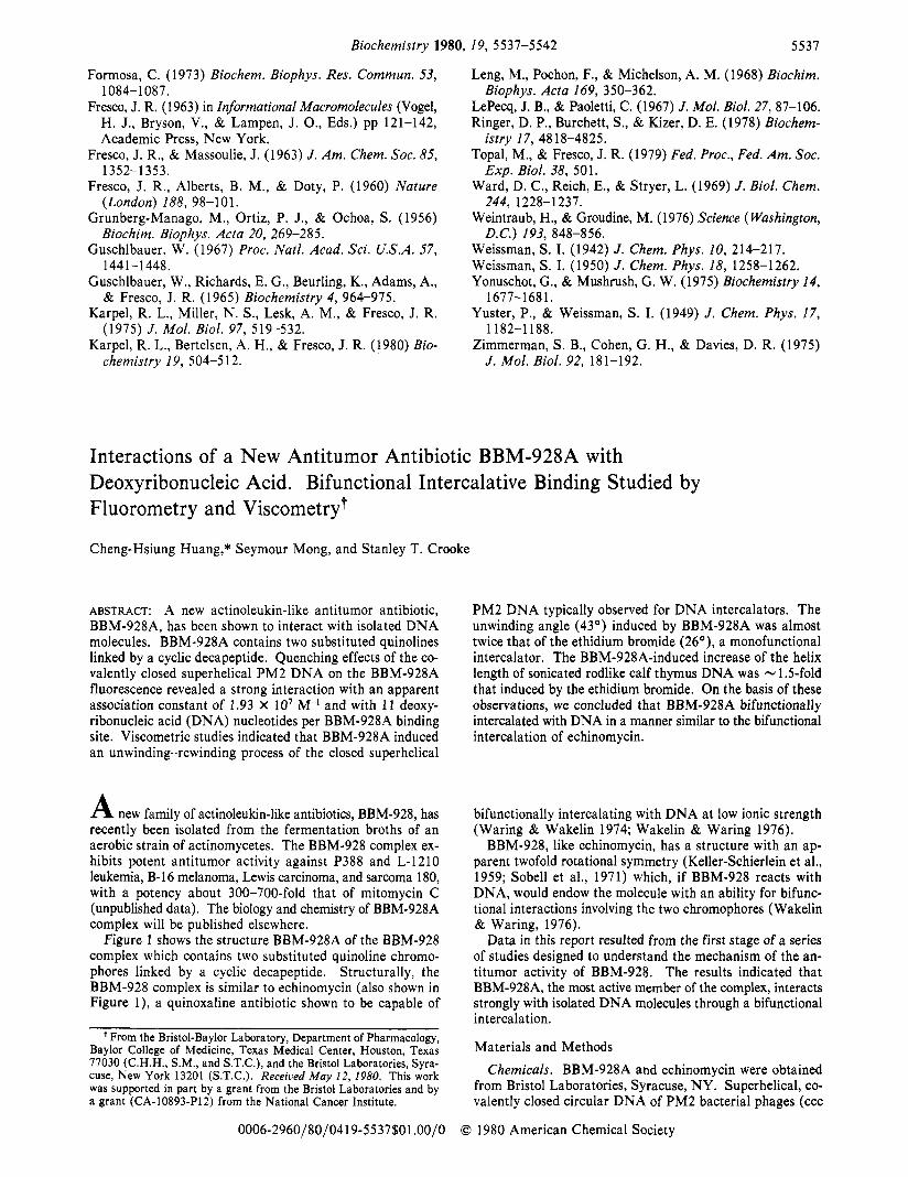
~ G U R E 2: Fluorescence spectra of BBM-928A (10 pg/mL or 6.9 pM) in 10 mM Tris-HCI, pH 7.4. (A) Excitation spectra [(e) emission at 520 nm; (0) emission at 675 nm]. (B) Emission spectrum [(a) excitation at 340 nm].
lished procedures or equations are described under Results. The sheared calf thymus DNA used for viscometric studies
was prepared either by forcing a DNA solution 30 times through a Yale 23G1 hypodermic needle (Pyeritz et al., 1972) or by pulsed sonications for a total of 10 min in a Heat Systems sonicator (Model W-375) with full output after the DNA solution was bubbled with N2 for 15 min. Results for both types of sheared DNA preparations were similar.
Determination of Drug Concentration. Drug concentrations were determined spectrophotometrically. For BBM-928A, a molecular weight of 1450 (by osmometry) and an extinction coefficient, ElCm1%, of 168 at 345 nm (in 95% ethanol) have been determined (unpublished data). Echinomycin (molecular weight = 1100) has a molar extinction coefficient of 12 400 mol-' cm-' at 325 nm (Wakelin & Waring, 1976). The molar extinction coefficient of EB is 4800 mol-' cm-l at 460 nm (Waring, 1965).
Results Fluorescence Properties of BBM-928A. Figure 2 shows the
excitation spectra (emission at 520 or 675 nm) and the emission spectrum (excitation at 340 nm) of BBM-928A. These spectra are due to the quinoline chromophores. When excited at 340 nm, the fluorescence spectrum (Figure 2B) showed a broad band at 520 nm and a sharp band at 675 nm. With an emission at 520 nm (Figure 2A), the excitation
A N T I T U M O R A N T I B I O T I C B B M - 9 2 8 A - D N A I N T E R C A L A T I O N V O L . 1 9 , N O . 2 4 , 1 9 8 0 5539
. r80.054
80
60
40
20
0
I20
400 500 600 700 0 20 40
n m ( D N ~ @rug3
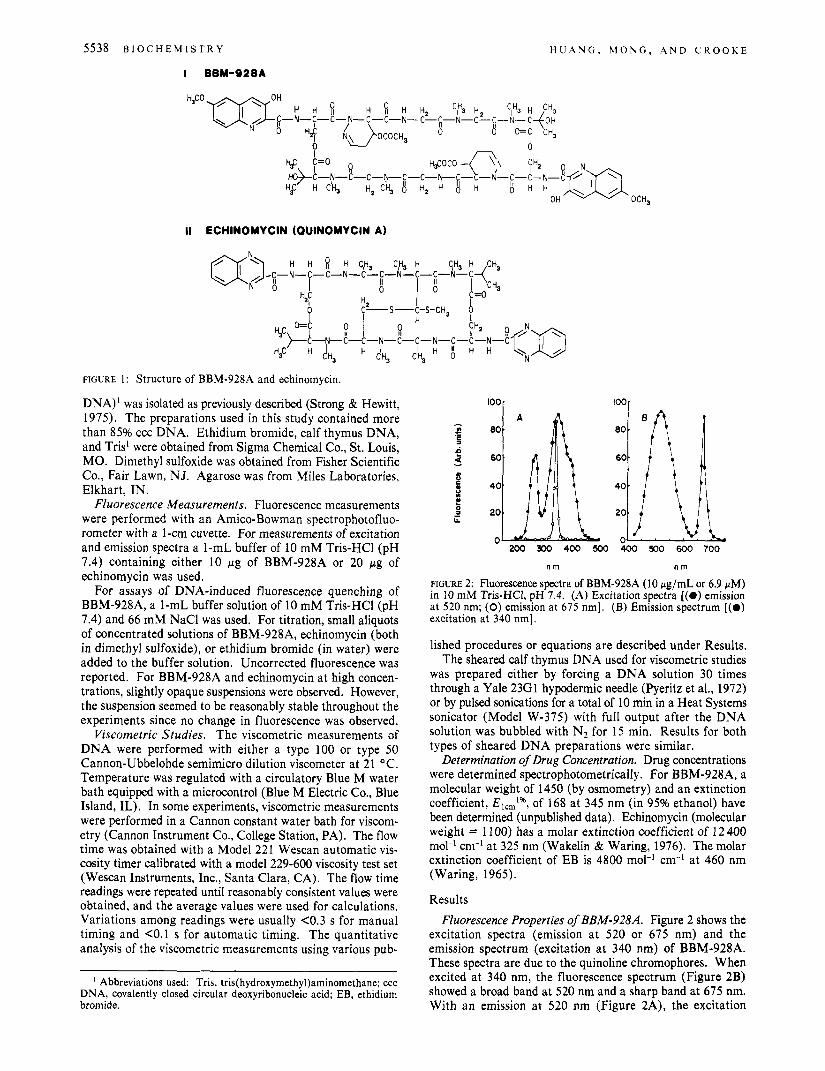
FIGURE 3: (A) Quenching effects of PM2 DNA on the fluorescence spectrum (excitation at 340 nm) of BBM-928A (6.9 pM) in 10 mM Tris-HCI (pH 7.4) and 75 mM NaC1. (0) -DNA; (0) +DNA. (B) Quenching effects of increasing DNA concentration on BBM-928A fluorescence at 520 nm (excitation at 340 nm) in 10 mM Tris-HC1 (pH 7.4) and 75 mM NaC1.
spectrum showed a major band at 350 nm, a minor band at 270 nm, and a shoulder around 400 nm. However, with an emission at 675 nm, only a sharp excitation band at 340 nm was observed [Figure 2A ( O ) ] . More detailed studies on the fluorescence properties of BBM-928A will be reported else- where.
Quenching of BBM-928A Fluorescence by DNA. The ad- dition of DNA induced a marked quenching effect on the fluorescence of BBM-928A. Figure 3A shows that the PM2 DNA induced quenching occurred throughout the entire spectral range. Figure 3B shows the progressive quenching effect of ccc PM2 DNA on the fluorescence of BBM-928A at 520 nm (excitation at 340 nm). At a BBM-928A ratio of 0.077), a 75% reduction in fluorescence was noted. These results indicate a strong interaction between BBM-928A and DNA molecules. The addition of calf thymus DNA induced similar fluorescence quenching effects (not shown).
Quantitative Analyses of Fluorescence Quenching Effect. The results of the PM2 DNA induced fluorescence quenching effect shown in Figure 3B were analyzed by a Scatchard (1949) analysis as previously described (Peacocke & Skerrett, 1956; LePecq & Paoletti, 1967). In the Scatchard equation rb/c = Kn - Krb, where rb is the number of drug molecules bound per DNA nucleotide phosphate, c is the free drug concentration, K is the apparent association constant, and n is the number of drug binding sites per nucleotide phosphate. A plot of rb /c vs. rb, as shown in Figure 4A, gave a K value of 1.93 X IO7 M-' from the slope and an n value of 0.091 from the intercept. The n value is equivalent to the binding of 1 molecule of BBM-928A/ll DNA nucleotide phosphate res- idues.
From the foregoing analysis, the bound drug to DNA ratio rb ( r 3 could be plotted against the input drug to DNA ratio ( r ) as shown in Figure 4B. The results showed that rb or r' values approach r values up to 0.075. This indicates that within a low input ratio ( r = 0.075), almost all the added BBM-928A molecules were bound to DNA molecules.
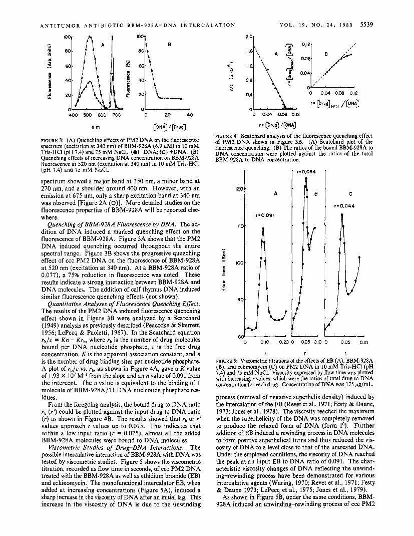
Viscometric Studies of Drug-DNA Interactions. The possible intercalative interaction of BBM-928A with DNA was tested by viscometric studies. Figure 5 shows the viscometric titration, recorded as flow time in seconds, of ccc PM2 DNA treated with the BBM-928A as well as ethidium bromide (EB) and echinomycin. The monofunctional intercalator EB, when added at increasing concentrations (Figure SA), induced a sharp increase in the viscosity of DNA after an initial lag. This increase in the viscosity of DNA is due to the unwinding
- A
r-0.091
2.01
0 0.04 0.08 0.12
o 0.04 0.08 0.12
r = [Druo) /[DNA)
FIGURE 4: Scatchard analysis of the fluorescence quenching effect of PM2 DNA shown in Figure 3B. (A) Scatchard plot of the fluorescence quenching. (B) The ratios of the bound BBM-928A to DNA concentration were plotted against the ratios of the total BBM-928A to DNA concentration.
80-
C
r 8 0.044
4
, I t
4 1
1
0 0.10 0.20 0 0.05 0.10 0 0.05 0.10
r r r
~ G U R E 5 : Viscometric titrations of the effects of EB (A), BBM-928A (B), and echinomycin (C) on PM2 DNA in 10 m M Tris-HC1 (pH 7.4) and 75 m M NaCl. Viscosity expressed by flow time was plotted with increasing r values, which were the ratios of total drug to DNA concentration for each drug. Concentration of DNA was 175 pg/mL.
process (removal of negative superhelix density) induced by the intercalation of the EB (Revet et al., 1971; Festy & Daune, 1973; Jones et al., 1978). The viscosity reached the maximum when the superhelicity of the DNA was completely removed to produce the relaxed form of DNA (form Io). Further addition of EB induced a rewinding process in DNA molecules to form positive superhelical turns and thus reduced the vis- cosity of DNA to a level close to that of the untreated DNA. Under the employed conditions, the viscosity of DNA reached the peak at an input EB to DNA ratio of 0.091. The char- acteristic viscosity changes of DNA reflecting the unwind- ing-rewinding process have been demonstrated for various intercalative agents (Waring, 1970; Revet et al., 1971; Festy & Daune 1973; LePecq et al., 1975; Jones et al., 1979).
As shown in Figure 5B, under the same conditions, BBM- 928A induced an unwinding-rewinding process of ccc PM2
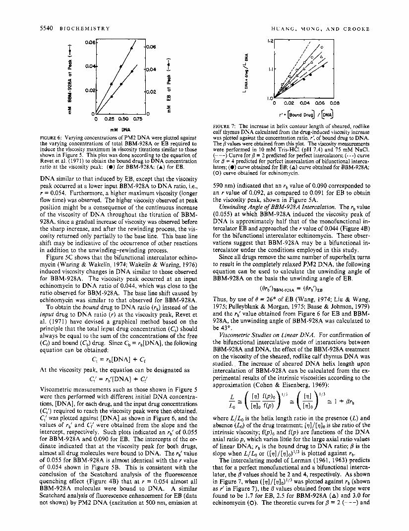
5540 B I O C H E M I S T R Y H U A N G , M O N G , A N D C R O O K E
i - X
L c 0
L E
B c 0
w m
I E
0 0.02 0.04 a06 0.08
mM DNA
FIGURE 6: Varying concentrations of PM2 DNA were plotted against the varying concentrations of total BBM-928A or EB required to induce the viscosity maximum in viscosity titrations similar to those shown in Figure 5 . This plot was done according to the equation of Revet et a]. (1971) to obtain the bound drug to DNA concentration ratio at the viscosity peak: (0) for BBM-928A; (A) for EB.
DNA similar to that induced by EB, except that the viscosity peak occurred at a lower input BBM-928A to DNA ratio, Le., r = 0.054. Furthermore, a higher maximum viscosity (longer flow time) was observed. The higher viscosity observed at peak position might be a consequence of the continuous increase of the viscosity of DNA throughout the titration of BBM- 928A, since a gradual increase of viscosity was observed before the sharp increase, and after the rewinding process, the vis- cosity returned only partially to the base line. This base line shift may be indicative of the occurrence of other reactions in addition to the unwinding-rewinding process.
Figure 5C shows that the bifunctional intercalator echino- mycin (Waring & Wakelin, 1974; Wakelin & Waring, 1976) induced viscosity changes in DNA similar to those observed for BBM-928A. The viscosity peak occurred at an input echinomycin to DNA ratio of 0.044, which was close to the ratio observed for BBM-928A. The base line shift caused by echinomycin was similar to that observed for BBM-928A.
To obtain the bound drug to DNA ratio (rb) instead of the input drug to DNA ratio (r) at the viscosity peak, Revet et al. (1971) have devised a graphical method based on the principle that the total input drug concentration (C,) should always be equal to the sum of the concentrations of the free (Cf) and bound (c,) drug. Since c b = rb[DNA], the following equation can be obtained:
C, = rb[DNA] + Cf At the viscosity peak, the equation can be designated as
C( = r{[DNA] + C,'
Viscometric measurements such as those shown in Figure 5 were then performed with different initial DNA concentra- tions, [DNA], for each drug, and the input drug concentrations (C;) required to reach the viscosity peak were then obtained. C,' was plotted against [DNA] as shown in Figure 6, and the values of rb) and C; were obtained from the slope and the intercept, respectively. Such plots indicated an rb) of 0.055 for BBM-928A and 0.090 for EB. The intercepts of the or- dinate indicated that at the viscosity peak for both drugs, almost all drug molecules were bound to DNA. The rd value of 0.055 for BBM-928A is almost identical with the r value of 0.054 shown in Figure 5B. This is consistent with the conclusion of the Scatchard analysis of the fluorescence quenching effect (Figure 4B) that at r = 0.054 almost all BBM-928A molecules were bound to DNA. A similar Scatchard analysis of fluorescence enhancement for EB (data not shown) by PM2 DNA (excitation at 500 nm, emission at
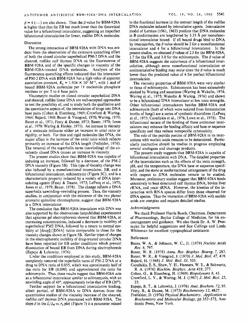
FIGURE 7: The increase in helix contour length of sheared, rodlike calf thymus DNA calculated from the drug-induced viscosity increase was plotted against the concentration ratio, r', of bound drug to DNA. The 0 values were obtained from this plot. The viscosity measurements were performed in 10 mM Tris-HC1 (pH 7.4) and 75 mM NaCI. (---) Curve for /3 = 2 predicted for perfect intercalators; (- - -) curve for 0 = 4 predicted for perfect intercalation of bifunctional interca- Iators; (0) curve obtained for EB; (A) curve obtained for BBM-928A; (0) curve obtained for echinomycin.
590 nm) indicated that an rb value of 0.090 corresponded to an r value of 0.092, as compared to 0.091 for EB to obtain the viscosity peak, shown in Figure 5A.
Unwinding Angle of BBM-928A Intercalation. The rb value (0.055) at which BBM-928A induced the viscosity peak of DNA is approximately half that of the monofunctional in- tercalator EB and approached the r value of 0.044 (Figure 4B) for the bifunctional intercalator echinomycin. These obser- vations suggest that BBM-928A may be a bifunctional in- tercalator under the conditions employed in this study.
Since all drugs remove the same number of superhelix turns to result in the completely relaxed PM2 DNA, the following equation can be used to calculate the unwinding angle of BBM-928A on the basis the unwinding angle of EB.
(%')BBM-~BA = (%')EB
Thus, by use of B = 26' of EB (Wang, 1974; Liu & Wang, 1975; Pulleyblank & Morgan, 1975; Baase & Johnson, 1979) and the r{ value obtained from Figure 6 for EB and BBM- 928A, the unwinding angle of BBM-928A was calculated to be 43O.
Viscometric Studies on Linear DNA. For confirmation of the bifunctional intercalative mode of interactions between BBM-928A and DNA, the effect of the BBM-928A treatment on the viscosity of the sheared, rodlike calf thymus DNA was studied.. The increase of sheared DNA helix length upon intercalation of BBM-928A can be calculated from the ex- perimental results of the intrinsic viscosities according to the approximation (Cohen & Eisenberg, 1969):
where L/Lo is the helix length ratio in the presence (L) and absence (&) of the drug treatment; [ 7 ] / [ 0 ] ~ is the ratio of the intrinsic viscosity; f(p)o and f(p) are functions of the DNA axial ratio p , which varies little for the large axial ratio values of linear DNA; rb is the bound drug to DNA ratio; P is the slope when L/Lo or ([V]/[V],,)'/~ is plotted against rb .
The intercalating model of Lerman (1961, 1963) predicts that for a perfect monofunctional and a bifunctional interca- lator, the 6 values should be 2 and 4, respectively. As shown in Figure 7, when ( [v ] / [v ]~ ) ' /~ was plotted against rb (shown as J in Figure 7), the P values obtained from the slope were found to be 1.7 for EB, 2.5 for BBM-928A (A) and 3.0 for echinomycin (0). The theoretic curves for /3 = 2 (---) and
A N T I T U M O R A N T I B I O T I C B B M - 9 2 8 A - D N A I N T E R C A L A T I O N V O L . 1 9 , N O . 2 4 , 1 9 8 0 5541
P = 4 (- - -) are also shown. Thus the P value for BBM-928A is higher than that for EB but much lower than the theoretical value for a bifunctional intercalator, suggesting an imperfect bifunctional intercalation for linear, rodlike DNA molecules.
Discussion The strong interaction of BBM-928A with DNA was evi-
dent from the observation of the extensive quenching effect of both the closed circular superhelical PM-2 DNA and the sheared, rodlike calf thymus DNA on the fluorescence of BBM-928A and of the specific changes in viscosity of the BBM-928A-treated DNA molecules, Analyses of the fluorescence quenching effects indicated that the interaction of PM-2 DNA with BBM-928A has a high value of apparent association constant, K, = 1.934 X lo7 M-l, with 1 mol of bound BBM-928A molecules per 11 nucleotide phosphate residues or per 5 to 6 base pairs.
Viscometric studies on closed circular superhelical DNA and sheared, rodlike linear DNA are well-accepted approaches to test the possibility of, and to study both the qualitative and the quantitative aspects of, the intercalation of drugs into DNA base pairs (Cohen & Eisenberg, 1969; Crawford & Waring, 1964; Bujard, 1968; Bauer & Vinograd, 1970; Waring, 1970; Revet et al., 1971; Festy & Daune, 1973; Bauer, 1978; Jones et al., 1979; Waring & Henley, 1975). An increase in viscosity of a molecule indicates either an increase in axial ratio or rigidity, or both. For thin and rigid molecules like DNA, the major effect is the increase of the axial ratio which reflects primarily an increase of the DNA length (Feifelder, 1976). The removal of the superhelix turns (unwinding) of the co- valently closed DNA causes an increase in viscosity.
The present studies show that BBM-928A was capable of inducing an increase, followed by a decrease, of the PM-2 DNA viscosity (Figure 5B). This type of viscosity change was also induced by a monofunctional intercalator, EB, and a bifunctional intercalator, echinomycin (Figure 5C), and is a characteristic property induced by a variety of DNA inter- calating agents (Festy & Daune, 1973; LePecq et al., 1975; Jones et al., 1979; Bauer, 1978). The change reflects a DNA superhelix unwinding-rewinding process. Thus, the viscosity studies, in conjunction with the existence of the two planar aromatic quinoline chromophores, suggest that BBM-928A is a DNA intercalator.
The conclusion that BBM-928A intercalates with DNA was also supported by the observations (unpublished experiments) that agrarose gel electrophoresis showed that BBM-928A, at increasing concentrations, induced a decrease in mobility of superhelical PM2 DNA, followed by a return to normal mo- bility at [drug]/[DNA] ratios comparable to those for the viscosity changes shown in Figure 5B. Similar types of changes in the electrophoretic mobility of drug-treated circular DNA have been reported for EB under conditions which prevent dissociation of bound EB from DNA during electrophoresis (Espejo & Lebowitz, 1976).
Under the conditions employed in this study, BBM-928A completely removed the superhelix turns of PM-2 DNA at a drug to DNA ratio of 0.055, which was aproximately half of the ratio for EB (0.090) and approximated the ratio for echinomycin. Thus, these results suggest that BBM-928A acts as a bifunctional intercalator similar to echinomycin, with an unwinding angle of 43', approximately twice that of EB (26').
Further support for a bifunctional intercalative binding, albeit partial, of BBM-928A to DNA derives from the quantitative studies of the viscosity increase of the sheared, rodlike calf thymus DNA pretreated with BBM-928A. The slope P in the L/& vs. rb plot (Figure 7) is a parameter related
to the fractional increase in the contour length of the rodlike DNA molecules induced by intercalative agents. Intercalative model of Lerman (1 961, 1963) predicts that DNA molecules in B conformation are lengthened by 3.35 A per monofunc- tional intercalator bound. If all bound drugs bind to DNA by intercalation, the p value should be 2 for a monofunctional intercalator and 4 for a bifunctional intercalator. In the present studies, we obtained p values of 2.5 for the BBM-928A, 1.7 for the EB, and 3.0 for the echinomycin. The /3 value of BBM-928A suggests the occurrence of a bifunctional inter- calation, although some monofunctional intercalation or nonintercalative binding may occur since the p value is much lower than the predicted value of 4 for perfect bifunctional intercalation.
The viscosity properties of BBM-928A were very similar to those of echinomycin. Echinomycin has been extensively studied by Waring and associates (Waring & Wakelin, 1974; Waring et al., 1975; Wakelin & Waring, 1976) and shown to be a bifunctional DNA intercalator at low ionic strengths. Other bifunctional intercalators besides BBM-928A and echinomycin (both of which were isolated from fermentation broths of fungi) are a series of synthetic diacridines (LePecq et al., 1975; Canellakis et al., 1976; Lown et al., 1978). The bifunctional nature of the binding of these antitumor inter- calators may enhance the binding affinity and base or sequence specificity and thus reduce nonspecific cytotoxicity.
The role of the peptide portion of BBM-928A in its inter- actions with nucleic acids remains to be determined. Partic- ularly instructive should be studies in progress employing several analogues and cleavage products.
The present study suggests that BBM-928A is capable of bifunctional intercalation with DNA. The detailed properties of the intercalation such as the effects of the ionic strength, pH, and the temperature, the DNA base or sequence specif- icity, and the steric or conformational arrangement of the drug with respect to DNA molecules remain to be studied. Moreover, preliminary studies suggest that BBM-928A binds extensively to heat denatured calf thymus DNA, bovine liver rRNA, and yeast tRNA. However, the kinetics of the in- teraction with RNA species differ from those observed for DNA species. Thus the interaction of BBM-928A with nucleic acids are complex and require detailed studies.
Acknowledgments We thank Professor Harris Busch, Chairman, Department
of Pharmacology, Baylor College of Medicine, for his en- couragement and guidance. We also thank Dr. A. W. Pres- tayko for helpful suggestions and Sue Collings and Linda Whiteman for excellent typographical assistance.
References Baase, W. A., & Johnson, W. C., Jr. (1979) Nucleic Acids
Bauer, W. R. (1978) Annu. Rev. Biophys. Bioeng. 7 , 287. Bauer, W. R., & Vinograd, J . (1970) J . Mol. Biol. 47, 419. Bujard, H. (1968) J . Mol. Biol. 33, 503. Canellakis, E. S., Shaw, Y. H., Hanners, W. E., & Schwartz,
Cohen, G., & Eisenberg, H. (1969) Biopolymers 8 , 45. Crawford, L. V., & Waring, M. J. (1967) J . Mol. Biol. 25,
Espejo, R. T., & Lebowitz, J. (1976) Anal. Biochem. 72, 95. Festy, B., & Daune, M. (1973) Biochemistry 12, 4827. Freifelder, D. (1976) Physical Biochemistry. Application to
Biochemistry and Molecular Biology, pp 352-373, Ace- demic Press, New York.
Res. 6, 797.
R. A. (1976) Biochim. Biophys. Acta 418, 277.
23.
5542 Biochemistry 1980, 19, 5542-5549
Jones, R. L., Davidson, M. W., & Wilson, W. D. (1979)
Keller-Schierlein, W., Mihailovic, M. D., & Prelog, V. (1959)
LePecq, J. B., & Paoletti, C. (1967) J . Mol. Biol. 27, 87. LePecq, J. B., LeBret, M., Barbet, J., & Roques, B. (1975)
Proc. Natl. Acad. Sci. U.S.A. 72, 2915. Lerman, L. S. (1961) J . Mol. Biol. 3, 18. Lerman, L. S. (1963) Proc. Natl. Acad. Sci. U.S.A. 49, 94. Liu, L. F., & Wang, J. C. (1975) Biochim. Biophys. Acta 395,
Lown, J. W., Gum, B. C., Chang, R. Y., Majumdar, K. C.,
Peacocke, A. R., & Skerret, J. N. H. (1956) Trans. Faraday
Pulleyblank, D. E., & Morgan, A. R. (1975) J . Mol. Biol. 91,
Pyeritz, R. E., Schlegel, R. A., & Thomas, C. A., Jr. (1972)
Biochim. Biophys. Acta 561, 77.
Helu. Chim. Acta 42, 305.
405.
& Lee, J. S . (1978) Can. J . Biochem. 56, 1006.
SOC. 52, 261.
1.
Biochim. Biophys. Acta 272, 504.
Revet, B. M. J., Schmir, M., & Vinograd, J. (1971) Nature
Scatchard, G. (1949) Ann. N .Y . Acad. Sci. 51, 660. Sobell, H. M., Jain, S. C., Sakore, T. D., & Nordman, C. E.
(1971) Nature (London), New Biol. 231, 200. Strong, J. E., & Hewitt, R. R. (1975) Isozymes Znt. Conf.,
3rd, 1974 3, 473. Wakelin, L. P. G., & Waring, M. J. (1976) Biochem. J . 157,
721. Wang, J. C. (1974) J . Mol. Biol. 89, 783. Waring, M. J. (1965) J . Mol. Biol. 13, 269. Waring, M. J. (1970) J . Mol. Biol. 54, 247. Waring, M. J., & Wakelin, L. P. G. (1974) Nature (London)
Waring, M. J., & Henley, S. M. (1975) Nucleic Acids Res.
Waring, M. J., Wakelin, L. P. G., & Lee, J . S. (1975) Bio-
(London), New Biol. 229, 10.
252, 653.
2, 567.
chim. Biophys. Acta 407, 200.
Reactions of 2-Thioribothymidine and 4-Thiouridine with Hydrogen Peroxide in Transfer Ribonucleic Acids from Thermus thermophilus and Escherichia coli As Studied by Circular Dichroismt
Kimitsuna Watanabe
ABSTRACT: The reaction of thiouridine derivatives with hy- drogen peroxide (H202) at the monomer level as well as in polynucleotides was investigated to check the possibility that the difference or variation in the environment surrounding the thiouridine residues may be simply a reflection of the difference in the reactivity of these residues with Hz02 At the monomer level, the reactivity is of the order of 4-thiouridine (s4U) > 5-[(methylamino)methyl]-2-thiouridine (mnm5s2U) > 2- thiouridine (s2U) N 2-thioribothymidine (s2T). When these residues are in polynucleotides, but without their C = S groups being hydrogen bonded, such as s4U in tRNAPhe and tRNA,Me', mnm5s2U in tRNA:'" from E. coli, and s2T in s2T\kCGp, their reactivity is reduced to no more than one-half of that of monomers. All these reactions proceeded by first-order kinetics. Poly(2-thiouridylic acid)/[poly(s2U)], in
E x t r e m e thermophilic bacteria, Thermus thermophilus HB 8 and HB 27, Thermus aquaticus YT 1, and Thermusflauus AT 62, all have 2-thioribothymidine (sZT)' in place of the usual ribothymidine (T) in the T W loop of their tRNAs (Watanabe et al., 1974; Oshima et al., 1977), and this modified nucleoside is thought to be the most responsible for the thermal stability of the tRNAs (Oshima et al., 1976; Watanabe et al., 1976a,b, 1979a, 1980; Davanloo et al., 1979).
On the basis of the tertiary structure of yeast tRNAPhe (Ladner et al., 1975a; Quigley et al., 1975; Rich & Kim,
From the Laboratory of Biochemical Reactions and Biocatalysts, Mitsubishi-Kasei Institute of Life Sciences, 11 Minamiooya, Machida- shi, Tokyo 194, Japan. Received March 10, 1980; revised manuscript received July 3, 1980.
0006-2960/80/0419-5.542$01 .OO/O
which both free and hydrogen-bonded C=S groups exist, reacted with H202, at first slowly (ksw = 0.1 1 X loT3 min-'), corresponding to the reaction of the free C=S group in the double helix, and then rapidly (knnal = 1.5 X min-I) due to denaturation of the strands. sZT in T. thermophilus tRNAPhe reacted easily when it was denatured by trans-1,2- diaminocyclohexanetetraacetic acid treatment, while it reacted very slowly in the native tRNAPhe [ k = (0.06-0.02) X min-'1. Even the larger rate constant is only one-half of the initial rate constant of poly(s2U). The charging activity of the H20z-treated tRNAPhc has good correlation with the re- sidual amount of s2T in the tRNAPhe. These results suggest that the s2T sulfur atom in the native tRNAPhc is probably involved in hydrogen bonding, as reported in the case of the tertiary structure of yeast tRNAPhe.
1978), it is speculated that the s2T54 residue of the thermophile tRNA is base paired with the m'A58 residue and that this base pair stacks on the neighboring common base pair G53-C6 1 and on the interloop base pair q55-Gm18 [the numbering of residues conforms to the proposed rule (Sprinzl et al., 1980)]. s2T nucleoside is known to take on the 3'-endo-gauche- gauche-anti form preferentially (Watanabe et al., 1979a; Yokoyama et al., 1979). This rigid conformation in the po-
l Abbreviations used: H202, hydrogen peroxide; C=S, thiocarbonyl group; k, rate constant; kinitial and knnal, initial and final rate constants, respectively; half-life; LC, high-pressure liquid chromatography; CDTA, trans- 1,2-diaminocyclohexanetetraacetic acid; s2U, 2-thiouridine; s2T, 2-thioribothymidine; mnm5s2U, 5-[(methylamino)methyl]-2-thio- uridine; poly(s2U), poly(2-thiouridylic acid); Cm, 02'-methylcytidine; Gm, @'-methylguanosine; mlA, l-methyladenosine.
0 1980 American Chemical Society