Embed Size (px)
Citation preview
Interactions between the Influenza A Virus RNA PolymeraseComponents and Retinoic Acid-Inducible Gene I
Weizhong Li, Hongjun Chen, Troy Sutton, Adebimpe Obadan, Daniel R. Perez
Department of Veterinary Medicine, University of Maryland, College Park, and Virginia-Maryland Regional College of Veterinary Medicine, College Park, Maryland, USA
ABSTRACT
The influenza A virus genome possesses eight negative-strand RNA segments in the form of viral ribonucleoprotein particles(vRNPs) in association with the three viral RNA polymerase subunits (PB2, PB1, and PA) and the nucleoprotein (NP). Throughinteractions with multiple host factors, the RNP subunits play vital roles in replication, host adaptation, interspecies transmis-sion, and pathogenicity. In order to gain insight into the potential roles of RNP subunits in the modulation of the host’s innateimmune response, the interactions of each RNP subunit with retinoic acid-inducible gene I protein (RIG-I) from mammalianand avian species were investigated. Studies using coimmunoprecipitation (co-IP), bimolecular fluorescence complementation(BiFc), and colocalization using confocal microscopy provided direct evidence for the RNA-independent binding of PB2, PB1,and PA with RIG-I from various hosts (human, swine, mouse, and duck). In contrast, the binding of NP with RIG-I was found tobe RNA dependent. Expression of the viral NS1 protein, which interacts with RIG-I, did not interfere with the association ofRNA polymerase subunits with RIG-I. The association of each individual virus polymerase component with RIG-I failed to sig-nificantly affect the interferon (IFN) induction elicited by RIG-I and 5= triphosphate (5=ppp) RNA in reporter assays, quantita-tive reverse transcription-PCR (RT-PCR), and IRF3 phosphorylation tests. Taken together, these findings indicate that viralRNA polymerase components PB2, PB1, and PA directly target RIG-I, but the exact biological significance of these interactionsin the replication and pathogenicity of influenza A virus needs to be further clarified.
IMPORTANCE
RIG-I is an important RNA sensor to elicit the innate immune response in mammals and some bird species (such as duck) uponinfluenza A virus infection. Although the 5=-triphosphate double-stranded RNA (dsRNA) panhandle structure at the end of viralgenome RNA is responsible for the binding and subsequent activation of RIG-I, this structure is supposedly wrapped by RNApolymerase complex (PB2, PB1, and PA), which may interfere with the induction of RIG-I signaling pathway. In the presentstudy, PB2, PB1, and PA were found to individually interact with RIG-Is from multiple mammalian and avian species in anRNA-independent manner, without significantly affecting the generation of IFN. The data suggest that although RIG-I bindingby RNA polymerase complex is conserved in different species, it does not appear to play crucial role in the modulation of IFN invitro.
Influenza A virus is a member of the Orthomyxoviridae familyand contains eight segments of negative-sense genomic RNA.
Within the virus particle, viral RNAs (vRNAs) are wrapped withmultiple copies of the nucleoprotein (NP) and are bound to aheterotrimeric polymerase complex composed of basic poly-merases 1 and 2 (PB1 and PB2, respectively) and the acidic poly-merase (PA) to collectively form viral ribonucleoprotein particles(vRNPs). Upon entry and uncoating, the vRNPs travel to the nu-cleus through interactions with importin-like factors and compo-nents of the nuclear pore complex. The vRNPs form the minimalunits for transcription and replication of viral RNAs. PB2 recog-nizes and binds the methylated cap of cellular pre-mRNAs (1),and then PA cleaves the cap and generates 5=-capped RNA frag-ments (2), which are then used to prime viral mRNA transcriptionby PB1 (3). The accumulation of NP during infection is thought tofavor the switching of viral RNA from transcription to replication(4) and promote the nuclear export of progeny vRNPs (in associ-ation with the viral proteins M1 and NEP and cellular factors) (5).The protein components of vRNPs are also involved in viru-lence, host adaptation, and interspecies transmission of influ-enza A virus, presumably due to interactions with numeroushost factors (6).
The innate immune response, especially the interferon (IFN)-
mediated response, makes up the first line of defense against theinvasion of pathogens. The production of type I IFN is primarilyinduced by Toll-like receptors (TLRs) located in the endosome orcellular membrane and RIG-I like receptors (RLRs) distributedmainly in the cytoplasm. RIG-I has been identified as the majorcellular sensor for IFN induction upon influenza virus infection(7, 8). RIG-I recognizes the 5= triphosphate (5=ppp) in viralgenomic RNAs (9, 10) or double-stranded RNA (dsRNA) inter-mediates synthesized during virus replication (7, 11). Recently,RIG-I was shown to preferentially associate with short sub-genomic viral segments in infected cells (12). Binding to its ligandinduces a conformational change in RIG-I that leads to the ubiq-uitination of CARD domains (caspase activation and recruitmentdomains) by TRIM25 (tripartite motif protein 25) or ubiquitina-
Received 12 May 2014 Accepted 12 June 2014
Published ahead of print 18 June 2014
Editor: M. S. Diamond
Address correspondence to Daniel R. Perez, [email protected].
Copyright © 2014, American Society for Microbiology. All Rights Reserved.
doi:10.1128/JVI.01383-14
10432 jvi.asm.org Journal of Virology p. 10432–10447 September 2014 Volume 88 Number 18
on May 25, 2018 by guest
http://jvi.asm.org/
Dow
nloaded from

tion of C-terminal regulatory domain by Riplet (RING finger pro-tein leading to RIG-I activation) and the subsequent complex for-mation between RIG-I and IPS-1 (beta interferon promoterstimulator 1, also known as MAVS, VISA, or Cardif) on the sur-face of the mitochondria (13). The ensuing signal cascade causesthe nuclear translocation of IRF3, IRF7, ATF-2/c-Jun, and NF-�B,which activate the IFN-� promoter and promote the expression ofIFN (14). Finally, hundreds of interferon-stimulated genes (ISGs),such as those encoding PKR, 2=,5=-OAS, Mx1, and ADAR1,among others, are produced and exert their antiviral function viamultiple mechanisms (15). Of note, RIG-I has been found notonly in humans and mammals but also in a wide variety of otheranimal species, such as zebrafish, amphibians, and birds (16, 17).Interestingly, Galliformes (e.g., chickens) appear to have lostthrough evolution the gene that encodes RIG-I, although it ismaintained in other avian species such as in Anseriformes (ducksand geese) (18, 19). This difference has been suggested as a possi-ble explanation for the increased susceptibility of chickens com-pared to that of ducks to highly pathogenic influenza viruses (18).
To survive in host cells, influenza A viruses have evolved vari-ous strategies to circumvent the IFN response. The NS1 protein, anonstructural protein generated during the early stages of virusinfection, is a well-characterized IFN inhibitor (20). NS1 can limitIFN induction by various strategies, including (i) sequestration ofviral dsRNA away from host-encoded RNA sensors (21), (ii) for-mation of complexes with RIG-I (9, 10, 22), (iii) suppression ofTRIM25- and/or Riplet-mediated RIG-I ubiquitination (16, 22),and (iv) blocking of cellular pre-mRNA processing and transportand thus decreasing synthesis of antiviral proteins involved in in-nate immunity (23, 24). Apart from NS1, other influenza virusproteins also possess IFN-antagonizing capabilities. PB1-F2 canact on MAVS (25, 26) and/or interfere with the RIG-I/MAVS pro-tein complex (27), thereby inhibiting IFN production. In addi-tion, the PB2 protein (28, 29) from human influenza A virus aloneor in the context of the three viral polymerase subunits has beenshown to interact with MAVS and impair host antiviral responsesmediated by IFN (30).
In the present study, evidence was obtained for the direct in-teractions of viral RNA polymerase components from an avianinfluenza A virus (H9N2 subtype) with RIG-I from various hostspecies (human, duck, mouse, and swine). NP was also shown tointeract with RIG-I, although this interaction is indirect and me-diated by RNA. Association of virus RNP subunits with RIG-I didnot remarkably contribute to the inhibition of the IFN signalingpathway. Collectively, these findings highlight the interactions ofviral polymerase components with RIG-I, although the biologicalsignificance of these interactions awaits clarification.
MATERIALS AND METHODSCells and viruses. Madin-Darby canine kidney (MDCK) cells, chickenembryonic fibroblasts (DF1), duck embryonic fibroblasts (CCL-141), amouse macrophage cell line (RAW264.7), a swine epithelial cell line(PK15), a human embryonic kidney cell line (293T), a human lung carci-noma cell line (A549), and African green monkey kidney (Vero) cells werecultured in OPTI-MEM supplemented with antibiotics and 10% fetal calfserum at 37°C in 5% CO2. Influenza A virus strain A/Guinea Fowl/HongKong/WF10/99 (H9N2) (WF10) has been previously described (31). Thevirus was propagated in 9-day-old embryonated eggs. Virus titer was de-termined by 50% tissue culture infective dose (TCID50) in MDCK cellsusing the method of Reed and Muench (32).
Plasmids. To construct plasmids used for biomolecular fluorescencecomplementation (BiFc) analysis, the sequences encoding the N terminusof enhanced yellow fluorescent protein (EYFP; containing residues 1 to155 and a Q70M point mutation to enhance stability of fluorescence sig-nal; NYFP), the C terminus of EYFP (comprising residues 156 to 239;CYFP), or the N terminus of Cerulean (a variant of EYFP) correspondingto residues 1 to 173 (NCe) were fused to PB2, PB1, PA, or NP at theC-terminal end via a linker (three repeats of the peptide sequence GGGGS). The chimeric genes were then cloned into the pDP2002 or pcDNA3vector to yield plasmids pDPPB2-NYFP, pDPPB1-NYFP, pDPPA-NYFP,pDPNP-NCe, pDPPB2-CYFP, pDPPA-CYFP, pc-PB2-NYFP, pc-PB1-NYFP, pc-PA-NYFP, and pc-NP-NYFP. Plasmid pcDNA3-NYFP wasconstructed via the insertion of NYFP-encoding sequence into pcDNA3.Based on this plasmid, a derivative construct was produced, pc-WFNS1(�S)-NYFP, which expresses NS1 protein (without a splicing site)flanked by a linker-NYFP segment at the C terminus. The full-length ofRIG-I open reading frame (ORF) from different species (duck, mouse,swine, or human) was amplified from WF10 virus-infected or polyI·C-stimulated cells (CCL-141, RAW264.7, PK15, or A549) using overlappingPCR. Different RIG-I genes were then ligated with pCMV-Flag-MAT-Tag-1 vector (abbreviated pflag vector; Sigma, St. Louis, MO) or pCMV-3Tag-2b vector (abbreviated pmyc-2b; Agilent Technologies, La Jolla,CA) to produce plasmids pflag-DRIG, pflag-MRIG, pflag-SRIG, pflag-HRIG, and pmyc-HRIG. The RIG-I genes were cloned into the pcDNA3vector, and the linker-CYFP sequence was subsequently inserted intothese constructs to produce plasmids pc-DRIG-CYFP, pc-MRIG-CYFP,pc-SRIG-CYFP, and pc-HRIG-CYFP, respectively. Using this strategy,recombinant plasmids pc-DRIG-NYFP, pc-MRIG-NYFP, pc-SRIG-NYFP, and pc-HRIG-NYFP were also generated; all these plasmids con-tain a linker and an N-terminal EYFP sequence with a Q70M point mu-tation.
To construct plasmids used for coimmunoprecipitation (co-IP), con-focal microscopy, and IRF3 phosphorylation assays, the PB2, PB1, PA, orNP gene from WF10 virus was amplified by reverse transcription-PCR(RT-PCR) and inserted in-frame into pCMV-3Tag-2a vector (termedpmyc-2a vector; Agilent Technologies). The corresponding plasmids en-code myc-tagged recombinant proteins and were labeled pmyc-PB2,pmyc-PB1, pmyc-PA, and pmyc-NP, respectively. In addition, the PB2,PB1, PA, and NP genes were also fused with pcDNA3 vector, generatingplasmids pcDNA3-PB2, pcDNA3-PB1, pcDNA3-PA, and pcDNA3-NP,respectively. Reporter plasmid pGluc-IFN�, which carries the Gaussialuciferase gene under the control of human IFN-� promoter, was createdby cloning the human IFN-� promoter (nucleotide [nt] �125 � �19)into pGluc-basic vector (New England BioLabs, Ipswich, MA). Anotherreporter plasmid, pISRE-Luc, was obtained from Agilent Technologies.Two negative-control plasmids, pflag-GFP and pmyc-GFP, were obtainedby fusing the gene encoding enhanced green fluorescent protein (EGFP)with the corresponding epitopes in pflag or pmyc-2a vectors via cloninginto the BamHI site.
To produce reverse genetics plasmid pDPNS2 (lacking the NS1 gene),the pDPNS-WF10 plasmid was used as a template and inverse PCR wasperformed first to remove the intron of the NS1 gene. Subsequently, thePCR product was digested by BsmBI and self-ligated. Plasmid pCAGGS-WFNS1(�S), encoding NS1 protein from WF10 virus, was generated bythe fusion of the NS1 ORF with the pCAGGS vector. The splicing site inthe NS1 ORF had been deleted via point mutations. Plasmid pCAGGS-GST, which served as a negative control, was constructed by the insertionof the glutathione S-transferase (GST) ORF into the pCAGGS vector viaEcoRI and BglII sites. pflag-IRF3, a recombinant plasmid used for IRF3phosphorylation analysis, was generated via the insertion of the full-length ORF of human IRF3 into pflag vector at the KpnI site. All plas-mid constructs were verified by Sanger sequencing using appropriateprimers and a 3500�L genetic analyzer from Applied Biosystems(Foster City, CA).
Viral RNA Polymerase Interacts with RIG-I
September 2014 Volume 88 Number 18 jvi.asm.org 10433
on May 25, 2018 by guest
http://jvi.asm.org/
Dow
nloaded from
Co-IP. Confluent 293T cells seeded in 10-cm dishes were transfectedwith different combinations of plasmids. After 48 h of cotransfection bythe indicated plasmids (or after 32 h of transfection followed by 14 h ofinfection with the WF10 virus), cells were lysed in cold 0.5% NP-40 lysisbuffer at 4°C for 30 min and then centrifuged at 14,000 � g for 10 min.Supernatants were collected and divided into two parts; one part wassupplied with RNase A (Invitrogen, Carlsbad, CA) at a final concentrationof 100 g/ml, while the other part was kept untreated. After 1 h of rotationof the samples at 4°C and 10 min of centrifugation at 14,000 � g, super-natants were harvested and precleared with Dynabeads-protein G (Invit-rogen) at 4°C for 4 h. Meanwhile, 5 l of mouse anti-myc antibody (CellSignaling Technology, Danvers, MA) or mouse anti-Flag antibody(Sigma, St. Louis, MO) was mixed with Dynabeads-protein G at roomtemperature for 1 h and at 4°C for 3 h to allow the binding of antibody tothe beads. Supernatants were subsequently incubated overnight with an-tibody-coated beads at 4°C with gentle rotation. After washing 4 timeswith radioimmunoprecipitation assay (RIPA) lysis buffer for 5 min eachtime, the precipitated proteins were eluted from beads with hot Laemmlisample buffer at 100°C for 7 min and resolved by 4 to 20% or 7.5%SDS-PAGE. Rabbit anti-Flag antibody (Sigma; 1:500), mouse anti-mycantibody (Cell Signaling Technology; 1:1,000), goat anti-PB2 antibody(Santa Cruz, CA; 1:200), goat anti-PB1 antibody (Santa Cruz; 1:150),rabbit anti-PA antibody (GeneTex, San Antonio, TX; 1:2,000), and rabbitanti-NP antibody (Novas Biologicals, Littleton, CO; 1:2,000) as well asperoxidase-conjugated secondary antibody (Southern Biotech, Birming-ham, AL; 1:4,000) were used to probe the eluted proteins. Ten percent ofcell lysates was kept as input samples and subjected to Western blottingusing the antibodies mentioned above.
SDS-PAGE and Western blotting. Cells were harvested and lysed with2� Laemmli sample buffer at 100°C for 7 min. After brief sonication, thelysates were separated on a 4 to 20% gradient polyacrylamide gel or 7.5%polyacrylamide gel (Bio-Rad, Hercules, CA) and transferred to a nitrocel-lulose membrane, followed by blocking in Tris-buffered saline (TBS) solu-tion containing 0.1% Tween 20 and 5% nonfat milk for 2 h. The membranewas incubated overnight at 4°C with the desired antibodies. After beingwashed three times with TBS containing 0.1% Tween 20, the membrane wasexposed to peroxidase-conjugated secondary antibody (Southern Biotech;1:4,000 dilution) for 2 h at room temperature. Immunoreactive proteins werevisualized using West Pico enhanced chemiluminescence reagent (Pierce,Rockford, IL) and autoradiography.
BiFc analysis. Tissue culture cells grown on 12-well plates were trans-fected with the selected pairs of bimolecular fluorescence complementa-tion (BiFc) constructs (1 g of each plasmid) using Transfectin (Bio-Rad,Hercules, CA) (for MDCK cells) or Transit-LT1 (Mirus, Madison, WI)(for 293T and DF1 cells) by following the procedures provided by themanufacturers. At 24 h posttransfection (hpt), cells were further subjectedto 14 h of incubation at 30°C. Then the cells were fixed with 4% parafor-maldehyde for 10 min and permeabilized with 0.2% Triton X-100 for 7min, followed by 10 min of nuclear staining with 4=,6-diamidino-2-phe-nylindole (DAPI; Thermo Scientific, Rockford, IL; 1:500). The digitizedimages were captured using a Zeiss SM510 confocal microscope (CarlZeiss Microscopy, Thornwood, NY). ImageJ software was used to quan-titate the frequency and intensities of BiFc signals if necessary.
Immunofluorescence analysis. RNP subunit-expressing plasmidswere introduced into 293T cells grown on coverslips along with differentRIG-I plasmids by Transit-LT1 reagent. Twenty-four hours later, cellswere subjected to 10 min of fixation with 4% paraformaldehyde and 7 minof permeabilization with 0.2% Triton X-100. After blocking for 40 min inphosphate-buffered saline (PBS) containing 3% bovine serum albumin(BSA), cells were stained with rabbit anti-Flag antibody (Sigma; 1:300)and mouse anti-myc antibody (Cell Signaling Technology; 1:700) dilutedin 3% BSA-PBS for 2 h. Then the cells were washed 3 times with PBS andincubated with Alexa Fluor 568-conjugated donkey anti-rabbit antibody(Invitrogen, Carlsbad, CA; 1:300) and Alexa Fluor 488-conjugated don-key anti-mouse antibody (Invitrogen; 1:300) for 1 h. Nuclei were stained
with DAPI (Thermo Scientific; 1:500) for 10 min, and the samples werevisualized using a Zeiss SM510 confocal microscope. Digital images wereprocessed with ZEN software.
Generation of NS1-deleted influenza virus. TheNS1-deleted WF10virus was rescued in Vero cells. In brief, Vero cells (95% confluent) weretransfected with plasmid pDPNS2 along with reverse genetics plasmidsencoding seven other segments of WF10 virus using Transfectin reagent.At 24 hpt, the medium was replaced by serum-free medium containing 1g/ml of tosylsulfonyl phenylalanyl chloromethyl ketone (TPCK)-tryp-sin. TPCK-trypsin was supplied every day at a final concentration of 1g/ml until cytopathic effect was observed. The virus was propagated in7-day-old eggs and titrated by TCID50 in MDCK cells. To verify whetherthe virus lacked NS1, Sanger sequencing and Western blot analyses wereperformed.
Localization of endogenous RIG-I during transfection or infection.A549 cells were transfected with appropriate plasmids (pmyc-PB2, pmyc-NP, or pmyc empty vector) for 24 h or infected with viruses (wild-type orNS1-deleted WF10 virus) for 14 h at a multiplicity of infection (MOI) of 5.Immunofluorescence assays (IFAs) were then performed as describedabove using rabbit anti-RIG-I antibody (Santa Cruz; 1:30) and mouseanti-myc antibody (Cell Signaling Technology; 1:250) or mouse anti-NPantibody (Santa Cruz; 1:250), followed by staining with Alexa Fluor 488-conjugated donkey anti-rabbit antibody (Invitrogen; 1:250) and AlexaFluor 568-conjugated donkey anti-mouse antibody (Invitrogen; 1:300).To investigate the location dynamics of endogenous RIG-I in the wholeinfection process, A549 cells were infected with wild-type or NS1-deletedWF10 virus at an MOI of 5. At 1, 2, 4, 6, 8, 10, 12, and 14 hours postin-fection (hpi), cells were fixed with 4% paraformaldehyde and permeabil-ized with 0.2% Triton X-100. RIG-I was probed with rabbit anti-RIG-Iantibody (Santa Cruz; 1:30) and Alexa Fluor 488-conjugated donkey anti-rabbit antibody (Invitrogen; 1:250).
Nuclear and cytosolic fractionation assay. Nuclear and cytosolic pro-teins were extracted from virus-infected A549 cells at various time pointspostinfection using the NE-PER nuclear and cytoplasmic extraction kit(Pierce) according to the manufacturer’s instructions. Briefly, the cells in12-well plates were suspended with 100 l of CER I and incubated for 10min on ice. Then, the cells were mixed with 5.5 l of CER II and centri-fuged for 5 min at 13,000 � g after 1 min of incubation on ice. Superna-tants (cytoplasmic fraction) were collected immediately and stored at�20°C. The pellets were resuspended with 25 l of NER and maintainedon ice for 40 min while being vortexed for 15 s at 10-min intervals. Nuclearproteins were harvested after 10 min of centrifugation at 13,000 � g todiscard the insoluble components. Nuclear and cytosolic fractions weresubsequently subjected to SDS-PAGE and Western blot analysis usingrabbit anti-RIG-I antibody (Santa Cruz; 1:1500), rabbit antitubulin anti-body (Sigma; 1:1,000), rabbit anti-PCNA antibody (Cell Signaling Tech-nology; 1:1,000), or rabbit anti-NP antibody (Novas Biologicals; 1:2,000).
IFN-� and interferon-stimulated response element (ISRE) pro-moter stimulation assays. 293T cells in 24-well plates were transfectedwith various amounts of pGluc-IFN�-, pCMV/SEAP-, pflag-HRIG-, andpcDNA3-based expression plasmids for PB2, PB1, PA, and NP, respec-tively. Plasmid pCAGGS-WFNS1(�S) was used as a positive control.Empty pcDNA3 vector was added to ensure that the total amount ofplasmid was the same for each well. Cells were incubated for 24 h, followedby transfection of 5=ppp RNA (Invivogen, San Diego, CA; 0.5 g/well)using Lipofectamine 2000 (Invitrogen). At 24 h after stimulation with5=ppp RNA, supernatants were harvested and analyzed for both luciferase(Gluc) and secreted alkaline phosphatase (SEAP) activities using the Bio-Lux Gaussia luciferase assay kit (New England BioLabs, Ipswich, MA) andthe Phospha-Light secreted alkaline phosphatase reporter gene kit (Ap-plied Biosystems) according to the manufacturers’ directions. All experi-ments were repeated independently three times, and the relative IFN-�promoter activities were expressed as the average values of Gluc normal-ized to SEAP.
For ISRE activity analysis, pISRE-Luc plasmids were introduced into
Li et al.
10434 jvi.asm.org Journal of Virology
on May 25, 2018 by guest
http://jvi.asm.org/
Dow
nloaded from
293T cells in combination with pflag-HRIG (or empty pflag vector) andplasmid pcDNA3-PB2, pcDNA3-PB1, pcDNA3-PA, pcDNA3-NP, orpCAGGS-WFNS1(�S) or empty pcDNA3 vector. 5=ppp RNA was admin-istered at a concentration of 0.5 g/well via transfection. At 24 hpt, thesupernatants were collected for SEAP detection as described above. At thesame time, the cells were lysed for determination of firefly luciferase ac-tivity using the Bright-Glo luciferase assay kit from Promega. Experimentswere performed in triplicate, and the relative ISRE activity was expressedas the ratio of Luc to SEAP.
Quantitative RT-PCR (qRT-PCR). 293T cells in 12-well plates weretransfected with the desired plasmids using Transit-LT1. Twenty-fourhours later, the cells were further transfected with 1 g of 5=ppp RNA(Invivogen) using Lipofectamine 2000. In another group, RNA extractedfrom WF10 virus-infected or mock-infected Vero cells was used as a stim-ulator (1 g/well). Total RNA was isolated from cells using the RNeasyminikit (Qiagen, Valencia, CA) after 11 h of stimulation and treated withDNase I to prevent the contamination of DNA. The cDNA was synthe-sized using oligo(dT) primer and the SuperScript III First-Strand Synthe-sis kit (Invitrogen). PCR was performed in a volume of 20 l for 40 cycles(95°C for 10 s, 58°C for 20 s, and 72°C for 25 s) using the LightCycler 480SYBR green I master mix kit (Roche, Indianapolis, IN). The primer sets weused were forward primer 5=-GATTCATCTAGCACTGGCTGG-3= plusreverse primer 5=-CTTCAGGTAATGCAGAATCC-3= for IFN-� and for-ward primer 5=-CCAAGGCCAACCGCGAGAAGATGAC-3= plus reverseprimer 5=-AGGGTACATGGTGGTGCCGCCAGAC-3= for �-actin. Thefluorescence signals were monitored by the LightCycler 480 real-timePCR system (Roche), and the specificity of PCR products was confirmedby melting-curve analysis. The mRNA expression levels of IFN-� relativeto �-actin were determined by the threshold cycle (2���CT) method.
Phosphorylation of IRF3. After 24 h of transfection of appropriateplasmids encoding RIG-I, IRF3, and myc-tagged viral proteins, 293T cellswere stimulated with 5=ppp RNA (via transfection) for 12 h and lysed withLaemmli buffer supplied with phosphatase inhibitor. Cellular lysates wereseparated in an SDS-PAGE gel, following by immunoblotting using anti-bodies targeting phospho-IRF3 (Ser396) (Cell Signaling Technology;1:1,000), total IRF3 (Cell Signaling Technology; 1:1,000), RIG-I (SantaCruz; 1:2,000), myc (Cell Signaling Technology; 1:1,000), and glyceralde-hyde-3-phosphate dehydrogenase (GAPDH; Santa Cruz; 1:3,000). Theprotein band intensities of phosphorylated IRF3 and total IRF3 werequantitated with ImageJ software.
Statistical analyses. Experimental data were expressed as means standard deviations (SD) and analyzed by GraphPad Prism 5 software (LaJolla, CA). One-way analysis of variance (ANOVA) was used, followed bythe Dunnett t test. A P value of �0.05 was considered statistically signifi-cant.
RESULTSAssociation between RNP subunits and RIG-I. To explore thepossible interactions among RNP subunits and RIG-I, co-IP testswere performed. The Flag-tagged human RIG-I (Flag-HRIG) andmyc-tagged PB2, PB1, PA, or NP were coexpressed in 293T cells.The myc-GFP expression plasmid was used as a negative control.RNase A treatment was used to evaluate the effect of RNA on thebinding of RNP subunits with RIG-I. As shown in Fig. 1A, RNaseA treatment completely degraded RNA extracted from cells. Re-gardless of RNase A treatment, Flag-HRIG could be pulled downby the myc antibody from cells coexpressing either myc-PB2,myc-PB1, or myc-PA but not from cells coexpressing myc-GFP(Fig. 1B), suggesting direct and RNA-independent interactionsbetween RNA polymerase subunits and human RIG-I. In contrast,Flag-HRIG interaction with myc-NP was RNA dependent, sinceRNase A treatment entirely abolished the binding of myc-NP withFlag-HRIG (Fig. 1C). Of note, the interactions of RNP subunitswith RIG-I were not limited to the human homolog, as similar
interactions were observed with constructs encoding duck,mouse, and swine RIG-I (Fig. 1D to G), despite sharing 52.6%,77.0%, and 78.1% amino acid homology with the human coun-terpart, respectively. These findings indicate that the binding ofRNP subunits with RIG-I from different species may be an inher-ent property of RIG-I and reflects the evolutionary conservationof this intracellular RNA sensor.
BiFc is a simple and reliable technique to study protein-proteininteractions. Two split fluorescent segments (e.g., N terminus ofEYFP or C terminus of EYFP) are fused to two candidate proteins.If the candidate proteins interact with each other, the split fluo-rescent segments will form a complex and emit a fluorescent signal(33). In this study, the N-terminal 155 residues of the Q70M EYFPvariant (NYFP), the N-terminal 173 residues of another variant,Cerulean EYFP (NCe), or the C-terminal 84 residues of EYFP(CYFP) were used to produce chimeric constructs with target pro-teins (PB2, PB1, PA, and NP) from the WF10 (31) virus or withRIG-I. Since previous studies showed that N-terminal fusion ofGFP to either PB1 or PA severely altered the function of theseproteins, while C-terminal fusion had no adverse effects (34, 35),all split fluorescent segments were placed on the C terminus oftarget proteins (Fig. 2A). Additionally, a short linker sequencewith three repeats of GGGGS was inserted between the target pro-teins and the split fluorescent segments to ensure flexibility andaccessibility of each protein product within the construct. Theputative interactions between different target proteins facilitatethe formation of NYFP/CYFP or NCe/CYFP complexes, whichcan produce stable fluorescence.
To test the associations between RNP subunits of influenza Avirus and RIG-I, different combinations of BiFc plasmids weretransfected into 293T cells. Obvious fluorescence was observedwhen HRIG-CYFP (human RIG-I fused to CYFP) and either PB2-NYFP, PB1-NYFP, PA-NYFP, or NP-NCe was coexpressed in thesame cells (Fig. 2B). Compared with the fluorescent signals causedby HRIG-CYFP/PA-NYFP and HRIG-CYFP/NP-NCe, the signalintensities that resulted from HRIG-CYFP/PB2-NYFP and HRIG-CYFP/PB1-NYFP are relatively weaker (Fig. 2B), which can beattributed to the lower abundance of PB2-NYFP and PB1-NYFPrelative to that of PA-NYFP and NP-NCe (Fig. 2F). Interestingly,fluorescence produced by HRIG-CYFP/PB2-NYFP as well asHRIG-CYFP/NP-NCe was predominantly nuclear, whereas fluo-rescence generated by HRIG-CYFP/PB1-NYFP as well as HRIG-CYFP/PA-NYFP had mostly a cytoplasmic distribution (Fig. 2B).Expression of RIG-I and individual vRNP protein components insingle-plasmid transfection studies (data not shown) showed that(i) RIG-I located nearly exclusively in the cytoplasm, (ii) PB1 andPA were present in the cytoplasm at high concentrations and inthe nucleus at low concentrations, and (iii) PB2 and NP predom-inantly distributed in the nucleus. Consistent with these results,RIG-I has already been shown to be a cytoplasmic protein (36),and individually expressed PB1 or PA was shown previously todisplay more cytoplasmic distribution than nuclear localization(35, 37). Thus, it is conceivable that the associations betweenRIG-I and PB1 or PA most probably occur in the cytoplasm. Incontrast, since PB2 and NP contain strong nuclear targeting se-quences (38, 39), they may bind RIG-I and sequester the latter inthe nuclear compartment (Fig. 2B).
Since initial BiFc assays were performed using clones from thereverse genetics vector pDP2002, which is able to produce viralRNA simultaneously, it was important to exclude the potential
Viral RNA Polymerase Interacts with RIG-I
September 2014 Volume 88 Number 18 jvi.asm.org 10435
on May 25, 2018 by guest
http://jvi.asm.org/
Dow
nloaded from

FIG 1 Interactions of viral RNP subunits with various RIG-Is in the presence or absence of RNA. (A) Degradation of cellular RNA by RNase A. (A)Cellular lysates from 293T cells were treated with RNase A (100 g/ml) for 1 h at 4°C and subjected to agarose electrophoresis. MW, 1-kb DNA ladder.(B) RNA-independent interactions of PB2, PB1, and PA with human RIG-I (HRIG). 293T cells were transfected for 48 h with Flag-HRIG plasmid alongwith myc-tagged expression plasmids for PB2, PB1, PA, or GFP. After lysis with NP-40 buffer, the cellular lysates were left untreated [(�)] or treated for1 h with RNase A [(�)]. Ten percent of cellular lysates was kept as input samples. The remaining cellular lysates were coimmunoprecipitated (co-IP) withmouse anti-myc antibody. Precipitated proteins were subjected to Western blot (WB) analysis using rabbit anti-Flag antibody or mouse anti-mycantibody. (C) RNase A treatment abolished the interaction between NP and human RIG-I. 293T cells were transfected with the indicated plasmids andlysed with NP-40 buffer, followed by RNase A digestion as indicated. Co-IP and WB were then conducted as described above. (D to G) Effect of RNase Atreatment on the binding of each RNP component with RIG-I from duck, mouse, and swine. 293T cells were transfected for 48 h with plasmids expressingFlag-RIG-I from either duck (D), mouse (M), or swine (S) along with myc-tagged expression plasmids for PB2, PB1, PA, NP, or GFP. Co-IP and WB werethen conducted as described above.
Li et al.
10436 jvi.asm.org Journal of Virology
on May 25, 2018 by guest
http://jvi.asm.org/
Dow
nloaded from
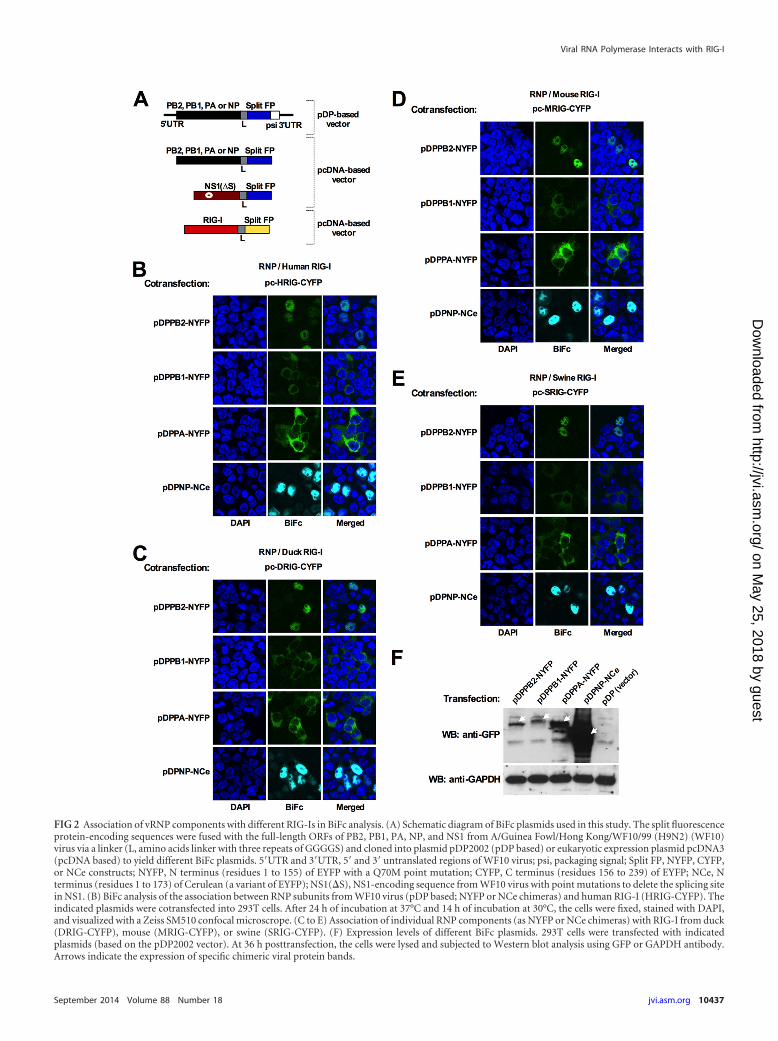
FIG 2 Association of vRNP components with different RIG-Is in BiFc analysis. (A) Schematic diagram of BiFc plasmids used in this study. The split fluorescenceprotein-encoding sequences were fused with the full-length ORFs of PB2, PB1, PA, NP, and NS1 from A/Guinea Fowl/Hong Kong/WF10/99 (H9N2) (WF10)virus via a linker (L, amino acids linker with three repeats of GGGGS) and cloned into plasmid pDP2002 (pDP based) or eukaryotic expression plasmid pcDNA3(pcDNA based) to yield different BiFc plasmids. 5=UTR and 3=UTR, 5= and 3= untranslated regions of WF10 virus; psi, packaging signal; Split FP, NYFP, CYFP,or NCe constructs; NYFP, N terminus (residues 1 to 155) of EYFP with a Q70M point mutation; CYFP, C terminus (residues 156 to 239) of EYFP; NCe, Nterminus (residues 1 to 173) of Cerulean (a variant of EYFP); NS1(�S), NS1-encoding sequence from WF10 virus with point mutations to delete the splicing sitein NS1. (B) BiFc analysis of the association between RNP subunits from WF10 virus (pDP based; NYFP or NCe chimeras) and human RIG-I (HRIG-CYFP). Theindicated plasmids were cotransfected into 293T cells. After 24 h of incubation at 37°C and 14 h of incubation at 30°C, the cells were fixed, stained with DAPI,and visualized with a Zeiss SM510 confocal microscrope. (C to E) Association of individual RNP components (as NYFP or NCe chimeras) with RIG-I from duck(DRIG-CYFP), mouse (MRIG-CYFP), or swine (SRIG-CYFP). (F) Expression levels of different BiFc plasmids. 293T cells were transfected with indicatedplasmids (based on the pDP2002 vector). At 36 h posttransfection, the cells were lysed and subjected to Western blot analysis using GFP or GAPDH antibody.Arrows indicate the expression of specific chimeric viral protein bands.
Viral RNA Polymerase Interacts with RIG-I
September 2014 Volume 88 Number 18 jvi.asm.org 10437
on May 25, 2018 by guest
http://jvi.asm.org/
Dow
nloaded from

Li et al.
10438 jvi.asm.org Journal of Virology
on May 25, 2018 by guest
http://jvi.asm.org/
Dow
nloaded from
participation of vRNA in the association of each vRNP compo-nent with RIG-I. Thus, four additional BiFc plasmids expressingRNP proteins but not vRNA were constructed based on the vectorpcDNA3 (Fig. 2A). In the context of this new set of plasmids, BiFcsignals were observed which were indistinguishable from thosedescribed above, suggesting direct associations of viral compo-nents with RIG-I in the absence of viral RNA. Similar interactionsof RNP subunits with RIG-I were also observed for the duck,mouse, and swine homologs (Fig. 2C to E).
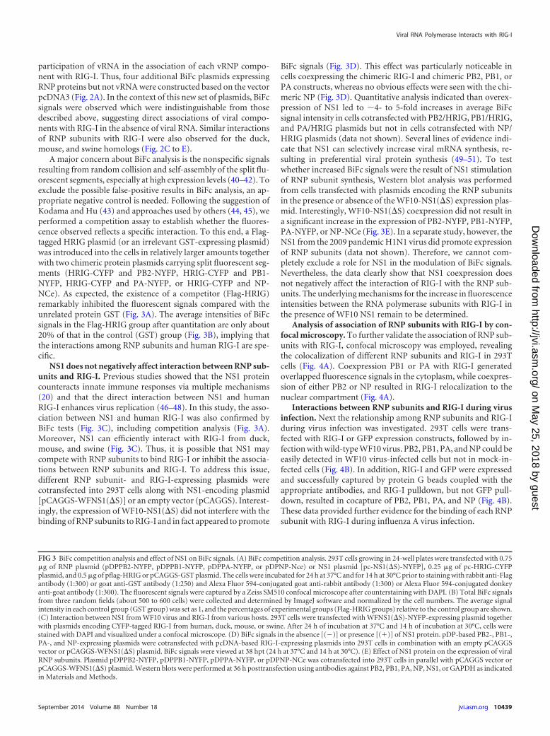
A major concern about BiFc analysis is the nonspecific signalsresulting from random collision and self-assembly of the split flu-orescent segments, especially at high expression levels (40–42). Toexclude the possible false-positive results in BiFc analysis, an ap-propriate negative control is needed. Following the suggestion ofKodama and Hu (43) and approaches used by others (44, 45), weperformed a competition assay to establish whether the fluores-cence observed reflects a specific interaction. To this end, a Flag-tagged HRIG plasmid (or an irrelevant GST-expressing plasmid)was introduced into the cells in relatively larger amounts togetherwith two chimeric protein plasmids carrying split fluorescent seg-ments (HRIG-CYFP and PB2-NYFP, HRIG-CYFP and PB1-NYFP, HRIG-CYFP and PA-NYFP, or HRIG-CYFP and NP-NCe). As expected, the existence of a competitor (Flag-HRIG)remarkably inhibited the fluorescent signals compared with theunrelated protein GST (Fig. 3A). The average intensities of BiFcsignals in the Flag-HRIG group after quantitation are only about20% of that in the control (GST) group (Fig. 3B), implying thatthe interactions among RNP subunits and human RIG-I are spe-cific.
NS1 does not negatively affect interaction between RNP sub-units and RIG-I. Previous studies showed that the NS1 proteincounteracts innate immune responses via multiple mechanisms(20) and that the direct interaction between NS1 and humanRIG-I enhances virus replication (46–48). In this study, the asso-ciation between NS1 and human RIG-I was also confirmed byBiFc tests (Fig. 3C), including competition analysis (Fig. 3A).Moreover, NS1 can efficiently interact with RIG-I from duck,mouse, and swine (Fig. 3C). Thus, it is possible that NS1 maycompete with RNP subunits to bind RIG-I or inhibit the associa-tions between RNP subunits and RIG-I. To address this issue,different RNP subunit- and RIG-I-expressing plasmids werecotransfected into 293T cells along with NS1-encoding plasmid[pCAGGS-WFNS1(�S)] or an empty vector (pCAGGS). Interest-ingly, the expression of WF10-NS1(�S) did not interfere with thebinding of RNP subunits to RIG-I and in fact appeared to promote
BiFc signals (Fig. 3D). This effect was particularly noticeable incells coexpressing the chimeric RIG-I and chimeric PB2, PB1, orPA constructs, whereas no obvious effects were seen with the chi-meric NP (Fig. 3D). Quantitative analysis indicated than overex-pression of NS1 led to �4- to 5-fold increases in average BiFcsignal intensity in cells cotransfected with PB2/HRIG, PB1/HRIG,and PA/HRIG plasmids but not in cells cotransfected with NP/HRIG plasmids (data not shown). Several lines of evidence indi-cate that NS1 can selectively increase viral mRNA synthesis, re-sulting in preferential viral protein synthesis (49–51). To testwhether increased BiFc signals were the result of NS1 stimulationof RNP subunit synthesis, Western blot analysis was performedfrom cells transfected with plasmids encoding the RNP subunitsin the presence or absence of the WF10-NS1(�S) expression plas-mid. Interestingly, WF10-NS1(�S) coexpression did not result ina significant increase in the expression of PB2-NYFP, PB1-NYFP,PA-NYFP, or NP-NCe (Fig. 3E). In a separate study, however, theNS1 from the 2009 pandemic H1N1 virus did promote expressionof RNP subunits (data not shown). Therefore, we cannot com-pletely exclude a role for NS1 in the modulation of BiFc signals.Nevertheless, the data clearly show that NS1 coexpression doesnot negatively affect the interaction of RIG-I with the RNP sub-units. The underlying mechanisms for the increase in fluorescenceintensities between the RNA polymerase subunits with RIG-I inthe presence of WF10 NS1 remain to be determined.
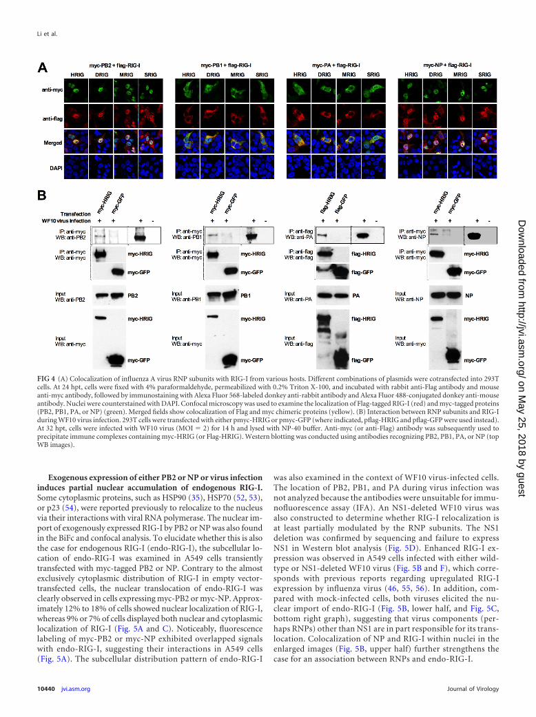
Analysis of association of RNP subunits with RIG-I by con-focal microscopy. To further validate the association of RNP sub-units with RIG-I, confocal microscopy was employed, revealingthe colocalization of different RNP subunits and RIG-I in 293Tcells (Fig. 4A). Coexpression PB1 or PA with RIG-I generatedoverlapped fluorescence signals in the cytoplasm, while coexpres-sion of either PB2 or NP resulted in RIG-I relocalization to thenuclear compartment (Fig. 4A).
Interactions between RNP subunits and RIG-I during virusinfection. Next the relationship among RNP subunits and RIG-Iduring virus infection was investigated. 293T cells were trans-fected with RIG-I or GFP expression constructs, followed by in-fection with wild-type WF10 virus. PB2, PB1, PA, and NP could beeasily detected in WF10 virus-infected cells but not in mock-in-fected cells (Fig. 4B). In addition, RIG-I and GFP were expressedand successfully captured by protein G beads coupled with theappropriate antibodies, and RIG-I pulldown, but not GFP pull-down, resulted in cocapture of PB2, PB1, PA, and NP (Fig. 4B).These data provided further evidence for the binding of each RNPsubunit with RIG-I during influenza A virus infection.
FIG 3 BiFc competition analysis and effect of NS1 on BiFc signals. (A) BiFc competition analysis. 293T cells growing in 24-well plates were transfected with 0.75g of RNP plasmid (pDPPB2-NYFP, pDPPB1-NYFP, pDPPA-NYFP, or pDPNP-Nce) or NS1 plasmid [pc-NS1(�S)-NYFP], 0.25 g of pc-HRIG-CYFPplasmid, and 0.5 g of pflag-HRIG or pCAGGS-GST plasmid. The cells were incubated for 24 h at 37°C and for 14 h at 30°C prior to staining with rabbit anti-Flagantibody (1:300) or goat anti-GST antibody (1:250) and Alexa Fluor 594-conjugated goat anti-rabbit antibody (1:300) or Alexa Fluor 594-conjugated donkeyanti-goat antibody (1:300). The fluorescent signals were captured by a Zeiss SM510 confocal microscope after counterstaining with DAPI. (B) Total BiFc signalsfrom three random fields (about 500 to 600 cells) were collected and determined by ImageJ software and normalized by the cell numbers. The average signalintensity in each control group (GST group) was set as 1, and the percentages of experimental groups (Flag-HRIG groups) relative to the control group are shown.(C) Interaction between NS1 from WF10 virus and RIG-I from various hosts. 293T cells were transfected with WFNS1(�S)-NYFP-expressing plasmid togetherwith plasmids encoding CYFP-tagged RIG-I from human, duck, mouse, or swine. After 24 h of incubation at 37°C and 14 h of incubation at 30°C, cells werestained with DAPI and visualized under a confocal microscope. (D) BiFc signals in the absence [(�)] or presence [(�)] of NS1 protein. pDP-based PB2-, PB1-,PA-, and NP-expressing plasmids were cotransfected with pcDNA-based RIG-I-expressing plasmids into 293T cells in combination with an empty pCAGGSvector or pCAGGS-WFNS1(�S) plasmid. BiFc signals were viewed at 38 hpt (24 h at 37°C and 14 h at 30°C). (E) Effect of NS1 protein on the expression of viralRNP subunits. Plasmid pDPPB2-NYFP, pDPPB1-NYFP, pDPPA-NYFP, or pDPNP-NCe was cotransfected into 293T cells in parallel with pCAGGS vector orpCAGGS-WFNS1(�S) plasmid. Western blots were performed at 36 h posttransfection using antibodies against PB2, PB1, PA, NP, NS1, or GAPDH as indicatedin Materials and Methods.
Viral RNA Polymerase Interacts with RIG-I
September 2014 Volume 88 Number 18 jvi.asm.org 10439
on May 25, 2018 by guest
http://jvi.asm.org/
Dow
nloaded from
Exogenous expression of either PB2 or NP or virus infectioninduces partial nuclear accumulation of endogenous RIG-I.Some cytoplasmic proteins, such as HSP90 (35), HSP70 (52, 53),or p23 (54), were reported previously to relocalize to the nucleusvia their interactions with viral RNA polymerase. The nuclear im-port of exogenously expressed RIG-I by PB2 or NP was also foundin the BiFc and confocal analysis. To elucidate whether this is alsothe case for endogenous RIG-I (endo-RIG-I), the subcellular lo-cation of endo-RIG-I was examined in A549 cells transientlytransfected with myc-tagged PB2 or NP. Contrary to the almostexclusively cytoplasmic distribution of RIG-I in empty vector-transfected cells, the nuclear translocation of endo-RIG-I wasclearly observed in cells expressing myc-PB2 or myc-NP. Approx-imately 12% to 18% of cells showed nuclear localization of RIG-I,whereas 9% or 7% of cells displayed both nuclear and cytoplasmiclocalization of RIG-I (Fig. 5A and C). Noticeably, fluorescencelabeling of myc-PB2 or myc-NP exhibited overlapped signalswith endo-RIG-I, suggesting their interactions in A549 cells(Fig. 5A). The subcellular distribution pattern of endo-RIG-I
was also examined in the context of WF10 virus-infected cells.The location of PB2, PB1, and PA during virus infection wasnot analyzed because the antibodies were unsuitable for immu-nofluorescence assay (IFA). An NS1-deleted WF10 virus wasalso constructed to determine whether RIG-I relocalization isat least partially modulated by the RNP subunits. The NS1deletion was confirmed by sequencing and failure to expressNS1 in Western blot analysis (Fig. 5D). Enhanced RIG-I ex-pression was observed in A549 cells infected with either wild-type or NS1-deleted WF10 virus (Fig. 5B and F), which corre-sponds with previous reports regarding upregulated RIG-Iexpression by influenza virus (46, 55, 56). In addition, com-pared with mock-infected cells, both viruses elicited the nu-clear import of endo-RIG-I (Fig. 5B, lower half, and Fig. 5C,bottom right graph), suggesting that virus components (per-haps RNPs) other than NS1 are in part responsible for its trans-location. Colocalization of NP and RIG-I within nuclei in theenlarged images (Fig. 5B, upper half) further strengthens thecase for an association between RNPs and endo-RIG-I.
FIG 4 (A) Colocalization of influenza A virus RNP subunits with RIG-I from various hosts. Different combinations of plasmids were cotransfected into 293Tcells. At 24 hpt, cells were fixed with 4% paraformaldehyde, permeabilized with 0.2% Triton X-100, and incubated with rabbit anti-Flag antibody and mouseanti-myc antibody, followed by immunostaining with Alexa Fluor 568-labeled donkey anti-rabbit antibody and Alexa Fluor 488-conjugated donkey anti-mouseantibody. Nuclei were counterstained with DAPI. Confocal microscopy was used to examine the localization of Flag-tagged RIG-I (red) and myc-tagged proteins(PB2, PB1, PA, or NP) (green). Merged fields show colocalization of Flag and myc chimeric proteins (yellow). (B) Interaction between RNP subunits and RIG-Iduring WF10 virus infection. 293T cells were transfected with either pmyc-HRIG or pmyc-GFP (where indicated, pflag-HRIG and pflag-GFP were used instead).At 32 hpt, cells were infected with WF10 virus (MOI � 2) for 14 h and lysed with NP-40 buffer. Anti-myc (or anti-Flag) antibody was subsequently used toprecipitate immune complexes containing myc-HRIG (or Flag-HRIG). Western blotting was conducted using antibodies recognizing PB2, PB1, PA, or NP (topWB images).
Li et al.
10440 jvi.asm.org Journal of Virology
on May 25, 2018 by guest
http://jvi.asm.org/
Dow
nloaded from

FIG 5 (A) Relocalization of endo-RIG-I in A549 cells during transfection with PB2- or NP-expressing plasmids. The myc-tagged PB2 or NP constructs or pmycvector was delivered into A549 cells. At 24 hpt, RIG-I was detected with rabbit anti-RIG-I antibody and Alexa Fluor 488-conjugated donkey anti-rabbit antibody,while myc-PB2 or myc-NP was probed with mouse anti-myc antibody and Alexa Fluor 568-conjugated donkey anti-mouse antibody. (B) Subcellular localizationof RIG-I and NP in virus-infected cells. After 14 h of infection of A549 cells with wild-type or NS1-deleted WF10 virus (MOI � 5), the cells were fixed and stainedwith rabbit anti-RIG-I and mouse anti-NP antibodies. The power of magnification is �600, with enlarged fields shown in the top three rows. (C) Quantitationanalysis of the percentage of cell populations based on the intracellular distribution pattern of RIG-I versus myc-PB2 or myc-NP (top left graph) or NP (bottomright graph) in virus-infected cells. (D) Confirmation of NS1-deleted virus by Western blotting. MDCK cells were infected with wild-type (MOI � 0.1) orNS1-deleted (MOI � 2) WF10 virus. At 16 hpi, Western blotting was conducted using NS1 or PB2 antibody. (E and F) Intracellular distribution of endo-RIG-Iat different times postinfection. A549 cells were infected (MOI � 5) with wild-type or NS1-deleted WF10 virus. At the indicated time points, cells were fixed andsubjected to IFA analysis using RIG-I antibody, while the cytoplasmic (C) or nuclear (N) fractions were isolated and subjected to WB analysis using antibodiesagainst RIG-I, tubulin, PCNA, and viral NP. Tubulin and PCNA serve as cytoplasmic and nuclear protein markers, respectively.
September 2014 Volume 88 Number 18 jvi.asm.org 10441
on May 25, 2018 by guest
http://jvi.asm.org/
Dow
nloaded from

Partial relocalization of RIG-I into the nucleus at late stagesof infection. In order to investigate the translocation kinetics ofendo-RIG-I in the infection process, A549 cells were infected withwild-type or NS1-deleted WF10 virus (MOI � 5). At differenttimes postinfection (1, 2, 4, 6, 8, 10, 12, and 14 hpi), the localiza-tion of endo-RIG-I was examined (Fig. 5E). In agreement withprevious studies (46, 55, 56), the levels of endo-RIG-I increasedsignificantly in cells infected with either virus (Fig. 5F). In addi-tion, endo-RIG-I was almost solely found in the cytoplasmic frac-tions in control cells and virus-infected cells prior to 8 hpi (Fig. 5Eand F). Then it started to move partially toward the nucleus. Atlater time points (e.g., 12 or 14 hpi), a proportion of endo-RIG-Iwas found in the nucleus (Fig. 5E and F) or an area surroundingthe nucleus (Fig. 5E), indicating that the nuclear import of endo-RIG-I is induced by the virus and that it is a late event and canoccur in the presence or absence of NS1. Interestingly, a minorproportion of endo-RIG-I was located on the plasma membraneruffle and formed a “hedgehog-like” shape at late infection phase(e.g., WF10 virus-infected cells at 14 hpi in Fig. 5E), presumablycaused by the surface bleb of apoptotic cells during virus infection(57–59).
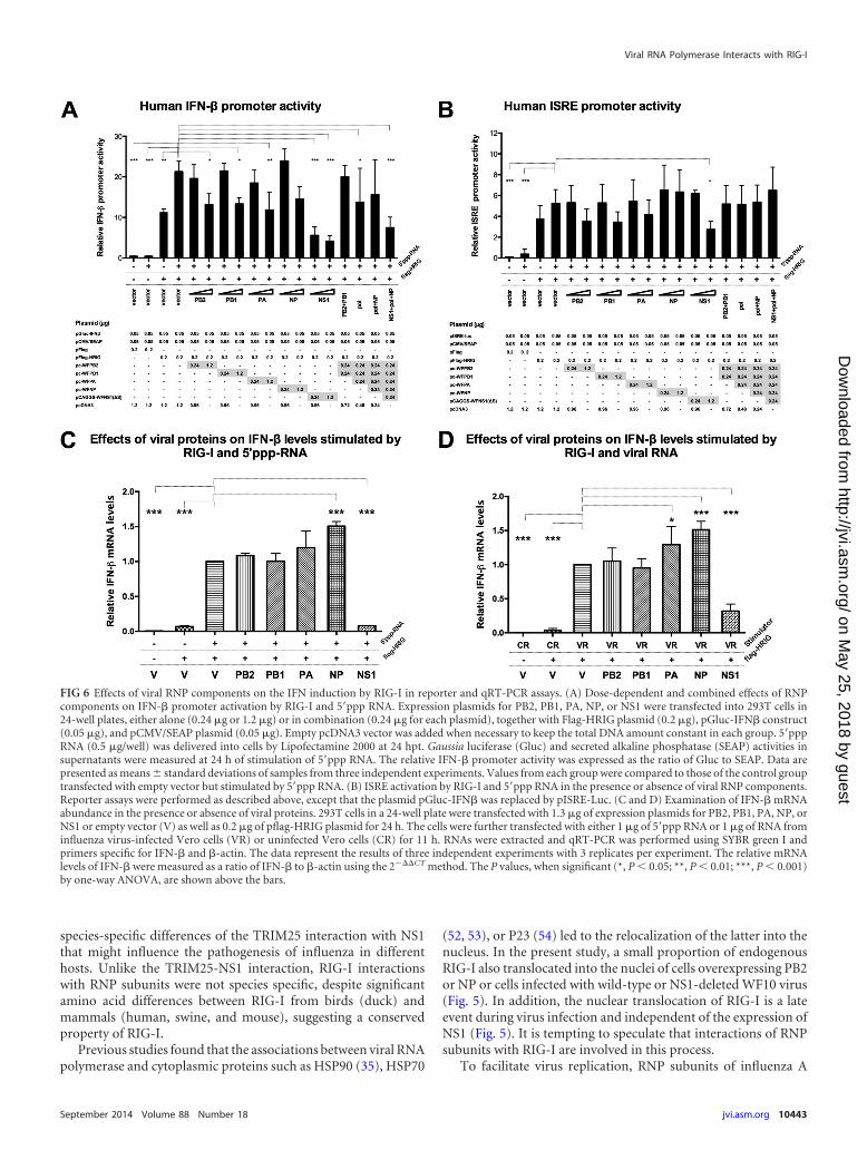
Effects of RNP subunits on the activation of IFN-� or ISREactivity by RIG-I. Interaction of NS1 with RIG-I, TRIM25 , orRiplet antagonizes IFN production and negatively modulates thehost’s antiviral defense (16, 22, 47). Furthermore, PB1-F2 inter-feres with IFN induction via MAVS (26, 27). The associationbetween RNP subunits and RIG-I may also play a previously un-defined role in interferon signaling. Therefore, IFN-� promoter-driven luciferase reporter gene assays were performed to deter-mine whether RNP subunits of influenza virus affect thegeneration of IFN triggered by RIG-I. 293T cells were transfectedwith a plasmid expressing Flag-tagged human RIG-I (Flag-HRIG)along with various amounts of PB2-, PB1-, PA- NP-, and NS1-expressing plasmids for 24 h and then stimulated with 5=ppp RNA(a strict RIG-I-specific ligand containing 19-mer double-strandedRNA with a 5= triphosphate) to induce IFN synthesis. Combinedtreatment of 293T cells with RIG-I and 5=ppp RNA resulted in amarked increase in IFN-� promoter induction (49.7-fold) com-pared with that in the mock-treated cells (Fig. 6A). In the positive-control group, NS1 from WF10 virus exhibited dramatic inhibi-tion of the IFN-� promoter (74% reduction) even at a lowtransfection dose (0.24 g). However, PB2, PB1, or PA alone onlyslightly decreased IFN-� promoter activity, by about 36 to �40%at a high transfection dose (1.2 g). NP did not show statisticallysignificant repression. Only mild (and even negligible) inhibitionwas observed if the mixtures of two components (PB2 and PB1) orthe RNA polymerase complex (PB2, PB1, and PA) or the vRNPcomplex (polymerase complex plus NP) were coexpressed withRIG-I and later stimulated by 5=ppp RNA (Fig. 6A). Results fromanother reporter assay using interferon-stimulated response ele-ment (ISRE) were even less prominent, with only suppression byNS1 at a high transfection dose having statistical significance.These results suggest a yet-to-be-defined biological role (if any)for the interaction of RNP subunits with RIG-I.
Examination of IFN induction using quantitative RT-PCRand IRF3 phosphorylation assay. To further clarify the role ofRNP subunits in IFN activation and IFN signaling transduction,the impact of each individual RNP subunit on mRNA levels ofhuman IFN-� was analyzed. To stimulate IFN, different inducerswere employed after 24 h of transfection of 293T cells with Flag-
HRIG plasmid or empty Flag vector. Either the specific RIG-Ibinding ligand (5=ppp RNA) or RNA from WF10 virus-infected oruninfected Vero cells was used as an IFN inducer (Fig. 6C and D),essentially as described previously (60, 61). As expected, IFN-�mRNA levels were substantially elevated by the coadministrationof RIG-I and either 5=ppp RNA or viral RNA (VR) but not withcontrol cellular RNA (CR). Moreover, IFN-� mRNA decreased to7.9% (Fig. 6C, 5=ppp RNA) or 32.0% (Fig. 6D, VR) compared tothe levels for controls in cells expressing NS1. In contrast, PB2,PB1, and PA did not repress IFN-� activation (Fig. 6C and D).Interestingly, NP upregulated IFN-� mRNA levels, the signifi-cance of which remains to be determined.
IRF3 is known to be an important adaptor of the RIG-I signal-ing pathway. The phosphorylation of IRF3 and the subsequentnuclear import of IRF3 play vital roles in IFN activation (62).Taking this into consideration, it was examined whether individ-ually expressed RNP subunits have an effect on levels of IRF3phosphorylation (via the monitoring of phosphorylated Ser396[p-IRF3] and total IRF3 [IRF3]). Quantitative analysis for the in-tensities of protein bands in Western blots showed that the ratio ofp-IRF3 to IRF3 remained unchanged upon the overexpression ofeach RNP subunit compared to the vector-alone control (Fig. 7).In contrast and as expected, the p-IRF3/IRF3 ratio was markedlydecreased by expression of NS1 compared to that with the nega-tive control (Fig. 7).
DISCUSSION
Interactions between viral proteins and host factors govern theadaptation and pathogenicity of viruses in different hosts. In thisregard, the influenza virus RNP complex plays a vital role (6, 63,64). Influenza virus polymerase complex subunits PB2, PB1, PA,and NP can interact, alone or in combination, with a multitude ofhost factors involved in various intracellular events such as cellu-lar transcription, nuclear-cytoplasmic transport, protein subcel-lular trafficking, and the host immune response (6, 64, 65). How-ever, the exact mechanisms that modulate vRNP-host factorinteractions and the outcome of infection remain largely elusive.
In this study, the associations of influenza A virus RNP sub-units with RIG-I were analyzed using different approaches. Co-IPtests showed the RNA-independent interactions of each viral RNApolymerase subunit (PB2, PB1, and PA) with RIG-I and RNA-dependent interaction of NP with RIG-I (Fig. 1). In BiFc analysis,expression of either PB2, PB1, PA, or NP chimeric constructs har-boring a split fluorescence protein sequence interacted with chi-meric RIG-I protein fused to a complementary split fluorescenceprotein, resulting in bright fluorescence signals in 293T cells (Fig.2). Interestingly, the BiFc signals emitted by PB1-RIG-I or PA-RIG-I interactions were predominantly distributed in the cyto-plasm, whereas the signals produced by PB2-RIG-I or NP-RIG-Iinteractions occurred mainly in the nuclei. Similar results wereobtained in confocal analysis (Fig. 4A). These findings are com-patible with the previous reports on the major subcellular local-ization of individual RNP subunits in cells (35, 37–39). Finally,co-IP results from infection experiments (Fig. 4B) provided fur-ther evidence for RIG-I binding to each of the RNP components.
The NS1 proteins from various influenza virus strains havebeen shown to differentially interact with TRIM25 (an impor-tant regulator of RIG-I) from different species (16). HumanTRIM25 was shown to bind NS1 proteins from human, swine, oravian viruses, whereas mouse TRIM25 did not (16), suggesting
Li et al.
10442 jvi.asm.org Journal of Virology
on May 25, 2018 by guest
http://jvi.asm.org/
Dow
nloaded from
species-specific differences of the TRIM25 interaction with NS1that might influence the pathogenesis of influenza in differenthosts. Unlike the TRIM25-NS1 interaction, RIG-I interactionswith RNP subunits were not species specific, despite significantamino acid differences between RIG-I from birds (duck) andmammals (human, swine, and mouse), suggesting a conservedproperty of RIG-I.
Previous studies found that the associations between viral RNApolymerase and cytoplasmic proteins such as HSP90 (35), HSP70
(52, 53), or P23 (54) led to the relocalization of the latter into thenucleus. In the present study, a small proportion of endogenousRIG-I also translocated into the nuclei of cells overexpressing PB2or NP or cells infected with wild-type or NS1-deleted WF10 virus(Fig. 5). In addition, the nuclear translocation of RIG-I is a lateevent during virus infection and independent of the expression ofNS1 (Fig. 5). It is tempting to speculate that interactions of RNPsubunits with RIG-I are involved in this process.
To facilitate virus replication, RNP subunits of influenza A
FIG 6 Effects of viral RNP components on the IFN induction by RIG-I in reporter and qRT-PCR assays. (A) Dose-dependent and combined effects of RNPcomponents on IFN-� promoter activation by RIG-I and 5=ppp RNA. Expression plasmids for PB2, PB1, PA, NP, or NS1 were transfected into 293T cells in24-well plates, either alone (0.24 g or 1.2 g) or in combination (0.24 g for each plasmid), together with Flag-HRIG plasmid (0.2 g), pGluc-IFN� construct(0.05 g), and pCMV/SEAP plasmid (0.05 g). Empty pcDNA3 vector was added when necessary to keep the total DNA amount constant in each group. 5=pppRNA (0.5 g/well) was delivered into cells by Lipofectamine 2000 at 24 hpt. Gaussia luciferase (Gluc) and secreted alkaline phosphatase (SEAP) activities insupernatants were measured at 24 h of stimulation of 5=ppp RNA. The relative IFN-� promoter activity was expressed as the ratio of Gluc to SEAP. Data arepresented as means standard deviations of samples from three independent experiments. Values from each group were compared to those of the control grouptransfected with empty vector but stimulated by 5=ppp RNA. (B) ISRE activation by RIG-I and 5=ppp RNA in the presence or absence of viral RNP components.Reporter assays were performed as described above, except that the plasmid pGluc-IFN� was replaced by pISRE-Luc. (C and D) Examination of IFN-� mRNAabundance in the presence or absence of viral proteins. 293T cells in a 24-well plate were transfected with 1.3 g of expression plasmids for PB2, PB1, PA, NP, orNS1 or empty vector (V) as well as 0.2 g of pflag-HRIG plasmid for 24 h. The cells were further transfected with either 1 g of 5=ppp RNA or 1 g of RNA frominfluenza virus-infected Vero cells (VR) or uninfected Vero cells (CR) for 11 h. RNAs were extracted and qRT-PCR was performed using SYBR green I andprimers specific for IFN-� and �-actin. The data represent the results of three independent experiments with 3 replicates per experiment. The relative mRNAlevels of IFN-� were measured as a ratio of IFN-� to �-actin using the 2���CT method. The P values, when significant (*, P � 0.05; **, P � 0.01; ***, P � 0.001)by one-way ANOVA, are shown above the bars.
Viral RNA Polymerase Interacts with RIG-I
September 2014 Volume 88 Number 18 jvi.asm.org 10443
on May 25, 2018 by guest
http://jvi.asm.org/
Dow
nloaded from

virus may manipulate IFN signaling transduction via their associ-ations with RIG-I. This effect would complement the activities ofsome other influenza virus proteins that have already been re-ported to affect the RIG-I signaling pathway at different steps (66).In addition, NS1 can directly target and inhibit RIG-I (9, 10, 22),TRIM25 (22), or Riplet (16); PB1-F2 acts on MAVS, henceblocking IFN production (25–27). More recently, overexpressionof PB2 (25, 28, 30), PB1 (25, 30), or PA (25, 30) from human virusstrains (PR8 or WSN) was found to repress IFN-� promoter-driven reporter activity via interactions with MAVS. In addition, ahigh-throughput screening for the interacting partners of influ-enza virus in human cells illustrated physical associations amongRNP subunits and a variety of regulators of interferon response(67). In this regard, the results presented here are in partial agree-ment with the above-mentioned studies, as PB2, PB1, or PA fromWF10 virus (an avian virus) individually reduced the activation ofIFN-� promoter in reporter assays (Fig. 6A and B). However, theRNA polymerase-mediated IFN-inhibiting effect is mild and hadno effect on IFN-� mRNA levels (Fig. 6C and D) or IRF3 phos-phorylation (Fig. 7). Altogether, the RNA polymerase subunitsfrom the WF10 virus do not have a significant impact on IFNactivation elicited by RIG-I in human cells in vitro. It remains to bedetermined if these interactions have any significant role in vivo.
Interestingly, NP neither suppressed the activation of theIFN-� promoter or ISRE (Fig. 6A and B) nor decreased the phos-phorylation of IRF3 (Fig. 7). On the contrary, qRT-PCR assayssuggest that NP upregulated IFN mRNA levels (Fig. 6C and D).Previous studies have shown that influenza virus vRNA wrapsaround the NP, being exposed to the surface and, thus, accessibleto RIG-I (68, 69).
It was presumed that interactions of RNP subunits with RIG-Icould have special significance for the early phase of influenza Avirus infection, when NS1 has yet to be synthesized. In addition,the incoming viral RNA is encapsidated with NP and the 5=pppdsRNA panhandle structure is bound by the viral polymerase.Little is known about how the host cell senses the invading viralgenome and triggers innate immune responses and how virusescounteract the immune recognition during this time. Using RiftValley fever virus (RVFV) and La Crosse virus (LACV), two mem-bers of Bunyaviridae family that possess 5=ppp dsRNA panhandlestructures in their negative-stranded genome RNA similar to thatin influenza A virus, Weber et al. showed that the interactionbetween RIG-I and viral nucleocapsids (including both viral nu-cleoprotein and the 5=ppp dsRNA structure) is necessary for theinitiation of innate immunity (70). In our study, although RNApolymerase components PB2, PB1, and PA were shown to directlyassociate with RIG-I, no significant effects on IFN productionwere observed. It is conceivable to speculate, then, that the viralRNA polymerase would not prevent access of RIG-I to the viral5=ppp dsRNA structure and the subsequent activation of IFN.Furthermore, it should be noted that the WF10 virus used in thisstudy belongs to the H9N2 subtype of viruses, responsible fordonating their internal genes to the highly pathogenic H5N1 virus(71), the novel H7N9 virus (72), and the newly identified H10N8virus that crossed to humans (73). However, in view that WF10 isstill a typical avian influenza virus, the possibility that RNA poly-merase complexes from avian influenza viruses do not efficientlyexert IFN-antagonizing ability in human cells cannot be com-pletely excluded.
Another possible scenario in the outcome of the interactionsbetween RIG-I and RNP components is the direct suppression ofviral RNA polymerase activity by RIG-I. RIG-I (also known asDDX58) belongs to the DExD/H helicase family. Some membersof this family have been shown to associate with viral RNP com-ponents and affect the polymerase activity. For instance, the asso-ciations of DDX3 (74), DDX5 (74, 75), and DDX17 (75, 76) withviral polymerase or NP have been identified, and DDX5 (76) andDDX17 (76) were found to facilitate viral RNA synthesis. In addi-tion, DDX39B (UAP56) (77, 78) and its paralog DDX39 (URH39)(78) interact with NP, and DDX39B helps virus replication viapromoting the formation of the NP-RNA complex or preventingthe formation of dsRNA and the subsequent activation of innateimmune response (78). Conversely, DDX21 restricts influenza vi-rus by binding PB1 and decreasing the assembly of viral polymer-ase (79). Our unpublished data from reporter assays also showedthat overexpression of RIG-I markedly suppressed virus polymer-ase activity in 293T cells, but the suppression could be attributedto IFN or other inflammatory cytokines elicited by RIG-I activa-tion. Previous studies indicated that a single mutation in residue183 (S183I) (80) or residue 55 (T55I) (81, 82) of RIG-I completelyeliminated its ability to transduce signal and trigger the innateimmune response. If these two RIG-I mutants still maintain thecapacity to interact with vRNP components, they can serve as
FIG 7 Effects of viral proteins on phosphorylation of IRF3. Myc-tagged con-structs encoding PB2, PB1, PA, NP, or NS1 from WF10 virus (2 g) or pmycempty vector (2 g) were transfected into 293T cells (12-well plate) along with0.3 g of pflag-HRIG plasmid and 0.7 g of pflag-IRF3 plasmid. Twenty-fourhours later, cells were further transfected with 5=ppp RNA for 12 h. The cellswere subsequently lysed and subjected to Western blot analysis using antibod-ies specific for phospho-IRF3 (Ser396), IRF3, RIG-I, myc tag, and GAPDH.The band intensities of p-IRF3 (phosphorylated IRF3) and IRF3 (total IRF3)were calculated by ImageJ software, and the ratios of p-IRF3 to IRF3 are shownat the bottom.
Li et al.
10444 jvi.asm.org Journal of Virology
on May 25, 2018 by guest
http://jvi.asm.org/
Dow
nloaded from

useful tools to investigate the direct effect of RIG-I on viral poly-merase activity in RIG-I knockout mouse embryo fibroblasts. Adetailed analysis of the relationship of RIG-I mutants with vRNPand their effects on virus replication will be undertaken in thefuture.
In conclusion, this study highlights the direct association ofinfluenza virus RNP subunits with RIG-I from various species.These interactions, however, seemed to be unrelated to the mod-ulation of the IFN signaling pathway. The significance of RNPsubunits in modulating immune responses and virulence of influ-enza A virus in vivo deserves further attention.
ACKNOWLEDGMENTS
We thank Andrea Ferrero, Johanna Lavigne, and Qiong Chen for theirlaboratory managerial skills and assistance.
D.R.P. and W.L. conceived and designed the experiments. W.L., H.C.,and A.O. performed the experiments. W.L., T.S., and D.R.P. analyzed thedata and prepared the manuscript. H.C. and T.S. contributed reagents,materials, and analysis tools.
This research was funded by NIAID/NIH contract HHSN26620070001.
REFERENCES1. Guilligay D, Tarendeau F, Resa-Infante P, Coloma R, Crepin T, Sehr P,
Lewis J, Ruigrok RW, Ortin J, Hart DJ, Cusack S. 2008. The structuralbasis for cap binding by influenza virus polymerase subunit PB2. Nat.Struct. Mol. Biol. 15:500 –506. http://dx.doi.org/10.1038/nsmb.1421.
2. Fodor E, Crow M, Mingay LJ, Deng T, Sharps J, Fechter P, BrownleeGG. 2002. A single amino acid mutation in the PA subunit of the influenzavirus RNA polymerase inhibits endonucleolytic cleavage of capped RNAs.J. Virol. 76:8989 –9001. http://dx.doi.org/10.1128/JVI.76.18.8989-9001.2002.
3. Biswas SK, Nayak DP. 1994. Mutational analysis of the conserved motifsof influenza A virus polymerase basic protein 1. J. Virol. 68:1819 –1826.
4. Portela A, Digard P. 2002. The influenza virus nucleoprotein: a multi-functional RNA-binding protein pivotal to virus replication. J. Gen. Virol.83:723–734.
5. Cros JF, Palese P. 2003. Trafficking of viral genomic RNA into and out ofthe nucleus: influenza, Thogoto and Borna disease viruses. Virus Res.95:3–12. http://dx.doi.org/10.1016/S0168-1702(03)00159-X.
6. Naffakh N, Tomoiu A, Rameix-Welti MA, van der Werf S. 2008. Hostrestriction of avian influenza viruses at the level of the ribonucleoproteins.Annu. Rev. Microbiol. 62:403– 424. http://dx.doi.org/10.1146/annurev.micro.62.081307.162746.
7. Kato H, Takeuchi O, Sato S, Yoneyama M, Yamamoto M, Matsui K,Uematsu S, Jung A, Kawai T, Ishii KJ, Yamaguchi O, Otsu K, TsujimuraT, Koh CS, Reis e Sousa C, Matsuura Y, Fujita T, Akira S. 2006.Differential roles of MDA5 and RIG-I helicases in the recognition of RNAviruses. Nature 441:101–105. http://dx.doi.org/10.1038/nature04734.
8. Loo YM, Fornek J, Crochet N, Bajwa G, Perwitasari O, Martinez-Sobrido L, Akira S, Gill MA, Garcia-Sastre A, Katze MG, Gale M, Jr.2008. Distinct RIG-I and MDA5 signaling by RNA viruses in innate im-munity. J. Virol. 82:335–345. http://dx.doi.org/10.1128/JVI.01080-07.
9. Pichlmair A, Schulz O, Tan CP, Naslund TI, Liljestrom P, Weber F,Reis e Sousa C. 2006. RIG-I-mediated antiviral responses to single-stranded RNA bearing 5=-phosphates. Science 314:997–1001. http://dx.doi.org/10.1126/science.1132998.
10. Rehwinkel J, Tan CP, Goubau D, Schulz O, Pichlmair A, Bier K, RobbN, Vreede F, Barclay W, Fodor E, Reis e Sousa C. 2010. RIG-I detectsviral genomic RNA during negative-strand RNA virus infection. Cell 140:397– 408. http://dx.doi.org/10.1016/j.cell.2010.01.020.
11. Kato H, Takeuchi O, Mikamo-Satoh E, Hirai R, Kawai T, Matsushita K,Hiiragi A, Dermody TS, Fujita T, Akira S. 2008. Length-dependentrecognition of double-stranded ribonucleic acids by retinoic acid-inducible gene-I and melanoma differentiation-associated gene 5. J. Exp.Med. 205:1601–1610. http://dx.doi.org/10.1084/jem.20080091.
12. Baum A, Sachidanandam R, Garcia-Sastre A. 2010. Preference of RIG-Ifor short viral RNA molecules in infected cells revealed by next-generation
sequencing. Proc. Natl. Acad. Sci. U. S. A. 107:16303–16308. http://dx.doi.org/10.1073/pnas.1005077107.
13. West AP, Shadel GS, Ghosh S. 2011. Mitochondria in innate immuneresponses. Nat. Rev. Immunol. 11:389 – 402. http://dx.doi.org/10.1038/nri2975.
14. Edwards MR, Slater L, Johnston SL. 2007. Signalling pathways mediatingtype I interferon gene expression. Microbes Infect. 9:1245–1251. http://dx.doi.org/10.1016/j.micinf.2007.06.008.
15. Schoggins JW, Wilson SJ, Panis M, Murphy MY, Jones CT, Bieniasz P,Rice CM. 2011. A diverse range of gene products are effectors of the typeI interferon antiviral response. Nature 472:481– 485. http://dx.doi.org/10.1038/nature09907.
16. Rajsbaum R, Albrecht RA, Wang MK, Maharaj NP, Versteeg GA,Nistal-Villan E, Garcia-Sastre A, Gack MU. 2012. Species-specific inhi-bition of RIG-I ubiquitination and IFN induction by the influenza A virusNS1 protein. PLoS Pathog. 8:e1003059. http://dx.doi.org/10.1371/journal.ppat.1003059.
17. Zou J, Chang M, Nie P, Secombes CJ. 2009. Origin and evolution of theRIG-I like RNA helicase gene family. BMC Evol. Biol. 9:85. http://dx.doi.org/10.1186/1471-2148-9-85.
18. Barber MR, Aldridge JR, Jr, Webster RG, Magor KE. 2010. Associationof RIG-I with innate immunity of ducks to influenza. Proc. Natl. Acad. Sci.U. S. A. 107:5913–5918. http://dx.doi.org/10.1073/pnas.1001755107.
19. Karpala AJ, Stewart C, McKay J, Lowenthal JW, Bean AG. 2011.Characterization of chicken Mda5 activity: regulation of IFN-beta in theabsence of RIG-I functionality. J. Immunol. 186:5397–5405. http://dx.doi.org/10.4049/jimmunol.1003712.
20. Hale BG, Randall RE, Ortin J, Jackson D. 2008. The multifunctional NS1protein of influenza A viruses. J. Gen. Virol. 89:2359 –2376. http://dx.doi.org/10.1099/vir.0.2008/004606-0.
21. Talon J, Horvath CM, Polley R, Basler CF, Muster T, Palese P, Garcia-Sastre A. 2000. Activation of interferon regulatory factor 3 is inhibited bythe influenza A virus NS1 protein. J. Virol. 74:7989 –7996. http://dx.doi.org/10.1128/JVI.74.17.7989-7996.2000.
22. Gack MU, Albrecht RA, Urano T, Inn KS, Huang IC, Carnero E, FarzanM, Inoue S, Jung JU, Garcia-Sastre A. 2009. Influenza A virus NS1 targetsthe ubiquitin ligase TRIM25 to evade recognition by the host viral RNAsensor RIG-I. Cell Host Microbe 5:439 – 449. http://dx.doi.org/10.1016/j.chom.2009.04.006.
23. Kochs G, Garcia-Sastre A, Martinez-Sobrido L. 2007. Multiple anti-interferon actions of the influenza A virus NS1 protein. J. Virol. 81:7011–7021. http://dx.doi.org/10.1128/JVI.02581-06.
24. Nemeroff ME, Barabino SM, Li Y, Keller W, Krug RM. 1998. Influenzavirus NS1 protein interacts with the cellular 30 kDa subunit of CPSF andinhibits 3=end formation of cellular pre-mRNAs. Mol. Cell 1:991–1000.http://dx.doi.org/10.1016/S1097-2765(00)80099-4.
25. Varga ZT, Grant A, Manicassamy B, Palese P. 2012. Influenza virusprotein PB1-F2 inhibits the induction of type I interferon by binding toMAVS and decreasing mitochondrial membrane potential. J. Virol. 86:8359 – 8366. http://dx.doi.org/10.1128/JVI.01122-12.
26. Varga ZT, Ramos I, Hai R, Schmolke M, Garcia-Sastre A, Fernan-dez-Sesma A, Palese P. 2011. The influenza virus protein PB1-F2inhibits the induction of type I interferon at the level of the MAVSadaptor protein. PLoS Pathog. 7:e1002067. http://dx.doi.org/10.1371/journal.ppat.1002067.
27. Dudek SE, Wixler L, Nordhoff C, Nordmann A, Anhlan D, Wixler V,Ludwig S. 2011. The influenza virus PB1-F2 protein has interferon-antagonistic activity. Biol. Chem. 392:1135–1144. http://dx.doi.org/10.1515/BC.2011.174.
28. Graef KM, Vreede FT, Lau YF, McCall AW, Carr SM, Subbarao K,Fodor E. 2010. The PB2 subunit of the influenza virus RNA polymeraseaffects virulence by interacting with the mitochondrial antiviral signalingprotein and inhibiting expression of beta interferon. J. Virol. 84:8433–8445. http://dx.doi.org/10.1128/JVI.00879-10.
29. Patel D, Schultz LW, Umland TC. 2013. Influenza A polymerase subunitPB2 possesses overlapping binding sites for polymerase subunit PB1 andhuman MAVS proteins. Virus Res. 172(1–2):75– 80. http://dx.doi.org/10.1016/j.virusres.2012.12.003.
30. Iwai A, Shiozaki T, Kawai T, Akira S, Kawaoka Y, Takada A, Kida H,Miyazaki T. 2010. Influenza A virus polymerase inhibits type I interferoninduction by binding to interferon beta promoter stimulator 1. J. Biol.Chem. 285:32064 –32074. http://dx.doi.org/10.1074/jbc.M110.112458.
31. Perez DR, Lim W, Seiler JP, Yi G, Peiris M, Shortridge KF, Webster RG.
Viral RNA Polymerase Interacts with RIG-I
September 2014 Volume 88 Number 18 jvi.asm.org 10445
on May 25, 2018 by guest
http://jvi.asm.org/
Dow
nloaded from

2003. Role of quail in the interspecies transmission of H9 influenza Aviruses: molecular changes on HA that correspond to adaptation fromducks to chickens. J. Virol. 77:3148 –3156. http://dx.doi.org/10.1128/JVI.77.5.3148-3156.2003.
32. Reed LJ, Muench H. 1938. A simple method for estimating fifty percentendpoints. Am. J. Hyg. 27:493– 497.
33. Shyu YJ, Liu H, Deng X, Hu CD. 2006. Identification of new fluorescentprotein fragments for bimolecular fluorescence complementation analysisunder physiological conditions. Biotechniques 40:61– 66. http://dx.doi.org/10.2144/000112036.
34. Fodor E, Smith M. 2004. The PA subunit is required for efficient nuclearaccumulation of the PB1 subunit of the influenza A virus RNA polymerasecomplex. J. Virol. 78:9144–9153. http://dx.doi.org/10.1128/JVI.78.17.9144-9153.2004.
35. Naito T, Momose F, Kawaguchi A, Nagata K. 2007. Involvement ofHsp90 in assembly and nuclear import of influenza virus RNA polymerasesubunits. J. Virol. 81:1339 –1349. http://dx.doi.org/10.1128/JVI.01917-06.
36. Kato H, Takahasi K, Fujita T. 2011. RIG-I-like receptors: cytoplasmicsensors for non-self RNA. Immunol. Rev. 243:91–98. http://dx.doi.org/10.1111/j.1600-065X.2011.01052.x.
37. Deng T, Sharps J, Fodor E, Brownlee GG. 2005. In vitro assembly of PB2with a PB1-PA dimer supports a new model of assembly of influenza Avirus polymerase subunits into a functional trimeric complex. J. Virol.79:8669 – 8674. http://dx.doi.org/10.1128/JVI.79.13.8669-8674.2005.
38. Huet S, Avilov SV, Ferbitz L, Daigle N, Cusack S, Ellenberg J. 2010.Nuclear import and assembly of influenza A virus RNA polymerase stud-ied in live cells by fluorescence cross-correlation spectroscopy. J. Virol.84:1254 –1264. http://dx.doi.org/10.1128/JVI.01533-09.
39. Loucaides EM, von Kirchbach JC, Foeglein A, Sharps J, Fodor E, DigardP. 2009. Nuclear dynamics of influenza A virus ribonucleoproteins re-vealed by live-cell imaging studies. Virology 394:154 –163. http://dx.doi.org/10.1016/j.virol.2009.08.015.
40. Ozalp C, Szczesna-Skorupa E, Kemper B. 2005. Bimolecular fluores-cence complementation analysis of cytochrome p450 2c2, 2e1, andNADPH-cytochrome p450 reductase molecular interactions in livingcells. Drug Metab. Dispos. 33:1382–1390. http://dx.doi.org/10.1124/dmd.105.005538.
41. Shyu YJ, Hu CD. 2008. Fluorescence complementation: an emerging toolfor biological research. Trends Biotechnol. 26:622– 630. http://dx.doi.org/10.1016/j.tibtech.2008.07.006.
42. Wilson CG, Magliery TJ, Regan L. 2004. Detecting protein-proteininteractions with GFP-fragment reassembly. Nat. Methods 1:255–262.http://dx.doi.org/10.1038/nmeth1204-255.
43. Kodama Y, Hu CD. 2012. Bimolecular fluorescence complementation(BiFC): a 5-year update and future perspectives. Biotechniques 53:285–298. http://dx.doi.org/10.2144/000113943.
44. Hudry B, Viala S, Graba Y, Merabet S. 2011. Visualization of proteininteractions in living Drosophila embryos by the bimolecular fluorescencecomplementation assay. BMC Biol. 9:5. http://dx.doi.org/10.1186/1741-7007-9-5.
45. Vidi PA, Chemel BR, Hu CD, Watts VJ. 2008. Ligand-dependent oli-gomerization of dopamine D(2) and adenosine A(2A) receptors in livingneuronal cells. Mol. Pharmacol. 74:544 –551. http://dx.doi.org/10.1124/mol.108.047472.
46. Guo Z, Chen LM, Zeng H, Gomez JA, Plowden J, Fujita T, Katz JM, DonisRO, Sambhara S. 2007. NS1 protein of influenza A virus inhibits the functionof intracytoplasmic pathogen sensor, RIG-I. Am. J. Respir. Cell Mol. Biol.36:263–269. http://dx.doi.org/10.1165/rcmb.2006-0283RC.
47. Mibayashi M, Martinez-Sobrido L, Loo YM, Cardenas WB, Gale M, Jr,Garcia-Sastre A. 2007. Inhibition of retinoic acid-inducible gene I-medi-ated induction of beta interferon by the NS1 protein of influenza A virus.J. Virol. 81:514 –524. http://dx.doi.org/10.1128/JVI.01265-06.
48. Opitz B, Rejaibi A, Dauber B, Eckhard J, Vinzing M, Schmeck B,Hippenstiel S, Suttorp N, Wolff T. 2007. IFNbeta induction by influenzaA virus is mediated by RIG-I which is regulated by the viral NS1 protein.Cell. Microbiol. 9:930 –938. http://dx.doi.org/10.1111/j.1462-5822.2006.00841.x.
49. de la Luna S, Fortes P, Beloso A, Ortin J. 1995. Influenza virus NS1protein enhances the rate of translation initiation of viral mRNAs. J. Virol.69:2427–2433.
50. Marión RM, Aragon T, Beloso A, Nieto A, Ortin J. 1997. The N-termi-nal half of the influenza virus NS1 protein is sufficient for nuclear reten-
tion of mRNA and enhancement of viral mRNA translation. Nucleic AcidsRes. 25:4271– 4277. http://dx.doi.org/10.1093/nar/25.21.4271.
51. Nivitchanyong T, Yongkiettrakul S, Kramyu J, Pannengpetch S, Wa-nasen N. 2011. Enhanced expression of secretable influenza virus neur-aminidase in suspension mammalian cells by influenza virus nonstruc-tural protein 1. J. Virol. Methods 178:44 –51. http://dx.doi.org/10.1016/j.jviromet.2011.08.010.
52. Li G, Zhang J, Tong X, Liu W, Ye X. 2011. Heat shock protein 70 inhibitsthe activity of influenza A virus ribonucleoprotein and blocks the replica-tion of virus in vitro and in vivo. PLoS One 6:e16546. http://dx.doi.org/10.1371/journal.pone.0016546.
53. Manzoor R, Kuroda K, Yoshida R, Tsuda Y, Fujikura D, Miyamoto H,Kajihara M, Kida H, Takada A. 2014. Heat shock protein 70 modulatesinfluenza A virus polymerase activity. J. Biol. Chem. 289:7599 –7614. http://dx.doi.org/10.1074/jbc.M113.507798.
54. Ge X, Rameix-Welti MA, Gault E, Chase G, dos Santos Afonso E, PicardD, Schwemmle M, Naffakh N. 2011. Influenza virus infection induces thenuclear relocalization of the Hsp90 co-chaperone p23 and inhibits theglucocorticoid receptor response. PLoS One 6:e23368. http://dx.doi.org/10.1371/journal.pone.0023368.
55. Ohman T, Rintahaka J, Kalkkinen N, Matikainen S, Nyman TA. 2009.Actin and RIG-I/MAVS signaling components translocate to mitochon-dria upon influenza A virus infection of human primary macrophages. J.Immunol. 182:5682–5692. http://dx.doi.org/10.4049/jimmunol.0803093.
56. Wu W, Zhang W, Booth JL, Metcalf JP. 2012. Influenza A(H1N1)pdm09virus suppresses RIG-I initiated innate antiviral responses in the human lung.PLoS One 7:e49856. http://dx.doi.org/10.1371/journal.pone.0049856.
57. Morosky SA, Zhu J, Mukherjee A, Sarkar SN, Coyne CB. 2011. Retinoicacid-induced gene-I (RIG-I) associates with nucleotide-binding oligomeriza-tion domain-2 (NOD2) to negatively regulate inflammatory signaling. J. Biol.Chem. 286:28574–28583. http://dx.doi.org/10.1074/jbc.M111.227942.
58. Mukherjee A, Morosky SA, Shen L, Weber CR, Turner JR, Kim KS,Wang T, Coyne CB. 2009. Retinoic acid-induced gene-1 (RIG-I) associ-ates with the actin cytoskeleton via caspase activation and recruitmentdomain-dependent interactions. J. Biol. Chem. 284:6486 – 6494. http://dx.doi.org/10.1074/jbc.M807547200.
59. Leverrier Y, Ridley AJ. 2001. Apoptosis: caspases orchestrate the ROCK‘n’ bleb. Nat. Cell Biol. 3:E91–E93. http://dx.doi.org/10.1038/35070151.
60. Pichlmair A, Schulz O, Tan CP, Rehwinkel J, Kato H, Takeuchi O,Akira S, Way M, Schiavo G, Reis e Sousa C. 2009. Activation of MDA5requires higher-order RNA structures generated during virus infection. J.Virol. 83:10761–10769. http://dx.doi.org/10.1128/JVI.00770-09.
61. Pauli EK, Schmolke M, Wolff T, Viemann D, Roth J, Bode JG, LudwigS. 2008. Influenza A virus inhibits type I IFN signaling via NF-kappaB-dependent induction of SOCS-3 expression. PLoS Pathog. 4:e1000196.http://dx.doi.org/10.1371/journal.ppat.1000196.
62. Eisenächer K, Krug A. 2012. Regulation of RLR-mediated innate immunesignaling—it is all about keeping the balance. Eur. J. Cell Biol. 91:36 – 47.http://dx.doi.org/10.1016/j.ejcb.2011.01.011.
63. Boivin S, Cusack S, Ruigrok RW, Hart DJ. 2010. Influenza A viruspolymerase: structural insights into replication and host adaptation mech-anisms. J. Biol. Chem. 285:28411–28417. http://dx.doi.org/10.1074/jbc.R110.117531.
64. Boulo S, Akarsu H, Ruigrok RW, Baudin F. 2007. Nuclear traffic ofinfluenza virus proteins and ribonucleoprotein complexes. Virus Res. 124:12–21. http://dx.doi.org/10.1016/j.virusres.2006.09.013.
65. Nagata K, Kawaguchi A, Naito T. 2008. Host factors for replication andtranscription of the influenza virus genome. Rev. Med. Virol. 18:247–260.http://dx.doi.org/10.1002/rmv.575.
66. van de Sandt CE, Kreijtz JH, Rimmelzwaan GF. 2012. Evasion of influ-enza A viruses from innate and adaptive immune responses. Viruses4:1438 –1476. http://dx.doi.org/10.3390/v4091438.
67. Shapira SD, Gat-Viks I, Shum BO, Dricot A, de Grace MM, Wu L,Gupta PB, Hao T, Silver SJ, Root DE, Hill DE, Regev A, Hacohen N.2009. A physical and regulatory map of host-influenza interactions revealspathways in H1N1 infection. Cell 139:1255–1267. http://dx.doi.org/10.1016/j.cell.2009.12.018.
68. Baudin F, Bach C, Cusack S, Ruigrok RW. 1994. Structure of influenzavirus RNP. I. Influenza virus nucleoprotein melts secondary structure inpanhandle RNA and exposes the bases to the solvent. EMBO J. 13:3158 –3165.
69. Ye Q, Guu TS, Mata DA, Kuo RL, Smith B, Krug RM, Tao YJ. 2012.Biochemical and structural evidence in support of a coherent model for
Li et al.
10446 jvi.asm.org Journal of Virology
on May 25, 2018 by guest
http://jvi.asm.org/
Dow
nloaded from

the formation of the double-helical influenza A virus ribonucleoprotein.mBio 4:e00467-12. http://dx.doi.org/10.1128/mBio.00467-12.
70. Weber M, Gawanbacht A, Habjan M, Rang A, Borner C, Schmidt AM,Veitinger S, Jacob R, Devignot S, Kochs G, Garcia-Sastre A, Weber F. 2013.Incoming RNA virus nucleocapsids containing a 5=-triphosphorylated ge-nome activate RIG-I and antiviral signaling. Cell Host Microbe 13:336–346.http://dx.doi.org/10.1016/j.chom.2013.01.012.
71. Guan Y, Shortridge KF, Krauss S, Webster RG. 1999. Molecular char-acterization of H9N2 influenza viruses: were they the donors of the “in-ternal” genes of H5N1 viruses in Hong Kong? Proc. Natl. Acad. Sci. U. S. A.96:9363–9367. http://dx.doi.org/10.1073/pnas.96.16.9363.
72. Gao R, Cao B, Hu Y, Feng Z, Wang D, Hu W, Chen J, Jie Z, Qiu H, XuK, Xu X, Lu H, Zhu W, Gao Z, Xiang N, Shen Y, He Z, Gu Y, Zhang Z,Yang Y, Zhao X, Zhou L, Li X, Zou S, Zhang Y, Li X, Yang L, Guo J, DongJ, Li Q, Dong L, Zhu Y, Bai T, Wang S, Hao P, Yang W, Zhang Y, Han J,Yu H, Li D, Gao GF, Wu G, Wang Y, Yuan Z, Shu Y. 2013. Humaninfection with a novel avian-origin influenza A (H7N9) virus. N. Engl. J. Med.368:1888–1897. http://dx.doi.org/10.1056/NEJMoa1304459.
73. Chen H, Yuan H, Gao R, Zhang J, Wang D, Xiong Y, Fan G, Yang F,Li X, Zhou J, Zou S, Yang L, Chen T, Dong L, Bo H, Zhao X, Zhang Y,Lan Y, Bai T, Dong J, Li Q, Wang S, Zhang Y, Li H, Gong T, Shi Y, NiX, Li J, Zhou J, Fan J, Wu J, Zhou X, Hu M, Wan J, Yang W, Li D, WuG, Feng Z, Gao GF, Wang Y, Jin Q, Liu M, Shu Y. 2014. Clinical andepidemiological characteristics of a fatal case of avian influenza A H10N8virus infection: a descriptive study. Lancet 383:714 –721. http://dx.doi.org/10.1016/S0140-6736(14)60111-2.
74. Jorba N, Juarez S, Torreira E, Gastaminza P, Zamarreno N, Albar JP,Ortin J. 2008. Analysis of the interaction of influenza virus polymerasecomplex with human cell factors. Proteomics 8:2077–2088. http://dx.doi.org/10.1002/pmic.200700508.
75. Tafforeau L, Chantier T, Pradezynski F, Pellet J, Mangeot PE, VidalainPO, Andre P, Rabourdin-Combe C, Lotteau V. 2011. Generation andcomprehensive analysis of an influenza virus polymerase cellular interac-
tion network. J. Virol. 85:13010 –13018. http://dx.doi.org/10.1128/JVI.02651-10.
76. Bortz E, Westera L, Maamary J, Steel J, Albrecht RA, Manicassamy B,Chase G, Martinez-Sobrido L, Schwemmle M, Garcia-Sastre A. 2011.Host- and strain-specific regulation of influenza virus polymerase activityby interacting cellular proteins. mBio 2(4):e00151-11. http://dx.doi.org/10.1128/mBio.00151-11.
77. Momose F, Basler CF, O’Neill RE, Iwamatsu A, Palese P, Nagata K. 2001.Cellular splicing factor RAF-2p48/NPI-5/BAT1/UAP56 interacts with the in-fluenza virus nucleoprotein and enhances viral RNA synthesis. J. Virol. 75:1899–1908. http://dx.doi.org/10.1128/JVI.75.4.1899-1908.2001.
78. Wisskirchen C, Ludersdorfer TH, Muller DA, Moritz E, Pavlovic J.2011. The cellular RNA helicase UAP56 is required for prevention of dou-ble-stranded RNA formation during influenza A virus infection. J. Virol.85:8646 – 8655. http://dx.doi.org/10.1128/JVI.02559-10.
79. Chen G, Liu CH, Zhou L, Krug RM. 2014. Cellular DDX21 RNA helicaseinhibits influenza A virus replication but is counteracted by the viral NS1protein. Cell Host Microbe 15:484 – 493. http://dx.doi.org/10.1016/j.chom.2014.03.002.
80. Shigemoto T, Kageyama M, Hirai R, Zheng J, Yoneyama M, Fujita T.2009. Identification of loss of function mutations in human genes encod-ing RIG-I and MDA5: implications for resistance to type I diabetes. J. Biol.Chem. 284:13348 –13354. http://dx.doi.org/10.1074/jbc.M809449200.
81. Sumpter R, Jr, Loo YM, Foy E, Li K, Yoneyama M, Fujita T, Lemon SM,Gale M, Jr. 2005. Regulating intracellular antiviral defense and permis-siveness to hepatitis C virus RNA replication through a cellular RNA he-licase, RIG-I. J. Virol. 79:2689 –2699. http://dx.doi.org/10.1128/JVI.79.5.2689-2699.2005.
82. Binder M, Kochs G, Bartenschlager R, Lohmann V. 2007. Hepatitis Cvirus escape from the interferon regulatory factor 3 pathway by a passiveand active evasion strategy. Hepatology 46:1365–1374. http://dx.doi.org/10.1002/hep.21829.
Viral RNA Polymerase Interacts with RIG-I
September 2014 Volume 88 Number 18 jvi.asm.org 10447
on May 25, 2018 by guest
http://jvi.asm.org/
Dow
nloaded from


![[XLS] · Web view005B PC Memory - 4MB 005C PC Memory - 6MB 005D PC Memory - 8MB 005E PC Memory - 10MB 005F PC Memory - 12MB 005G PC Memory - 14MB 005H PC Memory - 16MB 005I PC Memory](https://img.pdfslide.us/doc/110x75/5ab13df97f8b9ac66c8c4031/xls-view005b-pc-memory-4mb-005c-pc-memory-6mb-005d-pc-memory-8mb-005e-pc.jpg)