Embed Size (px)
Citation preview

· Advances in Medical Sciences · Vol. 58(1) · 2013 · pp 172-183 · DOI: 10.2478/v10039-012-0058-y© Medical University of Bialystok, Poland
Interactions between an M. tuberculosis strain overexpressing mtrA and mononuclear phagocytes
1 Department of Immunology and Infectious Biology, University of Lodz, Lodz, Poland2 Department of Genetics of Microorganisms, University of Lodz, Lodz, Poland
3 Department of Industrial Microbiology and Biotechnology, University of Lodz, Lodz, Poland4 Biomedical Research, The University of Texas Health Science Center at Tyler, TX, USA
5 Department of Head and Neck Neoplasms Surgery, Medical University of Lodz, Lodz, Poland
Fol M1*, Iwan-Barańska L1, Stączek P2, Krupiński M3, Różalska S3, Kowalewicz-Kulbat M1, Druszczyńska M1, Madiraju MVVS4, Kaczmarczyk D5, Rudnicka W1
ABSTRACT
Purpose: It was previously shown that the bacterial two-component regulatory signal transduction (2CR) system MtrAB may be associated with the ability of M. tuberculosis (Mtb) to survive in macrophages. In the present work Mtb mutants: Rv-78 with overexpression of mtrA and Rv-129 with elevated level of phosphorylation-defective MtrA were used for further investigation of the potential influence of the MtrAB system on Mtb interaction with human monocytes.Material/Methods: Flow cytometry was used to determine the expression of MHC class II molecules. The expression of genes for inducible nitric oxide synthase (iNOS) and cathepsin G was quantified by RT-PCR. The association of Mtb strains with Rab5 and Rab7 positive vacuoles was investigated applying confocal microscopy. IL-10 and IL-12 secretion by monocytes as well as the Mtb susceptibility to cathepsin G were investigated.Results: Mutation-carried and wild type Mtb strains inhibited MHC class II expression on monocytes to a similar extent. Monocyte stimulation with mycobacteria led to the increased production of IL-10 but no detectable amounts of IL-12 or NO were observed. Expression of the gene for iNOS was not detected while that for cathepsin G was shown, however its intensity was not associated with MtrA mutation. Mtb mutant strains were more effectively enclosed in phagosomes containing the late endosome marker Rab7 as compared to the control. Conclusions: The results may confirm the importance of the MtrAB system in mycobacterial capacity for successful survival in phagocytes, especially in the context of high degree of colocalization of Mtb Rv-78 to mature phagosomes.
Key words: M. tuberculosis, MtrA, mononuclear phagocytes, MHC class II, phagosome
* CORRESPONDING AUTHOR:Department of Immunology and Infectious Biology,University of Lodz,Banacha 12/16,90-237 Lodz, PolandTel.: +48 42 6354471Fax: +48 42 6655818e-mail: [email protected] (Marek Fol)
Received 22.05.2012 Accepted 08.10.2012Advances in Medical SciencesVol. 58(1) 2013 · pp 172-183DOI: 10.2478/v10039-012-0058-y© Medical University of Bialystok, Poland
INTRODUCTION
Mycobacterium tuberculosis (Mtb) is a facultative intracellular bacterium which has evolved sophisticated mechanisms to evade antimicrobial processes of macrophages. The balance between the bactericidal potency of macrophages and the evasion strategies of M. tuberculosis regulates the course of mycobacterial infection. However, there is an urgent need for a better understanding of the mechanisms that allow
mycobacteria to effectively avoid host immune mechanisms and for the identification of M. tuberculosis proteins which may serve as effective drug targets. The bacterial two-component regulatory signal transduction systems (2CRs) play an important role in the intracellular survival of M. tuberculosis. The systems consist of pairs of sensor and regulatory proteins [1-3] which detect signals from the external environment, transfer them to the bacterial cells resulting in transcriptional responses [4]. The Mtb genome
Fol M et al.
possesses 11 pairs of genes identified as those encoding 2CR systems [5], among them only the MtrAB system is absolutely essential for mycobacterial growth [6-8]. A previous study on M. tuberculosis mutant Rv-78, characterized by an elevated intracellular level of the MtrA regulator protein of the MtrAB system, showed that overexpression of the mtrA gene results in reduced Mtb survival in the monocytic THP-1 cell line and blood monocyte-derived macrophages. This phenomenon was not observed for the Mtb strains with overexpression of individual components of other 2CR systems. This allows suggesting that overexpression of mtrA gene is, at least partially, responsible for the attenuation of the mutant growth not only in vitro but also in the lungs and spleen of infected mice [9]. Moreover, the fact that MtrA protein targets the dnaA promoter (an essential replication initiator gene) and that dnaA transcription in vivo is promoted in the MtrA-phosphorylation dependent manner [9] confirms the importance of the MtrA overproduction in mycobacterial virulence. An M. tuberculosis strain showing an elevated level of phosphorylation-defective MtrA (Rv-129) did not multiply in macrophages and murine lungs as effectively as parental wild-type H37Rv (Rv-wt), however, the expression level of dnaA was similar in both strains [9]. It is interesting that M. bovis BCG also exhibits prominent upregulation of the MtrA protein during growth within macrophages [10-12].
In the present work we further explored a possible influence of the MtrAB system on the fate of ingested mycobacteria within macrophages. It has been reported that the survival of tuberculosis bacilli in macrophages is associated with: a) disturbances in the early/late endosome transfer or maturation [13-17], b) reduced acidification of mycobacterial phagosomes [18, 19], c) downregulation of MHC class II molecule expression critical for antigen presentation [14, 20, 21], and d) inefficient displacement of iNOS (inducible nitric oxide synthase) in the immediate vicinity of mycobacterial phagosomes [22, 23]. The results of this study show that the MtrAB system may influence the intracellular survival of Mtb. This may be accomplished by affecting trafficking of the bacteria to mature phagosomes, as mycobacterial phagosomes containing the Rv-78 mutant more effectively recruited late endosome marker Rab7 than did phagosomes containing the Rv-wt strain. No significant influence of mtrA expression disturbance on the susceptibility of mycobacteria to cathepsin G or on the level of MHC class II restriction was observed.
MATERIAL AND METHODS
Mycobacterial strains and culture conditionsThe laboratory, virulent wild-type strain of M. tuberculosis H37Rv (Rv-wt), M. tuberculosis Rv-wt strain expressing green fluorescent protein (Rv-wt GFP), M. tuberculosis Rv-78 – mutant overexpressing regulatory protein MtrA (Rv-
78), M. tuberculosis Rv-78 expressing GFP (Rv-78 GFP), M. tuberculosis Rv-129 mutant producing an elevated level of phosphorylation-defective MtrA (Rv-129), M. tuberculosis Rv-129 expressing GFP (Rv-129 GFP), attenuated strain M. bovis BCG and M. bovis BCG expressing GFP (BCG-GFP) were grown in Middlebrook 7H9 medium (Difco, Becton Dickinson) enriched with 10% oleic acid-albumin-dextrose-catalase (OADC, Becton Dickinson), 0.05% Tween 80. Antibiotics: hygromycin (Sigma) at 50μg/ml for Rv-78 and Rv-129, kanamycin (Sigma) at 25μg/ml for M. bovis BCG GFP, and both of them for Rv-78 GFP and Rv-129 GFP were used. M. tuberculosis H37Rv and M. bovis BCG were a gift kindly provided by the Institute of Tuberculosis and Lung Diseases, Warsaw, Poland, whereas all M. tuberculosis mutants and M. bovis BCG-GFP were generous gifts from the University of Texas Health Science Center at Tyler, USA. Bacterial growth was estimated by reading the optical density at 600nm.
THP-1 cells cultureThe monocyte-like cell line THP-1 (DSMZ, #ACC16, Germany) was grown as previously described [24] with minor modifications. Cells were maintained in RPMI-1640 medium (Sigma) supplemented with 10% fetal bovine serum, FBS (PAA, Austria), 2mM L-glutamine and 1mM sodium pyruvate and cultured at 37°C, 5% CO2. THP-1 cells were differentiated into adherent, well-spread macrophages by the addition of 50 nM phorbol myristate acetate, PMA (Sigma) and incubated for 48h before the experiment. Adhered macrophages (5×10⁵/well) were then washed three times with plain RPMI medium and next the cells (in culture medium) were exposed to bacteria for 3h at MOI (multiplicities of infection) of 5:1 for RT-PCR experiments or 20:1 for confocal microscopy. After that, noningested bacteria were removed by washing the cell monolayer (three times) with plain RPMI 1640 medium. Cells were resuspended in RPMI 1640 medium supplemented with 2% FBS and cultured 24h or 72h, respectively.
Monocyte isolation and culturingPeripheral blood mononuclear cells (PBMC) were isolated from venous blood of healthy adult volunteers by density sedimentation over LSM 1077 separation medium (PAA, Austria). Briefly, blood drawn into heparinized vacutainer tubes (Kima, Italy) was diluted 1:1 with RPMI 1640 medium (Sigma), next laid over the LSM 1077 medium (4:3) and centrifuged (1200×g for 20 min at room temp.). The obtained interphase containing PBMC was collected, washed twice with RPMI 1640, and then monocytes were isolated using the negative immunomagnetic separation MACS system (Miltenyi Biotech, Germany) according to the manufacturer’s instructions. 5×10⁵ monocytes in RPMI medium (supplemented with 10% FBS and L-Glu) were seeded onto a 12-well tissue culture plate and infected with different strains of Mycobacterium at MOI of 5:1 for 3h at
173

Monocyte response to Mtb with elevated MtrA
37˚C, 5% CO2. After that, noningested bacteria were removed by washing the cell monolayer (three times) with plain RPMI 1640 medium. Cells were resuspended in RPMI 1640 medium supplemented with 2% FBS and cultured for 24h at 37˚C, 5% CO2. The supernatants were collected (stored at -80°C for cytokine measurement) and monolayers were washed with PBS. Either cells were harvested, counted and used for flow cytometry, or treated with TriReagent (mrcGene, USA) for RNA isolation.
Flow cytometry analysisMonocytes stimulated with different mycobacterial strains were suspended in 200μl PBS and split into two parts. 5μl of fluorescein isothiocyanate (FITC) conjugated antibody anti-HLA-DR, DP, DQ (BD Bioscience) was added to the first aliquot, and 5μl of isotype-matched control antibody (BD Bioscience) was added to the second aliquot. Unstimulated monocytes were suspended in 400μl PBS and split into 4 samples, two of them were processed the same way as described, the other two were treated with FITC conjugated anti-CD14 and appropriate isotype antibody (BD Bioscience). All samples were incubated for 30 min at 4˚C in darkness, washed with cold PBS, centrifuged (460×g for 10 min, 4˚C) and fixed with 2% paraformaldehyde (PFA) for 15 min at 4˚C. Finally, after washing with cold PBS, centrifugation as above, the cells were resuspended in cold PBS (250μl) and analyzed by FACS LSR II (Becton Dickinson) flow cytometer. The emission wavelength was 530 nm, and the excitation was 488 nm. The obtained data were analyzed using FlowJo software version 7.5.5.
For the purpose of the intracellular MHC class II expression detection, pellets of monocytes stimulated with different strains of mycobacteria were suspended in 100μl of Cytofix/Cytoperm Buffer (BD Bioscience) and incubated for 20 min at 4˚C to permeabilize and fix the cells. Then, 1 ml of Perm/Wash Buffer (BD Bioscience) was added and the samples were centrifuged (260×g for 10 min, 4˚C). Cells were again suspended in Perm/Wash Buffer and stained with anti-HLA-DR, DP, DQ antibodies as described above (instead BPS Perm/Wash Buffer was used). Finally, the cells suspended in cold PBS were analyzed by flow cytometry.
Cathepsin G (CatG) mycobactericidal activity assessmentMycobacterial suspensions were adjusted to 1×10⁶ CFU/ml in Middlebrook 7H9 medium and 125μl aliquots were added onto a 48-well tissue culture plate. The cells were grown (24h up to 5 days, 37˚C) in the presence of cathepsin G (Sigma) at different concentrations. After that, serial 10-fold dilutions were prepared and 100μl aliquots were plated on 7H10 agar. CFU values were determined by colony counting after 2-3 weeks of incubation at 37˚C in 5% CO2.
Reverse transcription PCR (RT-PCR) for cathepsin G and iNOS encoding genesPrimers (Bionovo, Poland) specific for iNOS and Cathepsin G sequences were designed to obtain 671 bp and 300 bp products, respectively, as described [25, 26]. For iNOS: CTACTCCATCAGCTCCTCCC and ACAGCACCGAAGATATCTTC, for Cathepsin G: AGAAGAGTCAGACGGAACACTGA and CCCTGACGACTTTCCATAGGA primers were used (upstream and downstream, respectively). The primers for β-actin (upstream: TGGAGAAAATCTGGCACC, downstream: TGAGGTAGTCAGTCAGGT) to amplify a 300 bp product of the housekeeping gene served as a control to ensure similar starting amounts of cDNA. RNA was isolated from THP-1 or monocytes using TriReagent (mrcGene, USA). cDNA synthesis was performed using Reverse Transcription System (Promega, USA). The reaction mixture (final volume 20μl) contained 0.5μg RNA. Amplification of the desired genes was performed by adding 5μl of cDNA to 25μl Taq PCR Master Mix (Qiagen, USA) and 2μl of upstream and downstream appropriate primers. The final volume of each sample was adjusted to 50μl with nuclease-free water. The thermal cycling settings were: 94˚C for 5 min, and next: 40 cycles of 94˚C for 1 min, 60˚C for 1 min, and 72˚C for 1.5 min (in the case of iNOS) or 36 cycles of 94˚C for 45s, 55˚C for 45s, 72˚C for 45s (in the case of cathepsin G and β-actin). The final extension was conducted at 72˚C for 10 min. Products of RT-PCR were separated on 1.5% (w/v) agarose gel and visualized by ethidium bromide staining.
Nitric oxide assayA Griess-reaction based method, described previously [27], was used to determine the nitric oxide presence in supernatants as NO2¯ following the reduction of NO3 .̄
Cytokine measurementThe culture supernatants from blood isolated monocytes stimulated with different mycobacteria strains were collected and stored at -80°C. The IL-10 and IL-12 concentrations were measured using ELISA kits (eBioscience, USA) according to the manufacturer’s instructions. The absorbance of the samples was read on the multifunctional microplate reader Victor2 (Wallac, Great Britain) at 450nm.
Confocal microscopyThe microscope slides were prepared as previously described [9]. Briefly, 5×10⁵ THP-1 cells were seeded into 24-well tissue culture plates containing glass coverslips and infected at a MOI of 20:1 with mycobacterial strains expressing GFP. After 72h (no apparent damage to the macrophage monolayer was observed up to 3 days), the monolayers of infected phagocytes were fixed with 4% paraformaldehyde (20 min) and permeabilized with 0.1% saponin in PBS (20 min). Following the blocking with SBP buffer (0.1% saponin,
174

Fol M et al.
2.0% BSA, PBS; 20 min), the monolayers were treated with antibody to Rab5 (Abcam, UK), 250-fold diluted in SBP, or antibody to Rab7 (Abcam, UK), 50-fold diluted in SBP. After overnight incubation at 4°C the cells were washed three times with PBS, and then the monolayers were incubated for 4 h at room temperature with rhodamine conjugated secondary antibody (Millipore, USA), 100-fold diluted in SBP. Confocal analysis was performed in a blinded fashion. Images were generated and captured using LSM5 (Pascal) Laser Scanning Confocal Microscope (LSCM) equipped with Axiovert 2 (Zeiss) microscope with objective Plan-Apochromat 100× (1.4 oil). Excitation wavelength and filter used for GFP were: 488nm and BP 505-530nm, respectively, and for rhodamine: 543nm and BP 560-615nm, respectively. Digital image analysis was conducted with Pascal Zeiss software. Each bacterium was visually scored for colocalization (yellow) or non-colocalization (green) and at least 100 bacteria per strain were scored for measurements.
Statistical analysis Results are presented as the mean ± SD. The comparison of obtained values from different groups was analyzed as indicated in figure legends. Statistical significance was considered at p<0.05.
RESULTS
The intra- and extracellular expression of MHC class II molecules in monocytes stimulated by mycobacterial strains with normal or altered mtrA expressionAn effective immune response to bacterial infection requires the participation of the different parts of the immune system. During the course of infection, a prominent role is played by both macrophages and CD4+ T cells [28]. These lymphocytes recognize mycobacteria-infected mononuclear phagocytes through mycobacterial antigens presented to them via MHC class II molecules. Faulty transport and processing of MHC class II molecules has been postulated to be responsible for reducing the expression of these molecules during infection [21]. Hence the levels of monocytic MHC class II molecules both on the cell surface and intracellularly were determined after infection with the studied mycobacterial strains.
The expression of MHC class II molecules on the surface and inside the cells (extra- and intracellular expression) of monocytes infected with Mtb strains: Rv-wt, Rv-78, Rv-129 or M. bovis BCG was evaluated using a flow cytometry technique. Analyzed data were presented as mean value of median fluorescence intensity (MFI) (Fig. 1).
The monocytes stimulated with all mycobacterial strains showed a decrease in the MHC class II density not only on the cell surface but also inside the cell as compared to unstimulated phagocytes. However, no statistically significant differences
between the wild type and mutant strains, or between mutant strains and BCG, were found. This may be due to high inter-individual variability in the mycobacteria driven responses of monocytes isolated from independent blood donors. There was a trend towards stronger inhibition of MHC class II expression after infection with Rv-wt strain as compared to Rv-78 or Rv-129, but this was not statistically significant.
The survival of mycobacterial strains in the presence of cathepsin GCathepsin G belongs to the group of the host cationic antimicrobial peptides (CAMPs), which are a part of the innate defense mechanisms. Its production is limited mainly to cells of myeloid origin. Granulocytes are the main source of cathepsin G, but it is also produced by monocytes and mast cells. Amid numerous biological properties of cathepsin G, its antimicrobial activity against both Gram-negative and Gram-positive bacteria including M. tuberculosis has been postulated [29-31]. Here we sought to determine the possible effect of MtrA overexpression on Mtb sensitivity to cathepsin G.
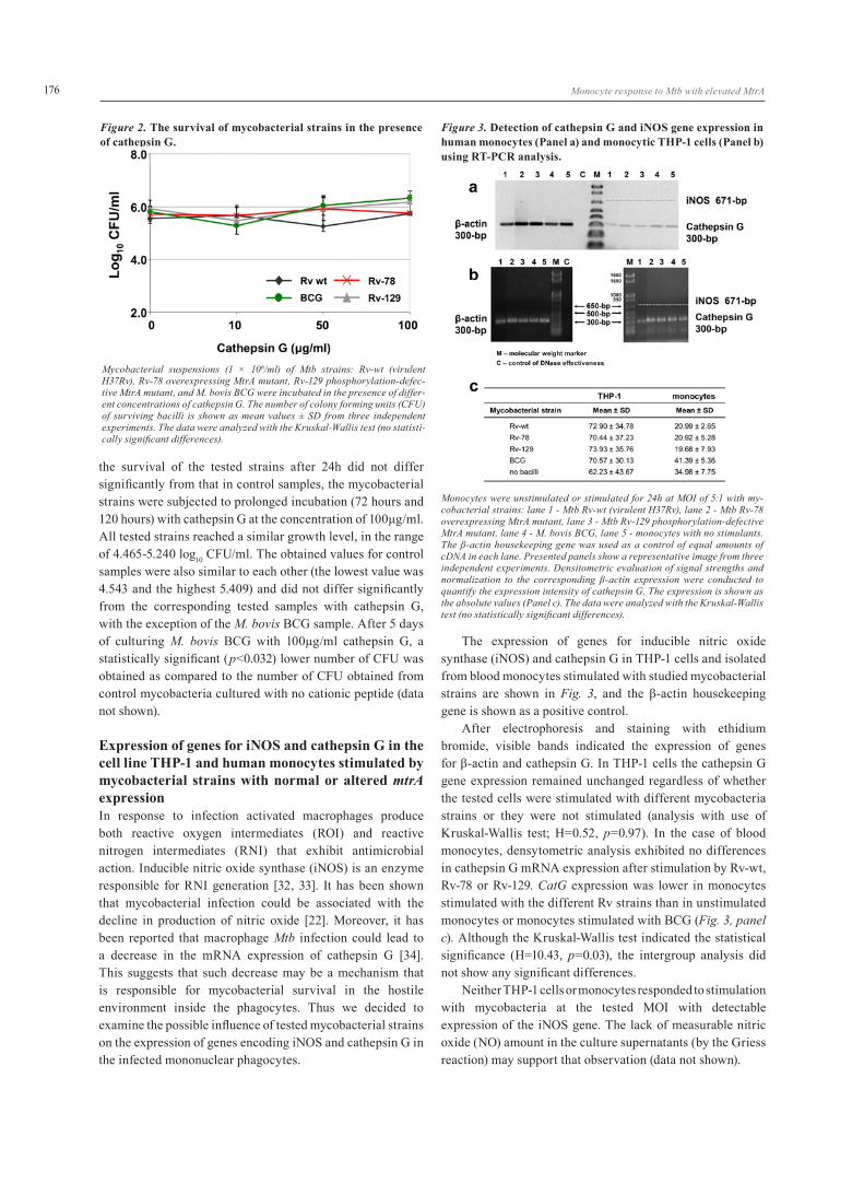
After 24 h incubation of investigated Mtb strains: Rv-wt, Rv-78, Rv-129, and M. bovis BCG with cathepsin G at the final concentrations: 10µg/ml, 50µg/ml, and 100µg/ml, bacteria were plated onto Middlebrook 7H10 solid medium and CFU values were calculated for each strain. The results are shown in Fig. 2.
All tested mycobacterial strains, regardless of the used cathepsin G concentrations, showed growth at similar levels, located in the range of 5.263-6.327 (log10 CFU/ml). Since
Figure 1. Flow cytometric analysis of MHC class II expression.
Panel a: Surface expression of MHC class II molecules on unstimulated monocytes (MØ) or monocytes stimulated for 24h at MOI of 5:1 with live my-cobacterial strains: Mtb Rv-wt (virulent H37Rv), Mtb Rv-78 overexpressing MtrA mutant, Mtb Rv-129 phosphorylation-defective MtrA mutant, M. bovis BCG. Data are representative FACS profiles of one experiment, which was repeated five additional times, using monocytes isolated from six independent donors. Isotype controls are shown in green and cells stained with antibody in red. Analogous experiments were performed to determine the degree of intracellular MHC class II molecules expression (not shown); Panel b: The levels of MHC class II intracellular and extracellular expression of mono-cytes stimulated with the indicated mycobacterial strains. Data are shown as median fluorescent intensity (MFI) values ± SD. The data were analyzed with the Kruskal-Wallis test (no statistically significant differences).
175
Monocyte response to Mtb with elevated MtrA
the survival of the tested strains after 24h did not differ significantly from that in control samples, the mycobacterial strains were subjected to prolonged incubation (72 hours and 120 hours) with cathepsin G at the concentration of 100µg/ml. All tested strains reached a similar growth level, in the range of 4.465-5.240 log10 CFU/ml. The obtained values for control samples were also similar to each other (the lowest value was 4.543 and the highest 5.409) and did not differ significantly from the corresponding tested samples with cathepsin G, with the exception of the M. bovis BCG sample. After 5 days of culturing M. bovis BCG with 100µg/ml cathepsin G, a statistically significant (p<0.032) lower number of CFU was obtained as compared to the number of CFU obtained from control mycobacteria cultured with no cationic peptide (data not shown).
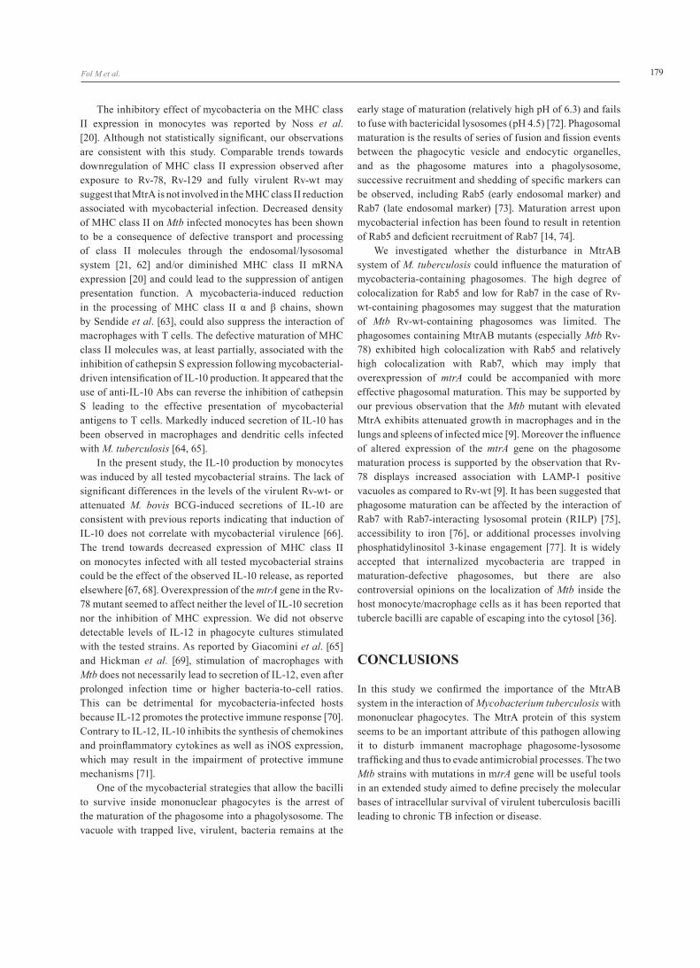
Expression of genes for iNOS and cathepsin G in the cell line THP-1 and human monocytes stimulated by mycobacterial strains with normal or altered mtrA expression In response to infection activated macrophages produce both reactive oxygen intermediates (ROI) and reactive nitrogen intermediates (RNI) that exhibit antimicrobial action. Inducible nitric oxide synthase (iNOS) is an enzyme responsible for RNI generation [32, 33]. It has been shown that mycobacterial infection could be associated with the decline in production of nitric oxide [22]. Moreover, it has been reported that macrophage Mtb infection could lead to a decrease in the mRNA expression of cathepsin G [34]. This suggests that such decrease may be a mechanism that is responsible for mycobacterial survival in the hostile environment inside the phagocytes. Thus we decided to examine the possible influence of tested mycobacterial strains on the expression of genes encoding iNOS and cathepsin G in the infected mononuclear phagocytes.
The expression of genes for inducible nitric oxide synthase (iNOS) and cathepsin G in THP-1 cells and isolated from blood monocytes stimulated with studied mycobacterial strains are shown in Fig. 3, and the β-actin housekeeping gene is shown as a positive control.
After electrophoresis and staining with ethidium bromide, visible bands indicated the expression of genes for β-actin and cathepsin G. In THP-1 cells the cathepsin G gene expression remained unchanged regardless of whether the tested cells were stimulated with different mycobacteria strains or they were not stimulated (analysis with use of Kruskal-Wallis test; H=0.52, p=0.97). In the case of blood monocytes, densytometric analysis exhibited no differences in cathepsin G mRNA expression after stimulation by Rv-wt, Rv-78 or Rv-129. CatG expression was lower in monocytes stimulated with the different Rv strains than in unstimulated monocytes or monocytes stimulated with BCG (Fig. 3, panel c). Although the Kruskal-Wallis test indicated the statistical significance (H=10.43, p=0.03), the intergroup analysis did not show any significant differences.
Neither THP-1 cells or monocytes responded to stimulation with mycobacteria at the tested MOI with detectable expression of the iNOS gene. The lack of measurable nitric oxide (NO) amount in the culture supernatants (by the Griess reaction) may support that observation (data not shown).
Figure 2. The survival of mycobacterial strains in the presence of cathepsin G.
Mycobacterial suspensions (1 × 10⁶/ml) of Mtb strains: Rv-wt (virulent H37Rv), Rv-78 overexpressing MtrA mutant, Rv-129 phosphorylation-defec-tive MtrA mutant, and M. bovis BCG were incubated in the presence of differ-ent concentrations of cathepsin G. The number of colony forming units (CFU) of surviving bacilli is shown as mean values ± SD from three independent experiments. The data were analyzed with the Kruskal-Wallis test (no statisti-cally significant differences).
Figure 3. Detection of cathepsin G and iNOS gene expression in human monocytes (Panel a) and monocytic THP-1 cells (Panel b) using RT-PCR analysis.
Monocytes were unstimulated or stimulated for 24h at MOI of 5:1 with my-cobacterial strains: lane 1 - Mtb Rv-wt (virulent H37Rv), lane 2 - Mtb Rv-78 overexpressing MtrA mutant, lane 3 - Mtb Rv-129 phosphorylation-defective MtrA mutant, lane 4 - M. bovis BCG, lane 5 - monocytes with no stimulants. The β-actin housekeeping gene was used as a control of equal amounts of cDNA in each lane. Presented panels show a representative image from three independent experiments. Densitometric evaluation of signal strengths and normalization to the corresponding β-actin expression were conducted to quantify the expression intensity of cathepsin G. The expression is shown as the absolute values (Panel c). The data were analyzed with the Kruskal-Wallis test (no statistically significant differences).
176

Fol M et al.
Mycobacteria driven IL-10 and Il-12 productionThe balance between the Th1 and Th2 response is considered to be crucial for determination of the outcome of an Mtb infection [35]. The amount of IL-12 and IL-10 (critical for Th1 and Th2 type cell response, respectively) secreted by infected monocytes was thus quantified.
In this study, no measurable IL-12 was observed in the culture supernatants from monocytes stimulated with Rv-wt, Rv-78, Rv-129 or M. bovis BCG. Nor did the unstimulated monocytes release any detectable amounts of IL-12. In contrast, significant amounts of IL-10 (which exceeded the sensitivity of the test: 2 pg/ml) were detected in the supernatants. The results are shown in Fig. 4.
Unstimulated monocytes produced IL-10 at a low level (6pg/ml) which was markedly increased after stimulation. Intergroup analysis showed significant differences between cells unstimulated and stimulated with Mtb Rv-wt as well as between unstimulated cells and cells stimulated with M. bovis BCG. No statistically significant differences between the tested mycobacterial strains were found. However, there was a trend towards Rv-78 and Rv-129 mutants being weaker stimuli for IL-10 production by monocytes than wild type. Interestingly, there were no significant differences in the production of IL-10 by monocytes stimulated with Rv-wt and the vaccine M. bowis BCG strain. The high standard deviation values may reflect high intra-individual variability in the monocyte response to mycobacterial stimulation.
Rv-78 colocalization with vacuoles containing Rab5 or Rab7 markersMononuclear phagocytes infected with Mtb display a deficiency in the fusion of mycobacterial phagosomes with lysosomes [14, 15, 16, 17]. Upon ingestion by macrophages,
live mycobacteria localize to phagosomes containing markers of early but not late endosomal structures [13, 17]. Using mycobacterial strains expressing green fluorescent protein (GFP), we examined intracellular trafficking of bacteria inside THP-1 macrophages. Phagocytes were stained with monoclonal antibodies to Rab5 or Rab7 followed by secondary antibodies conjugated to rhodamine.
We observed highly effective colocalization of all the mycobacterial strains tested and the early endosome marker Rab5 (70-80% of the bacteria resided in Rab5-positive vacuoles). In the case of colocalization with the late endosomal marker Rab7, however, significant differences between the strains appeared, particularly between Rv-78 and Rv-wt strains and between BCG bacilli and Rv-wt. The degree of colocalization of Rv-78 with Rab7 was high and comparable with that observed for Rab5. In contrast, less than 40% of Rv-wt-containing phagosomes colocalized with Rab7. Limited colocalization of Rv-wt with Rab7 suggests reduced phagosomal acquisition of this marker, and thus impaired phagosomal maturation.
DISCUSSION
Macrophages are among the first immune cells interacting with mycobacteria entering the host. The current view is that Mtb interactions with macrophages are dominated by the ability of the pathogen to inhibit phagosomal maturation [36]. However many questions remain to be answered regarding how mycobacteria mount the mechanisms that permit Mtb survival within macrophages. Genes encoding proteins involved in receiving and processing of information at both the inter- and intracellular levels are crucial for modulation
Figure 4. Production of IL-10 by monocytes infected with differ-ent mycobacterial strains.
The cells were unstimulated (only monocytes, MØ) or stimulated for 24h at MOI of 5:1 with mycobacterial strains: Mtb Rv-wt (virulent H37Rv), Mtb Rv-78 overexpressing MtrA mutant, Mtb Rv-129 phosphorylation-defective MtrA mutant or M. bovis BCG. Data (five independent experiments) were analyzed with Kruskal-Wallis test (H=13.18, p=0.01), post-hoc median test. *p=0.01 MØ vs Mtb Rv-wt, **p=0.02 MØ vs M. bovis BCG.
Figure 5. Colocalization of green fluorescence protein (GFP, green) expressing M. tuberculosis strains: wild-type (Rv-wt), overexpressing mtrA (Rv-78), phosphorylation-defective mtrA (Rv-129) and M. bovis BCG with Rab5 or Rab7 in THP-1 cells.
Infected cells were immunofluorescently stained for Rab5 and Rab7 (red). Yel-low color indicates colocalization. Values shown are mean percentages (±) of colocalization from three independent experiments. *p=0.015, **p=0.0041 (Kruskal-Wallis test, H=14.207, p=0.0026, post-hoc median test); ap=0.0012, bp=0.037 (Mann-Whitney U test).
177

Monocyte response to Mtb with elevated MtrA
of life processes in bacteria, allowing microorganisms to adapt to changing environmental conditions by regulating the expression of appropriate genes [4, 9]. The current study was aimed at further investigation of the MtrA protein, belonging to the MtrAB mycobacterial signal transduction system, in the interplay between M. tuberculosis and mononuclear phagocytes. Rv-78 (with overexpression of mtrA gene) and Rv-129 (with elevated level of phosphorylation-defective MtrA), derived from the virulent reference M. tuberculosis H37Rv strain, were used, as well as M. bovis BCG. Upregulation of MtrA has been observed during intramacrophage growth of M. bovis BCG whereas the level of MtrA in M. tuberculosis remains unchanged after entry into mononuclear phagocytes. We have previously shown that the Mtb mutant with an elevated level of MtrA exhibits attenuated growth not only in the THP-1 cell line but also in vivo in murine lungs and spleens, and its growth attenuation phenotype is accompanied by elevated expression of mtrA [9].
Phagocytes possess a number of mechanisms that allow them to inactivate ingested bacilli. Reactive nitrogen and oxygen intermediates (RNI, ROI) as well as anti-bacterial cationic peptides generated by phagocytes are important agents for intracellular killing. Additionally, macrophages bind processed bacterial peptides, which are recognized by specific T cells via MHC class II presentation. Both activated macrophages and T cells release cytokines regulating the fate of mycobacteria within macrophages and consequently in the infected organism [14, 15, 20, 28, 30, 32]. We observed that blood monocytes isolated from healthy donors as well as THP-1 cells, which are able to produce nitric oxide in response to LPS or silica [37], did not show expression of the gene for inducible nitric oxide synthase (iNOS) when stimulated by tested mycobacteria strains. We also did not detect the nitric oxide intermediates in monocyte culture supernatants. It can be suspected that in our experiments the phagocytes were not sufficiently stimulated to launch the expression of iNOS or that the commonly used colorimetric method of NO detection was not sensitive enough, as reported by Jagannath et al. [38]. Similarly to our results Aston et al. [26] reported the lack of iNOS mRNA in human macrophages after stimulation with mycobacteria or IFN-γ together with LPS. While the role of nitric oxide in the protection against mycobacteria in mouse macrophages has been well demonstrated, the role of nitric oxide in human mononuclear phagocytes is a debated issue [38]. Facchetti et al. [39] reported that monocytes isolated from healthy donors produced only a small, if any, amount of iNOS, whereas a high level of this enzyme was detected in the monocytes located in mycobacterial granulomae. This may indicate that iNOS gene expression requires signals only present in the granuloma microenvironment. The role of nitric oxide in the elimination of various bacterial [40, 41], parasite [42-44], fungal [45] or viral [46] pathogens has been reported but the exact mechanism by which nitric oxide participates in pathogen elimination remains obscure [47,
48]. One of the explanations could be apoptosis induction in infected host cells [49]. Nitric oxide has antibacterial effects on mycobacteria [38, 50, 51] however, the susceptibility of Mtb to nitric oxide intermediates depends on the bacterial strain, RNI concentration and time of interaction [52-54]. Surprisingly, in the case of human mononuclear phagocytes, despite using comparable techniques, variable results have been obtained in different research laboratories, possibly due to subject heterogeneity in terms of genetic polymorphisms of iNOS, or to the antagonistic effects of the used stimulatory agents depending on the concentration and sequence of the stimulation [55, 56].
It has been found that the presence of cationic sequences in the cathepsin G molecule provide the peptide with antimicrobial capacities against both gram-positive and gram-negative bacteria [57-59]. In addition, cathepsin G exhibits immunoregulatory propeties, e.g. in vitro it possesses chemotactic activity for monocytes and lymphocytes, and may act as a mitogen for T and B cells [30]. Rivera-Marrero et al. [25] observed a coincidence in downregulation of catG in Mtb infected THP-1 monocytes with enhanced bacillary multiplication within the cells. Herein, we show that in vitro cathepsin G, in the tested range, had no effect on the survival of mycobacteria, except for BCG, whose growth was limited after 5 days culturing with cathepsin G at 100µg/ml. It is possible that in vivo cathepsin G contributes to the killing of mycobacteria indirectly through its effect on the inflammatory response. The role of cathepsin G in the course of tuberculosis is not clear but cathepsin G seems to be of importance since mouse infection with M. tuberculosis leads to downregulation of catG expression, whereas the expression of genes for acidic-type cathepsins (D and B), as well as for cathepsin H are upregulated [34]. The limitation in cathepsin G production could be a mycobacterial defense mechanisms, enabling bacterial survival within the hostile phagocyte environment. Although cationic antimicrobial peptides (CAMPs) alone may not be effective at physiological concentrations, their use as the supplement to conventional anti-TB antibiotics can make them much more effective and has potential to improve the therapeutic effectiveness as it was shown by Fattorini et al. [60]. The THP-1 cells used in our study were a relevant model for cathepsin G investigation as this protein is one of the three most abundant gene products of these cells, in addition to neutrophil elastase 2 and proteinase 3 [61]. However there were no differences between catG expression in unstimulated THP-1 cells and cells stimulated with Mtb mutants or BCG. Experiments with blood monocytes also did not show any significant differences between the mycobacterial strains, although there was a trend towards lower catG expression after infection with Rv-wt, Rv-129 and Rv-78 than with M. bovis BCG or in unstimulated cells. This may suggest that disturbed functioning of the MtrAB system does not affect the bacterium’s ability to alter monocyte CatG expression.
178
Fol M et al.
The inhibitory effect of mycobacteria on the MHC class II expression in monocytes was reported by Noss et al. [20]. Although not statistically significant, our observations are consistent with this study. Comparable trends towards downregulation of MHC class II expression observed after exposure to Rv-78, Rv-129 and fully virulent Rv-wt may suggest that MtrA is not involved in the MHC class II reduction associated with mycobacterial infection. Decreased density of MHC class II on Mtb infected monocytes has been shown to be a consequence of defective transport and processing of class II molecules through the endosomal/lysosomal system [21, 62] and/or diminished MHC class II mRNA expression [20] and could lead to the suppression of antigen presentation function. A mycobacteria-induced reduction in the processing of MHC class II α and β chains, shown by Sendide et al. [63], could also suppress the interaction of macrophages with T cells. The defective maturation of MHC class II molecules was, at least partially, associated with the inhibition of cathepsin S expression following mycobacterial-driven intensification of IL-10 production. It appeared that the use of anti-IL-10 Abs can reverse the inhibition of cathepsin S leading to the effective presentation of mycobacterial antigens to T cells. Markedly induced secretion of IL-10 has been observed in macrophages and dendritic cells infected with M. tuberculosis [64, 65].
In the present study, the IL-10 production by monocytes was induced by all tested mycobacterial strains. The lack of significant differences in the levels of the virulent Rv-wt- or attenuated M. bovis BCG-induced secretions of IL-10 are consistent with previous reports indicating that induction of IL-10 does not correlate with mycobacterial virulence [66]. The trend towards decreased expression of MHC class II on monocytes infected with all tested mycobacterial strains could be the effect of the observed IL-10 release, as reported elsewhere [67, 68]. Overexpression of the mtrA gene in the Rv-78 mutant seemed to affect neither the level of IL-10 secretion nor the inhibition of MHC expression. We did not observe detectable levels of IL-12 in phagocyte cultures stimulated with the tested strains. As reported by Giacomini et al. [65] and Hickman et al. [69], stimulation of macrophages with Mtb does not necessarily lead to secretion of IL-12, even after prolonged infection time or higher bacteria-to-cell ratios. This can be detrimental for mycobacteria-infected hosts because IL-12 promotes the protective immune response [70]. Contrary to IL-12, IL-10 inhibits the synthesis of chemokines and proinflammatory cytokines as well as iNOS expression, which may result in the impairment of protective immune mechanisms [71].
One of the mycobacterial strategies that allow the bacilli to survive inside mononuclear phagocytes is the arrest of the maturation of the phagosome into a phagolysosome. The vacuole with trapped live, virulent, bacteria remains at the
early stage of maturation (relatively high pH of 6.3) and fails to fuse with bactericidal lysosomes (pH 4.5) [72]. Phagosomal maturation is the results of series of fusion and fission events between the phagocytic vesicle and endocytic organelles, and as the phagosome matures into a phagolysosome, successive recruitment and shedding of specific markers can be observed, including Rab5 (early endosomal marker) and Rab7 (late endosomal marker) [73]. Maturation arrest upon mycobacterial infection has been found to result in retention of Rab5 and deficient recruitment of Rab7 [14, 74].
We investigated whether the disturbance in MtrAB system of M. tuberculosis could influence the maturation of mycobacteria-containing phagosomes. The high degree of colocalization for Rab5 and low for Rab7 in the case of Rv-wt-containing phagosomes may suggest that the maturation of Mtb Rv-wt-containing phagosomes was limited. The phagosomes containing MtrAB mutants (especially Mtb Rv-78) exhibited high colocalization with Rab5 and relatively high colocalization with Rab7, which may imply that overexpression of mtrA could be accompanied with more effective phagosomal maturation. This may be supported by our previous observation that the Mtb mutant with elevated MtrA exhibits attenuated growth in macrophages and in the lungs and spleens of infected mice [9]. Moreover the influence of altered expression of the mtrA gene on the phagosome maturation process is supported by the observation that Rv-78 displays increased association with LAMP-1 positive vacuoles as compared to Rv-wt [9]. It has been suggested that phagosome maturation can be affected by the interaction of Rab7 with Rab7-interacting lysosomal protein (RILP) [75], accessibility to iron [76], or additional processes involving phosphatidylinositol 3-kinase engagement [77]. It is widely accepted that internalized mycobacteria are trapped in maturation-defective phagosomes, but there are also controversial opinions on the localization of Mtb inside the host monocyte/macrophage cells as it has been reported that tubercle bacilli are capable of escaping into the cytosol [36].
CONCLUSIONS
In this study we confirmed the importance of the MtrAB system in the interaction of Mycobacterium tuberculosis with mononuclear phagocytes. The MtrA protein of this system seems to be an important attribute of this pathogen allowing it to disturb immanent macrophage phagosome-lysosome trafficking and thus to evade antimicrobial processes. The two Mtb strains with mutations in mtrA gene will be useful tools in an extended study aimed to define precisely the molecular bases of intracellular survival of virulent tuberculosis bacilli leading to chronic TB infection or disease.
179

Monocyte response to Mtb with elevated MtrA
ACKNOWLEDGEMENTS
This work was supported by the Polish Ministry of Science and Higher Education, grant no. N N303 345035. The founder had no role in study design, data collection and analysis, decision to publish, or preparation of the manuscript.
REFERENCES1. Hoch JA. Two-component and phosphorelay signal
transduction. Curr Opin Microbiol. 2000 Apr;3(2):165-70.2. Parkinson JS, Kofoid EC. Communication modules
in bacterial signaling proteins. Annu Rev Genet. 1992;26:71-112.
3. Stock AM, Robinson VL, Goudreau PN. Two-component signal transduction. Annu Rev Biochem. 2000;69:183-215.
4. Chakraborti PK, Matange N, Nandicoori VK, Singh Y, Tyagi JS, Visweswariah SS. Signalling mechanisms in Mycobacteria. Tuberculosis (Edinb). 2011 Sep;91(5):432-40.
5. Cole ST, Brosch R, Parkhill J, Garnier T, Churcher C, Harris D, Gordon SV, Eiglmeier K, Gas S, Barry CE 3rd, Tekaia F, Badcock K, Basham D, Brown D, Chillingworth T, Connor R, Davies R, Devlin K, Feltwell T, Gentles S, Hamlin N, Holroyd S, Hornsby T, Jagels K, Krogh A, McLean J, Moule S, Murphy L, Oliver K, Osborne J, Quail MA, Rajandream MA, Rogers J, Rutter S, Seeger K, Skelton J, Squares R, Squares S, Sulston JE, Taylor K, Whitehead S, Barrell BG. Deciphering the biology of Mycobacterium tuberculosis from the complete genome sequence. Nature. 1998 Jun 11;393(6685):537-44.
6. Zahrt TC, Deretic V. An essential two-component signal transduction system in Mycobacterium tuberculosis. J Bacteriol. 2000 Jul;182(13):3832-8.
7. Via LE, Curcic R, Mudd MH, Dhandayuthapani S, Ulmer RJ, Deretic V. Elements of signal transduction in Mycobacterium tuberculosis: in vitro phosphorylation and in vivo expression of the response regulator MtrA. J Bacteriol. 1996 Jun;178(11): 3314-21.
8. Parish T, Smith DA. Dendall S. Deletion of two-component regulatory systems increases the virulence of the Mycobacterium tuberculosis. Infect Immun. 2003 Mar;71(3):1134-40.
9. Fol M, Chauchan A, Nair NK, Maloney E, Moomey M, Jagannath C, Madiraju MV, Rajagopalan M. Modulation of Mycobacterium tuberculosis proliferation by MtrA, an essential two-component response regulator. Mol Microbiol. 2006 May;60(3):643-57.
10. Curcic R, Dhandayuthapani S, Deretic V. Gene expression in mycobacteria: transcriptional fusions based on xylE and analysis of the promoter region on the response regulator mtrA from Mycobacterium tuberculosis. Mol Microbiol. 1994 Sep;13(6):1057-64.
11. Zahrt TC, Deretic V. An essential two-component signal transduction system in Mycobacterium tuberculosis. J Bacteriol. 2000 Jul;182(13):3832-8.
12. Zahrt TC, Deretic V. Mycobacterium tuberculosis signal transduction system required for persistent infections. Proc Natl Acad Sci USA, 2001 Oct;98(22):12706-11.
13. Deretic V, Fratti RA. Mycobacterium tuberculosis phagosome. Mol Microbiol. 1999 Mar;31(6):1603-09.
14. Flynn JL, Chan J. Immune evasion by Mycobacterium tuberculosis: living with the enemy. Curr Opin Immunol. 2003 Aug;15(4):450-5.
15. Raja A. Immunology of tuberculosis. Indian J Med Res. 2004 Oct;120(4): 213-32.
16. Russell DG. Phagosomes, fatty acids and tuberculosis. Nat Cell Biol. 2003 Sep;5(9):776-8.
17. Pieters J. Entry and survival of pathogenic mycobacteria in macrophages. Microbes Infect. 2001 Mar;3(3):249-55.
18. Hackam DJ, Rotstein OD, Zhang WJ, Demaurex N, Woodside M, Tsai O, Grinstein S. Regulation of phagosomal acidification. Differential targeting of Na+/H+ exchangers, Na+/K+-ATPases, and vacuolar-type H+-atpases. J Biol Chem. 1997 Nov 21;272(47):29810-20.
19. Sturgill-Koszycki S, Schlesinger PH, Chakraborty P, Haddix PL, Collins HL, Fok AK, Allen RD, Gluck SL, Heuser J, Russell DG. Lack of acidification in Mycobacterium phagosomes produced by exclusion of the vesicular proton-ATPase. Science. 1994 Feb 4;263(5147):678-81.
20. Noss EH, Harding CV, Boom WH. Mycobacterium tuberculosis inhibits MHC class II antigen processing in murine bone marrow macrophages. Cell Immunol. 2000 Apr 10;201(1):63-74.
21. Hmama Z, Gabathuler R, Jefferies WA, de Jong G, Reiner NE. Attenuation of HLA-DR expression by mononuclear phagocytes infected with Mycobacterium tuberculosis is related to intracellular sequestration of immature class II heterodimers. J Immunol. 1998 Nov 1;161(9):4882-93.
22. Da Silva TR, De Freitas JR, Silva QC, Figueira CP, Roxo E, Leão SC, De Freitas LA, Veras PS. Virulent Mycobacterium fortuitum restricts NO production by a gamma interferon-activated J774 cell line and phagosome-lysosome fusion. Infect Immun. 2002 Oct;70(10):5628-34.
23. Miller BH, Fratti RA, Poschet JF, Timmins GS, Master SS, Burgos M, Marletta MA, Deretic V. Mycobacteria inhibit nitric oxide synthase recruitment to phagosomes during macrophage infection. Infect Immun. 2004 May;72(5):2872-8.
24. Stokes RW, Doxsee D. The receptor-mediated uptake, survival, replication, and drug sensitivity of Mycobacterium tuberculosis within the macrophage-like cell line THP-1: a comparison with human monocyte-derived macrophages. Cell Immunol. 1999 Oct;197(1):1-9.
180

Fol M et al.
25. Rivera-Marrero CA, Stewart J, Shafer WM, Roman J. The down-regulation of cathepsin G in THP-1 monocytes after infection with Mycobacterium tuberculosis is associated with increased intracellular survival of bacilli. Infect Immun. 2004 Oct;72(10):5712-21.
26. Aston C, Rom WN, Talbot AT, Reibman J. Early inhibition of mycobacterial growth by human alveolar macrophages is not due to nitric oxide. Am J Respir Crit Care Med. 1998 Jun;157(6 Pt 1):1943-50.
27. Nims RW, Cook JC, Krishna MC, Christodoulou D, Poore CM, Miles AM, Grisham MB, Wink DA. Colorimetric assays for nitric oxide and nitrogen oxide species formed from nitric oxide stock solutions and donor compounds. Methods Emzymol. 1996;268:93-105.
28. Tsukaguchu K, Balaji KN, Boom WH. CD4+ alpha-beta T cell and gamma-delta T cell responses to Mycobacterium tuberculosis: similarities and differences in antigen recognition, cytotoxic effector function, and cytokine production. J Immunol. 1995 Feb;154(4):1786-96.
29. Wiesner J, Vilcinskas A. Antimicrobial peptides. The ancient arm of the human immune response. Virulence. 2010 Sep-Oct;1(5):440-64.
30. Chertov O, Yang D, Howard ZMO, Oppenheim JJ. Leukocyte granule proteins mobilize innate host defenses and adaptive immune responses. Immunol Rev. 2000 Oct;177:68-78.
31. Burster T, Macmillan H, Hou T, Boehm BO, Mellins ED. Cathepsin G: Roles in antigen presentation and beyond. Mol Immunol. 2010 Jan;47(4):658-65.
32. MacMicking J, Xie QW, Nathan C. Nitric oxide and macrophage function. Annu Rev Immunol. 1997;15:323-50.
33. Sato K, Akaki T, Tomioka H. Differential potentiation of anti-mycobacterial activity and reactive nitrogen intermediate-producing ability of murine peritoneal macrophages activated by interferon-gamma (IFN-γ) and tumor necrosis factor-alpha (TNF-α). Clin Exp Immunol. 1998 Apr;112(1):63-8.
34. Stewart JN, Rivera HN, Karls R, Quinn FD, Roman J, Rivera-Marrero CA. Increased pathology in lungs of mice after infection with an α-crystallin mutant of Mycobacterium tuberculosis: changes in cathepsin proteases and certain cytokines. Microbiology. 2006 Jan;152(Pt 1):233-44.
35. Sai Priya VH, Anuradha B, Latha Gaddam S, Hasnain SE, Murthy KJ, Valluri VL. In vitro levels of interleukin 10 (IL-10) and IL-12 in response to a recombinant 32-kilodalton antigen of Mycobacterium bovis BCG after treatment for tuberculosis. Clin Vaccine Immunol. 2009 Jan;16(1):111-5.
36. Welin A, Lerm M. Inside or outside the phagosome? The controversy of the intracellular localization of Mycobacterium tuberculosis. Tuberculosis (Edinb). 2012 Mar;92(2):113-20.
37. Chen F, Kuhn DC, Gaydos LJ, Demers LM. Induction of nitric oxide and nitric oxide synthase mRNA by
silica and lipopolysaccharide in PMA-primed THP-1 cells. APMIS 1996 Mar;104(3):176-82.
38. Jagannath C, Actor JK, Hunter RL Jr. Induction of nitric oxide in human monocytes and monocyte cell lines by Mycobacterium tuberculosis. Nitric Oxide. 1998;2(3):174-86.
39. Facchetti F, Vermi W, Fiorentini S, Chilosi M, Caruso A, Duse M, Notarangelo LD, Badolato R. Expression of inducible nitric oxide synthase in human granulomas and histiocitic reactions. Am J Pathol. 1999 Jan;154(1):145-51.
40. Boockvar KS, Granger DL, Poston RM, Maybodi M. Washington MK, Hibbs JB Jr, Kurlander RL. Nitric oxide produced during murine listeriosis is protective. Infect Immun. 1994 Mar;62(3):1089-100.
41. Brieland JK, Remick DG, Freeman PT, Hurley MC, Fantone JC, Engleberg NC. In vivo regulation of replicative Legionella pneumophila lung infection by endogenous tumor necrosis factor alpha and nitric oxide. Infect Immun. 1995 Sep;63(9):3253-8.
42. Gobert AP, Daulouede S, Lepoivre M, Boucher JL, Bouteille B, Buquet A, Cespuglio R, Veyret B, Vincendeau P. L-arginine availability modulates local nitric oxide production and parasite killing in experimental trypanosomiasis. Infect Immun. 2000 Aug;68(8):4653-7.
43. Romão PR, Fonseca SG, Hothersall JS, Noronha-Dutra AA, Ferreira SH, Cunha FQ. Glutathione protects macrophages and Leishmania major against nitric oxide-mediated cytotoxicity. Parasitology. 1999 Jun;118 ( Pt 6):559-66.
44. Panaro MA, Acquafredda A, Lisi S, Lofrumento DD, Trotta T, Satalino R, Saccia M, Mitolo V, Brandonisio O. Inducible nitric oxide synthase and nitric oxide production in Leishmania infatum-infected human macrophages stimulated with interferon-γ and bacterial lipopolysaccharide. Int J Clin Lab Res. 1999;29(3):122-7.
45. Rossi GR, Cervi LA, Sastre DA, Masih DT. Lack of involvement of nitric oxide in the macrophage-mediated inhibition of spleen cell proliferation during experimental cryptococcosis. Clin Immunol Immunopathol. 1998 Jan;86(1):16-26.
46. Saura M, Zaragoza C, McMillan A, Quick RA, Hohenadl C, Lowenstein JM, Lowenstein CJ. An antiviral mechanism of nitric oxide: inhibition of a viral protease. Immunity. 1999 Jan;10(1):21-8.
47. Axelrod S, Oschkinat H, Enders J, Schlegel B, Brinkmann V, Kaufmann SH, Haas A, Schaible EU. Delay of phagosome maturation by a mycobacterial lipid is reversed by nitric oxide. Cell Microbiol. 2008 Jul;10(7):1530-45.
48. Nathan C. Specificity of a third kind: reactive oxygen and nitrogen intermediates in cell signaling. J Clin Invest. 2003 Mar;111(6):769-78.
49. Herbst S, Schaible UE, Schneider BE. Interferon gamma activated macrophages kill mycobacteria by nitric oxide induced apoptosis. PLoS One. 2011 May 2;6(5):e19105.
181

Monocyte response to Mtb with elevated MtrA
50. Akaki T, Tomioka H, Shimizu T, Dekio S, Sato K. Comparative roles of free fatty acids with reactive nitrogen intermediates and reactive oxygen intermediates in expression of the anti-microbial activity of macrophages against Mycobacterium tuberculosis. Clin Exp Immunol. 2000 Aug;121(2):302-10.
51. Petricevich VL, Alves RC. Role of cytokines and nitric oxide in the induction of tuberculostatic macrophage function. Mediators Inflamm. 2000;9(6):261-9.
52. Long R, Light B, Talbot JA. Mycobacteriocidal action of exogenous nitric oxide. Antimicrob Agents Chemother. 1999 Feb;43(2):403-5.
53. O’Brien L, Carmichael J, Lowrie DB, Andrew PW. Strains of Mycobacterium tuberculosis differ in susceptibility to reactive nitrogen intermediates in vitro. Infect Immun. 1994 Nov;62(11):5187-90.
54. Rhoades ER, Orme IM. Susceptibility of a panel of virulent strains of Mycobacterium tuberculosis to reactive nitrogen intermediates. Infect Immun. 1997 Apr;65(4):1189-95.
55. Weinberg JB. Nitric oxide production and nitric oxide synthase type 2 expression by human mononuclear phagocytes: a review. Mol Med. 1998 Sep;4(9):557-91.
56. Bogdan C. The function of nitric oxide in the immune system. In: Mayer B, editor. Nitric Oxide (Handbook of Experimental Pharmacology). Vol. 143. Heidelberg (Germany): Springer; 2000. Chapter 18.
57. Shafer WM, Pohl J, Onunka VC, Bangalore N, Travis J. Human lysosomal catG and granzyme B share a functionally conserved broad spectrum antibacterial peptide. J Biol Chem. 1991 Jan;266(1):112-6.
58. Bangalore N, Travis J, Onunka VC, Pohl J, Shafer WM. Identification of the primary antimicrobi-al domains in human neutrohil catG. J Biol Chem. 1990 Aug;265(23):13584 -8.
59. Shafer WM, Shepherd ME, Boltin B, Wells L., Pohl J. Synthetic peptides of human lysosomal cathepsin G with potent antipseudomonal activity. Infect Immun. 1993 May;61(5):1900-8.
60. Fattorini L, Gennaro R, Zanetti M, Tan D, Brunori L, Giannoni F, Pardini M, Orefici G. In vitro activity of protegrin-1 and beta-defensin-1, alone and in combination with isoniazid, against Mycobacterium tuberculosis. Peptides. 2005 Jul;25(7):1075-7
61. Cassol E, Alfano M, Biswas P, Poli G. Monocyte-derived macrophages and myeloid cell lines as targets of HIV-1 replication and persistence. J Leukoc Biol. 2006 Nov;80(5):1018-30.
62. Chang ST, Linderman JJ, Kirschner DE. Multiple mechanisms allow Mycobacterium tuberculosis to continuously inhibit MHC class II-mediated antigen presentation by macrophages. Proc Natl Acad Sci USA. 2005 Mar;102(12):4530-5.
63. Sendide K, Deghmane AE, Pechkovsky D, Av-Gay Y, Talal A, Hmama Z. Mycobacterium bovis BCG attenuates surface expression of mature class II molecules through IL-10-dependent inhibition of cathepsin S. J Immunol. 2005 Oct;175(8):5324-32.
64. Shaw TC, Thomas LH, Friedland JS. Regulation of IL-10 secretion after phagocytosis of Mycobacterium tuberculosis by human monocytic cells. Cytokine. 2000 May;12(5):483-6.
65. Giacomini E, Iona E, Ferroni L, Miettinen M, Fattorini L, Orefici G, Julkunen I, Coccia EM. Infection of human macrophages and dendritic cells with Mycobacterium tuberculosis induces a differential cytokine gene expression that modulates T cell response. J Immunol. 2001 Jun;166(12):7033-41.
66. Silver RF, Li Q, Ellner JJ. Expression of virulence of Mycobacterium tuberculosis within human monocytes: virulence correlates with intracellular growth and induction of tumor necrosis factor alpha but not with evasion of lymphocyte-dependent monocyte effector functions. Infect Immun. 1998 Mar;66(3):1190-9.
67. de Waal Malefyt R, Haanen J, Spits H, Roncarolo MG, te Velde A, Figdor C, Johnson K, Kastelein R, Yssel H, de Vries JE. Interleukin 10 (IL-10) and viral IL-10 strongly reduce antigen-specific human T cell proliferation by diminishing the antigen-presenting capacity of monocytes via downregulation of class II major histocompatibility complex expression. J Exp Med. 1991 Oct 1;174(4):915-24.
68. Willems F, Marchant A, Delville JP, Gérard C, Delvaux A, Velu T, de Boer M, Goldman M. Interleukin-10 inhibits B7 and intercellular adhesion molecule-1 expression on human monocytes. Eur J Immunol. 1994 Apr;24(4):1007-9.
69. Hickman SP, Chan J, Salgame P. Mycobacterium tuberculosis induces differential cytokine production from dendritic and macrophages with divergent effects on naïve T cell polarization. J Immunol. 2002 May;168(9):4636-42.
70. Cooper AM, Solache A, Khader SA. Interleukin-12 and tuberculosis: an old story revisited. Curr Opin Immunol. 2007 Aug;19(4):441-7.
71. Reljic R, Stylianou E, Balu S, Ma JK. Cytokine interactions that determine the outcome of Mycobacterial infection of macrophages. Cytokine. 2010 Jul;51(1):42-6.
72. Russell DG. Phagosomes, fatty acids and tuberculosis. Nat Cell Biol. 2003 Sep;5(9):776-8.
73. Via LE, Deretic D, Ulmer RJ, Hibler NS, Huber LA, Deretic V. Arrest of mycobacterial phagosome maturation is caused by a block in vesicle fusion between stages controlled by rab5 and rab7. J Biol Chem. 1997 May;272(20):13326-31.
74. Rink J, Ghigo E, Kalaidzidis Y, Zerial M. Rab conversion as mechanism of progression from early to late endosomes. Cell. 2005 Sep;122(5):735-49.
75. Sun J, Deghmane AE, Soualhine H, Hong T, Bucci T, Solodkin A, Hmama Z. Mycobacterium bovis
182

Fol M et al.
disrupts the interaction of Rab7 with RILP contributing to inhibition of phagosome maturation. J Leukoc Biol. 2007 Dec;82(6):1437- 45.
76. Kelley VA, Schorey JS. Mycobacterium’s arrest of phagosome maturation in macrophages requires Rab5 activity and accessibility to iron. Mol Biol Cell. 2003 Aug;14(8):3366- 77.
77. Vieira OV, Bucci C, Harrison RE, Trimble WS, Lanzetti L, Gruenberg J, Schreiber AD, Stahl PD, Grinstein S. Modulation of Rab5 and Rab7 recruitment to phagosomes by phosphatidylinositol 3-kinase. Mol Cell Biol. 2003 Apr;23(7):2501-14.
183