Embed Size (px)
Citation preview
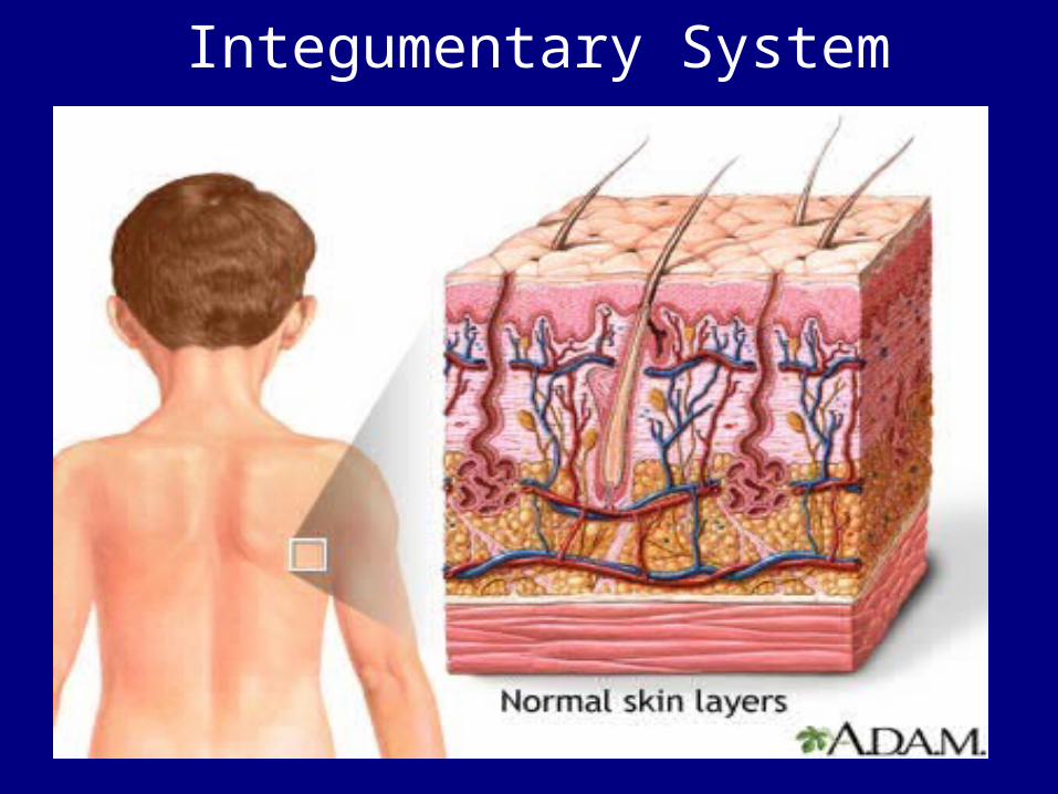
Integumentary System
Why hairy?Read article on p170.
• Why are we hairy?• Explain hypertrichosis.• What theories do scientists have
about why we have hair in the places we do?
• Is there an evolutionary relationship? Explain.
Functions of Skin?• Protects underlying tissues and organs• Excretes salts, water, and organic
wastes (glands)• Maintains body temperature (insulation
and evaporation)
• Synthesizes vitamin D3
• Stores lipids• Detects touch, pressure, pain, and
temperature by sensory receptors located in dermis
• First line of defense• Observed by physicians for health
analysis– Color – Elasticity– sensitivity
• The integument is the largest system of the body is 16% of body weight and 1.5 to 2 m2 in area.
Parts of the Integument
• The integument is made up of 2 parts:1. cutaneous membrane (skin)2. accessory structures3. Sub-Q (subcutaneous)
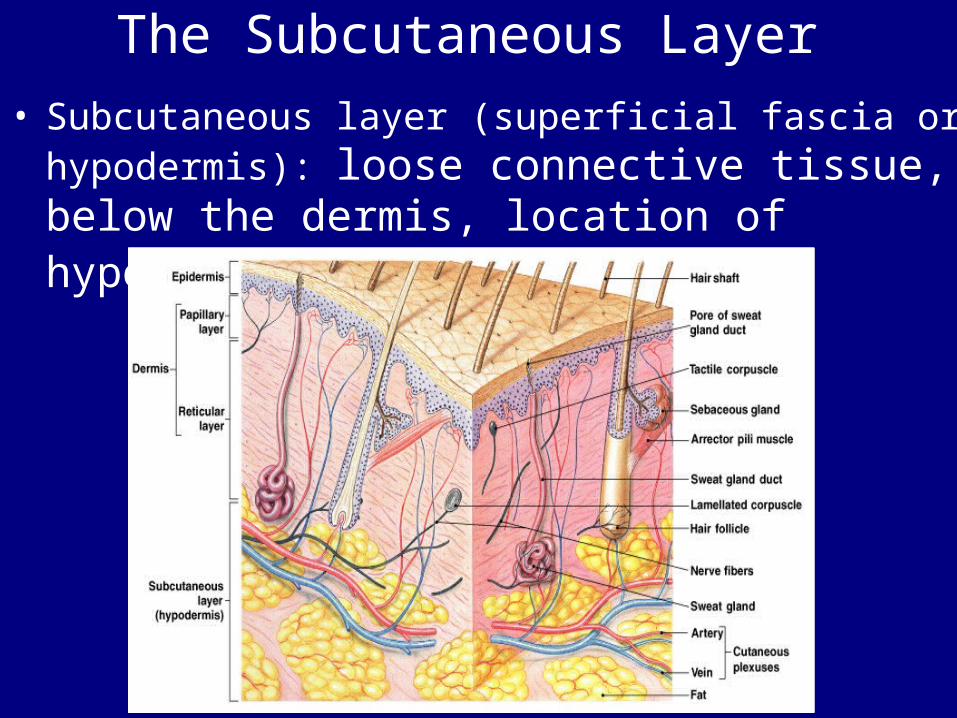
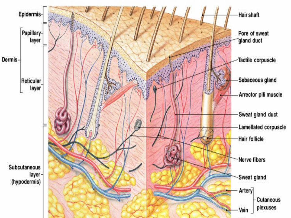
The Subcutaneous Layer• Subcutaneous layer (superficial fascia or
hypodermis): loose connective tissue, below the dermis, location of hypodermic injections
Accessory Structures• Originate in the dermis• Extend through the epidermis to skin
surface:– hair– nails– multicellular exocrine glands– Receptors Connections
• Circulatory system:– blood vessels in the dermis
• Nervous system:– sensory receptors for pain, touch, &
temperature
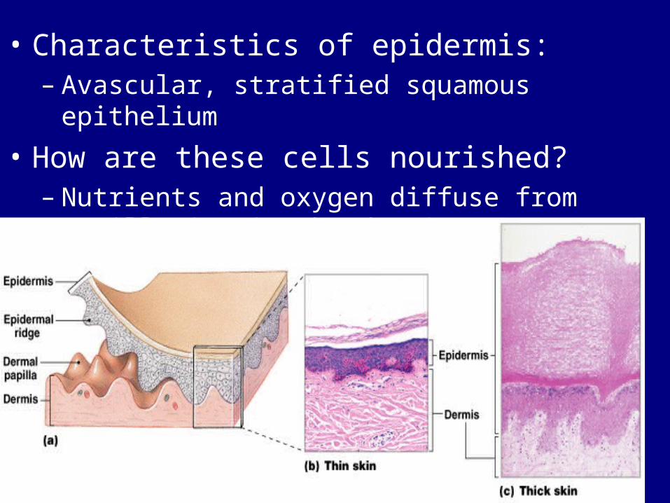
• Characteristics of epidermis: – Avascular, stratified squamous epithelium
• How are these cells nourished?– Nutrients and oxygen diffuse from capillaries
in the dermis
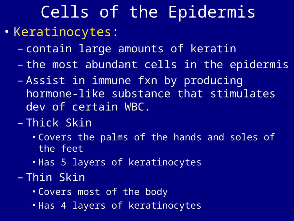
Cells of the Epidermis• Keratinocytes:
– contain large amounts of keratin– the most abundant cells in the epidermis– Assist in immune fxn by producing hormone-
like substance that stimulates dev of certain WBC.
– Thick Skin• Covers the palms of the hands and soles of the
feet• Has 5 layers of keratinocytes
– Thin Skin• Covers most of the body • Has 4 layers of keratinocytes
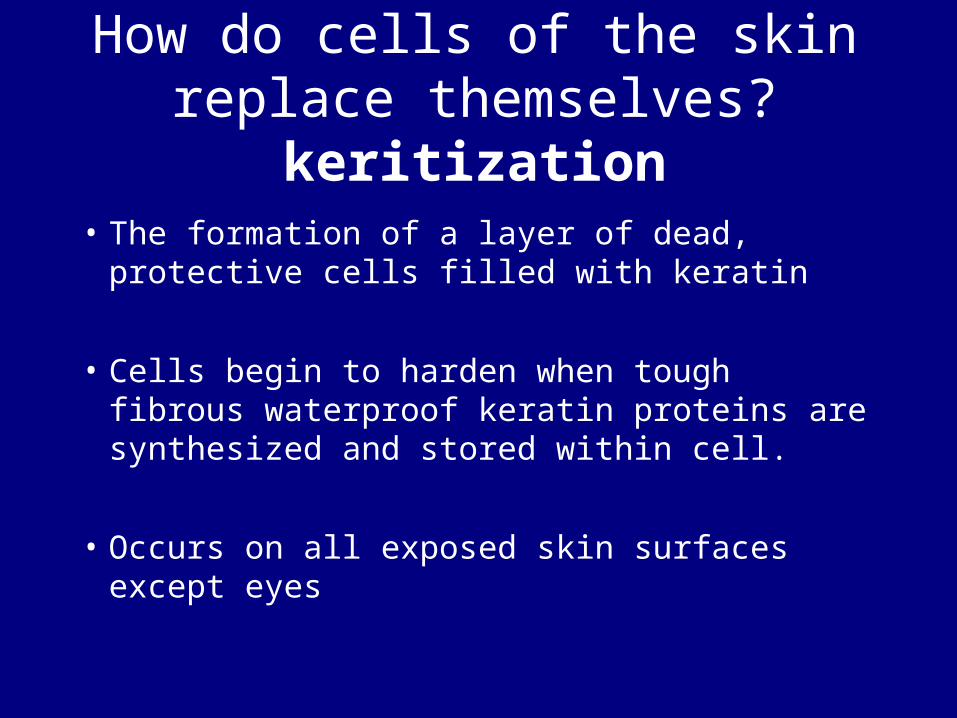
How do cells of the skin replace themselves?
keritization• The formation of a layer of dead,
protective cells filled with keratin
• Cells begin to harden when tough fibrous waterproof keratin proteins are synthesized and stored within cell.
• Occurs on all exposed skin surfaces except eyes

Figure 5–3
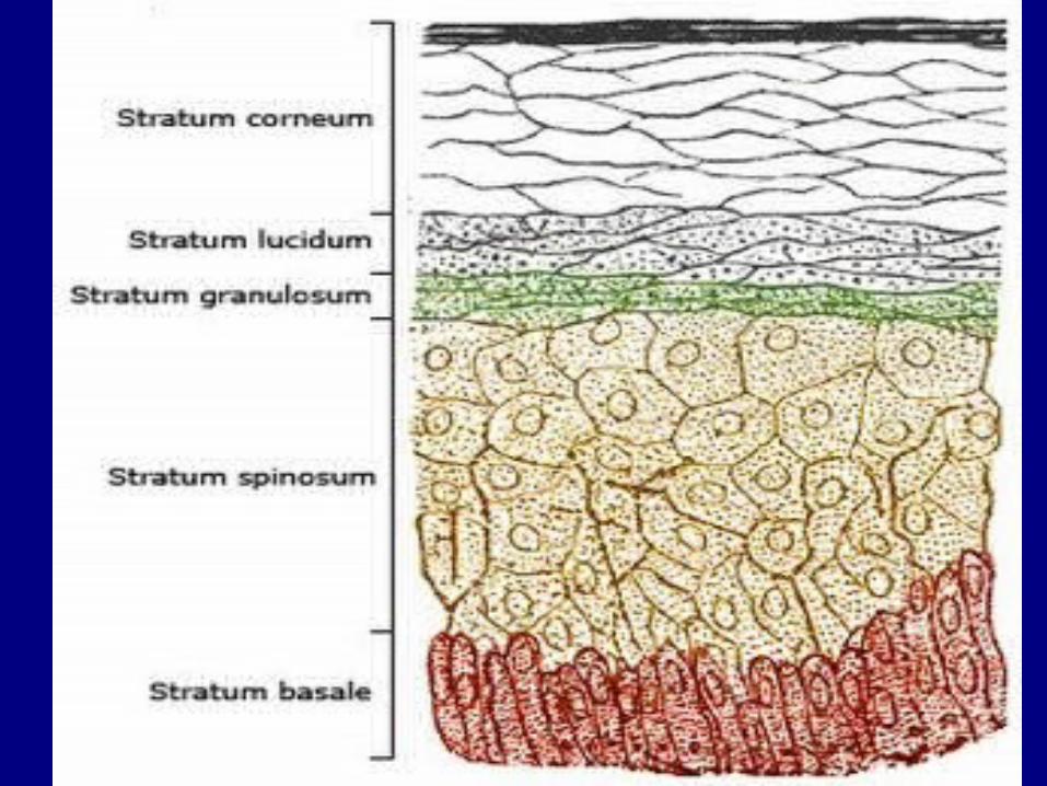
Layers of the Epidermis
• From basal lamina to free surface:– stratum germinativum/basale– stratum spinosum– stratum granulosum– stratum lucidum– stratum corneum
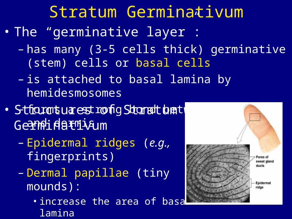
Stratum Germinativum• The “germinative layer”:
– has many (3-5 cells thick) germinative (stem) cells or basal cells
– is attached to basal lamina by hemidesmosomes
– forms a strong bond between epidermis and dermis
• Structures of Stratum Germinativum – Epidermal ridges (e.g.,
fingerprints) – Dermal papillae (tiny mounds):
• increase the area of basal lamina• strengthen attachment between
epidermis and dermis
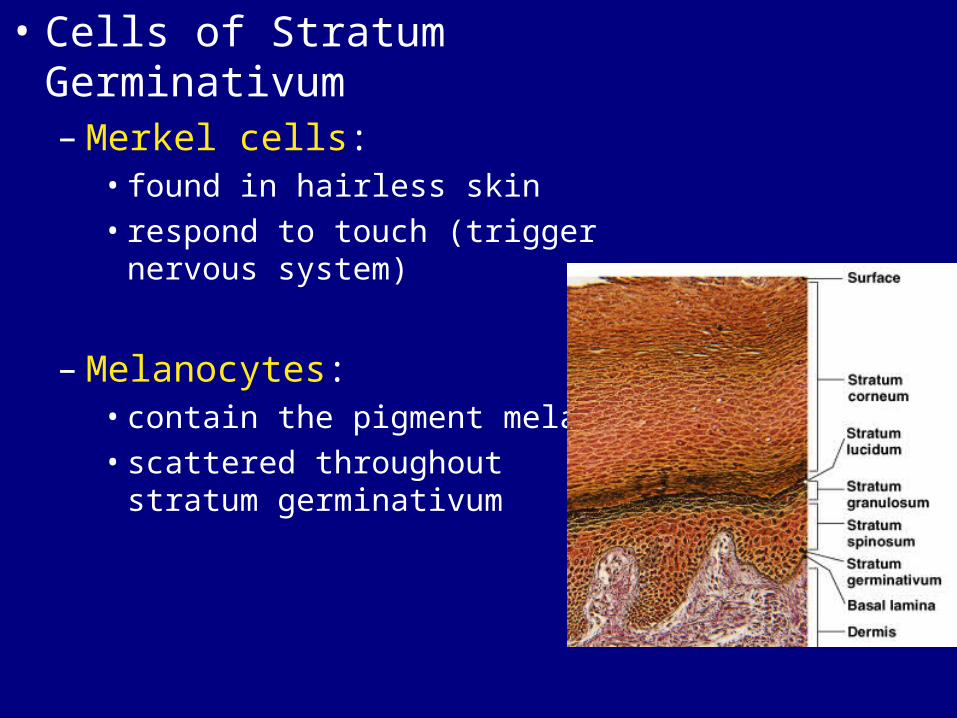
• Cells of Stratum Germinativum – Merkel cells:
• found in hairless skin • respond to touch (trigger
nervous system)
– Melanocytes:• contain the pigment melanin• scattered throughout stratum
germinativum
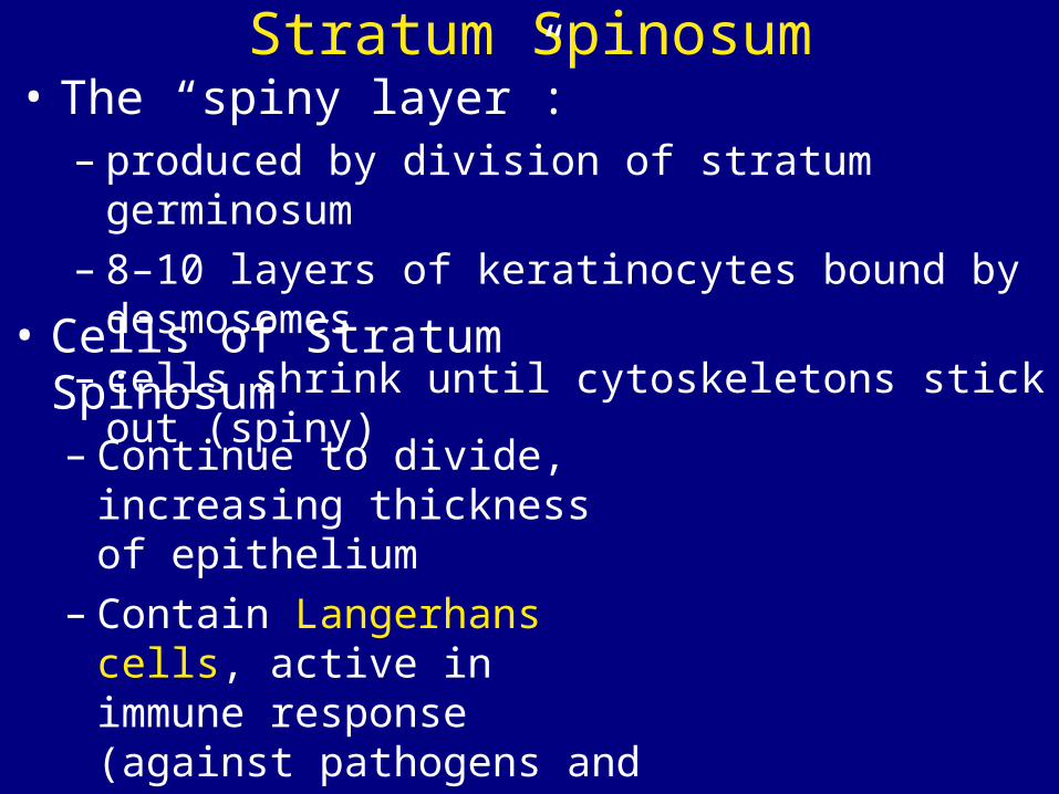
Stratum Spinosum• The “spiny layer”:
– produced by division of stratum germinosum– 8–10 layers of keratinocytes bound by
desmosomes– cells shrink until cytoskeletons stick out
(spiny)• Cells of Stratum
Spinosum – Continue to divide,
increasing thickness of epithelium
– Contain Langerhans cells, active in immune response (against pathogens and skin cancers)
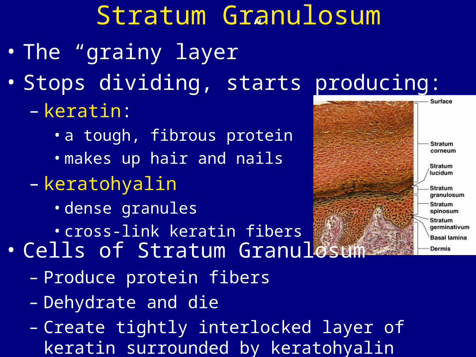
Stratum Granulosum• The “grainy layer” • Stops dividing, starts producing:
– keratin: • a tough, fibrous protein • makes up hair and nails
– keratohyalin • dense granules• cross-link keratin fibers
• Cells of Stratum Granulosum – Produce protein fibers– Dehydrate and die– Create tightly interlocked layer of keratin
surrounded by keratohyalin
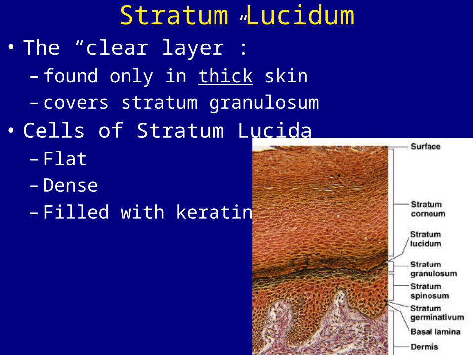
Stratum Lucidum• The “clear layer”:
– found only in thick skin– covers stratum granulosum
• Cells of Stratum Lucida – Flat– Dense– Filled with keratin
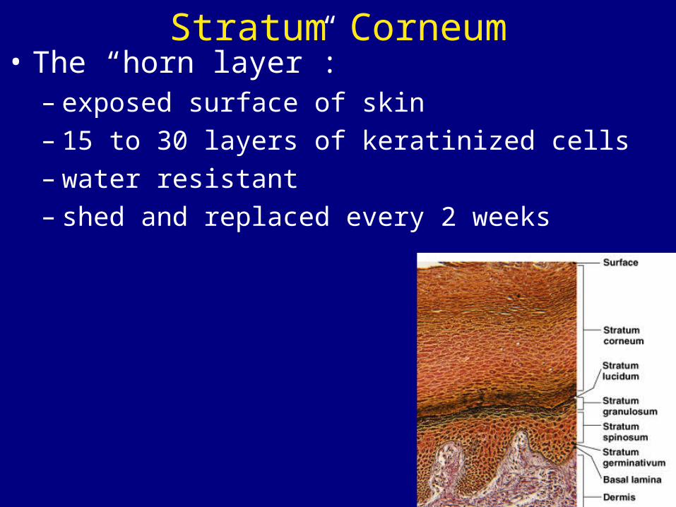
Stratum Corneum• The “horn layer”:
– exposed surface of skin – 15 to 30 layers of keratinized cells– water resistant– shed and replaced every 2 weeks
What are the structures & functions of the dermis?The Dermis
• Is located between epidermis & subcutaneous layer• Fxns to nourish epidermis and house accessory
organs of skin
• Has 2 components:– outer papillary layer – deep reticular layer
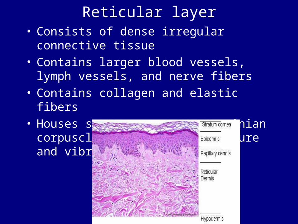
Reticular layer• Consists of dense irregular connective
tissue• Contains larger blood vessels, lymph
vessels, and nerve fibers• Contains collagen and elastic fibers• Houses sensory neurons: pacinian
corpuscle: detects deep pressure and vibration
The Papillary Layer
• Consists of areolar tissue
• Contains smaller capillaries, lymphatic vessels, and sensory neurons
• Sensory neurons: meissner’s corpuscle: detects ligh touch
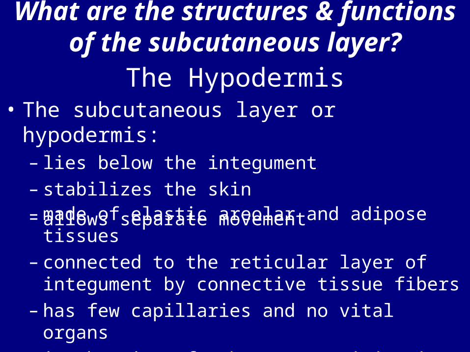
What are the structures & functions of the subcutaneous
layer?The Hypodermis
• The subcutaneous layer or hypodermis: – lies below the integument– stabilizes the skin
– allows separate movement – made of elastic areolar and adipose tissues– connected to the reticular layer of
integument by connective tissue fibers– has few capillaries and no vital organs– is the site of subcutaneous injections using
hypodermic needles
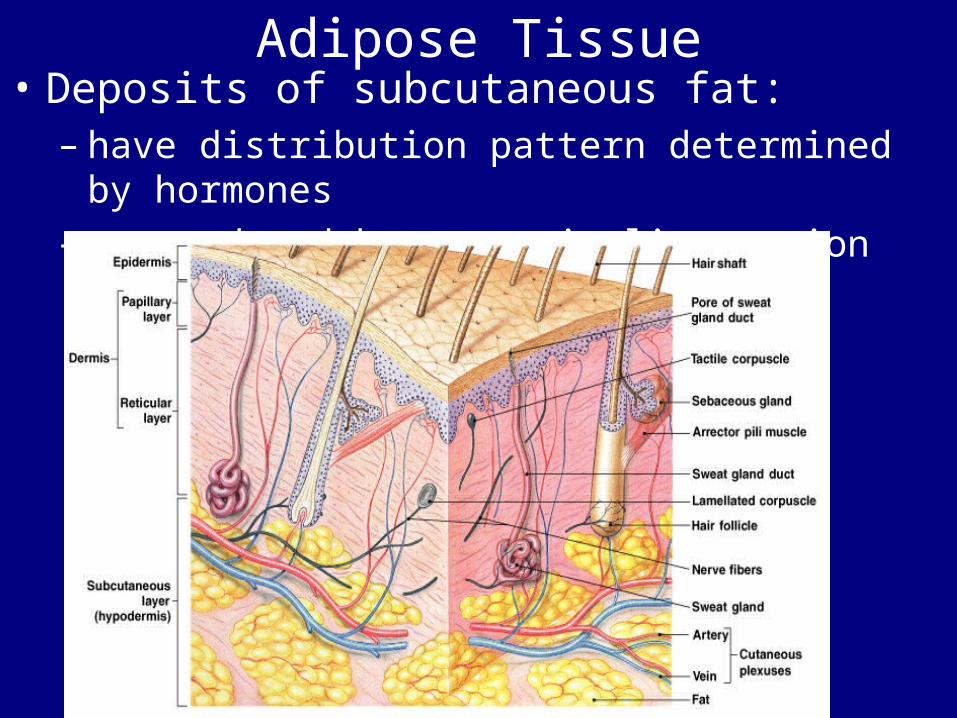
Adipose Tissue• Deposits of subcutaneous fat:
– have distribution pattern determined by hormones
– are reduced by cosmetic liposuction
Water Loss Through Skin• Dehydration results:
– from damage to stratum corneum, e.g., burns and blisters (insensible perspiration)
– from immersion in hypertonic solution, e.g., seawater (osmosis)
Water Gain Through Skin• Hydration: (skin is water resistant, not water proof)
– results from immersion in hypotonic solution, e.g., freshwater (osmosis)
– causes stretching and wrinkling skin
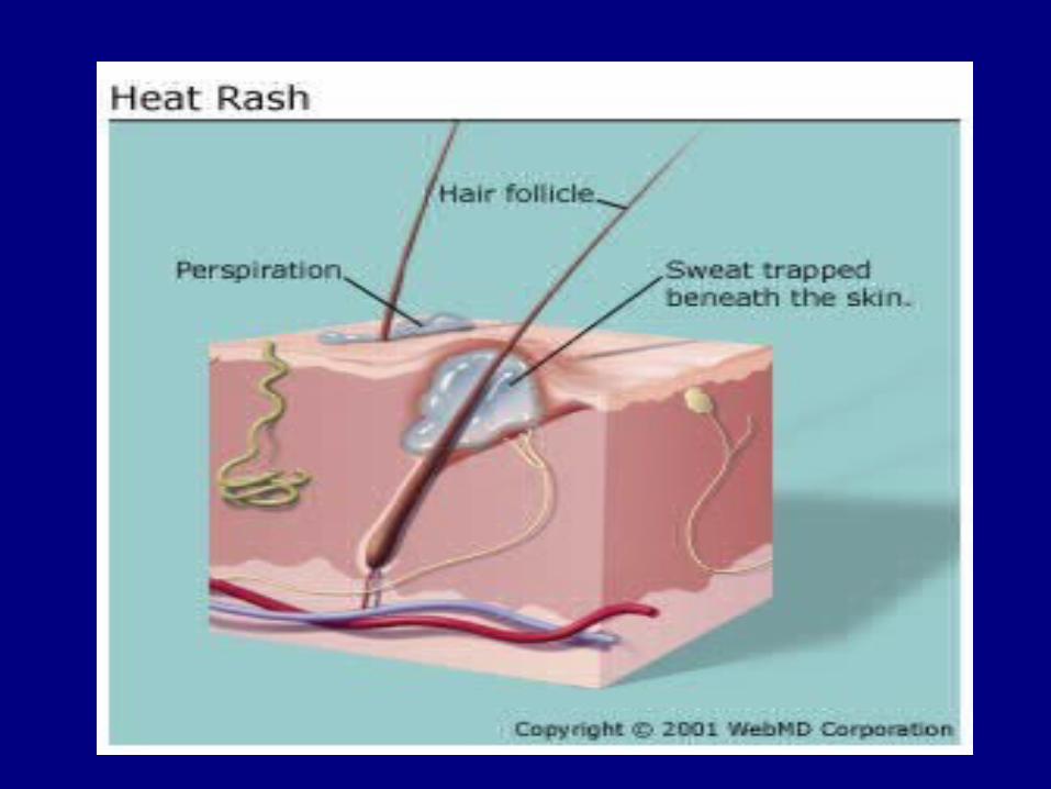
Perspiration• Insensible perspiration:
– interstitial fluid lost by evaporation through the stratum corneum
• Sensible perspiration: – water excreted by sweat glands
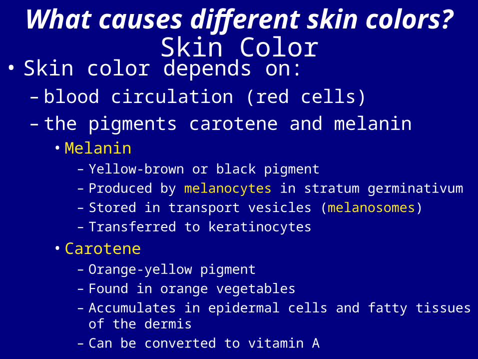
What causes different skin colors?Skin Color
• Skin color depends on:– blood circulation (red cells)– the pigments carotene and melanin
• Melanin– Yellow-brown or black pigment– Produced by melanocytes in stratum germinativum– Stored in transport vesicles (melanosomes)– Transferred to keratinocytes
• Carotene– Orange-yellow pigment– Found in orange vegetables– Accumulates in epidermal cells and fatty tissues of the
dermis– Can be converted to vitamin A
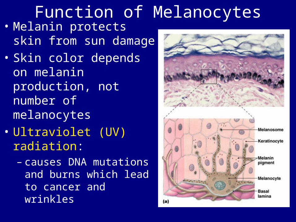
Function of Melanocytes• Melanin protects skin
from sun damage• Skin color depends on
melanin production, not number of melanocytes
• Ultraviolet (UV) radiation:– causes DNA mutations
and burns which lead to cancer and wrinkles
Capillaries and Skin Color• Oxygenated red blood contributes to skin color:
– blood vessels dilate from heat, skin reddens– blood flow decreases, skin pales
• Cyanosis– Bluish skin tint– Caused by severe reduction in blood flow or
oxygenation• Jaundice:
– buildup of bile produced by liver– yellow color
• Addison’s disease:– and other diseases of pituitary gland– skin darkening• Vitiglio:– loss of melanocytes– loss of color
Skin Damage• Sagging and wrinkles (reduced skin
elasticity) are caused by:– dehydration– age– hormonal changes– UV exposure
• Stretch Marks – Thickened tissue resulting from:
• excessive stretching of skin due to:– Pregnancy– weight gain
Vitamin D• Epidermal cells produce cholecalciferol
(vitamin D3):– in the presence of UV radiation
• Liver and kidneys convert vitamin D into calcitriol:– to aid absorption of calcium and phosphorus
• Insufficient vitamin D:– can cause rickets
Epidermal Growth Factor (EGF)• Is a powerful peptide growth factor
• Is produced by glands (salivary and duodenum)
• Is used in laboratories to grow skin graftsFunctions of EGF• Promotes division of germinative
cells• Accelerates keratin production• Stimulates epidermal repair• Stimulates glandular secretion
Dermatitis • An inflammation of the papillary layer• Caused by infection, radiation,
mechanical irritation, or chemicals (e.g., poison ivy)
• Characterized by itch or pain• Strong, due to collagen fibers• Elastic, due to elastic fibers• Flexible (skin turgor)
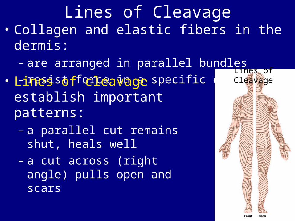
Lines of Cleavage• Collagen and elastic fibers in the dermis:
– are arranged in parallel bundles– resist force in a specific direction
• Lines of cleavage establish important patterns:– a parallel cut remains shut,
heals well– a cut across (right angle)
pulls open and scars
Lines of Cleavage
Dermal Circulation
Figure 5–8
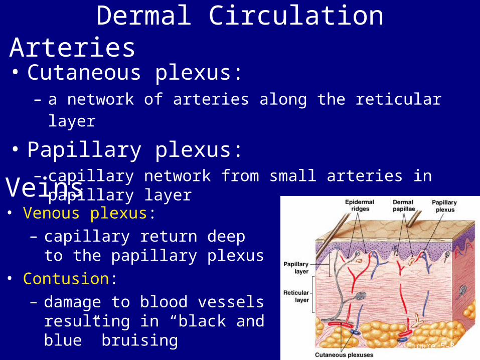
Arteries• Cutaneous plexus:
– a network of arteries along the reticular layer
• Papillary plexus: – capillary network from small arteries in papillary
layerVeins• Venous plexus:
– capillary return deep to the papillary plexus
• Contusion:– damage to blood vessels
resulting in “black and blue” bruising
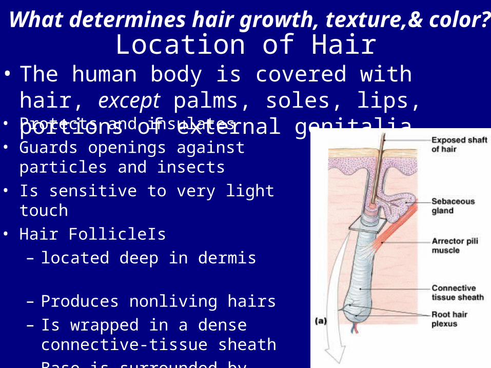
What determines hair growth, texture,& color?Location of Hair
• The human body is covered with hair, except palms, soles, lips, portions of external genitalia• Protects and insulates
• Guards openings against particles and insects
• Is sensitive to very light touch • Hair FollicleIs
– located deep in dermis– Produces nonliving hairs – Is wrapped in a dense
connective-tissue sheath– Base is surrounded by
sensory nerves (root hair plexus)
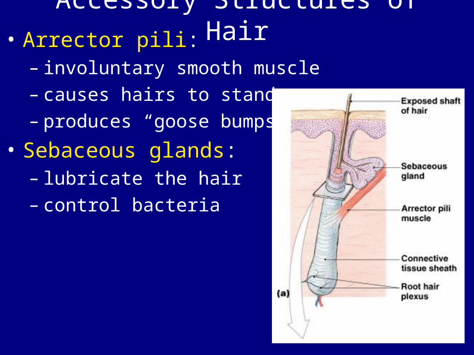
Accessory Structures of Hair• Arrector pili:
– involuntary smooth muscle – causes hairs to stand up– produces “goose bumps”
• Sebaceous glands: – lubricate the hair– control bacteria
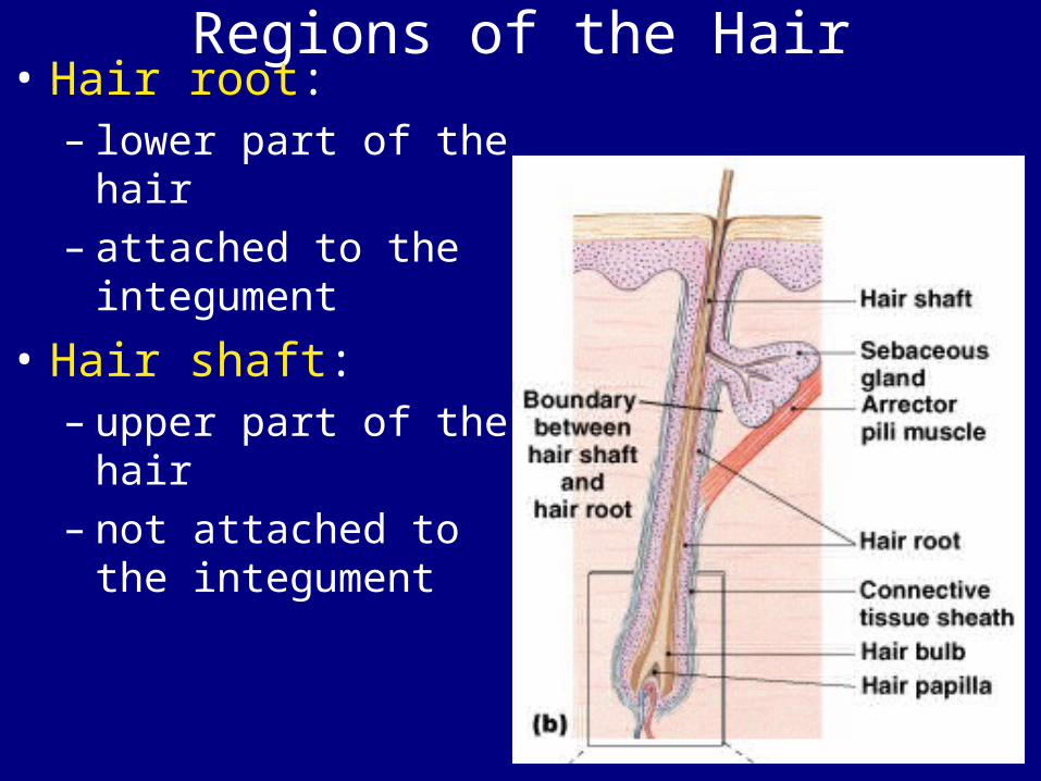
Regions of the Hair• Hair root:
– lower part of the hair
– attached to the integument
• Hair shaft:– upper part of the
hair– not attached to the
integument
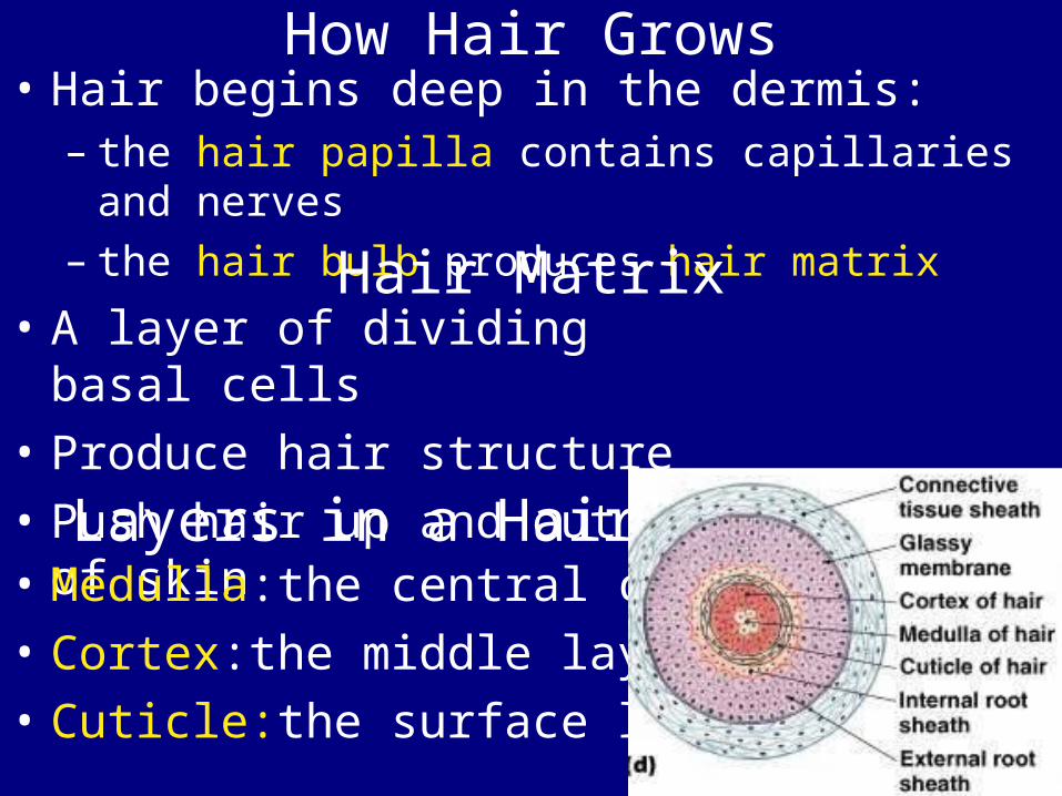
How Hair Grows• Hair begins deep in the dermis:
– the hair papilla contains capillaries and nerves
– the hair bulb produces hair matrixHair Matrix• A layer of dividing basal
cells• Produce hair structure• Push hair up and out of
skin Layers in a Hair
• Medulla:the central core • Cortex:the middle layer • Cuticle:the surface layer
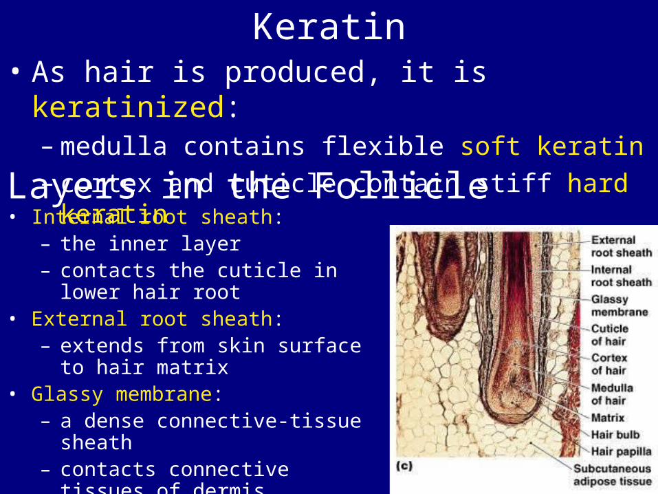
Keratin• As hair is produced, it is keratinized:
– medulla contains flexible soft keratin– cortex and cuticle contain stiff hard keratin
Layers in the Follicle• Internal root sheath:
– the inner layer– contacts the cuticle in lower
hair root• External root sheath:
– extends from skin surface to hair matrix
• Glassy membrane:– a dense connective-tissue
sheath– contacts connective tissues of
dermis
Hair Growth Cycle• Growing hair:
– is firmly attached to matrix
• Club hair:– is not growing– is attached to an inactive follicle
• New hair growth cycle:– follicle becomes active– produces new hair– club hair is shed
Types of Hairs• Vellus hairs:
– soft, fine – cover body surface
• Terminal hairs: – heavy, pigmented– head and eyebrows– other parts of body after puberty
Hair Color• Produced by melanocytes at the hair
papilla• Determined by genes
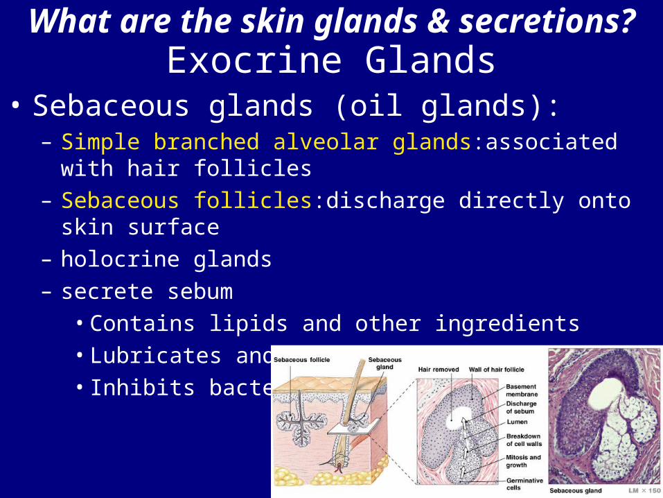
What are the skin glands & secretions?Exocrine Glands
• Sebaceous glands (oil glands):– Simple branched alveolar glands:associated with hair
follicles – Sebaceous follicles:discharge directly onto skin
surface– holocrine glands– secrete sebum
• Contains lipids and other ingredients• Lubricates and protects the epidermis• Inhibits bacteria
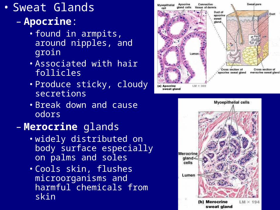
• Sweat Glands– Apocrine:
• found in armpits, around nipples, and groin
• Associated with hair follicles
• Produce sticky, cloudy secretions
• Break down and cause odors
– Merocrine glands• widely distributed on
body surface especially on palms and soles
• Cools skin, flushes microorganisms and harmful chemicals from skin
Other Integumentary Glands • Mammary glands:
– produce milk
• Ceruminous glands:– protect the eardrum– produce cerumen (earwax)
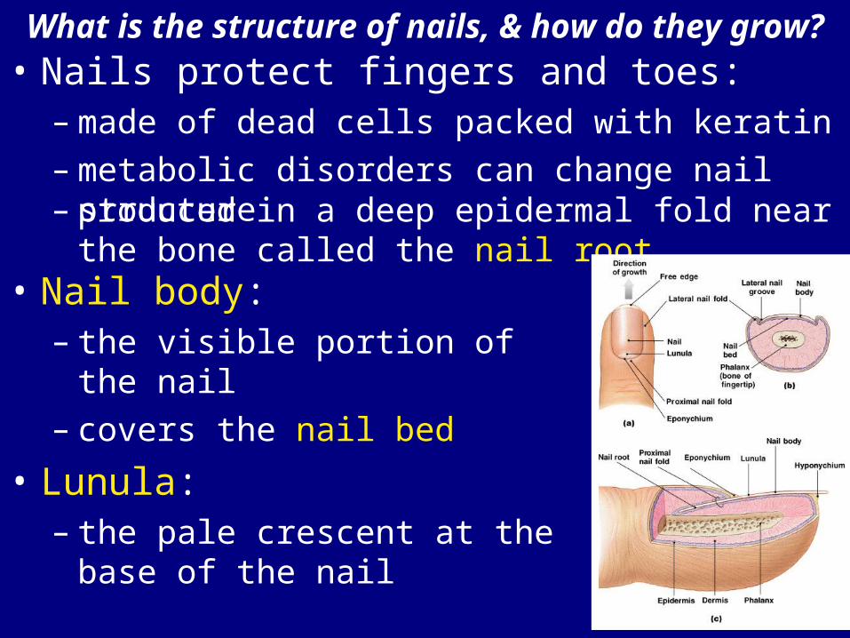
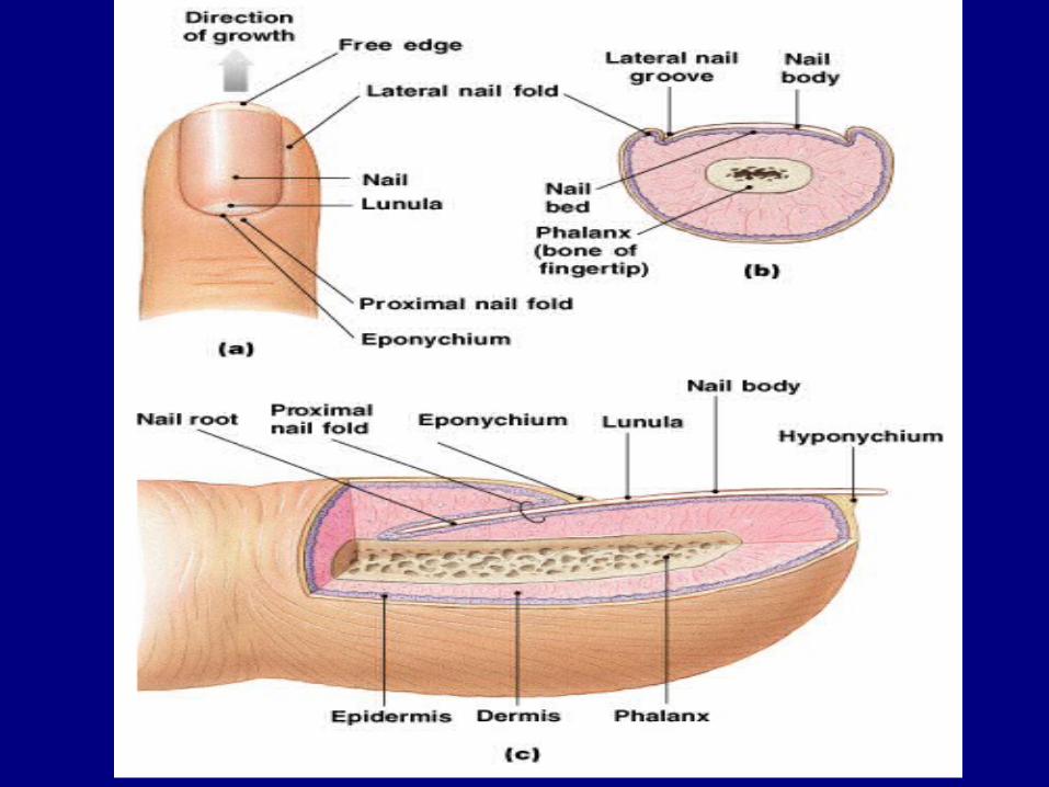
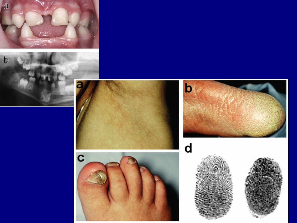
What is the structure of nails, & how do they grow?
• Nails protect fingers and toes:– made of dead cells packed with keratin – metabolic disorders can change nail structure– produced in a deep epidermal fold near the
bone called the nail root• Nail body:
– the visible portion of the nail– covers the nail bed
• Lunula:– the pale crescent at the base
of the nail