Embed Size (px)
Citation preview

PAPER www.rsc.org/loc | Lab on a Chip
Integrated microfluidic tmRNA purification and real-time NASBA devicefor molecular diagnostics†‡
Ivan K. Dimov,ab Jose L. Garcia-Cordero,a Justin O’Grady,c Claus R. Poulsen,a Caroline Viguier,a
Lorcan Kent,a Paul Daly,a Bryan Lincoln,a Majella Maher,c Richard O’Kennedy,a Terry J. Smith,c
Antonio J. Riccoa and Luke P. Lee*ad
Received 21st July 2008, Accepted 17th September 2008
First published as an Advance Article on the web 24th October 2008
DOI: 10.1039/b812515e
We demonstrate the first integrated microfluidic tmRNA purification and nucleic acid sequence-based
amplification (NASBA) device incorporating real-time detection. The real-time amplification and
detection step produces pathogen-specific response in < 3 min from the chip-purified RNA from 100
lysed bacteria. On-chip RNA purification uses a new silica bead immobilization method. On-chip
amplification uses custom-designed high-selectivity primers and real-time detection uses molecular
beacon fluorescent probe technology; both are integrated on-chip with NASBA. Present in all bacteria,
tmRNA (10Sa RNA) includes organism-specific identification sequences, exhibits unusually high
stability relative to mRNA, and has high copy number per organism; the latter two factors improve the
limit of detection, accelerate time-to-positive response, and suit this approach ideally to the detection of
small numbers of bacteria. Device efficacy was demonstrated by integrated on-chip purification,
amplification, and real-time detection of 100 E. coli bacteria in 100 mL of crude lysate in under 30 min
for the entire process.
Introduction
Enzymatic in-vitro amplification of nucleic acids has revolu-
tionised life science research and spurred numerous advances in
biotechnology and other disciplines.1–3 Nucleic-acid amplifica-
tion methods and DNA sequence-specific detection now enable
precise identification of many pathogens that previously could
not be unambiguously determined with traditional laboratory
techniques such as culture enrichment, plating, and visual
microscopy. Further advantages include precise quantification of
target pathogens and real-time detection of amplified products.
Nevertheless, conventional detection assays based on nucleic
acid amplification provide at best only an order of magnitude
lower limits of detection than other conventional methods.4 In
addition, manual handling and multiple liquid transfers can
introduce contamination, which leads to large errors.
A first step toward improving the overall genetic analysis
process is to separate, concentrate, and purify target pathogens
aBiomedical Diagnostics Institute, National Centre for Sensor Research,Research & Engineering Building, Dublin City University, Glasnevin,Dublin 9, IrelandbDepartment of Biomedical Engineering, Universidad de Valparaıso, ChilecBiomedical Diagnostics Institute, National Centre for BiomedicalEngineering Science, National University of Ireland, GalwaydBiomolecular Nanotechnology Center, Berkeley Sensor & ActuatorCenter, Department of Bioengineering, University of California,Berkeley, CA, USA. E-mail: [email protected]
† Part of a special issue on Point-of-care Microfluidic Diagnostics; GuestEditors - Professor Kricka and Professor Sia.
‡ Electronic supplementary information (ESI) available: Silica beadimmobilization method; Comparison: Microfluidic RNA purificationand real time NASBA protocol versus conventional RNA purificationand real time NASBA protocol. See DOI: 10.1039/b812515e
This journal is ª The Royal Society of Chemistry 2008
from raw samples. Next, the pathogens are lysed and, depending
on the sample matrix, volume, and pathogen concentration, an
additional step may be necessary to concentrate and purify the
nucleic acids. Matrix residues can inhibit amplification, reducing
assay efficiency. DNA concentration and purification can
enhance the limit of detection of nucleic acid amplification by two
orders of magnitude.4 Liquid–liquid extraction and solid-phase
extraction are two techniques to extract and purify nucleic acids.3
Microfluidic lab-on-a-chip technology promises to integrate
entire analytical/clinical chemistry processes on monolithic plat-
forms.5–7 Compared to conventional laboratory methods, inte-
grated microfluidic platforms offer potential advantages of lower
cost, higher speed, smaller sample and reagent volumes, and
automation of all processes from sample preparation to analytical
result: the ‘‘sample-to-answer’’ concept.8 For some bioanalytical
measurements, however, the most important consequences of
successfully implementing microfluidic lab-on-a-chip technology
will be enhanced assay reproducibility and more quantitative
results9 relative to classical analytical procedures.
Miniaturizing in-vitro amplification of nucleic acids can enable
the integration of multiple functional fluidic modules, potentially
enabling the analysis of smaller sample volumes, coupled with
increased reproducibility, more accurate quantification, and
faster analysis. While the number of micro-PCR-related publi-
cations has grown rapidly,2 many of the reported devices are
‘‘stand-alone’’ structures that replace only the roles of the
conventional thermocycler and sample tube. To more fully
realise the potential of microfluidic systems,10 multiple functions
can be combined on one chip. PCR has been integrated in a single
microdevice with pre-amplification modules, such as nucleic acid
extraction11 and purification plus pre-concentration,12 or with
Lab Chip, 2008, 8, 2071–2078 | 2071
post-amplification analytical modules, such as capillary electro-
phoresis (CE)13,14 and DNA microarrays.15 Fully integrated
microfluidic PCR devices with pre- and post-amplification
modules have also been reported,15–17 and portable versions of
fully integrated microfluidic PCR devices with all necessary
supporting systems, such as pumps and optical detection, have
been achieved.18,19
Nucleic acid sequence-based amplification (NASBA), a tran-
scription-based RNA amplification system, is more sensitive,
rapid, and ‘‘user-friendly’’ than PCR. Initially developed by
Compton in 1991,20 NASBA involves the simultaneous action of
three enzymes (avian myeloblastosis virus reverse transcriptase,
RNase H, and T7 RNA polymerase) that can produce more than
109 copies under isothermal (41 �C) conditions in 90 min. A range
of nucleic acid types, including mRNA, rRNA, tmRNA, and
ssDNA, as well as nucleic acids from virus particles, can be ana-
lysed with NASBA, enabling a range of diagnostics, along with
gene expression and cell viability measurements.21 In bacteria,
tmRNA is universally present and is encoded by the ssrA gene
(10Sa).22 It is significantly more stable than mRNA, coming close
to DNA in robustness. This high-copy-number target (RiboSEQ)
contains conserved and variable sequence regions, making it ideal
for application to microbial species identification in molecular
diagnostics.23–25 The isothermal, low-temperature NASBA process
offers multiple advantages relative to the thermal cycling between
three temperatures that is required by PCR: it simplifies the
thermal design of microfluidic chip, it expands the range of
materials and bonding methods that can be used, it increases the
number of (thermally sensitive) reagents (e.g., enzymes) or
components (e.g., certain valves) that can be included in close
proximity on the same chip, and it reduces both the complexity of
the instrument and power consumption. A further advantage of
NASBA, relative to RT-PCR, is direct amplification of RNA
templates without a separate reverse transcription step.
Real-time quantitative NASBA21 utilizes molecular beacon
probes26–28 that specifically hybridize to the target sequence
generated during amplification. With suitable optical excitation,
the hybridized beacons produce a fluorescent signal proportional
to the number of copies of the target sequence produced by the
NASBA process, simplifying the analytical procedure and
reducing the complexity of the microfluidic chip relative to other
detection approaches. The slope of the fluorescence-vs.-time
curve can yield a quantitative signal in under 3 minutes with
sufficient target copy numbers.
The first successful microfluidic NASBA chip, demonstrated
by Gulliksen et al.29 (2004) using silicon and glass chambers,
included real-time detection. They demonstrated an improve-
ment to the first design in 2005,30 showing that microfluidic
real-time NASBA performed as well as conventional off-chip
(in-tube) real-time NASBA.
Key aspects of many sample-to-answer systems include the
sample purification, amplification, and detection components. In
the case of an RNA analysis system, these elements pose signif-
icant technical challenges related to their miniaturisation and
integration.
Solid-phase extraction (SPE) is a sample purification tech-
nique that can be transferred to microfluidic systems.3 Various
implementations of SPE in microfluidics have been reported in
the literature, but the most efficient microfluidic-based SPE
2072 | Lab Chip, 2008, 8, 2071–2078
requires complex or in-situ chemistry-based fabrication.3 There is
a need for simpler fabrication techniques to integrate SPE more
easily with microfluidic systems.
To our knowledge, there are no prior reports of integrating
RNA purification, NASBA, and real-time detection all on
a single chip. Off-chip handling of RNA risks sample degrada-
tion, which can compromise assay accuracy, reliability, and
reproducibility. To avoid this, the sample preparation, amplifi-
cation, and detection steps can be integrated in a single micro-
fluidic device. For example, Legendre et al.31 showed that a single
glass device could extract and PCR amplify DNA from complex
lysates of blood, nasal swab, and semen samples; this design was
improved by Easley et al.17 by adding an electrophoresis channel
for PCR amplicon separation and detection. To minimize
contamination, different regions of the device were separated by
5 pressure-driven microfluidic valves. With the integration of
DNA extraction, PCR amplification, and microchip CE, Easley
et al.17 demonstrated a genetic analysis system with sample-to–
answer capability. However, it is desirable to avoid the
complexity of many unnecessary modules such as thermal cycler
for PCR or microfluidic CE, and find a solution for effective
nucleic acid-based diagnostics.
In this article, we report the monolithic integration of
a microfluidic sample-to-answer tmRNA analysis system that
includes RNA capture and purification, NASBA, and real-time
detection. A novel clog-free bead immobilisation method is used
to construct the RNA capture and purification module. The
device design also allows for future integration of cell lysis and
automated fluid-control functions.
Materials and methods
Design of integrated microfluidic NASABA chip
The integrated microfluidic NASBA chip, Fig. 1, is based on
microchannels and microchambers, with distinct functional
domains: a silica bead-bed RNA purification chamber (RPC,
volume 0.25 mL), and a NASBA chamber (NC, volume 2 mL).
The remaining channels and chambers have been implemented
for the future integration of on-chip chemical lysis. The height of
the microchannels and microchambers, 80 mm, was selected
because it is easily achievable with SU-8 2100 photoresist. The
RNA purification chamber enables capture of RNA present in
the sample by selective adsorption in silica, followed by removal
of the remaining crude cell lysate, which includes NASBA
inhibitors, by washing.
Detection of amplified tmRNA target from NASBA occurs in
real-time via molecular beacons whose fluorescence is
unquenched when hybridised to complementary RNA ampli-
cons. The device is characterized using a fluorescence microscope
to measure the change in fluorescence from the NASBA chamber
as a function of time. The device is mirrored (Fig. 1A) to allow
for 2 separate reactions with the same reagents but different
samples, nominally to implement control reactions. The rela-
tively large number of inlets (7) and outlets (4) is a consequence
of this device being designed as a subcomponent of a larger
system we are developing, where a number of the fluidic I/O
connections will be replaced with microchannel connections to
other components and/or on-chip reagent or waste reservoirs.
This journal is ª The Royal Society of Chemistry 2008

Fig. 1 Integrated microfluidic RNA purification chamber and real-time NASBA device. (A) Photograph of the device. The microfluidic architecture is
mirrored to allow for 2 separate reactions with the same reagents, but different samples, to incorporate controls. (B) Single device architecture showing
the distinct functional microfluidic modules: RNA purification chamber (RPC) and real-time NASBA chamber. The remaining channels and chambers
have been included for future integration of on-chip lysis. All channels and chambers are 80 mm high. Scale bar is 1 mm.
Fig. 2 Method for silica bead immobilisation on PDMS surface. (A)
Before loading the beads, all ports are sealed except for the Input and
Waste Output. (B) 3 mL of plain silica bead solution flows into the input,
left to dry, and exposed to UV-ozone for bonding. (C) Unbound beads
are washed away with dH2O, leaving a (D) layer of silica beads bonded to
the walls of the RPC (see ESI‡). (E–F) Bright-field micrographs of the
immobilised 10 mm silica beads on the PDMS walls of the RNA purifi-
cation chamber.
Microfabrication
PDMS is widely utilized for microfluidic devices due to the
ease of fabricating relatively complex structures,7 its general
biocompatibility, and the ease of attaching (sealing) it to glass,
silicon wafers, or a second PDMS structure. Microfluidic
channels were fabricated using standard soft lithography
replica molding techniques.32 A mold was created through
a single-layer process using negative photoresist, SU8-2100
(Microchem, USA), which was spun onto a clean silicon wafer
using a spinner (P6700 Specialty Coating Systems, Inc., USA).
The resist (5 mL) was spread onto the wafer at 500 rpm for 10 s,
and the rotation rate was then ramped at an acceleration of
300 rpm/s to 2,500 rpm, at which rate the sample was spun for
30 s to form an 80 mm layer. The wafer was then soft baked at
65 �C for 5 min and 95 �C for 30 min, then UV-exposed for 10 s
at 9.5 mW/cm2 using a Karl-Suss KSM MJB-55W mask
aligner. The wafer was post-exposure baked for 5 min at 65 �C
and 12 min at 95 �C, allowed to cool to room temperature,
developed in Microposit EC Solvent (Chestech Ltd., UK)
developer for 4 min, and finally blown dry with nitrogen.
PDMS (Sylgard 184, Dow Corning) was prepared according to
the instructions of the manufacturer, degassed in a vacuum
chamber for 30 min, then poured on the SU8 mold and cured
in a 60 �C oven for 10 h. The PDMS was then carefully peeled
off the mold. Fluid inlets and outlets were punched with a 1
mm outer diameter flat-tip needle for tube connections. Both
a 25 � 50 � 0.4 mm glass cover slide (VWR International Inc.,
USA) and the PDMS structures were treated with UV ozone
(PSD-UV, Novascan Technologies, Inc., Iowa, USA) for 10
min before bonding for 10 h at 90 �C.
Beads for RNA purification were immobilised within the RPC
by loading 3 mL of a 68% (bead: deionised H2O volume) solution
of 10 mm plain silica beads (PSi-10.0, G. Kisker Gbr., Germany)
into the input port (IP) (Fig. 2A), with all other ports on the chip
sealed except for the waste output (WO). The bead solution was
This journal is ª The Royal Society of Chemistry 2008
left to dry at room temperature for 3 hr, which packs the beads
into the extraction chamber. After the beads dried in place, they
were immobilised via bonding to the PDMS walls of the
Lab Chip, 2008, 8, 2071–2078 | 2073
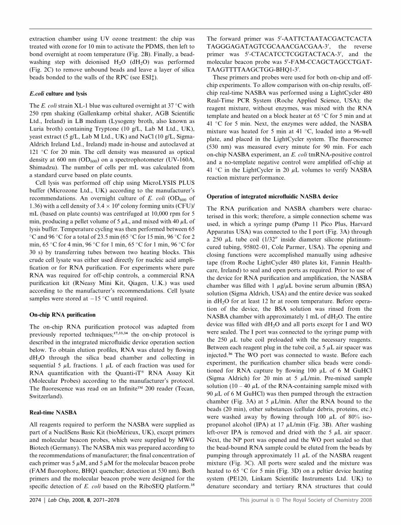
extraction chamber using UV ozone treatment: the chip was
treated with ozone for 10 min to activate the PDMS, then left to
bond overnight at room temperature (Fig. 2B). Finally, a bead-
washing step with deionised H2O (dH2O) was performed
(Fig. 2C) to remove unbound beads and leave a layer of silica
beads bonded to the walls of the RPC (see ESI‡).
E.coli culture and lysis
The E. coli strain XL-1 blue was cultured overnight at 37 �C with
250 rpm shaking (Gallenkamp orbital shaker, AGB Scientific
Ltd., Ireland) in LB medium (Lysogeny broth, also known as
Luria broth) containing Tryptone (10 g/L, Lab M Ltd., UK),
yeast extract (5 g/L, Lab M Ltd., UK) and NaCl (10 g/L, Sigma-
Aldrich Ireland Ltd., Ireland) made in-house and autoclaved at
121 �C for 20 min. The cell density was measured as optical
density at 600 nm (OD600) on a spectrophotometer (UV-160A,
Shimadzu). The number of cells per mL was calculated from
a standard curve based on plate counts.
Cell lysis was performed off chip using MicroLYSIS PLUS
buffer (Microzone Ltd., UK) according to the manufacturer’s
recommendations. An overnight culture of E. coli (OD600 of
1.36) with a cell density of 3.4 � 108 colony forming units (CFU)/
mL (based on plate counts) was centrifuged at 10,000 rpm for 5
min, producing a pellet volume of 5 mL, and mixed with 40 mL of
lysis buffer. Temperature cycling was then performed between 65�C and 96 �C for a total of 23.5 min (65 �C for 15 min, 96 �C for 2
min, 65 �C for 4 min, 96 �C for 1 min, 65 �C for 1 min, 96 �C for
30 s) by transferring tubes between two heating blocks. This
crude cell lysate was either used directly for nucleic acid ampli-
fication or for RNA purification. For experiments where pure
RNA was required for off-chip controls, a commercial RNA
purification kit (RNeasy Mini Kit, Qiagen, U.K.) was used
according to the manufacturer’s recommendations. Cell lysate
samples were stored at �15 �C until required.
On-chip RNA purification
The on-chip RNA purification protocol was adapted from
previously reported techniques;17,33,34 the on-chip protocol is
described in the integrated microfluidic device operation section
below. To obtain elution profiles, RNA was eluted by flowing
dH2O through the silica bead chamber and collecting in
sequential 5 mL fractions. 1 mL of each fraction was used for
RNA quantification with the Quanti-iT� RNA Assay Kit
(Molecular Probes) according to the manufacturer’s protocol.
The fluorescence was read on an Infinite� 200 reader (Tecan,
Switzerland).
Real-time NASBA
All reagents required to perform the NASBA were supplied as
part of a NucliSens Basic Kit (bioMerieux, UK), except primers
and molecular beacon probes, which were supplied by MWG
Biotech (Germany). The NASBA mix was prepared according to
the recommendations of manufacturer; the final concentration of
each primer was 5 mM, and 5 mM for the molecular beacon probe
(FAM fluorophore, BHQ1 quencher; detection at 530 nm). Both
primers and the molecular beacon probe were designed for the
specific detection of E. coli based on the RiboSEQ platform.35
2074 | Lab Chip, 2008, 8, 2071–2078
The forward primer was 50-AATTCTAATACGACTCACTA
TAGGGAGATAGTCGCAAACGACGAA-30, the reverse
primer was 50-CTACATCCTCGGTACTACA-30, and the
molecular beacon probe was 50-FAM-CCAGCTAGCCTGAT-
TAAGTTTTAAGCTGG-BHQ1-30.
These primers and probes were used for both on-chip and off-
chip experiments. To allow comparison with on-chip results, off-
chip real-time NASBA was performed using a LightCycler 480
Real-Time PCR System (Roche Applied Science, USA); the
reagent mixture, without enzymes, was mixed with the RNA
template and heated on a block heater at 65 �C for 5 min and at
41 �C for 5 min. Next, the enzymes were added, the NASBA
mixture was heated for 5 min at 41 �C, loaded into a 96-well
plate, and placed in the LightCycler system. The fluorescence
(530 nm) was measured every minute for 90 min. For each
on-chip NASBA experiment, an E. coli tmRNA-positive control
and a no-template negative control were amplified off-chip at
41 �C in the LightCycler in 20 mL volumes to verify NASBA
reaction mixture performance.
Operation of integrated microfluidic NASBA device
The RNA purification and NASBA chambers were charac-
terised in this work; therefore, a simple connection scheme was
used, in which a syringe pump (Pump 11 Pico Plus, Harvard
Apparatus USA) was connected to the I port (Fig. 3A) through
a 250 mL tube coil (1/3200 inside diameter silicone platinum-
cured tubing, 95802–01, Cole Parmer, USA). The opening and
closing functions were accomplished manually using adhesive
tape (from Roche LightCycler 480 plates kit, Fannin Health-
care, Ireland) to seal and open ports as required. Prior to use of
the device for RNA purification and amplification, the NASBA
chamber was filled with 1 mg/mL bovine serum albumin (BSA)
solution (Sigma Aldrich, USA) and the entire device was soaked
in dH2O for at least 12 hr at room temperature. Before opera-
tion of the device, the BSA solution was rinsed from the
NASBA chamber with approximately 1 mL of dH2O. The entire
device was filled with dH2O and all ports except for I and WO
were sealed. The I port was connected to the syringe pump with
the 250 mL tube coil preloaded with the necessary reagents.
Between each reagent plug in the tube coil, a 5 mL air spacer was
injected.36 The WO port was connected to waste. Before each
experiment, the purification chamber silica beads were condi-
tioned for RNA capture by flowing 100 mL of 6 M GuHCl
(Sigma Aldrich) for 20 min at 5 mL/min. Pre-mixed sample
solution (10 – 40 mL of the RNA-containing sample mixed with
90 mL of 6 M GuHCl) was then pumped through the extraction
chamber (Fig. 3A) at 5 mL/min. After the RNA bound to the
beads (20 min), other substances (cellular debris, proteins, etc.)
were washed away by flowing through 100 mL of 80% iso-
propanol alcohol (IPA) at 17 mL/min (Fig. 3B). After washing
left-over IPA is removed and dried with the 5 mL air spacer.
Next, the NP port was opened and the WO port sealed so that
the bead-bound RNA sample could be eluted from the beads by
pumping through approximately 11 mL of the NASBA reagent
mixture (Fig. 3C). All ports were sealed and the mixture was
heated to 65 �C for 5 min (Fig. 3D) on a peltier device heating
system (PE120, Linkam Scientific Instruments Ltd. UK) to
denature secondary and tertiary RNA structures that could
This journal is ª The Royal Society of Chemistry 2008
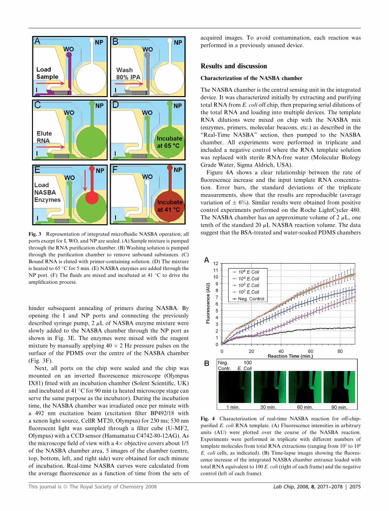
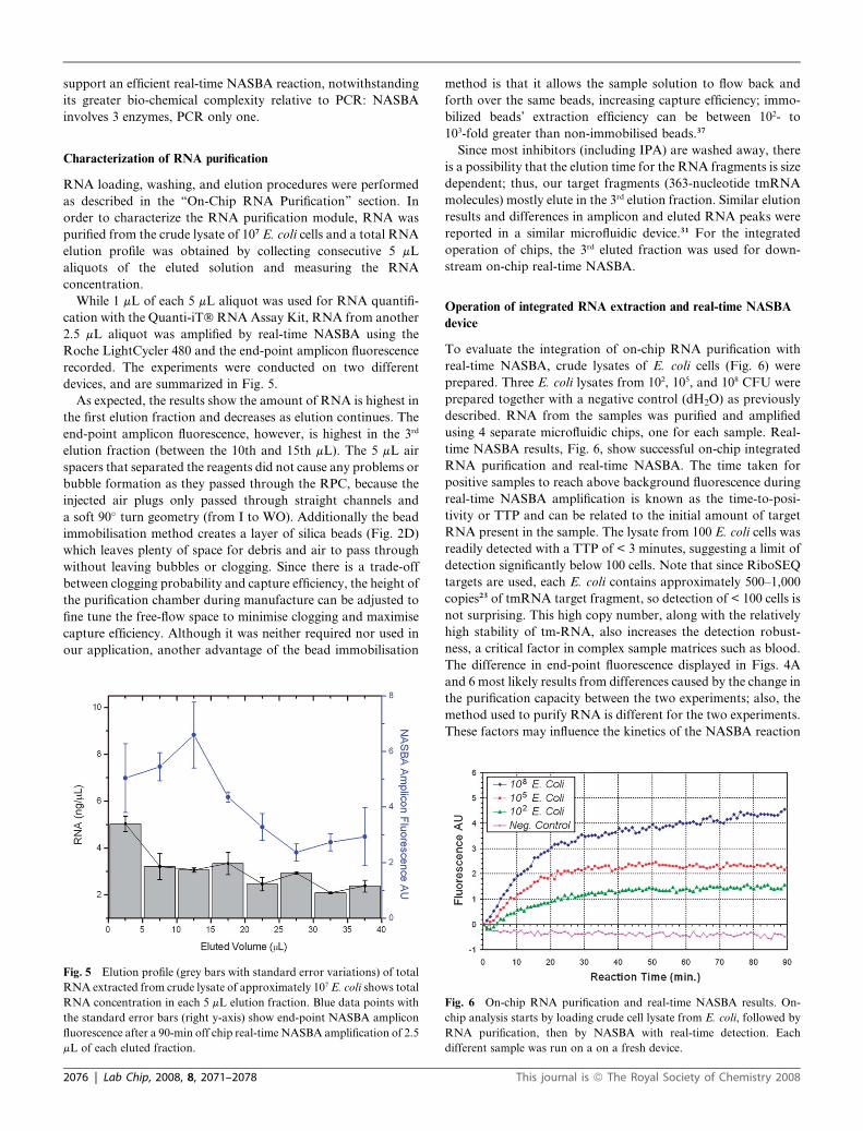
Fig. 4 Characterization of real-time NASBA reaction for off-chip-
purified E. coli RNA template. (A) Fluorescence intensities in arbitrary
units (AU) were plotted over the course of the NASBA reaction.
Experiments were performed in triplicate with different numbers of
template molecules from total RNA extractions (ranging from 102 to 106
E. coli cells, as indicated). (B) Time-lapse images showing the fluores-
cence increase of the integrated NASBA chamber entrance loaded with
total RNA equivalent to 100 E. coli (right of each frame) and the negative
control (left of each frame).
Fig. 3 Representation of integrated microfluidic NASBA operation; all
ports except for I, WO, and NP are sealed. (A) Sample mixture is pumped
through the RNA purification chamber. (B) Washing solution is pumped
through the purification chamber to remove unbound substances. (C)
Bound RNA is eluted with primer-containing solution. (D) The mixture
is heated to 65 �C for 5 min. (E) NASBA enzymes are added through the
NP port. (F) The fluids are mixed and incubated at 41 �C to drive the
amplification process.
hinder subsequent annealing of primers during NASBA. By
opening the I and NP ports and connecting the previously
described syringe pump, 2 mL of NASBA enzyme mixture were
slowly added to the NASBA chamber through the NP port as
shown in Fig. 3E. The enzymes were mixed with the reagent
mixture by manually applying 40 � 2 Hz pressure pulses on the
surface of the PDMS over the centre of the NASBA chamber
(Fig. 3F).
Next, all ports on the chip were sealed and the chip was
mounted on an inverted fluorescence microscope (Olympus
IX81) fitted with an incubation chamber (Solent Scientific, UK)
and incubated at 41 �C for 90 min (a heated microscope stage can
serve the same purpose as the incubator). During the incubation
time, the NASBA chamber was irradiated once per minute with
a 492 nm excitation beam (excitation filter BP492/18 with
a xenon light source, CellR MT20, Olympus) for 230 ms; 530 nm
fluorescent light was sampled through a filter cube (U-MF2,
Olympus) with a CCD sensor (Hamamatsu C4742-80-12AG). As
the microscope field of view with a 4� objective covers about 1/5
of the NASBA chamber area, 5 images of the chamber (centre,
top, bottom, left, and right side) were obtained for each minute
of incubation. Real-time NASBA curves were calculated from
the average fluorescence as a function of time from the sets of
This journal is ª The Royal Society of Chemistry 2008
acquired images. To avoid contamination, each reaction was
performed in a previously unused device.
Results and discussion
Characterization of the NASBA chamber
The NASBA chamber is the central sensing unit in the integrated
device. It was characterized initially by extracting and purifying
total RNA from E. coli off chip, then preparing serial dilutions of
the total RNA and loading into multiple devices. The template
RNA dilutions were mixed on chip with the NASBA mix
(enzymes, primers, molecular beacons, etc.) as described in the
‘‘Real-Time NASBA’’ section, then pumped to the NASBA
chamber. All experiments were performed in triplicate and
included a negative control where the RNA template solution
was replaced with sterile RNA-free water (Molecular Biology
Grade Water, Sigma Aldrich, USA).
Figure 4A shows a clear relationship between the rate of
fluorescence increase and the input template RNA concentra-
tion. Error bars, the standard deviations of the triplicate
measurements, show that the results are reproducible (average
variation of � 6%). Similar results were obtained from positive
control experiments performed on the Roche LightCycler 480.
The NASBA chamber has an approximate volume of 2 mL, one
tenth of the standard 20 mL NASBA reaction volume. The data
suggest that the BSA-treated and water-soaked PDMS chambers
Lab Chip, 2008, 8, 2071–2078 | 2075
support an efficient real-time NASBA reaction, notwithstanding
its greater bio-chemical complexity relative to PCR: NASBA
involves 3 enzymes, PCR only one.
Characterization of RNA purification
RNA loading, washing, and elution procedures were performed
as described in the ‘‘On-Chip RNA Purification’’ section. In
order to characterize the RNA purification module, RNA was
purified from the crude lysate of 107 E. coli cells and a total RNA
elution profile was obtained by collecting consecutive 5 mL
aliquots of the eluted solution and measuring the RNA
concentration.
While 1 mL of each 5 mL aliquot was used for RNA quantifi-
cation with the Quanti-iT� RNA Assay Kit, RNA from another
2.5 mL aliquot was amplified by real-time NASBA using the
Roche LightCycler 480 and the end-point amplicon fluorescence
recorded. The experiments were conducted on two different
devices, and are summarized in Fig. 5.
As expected, the results show the amount of RNA is highest in
the first elution fraction and decreases as elution continues. The
end-point amplicon fluorescence, however, is highest in the 3rd
elution fraction (between the 10th and 15th mL). The 5 mL air
spacers that separated the reagents did not cause any problems or
bubble formation as they passed through the RPC, because the
injected air plugs only passed through straight channels and
a soft 90� turn geometry (from I to WO). Additionally the bead
immobilisation method creates a layer of silica beads (Fig. 2D)
which leaves plenty of space for debris and air to pass through
without leaving bubbles or clogging. Since there is a trade-off
between clogging probability and capture efficiency, the height of
the purification chamber during manufacture can be adjusted to
fine tune the free-flow space to minimise clogging and maximise
capture efficiency. Although it was neither required nor used in
our application, another advantage of the bead immobilisation
Fig. 5 Elution profile (grey bars with standard error variations) of total
RNA extracted from crude lysate of approximately 107 E. coli shows total
RNA concentration in each 5 mL elution fraction. Blue data points with
the standard error bars (right y-axis) show end-point NASBA amplicon
fluorescence after a 90-min off chip real-time NASBA amplification of 2.5
mL of each eluted fraction.
2076 | Lab Chip, 2008, 8, 2071–2078
method is that it allows the sample solution to flow back and
forth over the same beads, increasing capture efficiency; immo-
bilized beads’ extraction efficiency can be between 102- to
103-fold greater than non-immobilised beads.37
Since most inhibitors (including IPA) are washed away, there
is a possibility that the elution time for the RNA fragments is size
dependent; thus, our target fragments (363-nucleotide tmRNA
molecules) mostly elute in the 3rd elution fraction. Similar elution
results and differences in amplicon and eluted RNA peaks were
reported in a similar microfluidic device.31 For the integrated
operation of chips, the 3rd eluted fraction was used for down-
stream on-chip real-time NASBA.
Operation of integrated RNA extraction and real-time NASBA
device
To evaluate the integration of on-chip RNA purification with
real-time NASBA, crude lysates of E. coli cells (Fig. 6) were
prepared. Three E. coli lysates from 102, 105, and 108 CFU were
prepared together with a negative control (dH2O) as previously
described. RNA from the samples was purified and amplified
using 4 separate microfluidic chips, one for each sample. Real-
time NASBA results, Fig. 6, show successful on-chip integrated
RNA purification and real-time NASBA. The time taken for
positive samples to reach above background fluorescence during
real-time NASBA amplification is known as the time-to-posi-
tivity or TTP and can be related to the initial amount of target
RNA present in the sample. The lysate from 100 E. coli cells was
readily detected with a TTP of < 3 minutes, suggesting a limit of
detection significantly below 100 cells. Note that since RiboSEQ
targets are used, each E. coli contains approximately 500–1,000
copies23 of tmRNA target fragment, so detection of < 100 cells is
not surprising. This high copy number, along with the relatively
high stability of tm-RNA, also increases the detection robust-
ness, a critical factor in complex sample matrices such as blood.
The difference in end-point fluorescence displayed in Figs. 4A
and 6 most likely results from differences caused by the change in
the purification capacity between the two experiments; also, the
method used to purify RNA is different for the two experiments.
These factors may influence the kinetics of the NASBA reaction
Fig. 6 On-chip RNA purification and real-time NASBA results. On-
chip analysis starts by loading crude cell lysate from E. coli, followed by
RNA purification, then by NASBA with real-time detection. Each
different sample was run on a on a fresh device.
This journal is ª The Royal Society of Chemistry 2008
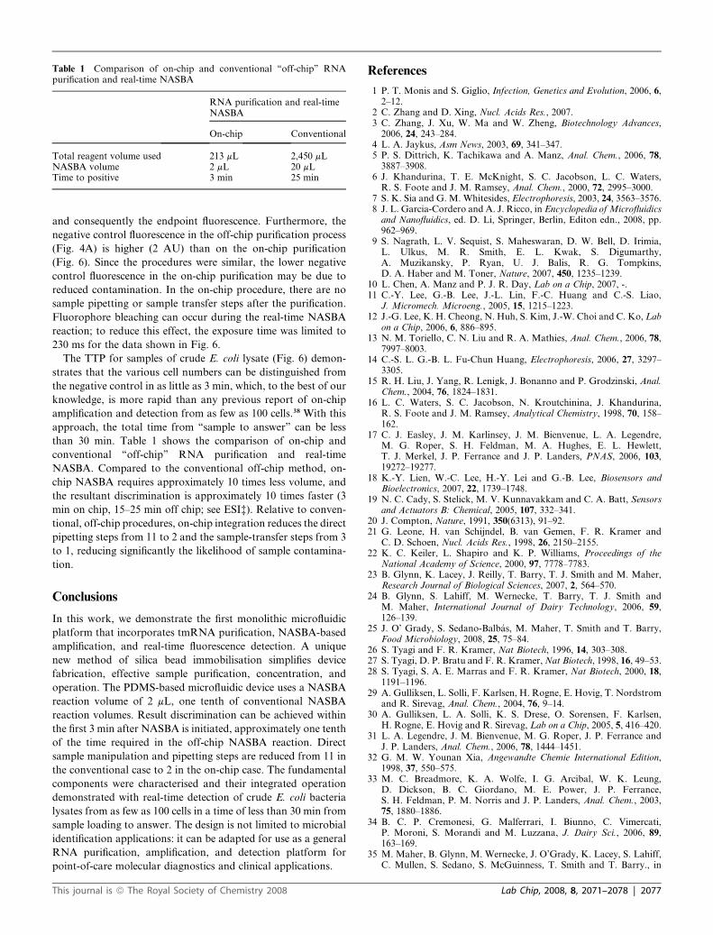
Table 1 Comparison of on-chip and conventional ‘‘off-chip’’ RNApurification and real-time NASBA
RNA purification and real-timeNASBA
On-chip Conventional
Total reagent volume used 213 mL 2,450 mLNASBA volume 2 mL 20 mLTime to positive 3 min 25 min
and consequently the endpoint fluorescence. Furthermore, the
negative control fluorescence in the off-chip purification process
(Fig. 4A) is higher (2 AU) than on the on-chip purification
(Fig. 6). Since the procedures were similar, the lower negative
control fluorescence in the on-chip purification may be due to
reduced contamination. In the on-chip procedure, there are no
sample pipetting or sample transfer steps after the purification.
Fluorophore bleaching can occur during the real-time NASBA
reaction; to reduce this effect, the exposure time was limited to
230 ms for the data shown in Fig. 6.
The TTP for samples of crude E. coli lysate (Fig. 6) demon-
strates that the various cell numbers can be distinguished from
the negative control in as little as 3 min, which, to the best of our
knowledge, is more rapid than any previous report of on-chip
amplification and detection from as few as 100 cells.38 With this
approach, the total time from ‘‘sample to answer’’ can be less
than 30 min. Table 1 shows the comparison of on-chip and
conventional ‘‘off-chip’’ RNA purification and real-time
NASBA. Compared to the conventional off-chip method, on-
chip NASBA requires approximately 10 times less volume, and
the resultant discrimination is approximately 10 times faster (3
min on chip, 15–25 min off chip; see ESI‡). Relative to conven-
tional, off-chip procedures, on-chip integration reduces the direct
pipetting steps from 11 to 2 and the sample-transfer steps from 3
to 1, reducing significantly the likelihood of sample contamina-
tion.
Conclusions
In this work, we demonstrate the first monolithic microfluidic
platform that incorporates tmRNA purification, NASBA-based
amplification, and real-time fluorescence detection. A unique
new method of silica bead immobilisation simplifies device
fabrication, effective sample purification, concentration, and
operation. The PDMS-based microfluidic device uses a NASBA
reaction volume of 2 mL, one tenth of conventional NASBA
reaction volumes. Result discrimination can be achieved within
the first 3 min after NASBA is initiated, approximately one tenth
of the time required in the off-chip NASBA reaction. Direct
sample manipulation and pipetting steps are reduced from 11 in
the conventional case to 2 in the on-chip case. The fundamental
components were characterised and their integrated operation
demonstrated with real-time detection of crude E. coli bacteria
lysates from as few as 100 cells in a time of less than 30 min from
sample loading to answer. The design is not limited to microbial
identification applications: it can be adapted for use as a general
RNA purification, amplification, and detection platform for
point-of-care molecular diagnostics and clinical applications.
This journal is ª The Royal Society of Chemistry 2008
References
1 P. T. Monis and S. Giglio, Infection, Genetics and Evolution, 2006, 6,2–12.
2 C. Zhang and D. Xing, Nucl. Acids Res., 2007.3 C. Zhang, J. Xu, W. Ma and W. Zheng, Biotechnology Advances,
2006, 24, 243–284.4 L. A. Jaykus, Asm News, 2003, 69, 341–347.5 P. S. Dittrich, K. Tachikawa and A. Manz, Anal. Chem., 2006, 78,
3887–3908.6 J. Khandurina, T. E. McKnight, S. C. Jacobson, L. C. Waters,
R. S. Foote and J. M. Ramsey, Anal. Chem., 2000, 72, 2995–3000.7 S. K. Sia and G. M. Whitesides, Electrophoresis, 2003, 24, 3563–3576.8 J. L. Garcia-Cordero and A. J. Ricco, in Encyclopedia of Microfluidicsand Nanofluidics, ed. D. Li, Springer, Berlin, Editon edn., 2008, pp.962–969.
9 S. Nagrath, L. V. Sequist, S. Maheswaran, D. W. Bell, D. Irimia,L. Ulkus, M. R. Smith, E. L. Kwak, S. Digumarthy,A. Muzikansky, P. Ryan, U. J. Balis, R. G. Tompkins,D. A. Haber and M. Toner, Nature, 2007, 450, 1235–1239.
10 L. Chen, A. Manz and P. J. R. Day, Lab on a Chip, 2007, -.11 C.-Y. Lee, G.-B. Lee, J.-L. Lin, F.-C. Huang and C.-S. Liao,
J. Micromech. Microeng., 2005, 15, 1215–1223.12 J.-G. Lee, K. H. Cheong, N. Huh, S. Kim, J.-W. Choi and C. Ko, Lab
on a Chip, 2006, 6, 886–895.13 N. M. Toriello, C. N. Liu and R. A. Mathies, Anal. Chem., 2006, 78,
7997–8003.14 C.-S. L. G.-B. L. Fu-Chun Huang, Electrophoresis, 2006, 27, 3297–
3305.15 R. H. Liu, J. Yang, R. Lenigk, J. Bonanno and P. Grodzinski, Anal.
Chem., 2004, 76, 1824–1831.16 L. C. Waters, S. C. Jacobson, N. Kroutchinina, J. Khandurina,
R. S. Foote and J. M. Ramsey, Analytical Chemistry, 1998, 70, 158–162.
17 C. J. Easley, J. M. Karlinsey, J. M. Bienvenue, L. A. Legendre,M. G. Roper, S. H. Feldman, M. A. Hughes, E. L. Hewlett,T. J. Merkel, J. P. Ferrance and J. P. Landers, PNAS, 2006, 103,19272–19277.
18 K.-Y. Lien, W.-C. Lee, H.-Y. Lei and G.-B. Lee, Biosensors andBioelectronics, 2007, 22, 1739–1748.
19 N. C. Cady, S. Stelick, M. V. Kunnavakkam and C. A. Batt, Sensorsand Actuators B: Chemical, 2005, 107, 332–341.
20 J. Compton, Nature, 1991, 350(6313), 91–92.21 G. Leone, H. van Schijndel, B. van Gemen, F. R. Kramer and
C. D. Schoen, Nucl. Acids Res., 1998, 26, 2150–2155.22 K. C. Keiler, L. Shapiro and K. P. Williams, Proceedings of the
National Academy of Science, 2000, 97, 7778–7783.23 B. Glynn, K. Lacey, J. Reilly, T. Barry, T. J. Smith and M. Maher,
Research Journal of Biological Sciences, 2007, 2, 564–570.24 B. Glynn, S. Lahiff, M. Wernecke, T. Barry, T. J. Smith and
M. Maher, International Journal of Dairy Technology, 2006, 59,126–139.
25 J. O’ Grady, S. Sedano-Balbas, M. Maher, T. Smith and T. Barry,Food Microbiology, 2008, 25, 75–84.
26 S. Tyagi and F. R. Kramer, Nat Biotech, 1996, 14, 303–308.27 S. Tyagi, D. P. Bratu and F. R. Kramer, Nat Biotech, 1998, 16, 49–53.28 S. Tyagi, S. A. E. Marras and F. R. Kramer, Nat Biotech, 2000, 18,
1191–1196.29 A. Gulliksen, L. Solli, F. Karlsen, H. Rogne, E. Hovig, T. Nordstrom
and R. Sirevag, Anal. Chem., 2004, 76, 9–14.30 A. Gulliksen, L. A. Solli, K. S. Drese, O. Sorensen, F. Karlsen,
H. Rogne, E. Hovig and R. Sirevag, Lab on a Chip, 2005, 5, 416–420.31 L. A. Legendre, J. M. Bienvenue, M. G. Roper, J. P. Ferrance and
J. P. Landers, Anal. Chem., 2006, 78, 1444–1451.32 G. M. W. Younan Xia, Angewandte Chemie International Edition,
1998, 37, 550–575.33 M. C. Breadmore, K. A. Wolfe, I. G. Arcibal, W. K. Leung,
D. Dickson, B. C. Giordano, M. E. Power, J. P. Ferrance,S. H. Feldman, P. M. Norris and J. P. Landers, Anal. Chem., 2003,75, 1880–1886.
34 B. C. P. Cremonesi, G. Malferrari, I. Biunno, C. Vimercati,P. Moroni, S. Morandi and M. Luzzana, J. Dairy Sci., 2006, 89,163–169.
35 M. Maher, B. Glynn, M. Wernecke, J. O’Grady, K. Lacey, S. Lahiff,C. Mullen, S. Sedano, S. McGuinness, T. Smith and T. Barry., in
Lab Chip, 2008, 8, 2071–2078 | 2077
Nucleic Acid-Based Technologies, Cambridge Healthtech Institute,Baltimore, Maryland, USA., Editon edn., 2007.
36 V. Linder, S. K. Sia and G. M. Whitesides, Anal. Chem., 2005, 77,64–71.
2078 | Lab Chip, 2008, 8, 2071–2078
37 Y.-C. Chung, M.-S. Jan, Y.-C. Lin, J.-H. Lin, W.-C. Cheng andC.-Y. Fan, Lab on a Chip, 2004, 4, 141–147.
38 L. Chen, A. Manz and P. J. R. Day, Lab on a Chip, 2007, 7, 1413–1423.
This journal is ª The Royal Society of Chemistry 2008