Embed Size (px)
Citation preview
Jour
nal o
f Cel
l Sci
ence
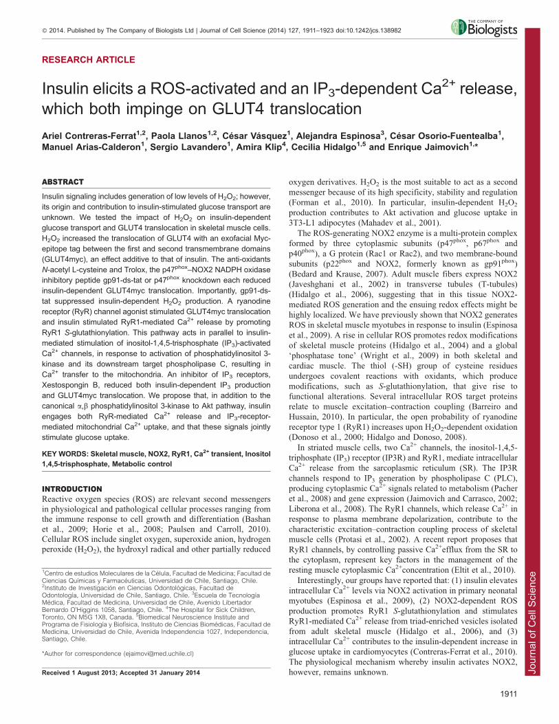
RESEARCH ARTICLE
Insulin elicits a ROS-activated and an IP3-dependent Ca2+ release,
which both impinge on GLUT4 translocation
Ariel Contreras-Ferrat1,2, Paola Llanos1,2, Cesar Vasquez1, Alejandra Espinosa3, Cesar Osorio-Fuentealba1,Manuel Arias-Calderon1, Sergio Lavandero1, Amira Klip4, Cecilia Hidalgo1,5 and Enrique Jaimovich1,*
ABSTRACT
Insulin signaling includes generation of low levels of H2O2; however,
its origin and contribution to insulin-stimulated glucose transport are
unknown. We tested the impact of H2O2 on insulin-dependent
glucose transport and GLUT4 translocation in skeletal muscle cells.
H2O2 increased the translocation of GLUT4 with an exofacial Myc-
epitope tag between the first and second transmembrane domains
(GLUT4myc), an effect additive to that of insulin. The anti-oxidants
N-acetyl L-cysteine and Trolox, the p47phox–NOX2 NADPH oxidase
inhibitory peptide gp91-ds-tat or p47phox knockdown each reduced
insulin-dependent GLUT4myc translocation. Importantly, gp91-ds-
tat suppressed insulin-dependent H2O2 production. A ryanodine
receptor (RyR) channel agonist stimulated GLUT4myc translocation
and insulin stimulated RyR1-mediated Ca2+ release by promoting
RyR1 S-glutathionylation. This pathway acts in parallel to insulin-
mediated stimulation of inositol-1,4,5-trisphosphate (IP3)-activated
Ca2+ channels, in response to activation of phosphatidylinositol 3-
kinase and its downstream target phospholipase C, resulting in
Ca2+ transfer to the mitochondria. An inhibitor of IP3 receptors,
Xestospongin B, reduced both insulin-dependent IP3 production
and GLUT4myc translocation. We propose that, in addition to the
canonical a,b phosphatidylinositol 3-kinase to Akt pathway, insulin
engages both RyR-mediated Ca2+ release and IP3-receptor-
mediated mitochondrial Ca2+ uptake, and that these signals jointly
stimulate glucose uptake.
KEY WORDS: Skeletal muscle, NOX2, RyR1, Ca2+ transient, Inositol
1,4,5-trisphosphate, Metabolic control
INTRODUCTIONReactive oxygen species (ROS) are relevant second messengersin physiological and pathological cellular processes ranging fromthe immune response to cell growth and differentiation (Bashan
et al., 2009; Horie et al., 2008; Paulsen and Carroll, 2010).Cellular ROS include singlet oxygen, superoxide anion, hydrogenperoxide (H2O2), the hydroxyl radical and other partially reduced
oxygen derivatives. H2O2 is the most suitable to act as a second
messenger because of its high specificity, stability and regulation
(Forman et al., 2010). In particular, insulin-dependent H2O2
production contributes to Akt activation and glucose uptake in
3T3-L1 adipocytes (Mahadev et al., 2001).
The ROS-generating NOX2 enzyme is a multi-protein complex
formed by three cytoplasmic subunits (p47phox, p67phox and
p40phox), a G protein (Rac1 or Rac2), and two membrane-bound
subunits (p22phox and NOX2, formerly known as gp91phox)
(Bedard and Krause, 2007). Adult muscle fibers express NOX2
(Javeshghani et al., 2002) in transverse tubules (T-tubules)
(Hidalgo et al., 2006), suggesting that in this tissue NOX2-
mediated ROS generation and the ensuing redox effects might be
highly localized. We have previously shown that NOX2 generates
ROS in skeletal muscle myotubes in response to insulin (Espinosa
et al., 2009). A rise in cellular ROS promotes redox modifications
of skeletal muscle proteins (Hidalgo et al., 2004) and a global
‘phosphatase tone’ (Wright et al., 2009) in both skeletal and
cardiac muscle. The thiol (-SH) group of cysteine residues
undergoes covalent reactions with oxidants, which produce
modifications, such as S-glutathionylation, that give rise to
functional alterations. Several intracellular ROS target proteins
relate to muscle excitation–contraction coupling (Barreiro and
Hussain, 2010). In particular, the open probability of ryanodine
receptor type 1 (RyR1) increases upon H2O2-dependent oxidation
(Donoso et al., 2000; Hidalgo and Donoso, 2008).
In striated muscle cells, two Ca2+ channels, the inositol-1,4,5-
triphosphate (IP3) receptor (IP3R) and RyR1, mediate intracellular
Ca2+ release from the sarcoplasmic reticulum (SR). The IP3R
channels respond to IP3 generation by phospholipase C (PLC),
producing cytoplasmic Ca2+ signals related to metabolism (Pacher
et al., 2008) and gene expression (Jaimovich and Carrasco, 2002;
Liberona et al., 2008). The RyR1 channels, which release Ca2+ in
response to plasma membrane depolarization, contribute to the
characteristic excitation–contraction coupling process of skeletal
muscle cells (Protasi et al., 2002). A recent report proposes that
RyR1 channels, by controlling passive Ca2+efflux from the SR to
the cytoplasm, represent key factors in the management of the
resting muscle cytoplasmic Ca2+concentration (Eltit et al., 2010).
Interestingly, our groups have reported that: (1) insulin elevates
intracellular Ca2+ levels via NOX2 activation in primary neonatal
myotubes (Espinosa et al., 2009), (2) NOX2-dependent ROS
production promotes RyR1 S-glutathionylation and stimulates
RyR1-mediated Ca2+ release from triad-enriched vesicles isolated
from adult skeletal muscle (Hidalgo et al., 2006), and (3)
intracellular Ca2+ contributes to the insulin-dependent increase in
glucose uptake in cardiomyocytes (Contreras-Ferrat et al., 2010).
The physiological mechanism whereby insulin activates NOX2,
however, remains unknown.
1Centro de estudios Moleculares de la Celula, Facultad de Medicina; Facultad deCiencias Quımicas y Farmaceuticas, Universidad de Chile, Santiago, Chile.2Instituto de Investigacion en Ciencias Odontologicas, Facultad deOdontologıa, Universidad de Chile, Santiago, Chile. 3Escuela de TecnologıaMedica, Facultad de Medicina, Universidad de Chile, Avenido LibertadorBernardo O’Higgins 1058, Santiago, Chile. 4The Hospital for Sick Children,Toronto, ON M5G 1X8, Canada. 5Biomedical Neuroscience Institute andPrograma de Fisiologıa y Biofısica, Instituto de Ciencias Biomedicas, Facultad deMedicina, Universidad de Chile, Avenida Independencia 1027, Independencia,Santiago, Chile.
*Author for correspondence ([email protected])
Received 1 August 2013; Accepted 31 January 2014
� 2014. Published by The Company of Biologists Ltd | Journal of Cell Science (2014) 127, 1911–1923 doi:10.1242/jcs.138982
1911
Jour
nal o
f Cel
l Sci
ence
An insulin-dependent Ca2+ increase, detected with the Ca2+
fluorescent dye Indo-1, occurs near the plasma membrane in
single muscle fibers isolated from the Flexor digitorum brevis
(FDB) muscle (Bruton et al., 1999), and this has been associatedwith insulin-stimulated Ca2+ influx via L-type voltage-dependentCa2+ channels (Cav1.1). In cultured myotubes from rat skeletal
muscle, insulin induces RyR1-mediated Ca2+ transients measuredwith the fluorescent Ca2+ indicator Fluo3 (Espinosa et al., 2004).In skeletal muscle fibers, 2-aminoethoxydiphenyl borate (2-APB)
inhibits both basal and insulin-dependent Ca2+ influx (Lanneret al., 2006). A contribution of intracellular Ca2+ stores to glucoseuptake was proposed on the basis that dantrolene, an inhibitor of
the interaction between Cav1.1 and RyR1 that inhibitsdepolarization-induced Ca2+ release (Bannister, 2013), preventsinsulin-dependent glucose uptake in adipocytes (Li et al., 2006).
In those cells, decreasing intracellular Ca2+ signals decreasedinsulin-stimulated glucose uptake and Akt phosphorylation(Whitehead et al., 2001). In skeletal muscle fibers, Ca2+ influxis important for full stimulation of glucose uptake (Lanner et al.,
2009; Lanner et al., 2006).Notably, it remains unknown how insulin elicits intracellular
Ca2+ signals and what are the sources of these signals. Given the
prominence of insulin action in skeletal muscle, unraveling thismechanism is paramount to understand the regulation of glucoseuptake by this hormone.
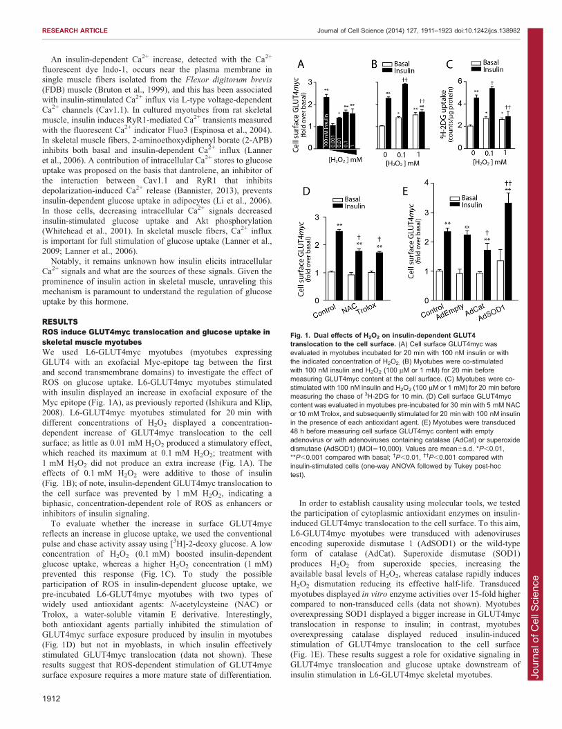
RESULTSROS induce GLUT4myc translocation and glucose uptake inskeletal muscle myotubesWe used L6-GLUT4myc myotubes (myotubes expressingGLUT4 with an exofacial Myc-epitope tag between the firstand second transmembrane domains) to investigate the effect of
ROS on glucose uptake. L6-GLUT4myc myotubes stimulatedwith insulin displayed an increase in exofacial exposure of theMyc epitope (Fig. 1A), as previously reported (Ishikura and Klip,
2008). L6-GLUT4myc myotubes stimulated for 20 min withdifferent concentrations of H2O2 displayed a concentration-dependent increase of GLUT4myc translocation to the cell
surface; as little as 0.01 mM H2O2 produced a stimulatory effect,which reached its maximum at 0.1 mM H2O2; treatment with1 mM H2O2 did not produce an extra increase (Fig. 1A). Theeffects of 0.1 mM H2O2 were additive to those of insulin
(Fig. 1B); of note, insulin-dependent GLUT4myc translocation tothe cell surface was prevented by 1 mM H2O2, indicating abiphasic, concentration-dependent role of ROS as enhancers or
inhibitors of insulin signaling.To evaluate whether the increase in surface GLUT4myc
reflects an increase in glucose uptake, we used the conventional
pulse and chase activity assay using [3H]-2-deoxy glucose. A lowconcentration of H2O2 (0.1 mM) boosted insulin-dependentglucose uptake, whereas a higher H2O2 concentration (1 mM)
prevented this response (Fig. 1C). To study the possibleparticipation of ROS in insulin-dependent glucose uptake, wepre-incubated L6-GLUT4myc myotubes with two types ofwidely used antioxidant agents: N-acetylcysteine (NAC) or
Trolox, a water-soluble vitamin E derivative. Interestingly,both antioxidant agents partially inhibited the stimulation ofGLUT4myc surface exposure produced by insulin in myotubes
(Fig. 1D) but not in myoblasts, in which insulin effectivelystimulated GLUT4myc translocation (data not shown). Theseresults suggest that ROS-dependent stimulation of GLUT4myc
surface exposure requires a more mature state of differentiation.
In order to establish causality using molecular tools, we testedthe participation of cytoplasmic antioxidant enzymes on insulin-induced GLUT4myc translocation to the cell surface. To this aim,
L6-GLUT4myc myotubes were transduced with adenovirusesencoding superoxide dismutase 1 (AdSOD1) or the wild-typeform of catalase (AdCat). Superoxide dismutase (SOD1)
produces H2O2 from superoxide species, increasing theavailable basal levels of H2O2, whereas catalase rapidly inducesH2O2 dismutation reducing its effective half-life. Transduced
myotubes displayed in vitro enzyme activities over 15-fold highercompared to non-transduced cells (data not shown). Myotubesoverexpressing SOD1 displayed a bigger increase in GLUT4myc
translocation in response to insulin; in contrast, myotubesoverexpressing catalase displayed reduced insulin-inducedstimulation of GLUT4myc translocation to the cell surface(Fig. 1E). These results suggest a role for oxidative signaling in
GLUT4myc translocation and glucose uptake downstream ofinsulin stimulation in L6-GLUT4myc skeletal myotubes.
Fig. 1. Dual effects of H2O2 on insulin-dependent GLUT4translocation to the cell surface. (A) Cell surface GLUT4myc wasevaluated in myotubes incubated for 20 min with 100 nM insulin or withthe indicated concentration of H2O2. (B) Myotubes were co-stimulatedwith 100 nM insulin and H2O2 (100 mM or 1 mM) for 20 min beforemeasuring GLUT4myc content at the cell surface. (C) Myotubes were co-stimulated with 100 nM insulin and H2O2 (100 mM or 1 mM) for 20 min beforemeasuring the chase of 3H-2DG for 10 min. (D) Cell surface GLUT4myccontent was evaluated in myotubes pre-incubated for 30 min with 5 mM NACor 10 mM Trolox, and subsequently stimulated for 20 min with 100 nM insulinin the presence of each antioxidant agent. (E) Myotubes were transduced48 h before measuring cell surface GLUT4myc content with emptyadenovirus or with adenoviruses containing catalase (AdCat) or superoxidedismutase (AdSOD1) (MOI510,000). Values are mean6s.d. *P,0.01,**P,0.001 compared with basal; �P,0.01, ��P,0.001 compared withinsulin-stimulated cells (one-way ANOVA followed by Tukey post-hoctest).
RESEARCH ARTICLE Journal of Cell Science (2014) 127, 1911–1923 doi:10.1242/jcs.138982
1912

Jour
nal o
f Cel
l Sci
ence
Insulin increases cytoplasmic H2O2 production through PKC-dependent NOX2 activation, which promotes GLUT4myctranslocation to the cell surfaceAs shown in Fig. 1, H2O2 induced an increase in bothGLUT4myc translocation and glucose uptake; the molecularentities involved in these responses are unknown, as is the
mechanism responsible for ROS generation downstream ofinsulin signaling in skeletal muscle cells. To determine whetherinsulin enhances cytoplasmic H2O2 production, we transiently
transfected L6-GLUT4myc myotubes with a plasmid vectorencoding the molecular H2O2 biosensor HyPer targeted to thecytoplasm (HyPer-cyto). Recording in real time revealed that
Hyper-cyto effectively sensed low H2O2 concentrations in L6-GLUT4myc myotubes (Fig. 2A); addition of vehicle did notchange the basal fluorescence levels (Fig. 2A). Addition of
insulin induced a fast and transient increase in HyPer-cytofluorescence emission, which reached a maximum at 763 s post
stimuli and took 143632 s to decay thereafter (mean6s.d.;Fig. 2B, black trace).
NOX2 is a protein complex canonically activated bytranslocation of p47phox, p67phox and the small GTP-binding
protein Rac1 to the plasma membrane, where they form the activeenzyme by interacting with the membrane integral proteins gp91and p22 (Maghzal et al., 2012). Indirect immunofluorescence
determinations showed p47phox subunits located in the cytoplasmof myotubes, showing poor expression in myoblasts (data notshown). To explore NOX2 participation in insulin-induced H2O2
generation, we inhibited NOX2 with the specific peptide gp91-ds-tat (Wei et al., 2008), which contains the gp91phox sequencelinked to the HIV tat peptide. L6-GLUT4myc myotubes
Fig. 2. Insulin-induced H2O2 production promotes GLUT4myc translocation through PKC-dependent NOX2 activation. (A) Myotubes transfected withthe cytoplasmic H2O2 sensor HyPer-cyto were treated with 100 mM H2O2 (black trace) or with vehicle (red trace). (B) Addition of 100 nM insulin increased theHyPer-cyto fluorescence ratio (black trace); treatment with 5 mM gp91-ds-tat (NOX2 inhibitory peptide, green trace) or pre-treatment with 1 mM of BIM(conventional and novel PKC inhibitor, red trace) abrogated the increase of the HyPer-cyto fluorescence ratio induced by 100 nM insulin. The results correspondto the fluorescence ratio 490 nm:420 nm. (C) The effects of adding 100 nM insulin on cell surface GLUT4myc translocation were tested in control myotubesor in myotubes pre-incubated for 30 min with 5 mM gp91-ds-tat or 5 mM scrambled (sc) gp91-ds-tat (inactive peptide). (D) Effects of 100 nM insulin on cellsurface GLUT4myc translocation were tested in control myotubes or in myotubes transfected with 50 nM p47phox siRNA (sip47phox) or 50 nM control siRNA(siSc). Myotubes transfected for 4 h with each siRNA were maintained for 48 h before adding 100 nM insulin; GLUT4myc translocation was measured 20 minafter insulin addition. (E) Myotubes transfected with the HyPer-cyto sensor and pre-incubated for 30 min with 5 mM gp91-ds-tat or 5 mM sc gp91-ds-tat asindicated were stimulated with 1 mM PMA (phorbol ester). As a control, 1 mM H2O2 was added at the end of the experiment. (F) Myotubes were stimulated with1 mM PMA for 20 min and surface GLUT4myc was evaluated in myotubes pre-incubated for 30 min with 5 mM gp91-ds-tat or 5 mM sc gp91-ds-tat. (G) L6-GLUT4myc myotubes were pre-incubated with 1 mM Go6976 (conventional PKC inhibitor) or 1 mM BIM (conventional and novel PKC inhibitor) during the30 min before insulin addition. In C, D, F and G, results are mean6s.d. Statistical analysis was performed with one-way ANOVA followed by Tukey post-hoc test.*P,0.01, **P,0.001 compared with basal; �P,0.01, ��P,0.001 compared with insulin-stimulated cells (one-way ANOVA followed by Tukey post-hoctest).
RESEARCH ARTICLE Journal of Cell Science (2014) 127, 1911–1923 doi:10.1242/jcs.138982
1913
Jour
nal o
f Cel
l Sci
ence
pre-incubated for 1 h with gp91-ds-tat or its inactive scrambledsequence sc-gp91-ds-tat were stimulated with insulin. Incubation
with the inhibitory peptide abolished insulin-dependent H2O2
production (Fig. 2B, red trace), whereas the inactive peptide didnot inhibit this response (data not shown). In order to explorewhether protein kinase C (PKC) contributes to NOX2 activation,
L6-GLUT4myc myotubes were pre-incubated with 1 mMbisindolylmaleimide (BIM), a conventional inhibitor of PKCaand PKCb, also described to inhibit PKCd. This PKC inhibitor
completely suppressed insulin-dependent H2O2 production(Fig. 2B, green trace). Moreover, addition of 5 mM NACdrastically reduced basal HyPer-cyto fluorescence and prevented
insulin-induced H2O2 production; NAC also moderately loweredthe fluorescent signal induced by addition of 100 mM H2O2, usedas positive control (data not shown). These combined results
strongly suggest that NOX2 mediates insulin-dependent ROSgeneration through PKC activation.
To elucidate whether NOX2 contributes to insulin-dependentGLUT4myc translocation, we inhibited NOX2 with gp91-ds-
tat. To this aim, L6-GLUT4myc myotubes were pre-incubatedfor 1 h with gp91-ds-tat or its inactive scrambled sequence sc-gp91-ds-tat and were stimulated with insulin for 20 min in
the presence of these synthetic peptides. Pre-incubation withthe gp91-ds-tat peptide partially reduced insulin-dependentGLUT4myc translocation; in contrast, transfection with the
scrambled sequence did not affect this response (Fig. 2C).Furthermore, the pharmacological NOX2 inhibitors apocynin ordiphenyliodonium (DPI) reduced but did not fully suppress
insulin-dependent GLUT4myc translocation to the cell surface(supplementary material Fig. S1A). Myotubes transientlytransfected with siRNA (50 nM) against the regulatory NOX2subunit p47phox displayed significantly reduced p47phox levels
(supplementary material Fig. S1C) and lower GLUT4myctranslocation following insulin addition than either controlmyotubes or myotubes transfected with a scrambled siRNA
(Fig. 2D).
Different PKC enzymes contribute to insulin signalingPrevious studies indicate that a diacylglycerol-mimicking phorbolester (Wright et al., 2004) and compounds that increaseintracellular Ca2+ (Wright et al., 2005) stimulate glucose uptakein cultured cells and isolated rodent muscles. In muscle cells, it
has been suggested that activation of PKCf and PKCl increasesGLUT4 translocation, although this finding has been somewhatcontroversial (Liu et al., 2006; Powell et al., 2003; Stretton et al.,
2010). Moreover, PKCa and PKCb promotes p47phox activationand plasma membrane translocation to assemble the NOX2activated complex in macrophages (San Jose et al., 2009). It has
been widely accepted that phosphorylation via PKC activatesNOX2 in many systems. To test whether direct PKC activationpromotes NOX2-dependent H2O2 production, we stimulated
myotubes with the phorbol ester 12-myristate 13-acetate (PMA,a universal activator of classical and novel PKCs). In L6-GLUT4myc myotubes, PMA induced a fast and transient increasein HyPer-cyto fluorescence, which reached a maximum value
362 s post stimuli (Fig. 2E), with a decay half time of 126641 s(mean6s.d.; presumably reflecting the breakage kinetics of theH2O2-induced HyPer-cyto disulfide bridge by cellular reducing
systems). Pre-incubation of myotubes with gp91-ds-tat but notwith the inactive peptide abolished PMA-dependent H2O2
production (Fig. 2E). Furthermore, incubation of myotubes with
PMA for 20 min strongly increased GLUT4myc translocation to
the cell surface; the synthetic inhibitory peptide gp91-ds-tatabolished this increase (Fig. 2F). Co-stimulation of myotubes
with PMA and insulin resulted in a significant extra increase inexofacial exposure of the Myc epitope over that produced byinsulin alone (supplementary material Fig. S1B). Both the PKCinhibitor Go6976 (specific for Ca2+-dependent PKCs) and more
so BIM (a general inhibitor of types a, b, c and d PKCs),significantly reduced GLUT4 translocation induced by insulin(Fig. 2G). Taken together, these results suggest that PKC is
involved in the increased NOX2-mediated cytoplasmic H2O2
generation and GLUT4myc translocation stimulated by insulin.
Exofacial exposure of GLUT4myc induced by insulin requiresRyR1-mediated Ca2+ releaseL6-GLUT4myc myotubes pre-incubated with the Ca2+ dye
indicator Fluo4 am were mounted in a chamber andintracellular Ca2+ levels were recorded frame-by-frame in aconfocal microscope. After 1 min recording, we replaced theCa2+-containing resting solution with a Ca2+-free solution
(Fig. 3A–C, blue dashed lines). Addition of 100 mM H2O2
produced a rapid increase in intracellular Ca2+ levels that didnot occur in cells pre-treated with 50 mM ryanodine, strongly
suggesting that ROS induced RyR1-mediated Ca2+ release(Fig. 3A). Insulin addition in Ca2+-free resting mediumgenerated whole-cell Ca2+ transients, which were not present in
cells pre-incubated with 50 mM ryanodine for 3 h or 5 mM gp91-ds-tat for 30 min (Fig. 3B). Addition of the Ca2+ ionophoreionomycin to cells kept in Ca2+-free solution induced a rapid rise
in fluorescence in all conditions, presumably by inducing Ca2+
release from intracellular stores (Fig. 3A,B). To induce RyR1-mediated Ca2+ release, we used the specific RyR agonist 4-chloro-m-cresol (4-CMC). In Ca2+-free resting medium, 4-CMC
induced a fast increase in intracellular Ca2+, which slowlydecreased to near basal levels after 5 min (Fig. 3C); ionomycinaddition was used as a positive control at the end of the recording.
4-CMC also induced an increase in GLUT4myc translocationto the cell surface, and this effect was more evident in insulin co-stimulated myotubes (Fig. 3D). Translocation of GLUT4myc
induced by 4-CMC was completely inhibited in myotubes pre-incubated with inhibitory doses of ryanodine (data not shown).Moreover, insulin-dependent GLUT4myc translocation was alsosignificantly lower in myotubes pre-incubated with ryanodine
(Fig. 3D), whereas myotubes pre-incubated jointly with theNOX2 gp91-ds-tat inhibitor and ryanodine did not showadditional inhibition over that produced by ryanodine alone
(Fig. 3D). We used ionomycin to determine whether an increasein intracellular Ca2+ levels stimulates GLUT4myc translocationto the cell surface. Myotubes stimulated with 1 mM ionomycin for
5 min showed increased exofacial exposure of the Myc epitopethat was additive to the stimulation induced by insulin (Fig. 3E).Insulin-dependent GLUT4myc translocation did not occur in L6-
GLUT4myc myotubes pre-incubated with BAPTA-AM for30 min; this intracellular Ca2+ chelator did not affect the basallevels of translocation (Fig. 3E). This strategy was complementedby overexpressing the parvalbumin Ca2+ buffer protein fused to
DsRed, as the cell tracker (PV–DsRed). A plasmid encoding onlyDsRed was used as control. Cells were fixed in non-permeabilizedconditions and the exofacial exposure of the Myc epitope was
assayed in single cells. PV–DsRed-expressing cells wereunresponsive to insulin and exhibited decreased basal exposureof the Myc epitope (Fig. 3F). Moreover, non-transfected myotubes
showed a notable increase in the amount of insulin-induced cell
RESEARCH ARTICLE Journal of Cell Science (2014) 127, 1911–1923 doi:10.1242/jcs.138982
1914

Jour
nal o
f Cel
l Sci
ence
surface GLUT4myc stain (Fig. 3F, arrowheads). These combined
experiments suggest that the cytoplasmic Ca2+ increase producedby stimulation of Ca2+ release through ROS-mediated RyR1activation forms part of the insulin signaling pathways thatpromote GLUT4myc translocation to the cell surface.
Insulin-dependent H2O2 production induced RyR1S-glutathionylation in skeletal muscle cellsROS generation induces reversible S-glutathionylation of reactivecysteine residues (Pastore and Piemonte, 2012). To detectwhether S-glutathionylation of proteins increases in insulin-
stimulated cells, myotubes were stimulated with insulin or H2O2
for 1 min and the indirect co-immunofluorescence against S-glutathionylated protein adducts or RyR1 was detected with
specific antibodies. In basal conditions, myotubes showed verylow fluorescence when tested with the antibody against S-glutathionylated protein adducts, indicating low basal levels ofthis redox modification (Fig. 4A). In contrast, myotubes
stimulated with 100 mM H2O2 exhibited significantly higher S-glutathionylated protein levels, which partially overlapped with
RyR1 immunofluorescence yielding a Pearson coefficient of
0.4460.06 and Mander’s coefficients M1 and M2 of 0.6860.09and 0.6160.09 (mean6s.d.), respectively (Fig. 4A). Of note,1 min exposure to 100 nM insulin increased S-glutathionylatedprotein adducts with a clear overlap with the RyR1 stain, reaching
a Pearson coefficient of 0.78260.07 and Mander’s coefficientsM1 and M2 of 0.9260.07 and 0.9160.02, respectively (Fig. 4A).As the resolution provided by co-immunofluorescence
experiments cannot ascertain the actual RyR1 S-glutathionylation levels, we used a novel high-resolutiontechnique that yields positive results for molecules located less
than 40 nm apart. Specific RyR1 S-glutathionylation wasascertained using in situ proximity ligation assay (PLA) probes,which detect closely positioned antibodies with an optimal
distance of 20–30 nm (Soderberg et al., 2006). To this aim,cultured myotubes stimulated with insulin or H2O2 for 1 min andrapidly fixed were probed both with anti-RyR1 and anti-S-glutathionylated protein adducts; RyR1 S-glutathionylation is
indicated by the appearance of fluorescent dots (Fig. 4B). Thebasal levels of RyR1 S-glutathionylation were 561.4 dots/
Fig. 3. Insulin-dependent translocation ofGLUT4myc to the cell surface requires RyR1-mediated intracellular Ca2+ increase. (A) Controlmyotubes (black trace) or myotubes pre-incubated for3 h with 50 mM ryanodine (RyR inhibitor, red trace)were incubated for 30 min with 5.4 mM Fluo4 am beforeaddition of 100 mM H2O2, as indicated by the arrows.(B) Control myotubes (black trace), myotubes pre-incubated for 3 h with 50 mM ryanodine (red trace) ormyotubes pre-incubated for 30 min with 5 mM gp91-ds-tat (NOX2 inhibitory peptide, green trace) wereincubated for 30 min with 5.4 mM Fluo4 am in thepresence of the respective compound before addition of100 nM insulin at the arrows. (C) Myotubes wereincubated for 30 min with 5.4 mM Fluo4 am beforeaddition (at the arrow) of 500 mM 4-CMC (specific RyRagonist, red trace); control (black trace). Ca2+ transientsdepicted in A, B and C were recorded in Ca2+-freemedium, the addition of which is indicated by thedashed blue line. At the end of the recording period,1 mM ionomycin (Ca2+ ionophore) was added as apositive control. (D) GLUT4myc translocation wasassayed in controls or after addition of 500 mM 4-CMC(RyR agonist), 100 nM insulin or both. Myotubes werepre-incubated for 3 h with 50 mM ryanodine (RyRinhibitor) or with 50 mM ryanodine for 3 h and 5 mMgp91-ds-tat, during the last 30 min. (E) GLUT4myctranslocation was assayed in control myotubes beforeor 5 min after addition of 100 nM insulin, 1 mMionomycin (Ca2+ ionophore) or both. In parallelexperiments, GLUT4myc translocation was assayed inmyotubes pre-incubated for 30 min with 50 mM BAPTA-AM (intracellular Ca2+ chelator), before or 5 min afteraddition of 100 nM insulin. (F) L6-GLUT4myc cellswere transiently transfected with parvalbumin fused toDsRed (PV–DsRed) and surface Myc epitope levelswere detected in non-permeabilized control single cells(empty bars) or in single cells stimulated with 100 nMinsulin for 20 min (solid bars). Cells transfected withDsRed were used as control. The image in the insetshows a representative experiment. Values aremean6s.d. *P,0.01, **P,0.001 compared with basal;�P,0.01, ��P,0.001 compared with insulin-stimulatedcells (one-way ANOVA followed by Tukey post-hoc test).
RESEARCH ARTICLE Journal of Cell Science (2014) 127, 1911–1923 doi:10.1242/jcs.138982
1915
Jour
nal o
f Cel
l Sci
ence
1000 mm3, whereas in insulin-stimulated myotubes these levelsincreased to 2462.5 dots/1000 mm3. Cells treated with 100 mM
H2O2 displayed 3764.2 dots/1000 mm3 (mean6s.d.; Fig. 4C).
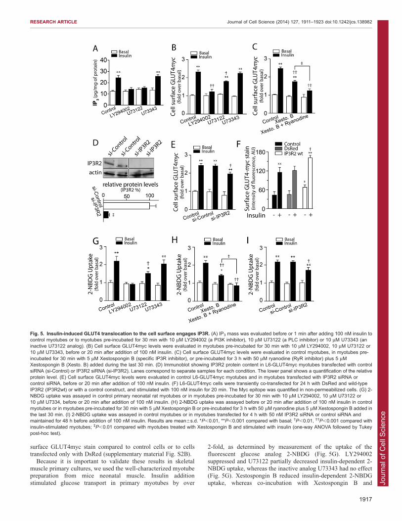
Insulin-dependent exofacial exposure of GLUT4myc andglucose uptake in skeletal myotubes require IP3R activationIP3R-dependent Ca2+ signaling plays a relevant role in cellphysiology (Foskett et al., 2007). The IP3R increases its openprobability in response to IP3 produced by PLC activation. We
explored whether IP3R-generated Ca2+ signals also contribute toinsulin-dependent GLUT4myc translocation in L6 and primaryrat neonatal myotubes. L6-GLUT4myc myotubes express all
three IP3R isoforms (supplementary material Fig. S2A). TheIP3R1 isoform displayed a cytoplasmic and nuclear distributionshowing diffuse stain with condensed highly fluorescent dots.Interestingly, the IP3R2 isoform presented a striated transversal
pattern in the cytoplasm. The IP3R3 isoform displayed poorfluorescence intensity compared to the other isoforms, detectedwith the same microscope settings (supplementary material Fig.
S2A).Insulin addition produced a fast increase in intracellular IP3
levels, which increased from the basal values of 12.361.6 to
24.862.4 pg/mg of protein in 30 s (mean6s.d.; Fig. 5A). Both,LY294002 and U-73122 [broad inhibitors of phosphoinositide 3-kinase (PI3K) and PLC, respectively] inhibited insulin-stimulated
IP3 production, whereas U-73343, an inactive analog of U-73122,did not affect IP3 production (Fig. 5A). Pre-incubation ofmyotubes with the PI3K inhibitor LY294002 did not affect the
basal myc epitope levels but abolished insulin-dependentGLUT4myc translocation (Fig. 5B). We found that the PLC
inhibitor U73122 partially inhibited, whereas the inactiveanalogue U73343 had no effect on, insulin-dependentGLUT4myc translocation in myotubes (Fig. 5B). The last stepin IP3-dependent Ca2+ release is the interaction of IP3 with the
IP3-binding site of IP3R. Myotubes pre-incubated withXestospongin B, a specific IP3R inhibitor, displayed a partialreduction of insulin-dependent GLUT4myc translocation
(Fig. 5C). Combined treatment with both intracellular Ca2+
channels inhibitors, Xestospongin B and ryanodine, stronglyinhibited insulin-dependent Myc epitope externalization, which
reached values not significantly different from the controls nottreated with insulin (Fig. 5C).
To further test the involvement of IP3R2, L6-GLUT4mycmyotubes were transfected with siRNA against IP3R2, which
produced a .90% reduction in protein levels compared with nontransfected cells or with a control non-targeting oligonucleotide(Fig. 5D). Insulin addition to L6-GLUT4myc myotubes
transfected with the IP3R2 siRNA produced a lower stimulationof Myc epitope exposure compared to the stimulation produced incontrol cells or in cells transfected with the control siRNAs
(Fig. 5E).Myotubes overexpressing IP3R2 wild type exhibited increased
basal levels of myc epitope exposure compared to control or
DsRed-transfected cells, detected by indirect immunofluorescencein non-permeabilized cells (Fig. 5F). Moreover, insulin-stimulatedmyoblasts overexpressing IP3R2 displayed significantly increased
Fig. 4. Insulin enhances RyR1 S-glutathionylation in L6-GLUT4myc myotubes. (A) Representative experiment showingco-immunofluorescence images against RyR1 (red) and S-glutathionylated protein adducts (green), in myotubes under basalconditions or 1 min after adding 100 mM H2O2 or 100 nM insulin.The panels on the right right show the corresponding values ofPearson (tP) and Mander’s coefficients (tM1 and tM2). For details,see text. (B) Proximity ligation assay (PLA) probes were used todetect specific RyR1 S-glutathionylation (red dots).Representative images taken from control myotubes, or frommyotubes incubated for 1 min with 100 nM insulin or 100 mMH2O2. (C) The positive red dots, such as those illustrated in B,were counted in all z-stack slices and normalized to the total stackvolume. In A and C, results are mean6s.d. *P,0.01, **P,0.001compared with basal; �P,0.01, ��P,0.001 compared with insulin-stimulated cells (one-way ANOVA followed by Tukey post-hoc test).
RESEARCH ARTICLE Journal of Cell Science (2014) 127, 1911–1923 doi:10.1242/jcs.138982
1916
Jour
nal o
f Cel
l Sci
ence
surface GLUT4myc stain compared to control cells or to cellstransfected only with DsRed (supplementary material Fig. S2B).
Because it is important to validate these results in skeletalmuscle primary cultures, we used the well-characterized myotubepreparation from mice neonatal muscle. Insulin addition
stimulated glucose transport in primary myotubes by over
2-fold, as determined by measurement of the uptake of thefluorescent glucose analog 2-NBDG (Fig. 5G). LY294002
suppressed and U73122 partially decreased insulin-dependent 2-NBDG uptake, whereas the inactive analog U73343 had no effect(Fig. 5G). Xestospongin B reduced insulin-dependent 2-NBDG
uptake, whereas co-incubation with Xestospongin B and
Fig. 5. Insulin-induced GLUT4 translocation to the cell surface engages IP3R. (A) IP3 mass was evaluated before or 1 min after adding 100 nM insulin tocontrol myotubes or to myotubes pre-incubated for 30 min with 10 mM LY294002 (a PI3K inhibitor), 10 mM U73122 (a PLC inhibitor) or 10 mM U73343 (aninactive U73122 analog). (B) Cell surface GLUT4myc levels were evaluated in myotubes pre-incubated for 30 min with 10 mM LY294002, 10 mM U73122 or10 mM U73343, before or 20 min after addition of 100 nM insulin. (C) Cell surface GLUT4myc levels were evaluated in control myotubes, in myotubes pre-incubated for 30 min with 5 mM Xestospongin B (specific IP3R inhibitor), or pre-incubated for 3 h with 50 mM ryanodine (RyR inhibitor) plus 5 mMXestospongin B (Xesto. B) added during the last 30 min. (D) Immunoblot showing IP3R2 protein content in L6-GLUT4myc myotubes transfected with controlsiRNA (si-Control) or IP3R2 siRNA (si-IP3R2). Lanes correspond to separate samples for each condition. The lower panel shows a quantification of the relativeprotein level. (E) Cell surface GLUT4myc levels were evaluated in control L6-GLUT4myc myotubes and in myotubes transfected with IP3R2 siRNA orcontrol siRNA, before or 20 min after addition of 100 nM insulin. (F) L6-GLUT4myc cells were transiently co-transfected for 24 h with DsRed and wild-typeIP3R2 (IP3R2wt) or with a control construct, and stimulated with 100 nM insulin for 20 min. The Myc epitope was quantified in non-permeabilized cells. (G) 2-NBDG uptake was assayed in control primary neonatal rat myotubes or in myotubes pre-incubated for 30 min with 10 mM LY294002, 10 mM U73122 or10 mM U7334, before or 20 min after addition of 100 nM insulin. (H) 2-NBDG uptake was assayed before or 20 min after addition of 100 nM insulin in controlmyotubes or in myotubes pre-incubated for 30 min with 5 mM Xestospongin B or pre-incubated for 3 h with 50 mM ryanodine plus 5 mM Xestospongin B added inthe last 30 min. (I) 2-NBDG uptake was assayed in control myotubes or in myotubes transfected for 4 h with 50 nM IP3R2 siRNA or control siRNA andmaintained for 48 h before addition of 100 nM insulin. Results are mean6s.d. *P,0.01, **P,0.001 compared with basal; �P,0.01, ��P,0.001 compared withinsulin-stimulated myotubes; `P,0.01 compared with myotubes treated with Xestospongin B and stimulated with insulin (one-way ANOVA followed by Tukeypost-hoc test).
RESEARCH ARTICLE Journal of Cell Science (2014) 127, 1911–1923 doi:10.1242/jcs.138982
1917
Jour
nal o
f Cel
l Sci
ence
ryanodine suppressed this response (Fig. 5H). The stimulatoryeffect of insulin on 2-NBDG uptake was partially reduced in
primary myotubes transfected with siRNA against IP3R2;transfection with the control siRNA did not induce changes(Fig. 5I). These combined results show that IP3R2 has a relevantparticipation in insulin-dependent GLUT4myc translocation and
2-NBDG uptake in skeletal muscle cells.
Insulin-dependent IP3R activation regulates mitochondrialCa2+ and pH levels in L6-GLUT4myc cellsTo investigate whether insulin promotes mitochondrial Ca2+
uptake, and hence stimulates mitochondrial function, we used the
mitochondrial-targeted ratiometric PeriCaM (mito-PeriCaM) as aCa2+ probe. This probe displays two excitation maxima (410–440 nm and 480–490 nm), each one presenting a maximal
emission wavelength of 535 nm (Fonteriz et al., 2010). The410–440 nm peak displays high Ca2+ sensitivity, as decreasingfluorescence emission, whereas the 480–490 nm is highlyresponsive to [H+], decreasing fluorescence emission when the
pH goes down. Following insulin addition, L6-GLUT4mycmyotubes transfected with mito-PeriCaM and mounted 24 hlater for viewing under a microscope, displayed fast and transient
increases in both mitochondrial Ca2+ levels and matrix pH(Fig. 6A). Ca2+ levels decreased towards the basal level at 10 minpost-stimuli (data not shown). The area under the curve,
corresponding to the first 5 min post insulin was measured(Fig. 6B). The serine/threonine kinase Akt inhibitor Akti1/2 didnot affect insulin-dependent mitochondrial Ca2+ uptake of L6-
GLUT4myc myotubes transiently transfected with mito-PeriCaM,whereas pre-incubation with inhibitory doses of ryanodine for 3 hproduced a partial decrease (Fig. 6C). Pre-incubation withXestospongin B or Ruthenium Red (RuRed) to inhibit the
mitochondrial Ca2+ uniporter (MCU), or co-transfection withsiRNA against IP3R2 significantly reduced insulin-dependentmitochondrial Ca2+ uptake (Fig. 6C). To associate the insulin-
dependent mitochondrial Ca2+ handling to GLUT4myctranslocation, myotubes were pre-incubated for 30 min withRuRed or Akti1/2; both inhibitors reduced insulin-dependent
GLUT4myc translocation (Fig. 6D). Interestingly, co-incubationwith Akti1/2 and RuRed, or with Akti1/2 and Xestospongin B, didnot increase the inhibitory effects of RuRed or Xestospongin Balone (Fig. 6D).
To test the participation of PI3K signaling on insulin-dependent mitochondrial Ca2+ uptake, primary myotubes wereco-transfected with Dp85dn, a dominant-negative form of the
PI3K IA regulatory subunit. These cells displayed significantlyreduced insulin-dependent mitochondrial Ca2 uptake (Fig. 6E).Co-transfection of the PH domain of centaurin a1 fused to eGFP
[a molecular inhibitor of phosphatidylinositol 3,4,5-trisphosphate(PIP3) signaling] together with mito-PeriCaM or of an IP3
sponge corresponding to the IP3-binding site of IP3R (M49-IP3
sponge), acting as competitive inhibitors of PIP3 signaling andIP3 production respectively, suppressed insulin-dependentmitochondrial Ca2+ uptake (Fig. 6E). Co-transfection with amutated construct without sponge capacity (M30-IP3 sponge) did
not alter the stimulatory effect of insulin (Fig. 6E). Additionalexperiments to inhibit mitochondrial Ca2+ uptake (RuRed) orIP3R function (M49-IP3 sponge) reduced insulin-dependent 2-
NBDG uptake in primary myotubes (Fig. 6F). All theseexperiments were made in labeled single living cells, asdescribed in the Materials and Methods, in order to avoid
dealing with the low transfection efficiency (less than 5%)
observed in primary myotubes. These data suggest that theincreased mitochondrial Ca2+ and pH levels induced by insulin
are associated with GLUT4 translocation to the cell surface andglucose uptake in muscle cells.
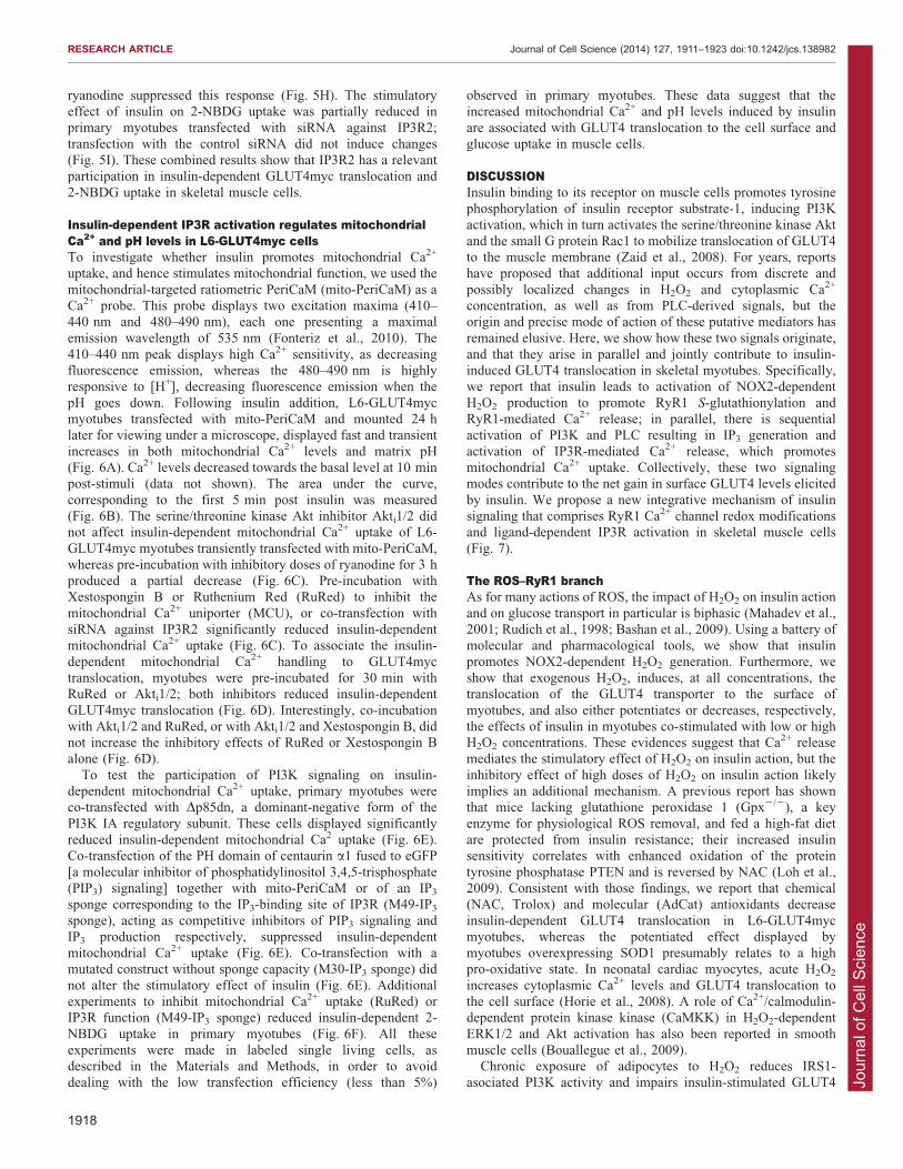
DISCUSSIONInsulin binding to its receptor on muscle cells promotes tyrosinephosphorylation of insulin receptor substrate-1, inducing PI3Kactivation, which in turn activates the serine/threonine kinase Akt
and the small G protein Rac1 to mobilize translocation of GLUT4to the muscle membrane (Zaid et al., 2008). For years, reportshave proposed that additional input occurs from discrete and
possibly localized changes in H2O2 and cytoplasmic Ca2+
concentration, as well as from PLC-derived signals, but theorigin and precise mode of action of these putative mediators has
remained elusive. Here, we show how these two signals originate,and that they arise in parallel and jointly contribute to insulin-induced GLUT4 translocation in skeletal myotubes. Specifically,we report that insulin leads to activation of NOX2-dependent
H2O2 production to promote RyR1 S-glutathionylation andRyR1-mediated Ca2+ release; in parallel, there is sequentialactivation of PI3K and PLC resulting in IP3 generation and
activation of IP3R-mediated Ca2+ release, which promotesmitochondrial Ca2+ uptake. Collectively, these two signalingmodes contribute to the net gain in surface GLUT4 levels elicited
by insulin. We propose a new integrative mechanism of insulinsignaling that comprises RyR1 Ca2+ channel redox modificationsand ligand-dependent IP3R activation in skeletal muscle cells
(Fig. 7).
The ROS–RyR1 branchAs for many actions of ROS, the impact of H2O2 on insulin action
and on glucose transport in particular is biphasic (Mahadev et al.,2001; Rudich et al., 1998; Bashan et al., 2009). Using a battery ofmolecular and pharmacological tools, we show that insulin
promotes NOX2-dependent H2O2 generation. Furthermore, weshow that exogenous H2O2, induces, at all concentrations, thetranslocation of the GLUT4 transporter to the surface of
myotubes, and also either potentiates or decreases, respectively,the effects of insulin in myotubes co-stimulated with low or highH2O2 concentrations. These evidences suggest that Ca2+ releasemediates the stimulatory effect of H2O2 on insulin action, but the
inhibitory effect of high doses of H2O2 on insulin action likelyimplies an additional mechanism. A previous report has shownthat mice lacking glutathione peroxidase 1 (Gpx2/2), a key
enzyme for physiological ROS removal, and fed a high-fat dietare protected from insulin resistance; their increased insulinsensitivity correlates with enhanced oxidation of the protein
tyrosine phosphatase PTEN and is reversed by NAC (Loh et al.,2009). Consistent with those findings, we report that chemical(NAC, Trolox) and molecular (AdCat) antioxidants decrease
insulin-dependent GLUT4 translocation in L6-GLUT4mycmyotubes, whereas the potentiated effect displayed bymyotubes overexpressing SOD1 presumably relates to a highpro-oxidative state. In neonatal cardiac myocytes, acute H2O2
increases cytoplasmic Ca2+ levels and GLUT4 translocation tothe cell surface (Horie et al., 2008). A role of Ca2+/calmodulin-dependent protein kinase kinase (CaMKK) in H2O2-dependent
ERK1/2 and Akt activation has also been reported in smoothmuscle cells (Bouallegue et al., 2009).
Chronic exposure of adipocytes to H2O2 reduces IRS1-
asociated PI3K activity and impairs insulin-stimulated GLUT4
RESEARCH ARTICLE Journal of Cell Science (2014) 127, 1911–1923 doi:10.1242/jcs.138982
1918

Jour
nal o
f Cel
l Sci
ence
Fig. 6. Insulin induces IP3R-dependent mitochondrial Ca2+ and pH increases in L6GLUT4myc myotubes, which enhance GLUT4 translocation tothe cell surface. (A) Representative fluorescence images recorded before and at different times after insulin addition. Myotubes transfected with mito-PeriCaMfor 4 h were maintained for 24 h before adding 100 nM insulin; mito-PeriCaM fluorescence was determined at 525 nm following excitation at 420 nm or 490 nm.The lower panel illustrates the respective changes in fluorescence versus time. Cells were treated with 100 nM insulin (vertical long-dashed line) and thenwith 1 mM ionomycin (a Ca2+ ionophore), as a positive control (vertical short-dashed line). (B) The mean area under the curves, such as those shown in A,illustrating the time-dependent Ca2+ and pH changes (excitation at 420 nm or 490 nm, respectively) caused by addition of 100 nM insulin was calculated. (C) Themean area under the curves illustrating insulin-induced mitochondrial Ca2+changes with time, was calculated from fluorescence traces recorded fromcontrol myotubes or from myotubes under the following conditions. Myotubes were pre-incubated with 10 mM Akti1/2 (an Akt inhibitor) for 30 min, 50 mMryanodine (an ryanodine receptor inhibitor) for 3 h, 5 mM Xestospongin B (an IP3R inhibitor) for 30 min, 5 mM Ruthenium Red (a mitochondrial Ca2+
uniporter inhibitor) for 30 min; records were obtained also from myotubes transfected with IP3R2 siRNA. (D) Cell surface GLUT4myc levels, evaluated before or20 min after 100 nM insulin addition, were determined in control myotubes and in myotubes pre-incubated as follows: 5 mM Ruthenium Red for 30 min, 10 mMAkti1/2 for 30 min, 10 mM Akti1/2 plus 5 mM RuRed for 30 min, or 10 mM Akti1/2 plus 5 mM Xestospongin B for 30 min. The effect of Xestospongin B aloneis shown in Fig. 5C. (E) Mean area under the curves, illustrating insulin-induced mitochondrial Ca2+ changes with time. Mean areas were calculated fromfluorescence traces recorded in control primary myotubes transfected with mito-PeriCaM or primary myotubes that after four days of differentiation were co-transfected with mito-PeriCaM plus Dp85dn (competitive inhibitor of regulatory subunit of PI3K), eGFP-tagged centaurin a1 (eGFP-Cent. a1, a PIP3 sequester),M49-IP3 (active IP3) sponge or M30-IP3 (inactive IP3 sponge). (F) Basal and insulin-induced 2-NBDG uptake was determined in primary myotubestransfected with plasmid encode RFP (red fluorescence protein), M49-IP3 sponge or M30-IP3 sponge (for further details, see the Materials and Methods).Results in B–F are mean6s.d. *P,0.01, **P,0.001 compared with basal; �P,0.01, ��P,0.001 compared with insulin-stimulated cells (one-way ANOVAfollowed by Tukey post-hoc test).
RESEARCH ARTICLE Journal of Cell Science (2014) 127, 1911–1923 doi:10.1242/jcs.138982
1919
Jour
nal o
f Cel
l Sci
ence
translocation (Rudich et al., 1998) and Akt and Rac1 activation(JeBailey et al., 2007). This is consistent with oxidative stress as acontributor factor to insulin resistance. Indeed, obese mice have a
higher intracellular oxidative environment (Anderson et al., 2009)that could produce a decrease in glucose uptake, such as thatpresented in Fig. 1, at high H2O2 concentration.
The participation of NOX2 in insulin signaling has begun toemerge in a number of cellular systems. We previously reportedthat NOX2 activation mediates insulin-dependent Ca2+ releasein primary myotubes (Espinosa et al., 2009); this study suggested
that PKC participates in NOX2 activation, and it is known PKCpromotes NOX2 activation in other systems (Gupte et al., 2009).In the present work, we confirmed the participation of PKC
in insulin signaling. In fact, PKC inhibitors completely preventedinsulin-induced H2O2 production, whereas PKC activationwith PMA induced GLUT4 translocation. The fact that the
stimulation produced by PMA was higher than that producedby insulin suggests that several different PKC isoformsparticipate in this process, not all of them sensitive to insulin
signaling.We further describe a specific role of insulin-dependent H2O2
production on RyR1 redox state. Sulfhydryl reagents modifyselective RyR1 cysteine residues known as the ‘hyper-reactive’
cysteine residues (Zable et al., 1997). Modification of these hyper-reactive cysteine residues increases the open probability of RyR1single channels (Marengo et al., 1998) and enhances RyR1-
mediated Ca2+ release (Hidalgo et al., 2006). These hyper-reactivecysteine residues on RyR1 are targets for disulfide cross-linking, S-nitrosylation and/or S-glutathionylation (Aracena-Parks et al.,
2006). In particular, a selective decrease in RyR1 channel
inhibition by Mg2+ underlies the increased RyR1 activity inducedby S-glutathionylation (Aracena et al., 2003). Protein modifications
by S-glutathionylation have been implicated in the regulation ofgene expression, cell signaling, ion channels, energy metabolism,mitochondria function, cell death and survival, cytoskeleton,folding and degradation (Pastore and Piemonte, 2012).
Based on those findings, it became important to assess thepotential contribution of Ca2+ to downstream insulin action.Indeed, there are reports that GLUT4 exocytosis requires
intracellular Ca2+ in adipocytes, allowing activation of Akt(Whitehead et al., 2001) and CDP138, a Ca2+-dependent proteindownstream of Akt (Xie et al., 2011). In skeletal muscle,
however, direct evidence showing intracellular Ca2+-dependentGLUT4 translocation was missing, although results havesuggested that it occurs (Lanner et al., 2006; Wijesekara et al.,
2006), and localized, sub-membrane changes in Ca2+ levels havebeen reported in response to the hormone (Bruton et al., 1999).
Here, we show that RyR1-mediated Ca2+ release, presumablythrough enhanced RyR1 S-glutathionylation, contributes solely to
the cytoplasmic Ca2+ increase produced by insulin, whichpromotes insulin-induced GLUT4 translocation to the cellsurface. Given that NOX2-dependent ROS generation leading
to RyR1 S-glutathionylation and activation occurs in the skeletalmuscle T-tubule membranes (Hidalgo et al., 2006), and GLUT4preferentially inserts into the T-tubules in response to insulin
(Marette et al., 1992), we speculate that short-term insulin-mediated cytoplasmic Ca2+ spikes near the T-tubule membraneallow vesicles containing the GLUT4 transporter to fuse into the
T-tubule muscle membranes.
The IP3R–mitochondria branchIntriguingly, we found that insulin also stimulates IP3 production
in myotubes, raising the question as to whether this intracellularmediator might also contribute to insulin-induced GLUT4translocation. The observed reduction in GLUT4 translocation
brought about by strategies designed to inhibit IP3R or dampenIP3 levels support this prediction (Figs 5 and 6). Inhibition ofRyR1 abolished the insulin-dependent Ca2+ spikes, indicating that
IP3R-mediated Ca2+ release does not make a substantialcontribution to the global cytoplasmic Ca2+ increase induced byinsulin. Instead, our results show significant increases inmitochondrial Ca2+ originating from insulin-stimulated IP3R-
mediated Ca2+ release, suggesting that mitochondria rapidlybuffer the local Ca2+ changes produced by this pathway. We havenot explored, however, whether H2O2 affects IP3-dependent
mitochondrial Ca2+ signals; this interesting possibility should bethe subject of future studies. In addition, the impact of the IP3R–mitochondrial shunt might contribute to maintenance of
mitochondrial function, adding to the overall anabolic functionof insulin. It is interesting to note that incubation with ryanodinealso has a partial effect on insulin-dependent mitochondria Ca2+
uptake (Fig. 6C); this can be interpreted in terms of a modulationof IP3Rs by Ca2+ coming from RyR, or a direct Ca2+ transferfrom RyR to the mitochondria.
In conclusion, we find that insulin-activated NOX2 leads to S-
glutathionylation and consequent activation of RyR1 that, inconjunction with activation of IP3R, contribute to insulin-dependent GLUT4 translocation. Future work should examine
whether the transient increase in cytoplasmic Ca2+ potentiateselements in the canonical insulin signaling pathway constitutedby PI3K and Akt and/or whether it directly promotes vesicle
fusion with the surface membrane.
Fig. 7. Proposed model of Ca2+-dependent glucose transport. Insulin-dependent GLUT4 translocation to the cell surface requires a complexmechanism involving Ca2+ release through both RyR and IP3R intracellularCa2+ channels, which coordinately regulate insulin-induced stimulation ofglucose transport. Insulin-mediated PI3K activation is common to bothpathways. In one branch, insulin activates the NADPH oxidase NOX2isoform through PKC-dependent stimulation, enhancing local ROSproduction (superoxide anion) that rapidly dismutates to H2O2, resulting inmodification of RyR1 reactive cysteine residues. The ensuing Ca2+ releaseprovides the Ca2+ signals required for GLUT4 translocation. In the parallelbranch, insulin promotes IP3 generation by PLC, which activates IP3R,causing local Ca2+ release and increasing mitochondrial Ca2+ uptake. Thisnovel retrograde signal from mitochondria appears also to be involved in theregulation of GLUT4 translocation.
RESEARCH ARTICLE Journal of Cell Science (2014) 127, 1911–1923 doi:10.1242/jcs.138982
1920
Jour
nal o
f Cel
l Sci
ence
MATERIALS AND METHODSReagentsPenicillin-streptomycin and amphotericin B were obtained from Sigma-
Aldrich. Dulbecco’s modified Eagle’s medium-F12, aMEM, bovine serum
and fetal bovine serum (FBS) were from Invitrogen. Collagenase type II was
from Worthington Biochemical Corp. Mini protease inhibitors were from
Roche Applied Science. Secondary horseradish-peroxidase-conjugated anti-
rabbit and anti-mouse Ig antibodies were from Pierce Biotechnology.
Enhanced chemiluminescence reagents were from Amersham Pharmacia
Biotech (Amersham, UK). Polyvinylidenedifluoride (PVDF) membranes
were from Millipore. All other reagents were obtained from Sigma, Merck
(Darmstadt, Germany) or Invitrogen. 2-NBDG and anti-rabbit or anti-mouse
conjugated to Alexa Fluor 488 or 546 were from Molecular Probes. LY-
294002 and Akt inhibitor VIII were from Calbiochem. Anti-Myc antibodies
used were both polyclonal (Sigma-Aldrich) or monoclonal (sc40 clone,
Santa Cruz Biotechnology). siRNA against p47phox was from Santa Cruz
Biotechnology, siRNA against IP3R2 and Dharmafect was from Thermo
Scientific.
AnimalsNewborn rats were bred in the Animal Breeding Facility, Faculty of
Medicine, Universidad de Chile (Santiago, Chile). Studies were approved
by the Institutional Bioethical Committee, Faculty of Medicine,
Universidad de Chile, in accordance with the ‘Guide for the Care and
Use of Laboratory Animals’ (Bayne, 1996).
Cell culturesPrimary cultures of skeletal muscle cells were prepared from Sprague-
Dawley neonatal rats as previously reported (Jaimovich et al., 2000). Six-
to seven-day-old cultures were employed for the experiments. L6 muscle
cells stable expressing GLUT4 with an exofacial Myc epitope (L6-
GLUT4myc) were cultured as described previously (Wang et al., 1998).
Protein immunodetectionWestern blot analysis was performed as previously reported (Contreras-
Ferrat et al., 2010). The following primary antibodies and dilutions were
used: anti-b-actin (1:3000 Cell Signaling) and anti-IP3R2 (1:2000; ABR,
PA1-904). After scanning the films, densitometry analysis of the bands
was performed with the Image J program (NIH, Bethesda, MD, USA).
3H-2DG uptakeMyotubes were pre-incubated with inhibitors for 30 min and with insulin
for 20 min in presence of each inhibitor. Glucose uptake was measured
using 10 mM [3H]2-deoxyglucose (3H-2DG). To quantify 3H-2DG uptake
(0.1 mCi/well), myotubes were grown on 12-well plates (106 cells per
well) and treated with insulin in different conditions as detailed in the
text. After stimulation, cells were washed with cold HEPES buffer
solution and were later incubated with 3H-2DG for 10 min on ice. Cold
glucose solution (30 mM) was added to stop 3H-2DG uptake. Cells were
washed with 30 mM HEPES/glucose buffer and incubated with 0.05 M
NaOH. Radioactivity was measured by liquid scintillation counting.
Glucose uptake was expressed as fold over basal.
Intracellular Ca2+ measurementsImages were collected for 10 min using laser scanning confocal
microscope in the frame-by-frame mode as previously reported
(Contreras-Ferrat et al., 2010). L6 myotubes were preloaded with
Fluo4 am (5 mM) at 37˚ for 30 min in Krebs Ringer buffer and stimulus
were added to the microscopy chamber as indicated by arrows. BAPTA-
AM (30 mM) was pre-incubated together with Fluo3 am whereas
ryanodine (50 mM) was pre-incubated during 3 h. The dashed line in
Fig. 3 represents medium replacement from Ca2+-containing to Ca2+-free
conditions. Data were analyzed using ImageJ software.
Single-cell fluorescent hexose uptake assayAnalysis uptake of 2-NBDG was performed as previously reported
(Osorio-Fuentealba et al., 2012). Approximately 100 cells in each
condition were analyzed in different myotube cultures. The ImageJ
software (NIH, Bethesda, MD, USA) was used to quantify 2-NBDG
uptake in stimulated and non-stimulated cells. Glucose uptake is
expressed as fold over basal (non-stimulated cells).
Recombinant adenovirusesAdenovirus for catalase (AdCat) (Lam et al., 1999), cytosolic superoxide
dismutase 1 (AdSOD1) (Zwacka et al., 1998) and empty construct
(AdEmpty) were used to transduce myotubes at a multiplicity of infection
(MOI) equivalent to 10,000 adenoviral particles per myotube for 24 h.
Transduction efficiency was over 95% as monitored with adenovirus.
Myotubes were infected with adenoviral vectors at a multiplicity of
infection (MOI) of 1000 at least 24 h before use.
Quantification of cell surface GLUT4mycA previously described assay was used (Wang et al., 1998). Briefly, cells
grown in 12- or 24-well plates and serum starved for 3 h were treated with
100 nM insulin. The reaction was stopped with 0.25 ml per well of 3 M
HCl. Supernatants were collected and absorbance was measured at 492 nm.
To determine the level of GLUT4myc stain in single cells, the experiments
were performed as previously described by (Wang et al., 1999).
Plasmid transfection, immunofluorescence microscopy andintracellular H2O2 detectionMyotubes were transfected with 3 mg of NES-PV-DsRed, Hyper-cyto,
M49-IP3 sponge, M30-IP3 sponge, Dp85dn, eGFP-tagged centaurin a1,
RFP or mitoPeriCaM using 6 mL/ml LipofectamineTM 2000 (Invitrogen).
Transfection efficiency was over 80%. Plasmid-treated cells were incubated
with transfection mixture for 4 h in penicillin-streptomycin and FBS-free
OPTI-MEM (Invitrogen) medium. Myotubes were incubated in aMEM or
F12-DMEM (1:1) for 24 h, and stimulated and processed according to each
experimental protocol. The level of cell surface GLUT4myc was determined
as described previously (Contreras-Ferrat et al., 2010; Wang et al., 1999).
1 mm z-stack images were acquired by laser scanning confocal microscope
(CarlZeissPascal5, 636 NA1.4 objectives, Oberkochen, Germany). All
materials used were free of detergent to avoid cell permeabilization. For
GLUT4myc cell surface detection, .20 cells were analyzed in each
condition from four different cultures. The HyPer-cytoplasmid has cpYFP
inserted into OxyR-RD and displays submicromolar affinity for H2O2
(Belousov et al., 2006). Addition of 100 mM H2O2 to myotubes 24 h post-
transfection caused a rapid and transient increase in intracellular HyPer-cyto
fluorescence (ratio 490:420 nm).
Determination of intracellular IP3 productionControl or experimental L6-GLUT4myc myotubes were quickly frozen
in liquid nitrogen and were homogenized in 20 mM Tris-HCl pH 7.5,
2 mM EDTA, 150 mM NaCl and 0.5% Triton X-100. Determinations of
IP3 production were performed with an IP3 ELISA Kit (Cusabio Biotech)
following the manufacturer’s instructions.
Statistical analysisData from at least four independent experiments are expressed as
mean6s.d. The significance of difference among treatments was
evaluated using a Student’s t-test for unpaired data or by analysis of
variance followed by Tukey post-hoc test. P,0.01 was considered
statistically significant.
Competing interestsThe authors declare no competing interests.
Author contributionsA.C.F. proposed the original idea; A.E. and E.J. contributed to the experimentaldesign; A.C.F., P.L.l., C.V. performed Ca2+ measurements, GLUT4myctranslocations and transfections; A.C.F., C.O.F. performed 2-NBDG uptake;A.C.F., M.A.C. and E.J. performed P.L.A. imaging and analysis; A.C.F., A.E., S.L.,A.K., C.H. and E.J. analyzed results and wrote the manuscript.
FundingThis work was supported by FONDECYT [grant numbers 3110170 and 11130267to A.C.-F., 3110105 to P.L., ACT1111 to E.J., S.L., A.E., 1100052 and BNI
RESEARCH ARTICLE Journal of Cell Science (2014) 127, 1911–1923 doi:10.1242/jcs.138982
1921

Jour
nal o
f Cel
l Sci
ence
P-09-015 to C.H.]; and Canadian Institutes of Health Research [grant number MT7307 to A.K.]. C.V. and M.A-C. hold PhD fellowships from CONICYT, Chile.
FundingThis work was supported by FONDECYT [grant numbers 3110170 and 11130267to A.C.-F., 3110105 to P.L., ACT1111 to E.J., S.L., A.E., 1100052 and BNIP-09-015 to C.H.]; and Canadian Institutes of Health Research [grant number MT7307 to A.K.]. C.V. and M.A-C. hold PhD fellowships from CONICYT, Chile.
Supplementary materialSupplementary material available online athttp://jcs.biologists.org/lookup/suppl/doi:10.1242/jcs.138982/-/DC1
ReferencesAnderson, E. J., Lustig, M. E., Boyle, K. E., Woodlief, T. L., Kane, D. A., Lin,C. T., Price, J. W., 3rd, Kang, L., Rabinovitch, P. S., Szeto, H. H. et al. (2009).Mitochondrial H2O2 emission and cellular redox state link excess fat intake toinsulin resistance in both rodents and humans. J. Clin. Invest. 119, 573-581.
Aracena, P., Sanchez, G., Donoso, P., Hamilton, S. L. and Hidalgo, C. (2003).S-glutathionylation decreases Mg2+ inhibition and S-nitrosylation enhancesCa2+ activation of RyR1 channels. J. Biol. Chem. 278, 42927-42935.
Aracena-Parks, P., Goonasekera, S. A., Gilman, C. P., Dirksen, R. T., Hidalgo,C. and Hamilton, S. L. (2006). Identification of cysteines involved in S-nitrosylation, S-glutathionylation, and oxidation to disulfides in ryanodinereceptor type 1. J. Biol. Chem. 281, 40354-40368.
Bannister, R. A. (2013). Dantrolene-induced inhibition of skeletal L-type Ca2+current requires RyR1 expression. Biomed Res. Int. 2013, 390493.
Barreiro, E. and Hussain, S. N. (2010). Protein carbonylation in skeletal muscles:impact on function. Antioxid. Redox Signal. 12, 417-429.
Bashan, N., Kovsan, J., Kachko, I., Ovadia, H. and Rudich, A. (2009). Positiveand negative regulation of insulin signaling by reactive oxygen and nitrogenspecies. Physiol. Rev. 89, 27-71.
Bayne, K.; American Physiological Society (1996). Revised guide for the careand use of laboratory animals available. Physiologist 39, 199, 208-211.
Bedard, K. and Krause, K. H. (2007). The NOX family of ROS-generatingNADPH oxidases: physiology and pathophysiology. Physiol. Rev. 87, 245-313.
Belousov, V. V., Fradkov, A. F., Lukyanov, K. A., Staroverov, D. B., Shakhbazov,K. S., Terskikh, A. V. and Lukyanov, S. (2006). Genetically encoded fluorescentindicator for intracellular hydrogen peroxide. Nat. Methods 3, 281-286.
Bouallegue, A., Pandey, N. R. and Srivastava, A. K. (2009). CaMKII knockdownattenuates H2O2-induced phosphorylation of ERK1/2, PKB/Akt, and IGF-1R invascular smooth muscle cells. Free Radic. Biol. Med. 47, 858-866.
Bruton, J. D., Katz, A. and Westerblad, H. (1999). Insulin increases near-membrane but not global Ca2+ in isolated skeletal muscle. Proc. Natl. Acad.Sci. USA 96, 3281-3286.
Contreras-Ferrat, A. E., Toro, B., Bravo, R., Parra, V., Vasquez, C., Ibarra, C.,Mears, D., Chiong, M., Jaimovich, E., Klip, A. et al. (2010). An inositol 1,4,5-triphosphate (IP3)-IP3 receptor pathway is required for insulin-stimulatedglucose transporter 4 translocation and glucose uptake in cardiomyocytes.Endocrinology 151, 4665-4677.
Donoso, P., Aracena, P. and Hidalgo, C. (2000). Sulfhydryl oxidation overridesMg(2+) inhibition of calcium-induced calcium release in skeletal muscle triads.Biophys. J. 79, 279-286.
Eltit, J. M., Yang, T., Li, H., Molinski, T. F., Pessah, I. N., Allen, P. D. and Lopez,J. R. (2010). RyR1-mediated Ca2+ leak and Ca2+ entry determine restingintracellular Ca2+ in skeletal myotubes. J. Biol. Chem. 285, 13781-13787.
Espinosa, A., Estrada, M. and Jaimovich, E. (2004). IGF-I and insulin inducedifferent intracellular calcium signals in skeletal muscle cells. J. Endocrinol. 182,339-352.
Espinosa, A., Garcıa, A., Hartel, S., Hidalgo, C. and Jaimovich, E. (2009).NADPH oxidase and hydrogen peroxide mediate insulin-induced calciumincrease in skeletal muscle cells. J. Biol. Chem. 284, 2568-2575.
Fonteriz, R. I., de la Fuente, S., Moreno, A., Lobaton, C. D., Montero, M. andAlvarez, J. (2010). Monitoring mitochondrial [Ca(2+)] dynamics with rhod-2,ratiometric pericam and aequorin. Cell Calcium 48, 61-69.
Forman, H. J., Maiorino, M. and Ursini, F. (2010). Signaling functions of reactiveoxygen species. Biochemistry 49, 835-842.
Foskett, J. K., White, C., Cheung, K. H. and Mak, D. O. (2007). Inositoltrisphosphate receptor Ca2+ release channels. Physiol. Rev. 87, 593-658.
Gupte, S. A., Kaminski, P. M., George, S., Kouznestova, L., Olson, S. C.,Mathew, R., Hintze, T. H. and Wolin, M. S. (2009). Peroxide generation byp47phox-Src activation of Nox2 has a key role in protein kinase C-inducedarterial smooth muscle contraction. Am. J. Physiol. 296, H1048-H1057.
Hidalgo, C. and Donoso, P. (2008). Crosstalk between calcium and redoxsignaling: from molecular mechanisms to health implications. Antioxid. RedoxSignal. 10, 1275-1312.
Hidalgo, C., Bull, R., Behrens, M. I. and Donoso, P. (2004). Redox regulation ofRyR-mediated Ca2+ release in muscle and neurons. Biol. Res. 37, 539-552.
Hidalgo, C., Sanchez, G., Barrientos, G. and Aracena-Parks, P. (2006). Atransverse tubule NADPH oxidase activity stimulates calcium release fromisolated triads via ryanodine receptor type 1 S -glutathionylation. J. Biol. Chem.281, 26473-26482.
Horie, T., Ono, K., Nagao, K., Nishi, H., Kinoshita, M., Kawamura, T., Wada, H.,Shimatsu, A., Kita, T. and Hasegawa, K. (2008). Oxidative stress inducesGLUT4 translocation by activation of PI3-K/Akt and dual AMPK kinase incardiac myocytes. J. Cell. Physiol. 215, 733-742.
Ishikura, S. and Klip, A. (2008). Muscle cells engage Rab8A and myosin Vbin insulin-dependent GLUT4 translocation. Am. J. Physiol. 295, C1016-C1025.
Jaimovich, E. and Carrasco, M. A. (2002). IP3 dependent Ca2+ signals inmuscle cells are involved in regulation of gene expression. Biol. Res. 35, 195-202.
Jaimovich, E., Reyes, R., Liberona, J. L. and Powell, J. A. (2000). IP(3)receptors, IP(3) transients, and nucleus-associated Ca(2+) signals in culturedskeletal muscle. Am. J. Physiol. 278, C998-C1010.
Javeshghani, D., Magder, S. A., Barreiro, E., Quinn, M. T. and Hussain, S. N.(2002). Molecular characterization of a superoxide-generating NAD(P)Hoxidase in the ventilatory muscles. Am. J. Respir. Crit. Care Med. 165, 412-418.
JeBailey, L., Wanono, O., Niu, W., Roessler, J., Rudich, A. and Klip, A. (2007).Ceramide- and oxidant-induced insulin resistance involve loss of insulin-dependent Rac-activation and actin remodeling in muscle cells. Diabetes 56,394-403.
Lam, E. W., Zwacka, R., Seftor, E. A., Nieva, D. R., Davidson, B. L., Engelhardt,J. F., Hendrix, M. J. and Oberley, L. W. (1999). Effects of antioxidant enzymeoverexpression on the invasive phenotype of hamster cheek pouch carcinomacells. Free Radic. Biol. Med. 27, 572-579.
Lanner, J. T., Katz, A., Tavi, P., Sandstrom, M. E., Zhang, S. J., Wretman, C.,James, S., Fauconnier, J., Lannergren, J., Bruton, J. D. et al. (2006). Therole of Ca2+ influx for insulin-mediated glucose uptake in skeletal muscle.Diabetes 55, 2077-2083.
Lanner, J. T., Bruton, J. D., Assefaw-Redda, Y., Andronache, Z., Zhang, S. J.,Severa, D., Zhang, Z. B., Melzer, W., Zhang, S. L., Katz, A. et al. (2009).Knockdown of TRPC3 with siRNA coupled to carbon nanotubes results indecreased insulin-mediated glucose uptake in adult skeletal muscle cells.FASEB J. 23, 1728-1738.
Li, Y., Wang, P., Xu, J. and Desir, G. V. (2006). Voltage-gated potassium channelKv1.3 regulates GLUT4 trafficking to the plasma membrane via a Ca2+-dependent mechanism. Am. J. Physiol. 290, C345-C351.
Liberona, J. L., Cardenas, J. C., Reyes, R., Hidalgo, J., Molgo, J. andJaimovich, E. (2008). Sodium-dependent action potentials induced bybrevetoxin-3 trigger both IP3 increase and intracellular Ca2+ release in ratskeletal myotubes. Cell Calcium 44, 289-297.
Liu, L. Z., Zhao, H. L., Zuo, J., Ho, S. K., Chan, J. C., Meng, Y., Fang, F. D. andTong, P. C. (2006). Protein kinase Czeta mediates insulin-induced glucosetransport through actin remodeling in L6 muscle cells. Mol. Biol. Cell 17, 2322-2330.
Loh, K., Deng, H., Fukushima, A., Cai, X., Boivin, B., Galic, S., Bruce, C.,Shields, B. J., Skiba, B., Ooms, L. M. et al. (2009). Reactive oxygen speciesenhance insulin sensitivity. Cell Metab. 10, 260-272.
Maghzal, G. J., Krause, K. H., Stocker, R. and Jaquet, V. (2012). Detection ofreactive oxygen species derived from the family of NOX NADPH oxidases. FreeRadic. Biol. Med. 53, 1903-1918.
Mahadev, K., Wu, X., Zilbering, A., Zhu, L., Lawrence, J. T. and Goldstein, B. J.(2001). Hydrogen peroxide generated during cellular insulin stimulation isintegral to activation of the distal insulin signaling cascade in 3T3-L1 adipocytes.J. Biol. Chem. 276, 48662-48669.
Marengo, J. J., Hidalgo, C. and Bull, R. (1998). Sulfhydryl oxidation modifies thecalcium dependence of ryanodine-sensitive calcium channels of excitable cells.Biophys. J. 74, 1263-1277.
Marette, A., Burdett, E., Douen, A., Vranic, M. and Klip, A. (1992). Insulininduces the translocation of GLUT4 from a unique intracellular organelle totransverse tubules in rat skeletal muscle. Diabetes 41, 1562-1569.
Osorio-Fuentealba, C., Contreras-Ferrat, A. E., Altamirano, F., Espinosa, A.,Li, Q., Niu, W., Lavandero, S., Klip, A. and Jaimovich, E. (2012). Electricalstimuli release ATP to increase GLUT4 translocation and glucose uptakevia PI3Kgamma-Akt-AS160 in skeletal muscle cells. Diabetes. 62, 1519-1526.
Pacher, P., Sharma, K., Csordas, G., Zhu, Y. and Hajnoczky, G. (2008).Uncoupling of ER-mitochondrial calcium communication by transforming growthfactor-beta. Am. J. Physiol. 295, F1303-F1312.
Pastore, A. and Piemonte, F. (2012). S-Glutathionylation signaling in cell biology:progress and prospects. Eur. J. Pharm. Sci. 46, 279-292.
Paulsen, C. E. and Carroll, K. S. (2010). Orchestrating redox signaling networksthrough regulatory cysteine switches. ACS Chem. Biol. 5, 47-62.
Powell, D. J., Hajduch, E., Kular, G. and Hundal, H. S. (2003). Ceramidedisables 3-phosphoinositide binding to the pleckstrin homology domain ofprotein kinase B (PKB)/Akt by a PKCzeta-dependent mechanism. Mol. Cell.Biol. 23, 7794-7808.
Protasi, F., Paolini, C., Nakai, J., Beam, K. G., Franzini-Armstrong, C. andAllen, P. D. (2002). Multiple regions of RyR1 mediate functional and structuralinteractions with alpha(1S)-dihydropyridine receptors in skeletal muscle.Biophys. J. 83, 3230-3244.
Rudich, A., Tirosh, A., Potashnik, R., Hemi, R., Kanety, H. and Bashan, N.(1998). Prolonged oxidative stress impairs insulin-induced GLUT4 translocationin 3T3-L1 adipocytes. Diabetes 47, 1562-1569.
San Jose, G., Bidegain, J., Robador, P. A., Dıez, J., Fortuno, A. and Zalba, G.(2009). Insulin-induced NADPH oxidase activation promotes proliferation and
RESEARCH ARTICLE Journal of Cell Science (2014) 127, 1911–1923 doi:10.1242/jcs.138982
1922
Jour
nal o
f Cel
l Sci
ence
matrix metalloproteinase activation in monocytes/macrophages. Free Radic.Biol. Med. 46, 1058-1067.
Soderberg, O., Gullberg, M., Jarvius, M., Ridderstrale, K., Leuchowius, K. J.,Jarvius, J., Wester, K., Hydbring, P., Bahram, F., Larsson, L. G. et al. (2006).Direct observation of individual endogenous protein complexes in situ byproximity ligation. Nat. Methods 3, 995-1000.
Stretton, C., Evans, A. and Hundal, H. S. (2010). Cellular depletion of atypicalPKClambda is associated with enhanced insulin sensitivity and glucose uptakein L6 rat skeletal muscle cells. Am. J. Physiol. 299, E402-E412.
Wang, Q., Khayat, Z., Kishi, K., Ebina, Y. and Klip, A. (1998). GLUT4translocation by insulin in intact muscle cells: detection by a fast and quantitativeassay. FEBS Lett. 427, 193-197.
Wang, Q., Somwar, R., Bilan, P. J., Liu, Z., Jin, J., Woodgett, J. R. and Klip, A.(1999). Protein kinase B/Akt participates in GLUT4 translocation by insulin in L6myoblasts. Mol. Cell. Biol. 19, 4008-4018.
Wei, Y., Sowers, J. R., Clark, S. E., Li, W., Ferrario, C. M. and Stump, C. S.(2008). Angiotensin II-induced skeletal muscle insulin resistance mediated byNF-kappaB activation via NADPH oxidase. Am. J. Physiol. 294, E345-E351.
Whitehead, J. P., Molero, J. C., Clark, S., Martin, S., Meneilly, G. and James,D. E. (2001). The role of Ca2+ in insulin-stimulated glucose transport in 3T3-L1cells. J. Biol. Chem. 276, 27816-27824.
Wijesekara, N., Tung, A., Thong, F. and Klip, A. (2006). Muscle celldepolarization induces a gain in surface GLUT4 via reduced endocytosisindependently of AMPK. Am. J. Physiol. 290, E1276-E1286.
Wright, D. C., Geiger, P. C., Rheinheimer, M. J., Han, D. H. and Holloszy, J. O.(2004). Phorbol esters affect skeletal muscle glucose transport in a fiber type-specific manner. Am. J. Physiol. 287, E305-E309.
Wright, D. C., Geiger, P. C., Holloszy, J. O. and Han, D. H. (2005). Contraction-and hypoxia-stimulated glucose transport is mediated by a Ca2+-dependentmechanism in slow-twitch rat soleus muscle. Am. J. Physiol. 288, E1062-E1066.
Wright, V. P., Reiser, P. J. and Clanton, T. L. (2009). Redox modulation of globalphosphatase activity and protein phosphorylation in intact skeletal muscle.J. Physiol. 587, 5767-5781.
Xie, X., Gong, Z., Mansuy-Aubert, V., Zhou, Q. L., Tatulian, S. A., Sehrt, D.,Gnad, F., Brill, L. M., Motamedchaboki, K., Chen, Y. et al. (2011). C2 domain-containing phosphoprotein CDP138 regulates GLUT4 insertion into the plasmamembrane. Cell Metab. 14, 378-389.
Zable, A. C., Favero, T. G. and Abramson, J. J. (1997). Glutathione modulatesryanodine receptor from skeletal muscle sarcoplasmic reticulum. Evidence forredox regulation of the Ca2+ release mechanism. J. Biol. Chem. 272, 7069-7077.
Zaid, H., Antonescu, C. N., Randhawa, V. K. and Klip, A. (2008). Insulin actionon glucose transporters through molecular switches, tracks and tethers.Biochem. J. 413, 201-215.
Zwacka, R. M., Dudus, L., Epperly, M. W., Greenberger, J. S. and Engelhardt,J. F. (1998). Redox gene therapy protects human IB-3 lung epithelial cellsagainst ionizing radiation-induced apoptosis. Hum. Gene Ther. 9, 1381-1386.
RESEARCH ARTICLE Journal of Cell Science (2014) 127, 1911–1923 doi:10.1242/jcs.138982
1923