Embed Size (px)
Citation preview
J ournal afGlaciology, V ol. 18, No. 79, 1977
INSTRUMENTS AND METHODS
TECH N IQ U E FOR ST U DYI N G STRUCT U RE OF SEA ICE
By N. K . SINHA
(Division of Building R esearch , National R esearch Council of Canada , Ottawa, Ontario KIA oR 6, Canada )
ABSTRACT. A micro toming a nd repl ica ting technique has bee n d eveloped for exa mining the microstructure of sea ice optica lly a nd by scann ing e lec tron microscope. This dual observa tio n m ethod is useful for studying the grain and su b-grain structure o f sea ice, the na ture o f the brine pockets, a nd the precipita tio n. pattern o f the sa lt crystals a t low temperatures.
REsuME. Une technique /)our l 'etude de la structure de la glace d'eau de mer. U ne technique d e microtomie e t de replique a e te deveioppee a fin d 'examiner la micros tructure de la glace d 'cau de mer o ptiquement et avec un microscope it balayage electronique. Cette double methode d 'observa tion es t util e pour etudier la structure granulaire et subgra nulaire de la g lacc d 'eau de mer, la nature de, inclusions d e saumure et le mod e d e precipita tion des cris taux de sel it basses tempera tures.
Z USAMM E NFASSUNG . Eine M ethode .<urn Studillln der Struktur von M eereis. Zur Untersu chung der Mickrostruktur d es M eereises auf optischem Wege und durch Abtasten mit d em Elektronenmikroskop wurde eine Diinnschliff- und Nachbildungstechnik entwic kelt. Diese doppelte Beobachtungsm e thode ka nn zum Studium d er K orn- und M olekula rstruktur d es M eereises, der Natur d er Salzwassertasch e n und des Ablagerungsmusters der Salzkris ta lle bei ti efen T empera turen herangezogen werden.
I . INTROD UCTION
Recent interes t in off-shore drilling for o il a nd natural gas in the Arcti c has increased the need for solutions to engineering problems associated with sea ice. Knowledge of the structure of sea ice is essential for an understanding o f its m echanical behaviour, class ification , structUl'e and texture.
This paper briefly d escribes a microtoming and replicating technique that permits examination of the substruc ture of sea ice with greater clarity than has yet been achieved. Successful observations have been made, both optically and electron-optically, of the shape and size of brine pockets and the pattern o f precipitation of the enclosed sa lts.
2. SPEC IMEN AND STORAGE
First-year sea-ice cores were obta ined from Strathcona Sound , N .W.T. , Canada, during November, F ebruary and June. They wer e packed in cylindrical polyethyl ene bags, transported in an insulated box fill ed with dry ice and stored in special refrigerators at - 40 to - 45°C . Salinity measurem ents were taken in the field and again in the laboratory to make sure that no brine drainage occurred in the intervening period. Structura l analyses reported in this writing were made within two or three weeks of removal of the specimens from the sea. Laboratory examination was carried out at var ious temperatures, but this presentation will be limi ted mainly to findings at - 30°C, which is below the eutectic point of the predominant salts in the brine pockets .
3. SPECIMEN PREPARATION AND OBSERVATION TECHNIQUES
The specimens were thermally stabilized at - 30°C before any examination commenced . Thick sec tions (5 mm) were cut both vertically and horizonta lly from the cores with a band saw. Each was mounted on a clear glass plate by freezing a few drops of water at the edge, making sure that no water entered the space between the glass and the specimen. The exposed surface of the section was then microtomed to a mirror fini sh in three stages. First ,
3 1 5
JOURNAL OF GLACIOLOGY
500 fLm was removed from the surface, taking only 10 fLm at a time. This was followed by removal of the next 200 fLm in 5 [Lm layers, cleaning the microtome blade with a soft tissue paper after every pass. Final finish was given by taking another 50 [Lm from the surface in I to 2 fLm layers, ensuring that the blade was clean before each pass . The quality of the surface was examined visually, u sing refl ected light from a distant source. The sp ecimen was then removed from the glass plate by careful cutting of the bonding ice at the edge with a sharp razor blade, remounted on another clean glass plate with the prepared surface facing the glass, and microtomed until the second surface had a mirror finish. The sections were microtomed to a thickness of about 0.5 mm for photographing with polarized light transmitted through the sections.
Freshly prepared sections were kept in a transparent box containing crushed ice to
minimize the rate of sublimation and were examined under a microscope with both polarized and unpolarized, filtered monochromatic light. Areas of interest were selected [or making replicas to be later examined with a scanning electron microscope. The replicas were made by pouring a 5 % solution of Formvar (polyvinyl formar) in ethylene dichloride on the mirrorfinish ice surface and allowing the plastic film to dry. The technique of examiring the replica with the scanning electron microscope, essentially followed in this study, has been described by Kuroiwa (1969). Microtoming the surface before replicating, however, influences the quality of the replica, particularly that of sea ice at low temperature. The first replica of sea ice almost always contains d ebris from the microtoming procedure. In the present instance, a second replica of the same area of the specimen was found to be relatively free of debris. Th us the surface was further cleaned for subsequent observations by additional applications of this replicating technique.
4. R ESULTS AND DISCUSSION
Sea ice normally has a characteristic columnar-grained structure established a few centimetres beneath th e surface. The crystallographi c c-axes of the columnar grains usually tend to be in the hori zontal plane, with random direction in that plane (Pounder, 1965 ; Weeks and Assur, 1967) . Peyton (1966) pointed out that the structure of columnar ice 50 cm or more beneath the surface is characterized by a horizontal c-axis and strongly preferred c-axis orientation in that plane. Peyton called this "bottom" ice.
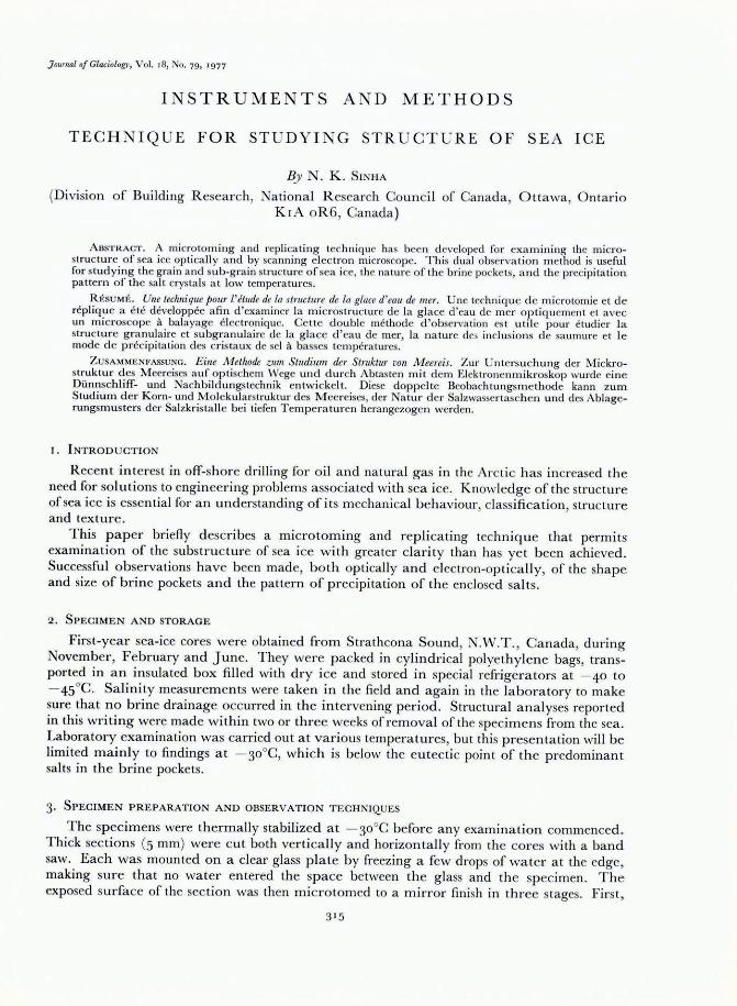
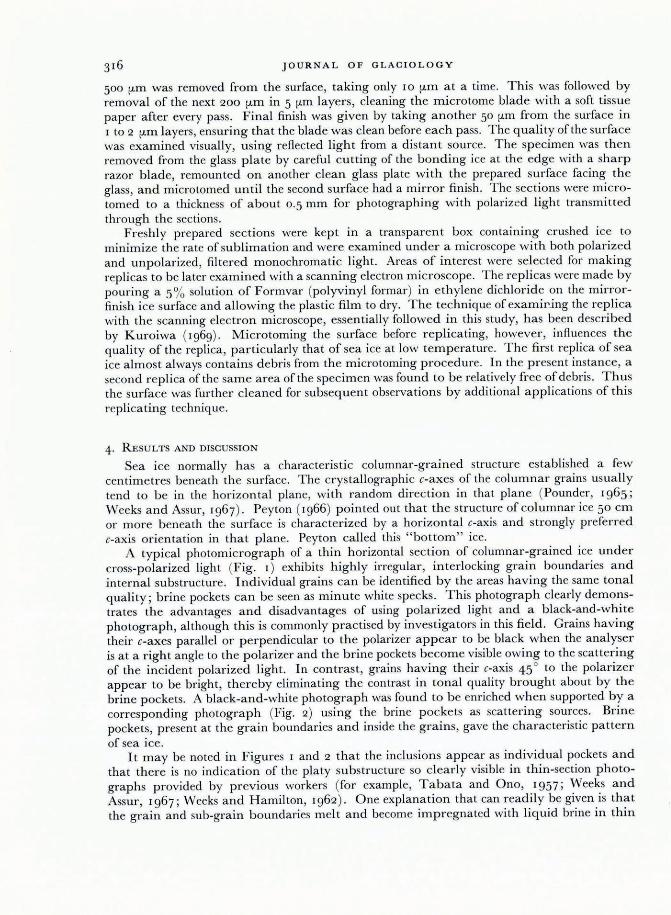
A typical photomicrograph of a thin horizontal section of columnar-grained ice under cross-polarized light (Fig. I) exhibits highly irregular, interlocking grain boundaries and internal substructure. Individual grains can be identified by the areas having the same tonal quality; brine pockets can be seen as minute white specks . This photograph clearly demonstrates the advantages and disadvantages of using polarized light and a black-and-white photograph, although this is commonly practised by investigators in this field. Grains having their c-axes parallel or perpendicular to the polarizer appear to be black when the analyser is at a right angle to the polarizer and the brine pockets become visible owing to the scattering of the incident polarized light. In contrast, grains having their c-axis 45 0 to the polarizer appear to be bright, thereby eliminating the contrast in tonal quality brought about by the brine pockets. A black-and-white photograph was found to be enriched when supported by a corresponding photograph (Fig. 2) using the brine pockets as scattering sources. Brine pockets, present at the grain boundaries and inside the grains, gave the characteristic pattern of sea ice.
It may be noted in Figures I and 2 that the inclusions appear as individual pockets and that there is no indication of the platy substructure so clearly visible in thin-section photographs provided by previous workers (for example, Tabata and Ono, 1957 ; Weeks and Assur, 1967 ; Weeks and Hamilton, 1962) . One explanation that can readily be given is that the grain and sub-grain boundaries melt and become impregnated with liquid brine in thin
IN STR U M E NT S AND METHOD S
Fig. I . Photograph oJ (I horiz ontal thin sectioll oJ columnar-grained sea ice at -30°C, IInder /Jolarized light.
sections prepa red a t e leva ted tem pera tures o r by the hot pla te techn iq ue commonl y used . Little a ttention has been p a id in the pas t to th e deta ils of prepa ra ti on and examina tion of thin secti ons, but it was observed during this in ves tigation th a t even the choice o f the source ofligh t influenced the na ture o f the thin secti ons. T he absorption of infra-red ra diation from the li ght sou rce was found to introduce und esira ble morphologica l changes in the thin sections. Green (546 nm fil ter) m onochroma ti c li g ht, near which th e normal eye is m ost sensi tive a nd the a bsorption coeffi cient o f ice is neglig ibl e, was therefore used.
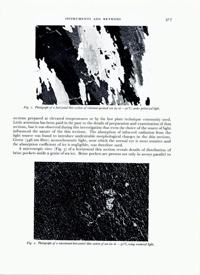
A mic roscopic view (Fig . 3) of a hor izonta l thin section reveals deta il s o f di stribution of brine pocke ts inside a grain of sea ice. Brine pockets a re presen t not onl y in a lTays para ll el to
F ig. 2 . Photograph oJ a microtomed horizontal thin section oJ sea ice at - 30°C, using sca ttered light.
JO U RNAL OF GLAC10LOGY
the basal plane of the grain but also along the c-axis. This distribution seems to outline the original boundaries of the dendritic structure formed at the ice- water interface during freezing . The entrapment process of brine in the sea ice is usually explained by the constitutional supercooling at the ice- water interface, causing a planar interface to become unstable and change to a highly cellular or dendritic interface (Harrison and Tiller, 1963; Lofgren and Weeks , 1967).
Vertical sections were also prepared to p ermit examination of the distribution of the brine pockets along the length of the grains. Comparison of the two sections revealed that the majority of brine pockets were quite irregular, although cylindrical and ellipsoidal-tabular shapes were also present.
According to phase relations of sea-water (Assur, 1958), precipitation of salts depends upon the temperature of the ice. Although significant discrepancies of the phase diagram at low temperatures have been discussed by W eeks ( [967), they are not important, at least not down to about - 30°C. Precipitation of the salts from sea-ice brine at various temperatures was deduced from changes in brine composition and stability ranges of individual salts in the corresponding pure-salt- water systems. A survey of the literature, however, indicates that no one has yet observed under controlled conditions the location and morphology of the precipitated salt crystals in brine pockets.
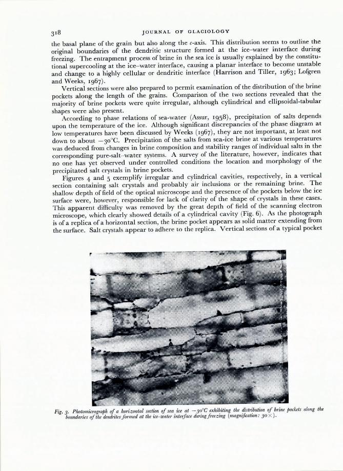
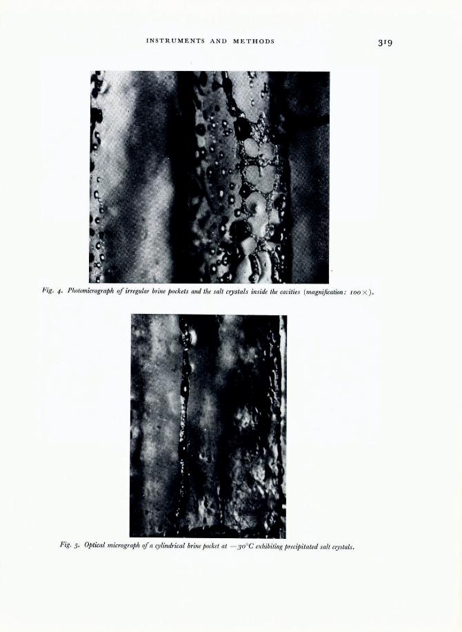
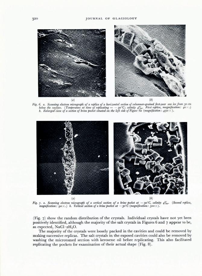
Figures 4 and 5 exemplify irregular and cylindrical cavities, respectively, in a vertical section containing salt crystals and probably air inclusions or the remaining brine. The shallow depth of field of the optical microscope and the presence of the pockets below the ice surface were, however, responsible for lack of clarity of the shape of crystals in these cases. T his apparent difficulty was removed by the great depth of field of the scanning electron microscope, which clearly showed details of a cylindrical cavity (Fig. 6). As the photograph is of a replica of a horizontal section, the brine pocket appears as solid matter extending from the surface. Salt crys tals appear to adhere to the replica. Vertical sections of a typical pocket
Fig. 3. Photomicrograph of a horizontal section of sea ice at -30°C exhibiting the distribution of brine pockets along the boundaries of the dendrites formed at the ice- water inteiface during freezing (magnification: 30 X ).
INSTR U MENT S AND METHOD S 3 19
Fig. 4. Photomicrograph of irregular brille pockets alld the salt crystals illside the cavities (magnification: I OO X ).
Fig. 5. Optical micrograph oJ a cylindrical brille pocket at - 30°C exhibitillg precipitated salt crystals.
JOURNAL OF GLACIOLOGY
(a) (b) Fig. 6. a. Scanning electron micrograph of a replica of a horizontal section of columnar-grained first-year sea ice from 30 cm
below the surface. ( T emperature at time of replicating = - 30°C; salinity 1- %0 ' First replica , magnification: 40 x .) b. ElIlarged view of a section of brine pocket situated on the lift side of Figure 6a (magnification: , 1-50 X ) .
(a) (b) Fig. 7. a. Scanning electron micrograph of a vertical section of a brine pocket at - 30°C, salinity 1- %0 ' (Second replica,
magnification : 90 X .) b. Vertical section of a brine pocket at - 30°C (magnification : 500 X ) .
(Fig. 7) show the random distribution of the crystals . Individual crystals have not yet been positively identified , although the majority of the salt crystals in Figures 6 and 7 appear to be, as expected , aCl· 2H20.
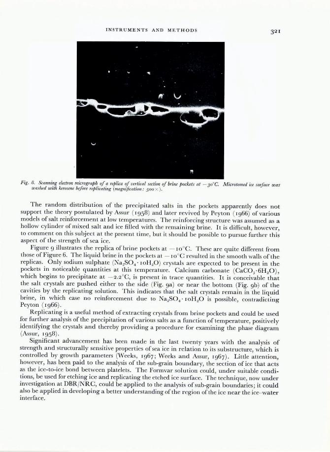
The majority of the crys tals were loosely packed in the cavities and could be removed by making successive replicas. The salt crystals in the exposed cavities could also be removed by washing the microtomed section with kerosen e oil before replicating. This also facilitated replicating the p ockets for examination of their actual shape (Fig. 8).
IN STR U MENT S AND M E THOD S 321
Fig. 8. Scanning electron micrograplz o.f a replica o.f vertical sectioll o.f brine jJockets at - 30oe . Microtollled ice sur.face was washed with kerosene bifore replicating (magnification,' 500 X ) .
The random distribution of the precipitated a lts in the pockets apparently does not support the theory postulated by Assu r ( 1958) and later rev ived by Peyton ( 1966) of various models of salt reinforcem ent at low temperatures. T hc reinfo rcing structure was assumed as a hollow cylinder of mixed sal t and ice fill ed with the remain ing brine. It is difficult, however , to comment on this subj ect at the present time, but it should be poss ible to pursue furth er this aspect of the strength of sea ice.
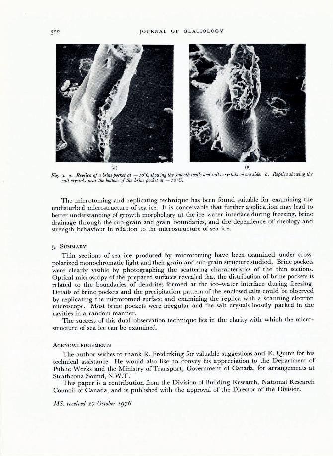
Figure 9 illustrates th e replica of brine pockets at - IO°e. These are quite different fmm those of Figure 6. T he liq uid brine in th e pockets at - IOoe resul ted in the smooth walls of the replicas . Only sodium sulphate (Na2S04 ' IOH 20 ) crystals a re expected to be present in the pockets in noticeable quantiti es at this tempera ture. Calcium carbonate (CaCO J '6H2 0 ) , which begins to precipitate at - 2.2 °C, is present in trace quantities. It is conceivable that the salt crystals are pushed either to the side (Fig. ga) o r near the bottom (Fig. 9b) of the cavities by the replicating solu tion . This indicates that the a lt cry tals remain in the liquid brine, in which case no reinforcement due to Na2S04 · loH20 is possible, contradicting Peyton ( 1966).
R epli cating is a useful method of extract ing crystals from brine pocke ts and could be used for furth er analysis of the precipitation of various salts as a function of temperature, positively identifying the crystals and thereby prov iding a procedure for examining the phase diagram (Assur, 1958).
Significant advancem ent has been made in the last twenty years with the analysis of strength and structurally sensitive propertie of ea ice in relation to its substructure, which is controll ed by growth parameters (Weeks, 1967; Weeks and Assur, Ig67) . Little attention, however, has been paid to the analysis of the ub-gra in boundary, the section of ice that acts as the ice-to-ice bond between platelets . The Formvar soluti on could , under suitable conditions, be used for etching ice and replicating the etched ice surface. The technique, now under inves tigation at DBRjNRC, could be applied to the analysis o f sub-grain boundaries; it could a lso be applied in developing a better understanding of tht: region of the ice near the ice- water interface.
JOURNAL OF GLACIOLOGY
(a) (b) Fig. 9. a. R eplica of a brine pocket at - IOoG showing the smooth walls and salts crystals on one side. b. R eplica showing the
salt crystals near the bottom of the brine pocket at - I Ooe.
The microtoming and replicating technique has been found suitable for examining the undisturbed microstructure of sea ice. It is conceivable that further application may lead to better understanding of growth morphology at the ice- water interface during freezing, brine drainage through the sub-grain and grain boundaries, and the dependence of rheology and strength behaviour in relation to the microstructure of sea ice.
5. SUMMARY
Thin sections of sea ice produced by microtoming have been examined under crosspolarized monochromatic light and their grain and sub-grain structure studied . Brine pockets were clearly visible by photographing the scattering characteristics of the thin sections. Optical microscopy of the prepared surfaces revealed that the distribution of brine pockets is related to the boundaries of dendrites formed at the ice- water interface during freezing. Details of brine pockets and the precipitation pattern of the enclosed salts could be observed by replicating the microtom ed surface and examining the r eplica with a scanning electron microscop e. Most brine pockets were irregular and the salt crystals loosely packed in the cavities in a random manner.
The success of this dual observation technique lies in the clarity with which the microstructure of sea ice can be examined.
ACKNOWLEDGEMENTS
The author wishes to thank R. Frederking for valuable suggestions and E. Quinn for his technical assistance. H e would also like to convey his appreciation to the D epartment of Public Works and the Ministry of Transport, Government of Canada, for arrangements at Strathcona Sound, N.W.T.
This paper is a contribution from the Division of Building R esearch, National Research Council of Canada, and is published with the approval of the Director of the Division.
MS. received 27 October 1976
INSTR U MENTS AND METHODS
REFERENCES
Assur, A. 1958. Composition of sea ice and its tensile strength. (In Arctic sea ice. Washington , D. C., p. 106-38. ([U .S.] National Academy o f Sciences- Nationa l R esearch Counci l Publica tion 598.))
H a rrison, J. D. , and T iller, \V. A. 1963. Controlled freezing of wate r. (In Kingery, W . D. , ed. Ice and snow; properties, processes, and applications : proceedings of a cOliference held at the Massachusetts institute of T echnology, February J2- J6, 1962. Cambridge, M ass., M .LT. Press, p. 215- 25.)
Kuroiwa , D . 1969. Surface topogra phy of etched ice crystals observed by a scanning electron microscope. J ournal of Glaciology, Vo!. 8, No. 54, p. 475- 83.
Lofgren, G. , and Weeks, W. F. 1967. Effect of growth parameters on substructure spacing in NaCI ice crys ta ls. U.S. Cold R egions Research and Engineering Laboratory. R esearch Report 195.
Peyton, H . R. 1966. Sea ice strength. College, Alaska , Geophysical Institute, Univers ity of Alaska. (UAC R- 182. ) Pounder, E. R. 1965. The physics of ice. Oxford, e tc., Pergamon Press. (The Commonwealth and International
Library. G eophysics Division. ) Tabata, T. , and Ono, N. 1957. K a ihyo no kozo tsuite [On the structure of sea ice]. T eion-kagaku: Low Temperature
Science, Ser. A, [No.] 16, p . 197- 210. Weeks, W. F. 1967. Understanding the variations o f the physical properties of sea ice. U.S. Cold Regions Research
and Engineering Laboratory. Special R eport 112. Weeks, W . F ., and Assur, A. 1967. The mechanical properties of sea ice. V.S. Cold R egions Research and
Engineering Laboratory. Cold regions science and engineering. Hanover, N.R., Pt. 11 , Sect. C3. Weeks, W. F. , and Hamilton, W . L. 1962. Petrographic characteristi cs of young sea ice, Point Barrow, Alaska.
American Mineralogist, Vo!. 47, Nos 7- 8, p. 945- 6 1.
6