Embed Size (px)
Citation preview
Instructions for use
Title Nanoparticulation of BCG-CWS for application to bladder cancer therapy
Author(s) Nakamura, Takashi; Fukiage, Masafumi; Higuchi, Megumi; Nakaya, Akihiro; Yano, Ikuya; Miyazaki, Jun; Nishiyama,Hiroyuki; Akaza, Hideyuki; Ito, Toshihiro; Hosokawa, Hiroyuki; Nakayama, Toshinori; Harashima, Hideyoshi
Citation Journal of controlled release, 176, 44-53https://doi.org/10.1016/j.jconrel.2013.12.027
Issue Date 2014-02-28
Doc URL http://hdl.handle.net/2115/57253
Type article (author version)
File Information WoS_64966_Nakamura.pdf
Hokkaido University Collection of Scholarly and Academic Papers : HUSCAP
1
Title
Nanoparticulation of BCG-CWS for application to bladder cancer therapy
Authors
Takashi Nakamuraa, Masafumi Fukiagea, Megumi Higuchia, Akihiro Nakayab, Ikuya Yanob, Jun Miyazakic,
Hiroyuki Nishiyamac, Hideyuki Akazad, Toshihiro Itoe, Hiroyuki Hosokawae, Toshinori Nakayamae,
Hideyoshi Harashimaa,*
aFaculty of Pharmaceutical Sciences, Hokkaido University, bJapan BCG Central Laboratory, cDepartment of
Urology, Faculty of Medicine, Tsukuba University, dTokyo University, Laboratory for systems, Biology and
Medicine, Research Center for Advanced Science and Technology, eDepartment of Immunology, Graduate
School of Medicine, Chiba University
*Correspondence:
Hideyoshi Harashima, Faculty of Pharmaceutical Sciences, Hokkaido University, Sapporo, Hokkaido
060-0812, Japan.
Telephone: +81-11-706-3919, Fax: +81-11-706-4879
E-mail: [email protected]

2
Abstract
The Mycobacterium bovis Bacille Calmett-Guerin cell wall skeleton (BCG-CWS) could be used to
replace live BCG as a bladder cancer drug. However, because BCG-CWS is poorly soluble, has a
strong-negative charge, very high molecular weight and heterogeneity in size of tens of μm, it cannot be used
in such an application. We report herein on the development of a novel packaging method that permits
BCG-CWS to be encapsulated into 166 nm-sized lipid particles. The BCG-CWS encapsulated nano particle
(CWS-NP) has a high uniformity and can be easily dispersed. Thus, it has the potential for use as a packaging
method that would advance the scope of applications of BCG-CWS as a bladder cancer drug. In a functional
evaluation, CWS-NP was efficiently taken up by mouse bladder tumor cells (MBT-2) in vitro and inhibited
tumor growth in mice bearing MBT-2 tumors. Moreover, intravesically administered CWS-NP showed
significant antitumor effects in a rat model with in naturally developed bladder cancer. An enhancement in
Th1 differentiation by CWS-NP was also confirmed in human T cells. In conclusion, CWS-NP represents a
promising delivery system for BCG-CWS for clinical development as a potent bladder cancer drug.
Keywords: packaging; nanoparticle; delivery system; bladder cancer; BCG-CWS; cancer immunotherapy
3
1. Introduction
Bacillus Calmette-Guerin (BCG) is a strain of Mycobacterium bovis that has been used to vaccinate
against tuberculosis. In addition, intravesical live BCG instillation is used in the treatment of non-muscle
invasive bladder cancer (NMIBC), particularly, superficial bladder cancer and carcinoma in situ (CIS) [1,2].
Based on results of clinical studies of Immunobladder® (live BCG), BCG therapy showed a complete
response rate of 66.0% and 79.1% against superficial bladder cancer and CIS, respectively and the
recurrence-free survival rate was 71.8% [3,4]. Although this form of treatment is an effective therapy, serious
side-effects associated with the use of live mycobacteria, such as the development of systemic BCG
infections, sepsis and even death, pose a significant concern [5]. In clinical studies, the incidence rate of
subjective and/or objective symptoms was 78.3% [3,4]. These side-effects can lead to terminating BCG
treatment in approximately 20% of patients. To avoid these issues, it is necessary to develop a non-infectious
and less toxic immunotherapeutic drug.
The BCG cell wall skeleton (BCG-CWS), the main immune active center of BCG [6], has the
potential to function as a substitute for live BCG. However, the clinical use of BCG-CWS has not yet been
achieved. The following reasons hamper such a use. It is very difficult to formulate a suitable water-soluble
drug without the BCG-CWS undergoing aggregation. The efficiency of uptake of BCG-CWS by cancer cells
is extremely low. Moreover, an oil-in-water (O/W) emulsion including a detergent is generally used for the
forced dispersion of BCG-CWS, when BCG-CWS is applied to animal and human studies. However the
emulsified BCG-CWS induces strong inflammation [7]. Because of this, it is extremely-difficult to apply an
O/W emulsion of BCG-CWS in the treatment of bladder cancer. Thus, it is essential to develop a delivery
system based on a novel concept for circumventing the problems that are currently associated with the use of
BCG-CWS.
An interesting report recently appeared regarding the size of BCG-CWS. Uenish and colleagues
reported that the apparent size of BCG-CWS varied drastically, depending on the polarity of the solvent used
[8]. BCG-CWS in a hydrophilic solvent such as saline formed a double folded sheet structure the size of
which was in the range from 4.7 to 67.8 μm. The larger size and wide size distribution were due to the
aggregation of BCG-CWS caused by interactions between hydrophobic moieties. On the other hand,
BCG-CWS became smaller and more compact (about 0.4 to 1.1 μm) when suspended in hydrophobic
solvents. The BCG-CWS took the form of a multilayered rolled sheet by rolling its hydrophilic moieties
(peptidoglycan layer) inward. In other words, the size of BCG-CWS particles can be controlled by the
polarity of the solvent. Thus, we hypothesized that a small sized and homogenous BCG-CWS formulation
could be prepared by encapsulating BCG-CWS under a hydrophobic environment within a lipid vesicle.
However, no methodology for accomplishing this is currently available.
Here, we report that the alternative packaging method for BCG-CWS without an oil and detergent
leads to the formation of nano- and uniformly-sized BCG-CWS. We succeeded in producing a BCG-CWS
encapsulated nano particle (CWS-NP) with a size of 166 nm and a high uniformity. We refer to this method
4
as the liposome evaporated via emulsified lipid (LEEL) method (Fig. 1). The nanoparticulation of BCG-CWS
by the LEEL method allowed us to demonstrate the efficient delivery of BCG-CWS into mouse bladder
tumor (MBT-2) cells and the induction of a strong antitumor effect in MBT-2 bearing mice. To direct the
CWS-NP prepared by the LEEL method (CWS-NP/LEEL) system toward clinical applications, the antitumor
effect of the intravesical CWS-NP/LEEL instillation and the immune effects of CWS-NP/LEEL in human
cells were confirmed. The CWS-NP/LEEL system showed the potential for use in intravesical instillation
therapy without any observable side-effects against naturally-occurring bladder tumors in a rat model. We
also demonstrated a proof-of-concept (POC) for CWS-NP/LEEL in human T cells, resulting in
CWS-NP/LEEL enhanced Th1 responses. Thus, this report demonstrates that CWS-NP/LEEL represents a
potentially promising formulation for use against bladder cancer as an alternative to the use of live BCG.
5
2. Methods and methods
2.1. Materials.
Egg phosphatidylcholine (EPC) was purchased from NOF Corporation (Tokyo, Japan). Cholesterol
(Chol) and N-(7-nitro-2,1,3-benzoxadiazol-4-yl) labeled dioleoyl phosphatidyl ethanolamine (NBD-DOPE)
was purchased from AVANTI Polar Lipids Inc. Stearylated octaarginine (STR-R8) was synthesized by
KURABO. BCG-CWS (SMP-105) was provided from MBR Co., Ltd. SMP-150’ starting raw material is a
killed BCG (Tokyo 172 strain) which strain can only be supplied by Japan BCG Laboratory.
2.2. Preparation of CWS-NP by the LEEL method and the hydration method
R8-modified liposomes (R8-Lip), composed of EPC/Chol/STR-R8 (70:30:2 molar ratio), were
prepared by the hydration method. Chloroform solutions of lipids were initially mixed in a flask, and the
solvent was removed with a rotary evaporator under reduced pressure to produce a lipid film. Hydration of
the lipid film was done by adding 5 mM HEPES buffer saline (HBS), and the lipid dispersion was extruded
through polycarbonate membrane filters (400-nm pore size; Nucleopore) with a Mini-Extruder (Avanti Polar
Lipids Inc.) for sizing of liposomes. The diameter and zeta-potential of R8-Lip were 283±16 nm and 48±2
mV, respectively (mean±SEM). BCG-CWS was dissolved in pentane, and the BCG-CWS in pentane and
R8-Lip were mixed. The mixture was sonicated using a probe type sonicator (BRANSON) to prepare an
O/W lipid emulsion. The pentane was removed with a rotary evaporator under reduced pressure to prepare
CWS-NP/LEEL. The CWS-NP/LEEL was finally extruded through polycarbonate membrane filters with
pore sizes varying from 0.4 to 0.2 μm using Mini-Extruder. The diameter, polydispersity index (PDI) and
zeta-potential of CWS-NP/LEEL were 166±2 nm, 0.257±0.021 and 31±0.4 mV, respectively (mean±SEM).
CWS-NP/Hyd was prepared by a previously described method [9]. The diameter, PDI and
zeta-potential of CWS-NP/Hyd were 495±59 nm, 0.453±0.037 and 34±5 mV, respectively (mean±SEM).
Diameter was measured by dynamic light scattering, and zeta-potential was determined by
laser-Doppler velocimetry with a ZETASIZER Nano (ZEN3600, Malvern Instruments).
To measure the encapsulating ratio of BCG-CWS in the CWS-NP/LEEL, the CWS-NP/LEEL was
dissolved in ethanol to disrupt the liposome structure, and BCG-CWS was obtained as a precipitate after
centrifugation. The pelleted BCG-CWS was suspended in hexane. The suspension of BCG-CWS was mixed
with 0.55% carbol-fuchsin solution. After mixing for 4 min, the absorbance of the hexane fraction was
measured at 530 nm. The encapsulating ratio of BCG-CWS in the CWS-NP/LEEL was 57±2%.
2.3. Negative staining and TEM observation
The samples were mixed with a 2% phosphotungstic acid solution and dropped onto a 400 mesh
carbon coated grid and dried in the air immediately after removing any excess solution. The samples were
then observed by transmission electron microscopy (TEM) (JEM-1200EX; JEOL Ltd.) at an acceleration
voltage of 80 kV. Digital images (2048 × 2048 pixels) were taken with a CCD camera (VELETA, Olympus

6
Soft Imaging Solutions GmbH).
2.4. Measurement of amount of aqueous phase in CWS-NP
CWS-NP/LEEL was prepared with 5 mM HBS including 0.1 mM calcein. Empty liposomes
(Empty-Lip), composed of EPC/Chol/STR-R8 (70:30:2 molar ratio), were prepared with 5 mM HBS
including 0.1 mM calcein by the hydration method. The empty liposomes were finally extruded through
polycarbonate membrane filters with 0.4 or 0.2 μm of pore sizes using Mini-Extruder. CWS-Lip/LEEL and
Empty-lip were diluted with 100 times by HBS. Two types of fluorescent intensities (FI) were measured. The
diluted CWS-NP/LEEL and Empty-Lip were measured FI (460 nm/550 nm) in the presence of 0.1 mM
CoCl2 (FI-A) or 0.1 mM CoCl2 and 1% Triton X-100 (FI-B). The amount of aqueous phase was calculated
using following formula: (FI/lipid nmol) = (FI-A - FI-B)/amount of lipid (nmol). Amount of lipid were
determined by means of a phospholipid assay kit (Wako).
2.5. Cell
MBT-2 derived from C3H/HeN mice were obtained from RIKEN. The cells were maintained at 37
oC in air with 5% CO2 in RPMI 1640 medium supplemented with 10% fetal calf serum.
2.6. Animals
Female C3H/HeN mice (7 weeks old) were purchased from Japan SLC, Inc. Male F344/DuCrlCrlj
rats (6 weeks old) were obtained from Charles River Japan. Mice and rats were housed in plastic cages and
maintained under standard conditions of temperature, humidity, and a 12:12-h light-dark cycle daily. Mice
and rats had free access to a standard diet and water. The Guidelines for the University Council for Animal
Care was followed at all times.
2.7. Analysis of cellular internalization in MBT-2 cells
MBT-2 cells were incubated with CWS-NP/LEEL labeled with the green fluorescence marker,
NBD-DOPE, in serum-free medium for 1 hour. In the inhibition experiment, the cells were incubated with
chlorpromazine or amiloride for 1 hour or 30 minutes, respectively, before the addition of CWS-NP/LEEL.
MBT-2 cells were then washed with 20 U/ml of heparin-PBS and culture medium. After an 1 hour incubation
in culture medium, the MBT-2 cells were collected and analyzed by FACSCalibur (BD Bioscience).
In the confocal laser scanning microscopy (CLSM) experiments, MBT-2 cell were incubated with
CWS-NP/LEEL labeled with green fluorescence marker, NBD-DOPE, in serum-free medium for 1 hour.
Then, the MBT-2 cells were washed by 20 U/ml of heparin-PBS and culture medium. After following 1 hour
incubation in culture medium, the MBT-2 cells were observed by CLSM (LSM510, Carl Zeiss) after staining
with LysoTracker Red.

7
2.8. Antitumor effects on mouse model
C3H/HeN mice were subcutaneously inoculated with a mixture of 3.5 × 10⁶ MBT-2 cells and
CWS-NP/LEEL (equivalent to 0.3 mg of BCG-CWS), CWS-NP/LEEL (equivalent to 0.1 mg of BCG-CWS)
or NP without CWS (equivalent to 0.3 mg dose). Tumor volume was calculated using the following formula:
(major axis × minor axis2) × 0.52.
2.9. Antitumor effects on rat model
BBN bladder carcinogenesis is generally considered to be a model for superficial bladder tumor.
Rats were given 0.05% N-butyl-N-(4-hydroxybutyl) nitrosamine (BBN) in the drinking water and diets
containing 5% (w/w) sodium ascorbate, cancer-causing promoter, for 8 weeks. The rats were divided into 4
groups: group 1, Vehicle; group 2, NP without CWS (equivalent to 0.1 mg dose); group 3, CWS-NPLEEL
(equivalent to 0.03 mg of BCG-CWS); group 4, CWS-NP/LEEL (equivalent to 0.1 mg of BCG-CWS). Each
sample (0.1 ml/rat) was intravesically administered to the rat at weekly intervals for 8 weeks. At the end of
the treatment, the rats were killed under anesthesia. Before removal of the bladder from each rat, the bladder
was intravesically injected with 10% neutral buffer formalin as a fixative for histological analyses. A ligature
was placed around the bladder neck to maintain the proper distention. After paraffin embedding, 3 μm tissue
sections were prepared. The sections were then stained with hematoxylin-eosin and observed
microscopically.
2.10. Human Th1/Th2 differentiation culture
Human Th1/Th2 differentiation culture was performed by the method described previously [10].
Whole blood was obtained from 4 healthy donor volunteers between 24 and 34 years old. The protocol was
approved by the Institutional Ethics Committee (No. 1972). PBMCs were isolated by Ficol-Paque
(Pharmacia-Upjohn) gradient centrifugation. Naïve CD4+ T cells were stained with anti-CD8/CD45RO-FITC
and then purified using anti-FITC magnetic beads (Miltenyi Biotec) and Auto-MACS cell sorter (Miltenyi
Biotec) by negative sorting. Naïve CD 4 T cells (7.5 × 10⁵ per well) were stimulated with 20 µg/ml
immobilized anti-CD3 antibody (Raritan, Somerset Country, UJ, USA) for 2 days in the presence of 50 U ml
IL-2 (Shionogi & Co., Ltd), 1 ng/ml IL-12 (R&D systems) and 5 µg/ml anti-IL-4 antibody (BD Bioscience)
under Th1 conditions. For Th2 conditions, the cells were stimulated with 20 µg/ml immobilized anti-CD3
antibody in the presence of 50 U ml IL-2, 1 ng/ml IL-4 (R&D systems) and 5 µg/ml anti-IFN-γ antibody (BD
Bioscience). The cells were then transferred to new plates and cultured for another 5 days in the presence of
cytokines and antibodies used in the same culture conditions. The final concentrations of BCG-CW,
CWS-NP/LEEL and NP without CWS were adjusted to 1, 3, 10 and 30 μg/ml.
2.11. Statistical analysis
Comparisons between multiple treatments were made by one-way analysis of variance, followed by
8
Tukey-Kramer test or Dunnett test. Comparisons between two treatments were performed by unpaired t-test.
In the case of the comparison of tumor volumes, the two-way repeated analysis of variance was used,
followed by Dunnett test. The comparison in survival experiment was made by Kaplan-Meier, followed by
Longrank test.
9
3. Results
3.1. Characterization of CWS-NP/LEEL
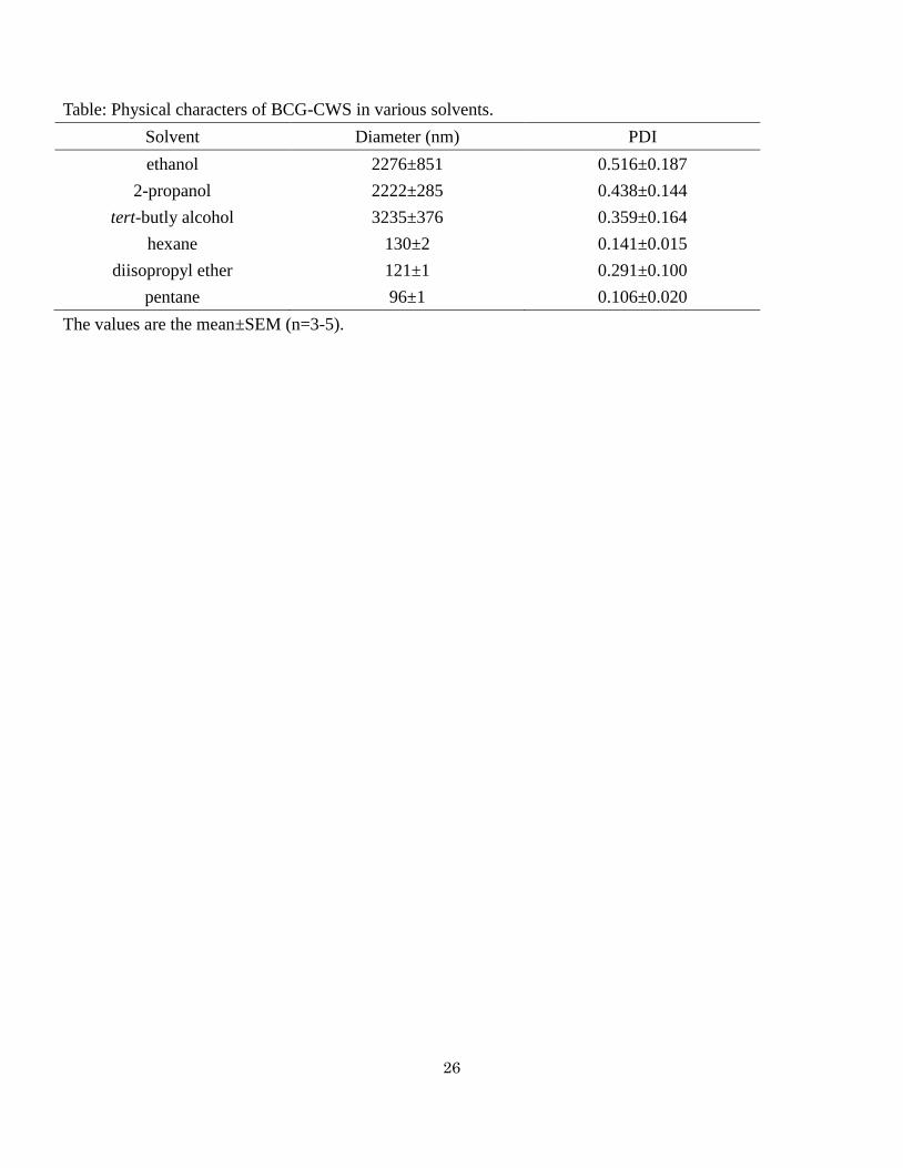
We first examined the use of various hydrophobic solvents in forming compact BCG-CWS particles.
BCG-CWS was suspended in several solvents and the diameter and PDI were measured (Table). As expected,
BCG-CWS in hydrophilic solvents, such as ethanol, 1-propanol, tert-butyl alcohol, underwent aggregation to
sizes in excess of a μm. On the other hand, in hydrophobic solvents, such as hexane, diisopropyl ether,
pentane, BCG-CWS formed compact sized particles of around 100 nm. In particular, BCG-CWS in pentane
showed the most compact size (96 nm) and a high homogeneity (PDI=0.106). Thus, we selected pentane as
the hydrophobic solvent. Figure 2a also clearly shows that, when BCG-CWS is suspended in pentane, a
translucent suspension is produced, while extensive aggregation in HBS and μm sized of aggregation in
ethanol were observed. Subsequently, a pentane solution including BCG-CWS was loaded on HBS
containing R8-Lip the size of which was around 280 nm (Fig. 1). The surface of the liposomes was modified
with the octaarginine (R8) peptide in the form of STR-R8. The R8 peptide is a type of cell-penetrating
peptide and is highly positively charged [11], permitting the efficient delivery of CWS-NP/LEEL into cells.
The loaded solution was then emulsified by means of a probe-type sonicator to form an O/W emulsion.
Lipids and STR-R8 function as emulsifiers, because they are amphipathic molecules. Their hydrophilic and
hydrophobic groups are likely to be directed to buffer phase and pentane phase, respectively. Therefore,
BCG-CWS which is kept in compact form in the pentane phase is covered with lipids and STR-R8 in the
case of an O/W emulsion. Finally, the pentane is removed by evaporation. The LEEL method makes it
possible to cover BCG-CWS with a lipid vesicle without interaction with a hydrophilic environment. The
solution of CWS-NP/LEEL was a translucent suspension (Fig. 2a). The diameter and PDI were 166 nm and
0.257, respectively. The encapsulating ratio of BCG-CWS in CWS-NP/LEEL was 57%.
We also prepared CWS-NP by the hydration method (CWS-NP/Hyd) to compare the characteristics
of the product with the CWS-NP/LEEL [9]. In the hydration method, BCG-CWS undergoes aggregation
during hydration with HBS, because the BCG-CWS in a lipid film is in a hydrophilic environment. The size
distribution of the CWS-NP/LEEL became sharp (size range from 33 nm to 531 nm) in comparison with that
of CWS-NP/Hyd (size range from 79 nm to 5560 nm) (Fig. 2b). This result clearly shows that the LEEL
method increased the homogeneity of the particle population, compared with that of the hydration method.
We also confirmed the structure of the CWS-NP/LEEL by TEM. In the case of CWS-NP/Hyd, a huge
structure that included multi-layered strings was observed instead of lipid vehicles (Fig. 2c). This finding
suggests that BCG-CWS forms multi-layered strings with lipids, but not be covered with a lipid bilayer. On
the other hand, in the case of CWS-NP/LEEL, lipid vesicles with diameters of approximately 200 nm were
observed and the condensed structure was present in the center of the lipid vesicle (Fig. 2d). This suggests
that the CWS-NP/LEEL had compact-shaped BCG-CWS in the center, which was covered with lipid layers.
Moreover, CWS-NP/LEEL is particularly marked by the nature of its internal structure. It is
expected that the central phase of the lipid vesicle remains hydrophobic since the hydrophobic core of
10
BCG-CWS is covered with a lipid fatty acid chain after removal of the hydrophobic solvent, based on Figure
1 and Figure 2d. We then compared the amount of aqueous phase between CWS-NP/LEEL and general
liposomes (Fig. 2e). The amount of aqueous phase for the CWS-NP/LEEL was significantly lower than that
of empty liposomes which have the same particle size as the CWS-NP/LEEL. The amount of aqueous phase
of the liposome did not drastically change, when the particle size of the liposome became large by increasing
the number of lamellar structures (Empty-Lip 248 nm). That is, the difference in the amount of aqueous
phase between CWS-NP/LEEL and the 160 nm-sized Empty-Lip presumably reflects the amount of central
aqueous phase. These data indicate that the CWS-NP/LEEL contains no central aqueous phase. In Figure 2d,
we showed TEM images of the CWS-NP/LEEL. However, the CWS-NP/LEEL solution actually includes
lipid vesicles without BCG-CWS. In the TEM observations, the lipid vesicles without BCG-CWS were
devoid of any central aqueous phase (Fig. 2f). These results also suggest that nanoparticles prepared by
LEEL method do not contain a central aqueous phase, that is, the structure of the CWS-NP/LEEL is different
from that of a typical liposome.
Collectively, it is clear that the LEEL method resulted in the formation of nano- and uniformly-sized
BCG-CWS nanoparticles by encapsulating BCG-CWS into sub-200 nm sized lipid vehicles.
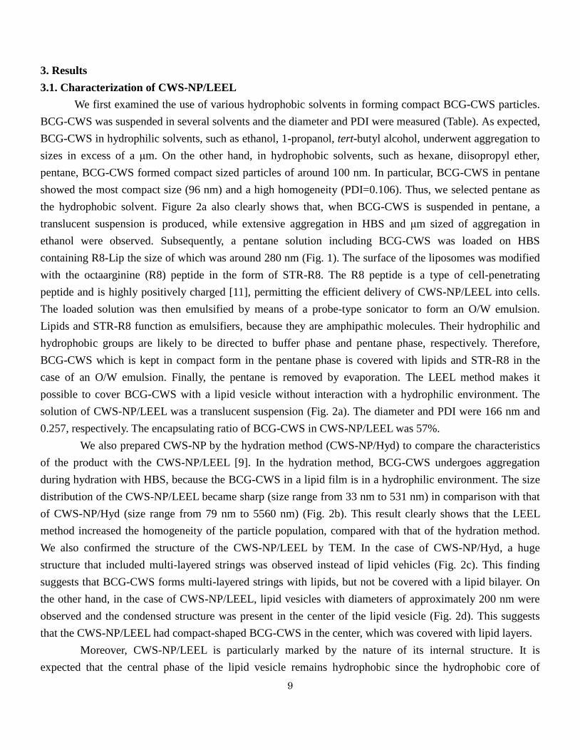
Figure 1: Schema of liposomes evaporated via the emulsified lipid (LEEL) method.
BCG-CWS forms compact particles in pentane. The BCG-CWS included pentane solution then loaded on HEPES
buffer saline (HBS) containing liposomes of which size was around 280 nm. The liposome contains stearylated octaarginine
(STR-R8) and is modified with octaarginine (R8) peptide on lipid surface. The loaded solution was then emulsified by the
means of a probe-type sonicator to form an oil in water (O/W) emulsion. Lipids and STR-R8 work as emulsifier and their
11
hydrophilic group and hydrophobic group are directed to buffer phase and pentane phase, respectively. That is, the drop of
pentane containing compact BCG-CWS is surrounded by lipid layers. Finally, the pentane is removed by evaporation.
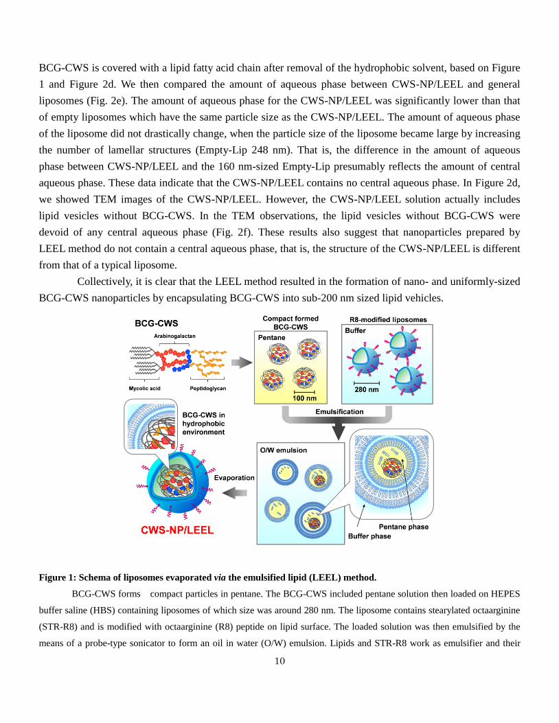
Figure 2: Characters of CWS-NP prepared by LEEL method.
(A) Physical appearances of BCG-CWS in HBS, EtOH and Pentane, and CWS-NP/LEEL. (B) Size distribution of
CWS-NP/LEEL and CWS-NP/Hyd. (C) TEM image of CWS-NP/Hyd. Bar shows 200 nm. (D) TEM images of
CWS-NP/LEEL (left: whole, right: magnification). Bars show 200 nm. (E) Fluorescent intensities of empty liposomes
(Empty-lip) and CWS-NP/LEEL. The values are the mean± SEM of at least three different experiments (**P<0.01). (F)
TEM image of CWS-NP/LEEL without BCG-CWS. Bar shows 100 nm.
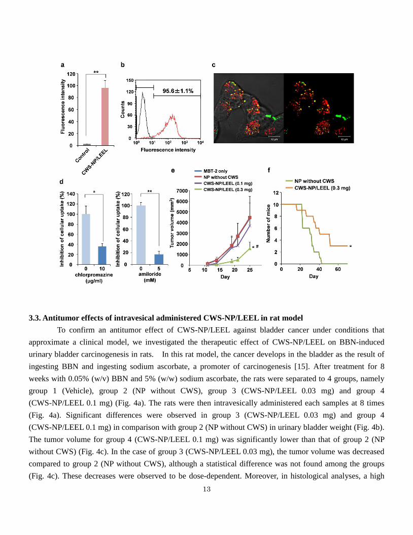
3.2. Functional analysis of CWS-NP/LEEL with MBT-2 cells
The direct internalization of live BCG into bladder cancer cells seems to be important for an
antitumor immune response in BCG therapy [12,13]. We then prepared fluorescence-labeled CWS-NP/LEEL
particles and investigated their cellular uptake by MBT-2 cells by flow cytometry and CLSM. The
fluorescent intensity of MBT-2 cells was drastically increased after treatment with CWS-NP/LEEL (Fig. 3a).
12
The CWS-NP/LEEL particles were taken up by more than 95% MBT-2 cells (Fig. 3b). In a CLSM analysis,
MBT-2 cells were stained with a red fluorescence (LysoTracker) 2 hours after a green fluorescence-labeled
CWS-NP/LEEL was pulsed. Numerous yellow signals were observed in MBT-2 cells (Fig. 3c). This indicates
that the internalized CWS-NP/LEEL particles were mainly localized in lysosomes. Thus, these results
suggest that CWS-NP/LEEL is efficiently taken up by MBT-2 cells.
It has been reported that R8-Lip particles are taken up via macropinocytosis and clathrin-mediated
endocytosis, depending on the ratio of R8 used to modify them [14]. Thus, we examined the cellular entrance
pathway of CWS-NP/LEEL in MBT-2 cells by chlorpromazine, a clathrin-mediated endocytosis inhibitor,
and amiloride, an inhibitor of macropinocytosis. In the presence of 10 μg/ml chlorpromazine, the uptake of
CWS-NP/LEEL was significantly inhibited (Fig. 3d). Moreover, 5 mM amiloride had a remarkable effect on
the uptake of CWS-NP/LEEL (Fig. 3d). It can therefore be concluded that the cellular uptake of the
CWS-NP/LEEL in MBT-2 involves clathrin-mediated endocytosis and macropinocytosis.
We next investigated the antitumor effects of CWS-NP/LEEL against MBT-2 tumors in vivo. Mice
were inoculated with the mixture of MBT-2 cells and each sample, and tumor growth was monitored. The
growth of tumors of MBT-2 cells in the presence CWS-NP/LEEL (BCG-CWS 0.3 mg) was significantly
suppressed, while that for MBT-2 cells mixed with CWS-NP/LEEL (BCG-CWS 0.1mg) or CWS-NP/LEEL
without BCG-CWS (NP without CWS) was not suppressed, in comparison with the growth of tumors of only
MBT-2 cells (Fig. 3e). Moreover, in the case of CWS-NP/LEEL (BCG-CWS 0.3 mg), the mice showed a
significant improvement in the median time of survival compared with the control mice, which had been
inoculated with a mixture of NP without CWS and MBT-2 cells (Fig. 3f). These results indicate that the
CWS-NP/LEEL preparation efficiently induced anti-tumor effects against mouse bladder carcinomas.
Figure 3: Functional analysis of CWS-NP/LEEL with MBT-2 cell.
(A) The amounts of CWS-NP/LEEL taken up by MBT-2 cells. Fluorescence labeled CWS-NP/LEEL was pulsed to
MBT-2 cells and the fluorescence intensity was measured by flowcytometer. The values are the mean± SEM of at least three
different experiments (**P<0.01). (B) Histogram analysis of cellular uptake of CWS-NP/LEEL by flowcytometer. The
values are the mean± SEM of at least three different experiments. (C) CLSM analysis of cellular uptake of CWS-NP/LEEL.
Green signals and red signals show CWS-NP/LEEL and lysosome, respectively. White bars means scale of 10 μm. (D)
Analysis of uptake pathway of CWS-NP/LEEL with inhibitors. Cellular uptake of CWS-NP/LEEL in MBT-2 cells were
measured in the presence of chlorpromazine (left) or amiloride (right) using a flowcytometer. The values are the mean ±
SEM of at least three different experiments (*P<0.05, **P<0.01). (E) Inhibition of MBT-2 tumor growth. The mixture of
MBT-2 cells and NP without CWS or CWS-NP/LEEL (0.1 mg) or CWS-NP/LEEL (0.3 mg) were subcutaneously inoculated
to mice and the tumor volumes were measured. The values are the means (n=4-6, *P<0.05: vs MBT-2 only, #P<0.05: vs NP
without CWS). (F) Survival number of mice after inoculation of the mixture of MBT-2 cells and NP without CWS or
CWS-NP/LEEL (0.3 mg) (n=10, *P<0.05 vs NP without CWS).
13
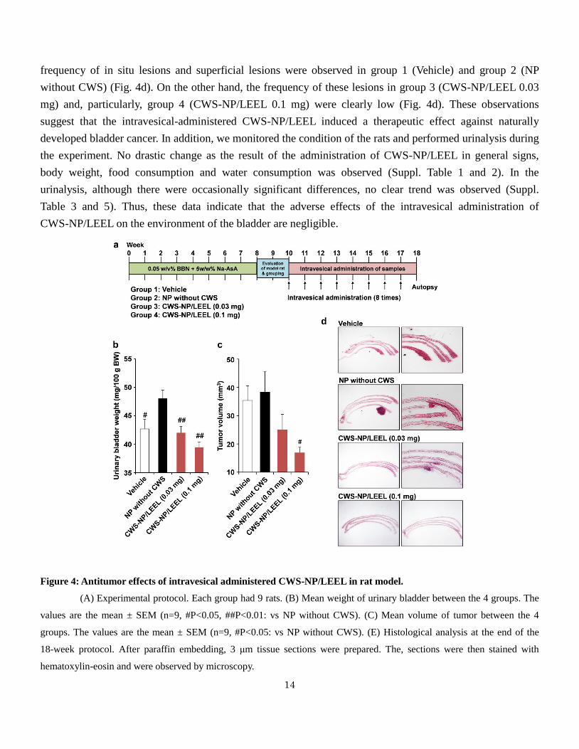
3.3. Antitumor effects of intravesical administered CWS-NP/LEEL in rat model
To confirm an antitumor effect of CWS-NP/LEEL against bladder cancer under conditions that
approximate a clinical model, we investigated the therapeutic effect of CWS-NP/LEEL on BBN-induced
urinary bladder carcinogenesis in rats. In this rat model, the cancer develops in the bladder as the result of
ingesting BBN and ingesting sodium ascorbate, a promoter of carcinogenesis [15]. After treatment for 8
weeks with 0.05% (w/v) BBN and 5% (w/w) sodium ascorbate, the rats were separated to 4 groups, namely
group 1 (Vehicle), group 2 (NP without CWS), group 3 (CWS-NP/LEEL 0.03 mg) and group 4
(CWS-NP/LEEL 0.1 mg) (Fig. 4a). The rats were then intravesically administered each samples at 8 times
(Fig. 4a). Significant differences were observed in group 3 (CWS-NP/LEEL 0.03 mg) and group 4
(CWS-NP/LEEL 0.1 mg) in comparison with group 2 (NP without CWS) in urinary bladder weight (Fig. 4b).
The tumor volume for group 4 (CWS-NP/LEEL 0.1 mg) was significantly lower than that of group 2 (NP
without CWS) (Fig. 4c). In the case of group 3 (CWS-NP/LEEL 0.03 mg), the tumor volume was decreased
compared to group 2 (NP without CWS), although a statistical difference was not found among the groups
(Fig. 4c). These decreases were observed to be dose-dependent. Moreover, in histological analyses, a high
14
frequency of in situ lesions and superficial lesions were observed in group 1 (Vehicle) and group 2 (NP
without CWS) (Fig. 4d). On the other hand, the frequency of these lesions in group 3 (CWS-NP/LEEL 0.03
mg) and, particularly, group 4 (CWS-NP/LEEL 0.1 mg) were clearly low (Fig. 4d). These observations
suggest that the intravesical-administered CWS-NP/LEEL induced a therapeutic effect against naturally
developed bladder cancer. In addition, we monitored the condition of the rats and performed urinalysis during
the experiment. No drastic change as the result of the administration of CWS-NP/LEEL in general signs,
body weight, food consumption and water consumption was observed (Suppl. Table 1 and 2). In the
urinalysis, although there were occasionally significant differences, no clear trend was observed (Suppl.
Table 3 and 5). Thus, these data indicate that the adverse effects of the intravesical administration of
CWS-NP/LEEL on the environment of the bladder are negligible.
Figure 4: Antitumor effects of intravesical administered CWS-NP/LEEL in rat model.
(A) Experimental protocol. Each group had 9 rats. (B) Mean weight of urinary bladder between the 4 groups. The
values are the mean ± SEM (n=9, #P<0.05, ##P<0.01: vs NP without CWS). (C) Mean volume of tumor between the 4
groups. The values are the mean ± SEM (n=9, #P<0.05: vs NP without CWS). (E) Histological analysis at the end of the
18-week protocol. After paraffin embedding, 3 μm tissue sections were prepared. The, sections were then stained with
hematoxylin-eosin and were observed by microscopy.
15
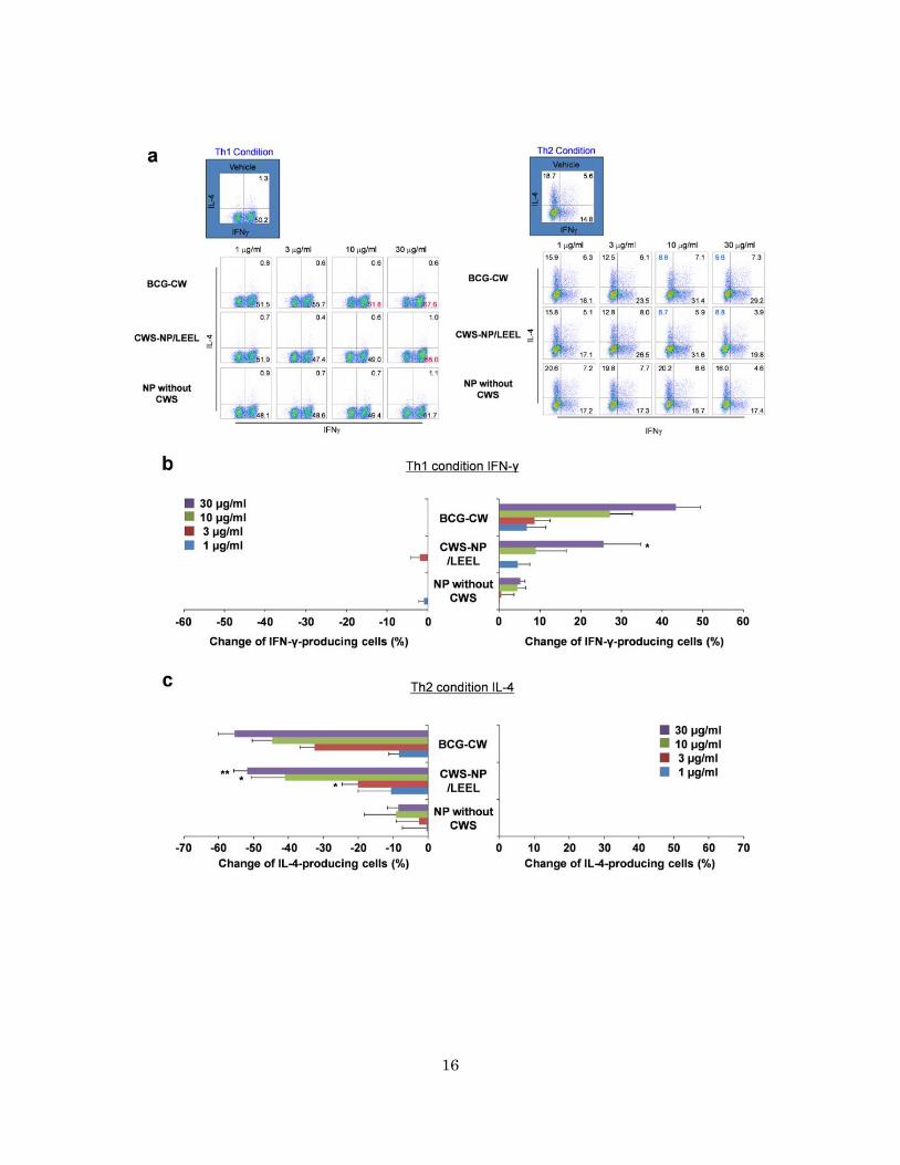
3.4. Effects of CWS-NP/LEEL in human Th1/Th2 cell differentiation
To achieve clinical applications of CWS-NP/LEEL, it is necessary to examine the antitumor ability
of CWS-NP/LEEL in human cells. To accomplish this, we investigated the promotional effect of a Th1
response in human peripheral blood naïve CD4 T cells. Naïve CD4 T cells differentiate to Th1 cells on
stimulation with Th1 cytokines, such as IL-12. Th1 cells then produce IFN-γ and promote antitumor
responses. An in vitro Th1/Th2 differentiation culture system was used with human peripheral blood naïve
CD4 T cells to evaluate the effects of CWS-NP/LEEL on the differentiation of human Th1/Th2 cells [10]. In
this system, a change in Th1/Th2 balance is indicative of a Th1-dominant condition or a Th2-domonant
condition, based on the number of cells producing IFN-γ and IL-4. BCG cell walls (BCG-CW) were
homogenously suspended in PBS and used as a positive control. NP without CWS was used as a negative
control. Figure 5a shows typical dot plots for the Th1 condition and the Th2 condition. Under Th1 culture
conditions, the generation of Th1 cells (low left: IFN-γ positive and IL-4 negative) was remarkably enhanced
in the presence of 30 μg/ml of CWS-NP/LEEL (Fig. 5a). On the other hand, the generation of Th2 cells (IL-4
positive and IFN-γ negative) was inhibited in the presence of 10 μg/ml and 30 μg/ml of CWS-NP/LEEL
under Th2 culture conditions (Fig. 5a).
The analysis was then extended to naïve CD4 T cells obtained from 4 healthy volunteers (with their
permission). Under Th1 culture conditions, 30 μg/ml of CWS-NP/LEEL significantly increased the number
of IFN-γ-producing cells compared to that of NP without CWS (Fig. 5b). On the other hand, 3 μmg/ml, 10
μg/ml and 30 μg/ml of CWS-NP/LEEL dose-dependently decreased the number of IL-4-producing cells in
comparison with these of NP without CWS (Fig. 5c). This inhibition by CWS-NP/LEEL was similar to that
by BCG-CW, a positive control. Therefore, these results indicate that CWS-NP/LEEL enhances the
generation of IFN-γ-producing cells under Th1 culture conditions and inhibits the generation of
IL-4-producing cell under Th2 culture conditions, that is, CWS-NP/LEEL directs the Th1/Th2 balance
toward Th1 immunity.
Figure 5: Influence of CWS-NP on human Th1/Th2 differentiation.
Naïve T cells from PBMC were stimulated with anti-CD3 in the presence of anti-IL-4 antibody, IL-12 and IL-2
(Th1 conditions) or in the presence of anti-IFN-γ antibody, IL-4 and IL-2 (Th2 conditions). The cultured cells were subjected
to intracellular staining with anti- IFN-γ and anti-IL-4. BCG-CW, NP without CWS and CWS-NP/LEEL were added to the
culture. (A) Typical dot plots of Th1 condition and Th2 condition. (B) Effect on Th1 differentiation using PBMCs obtained
from four human healthy donors. The abscissa axis shows a rate of change for IFN-γ positive cells in vehicle group. The
values are the mean ± SEM (n=4, *P<0.05: vs NP without CWS). (C) Effect on Th2 differentiation using four human healthy
donor PBMCs. The abscissa axis shows the rate of change for IL-4 positive cells in the vehicle group. The values are the
mean ± SEM (n=4, *P<0.05, **P<0.01: vs NP without CWS).
16
17
4. Discussion
The potential value of CWS-NP/LEEL in the bladder cancer market is significant and of interest.
According to GLOBOCAN 2008, published by the International Agency for Research on Cancer, it is
estimated that 382,660 new case of bladder cancer were diagnosed and the mortality was 150,282 worldwide
[16]. In Europe and the USA, bladder cancer is the fourth most common form of cancer in men. For example,
437,180 men were bladder cancer patients on January 1, 2012 in the USA [17]. NMIBC represents 80% of
the incident cases of bladder cancer [18]. Intravesical BCG instillation is recommended for patients who are
at intermediate and high risk. That is, many patients who are diagnosed with NMIBC receive BCG therapy.
In general, bladder cancer treatment is a lengthy procedure, takes time in comparison with other type of
cancers. The BCG immunologic activities against bladder cancer persist for more than 1 year after the initial
6- to 8-week therapeutic course. However, the effects begin to wane after 3 to 6 months. Thus, maintenance
therapy is needed. After the 6-week initial course, maintenance therapy is given as 3 weekly instillations at 3,
6, 12, 18, 24, 30, and 36 months for a total 27 instillation over a period of 3 years. Moreover, the high
recurrence rate of bladder cancer causes a further prolongation of the treatment period. Although BCG
therapy shows prominent therapeutic effects and the inhibition of recurrence, BCG therapy is a considerable
burden for patients, because it often induces various side-effects, both small and large. Occasionally, patients
are compelled to terminate BCG therapy due to serious side-effects. If a non-infectious immunotherapeutic
drug without side-effects were to be developed, approximately 80% of bladder cancer patients would likely
be the recipients of a significant benefit, namely, the alleviation of side-effects. The alleviation of side-effects
would result in improved therapeutic effects, the expanding of applications to low-risk patients and a
reduction in overall cost. Therefore, the clinical application of BCG-CWS promises to drastically change the
QOL of patients.
The formulation of BCG-CWS has been bottleneck for clinical applications of BCG-CWS. In this
study, we developed the LEEL method and succeeded in constructing CWS-NP which had a size of 166 nm
and a high uniformity. The concept of the LEEL method involves the packaging of BCG-CWS in the state of
hydrophobic-compact form into a lipid vesicle. Thus, it is easily inferred that the size of BCG-CWS in
hydrophobic solvent affects to the size of CWS-NP/LEEL. As shown in Table, we selected pentane as a
hydrophobic solvent. Pentane allowed BCG-CWS to form smallest and most uniformly-sized particle.
Subsequently, O/W emulsion is prepared by sonication after adding a hydrophilic solution including R8-Lip
which functions as an emulsifier. As shown in Figure 1, it is likely that four types of O/W emulsions (i.e.
mono-layered emulsion including BCG-CWS, multi-layered emulsion including BCG-CWS, mono-layered
emulsion without BCG-CWS, and multi-layered emulsion without BCG-CWS) are formed. However, it can
be inferred that a multi-layered O/W emulsion including BCG-CWS and a multi-layered O/W emulsion
without BCG-CWS are actually formed, because a multi-layered lipid vesicle encapsulating BCG-CWS
(CWS-NP/LEEL, Fig. 2d) and a multi-layered lipid vesicle without BCG-CWS (Fig. 2f) were mainly
observed. After removing the pentane and sizing, the CWS-NP/LEEL is prepared. In Figure 2d, the
18
CWS-NP/LEEL particles appeared to be weakly attached to each other. We consider that this aggregation is
induced by simply physically adhering the each other, which is different from general aggregation mediated
by the fusion of each particle, such as CWS-NP/Hyd. It is also likely that the treatment with the 2%
phosphotungstic acid solution or the drying procedure resulted in an enhancement in the interaction between
each particle. Thus, it is likely that this aggregation does not occur in normal buffer (HBS), because it was
due to the treatment procedure used for the TEM observations. By the way, based on the TEM images, it is
likely that CWS-NP/LEEL is present in the form of a lipid vesicle in which the hydrophobic core of
BCG-CWS covered with the lipid fatty acid chain is coated with a multi lipid bilayer. Figure 2e also indicates
the structure of CSW-NP/LEEL described above in the term of the amount of aqueous phase. The different
amount of aqueous phase between CWS-NP/LEEL and the 160 nm-sized Empty-Lip indicates the amount of
central aqueous phase (Fig. 2e). The difference in the amount of aqueous phase between the 160 nm-sized
Empty-Lip and the 248 nm-sized Empty-Lip probably reflects the amount of aqueous phase in the space
multi-layered lipid bilayer, because the difference in liposomal size is mainly derived from the number of
lipid bilayer in these empty liposomes. This therefore suggests that the aqueous phase of CWS-NP/LEEL is
derived from the aqueous phase in the space multi-layered lipid bilayer. We can also conclude that the
construction of CWS-NP/LEEL by LEEL method is performed by the following processes: i) formulation of
multi-layered O/W emulsion which includes BCG-CWS in pentane phase; ii) coating the hydrophobic
BCG-CWS core with the hydrophobic moieties of mono-layer after removing the pentane phase.
The structure of the CWS-NP/LEEL mediated by the LEEL method is advantageous in terms of
inhibiting aggregation that can occur in a hydrophilic environment and size control of BCG-CWS
incorporating nanoparticle. Although BCG-CWS readily undergoes aggregation when in contact with a
hydrophilic solution or environment (Fig. 2a), no evidence for this was found in the case of the
CWS-NP/LEEL because the lipid vesicle protected BCG-CWS from coming into contact with a hydrophilic
solution or environment. In addition, the size of the CWS-NP/LEEL was less than 200 nm, because it was
kept compact by virtue of being in a lipid vesicle. On the other hand, as shown in Figure 2b and 2c,
CWS-NP/Hyd prepared by the hydration method, underwent aggregation, had a large size and the particles
were non-uniform. The reason for this is that BCG-CWS in the form of a lipid film comes into contact with
the hydrophilic buffer after hydration, resulting in the formation of large sized, BCG-CWS aggregates.
BCG-CWS is much larger than the thickness of the lipid bilayer. It is likely that BCG-CWS predominates the
character of delivery system. We solved this problem by encapsulating BCG-CWS in the hydrophobic state
into a lipid vesicle.
A BCG-induced infection of bladder tumor cells is a key for the induction of an effective antitumor
effect against bladder cancer [12,13]. Thus, one important role of CWS-NP/LEEL is the delivery of
BCG-CWS into bladder tumor cells. To successfully deliver BCG-CWS, we incorporated STR-R8 in the
CWS-NP/LEEL. The R8 peptide is a synthetic peptide that mimics the trans-activating transcriptional
activator of the human immunodeficiency virus [19], and has a highly positive charge and a high cellular
19
affinity. Nanoparticles modified with the R8 peptide have been successful in delivering several substances,
such as low molecular drugs, proteins and nucleic acids, under in vitro and in vivo conditions [11]. The high
cellular affinity of R8 peptide can be used for most type of cells: fibroblast cells [14], hepatic cells [20],
endothelial cells [21], tumor cells [22], immune cells [23,24] and so on. In this study, CWS-NP/LEEL was
efficiently internalized into MBT-2 cells (Figure 3A-3C). The findings suggest that the efficient
internalization into bladder tumor cells results in significant antitumor effects. Therefore, the R8 peptide is a
great advantage and is an essential-functional device, for use in the CWS-NP/LEEL.
In the functions of CWS-NP/LEEL as immunotherapeutic drugs against bladder cancer,
dose-dependent antitumor effects were also observed against MBT-2 cells which are a high-grade bladder
carcinoma and the survival rate of the CWS-NP/LEEL treated group was significantly extended (Figure
3E&3F). It is difficult to compare the antitumor effect of CWS-NP/LEEL based on currently published data,
because antitumor effects against bladder cancer by BCG-CWS have been performed in only a few
incidences [15,25,26]. In particular, studies using a delivery system (liposomal BCG-CWS prepared by the
hydration method) were performed by only our group [15,26]. On the other hand, in the case of live BCG, the
doses ranged between 0.1 mg and 1 mg/mouse against MBT-2 cells [27-30]. Live BCG at doses of 0.5 mg
and 1 mg strongly inhibited the growth of MBT-2 tumor. However, BCG at a dose of 0.1 mg showed no
antitumor effect, which is consistent with the results reported here [27,30]. Although the CWS-NP/LEEL also
showed no antitumor effect at a dose of 0.1 mg, CWS-NP/LEEL at a dose of 0.3 mg significantly inhibited
the growth of MBT-2 tumors (Fig. 3e). Thus, these findings suggest that the antitumor effect of
CWS-NP/LEEL against MBT-2 tumors may be comparable to that of live BCG.
It is important to confirm whether CWS-NP/LEEL has an antitumor effect in a model that
approaches a clinical condition. That is, it is necessary to investigate the antitumor effects of
intravesical-administered CWS-NP/LEEL against orthotopic bladder cancer. In this study, we selected a rat
model of BBN-induced bladder cancer, because feeding BBN induces NMIBC in the rat. The oral
administration of the carcinogen BBN induces urinary bladder tumor within a short time [31]. In addition to
BBN, sodium ascorbate was administered to promote urinary bladder carcinogenesis [32]. BBN
carcinogenesis also appears to be related to a risk factor for bladder cancer. Cigarette smoking is the primary
risk factor for bladder cancer. Cigarette smoke contains high levels of carcinogens, and nitrosamine one such
substance. Thus, it would be expected that the BBN carcinogenesis in a rat model would be similar to clinical
bladder cancer. CWS-NP/LEEL showed a therapeutic antitumor effect by intravesical instillation in the
bladder carcinogenesis rat model (Figure 4). The nanoparticulation of BCG-CWS appears to prevent the
aggregation of BCG-CWS in the bladder. In addition, the R8 peptide appears to promote cellular uptake by
bladder cancer cells. For this reason, the CWS-NP/LEEL appeared to show an antitumor effect in the bladder.
The results shown in Figure 4 clearly indicate that CWS-NP/LEEL functions as an immunotherapeutic agent
in the bladder.
In addition to therapeutic effects, low toxicity is another advantage of a non-infectious drug, such as
20
CWS-NP/LEEL. Intravesical BCG instillation involves several local and systemic side-effects [33]. The most
frequent local side-effects are BCG induced cystitis, irritative voiding symptoms and hematuria, which occur
in approximately 75% of all patients. Systemic side-effects include flu-like symptoms, such as general
malaise and fever, and occur in approximately 40% of patients. These local and systemic side-effects might
lead to terminating intravesical BCG treatment in approximately 20% of patients. On the other hand, no
drastic change was observed in the case of the intravesical administration of CWS-NP/LEEL between the
start and the end of the period in general signs, body weight, food consumption, water consumption, urinary
volume, specific gravity of urine and pH of urine (Supplemental Table 1, 2 and 3). In a urinalysis, although
occult blood was observed in 10-20% of the rats in each group (Supplemental Table 5), it is highly unlikely
that this side-effect was caused by the intravesical administration of CWS-NP/LEEL, because occult blood
also occurred in the vehicle treated group. In contrast with BCG, CWS-NP/LEEL did not induce serious
side-effects in the case of multiple administrations. Hence, this suggests that CWS-NP/LEEL is promising
drug for the treatment of bladder cancer in terms of its low side-effects.
It should also be noted that bladder cancer cells and normal cells are present in the bladder of cancer
patients or cancer model animals. This discussion deals with the effect of CWS-NP/LEEL in normal cells and
healthy animals, when CWE-NP/LEEL is intravesically administered. The inner cavity of the urinary bladder
is covered with urothelium (transitional epithelium). Urothelium linking the luminal surface, known as
umbrella cells, are engaged in tight junctions that prevent access to the lower transitional cell layers [34]. The
umbrella cells express characteristic extracellular proteins (uroplakins) that assemble into semi-rigid plaques
that provide effective shielding for the apical surface. Furthermore, a glycosaminoglycan (GAG)-rich mucin
layer which is produced and assembled on the apical surface of the umbrella cells isolates the urothelium
from the bladder lumen. In the contract, bladder cancer cells are usually less differentiated, less polarized,
exhibit a diminished expression of uroprakins and a low-GAG layer. Thus, as opposed to the normal
urothelium, bladder cancer cells are exposed to the lumen of the bladder [35]. It is likely that this leads to an
increased accessibility of live BCG or CWS-NP/LEEL to tumor lesions compared to the well-protected
normal regions of the bladder. On the other hand, R8-modified nanoparticles are not able to penetrate the
mucosal layer in the small intestine (Nakamura T, et al., unpublished data). Thus, the action of the
CWS-NP/LEEL can be prevented by uroplakins and GAG-rich mucin layers from internalizing into normal
bladder cells. When CWS-NP/LEEL is intravesically administered, it appears to have a minor effect against
normal cells and healthy animals. Collectively, CWS-NP/LEEL can reduce the side-effects while also being
effective, because CWS-NP/LEEL appears to be nearly completely internalized into bladder cancer cells.
Since CWS-NP/LEEL might be a valid candidate for a clinical drug against bladder cancer, we
examined the POC of CWS-NP/LEEL in human cells. Although the exact mechanisms by which BCG
mediates antitumor immunity remain unclear, a viable induction of a Th1 immune response appears to be
indispensable for successful BCG therapy. Therefore, we used the human Th1/Th2 differentiation culture
system and the results showed that CWS-NP/LEEL acted directly on naïve CD4 T cells to enhance Th1
21
differentiation under Th1 culture conditions (Figure 5A and B). In addition, CWS-NP/LEEL negatively
regulated Th2 differentiation through the enhancement of Th1 under Th2 conditions. In general, Th2 is
dominant in tumor environments, resulting in the inhibition of cellular immune responses. Even patients with
bladder cancer are no exception, Th2 polarization occurs, and the shift from Th1 to Th2 cytokine production
might facilitate tumor progression [36]. That is, the rescue from Th2 dominance to Th1 dominance is
indispensable for achieving immunotherapy. In the antitumor effect of BCG, the role of Th1 cell-mediated
immunity including CD4 T cells and CD8 cytotoxic T cell is important [2]. We showed that CWS-NP/LEEL
enhanced Th1 responses in human T cells. Thus, it would be expected that CWS-NP/LEEL would promote
Th1 immune responses in patients with bladder cancer.
BCG-CWS shows antitumor effects against several type of cancer [6,37,38]. However, an
oil-in-water (O/W) emulsion including a detergent is generally used for the forced dispersion of BCG-CWS.
This fact restricts the clinical application of BCG-CWS for cancer therapy. On the other hand,
CWS-NP/LEEL has potential advantages in terms of formulation, and would be expected to be applicable to
other type of cancer therapy in near future. In addition, the LEEL method promises to be an important
technology when hydrophobic macromolecules are nanopaticulated.
In this study, the LEEL method, our breakthrough technology, first enabled the formulation of a
nano-structure “encapsulating” BCG-CWS and allowed us to confirm the usefulness of CWS-NP/LEEL as a
candidate drug for bladder cancer. Consequently, we succeeded in proving this concept in mouse, rat and
human cells. A further direction of this study will be to manufacture CWS-NP/LEEL under GMP control and
to perform non-clinical and clinical trials.
22
Acknowledgements
This work was supported by a grant from the New Energy and Industrial Technology Development
Organization (AGE21079). We also appreciate Milton S. Feather for this helpful advice in writing the
English manuscript.
23
References
[1] A.B. Alexandroff, A.M. Jackson, M.A. O'Donnell, K. James, BCG immunotherapy of bladder cancer:
20 years on, Lancet 353 (1999) 1689-1694.
[2] K. Kawai, J. Miyazaki, A. Joraku, H. Nishiyama, H. Akaza, Bacillus Calmette-Guerin (BCG)
immunotherapy for bladder cancer: current understanding and perspectives on engineered BCG
vaccine, Cancer Sci 104 (2013) 22-27.
[3] H. Akaza, S. Kameyama, K. Koiso, T. Kakizoe, H. Kojima, T. Umeda, K. Kawabe, K. Fujita, Y.
Nishimura, M. Yokoyama, et al., [Analyses of the effects of intravesical Bacillus Calmette-Guerin
(Tokyo 172 strain) therapy of superficial bladder cancer], Nihon Hinyokika Gakkai Zasshi 80
(1989) 167-174.
[4] H. Akaza, S. Kameyama, T. Kakizoe, H. Kojima, K. Koiso, Y. Aso, T. Niijima, [Ablative and
prophylactic effects of BCG Tokyo 172 strain for intravesical treatment in patients with superficial
bladder cancer and carcinoma in situ of the bladder. Bladder cancer BCG Study Group], Nihon
Hinyokika Gakkai Zasshi 83 (1992) 183-189.
[5] D.L. Lamm, P.M. van der Meijden, A. Morales, S.A. Brosman, W.J. Catalona, H.W. Herr, M.S.
Soloway, A. Steg, F.M. Debruyne, Incidence and treatment of complications of bacillus
Calmette-Guerin intravesical therapy in superficial bladder cancer, J Urol 147 (1992) 596-600.
[6] I. Azuma, T. Seya, Development of immunoadjuvants for immunotherapy of cancer, Int
Immunopharmacol 1 (2001) 1249-1259.
[7] T. Ochiai, H. Sato, R. Hayashi, T. Asano, Y. Yamamura, Postoperative adjuvant immunotherapy of
gastric cancer with BCG-cell wall skeleton. 3- to 6-year follow up of a randomized clinical trial,
Cancer Immunol Immunother 14 (1983) 167-171.
[8] Y. Uenishi, K. Kawabe, T. Nomura, M. Nakai, M. Sunagawa, Morphological study on
Mycobacterium bovis BCG Tokyo 172 cell wall skeleton (SMP-105), J Microbiol Methods 77
(2009) 139-144.
[9] A. Homhuan, K. Kogure, H. Akaza, S. Futaki, T. Naka, Y. Fujita, I. Yano, H. Harashima, New
packaging method of mycobacterial cell wall using octaarginine-modified liposomes: enhanced
uptake by and immunostimulatory activity of dendritic cells, J Control Release 120 (2007) 60-69.
[10] T. Ito, A. Hasegawa, H. Hosokawa, M. Yamashita, S. Motohashi, T. Naka, Y. Okamoto, Y. Fujita, Y.
Ishii, M. Taniguchi, I. Yano, T. Nakayama, Human Th1 differentiation induced by
lipoarabinomannan/lipomannan from Mycobacterium bovis BCG Tokyo-172, Int Immunol 20
(2008) 849-860.
[11] T. Nakamura, H. Akita, Y. Yamada, H. Hatakeyama, H. Harashima, A Multifunctional Envelope-type
Nanodevice for Use in Nanomedicine: Concept and Applications, Acc Chem Res 45 (2012)
1113-1121.
[12] A.B. Alexandroff, S. Nicholson, P.M. Patel, A.M. Jackson, Recent advances in bacillus

24
Calmette-Guerin immunotherapy in bladder cancer, Immunotherapy 2 (2010) 551-560.
[13] R.F. Bevers, K.H. Kurth, D.H. Schamhart, Role of urothelial cells in BCG immunotherapy for
superficial bladder cancer, Br J Cancer 91 (2004) 607-612.
[14] I.A. Khalil, K. Kogure, S. Futaki, H. Harashima, High density of octaarginine stimulates
macropinocytosis leading to efficient intracellular trafficking for gene expression, J Biol Chem 281
(2006) 3544-3551.
[15] J. Miyazaki, H. Nishiyama, I. Yano, A. Nakaya, H. Kohama, K. Kawai, A. Joraku, T. Nakamura, H.
Harashima, H. Akaza, The therapeutic effects of R8-liposome-BCG-CWS on BBN-induced rat
urinary bladder carcinoma, Anticancer Res 31 (2011) 2065-2071.
[16] S.H. Ferlay J, Bray F, Forman D, Malthers C and Parkin DM. (International Agency for
Research on Cancer, 2010).
[17] R. Siegel, C. DeSantis, K. Virgo, K. Stein, A. Mariotto, T. Smith, D. Cooper, T. Gansler, C. Lerro, S.
Fedewa, C. Lin, C. Leach, R.S. Cannady, H. Cho, S. Scoppa, M. Hachey, R. Kirch, A. Jemal, E. Ward,
Cancer treatment and survivorship statistics, 2012, CA Cancer J Clin 62 (2012) 220-241.
[18] V.H. Nargund, C.K. Tanabalan, M.N. Kabir, Management of non-muscle-invasive (superficial)
bladder cancer, Semin Oncol 39 (2012) 559-572.
[19] S. Futaki, W. Ohashi, T. Suzuki, M. Niwa, S. Tanaka, K. Ueda, H. Harashima, Y. Sugiura, Stearylated
arginine-rich peptides: a new class of transfection systems, Bioconjug Chem 12 (2001) 1005-1011.
[20] I.A. Khalil, Y. Hayashi, R. Mizuno, H. Harashima, Octaarginine- and pH sensitive fusogenic
peptide-modified nanoparticles for liver gene delivery, J Control Release 156 (2011) 374-380.
[21] G. Kibria, H. Hatakeyama, H. Harashima, A new peptide motif present in the protective antigen of
anthrax toxin exerts its efficiency on the cellular uptake of liposomes and applications for a
dual-ligand system, Int J Pharm 412 (2011) 106-114.
[22] Y. Nakamura, K. Kogure, S. Futaki, H. Harashima, Octaarginine-modified multifunctional
envelope-type nano device for siRNA, J Control Release 119 (2007) 360-367.
[23] T. Nakamura, R. Moriguchi, K. Kogure, N. Shastri, H. Harashima, Efficient MHC class I presentation
by controlled intracellular trafficking of antigens in octaarginine-modified liposomes, Mol Ther 16
(2008) 1507-1514.
[24] T. Nakamura, D. Yamazaki, J. Yamauchi, H. Harashima, The nanoparticulation by
octaarginine-modified liposome improves alpha-galactosylceramide-mediated antitumor therapy via
systemic administration, J Control Release 171 (2013) 216-224.
[25] T. Kato, V. Bilim, K. Yuuki, S. Naito, T. Yamanobe, A. Nagaoka, I. Yano, H. Akaza, Y. Tomita,
Bacillus Calmette-Guerin and BCG cell wall skeleton suppressed viability of bladder cancer cells in
vitro, Anticancer Res 30 (2010) 4089-4096.
[26] J. Miyazaki, K. Kawai, T. Kojima, T. Oikawa, A. Joraku, T. Shimazui, A. Nakaya, I. Yano, T.
Nakamura, H. Harashima, H. Akaza, The liposome-incorporating cell wall skeleton of
25
Mycobacterium bovis bacillus Calmette-Guein can directly enhance the susceptibility of cancer cells
to lymphokine-activated killer cells through up-regulation of natural-killer group 2, member D
ligands, BJU Int 108 (2011) 1520-1526.
[27] T. Takahashi, A. Kushiro, K. Nomoto, K. Uchida, M. Morotomi, T. Yokokura, H. Akaza, Antitumor
effects of the intravesical instillation of heat killed cells of the Lactobacillus casei strain Shirota on
the murine orthotopic bladder tumor MBT-2, J Urol 166 (2001) 2506-2511.
[28] M. Horinaga, K.M. Harsch, R. Fukuyama, W. Heston, W. Larchian, Intravesical interleukin-12 gene
therapy in an orthotopic bladder cancer model, Urology 66 (2005) 461-466.
[29] M. Horinaga, R. Fukuyama, M. Iida, H. Yanaihara, Y. Nakahira, S. Nonaka, N. Deguchi, H. Asakura,
Enhanced antitumor effect of coincident intravesical gemcitabine plus BCG therapy in an orthotopic
bladder cancer model, Urology 76 (2010) 1267 e1261-1266.
[30] A. Joraku, A. Homhuan, K. Kawai, T. Yamamoto, J. Miyazaki, K. Kogure, I. Yano, H. Harashima, H.
Akaza, Immunoprotection against murine bladder carcinoma by octaarginine-modified liposomes
incorporating cell wall of Mycobacterium bovis bacillus Calmette-Guerin, BJU Int 103 (2009)
686-693.
[31] N. Ito, Early changes caused by N-butyl-N-(4-hydroxybutyl)nitrosamine in the bladder epithelium of
different animal species, Cancer Res 36 (1976) 2528-2531.
[32] N. Ito, S. Fukushima, T. Shirai, K. Nakanishi, R. Hasegawa, K. Imaida, Modifying factors in urinary
bladder carcinogenesis, Environ Health Perspect 49 (1983) 217-222.
[33] R.J. Sylvester, Bacillus Calmette-Guerin treatment of non-muscle invasive bladder cancer, Int J
Urol 18 (2011) 113-120.
[34] R. Romih, P. Korosec, W. de Mello, Jr., K. Jezernik, Differentiation of epithelial cells in the urinary
tract, Cell Tissue Res 320 (2005) 259-268.
[35] B.G. Coon, S. Crist, A.M. Gonzalez-Bonet, H.K. Kim, J. Sowa, D.H. Thompson, T.L. Ratliff, R.C.
Aguilar, Fibronectin attachment protein from bacillus Calmette-Guerin as targeting agent for bladder
tumor cells, Int J Cancer 131 (2012) 591-600.
[36] A. Satyam, P. Singh, N. Badjatia, A. Seth, A. Sharma, A disproportion of TH1/TH2 cytokines with
predominance of TH2, in urothelial carcinoma of bladder, Urol Oncol 29 (2011) 58-65.
[37] M. Nishioka, A. Tanemura, S. Nishida, A. Nakano, A. Tsuboi, Y. Oji, Y. Oka, I. Azuma, H. Sugiyama,
I. Katayama, Vaccination with WT-1 (Wilms' tumor gene-1) peptide and BCG-CWS in melanoma,
Eur J Dermatol 22 (2012) 258-259.
[38] M. Miyauchi, M. Murata, A. Fukushima, T. Sato, M. Nakagawa, T. Fujii, N. Koseki, N. Chiba, Y.
Kashiwazaki, Optimization of cell-wall skeleton derived from Mycobacterium bovis BCG Tokyo 172
(SMP-105) emulsion in delayed-type hypersensitivity and antitumor models, Drug Discov Ther 6
(2012) 218-225.
26
Table: Physical characters of BCG-CWS in various solvents.
Solvent Diameter (nm) PDI
ethanol 2276±851 0.516±0.187
2-propanol 2222±285 0.438±0.144
tert-butly alcohol 3235±376 0.359±0.164
hexane 130±2 0.141±0.015
diisopropyl ether 121±1 0.291±0.100
pentane 96±1 0.106±0.020
The values are the mean±SEM (n=3-5).