Embed Size (px)
Citation preview
LBL-01039 REV 14 COPYRIGHT © 2016 ALCON LABORATORIES, INC 1 JULY 26, 2016
INSTRUCTIONS FOR USE
CyPass® System REF # CYP-241-S
Caution: Federal law restricts this device to sale by or on the order of a physician.
A. DEVICE DESCRIPTION
The CyPass® system consists of the CyPass Micro-Stent, which is
contained in a loading device (loader),
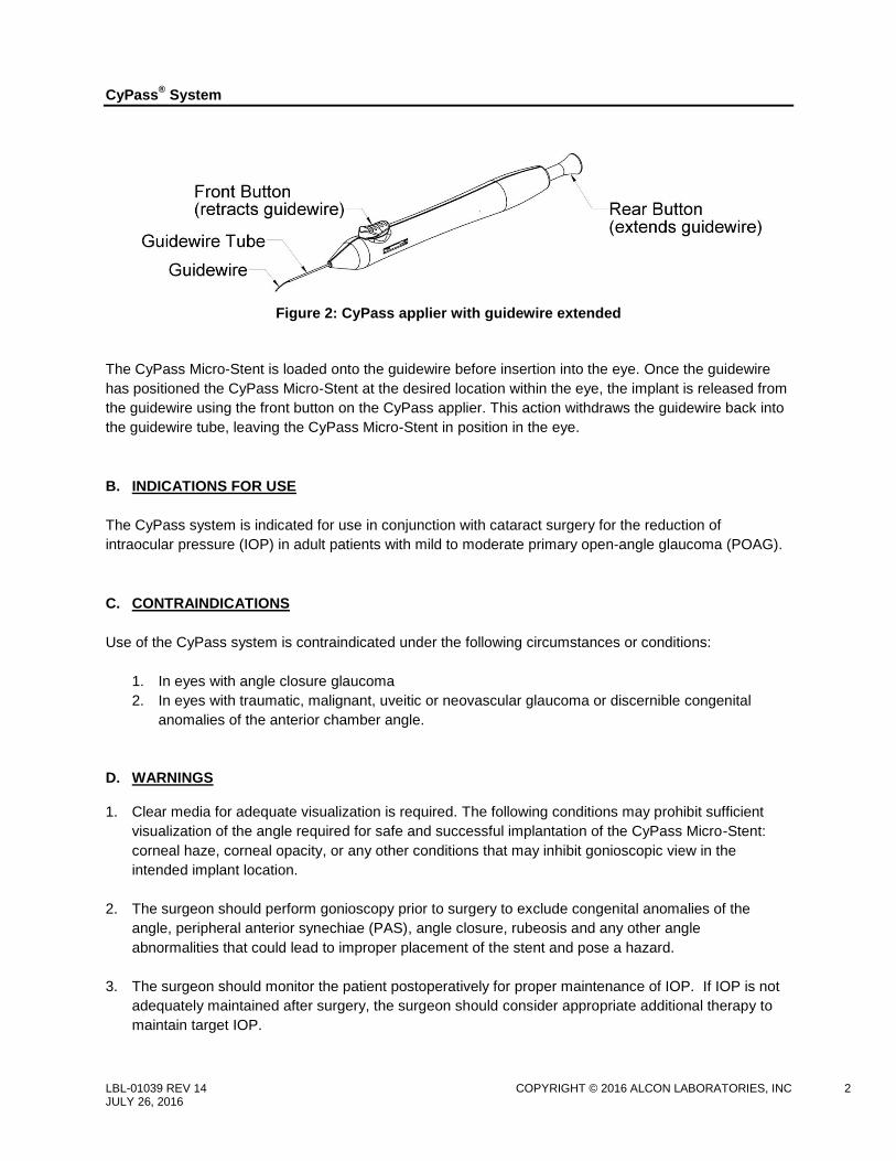
and the CyPass applier. The CyPass Micro-Stent (Figure 1) is a polyimide tube with a fenestrated lumen.
The stent has a single piece design and is 0.25” (6.35 mm) long. The inner diameter of the stent is 0.012”
(0.30 mm) and the outer diameter is 0.017” (0.43 mm). The CyPass Micro-Stent is designed for
placement in the angle of the eye, with the proximal end extending from the angle into the anterior
chamber (AC) and the distal end residing in the supraciliary space.
Figure 1: CyPass Micro-Stent
When properly implanted, the CyPass Micro-Stent is intended to allow outflow of aqueous fluid from the
AC, where the device proximal end resides, through and around the fenestrated lumen and distal end of
the tube into the supraciliary and suprachoroidal space via the uveoscleral pathway.
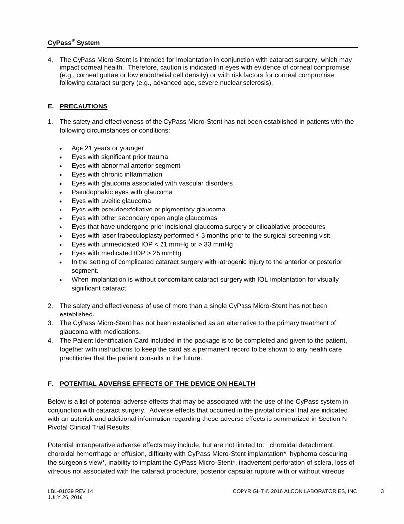
The CyPass applier (Figure 2) is the hand-held surgical instrument used to implant the CyPass Micro-
Stent. The applier consists of a medical-grade polymer handpiece with a guidewire assembly. The
guidewire assembly includes a nitinol implant delivery guidewire extending from inside the handpiece
through and beyond the distal end of a stainless steel tube (guidewire tube) that supports the guidewire.
The guidewire is 0.011” (0.28 mm) in diameter and formed with a 0.48” (12 mm) radius of distal curvature
and a blunt distal tip to facilitate location and blunt dissection of the plane between the ciliary body and
sclera. The CyPass applier delivers the CyPass Micro-Stent to the desired location within the eye.
6.35 mm Length
CyPass
® System
LBL-01039 REV 14 COPYRIGHT © 2016 ALCON LABORATORIES, INC 2 JULY 26, 2016
Figure 2: CyPass applier with guidewire extended
The CyPass Micro-Stent is loaded onto the guidewire before insertion into the eye. Once the guidewire
has positioned the CyPass Micro-Stent at the desired location within the eye, the implant is released from
the guidewire using the front button on the CyPass applier. This action withdraws the guidewire back into
the guidewire tube, leaving the CyPass Micro-Stent in position in the eye.
B. INDICATIONS FOR USE
The CyPass system is indicated for use in conjunction with cataract surgery for the reduction of
intraocular pressure (IOP) in adult patients with mild to moderate primary open-angle glaucoma (POAG).
C. CONTRAINDICATIONS
Use of the CyPass system is contraindicated under the following circumstances or conditions:
1. In eyes with angle closure glaucoma
2. In eyes with traumatic, malignant, uveitic or neovascular glaucoma or discernible congenital
anomalies of the anterior chamber angle.
D. WARNINGS
1. Clear media for adequate visualization is required. The following conditions may prohibit sufficient
visualization of the angle required for safe and successful implantation of the CyPass Micro-Stent:
corneal haze, corneal opacity, or any other conditions that may inhibit gonioscopic view in the
intended implant location.
2. The surgeon should perform gonioscopy prior to surgery to exclude congenital anomalies of the
angle, peripheral anterior synechiae (PAS), angle closure, rubeosis and any other angle
abnormalities that could lead to improper placement of the stent and pose a hazard.
3. The surgeon should monitor the patient postoperatively for proper maintenance of IOP. If IOP is not
adequately maintained after surgery, the surgeon should consider appropriate additional therapy to
maintain target IOP.
CyPass
® System
LBL-01039 REV 14 COPYRIGHT © 2016 ALCON LABORATORIES, INC 3 JULY 26, 2016
4. The CyPass Micro-Stent is intended for implantation in conjunction with cataract surgery, which may impact corneal health. Therefore, caution is indicated in eyes with evidence of corneal compromise (e.g., corneal guttae or low endothelial cell density) or with risk factors for corneal compromise following cataract surgery (e.g., advanced age, severe nuclear sclerosis).
E. PRECAUTIONS
1. The safety and effectiveness of the CyPass Micro-Stent has not been established in patients with the
following circumstances or conditions:
Age 21 years or younger
Eyes with significant prior trauma
Eyes with abnormal anterior segment
Eyes with chronic inflammation
Eyes with glaucoma associated with vascular disorders
Pseudophakic eyes with glaucoma
Eyes with uveitic glaucoma
Eyes with pseudoexfoliative or pigmentary glaucoma
Eyes with other secondary open angle glaucomas
Eyes that have undergone prior incisional glaucoma surgery or cilioablative procedures
Eyes with laser trabeculoplasty performed ≤ 3 months prior to the surgical screening visit
Eyes with unmedicated IOP < 21 mmHg or > 33 mmHg
Eyes with medicated IOP > 25 mmHg
In the setting of complicated cataract surgery with iatrogenic injury to the anterior or posterior
segment.
When implantation is without concomitant cataract surgery with IOL implantation for visually
significant cataract
2. The safety and effectiveness of use of more than a single CyPass Micro-Stent has not been
established.
3. The CyPass Micro-Stent has not been established as an alternative to the primary treatment of
glaucoma with medications.
4. The Patient Identification Card included in the package is to be completed and given to the patient,
together with instructions to keep the card as a permanent record to be shown to any health care
practitioner that the patient consults in the future.
F. POTENTIAL ADVERSE EFFECTS OF THE DEVICE ON HEALTH
Below is a list of potential adverse effects that may be associated with the use of the CyPass system in
conjunction with cataract surgery. Adverse effects that occurred in the pivotal clinical trial are indicated
with an asterisk and additional information regarding these adverse effects is summarized in Section N -
Pivotal Clinical Trial Results.
Potential intraoperative adverse effects may include, but are not limited to: choroidal detachment,
choroidal hemorrhage or effusion, difficulty with CyPass Micro-Stent implantation*, hyphema obscuring
the surgeon’s view*, inability to implant the CyPass Micro-Stent*, inadvertent perforation of sclera, loss of
vitreous not associated with the cataract procedure, posterior capsular rupture with or without vitreous
CyPass
® System
LBL-01039 REV 14 COPYRIGHT © 2016 ALCON LABORATORIES, INC 4 JULY 26, 2016
loss resulting from cataract surgery*, significant corneal damage, significant iris injury or trauma*, and
zonular dialysis.
Potential postoperative adverse effects may include, but are not limited to: AC cell and flare requiring
either extension of the standard postoperative steroid regimen or re-initiation of steroids after steroid
regimen completion*; AC flattening with lens/cornea touch; AC shallowing with iridocorneal apposition;
atrophy/phthisis; choroidal hemorrhage or effusion; chronic pain in the implanted eye; corneal edema*;
corneal opacification; corneal decompensation*; CyPass Micro-Stent malposition, dislodgement or
movement*; CyPass Micro-Stent explantation*; CyPass Micro-Stent obstruction*; elevated IOP requiring
treatment with oral or intravenous medications or with surgical intervention*; endophthalmitis; hypotonic
maculopathy*; increase in C:D ratio; loss of best-corrected visual acuity (BCVA)*; persistent significant
foreign body sensation*; persistent hyphema*; persistent hypotony*; maculopathy*; retinal complications
(dialysis, flap tears, retinal detachment or proliferative vitreoretinopathy)*; significant ptosis; worsening in
visual field loss*; unplanned secondary ocular surgical intervention*; and wound dehiscence (persistent
aqueous leak or fistula formation).
CyPass
® System
LBL-01039 REV 14 COPYRIGHT © 2016 ALCON LABORATORIES, INC 5 JULY 26, 2016
G. DIRECTIONS FOR USE
Surgical Procedure
The implantation procedure is performed as follows, after completion of cataract extraction and
intraocular lens implantation.
1. Instill a miotic agent to constrict the pupil.
2. Tilt the microscope approximately 35 - 45° towards the surgeon and rotate the patient’s head
approximately 10° away from the surgeon to facilitate direct visualization of the anterior angle.
3. Confirm the AC angle is open.
4. Open the tray containing the CyPass system onto a sterile field. Do not use either the CyPass
Micro-Stent or the CyPass applier if the packaging has been opened or damaged.
5. Remove the CyPass applier from the sterile tray and examine its condition. First, press the rear
button on the handle to verify the guidewire extends from the guidewire tube. Then, press the
front button on the handle to confirm the guidewire fully retracts into the guidewire tube.
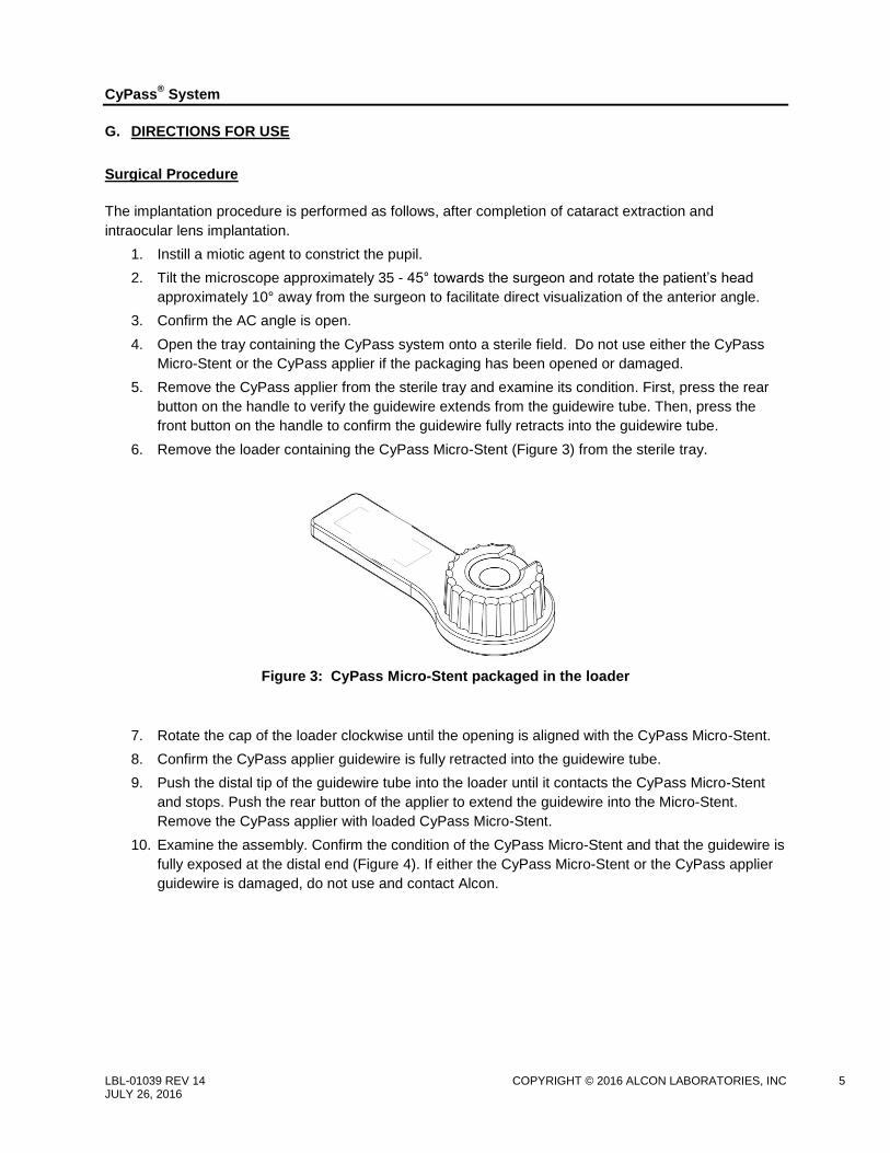
6. Remove the loader containing the CyPass Micro-Stent (Figure 3) from the sterile tray.
Figure 3: CyPass Micro-Stent packaged in the loader
7. Rotate the cap of the loader clockwise until the opening is aligned with the CyPass Micro-Stent.
8. Confirm the CyPass applier guidewire is fully retracted into the guidewire tube.
9. Push the distal tip of the guidewire tube into the loader until it contacts the CyPass Micro-Stent
and stops. Push the rear button of the applier to extend the guidewire into the Micro-Stent.
Remove the CyPass applier with loaded CyPass Micro-Stent.
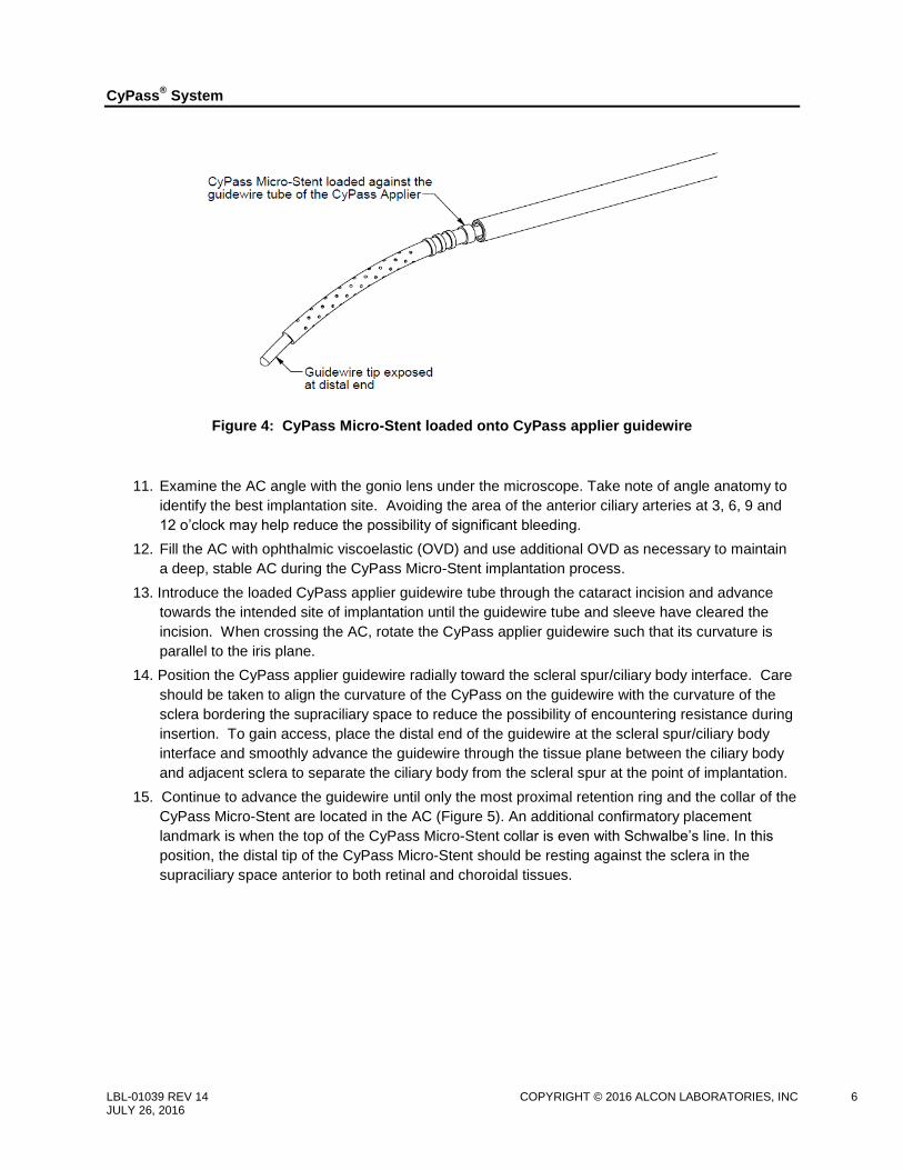
10. Examine the assembly. Confirm the condition of the CyPass Micro-Stent and that the guidewire is
fully exposed at the distal end (Figure 4). If either the CyPass Micro-Stent or the CyPass applier
guidewire is damaged, do not use and contact Alcon.
CyPass
® System
LBL-01039 REV 14 COPYRIGHT © 2016 ALCON LABORATORIES, INC 6 JULY 26, 2016
Figure 4: CyPass Micro-Stent loaded onto CyPass applier guidewire
11. Examine the AC angle with the gonio lens under the microscope. Take note of angle anatomy to
identify the best implantation site. Avoiding the area of the anterior ciliary arteries at 3, 6, 9 and
12 o’clock may help reduce the possibility of significant bleeding.
12. Fill the AC with ophthalmic viscoelastic (OVD) and use additional OVD as necessary to maintain
a deep, stable AC during the CyPass Micro-Stent implantation process.
13. Introduce the loaded CyPass applier guidewire tube through the cataract incision and advance
towards the intended site of implantation until the guidewire tube and sleeve have cleared the
incision. When crossing the AC, rotate the CyPass applier guidewire such that its curvature is
parallel to the iris plane.
14. Position the CyPass applier guidewire radially toward the scleral spur/ciliary body interface. Care
should be taken to align the curvature of the CyPass on the guidewire with the curvature of the
sclera bordering the supraciliary space to reduce the possibility of encountering resistance during
insertion. To gain access, place the distal end of the guidewire at the scleral spur/ciliary body
interface and smoothly advance the guidewire through the tissue plane between the ciliary body
and adjacent sclera to separate the ciliary body from the scleral spur at the point of implantation.
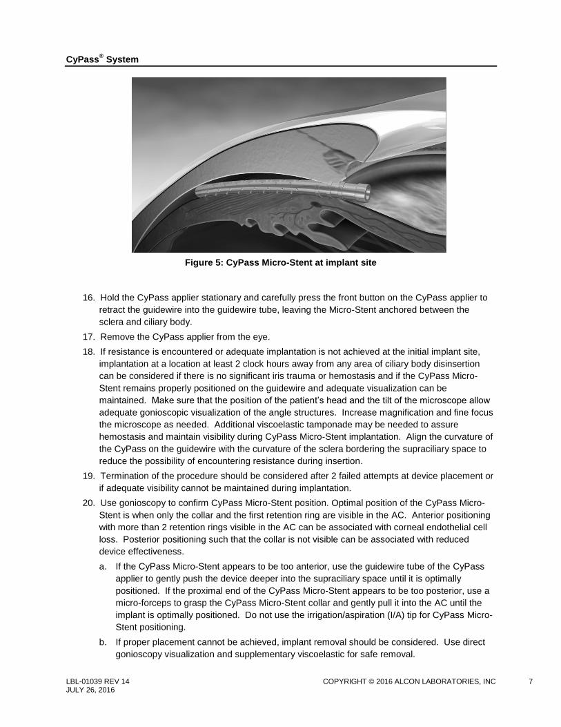
15. Continue to advance the guidewire until only the most proximal retention ring and the collar of the
CyPass Micro-Stent are located in the AC (Figure 5). An additional confirmatory placement
landmark is when the top of the CyPass Micro-Stent collar is even with Schwalbe’s line. In this
position, the distal tip of the CyPass Micro-Stent should be resting against the sclera in the
supraciliary space anterior to both retinal and choroidal tissues.
CyPass
® System
LBL-01039 REV 14 COPYRIGHT © 2016 ALCON LABORATORIES, INC 7 JULY 26, 2016
Figure 5: CyPass Micro-Stent at implant site
16. Hold the CyPass applier stationary and carefully press the front button on the CyPass applier to
retract the guidewire into the guidewire tube, leaving the Micro-Stent anchored between the
sclera and ciliary body.
17. Remove the CyPass applier from the eye.
18. If resistance is encountered or adequate implantation is not achieved at the initial implant site,
implantation at a location at least 2 clock hours away from any area of ciliary body disinsertion
can be considered if there is no significant iris trauma or hemostasis and if the CyPass Micro-
Stent remains properly positioned on the guidewire and adequate visualization can be
maintained. Make sure that the position of the patient’s head and the tilt of the microscope allow
adequate gonioscopic visualization of the angle structures. Increase magnification and fine focus
the microscope as needed. Additional viscoelastic tamponade may be needed to assure
hemostasis and maintain visibility during CyPass Micro-Stent implantation. Align the curvature of
the CyPass on the guidewire with the curvature of the sclera bordering the supraciliary space to
reduce the possibility of encountering resistance during insertion.
19. Termination of the procedure should be considered after 2 failed attempts at device placement or
if adequate visibility cannot be maintained during implantation.
20. Use gonioscopy to confirm CyPass Micro-Stent position. Optimal position of the CyPass Micro-
Stent is when only the collar and the first retention ring are visible in the AC. Anterior positioning
with more than 2 retention rings visible in the AC can be associated with corneal endothelial cell
loss. Posterior positioning such that the collar is not visible can be associated with reduced
device effectiveness.
a. If the CyPass Micro-Stent appears to be too anterior, use the guidewire tube of the CyPass
applier to gently push the device deeper into the supraciliary space until it is optimally
positioned. If the proximal end of the CyPass Micro-Stent appears to be too posterior, use a
micro-forceps to grasp the CyPass Micro-Stent collar and gently pull it into the AC until the
implant is optimally positioned. Do not use the irrigation/aspiration (I/A) tip for CyPass Micro-
Stent positioning.
b. If proper placement cannot be achieved, implant removal should be considered. Use direct
gonioscopy visualization and supplementary viscoelastic for safe removal.
CyPass
® System
LBL-01039 REV 14 COPYRIGHT © 2016 ALCON LABORATORIES, INC 8 JULY 26, 2016
21. Since retained viscoelastic can lead to elevated IOP in the early postoperative period, irrigate and aspirate viscoelastic from the AC, taking care to avoid irrigation/aspiration (I/A) tip proximity to the CyPass Micro-Stent. Note: The flow of irrigation fluid near the Micro-Stent may cause implant movement.
22. After completion of I/A, verify CyPass Micro-Stent location and confirm the absence of CyPass
Micro-Stent lumen obstruction.
23. Confirm that the surgical incision is sealed by either pressure challenge testing or Seidel testing.
Use a suture or ocular sealant for closure, if needed.
Postoperative Instructions
Patients should be observed postoperatively for IOP increases that may occur in the early postoperative
period as a possible sequelae following intraocular surgery in patients with glaucoma.
Gonioscopy should be performed to assess CyPass Micro-Stent position postoperatively. Anterior
positioning of the CyPass Micro-Stent such that more than 2 retention rings are visible in the AC may
result in reduced endothelial cell density and a need for secondary surgical intervention (e.g., device
repositioning, device trimming, or device removal). If the CyPass Micro-Stent is close to the corneal
endothelium, early repositioning or removal of the CyPass Micro-Stent and/or periodic follow-up
assessments of endothelial cell density using specular microscopy should be considered.
If ciliary body edema is suspected due to forward movement of the ciliary body-lens diaphragm,
ultrasound biomicroscopy (UBM) may be a useful adjunctive diagnostic aid in the evaluation of the ciliary
body and suprachoroidal space.
Postoperative CyPass Micro-Stent Adjustment or Removal
Situations that may merit consideration of CyPass Micro-Stent position adjustment or removal include, but
are not limited to: intermittent or persistent contact between the CyPass Micro-Stent and the corneal
endothelium; significant decrease in endothelial cell density that appears related to CyPass Micro-Stent
positioning or stability; iris-cornea touch; persistent hypotony; persistent uncontrolled uveitis; recurrent or
persistent hyphema with IOP elevation above target pressure; or any anatomic or functional clinical
sequelae of the anterior or posterior segment that may cause a threat to vision. Variations in gonioscopic
visualization or other alterations in angle anatomy may be interpreted as micro-movement of the CyPass
Micro-Stent; however, in the absence of clinical sequelae, device adjustment or removal is not
recommended.
Healing response and progressive engagement of implant retention features must be factored into the
decision to remove the CyPass Micro-Stent after the immediate postoperative period (e.g., after 1 month
postoperative). It is advised to consider less invasive intervention, such as positional adjustment or
trimming of the CyPass Micro-Stent proximal end (if the device is considered too anteriorly positioned) as
a first alternative to device removal. It is also highly recommended that Alcon be contacted prior to
device removal.
CyPass
® System
LBL-01039 REV 14 COPYRIGHT © 2016 ALCON LABORATORIES, INC 9 JULY 26, 2016
Procedure for CyPass Micro-Stent Repositioning or Removal
Surgical access for CyPass Micro-Stent repositioning or removal is primarily ab-interno through a
minimum 1.5 mm clear cornea incision under direct gonioscopy and AC viscoelastic stabilization. Use of
retinal micro-forceps and retinal instrumentation is encouraged for optimal surgical control and access.
The steps for this procedure are as follows:
1. Construct the clear corneal incision and instill OVD into the AC.
2. Utilizing a gonioprism for visualization, grasp the CyPass Micro-Stent with toothed micro forceps
around the anterior rim and gently either reposition until optimal positioning is achieved, or remove
the device from its position. Carefully observe any tension or traction on surrounding tissues.
3. Remove remaining OVD from the AC and adjust the tension in the eye by injecting and/or allowing
egress of BSS; then close the incision.
Procedure for CyPass Micro-Stent Proximal End Trimming
After the immediate postoperative period, trimming of the proximal end of the CyPass Micro-Stent may be
considered when anterior positioning of the CyPass is likely to compromise corneal endothelial
health. The steps for this procedure are as follows:
1. Construct 2 clear corneal incisions under OVD, utilizing either direct observation or a gonioprism
for visualization.
2. Hold the proximal portion of the CyPass Micro-Stent with micro-forceps, and incise distally with
micro-scissors.
3. Remove the separated proximal portion through the corneal incision.
4. Inspect the remaining distal portion of the CyPass Micro-Stent with a gonioprism to confirm
optimal positioning.
5. Remove remaining OVD from the AC utilizing irrigation alone or automated irrigation/aspiration,
then adjust the tension in the eye by allowing egress of aqueous and seal the incision.
After surgery, patients should be monitored for IOP changes that may occur as possible sequelae
following intraocular surgery in patients with glaucoma. Patients should also be periodically evaluated
using specular microscopy to assess changes in endothelial cell density.
H. ADVERSE EVENT REPORTING
Adverse events and/or potentially sight-threatening complications that may reasonably be regarded as
related to the CyPass system must be reported to Alcon Medical Safety.
Alcon Laboratories, Inc.,
Medical Safety (AB 2-6),
6201 South Freeway, Fort Worth, TX 76134-2099
Call Toll free: 1-800-757-9780
CyPass
® System
LBL-01039 REV 14 COPYRIGHT © 2016 ALCON LABORATORIES, INC 10 JULY 26, 2016
I. HOW SUPPLIED
The CyPass system is supplied sterile and non-pyrogenic in a sealed tray. The sealed tray is placed in a
unit box containing product labeling and product information. The CyPass system has been sterilized
using radiation.
The CyPass system is designed for single use only, and is intended to be used only on a single patient.
The safety and effectiveness of cleaning, re-sterilization and/or reuse has not been evaluated and may
adversely impact device integrity and patient safety.
The CyPass system and manufacturing processes do not contain latex.
Used CyPass appliers should be discarded only in a suitable, biohazardous sharps container.
J. STORAGE REQUIREMENTS
The CyPass system should be stored at room temperature in the range of 68-75°F. K. EXPIRATION DATE
The sterility expiration date (year, month and day) is clearly indicated both on the sealed tray and the
outside of the unit box. Sterility is assured until the expiration date as long as the tray seal is not
punctured or damaged. The CyPass system should not be used past the date indicated.
L. MRI INFORMATION
The CyPass Micro-Stent is magnetic resonance (MR) Safe: the implant is constructed of polyimide
material, a non-conducting, non-metallic, non-magnetic polymer that poses no known hazards in all
magnetic resonance imaging environments.
M. RETURNED GOODS POLICY
In the United States, returned product will only be accepted in exchange for other products, not credit. All
returns must be accompanied by an Alcon Laboratories, Inc. Returned Goods Number and be shipped
via traceable means. A Returned Goods Number is obtained by contacting Alcon’s Customer Service
Department. Issuance of this number does not constitute final acceptance of the returned products. For
detailed policy guidelines including exchange, please contact your Sales or Customer Service
Representative. Outside the United States, contact local Alcon offices or distributors regarding returned
goods policy.
CyPass
® System
LBL-01039 REV 14 COPYRIGHT © 2016 ALCON LABORATORIES, INC 11 JULY 26, 2016
N. PIVOTAL CLINICAL TRIAL RESULTS
The pivotal study for the CyPass Micro-Stent, known as the COMPASS Trial (Protocol TMI-09-01), was a
prospective, randomized, comparative, multicenter investigation conducted in the United States, in which
a total of 505 subjects from 24 sites were randomized in a 3:1 fashion to undergo either implantation of
the CyPass Micro-Stent after uncomplicated cataract surgery (CyPass group) or to undergo cataract
surgery without implantation of the CyPass Micro-Stent (Control group). A total of 374 subjects were
randomized to the CyPass group and 131 subjects were randomized to the Control group. In each
subject, only one eye was considered to be the study eye. Enrollment in the study began in September of
2009 and the last study subject was randomized in March of 2013. Randomized subjects were followed
for 2 years postoperatively.
To participate in the study, subjects were required to have a diagnosis of POAG in the study eye.
Additional study eye key inclusion criteria were:
At the Screening Visit, a mean (or median) medicated IOP ≤ 25.0 mmHg or an unmedicated IOP ≥
21.0 mmHg and ≤ 33.0 mmHg.
At the Baseline Visit, an unmedicated mean diurnal IOP ≥ 21.0 mmHg and ≤ 33.0 mmHg, which was
≥ 3.0 mmHg higher than the Screening Visit medicated IOP.
Gonioscopy confirming normal angle anatomy at the anticipated site of CyPass implantation and
Shaffer grade ≥ III in all 4 quadrants of the eye.
Subjects could not participate in the study if they had a diagnosis of acute angle closure, traumatic,
congenital, malignant, uveitic, pseudoexfoliative, and pigmentary or neovascular glaucoma in the
designated study eye. Subjects who had undergone previous incisional or cilioablative glaucoma
procedures were also excluded from the study. Also, subjects who experienced cataract surgery
complications were exited from the study prior to randomization. Subjects were not required to be using
ocular hypotensive medications for inclusion in the study; however use of oral ocular hypotensive
medication or more than 3 ocular hypotensive medications was an exclusion criterion.
The mean age of the study population was 70.3 years, with the majority (76.0%) between 60 and 79
years of age at the time of enrollment. Slightly more females (n = 269) than males (n = 236) were
randomized. The majority of subjects were White (83.6%), with Black/African American subjects being
the second most frequently enrolled race (9.3%). Screening and baseline characteristics were balanced
between the 2 study groups.
The primary effectiveness endpoint was defined as the proportion of subject eyes with ≥ 20% decrease in
the 24-month unmedicated mean diurnal IOP (DIOP) compared to the baseline DIOP. Subjects were
defined as IOP non-responders if:
they did not achieve the primary effectiveness endpoint,
they missed the 24-month IOP assessment,
ocular hypotensive medication use was not terminated prior to the 24-month DIOP assessment,
they underwent an IOP-affecting secondary surgical procedure (i.e., iridotomy, iridectomy,
trabeculectomy, glaucoma shunt implantation, argon laser trabeculoplasty, selective laser
trabeculoplasty), or other surgery that would affect IOP, or
CyPass
® System
LBL-01039 REV 14 COPYRIGHT © 2016 ALCON LABORATORIES, INC 12 JULY 26, 2016
they underwent CyPass explantation or repositioning. Subjects in which CyPass implantation
was not successful were also considered to be IOP non-responders.
The first secondary effectiveness endpoint was the mean change in 24-month DIOP from baseline, and
the second secondary effectiveness endpoint was defined as the proportion of subject eyes with 24-
month unmedicated mean DIOP ≥ 6 mmHg and ≤ 18 mmHg.
Each endpoint required a comparison between the CyPass and Control groups. The primary and
secondary effectiveness analyses were performed using the Intent to Treat (ITT) population, comprised of
all randomized subjects grouped according to their randomization assignment.
During the COMPASS Trial, the CyPass Micro-Stent was implanted using a CyPass applier slightly
different from the commercially available applier. The applier used in the COMPASS Trial used a
different mechanism to hold the CyPass on the delivery guidewire prior to implantation, and guidewire
retraction was performed by sliding the retraction button toward the back of the applier hand piece. The
commercially available CyPass applier was clinically tested in a multi-surgeon case series of 91 subjects
evaluating the intraoperative safety and performance in a similar patient population to that of the
COMPASS Trial. In this study, the CyPass Micro-Stent was successfully implanted in all cases. Device
implantation was successful on the initial placement attempt in 87 cases (95.6%); 1 case required 2
implantation attempts, while the number of attempts was undocumented for the remaining 3 cases.
Postoperative gonioscopy revealed the CyPass to be visible, adequately positioned and without
obstruction in the vast majority of cases reviewed. Adverse events (AE) related to device placement and
stability were similar in nature to AEs observed in the CyPass arm of the COMPASS Trial.
Effectiveness Results
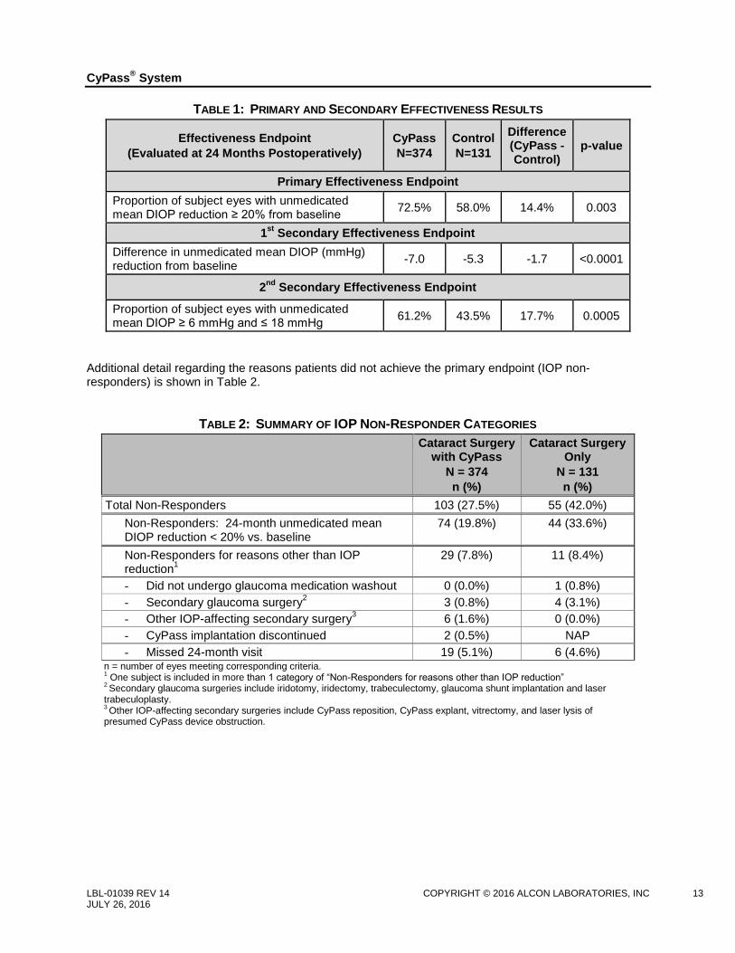
Results from the primary and secondary endpoints are shown in Table 1. The primary effectiveness
endpoint was met, with 72.5% in the CyPass group and 58.0% in the Control group achieving a clinically
significant (≥ 20%) decrease in unmedicated mean DIOP from baseline to the hypotensive medication-
free 24-month postoperative examination. This difference between groups was statistically significant
(p=0.0030).
The first secondary endpoint, a clinically significant mean change in IOP between baseline and
hypotensive medication-free 24-month postoperative examination, was met. The mean reduction in
unmedicated mean DIOP from baseline to 24 months was 7.0 mmHg (SD 4.5) in the CyPass group
compared to 5.3 mmHg (SD 4.0) in the Control group (p < 0.0001).
The second secondary effectiveness endpoint was also met, with 61.2% in the CyPass group and 43.5%
in the Control group achieving unmedicated mean DIOP ≥ 6 mmHg and ≤ 18 mmHg at the hypotensive
medication-free 24-month postoperative examination. The difference between groups was statistically
significant (p=0.0005).
CyPass
® System
LBL-01039 REV 14 COPYRIGHT © 2016 ALCON LABORATORIES, INC 13 JULY 26, 2016
TABLE 1: PRIMARY AND SECONDARY EFFECTIVENESS RESULTS
Effectiveness Endpoint
(Evaluated at 24 Months Postoperatively)
CyPass
N=374
Control
N=131
Difference (CyPass - Control)
p-value
Primary Effectiveness Endpoint
Proportion of subject eyes with unmedicated mean DIOP reduction ≥ 20% from baseline
72.5% 58.0% 14.4% 0.003
1st
Secondary Effectiveness Endpoint
Difference in unmedicated mean DIOP (mmHg) reduction from baseline
-7.0 -5.3 -1.7 <0.0001
2nd
Secondary Effectiveness Endpoint
Proportion of subject eyes with unmedicated mean DIOP ≥ 6 mmHg and ≤ 18 mmHg
61.2% 43.5% 17.7% 0.0005
Additional detail regarding the reasons patients did not achieve the primary endpoint (IOP non-responders) is shown in Table 2.
TABLE 2: SUMMARY OF IOP NON-RESPONDER CATEGORIES
Cataract Surgery with CyPass
N = 374
n (%)
Cataract Surgery Only
N = 131
n (%)
Total Non-Responders 103 (27.5%) 55 (42.0%)
Non-Responders: 24-month unmedicated mean DIOP reduction < 20% vs. baseline
74 (19.8%) 44 (33.6%)
Non-Responders for reasons other than IOP reduction
1
29 (7.8%) 11 (8.4%)
- Did not undergo glaucoma medication washout 0 (0.0%) 1 (0.8%)
- Secondary glaucoma surgery2 3 (0.8%) 4 (3.1%)
- Other IOP-affecting secondary surgery3 6 (1.6%) 0 (0.0%)
- CyPass implantation discontinued 2 (0.5%) NAP
- Missed 24-month visit 19 (5.1%) 6 (4.6%) n = number of eyes meeting corresponding criteria. 1 One subject is included in more than 1 category of “Non-Responders for reasons other than IOP reduction”
2 Secondary glaucoma surgeries include iridotomy, iridectomy, trabeculectomy, glaucoma shunt implantation and laser
trabeculoplasty. 3 Other IOP-affecting secondary surgeries include CyPass reposition, CyPass explant, vitrectomy, and laser lysis of
presumed CyPass device obstruction.
CyPass
® System
LBL-01039 REV 14 COPYRIGHT © 2016 ALCON LABORATORIES, INC 14 JULY 26, 2016
Safety Results
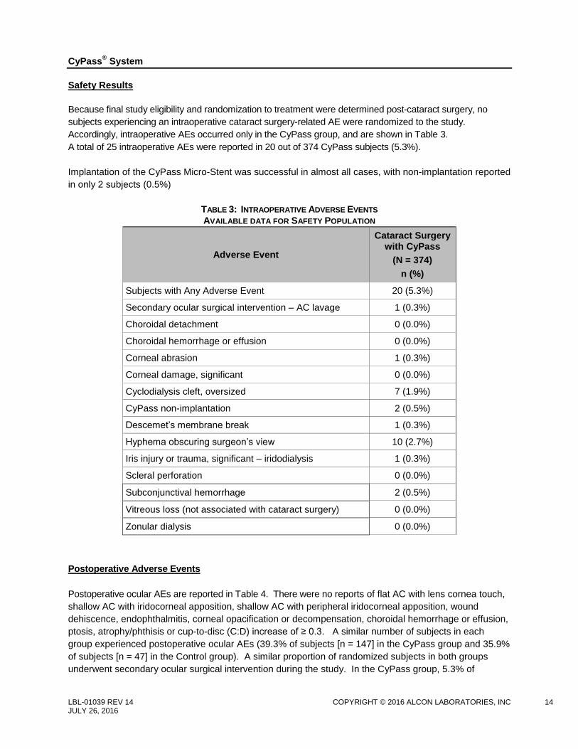
Because final study eligibility and randomization to treatment were determined post-cataract surgery, no
subjects experiencing an intraoperative cataract surgery-related AE were randomized to the study.
Accordingly, intraoperative AEs occurred only in the CyPass group, and are shown in Table 3.
A total of 25 intraoperative AEs were reported in 20 out of 374 CyPass subjects (5.3%).
Implantation of the CyPass Micro-Stent was successful in almost all cases, with non-implantation reported
in only 2 subjects (0.5%)
TABLE 3: INTRAOPERATIVE ADVERSE EVENTS
AVAILABLE DATA FOR SAFETY POPULATION
Adverse Event
Cataract Surgery with CyPass
(N = 374)
n (%)
Subjects with Any Adverse Event 20 (5.3%)
Secondary ocular surgical intervention – AC lavage 1 (0.3%)
Choroidal detachment 0 (0.0%)
Choroidal hemorrhage or effusion 0 (0.0%)
Corneal abrasion 1 (0.3%)
Corneal damage, significant 0 (0.0%)
Cyclodialysis cleft, oversized 7 (1.9%)
CyPass non-implantation 2 (0.5%)
Descemet’s membrane break 1 (0.3%)
Hyphema obscuring surgeon’s view 10 (2.7%)
Iris injury or trauma, significant – iridodialysis 1 (0.3%)
Scleral perforation 0 (0.0%)
Subconjunctival hemorrhage 2 (0.5%)
Vitreous loss (not associated with cataract surgery) 0 (0.0%)
Zonular dialysis 0 (0.0%)
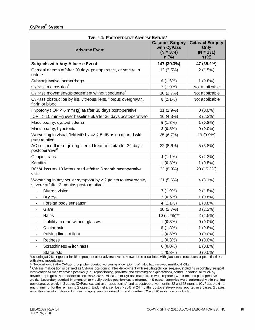
Postoperative Adverse Events
Postoperative ocular AEs are reported in Table 4. There were no reports of flat AC with lens cornea touch,
shallow AC with iridocorneal apposition, shallow AC with peripheral iridocorneal apposition, wound
dehiscence, endophthalmitis, corneal opacification or decompensation, choroidal hemorrhage or effusion,
ptosis, atrophy/phthisis or cup-to-disc (C:D) increase of ≥ 0.3. A similar number of subjects in each
group experienced postoperative ocular AEs (39.3% of subjects [n = 147] in the CyPass group and 35.9%
of subjects [n = 47] in the Control group). A similar proportion of randomized subjects in both groups
underwent secondary ocular surgical intervention during the study. In the CyPass group, 5.3% of
CyPass
® System
LBL-01039 REV 14 COPYRIGHT © 2016 ALCON LABORATORIES, INC 15 JULY 26, 2016
subjects (n = 20) underwent secondary ocular surgeries, while in the Control group, 5.3% of subjects (n =
7) had secondary ocular surgeries.
Corneal edema associated with the surgical procedure resolved within the first postoperative month in
98% of CyPass group subjects. Only a single case of persistent corneal edema occurred, an incidence of
0.27% in the CyPass group. Anterior segment inflammation, which was generally mild, resolved in 95%
of CyPass subjects by 3 months. Of note, subjects in the CyPass group underwent two interventions
(cataract surgery and CyPass implantation) but had the same standard course of prophylactic steroids
and NSAIDs that was used for cataract surgery alone. No cases of retinal detachment, pupillary block,
endophthalmitis, or hypopyon occurred during the study. Based on available data at the 24-month visit,
BCVA was 20/40 or better for 98.6% of subjects (n = 350) in the CyPass group and 98.4% of subjects (n =
126) in the Control group.
Only 2 ocular serious adverse events (SAE) were reported during the course of the study. One of these
SAEs, peripheral anterior choroidal effusion, was considered to be related to the CyPass Micro-Stent.
This event was not reported as “choroidal hemorrhage or effusion” because it did not meet the pre-
specified criteria for that adverse event, i.e., there was no hemorrhagic component obstructing vision or
causing pain; in addition, the effusion was restricted to the anterior supraciliary space and was only
detectable with ultrasound imaging. This SAE was associated with IOL subluxation, which was
successfully addressed with IOL repositioning and other complications, all of which resolved without
sequelae by the 24-month visit. The other SAE was pseudophakic bullous keratopathy (PBK). Descemet
stripping endothelial keratoplasty (DSEK) was performed approximately 1 year postoperatively.
CyPass
® System
LBL-01039 REV 14 COPYRIGHT © 2016 ALCON LABORATORIES, INC 16 JULY 26, 2016
TABLE 4: POSTOPERATIVE ADVERSE EVENTS*
Adverse Event
Cataract Surgery with CyPass
(N = 374) n (%)
Cataract Surgery Only
(N = 131) n (%)
Subjects with Any Adverse Event 147 (39.3%) 47 (35.9%)
Corneal edema at/after 30 days postoperative, or severe in nature
13 (3.5%) 2 (1.5%)
Subconjunctival hemorrhage 6 (1.6%) 1 (0.8%)
CyPass malposition1
7 (1.9%) Not applicable
CyPass movement/dislodgement without sequelae2
10 (2.7%) Not applicable
CyPass obstruction by iris, vitreous, lens, fibrous overgrowth, fibrin or blood
8 (2.1%) Not applicable
Hypotony (IOP < 6 mmHg) at/after 30 days postoperative 11 (2.9%) 0 (0.0%)
IOP => 10 mmHg over baseline at/after 30 days postoperative^ 16 (4.3%) 3 (2.3%)
Maculopathy, cystoid edema 5 (1.3%) 1 (0.8%)
Maculopathy, hypotonic 3 (0.8%) 0 (0.0%)
Worsening in visual field MD by => 2.5 dB as compared with preoperative
25 (6.7%) 13 (9.9%)
AC cell and flare requiring steroid treatment at/after 30 days postoperative
3 32 (8.6%) 5 (3.8%)
Conjunctivitis 4 (1.1%) 3 (2.3%)
Keratitis 1 (0.3%) 1 (0.8%)
BCVA loss => 10 letters read at/after 3 month postoperative visit
33 (8.8%) 20 (15.3%)
Worsening in any ocular symptom by ≥ 2 points to severe/very severe at/after 3 months postoperative:
21 (5.6%) 4 (3.1%)
- Blurred vision 7 (1.9%) 2 (1.5%)
- Dry eye 2 (0.5%) 1 (0.8%)
- Foreign body sensation 4 (1.1%) 1 (0.8%)
- Glare 10 (2.7%) 3 (2.3%)
- Halos 10 (2.7%)** 2 (1.5%)
- Inability to read without glasses 1 (0.3%) 0 (0.0%)
- Ocular pain 5 (1.3%) 1 (0.8%)
- Pulsing lines of light 1 (0.3%) 0 (0.0%)
- Redness 1 (0.3%) 0 (0.0%)
- Scratchiness & itchiness 0 (0.0%) 1 (0.8%)
- Starbursts 1 (0.3%) 0 (0.0%) *occurring at 2% or greater in either group, or other adverse events known to be associated with glaucoma procedures or potential risks with stent implantations ** Two subjects in the CyPass group who reported worsening of symptoms of halos had received multifocal IOLs. 1 CyPass malposition is defined as CyPass positioning after deployment with resulting clinical sequela, including secondary surgical
intervention to modify device position (e.g., repositioning, proximal end trimming or explantation), corneal endothelial touch by device, or progressive endothelial cell loss > 30%. All cases of CyPass malposition were reported within the first postoperative week. Secondary surgical intervention to modify device position was performed in 5 cases: surgeries were performed within the first postoperative week in 3 cases (CyPass explant and repositioning) and at postoperative months 32 and 48 months (CyPass proximal end trimming) for the remaining 2 cases. Endothelial cell loss > 30% at 24 months postoperatively was reported in 3 cases; 2 cases were those in which device trimming surgery was performed at postoperative 32 and 48 months respectively.
CyPass
® System
LBL-01039 REV 14 COPYRIGHT © 2016 ALCON LABORATORIES, INC 17 JULY 26, 2016
2 One case of CyPass movement without sequelae was reported within the first postoperative week, while the remaining 9 cases
were reported between 3 and 24 months postoperatively. No secondary surgeries to modify device positioning were performed in these cases and none of the cases had endothelial cell loss > 30% at 24 months postoperatively. 3 One case of chronic anterior uveitis was reported in a subject in whom CyPass implantation was complicated by an oversized
cyclodialysis cleft. ^ While not reported as an AE, IOP ≥ 10 mmHg over baseline within the initial 30 days postoperatively occurred in 6.4% (n = 24) of CyPass subjects and 20.6% (n = 27) of Control subjects. Short-term use of oral ocular hypotensive medication for pressure control was employed for 3 subjects in the CyPass group and 3 subjects in the Control group; paracentesis on postoperative day 1 was performed in 1 additional subject in the Control group.
In addition to the AEs reported in Table 4, AEs that occurred at < 2% in both groups included ocular
medication allergy, corneal abrasion, non-proliferative diabetic retinopathy, proliferative diabetic
retinopathy, diabetic macular edema, eyelid dermatitis and significant foreign body sensation at or after 3
months postoperative. Adverse events that occurred at <2% in the CyPass group included environmental
allergy, map-dot fingerprint dystrophy, dry eye syndrome, hordeolum, trichiasis, foreign body with mucus
filament, peripheral anterior choroidal effusion, ciliary body edema, hyphema > 2mm after 1 day
postoperative, IOL complication (Crystalens Z syndrome, anterior malposition, and subluxation),
worsening in slit lamp findings at or after 3 months postoperative (corneal staining and pigment
deposition), choroidal folds without hypotony, wet age-related maculopathy, posterior vitreous
detachment, vitreal-macular traction, blepharitis, episcleritis, ocular migraine, and vertical binocular
diplopia. Adverse events that occurred at < 2% in the control group included chalazion, metallic foreign
body, ectropion, and chronic ocular pain after 3 months postoperative.
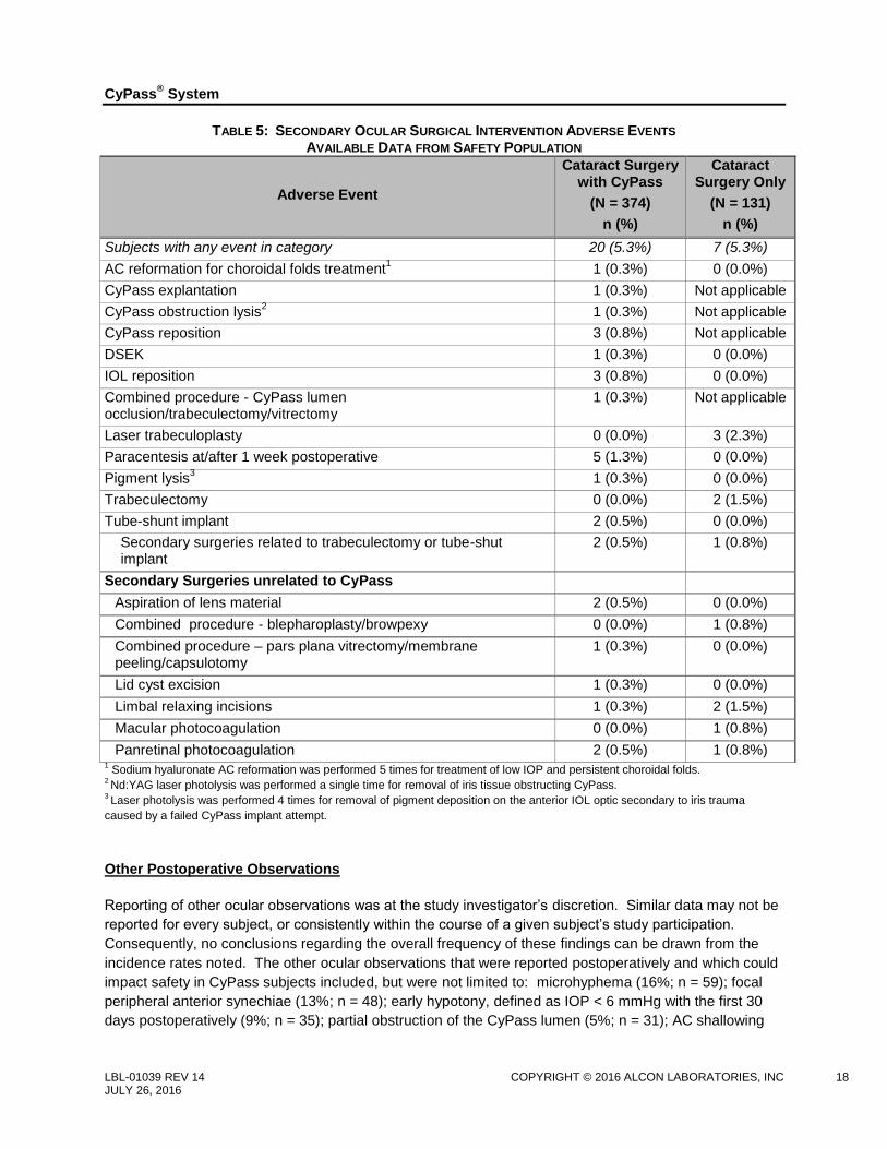
Secondary Ocular Surgical Interventions
Secondary ocular surgeries during the course of the study occurred in 5.3% of CyPass group subjects
(n = 20) and 5.3% (n = 7) of subjects in the Control group. Three additional subjects had CyPass-related
surgical procedures after completion of study participation. Secondary surgeries reported in both groups
are shown in Table 5.
CyPass
® System
LBL-01039 REV 14 COPYRIGHT © 2016 ALCON LABORATORIES, INC 18 JULY 26, 2016
TABLE 5: SECONDARY OCULAR SURGICAL INTERVENTION ADVERSE EVENTS AVAILABLE DATA FROM SAFETY POPULATION
Adverse Event
Cataract Surgery with CyPass
(N = 374)
n (%)
Cataract Surgery Only
(N = 131)
n (%)
Subjects with any event in category 20 (5.3%) 7 (5.3%)
AC reformation for choroidal folds treatment1
1 (0.3%) 0 (0.0%)
CyPass explantation 1 (0.3%) Not applicable
CyPass obstruction lysis2
1 (0.3%) Not applicable
CyPass reposition 3 (0.8%) Not applicable
DSEK 1 (0.3%) 0 (0.0%)
IOL reposition 3 (0.8%) 0 (0.0%)
Combined procedure - CyPass lumen occlusion/trabeculectomy/vitrectomy
1 (0.3%) Not applicable
Laser trabeculoplasty 0 (0.0%) 3 (2.3%)
Paracentesis at/after 1 week postoperative 5 (1.3%) 0 (0.0%)
Pigment lysis3
1 (0.3%) 0 (0.0%)
Trabeculectomy 0 (0.0%) 2 (1.5%)
Tube-shunt implant 2 (0.5%) 0 (0.0%)
Secondary surgeries related to trabeculectomy or tube-shut implant
2 (0.5%) 1 (0.8%)
Secondary Surgeries unrelated to CyPass
Aspiration of lens material 2 (0.5%) 0 (0.0%)
Combined procedure - blepharoplasty/browpexy 0 (0.0%) 1 (0.8%)
Combined procedure – pars plana vitrectomy/membrane peeling/capsulotomy
1 (0.3%) 0 (0.0%)
Lid cyst excision 1 (0.3%) 0 (0.0%)
Limbal relaxing incisions 1 (0.3%) 2 (1.5%)
Macular photocoagulation 0 (0.0%) 1 (0.8%)
Panretinal photocoagulation 2 (0.5%) 1 (0.8%) 1 Sodium hyaluronate AC reformation was performed 5 times for treatment of low IOP and persistent choroidal folds.
2 Nd:YAG laser photolysis was performed a single time for removal of iris tissue obstructing CyPass.
3 Laser photolysis was performed 4 times for removal of pigment deposition on the anterior IOL optic secondary to iris trauma
caused by a failed CyPass implant attempt.
Other Postoperative Observations
Reporting of other ocular observations was at the study investigator’s discretion. Similar data may not be
reported for every subject, or consistently within the course of a given subject’s study participation.
Consequently, no conclusions regarding the overall frequency of these findings can be drawn from the
incidence rates noted. The other ocular observations that were reported postoperatively and which could
impact safety in CyPass subjects included, but were not limited to: microhyphema (16%; n = 59); focal
peripheral anterior synechiae (13%; n = 48); early hypotony, defined as IOP < 6 mmHg with the first 30
days postoperatively (9%; n = 35); partial obstruction of the CyPass lumen (5%; n = 31); AC shallowing
CyPass
® System
LBL-01039 REV 14 COPYRIGHT © 2016 ALCON LABORATORIES, INC 19 JULY 26, 2016
(6%; n = 21); and pigment dispersion (4%; n = 16). Other ocular observations reported at a rate < 2%
include: chemosis (n = 9), CyPass intraluminal blood (n = 7), Schlemm’s intracanalicular blood (n = 6),
pseudophacodonesis (n = 2), posterior synechiae (n = 2), pupillary miosis (n = 1), and transient forward
IOL movement related to AC shallowing (n = 1).
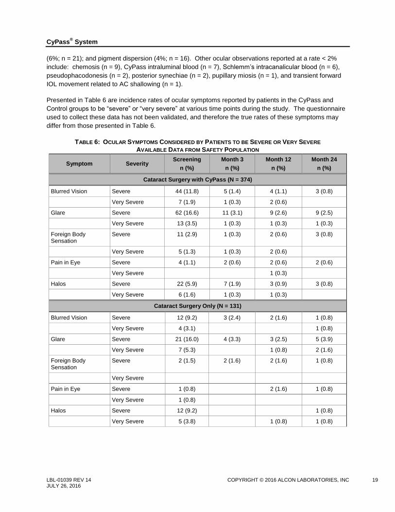
Presented in Table 6 are incidence rates of ocular symptoms reported by patients in the CyPass and
Control groups to be “severe” or “very severe” at various time points during the study. The questionnaire
used to collect these data has not been validated, and therefore the true rates of these symptoms may
differ from those presented in Table 6.
TABLE 6: OCULAR SYMPTOMS CONSIDERED BY PATIENTS TO BE SEVERE OR VERY SEVERE
AVAILABLE DATA FROM SAFETY POPULATION
Symptom Severity Screening
n (%)
Month 3
n (%)
Month 12
n (%)
Month 24
n (%)
Cataract Surgery with CyPass (N = 374)
Blurred Vision Severe 44 (11.8) 5 (1.4) 4 (1.1) 3 (0.8)
Very Severe 7 (1.9) 1 (0.3) 2 (0.6)
Glare Severe 62 (16.6) 11 (3.1) 9 (2.6) 9 (2.5)
Very Severe 13 (3.5) 1 (0.3) 1 (0.3) 1 (0.3)
Foreign Body Sensation
Severe 11 (2.9) 1 (0.3) 2 (0.6) 3 (0.8)
Very Severe 5 (1.3) 1 (0.3) 2 (0.6)
Pain in Eye Severe 4 (1.1) 2 (0.6) 2 (0.6) 2 (0.6)
Very Severe 1 (0.3)
Halos Severe 22 (5.9) 7 (1.9) 3 (0.9) 3 (0.8)
Very Severe 6 (1.6) 1 (0.3) 1 (0.3)
Cataract Surgery Only (N = 131)
Blurred Vision Severe 12 (9.2) 3 (2.4) 2 (1.6) 1 (0.8)
Very Severe 4 (3.1) 1 (0.8)
Glare Severe 21 (16.0) 4 (3.3) 3 (2.5) 5 (3.9)
Very Severe 7 (5.3) 1 (0.8) 2 (1.6)
Foreign Body Sensation
Severe 2 (1.5) 2 (1.6) 2 (1.6) 1 (0.8)
Very Severe
Pain in Eye Severe 1 (0.8) 2 (1.6) 1 (0.8)
Very Severe 1 (0.8)
Halos Severe 12 (9.2) 1 (0.8)
Very Severe 5 (3.8) 1 (0.8) 1 (0.8)
CyPass
® System
LBL-01039 REV 14 COPYRIGHT © 2016 ALCON LABORATORIES, INC 20 JULY 26, 2016
Corneal Endothelial Cell Density
There was little difference in endothelial cell loss (ECL) between the CyPass and Control groups and
results were consistent with the peer-review literature benchmarks of cataract-related ECL1,2
. In the 24-
month consistent cohort of subjects in the safety population (n = 322 CyPass subjects and 114 Control
subjects), mean endothelial cell density (ECD) was 2107 cells/mm2 (± 482) at 24 months in comparison
with 2422 cells/mm2 (± 409) at baseline in the CyPass group. Mean ECD was 2181 cells/mm
2 (± 441) at
24 months in comparison with 2427 cells/mm2 (± 359) at baseline in the Control group. Mean ECD
change at 24 months in the CyPass group was -13% (± 14). Mean ECD change at 24 months in the
Control group was -10% (± 14). A similar proportion of subjects in each group (11% [95% CI: 7.2, 13.9] in
the CyPass group and 9% [95% CI: 3.6, 14.0] in the Control group) experienced significant ECL (e.g.,
ECL > 30%) at 24 months postoperatively.
Study data collection methodology was a confounding factor in the determination of a relationship
between CyPass positioning and ECL. The primary landmark used for determination of CyPass
positioning was the number of CyPass retention rings visible; however, these data were not reported for
every subject, nor were they reported consistently within the course of a given subject’s study
participation.
Endothelial cell loss > 30% was noted in 9.6% (16/166) of subjects for whom 1 CyPass retention ring was
reported visible at 2 or more postoperative examinations and there were no reports of 2 or more rings
visible at any examination for these subjects. In the group of subjects for whom 2 CyPass rings were
visible at 2 or more postoperative examinations, ECL > 30% was noted in 9.8% (4/41) of subjects.
Additional Safety Data Gathered after Study Exit
Post-study exit, the following CyPass-related secondary ocular surgeries have been reported, which are
not included in Table 5:
- One subject underwent planned surgical occlusion of the CyPass lumen with Prolene suture for IOP
control approximately 28 months post-CyPass implantation.
- Another subject underwent secondary surgery for CyPass proximal end trimming due to anterior
device positioning associated with significant endothelial cell loss approximately 32 months post-
CyPass implantation. Anterior device positioning was reported within the first postoperative week.
- One subject underwent secondary surgery for CyPass proximal end trimming due to anterior device
positioning associated with significant endothelial cell loss approximately 48 months post-CyPass
implantation. Anterior device positioning was reported within the first postoperative week.
1
Reuschel, A., et al., Comparison of endothelial changes and power settings between torsional and longitudinal phacoemulsification. J Cataract Refract Surg, 2010. 36(11): p. 1855-61. 2
Buys, Y.M., et al., Prospective randomized comparison of one- versus two-site Phacotrabeculectomy two-year results. Ophthalmology, 2008. 115(7): p. 1130-1133 e1.
CyPass
® System
LBL-01039 REV 14 COPYRIGHT © 2016 ALCON LABORATORIES, INC 21 JULY 26, 2016
Medication Usage
Of subjects who were responders (e.g., 24-month unmedicated mean DIOP was reduced by ≥ 20% as
compared with baseline in the absence of IOP-affecting surgery during the study), 96.5% of subjects in
the CyPass group (251/271) and 72.4% of subjects in the Control group (55/74) were not using ocular
hypotensive medication at 24 months.
O. PHYSICIAN TRAINING
Physician training by certified Alcon personnel is required prior to use of this device. Training consists of
three main parts:
Didactic session
Simulated implantation of CyPass Micro-Stent
Supervised CyPass Micro-Stent implantation clinical cases until implantation proficiency is
demonstrated.
P. MANUFACTURER
Alcon Laboratories, Inc.,
6201 South Freeway, Fort Worth, TX 76134-2099
CyPass
® System
LBL-01039 REV 14 COPYRIGHT © 2016 ALCON LABORATORIES, INC 22 JULY 26, 2016
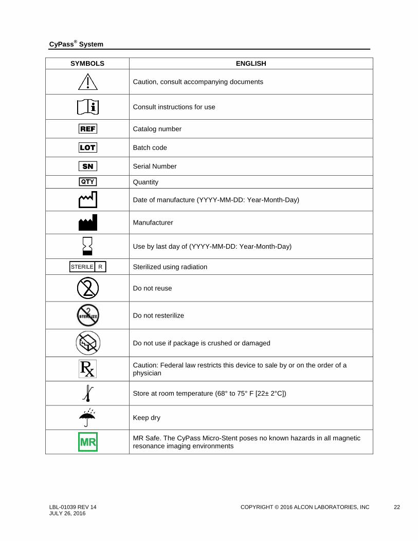
SYMBOLS ENGLISH
Caution, consult accompanying documents
Consult instructions for use
Catalog number
Batch code
Serial Number
Quantity
Date of manufacture (YYYY-MM-DD: Year-Month-Day)
Manufacturer
Use by last day of (YYYY-MM-DD: Year-Month-Day)
Sterilized using radiation
Do not reuse
Do not resterilize
Do not use if package is crushed or damaged
Caution: Federal law restricts this device to sale by or on the order of a physician
Store at room temperature (68° to 75° F [22± 2°C])
Keep dry
MR Safe. The CyPass Micro-Stent poses no known hazards in all magnetic resonance imaging environments

CyPass
® System
LBL-01039 REV 14 COPYRIGHT © 2016 ALCON LABORATORIES, INC 23 JULY 26, 2016
U.S. Pat. www.alconpatents.com
© 2016 Novartis


![Y FFS fee for service HDE = humanitarian device exemptionPotential Eye Damage From Alcon CyPass Micro-Stent Used to Treat Open-Angle Glaucoma: FDA Safety Communication], iStent®,](https://img.pdfslide.us/doc/110x75/5e536b6ef6fc066b0e676e5a/y-ffs-fee-for-service-hde-humanitarian-device-exemption-potential-eye-damage-from.jpg)