Embed Size (px)
Citation preview

Inspiratory Muscle Dysfunction in Patients with Severe Obstructive Sleep
Apnea
Meng-Yueh Chien1,2, Ying-Tai Wu1,2, * , Pei-Lin Lee3,4, Ya-Ju Chang5,
Pan-Chyr Yang4, *
1 School and Graduate Institute of Physical Therapy, College of Medicine, National Taiwan
University, Taipei, Taiwan.
2 Physical Therapy Center, National Taiwan University Hospital, Taipei, Taiwan.
3 Center of Sleep Disorder, National Taiwan University Hospital, Taipei, Taiwan.
4 Division of Pulmonary and Critical Care Medicine, Department of Internal Medicine,
National Taiwan University Hospital, Taipei, Taiwan.
5 Department of Physical Therapy, Chang Gung University, Tao-Yuan, Taiwan.
* These two authors contribute equally as corresponding authors.
Correspondence and requests for reprints should be addressed to Dr. Pan-Chyr Yang, Ph.D.,
Department of Internal Medicine, National Taiwan University Hospital, No 1, Jen-Ai Road,
Section 1, Taipei 10051, Taiwan and Dr. Ying-Tai Wu, Ph. D., School and Graduate Institute
of Physical Therapy, College of Medicine, National Taiwan University, Taipei, Taiwan.
E-mail: [email protected] and [email protected]
Running title: Muscle dysfunction in obstructive sleep apnea
. Published on July 30, 2009 as doi: 10.1183/09031936.00190208ERJ Express
Copyright 2009 by the European Respiratory Society.

ABSTRACT
Repetitive inspiratory effort against an obstructed airway and intermittent hypoxia may
be deleterious to the inspiratory muscles in patients with obstructive sleep apnea (OSA).
We investigated muscular dysfunction by comparing the strength, endurance, and
fatigability of inspiratory muscles and knee extensors in patients with newly diagnosed severe
OSA compared with matched controls. The measurements included strength and endurance
tests of both muscles, and a fatigue trial with simultaneous surface electromyography of the
diaphragm and the vastus lateralis during voluntary contractions and in response to magnetic
stimulation. This is the first investigation that we know of in assessing peripheral muscle
performance in severe OSA patients versus controls.
Patients in the OSA group exhibited significantly lower strength and endurance in both
muscles than the control group. The fatigue index decreased significantly exclusively in the
inspiratory muscles of OSA patients. Magnetic stimulation-evoked compound muscle action
potential latencies increased and the amplitudes decreased significantly in the diaphragm, but
not in the vastus lateralis after a fatigue test in the OSA group.
In conclusion, a significantly lower functional performance was shown for both
inspiratory muscles and knee extensors in OSA group. However, higher fatigability exhibited
only in the inspiratory muscles of patients with severe OSA.

KEYWORDS: inspiratory muscle; knee extensors; magnetic stimulation; nerve conduction
delay; obstructive sleep apnea; surface electromyography

Repetitive obstruction of the upper airway and apnea-hypopnea during sleep is a feature
of obstructive sleep apnea (OSA) syndrome [1]. An obstructed airway and the subsequent
asphyxia may lead to increased inspiratory efforts and hence, the chronic overload of
inspiratory muscles [2]. The chronic overuse of diaphragm was reported to put OSA subjects
at risk of inspiratory muscles fatigue [3]. However, whether diaphragm fatigue actually
occurs in OSA patients is still controversial [4-6]. Griggs and coworkers have shown that
pleural pressure relaxation rates during voluntary sniff maneuvers were prolonged in the
morning compared with the preceding night prior to sleep in OSA patients [4]. However,
Montserrant and coworkers reported lack of evidence for diaphragmatic fatigue over the night
in these patients [5]. They explained that the fatiguing levels of inspiratory effort generated
throughout the night did not sustain for sufficiently long periods to develop impaired
contractility.
The unique repetitive deoxygenation- reoxygenation pattern in OSA patients may induce
free radical production and oxidative stress, which causes muscle damage [7]. OSA has been
considered a systemic oxidative disorder [8]. Therefore, it is logical to hypothesize that
systemic skeletal muscle dysfunction may develop in patients with OSA, in addition to
disorders of the upper airway muscles. However, studies addressing this issue were
conducted in animal models and have shown conflicting results [9, 10].
The purpose of this study was to investigate the effect of OSA on muscular strength,
endurance, and the fatigability of inspiratory muscles and peripheral knee extensors in
patients with severe OSA as compared with those in the control group. Comparisons made
between these two types of muscle were aimed to determine whether the effects of OSA were
generalized or specific to the muscles subjected to increased use.
METHODS
Experimental design and participants
Fifteen men aged 40-65 years newly diagnosed with severe OSA with an apnea-hypopnea
index (AHI) ≥ 30 h–1and an Epworth sleepiness scale score ≥10 were recruited at the Sleep
Research Center in a teaching hospital. The control group consisted of 15 age-, height-, and
weight-matched subjects (AHI < 5 h–1) not diagnosed as OSA from the Sleep Research
Center. Subjects were excluded if presented with any active medical diseases, nervous system
diseases, major pulmonary dysfunction, morbid obesity, diabetes under oral hypoglycemic
agents, alcoholism (≥ 50 ml per day), or recent infection. All the participants at the time of
the study were not receiving pharmacological or mechanical treatment. The study was
approved by the Institutional Ethics Committee (NCT00813852), and all subjects were
informed of the procedures in detail and gave their written informed consent prior to
enrollment.
Diagnosis of OSA
The definitive diagnosis of OSA depended on an in-laboratory overnight
polysomnography recording system (Embla, Medcare, Iceland) demonstrating an elevated
AHI, which was calculated as the total episodes of hypopnea and apnea per hour of sleep
according to the criteria of the American Academy of Sleep Medicine [1].
The level of daytime sleepiness was assessed using the Epworth sleepiness scale in the
morning after nocturnal polysomnography. Normal values ranged from 2 to 10, with scores
≥10 indicating daytime sleepiness [11].
Anthropometrics, pulmonary function tests, and physical activity evaluation
Body weight and height, and circumferences of the neck, waist, and hip were measured.
Body mass index (BMI) and waist-hip ratio (WHR) were also calculated. Pulmonary function
tests (PFT) were performed by using a computerized spirometer (Chestgraph HI-701, Chest
MI Inc., Tokyo, Japan) with the subjects in a sitting position, according to the standardized
procedure [12]. The forced expiratory volume in the first second (FEV1), forced vital capacity
(FVC) and FEV1/FVC ratio were recorded.
Physical activity was evaluated with a 7-day recall questionnaire, which was designed to
determine calories expended on all activities during the previous 7-day period [13]. The total

energy expenditure was the sum of energy consumed by all activities.
Functional performance and fatigue index of inspiratory muscles
The maximal inspiratory pressure (PImax) indicative of muscle strength was measured at
the mouth near the residual volume with an aneroid pressure gauge (Model 4103, Boehringer,
Norristown, PA, USA) using standard procedures [12].
Maximal voluntary ventilation (MVV) maneuvers were used as the index of inspiratory
muscle endurance and for the fatigue-induced protocol according to the diagnostic
recommendations by the Chestgraph HI-701 spirometer [12]. Ventilation time was calculated
by the computer automatically. Our previous study reported diaphragmatic fatigue could be
induced by the 2-set MVV maneuvers with a five-minute rest in between [14]. The fatigue
index was defined as the post-MVV maneuvers PImax divided by the pre-MVV maneuvers
PImax which represents how much the muscle tension declined upon repetitive contraction.
For routine muscle fatigue measurements, the decrease in force generated from a maximal
volitional contraction is often used as a fatigue index.
Functional performance and fatigue index of knee extensors
A Cybex 6000 (Cybex, Division of Lumex Inc., Ronkonkoma, NY, USA) was used to
measure the strength and endurance of the right knee extensors. Each subject was instructed

to perform five maximal isometric knee extension contractions at a 60° knee flexion angle.
Each contraction lasted for five seconds, with a resting period of at least 15 seconds between
trials [15]. The means of five maximal isometric contraction peak torques (Newton-meter,
N-m) were calculated for data analysis.
The endurance test consisted of 30 cycles of alternative knee extension / flexion
isokinetic contractions at the speed of 180°/sec. Before each test, subjects performed three
submaximal trials to become familiarized with the test. The muscular endurance was
evaluated by the total work (N-m) generated during 30 cycles of contractions as the total area
under the torque curve for knee extension movement which would be automatically provided
by the Cybex system [15].
After 30 cycles of isokinetic contractions, each participant performed three maximal
isometric knee extension contractions at 60° knee flexion angle again. The fatigue index was
defined as the mean of the peak torques of isometric contractions at post-endurance test
divided by the pre-endurance value.
Magnetic stimulation
Cervical magnetic stimulation (CMS) was performed by a Magstim 220 stimulator
equipped with a circular doughnut-shaped 90 mm coil producing a maximum output of 2.5
Tesla (Magstim, Whitland, Dyfed, UK) using standard techniques described before [16].

Stimulations were delivered with the maximal output of the stimulator. Subjects were asked
to keep their neck slightly forward bent for stimulating the phrenic nerve [16], and lying on
the Cybex table with the coil placed below the inguinal ligament and 2 cm laterally from the
femoral artery for stimulating the femoral nerve [17].
Surface EMG recordings for the diaphragm and vastus lateralis
The surface EMG signals were simultaneously and continuously acquired by means of
bipolar electrodes from the right diaphragm and the right vastus lateralis (VL). For the
diaphragm, the electrodes were placed in the seventh or eighth intercostal space on the right
side of the body at the midclavicular line [18, 19]. The electrodes were also placed on the
skin overlying the lower third of the VL muscle (10-12 cm above the knee joint) and the
electrodes were aligned longitudinally to the direction of the muscle fibers. The distance
between the two electrodes was kept below 2 cm and the reference electrodes were placed on
the sternum and patella. The skin surface was well prepared and cleaned with alcohol to
obtain a low interelectrode resistance prior to positioning the electrodes.
Experimental protocol
All the subjects performed five maximal voluntary contractions (MVCs), followed by five
magnetic stimulations. Subsequently, inspiratory muscle fatigue was induced by two MVV

maneuvers with a 5 minute rest between each trial, while knee extensor fatigue was induced
by 30 repetitions of isokinetic contractions. Immediately after the fatigue protocols, another
five magnetic stimulations were performed as described above and were followed by five
MVCs (about 1 minute after fatigue test). The testing protocol is illustrated in Figure 1.
EMG data analysis
All EMG signals were amplified (EMG100A, Biopac Systems Co., Goleta, CA, USA),
band-pass filtered between 10-Hz to 1000-Hz, and digitized at a sampling rate of 2 kHz.
Subsequently, the final signal was acquired and analyzed off-line later using commercially
available software (AcqKnowledge 3.9.1 software for Windows). The parameters of the
sEMG signals during maximal voluntary contractions included root mean square (RMS)
values of the amplitude and median power frequency (MPF). The means of the first three
voluntary sEMG signals (pre-, and post-MVV PImax, pre- and post-test maximal isometric
knee contractions, Figure 2), and the first three and the last three breaths during MVV
maneuvers and during 30-cycle isokinetic endurance test were calculated. RMS was used to
quantify the diaphragm activation which means combined processes of diaphragm motor unit
recruitment and motor firing rate [20]. The influence of the EKG on sEMG signals was
minimized by recording from the right side of the body. To further minimize any signal
contamination by EKG on the diaphragmatic sEMG signals during PImax effort, RMS was
measured from the segments between QRS complexes.
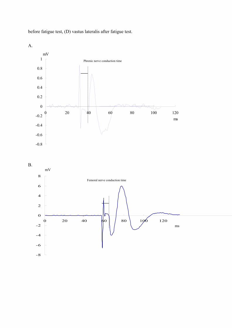
The conduction time of the phrenic nerve and femoral nerve were measured as the time
elapsed between the stimuli delivered by CMS and the onset of the compound muscle action
potential (CMAP), which was specifically marked by the first deviation of the signal from
baseline (CMAP latencies, Figure 3). CMAP latencies and amplitudes provided thereafter
corresponded to the average of five stimulations. The voluntary EMG and the diaphragm
CMAPs are myoelectric signals that represent different meanings: the voluntary signal
represents the summated electrical activity generated by asynchronously firing motor units,
whereas the diaphragm CMAPs represents the summated synchronized electrical activity
generated by all motor units after a supramaximal stimulus of the phrenic nerve [21].
Data analysis and statistics
Statistical analyses were performed using SPSS v11.5 for Windows (SPSS Inc., Chicago,
IL, USA). Values were expressed as mean ± SD. Comparisons of basic characteristics
between the two groups were made by independent t-tests. The maximum voluntary
contraction force and sEMG parameters were analyzed using a repeated measures analysis of
variance (ANOVA) to assess the difference between the baseline and the post fatigue test in
each group. α value was set at 0.05.
RESULTS
Baseline characteristics of the study group
There was no significant difference in the basic demographic characteristics, current
smokers, and energy expenditure of daily activities between the two groups except that the
OSA group had significantly higher values of AHI and ESS (Table 1). All the participants had
normal FVC, FEV1, and FEV1/FVC ratio. However, the patients with severe OSA had
significantly lower values o f both FVC and FEV1 than the controls.
Functional performance and sEMG parameters at baseline
Patients with severe OSA had significantly lower baseline strength (PImax and peak torque
values for the knee extensors) and endurance than the controls at baseline than their controls
(Table 2, P<0.05). Diaphragmatic sEMG revealed significantly lower MPF in patients with
severe OSA than those in the control group (P<0.05). sEMG recordings for VL revealed
significantly lower RMS in patients with severe OSA than those in the control group
(P<0.05).The evoked sEMG for the diaphragm response to CMS showed a significantly
longer CMAP latency (7.28±0.66 ms) and lower CMAP amplitude (1.4±0.5 mV) in patients
with severe OSA than those in the control group (P<0.05). It also showed a significantly
longer CMAP latency (5.41±0.74 ms) of evoked sEMG for the VL response to magnetic
stimulation in patients with severe OSA than those in the control group (P<0.05).
Functional performance and sEMG parameters after the fatigue test
A similar trend was found for the strength decline and voluntary sEMG parameters of
inspiratory muscles and knee extensors in the two groups (Table 2). As the muscles of OSA
patients were weaker than those of the controls, the level of fatiguing tasks might be
relatively higher for the OSA group. The fatigue index derived during maximal voluntary
efforts was significantly different between the two groups in the inspiratory muscles (0.86 vs
0.92, P<0.05), but not in the knee extensors (0.94 vs 0.95, P>0.05).
The diaphragmatic voluntary sEMG MPF declined significantly from baseline to the end
of the second MVV maneuver and slightly returned at post-MVV maneuvers PImax (P<0.05,
Figure 4). Similar trend was shown in VL, however, no statistical significance was detected.
The diaphragmatic CMAP latency and amplitude showed significant changes after the
MVV maneuvers in the severe OSA group (latency: 7.28±0.66 vs 7.48±0.65 ms; amplitude:
1.4±0.5 vs 1.2±0.5 mV, P<0.05). However, there were no significant changes in CMAP
latency and amplitude of the VL after the endurance test.
DISCUSSION
This study demonstrated that severe OSA patients have significantly lower functional
performance in strength and endurance, and a prolonged CMAP latency in response to
magnetic stimulation of both the inspiratory muscles and knee extensors, but only the
inspiratory muscles showed significantly increased fatigability in performance and sEMG
assessments. This is the first investigation that we know of in assessing peripheral muscle
performance in severe OSA patients versus controls.
Methodological considerations
Diaphragm muscle activity can be assessed either as a pressure or an EMG response
which was recorded with surface, intramuscular needle or esophageal electrodes. However,
transdiaphragmatic pressure method and esophageal EMG have the disadvantage of their
invasiveness and discomfort. Use of intramuscular needle electrodes avoids cross-talk but
contains disadvantages of invasiveness and sampling bias. sEMG recordings provide a
popular, routine tool to investigate chest wall muscle function but can be confounded by
noise and cross-talk [22]. This is a potential limitation of sEMG. Thus, electrodes were
placed in the way that minimizes cross-talk suggested by previous researches [18, 19].
Subjects were well supported during testing to minimize the activities of the adjacent trunk
muscles.
It was well established that force production by the diaphragm could be significantly
reduced by fatigue induced by periods of high-intensity voluntary isocapnic ventilation [23].

During a 2-minute MVV maneuver there was a progressive reduction in ventilation and
transdiaphragmatic pressure generation associated with the development of fatigue of the
diaphragm [23]. Isocapnic maximal sustained ventilation for 8 minutes has also been
suggested as fatiguing procedures [12]. Our preliminary study have also demonstrated 2-set
15-second MVV maneuvers could induce diaphragmatic fatigue in young healthy participants
[14].
Our study employed MVV maneuvers and 30 cycles of isokinetic knee contractions for
determining the endurance of inspiratory muscles and knee extensors, respectively, which,
strictly speaking, were not standardized endurance tests by the definition. However, they
were widely utilized for evaluating functional muscle endurance clinically [12, 15].
Functional performance of inspiratory muscles and knee extensors
In this study, inspiratory muscles (diaphragm) and knee extensors (VL) were selected to
explore the effect of the functional changes observed in OSA patients. The chronic
overloading of inspiratory muscles against an obstructed upper airway could lead to structural
and metabolic adaptations. Peripheral muscle was chosen as control because it was
considered to be not overloaded during sleep. Few studies compared the inspiratory muscles
and peripheral muscles in patients with OSA.

Mezzanotte and coworkers reported a significantly lower PImax in severe OSA subjects
[24], but Shepherd and coworkers reported that the PImax was not different between the
subjects whose AHI was below 20 h-1 and above 20 h-1 [25]. In this study, we found
significantly lowered values of PImax in patients with severe OSA compared to those without
OSA. The weakened diaphragm would reduce the collapsing force during apneas thereby
offset the large negative pressure generated by the diaphragm. However, the cause-effect
relationship could not be determined in the present observation.
In regard to the performance of the lower extremity muscles among OSA patients, little
study has been conducted in human. Sauleda and coworkers obtained a needle biopsy of
quadriceps femoris in severe OSA patients and found structural and bioenergetic changes in
the skeletal muscles [26]. Nevertheless, some studies have shown conflicting results
regarding limb muscle performance in OSA animals [9, 10]. The present study is the first
investigation that we know of in showing lower strength and endurance of knee extensors in
the OSA group, but not fatigability. Future studies were needed to confirm this observation.
It is noteworthy that factors such as nutrition, obesity state, and physical activity have
profound effects on muscle performance [27]. In our study, the groups were matched for the
anthropometric parameters; therefore, the difference in muscular strength could not be
attributed to them. However, the relatively lower physical activity levels might be related to
the lower baseline muscular strength of both examined muscles in the severe OSA group. In

addition, the possibility that lower values of PImax and peak torques represent difference in
effort and cooperation between two groups could not be excluded, even though we gave
maximal encouragement to each participant during all the tests.
Fatigability of the inspiratory muscles
Previous studies have shown conflicting results regarding whether inspiratory muscles
fatigue during OSA [4-6]. One possible reason is the fatigue index may not distinguish
between different types of fatigue, i.e. central fatigue and peripheral neuromuscular fatigue.
In contrast to the conventional fatigue test induced by MVCs, externally applied electrical or
magnetic stimulations for peripheral nerve roots measures the CMAP after stimulation have
long been known to evaluate peripheral neuromuscular function [16, 17]. CMAP measures
the peripheral neuromuscular fatigue (especially neurotransmitters) in the muscles bypassing
the central nervous system. El-Kabir et al. have shown that in patients with OSA treatment
with nasal CPAP did not improve the twitch transdiaphragmatic pressure in response to
bilateral CMS [28]. However, the study included lack of repeated polysomnography and
compliance data of CPAPA treatment, and no control group. Our results revealed a
significant decrease in the sEMG RMS amplitude after MVV maneuvers but not CMAP
amplitudes in control subjects. These findings indicate that MVV maneuvers might induce

central fatigue which was considered as a preemptive fatigue induced by the central nerve
system to prevent further inspiratory muscle fatigue; thus acting as a protective mechanism.
However, significant decreases in CMAP latencies and amplitudes after MVV maneuvers
developed in patients in the severe OSA group, thereby suggesting the diaphragm muscle was
responsible for inspiratory muscle fatigue in these patients. Because peripheral muscle fatigue
can result in persistent muscle fatigue and longer recovery periods, it is likely that this
peripheral neuromuscular fatigue was responsible for the decline in function of inspiratory
muscles, i.e. decreased PImax and endurance.
The decrease of sEMG amplitudes and power spectra of the inspiratory muscles found in
this study may reflect decreased motor unit recruitment from a decreased activation [29], or
hypoxia induced decreases in muscle pH that promote fatigue [30]. Further studies are needed
to explore the associated mechanisms.
Study limitations
This study had several limitations. Firstly, since we did not measure twitch pressure in
response to the stimulation of phrenic nerves and femoral nerve, this study could not answer
whether the decreased strength of the inspiratory muscle and knee extensors was due to
decreased recruitment and/or decreased contractility per se. Nevertheless, the study
demonstrated decreased CMAP amplitude that could represent the delayed neurotransmitter

in OSA patients compared with the controls.
Secondly, we did not measure lung volume changes before and immediately after MVV
maneuvers. Thus it may be difficult to thoroughly interpret the changes in CMAP
characteristics. Previous studies had reported that CMAP amplitude and latency would be
affected by different lung volumes [21, 22]. We assumed both groups in our study were
influenced to the same extent; however, the result showing significant difference of CMAP
amplitude and latency could provide important information regarding the neuromuscular
characteristics inspiratory muscles in OSA patients. Future studies should examine the effect
of OSA on inspiratory muscles fatigue by using comprehensive measurement tools.
Thirdly, the unequal fatiguing tasks produced by OSA and control participants might
complicate the comparison of fatigue indices. The lower baseline PImax and peak torques of
knee extensors in the OSA group would bring about the different fatigue tasks for the OSA
and the control group. According to that, future study should strive to employ a more
standardized fatigue tests for further refinement of this study.
Implications
A significantly lower strength and endurance for inspiratory muscles and knee extensors
of severe OSA patients were found in comparison with their age and BMI matched controls.
Therefore, systemic effects of chronic intermittent hypoxia and reoxygenation on skeletal
muscles in OSA populations could not be completely ruled out. However, higher fatigability
either during voluntary contractions or in response to magnetic stimulations was only
exhibited in the inspiratory muscles of patients with severe OSA. Peripheral neuromuscular
fatigue of the diaphragm might contribute to this increased fatigability; therefore, we
speculated that the underlying mechanisms of muscular adaptation to chronic increase use
played a critical role in this pathological state when compared to the systemic effects on
skeletal muscles occurring in OSA patients.
Conflict of Interest Statement: None of the authors has a financial relationship with a
commercial entity that has an interest in the subject of this manuscript.
Acknowledgement: The authors thank the National Science Council (Taiwan) for financial
support (NSC 96-2314-B-002-022-MY3).
REFERENCES:
1. American Academy of Sleep Medicine. Sleep-related breathing disorders in adults:
recommendations for syndrome definition and measurement techniques in clinical
research. The Report of an American Academy of Sleep Medicine Task Force. Sleep
1999; 22: 667-689.
2. Wilcox PG, Paré PD, Road JD, Fleetham JA. Respiratory muscle function during
obstructive sleep apnea. Am Rev Respir Dis 1990; 142: 533-539.
3. MacIntyre NR. Muscle dysfunction associated with obstructive pulmonary disease.
Respir Care 2006; 51: 840-848.
4. Griggs GA, Findley LJ, Suratt PM, Esau SA, Wilhoit SC, Rochester DF. Prolonged
relaxation rate of inspiratory muscles in patients with sleep apnea. Am Rev Respir Dis
1989; 140: 706-710.
5. Montserrat JM, Kosmas EN, Cosio MG, Kimoff RJ. Lack of evidence for diaphragmatic
fatigue over the course of the night in obstructive sleep apnea. Eur Respir J 1997; 10:
133-138.
6. Cibella F, Cuttitta G, Romano S, Bellia V, Bonsignore G. Evaluation of diaphragmatic
fatigue in obstructive sleep apnoeas during non-REM sleep. Thorax 1997; 52: 731-735.
7. Jackson MJ, O’Farrell S. Free radicals and muscle damage. Br Med Bull 1993; 49:
630-641.
8. Lavie L. Obstructive sleep apnea syndrome: an oxidative stress disorder. Sleep Med Rev
2003; 7: 35-51.
9. Petrof BJ, Pack AI, Kelly AM, Eby J, Hendricks JC. Pharyngeal myopathy of loaded
upper airway in dogs with sleep apnoea. J Appl Physiol 1994; 76: 1746-1752.
10. McGuire M, MacDermott M, Bradfold A. Effects of chronic intermittent asphyxia on rat
diaphragm and limb muscle contractility. Chest 2003; 123: 875-881.
11. John MW. A new method for measuring daytime sleepiness: the Epworth sleepiness
scale. Sleep 1991; 14: 540-545.
12. American Thoracic Society/European Respiratory Society. ATS/ERS statement on
respiratory muscle testing. Am J Respir Crit Care Med 2002; 166: 518-628.
13. Sallis JF, Haskell WL, Wood PD, Fortmann SP, Rogers T, Blair SN, Paffenbarger RS Jr.
Physical activity assessment methodology in Five-City Project. Am J Epidemiol 1985;
121: 91-106.
14. Chien MY, Wu YT, Chang YJ. Assessment of diaphragm and external intercostals
fatigue from surface EMG using cervical magnetic stimulation. Sensors 2008; 8:
2174-2187.
15. Wilcock A, Maddocks M, Lewis M, et al. Use of a Cybex NORM dynamometer to
assess muscle function in patients with thoracic cancer. BMC Palliat Care 2008; 7: 3.
16. Similowski T, Fleury B, Launois S, Cathala HP, Bouche P, Derenne JP. Cervical

magnetic stimulation: a new painless method for bilateral phrenic nerve stimulation in
conscious humans. J Appl Physiol 1989; 67: 1311-1318.
17. Alisauskiene M, Magistris MR, Vaiciene N, Truffert A. Electrophysiological evaluation
of motor pathways to proximal lower limb muscles: a combined method and reference
values. Clin Neurophysiol 2007; 118: 513-524.
18. Verin E, Straus C, Demoule A, Mialon P, Derenne JP, Similowski T. Validation of
improved recording site to measure phrenic conduction from surface electrodes in
humans. J Appl Physiol 2002; 92: 967-974.
19. Demoule A, Verin E, Locher C, Derenne JP, Similowski T. Validation of surface
recordings of the diaphragm response to transcranial magnetic stimulation in humans. J
Appl Physiol 2003; 94: 453-461.
20. Beck J, Sinerby C, Lindstrom L, Grassino A. Effect of lung volume on diaphragm EMG
signal strength during voluntary contractions. J Appl Physiol 1998; 85: 1123-1134.
21. Beck J, Sinerby C, Lindstrom L, Grassino A. Diaphragm interference pattern EMG and
compound action potentials: effects of chest wall configuration. J Appl Phsyiol 1997; 82:
520-530.
22. Sinderby C, Friberg S, Comtois N, Grassino A, Chest wall muscle cross talk in Canine
costal diaphragm electromyogram. J Appl Physiol; 1996; 81: 2312-2327.
23. Hammegård C, Wragg S, Kryoussis D, Mills GH, Polky MI, Moran J, Road J, Bake B,
Green M, Moxham J. Diaphragm fatigue following maximal ventilation in man. Eur
Respir J 1996; 9; 241-247.
24. Mezzanotte WS, Tangel DJ, White DP. Waking genioglossal electromyogram in sleep
apnea patients versus normal controls (a neuromuscular compensatory mechanism). J
Clin Invest 1992; 89: 1571-1579.
25. Shepherd KL, Jensen CM, Maddison KJ, Hillman DR, Eastwood PR. Relationship
between upper airway and inspiratory pump muscle force in obstructive sleep apnea.
Chest 2006; 130: 1757-1764.
26. Sauleda J, Garcia-palmer FJ, Tarraga S, Maimo A, Palou A, Agusti AG. Skeletal muscle
changes in patients with obstructive sleep apnoea syndrome. Respir Med 2003; 97:
804-810.
27. Morley JE, Baumgartner RN, Roubenoff R et al. Sarcopenia. J Lab Clin Med 2001; 137:
231-243.
28. El-Kabir DR, Polkey MI, Lyall RA, Williams AJ, Moxham J. The effect of treatment on
diaphragm contractility in obstructive sleep apnea syndrome. Respir Med 2003; 97:
1021-1026.
29. Bigland-Ritchie B, Donovan EF, Roussos CS. Conduction velocity and EMG power
spectrum changes in fatigue of sustained maximal efforts. J Appl Physiol 1981; 51:
1300-1305.
30. Reid MB, Haack KE, Franchek KM, Valberg PA, Kobzik L, West MS. Reactive oxygen
in skeletal muscle. I. Intracellular oxidant kinetics and fatigue in vitro. J Appl Physiol
1992; 73: 1797-1804.

Table 1. Baseline characteristics of the subjects
OSA group (n=15)
Control group (n=15)
P value
Age (yrs) 51.3±6.5 51.2±7.0 0.979 Current smoker (n)* 8 5 0.462 Anthropometrics
Body weight (kg) 75.0±8.1 73.6±7.8 0.625 Body height (cm) 168.1±5.8 167.4±3.7 0.698 Body mass index (kg/m2) 26.6±3.1 26.3±3.0 0.786 Neck circumference (cm) 38.3±3.2 37.9±2.1 0.463 Waist circumference (cm) 90.6±10.4 89.2±10.6 0.724 Hip circumference (cm) 99.8±6.9 99.1±7.2 0.656 Waist-hip ratio (%) 90.8±8.2 90.0±8.3 0.829
Sleep examination Apnea-hypopnea index (h-1) 54.0±21.7 2.3±1.0 <0.001 Epworth sleepiness scale 11.1±3.1 8.3±3.6 0.033 Sleep efficiency (%) 83.2±8.7 92.0±4.1 0.002 Average SaO2 (%) 92.3±4.5 96.2±7.8 0.005 Lowest SaO2 (%) 71.0±9.0 88.9±2.7 <0.001
Pulmonary function test FVC (l) 3.8±0.7 4.3±0.6 0.040 FVC, % of predicted 102.3±9.4 111.7±11.4 0.020 FEV1 (l) 3.1±0.6 3.5±0.5 0.064 FEV1, % of predicted 100.8±11.1 108.2±8.2 0.047 FEV1/FVC (%) 81.6±3.7 80.8±6.6 0.926
Physical activity (kcal/kg/d) 35.1±2.9 38.6±6.3 0.059 Values are as group means±SD. *: tested by Chi-square.
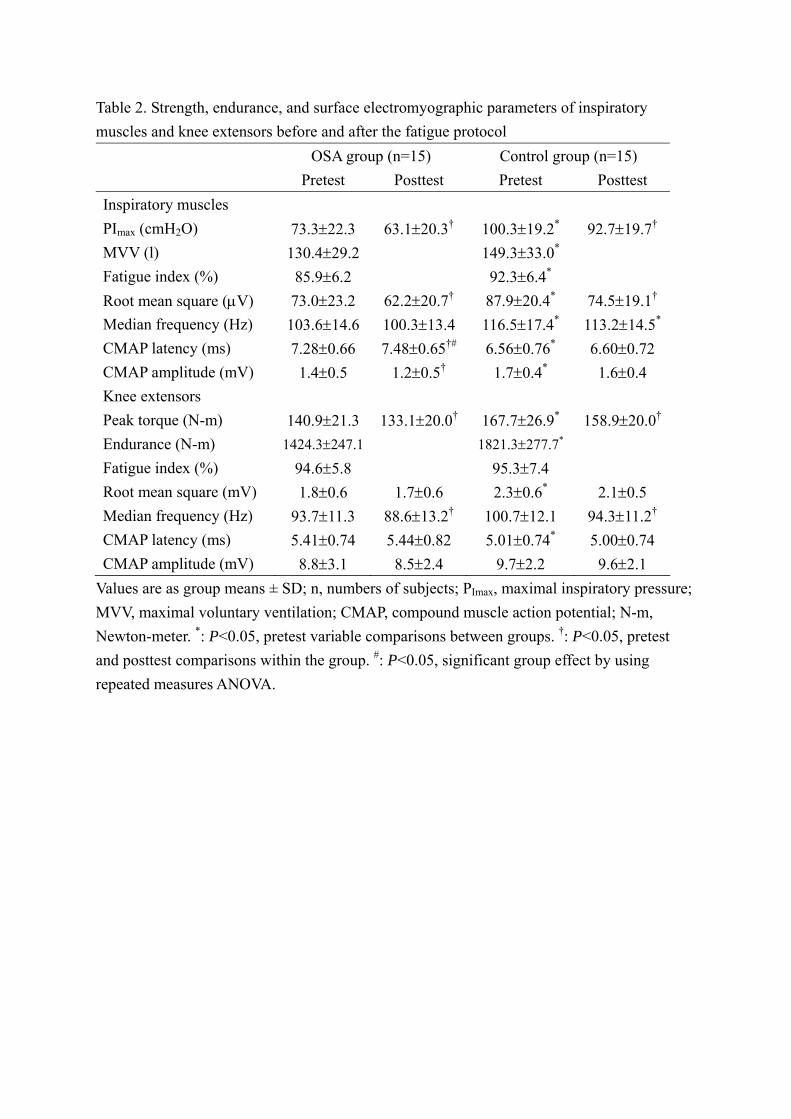
Table 2. Strength, endurance, and surface electromyographic parameters of inspiratory muscles and knee extensors before and after the fatigue protocol OSA group (n=15) Control group (n=15) Pretest Posttest Pretest Posttest Inspiratory muscles PImax (cmH2O) 73.3±22.3 63.1±20.3† 100.3±19.2* 92.7±19.7† MVV (l) 130.4±29.2 149.3±33.0* Fatigue index (%) 85.9±6.2 92.3±6.4* Root mean square (μV) 73.0±23.2 62.2±20.7† 87.9±20.4* 74.5±19.1† Median frequency (Hz) 103.6±14.6 100.3±13.4 116.5±17.4* 113.2±14.5* CMAP latency (ms) 7.28±0.66 7.48±0.65†# 6.56±0.76* 6.60±0.72 CMAP amplitude (mV) 1.4±0.5 1.2±0.5† 1.7±0.4* 1.6±0.4 Knee extensors Peak torque (N-m) 140.9±21.3 133.1±20.0† 167.7±26.9* 158.9±20.0† Endurance (N-m) 1424.3±247.1 1821.3±277.7* Fatigue index (%) 94.6±5.8 95.3±7.4 Root mean square (mV) 1.8±0.6 1.7±0.6 2.3±0.6* 2.1±0.5 Median frequency (Hz) 93.7±11.3 88.6±13.2† 100.7±12.1 94.3±11.2† CMAP latency (ms) 5.41±0.74 5.44±0.82 5.01±0.74* 5.00±0.74 CMAP amplitude (mV) 8.8±3.1 8.5±2.4 9.7±2.2 9.6±2.1
Values are as group means ± SD; n, numbers of subjects; PImax, maximal inspiratory pressure; MVV, maximal voluntary ventilation; CMAP, compound muscle action potential; N-m, Newton-meter. *: P<0.05, pretest variable comparisons between groups. †: P<0.05, pretest and posttest comparisons within the group. #: P<0.05, significant group effect by using repeated measures ANOVA.

Protocol Maximal voluntary
contractions
Magnetic
stimulation
Fatigue
test
Magnetic
stimulation
Maximal voluntary
contractions
Parameters Root mean square
Median frequency
CMAP amplitude
CMAP latency
CMAP amplitude
CMAP latency
Root mean square
Median frequency
Figure 1. Illustration of the protocol for the fatigue test
Figure 2. Examples of raw electromyopgraphic recordings (top) and root mean square (bottom) during maximal voluntary contractions in one patient with severe OSA. (A) diaphragm before fatigue test, (B) diaphragm after fatigue test, (C) vastus lateralis
before fatigue test, (D) vastus lateralis after fatigue test. A.
-0.8
-0.6
-0.4
-0.2
0
0.2
0.4
0.6
0.8
1
0 20 40 60 80 100 120
mV
ms
B.
-8
-6
-4
-2
0
2
4
6
8
0 20 40 60 80 100 120
ms
mV
Femoral nerve conduction time
Phrenic nerve conduction time

Figure 3. Example of EMG recordings of the right diaphragm (a) and VL (b) for motor responses (CMAP) with magnetic stimulation in a patient with severe OSA. a.
0
20
40
60
80
100
120
140
PImax_pre MVVearly MVVend PImax_post
OSACTN
b.
0
20
40
60
80
100
120
Voluntary_pre Isokinetic_early Isokinetic_end Voluntary_post
OSACTN
Figure 4. Group mean values of median power frequency of the right diaphragm (a) and vastus lateralis (b) for voluntary contractions before, during, and after fatigue test by MVV maneuvers (for inspiratory muscles) and isokinetic test (for knee extensors). #: P<0.05, significant group effect by using repeated measures ANOVA.
#
#