Embed Size (px)
Citation preview
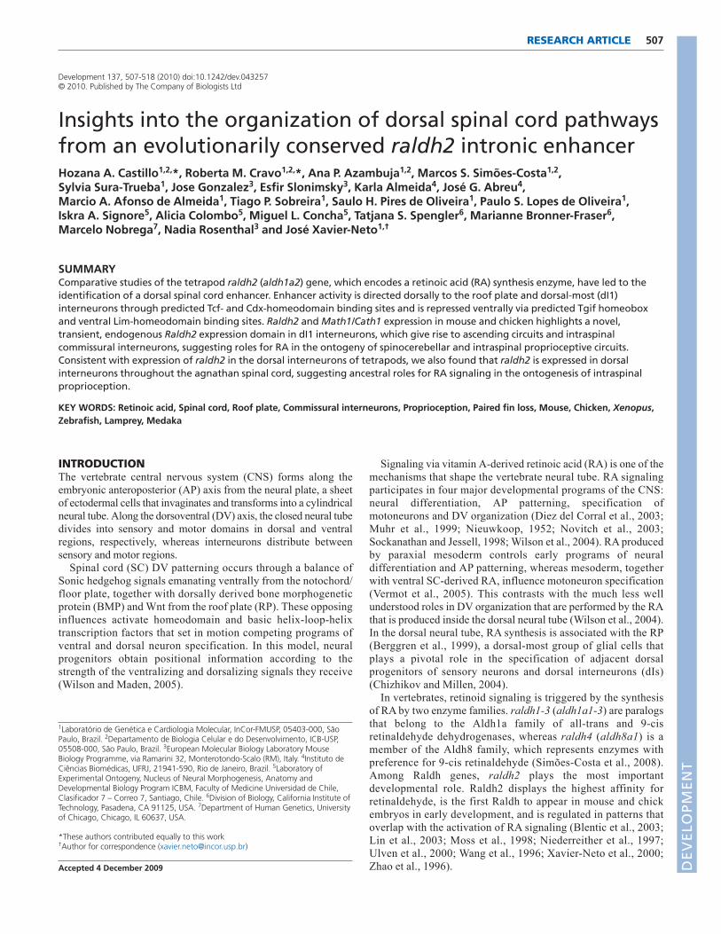
507RESEARCH ARTICLE
INTRODUCTIONThe vertebrate central nervous system (CNS) forms along theembryonic anteroposterior (AP) axis from the neural plate, a sheetof ectodermal cells that invaginates and transforms into a cylindricalneural tube. Along the dorsoventral (DV) axis, the closed neural tubedivides into sensory and motor domains in dorsal and ventralregions, respectively, whereas interneurons distribute betweensensory and motor regions.
Spinal cord (SC) DV patterning occurs through a balance ofSonic hedgehog signals emanating ventrally from the notochord/floor plate, together with dorsally derived bone morphogeneticprotein (BMP) and Wnt from the roof plate (RP). These opposinginfluences activate homeodomain and basic helix-loop-helixtranscription factors that set in motion competing programs ofventral and dorsal neuron specification. In this model, neuralprogenitors obtain positional information according to thestrength of the ventralizing and dorsalizing signals they receive(Wilson and Maden, 2005).
Signaling via vitamin A-derived retinoic acid (RA) is one of themechanisms that shape the vertebrate neural tube. RA signalingparticipates in four major developmental programs of the CNS:neural differentiation, AP patterning, specification ofmotoneurons and DV organization (Diez del Corral et al., 2003;Muhr et al., 1999; Nieuwkoop, 1952; Novitch et al., 2003;Sockanathan and Jessell, 1998; Wilson et al., 2004). RA producedby paraxial mesoderm controls early programs of neuraldifferentiation and AP patterning, whereas mesoderm, togetherwith ventral SC-derived RA, influence motoneuron specification(Vermot et al., 2005). This contrasts with the much less wellunderstood roles in DV organization that are performed by the RAthat is produced inside the dorsal neural tube (Wilson et al., 2004).In the dorsal neural tube, RA synthesis is associated with the RP(Berggren et al., 1999), a dorsal-most group of glial cells thatplays a pivotal role in the specification of adjacent dorsalprogenitors of sensory neurons and dorsal interneurons (dIs)(Chizhikov and Millen, 2004).
In vertebrates, retinoid signaling is triggered by the synthesisof RA by two enzyme families. raldh1-3 (aldh1a1-3) are paralogsthat belong to the Aldh1a family of all-trans and 9-cisretinaldehyde dehydrogenases, whereas raldh4 (aldh8a1) is amember of the Aldh8 family, which represents enzymes withpreference for 9-cis retinaldehyde (Simões-Costa et al., 2008).Among Raldh genes, raldh2 plays the most importantdevelopmental role. Raldh2 displays the highest affinity forretinaldehyde, is the first Raldh to appear in mouse and chickembryos in early development, and is regulated in patterns thatoverlap with the activation of RA signaling (Blentic et al., 2003;Lin et al., 2003; Moss et al., 1998; Niederreither et al., 1997;Ulven et al., 2000; Wang et al., 1996; Xavier-Neto et al., 2000;Zhao et al., 1996).
Development 137, 507-518 (2010) doi:10.1242/dev.043257© 2010. Published by The Company of Biologists Ltd
1Laboratório de Genética e Cardiologia Molecular, InCor-FMUSP, 05403-000, SãoPaulo, Brazil. 2Departamento de Biologia Celular e do Desenvolvimento, ICB-USP,05508-000, São Paulo, Brazil. 3European Molecular Biology Laboratory MouseBiology Programme, via Ramarini 32, Monterotondo-Scalo (RM), Italy. 4Instituto deCiências Biomédicas, UFRJ, 21941-590, Rio de Janeiro, Brazil. 5Laboratory ofExperimental Ontogeny, Nucleus of Neural Morphogenesis, Anatomy andDevelopmental Biology Program ICBM, Faculty of Medicine Universidad de Chile,Clasificador 7 – Correo 7, Santiago, Chile. 6Division of Biology, California Institute ofTechnology, Pasadena, CA 91125, USA. 7Department of Human Genetics, Universityof Chicago, Chicago, IL 60637, USA.
*These authors contributed equally to this work†Author for correspondence ([email protected])
Accepted 4 December 2009
SUMMARYComparative studies of the tetrapod raldh2 (aldh1a2) gene, which encodes a retinoic acid (RA) synthesis enzyme, have led to theidentification of a dorsal spinal cord enhancer. Enhancer activity is directed dorsally to the roof plate and dorsal-most (dI1)interneurons through predicted Tcf- and Cdx-homeodomain binding sites and is repressed ventrally via predicted Tgif homeoboxand ventral Lim-homeodomain binding sites. Raldh2 and Math1/Cath1 expression in mouse and chicken highlights a novel,transient, endogenous Raldh2 expression domain in dI1 interneurons, which give rise to ascending circuits and intraspinalcommissural interneurons, suggesting roles for RA in the ontogeny of spinocerebellar and intraspinal proprioceptive circuits.Consistent with expression of raldh2 in the dorsal interneurons of tetrapods, we also found that raldh2 is expressed in dorsalinterneurons throughout the agnathan spinal cord, suggesting ancestral roles for RA signaling in the ontogenesis of intraspinalproprioception.
KEY WORDS: Retinoic acid, Spinal cord, Roof plate, Commissural interneurons, Proprioception, Paired fin loss, Mouse, Chicken, Xenopus,Zebrafish, Lamprey, Medaka
Insights into the organization of dorsal spinal cord pathwaysfrom an evolutionarily conserved raldh2 intronic enhancerHozana A. Castillo1,2,*, Roberta M. Cravo1,2,*, Ana P. Azambuja1,2, Marcos S. Simões-Costa1,2,Sylvia Sura-Trueba1, Jose Gonzalez3, Esfir Slonimsky3, Karla Almeida4, José G. Abreu4,Marcio A. Afonso de Almeida1, Tiago P. Sobreira1, Saulo H. Pires de Oliveira1, Paulo S. Lopes de Oliveira1,Iskra A. Signore5, Alicia Colombo5, Miguel L. Concha5, Tatjana S. Spengler6, Marianne Bronner-Fraser6,Marcelo Nobrega7, Nadia Rosenthal3 and José Xavier-Neto1,†
DEVELO
PMENT
508
Here we identify a conserved intronic enhancer that drivesraldh2 expression in the RP and dIs of the frog, mouse andchicken dorsal SC, suggesting that DV programs of neural tubedevelopment regulated by RA are encoded by evolutionarilyconserved cis-regulatory modules. The combined activation ofraldh2 in the RP and dIs by a single enhancer suggests that thesetwo RA signaling domains play related roles and this prompted usto utilize a comparative approach to gain insight into thesefunctions. By investigating the patterns of raldh2 expression inthe SC of agnathan, teleost and tetrapod embryos, we discovereda novel, transient, field of RA synthesis in dI precursors. Bycomparing the patterns of raldh2 expression in agnathans andteleosts with those of amniotes, we provide evidence that RAsignaling might be involved in the ontogeny of two specific SCsensory functions: proprioception from vertebrate pairedappendages and modulation of the intrinsic SC locomotorcircuitry.
MATERIALS AND METHODSBioinformaticsAldh gene sequences were obtained from NCBI, the Ensembl genomebrowser, JGI Eukaryotic Genomes and through the method of Sobreira andGruber (Sobreira and Gruber, 2008). Conserved non-coding elements(CNEs) were identified using the ECR Browser and BLAST 2 sequences.The CNE search was limited to between 30 kb upstream of the transcriptionstart site and 30 kb downstream of the stop codon. Transcription factorbinding site (TFBS) prediction was performed using a locally availablesoftware that implements the MatInspector algorithm (Quandt et al., 1995)with TFBS matrices from TRANSFAC 6.0 (Wingender et al., 1996).TFBSs were accepted if at least 90% similar to a matrix core/whole matrixscore.
Phylogenetic analysisThirty Aldh protein sequences from six vertebrate species ranging fromagnathans to primates were aligned using MUSCLE (Edgar, 2004).The alignment (available on request) consisted of 464 amino acidpositions, manually refined to eliminate gaps. Phylogenetic trees weregenerated using neighbor-joining (NJ) (Saitou and Nei, 1987), maximumparsimony (MP) (Swofford, 2000), maximum likelihood (ML) (Schmidtet al., 2002) and Bayesian inference (BI) (Ronquist and Huelsenbeck,2003) methods. The WAG model was selected by ProtTest (Abascal et al.,2005). Node support was assessed in NJ and MP trees by 2000 bootstrapreplicates. ML was performed with 100,000 puzzling steps. For BIwe used two runs of 5,000,000 generations. Convergence was verifiedand an appropriate burn-in period of 2000 was determined. Consensustrees and posterior probabilities were calculated using the 50% majorityrule.
Plasmids and constructsraldh2 intron 1G CNEs from mouse, chicken and X. laevis were PCRamplified from genomic DNA and cloned into HSP68-lacZ and pTkeGFPvectors (Kothary et al., 1989; Rossant et al., 1991).
MutagenesisNested deletion mutants of the chicken Raldh2 intron 1G enhancer with734 bp, 450 bp, 355 bp and 149 bp fragments, as well as a 279 bp fragmentcontaining RP and interneuron activator elements (RPIE), plusoverlapping fragments A (62 bp), B (91 bp), C (92 bp) and D (114 bp),were PCR amplified and cloned into pTkeGFP. For site-directedmutagenesis we utilized QuickChange II (Stratagene, 200524).Nucleotides belonging to a predicted binding site for the Lim-homeodomain protein Lhx3 (116-126 bp) in the enhancer were substitutedby an AscI restriction site. The Tcf/Cdx site (207-221 bp) was substitutedby an AscI restriction site. A second Tcf/Cdx motif (353-367 bp) wassubstituted by an FseI restriction site. A Tcf/Tgif site (427-441 bp) wassubstituted by an AscI restriction site. All constructs were confirmed byDNA sequencing.
Transgenic miceThe mouse Raldh2 intron 1G CNE-HSP68-lacZ reporter was excised bySalI digestion. Pronuclear injection was as described (Xavier-Neto et al.,1999).
ElectroporationEnhancer-eGFP plasmid (2 mg/ml) plus pCA-RFP DNA (0.5-1.0 mg/ml) in0.5% Fast Green in water were injected into the lumen of the chicken neuraltube (HH17-18). In ovo electroporation was performed by applying six 60-to 100-millisecond pulses at 15-18 V with a 0.5 mm platinum electrode.
Xenopus laevis embryo injectionsThe Xenopus intronic enhancer, or the negative control pTkeGFP plasmid(30 pg), were co-injected with -galactosidase mRNA (250 pg total) at the4-cell stage into both dorsal blastomeres. Injections were performed at theanimal pole ~45° from the top.
Staining and in situ hybridizationStains were performed as described previously (Xavier-Neto et al., 1999)using an anti-GFP rabbit polyclonal antibody (1:500; Abcam, ab6556) anddonkey anti-rabbit IgG conjugated with Alexa Fluor 488 (1:1000; MolecularProbes, A21206). In situ hybridization (ISH) (Wilkinson, 1992) and doubleISH (Stern, 1998) were preformed as described previously, using mouse(Zhao et al., 1996), chicken, Xenopus (Chen et al., 2001), zebrafish (viaDeborah Yelon, New York University School of Medicine, New York, USA)and lamprey raldh2 probes. Other probes included lamprey zic-1 (Sauka-Spengler et al., 2007), mouse Math1 (Helms and Johnson, 1998) and chickenCath1 (Wilson and Wingate, 2006). Lamprey raldh2 was cloned by low-stringency hybridization with a 32P-labeled zebrafish raldh2 probe (Sauka-Spengler et al., 2007). The lamprey raldh2 sequence was deposited inGenBank as FJ536260. An in situ probe for medaka raldh2 was PCRamplified using medaka cDNA.
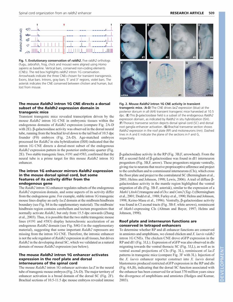
RESULTSraldh2 intron 1G is a conserved non-codingelement enriched with highly conservedtranscription factor binding sitesTo identify conserved non-coding elements (CNEs) potentiallycarrying raldh2 regulatory sequences, we aligned raldh2 orthologsfrom vertebrates including fugu, zebrafish, frog, chick and mouse,using human as baseline. This analysis revealed 72 CNEsdisplaying more than 75% identity over 183±16 bp (range 32-905bp) in 5�, intronic and 3� regions (Fig. 1). To identify sequencemodules that regulate raldh2 expression, we screened vertebrateraldh2 CNEs for enhancer function. We selected three CNEs: a 5�and a 3� CNE conserved in all species (Fig. 1, gray arrowheads),plus raldh2 intron 1G, the largest raldh2 CNE (Fig. 1, redarrowhead). Of those, only mouse Raldh2 intron 1G displayedenhancer activity in 10.5 days post-coitum (dpc) transienttransgenic mice. raldh2 intron 1G is conserved in amphibians,avians, rodents and primates and spans an average of 843±47 bp(702-905 bp) (Fig. 1, red box; see Fig. S1 in the supplementarymaterial), but could not be detected in teleosts (see Fig. S2 in thesupplementary material).
For a list of transcription factor binding sites (TFBSs) that displaymatrix identities to the matrix core that are higher than 90% and thatare conserved across amphibian, avian, marsupial, rodent and primateraldh2 intron 1G enhancers, see Fig. S3 in the supplementary material.There is deep conservation for TFBSs associated with Wnt signaling(i.e. Tcf binding sites), for homeodomain and Lim-homeodomainfactors, and for factors such as Pax, Pou and basic helix-loop-helix.Some predicted sites for Forkhead factors, Vsx2 (Chx10), Lhx3,Pou3f1 and Klf4 are so rare that their presence in the enhancer reflectsa statistically significant event when compared with their occurrencein a billion-bp random set.
RESEARCH ARTICLE Development 137 (3)
DEVELO
PMENT
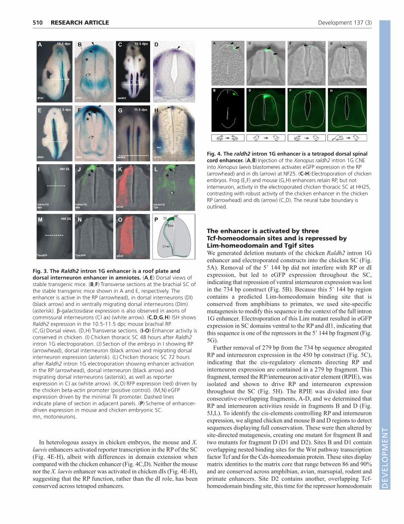
The mouse Raldh2 intron 1G CNE directs a dorsalsubset of the Raldh2 expression domain intransgenic miceTransient transgenic mice revealed transcription driven by themouse Raldh2 intron 1G CNE in embryonic tissues within theendogenous domains of Raldh2 expression (compare Fig. 2A-Dwith 2E). -galactosidase activity was observed in the dorsal neuraltube, running from the brachial level down to the tail bud of 10.5 dpcfounder (F0) embryos (Fig. 2A-D). Age-matched embryosprocessed for Raldh2 in situ hybridization (ISH) indicated that theintron 1G CNE directs a dorsal-most subset of the endogenousRaldh2 expression pattern in the posterior embryonic quarter (Fig.2E). Two stable transgenic lines, #191 and #583, confirmed that theneural tube is a prime target for this mouse Raldh2 intron 1Genhancer.
The intron 1G enhancer mirrors Raldh2 expressionin the mouse dorsal spinal cord, but somefeatures of its activity differ from theendogenous geneThe Raldh2 intron 1G enhancer regulates subsets of the endogenousRaldh2 expression domain, and some aspects of its activity differfrom the endogenous gene. For example, stable intron 1G transgenicmouse lines display an early lacZ domain at the midbrain/hindbrainboundary (see Fig. S4 in the supplementary material). The midbrain/hindbrain region contains cerebellum and tectum progenitors thatnormally activate Raldh2, but only from 15.5 dpc onwards (Zhanget al., 2003). Thus, it is possible that the two stable transgenic mouselines (#191 and #583) display heterochronic acceleration of anendogenous Raldh2 domain (see Fig. S4G-I in the supplementarymaterial), suggesting that some important Raldh2 repressors aremissing from the intron 1G CNE. Therefore, the intronic enhanceris not the sole regulator of Raldh2 expression in all tissues, but drivesRaldh2 in the developing dorsal SC, which we validated as a naturaldomain of mouse Raldh2 expression (see below).
The mouse Raldh2 intron 1G enhancer activatesexpression in the roof plate and dorsalinterneurons of the spinal cordThe mouse Raldh2 intron 1G enhancer activates lacZ in the neuraltube of transgenic mouse embryos (Fig. 2A-D). The major territory ofenhancer activation is a broad domain of the dorsal SC (Fig. 2F).Brachial sections of 10.5-11.5 dpc mouse embryos revealed intense
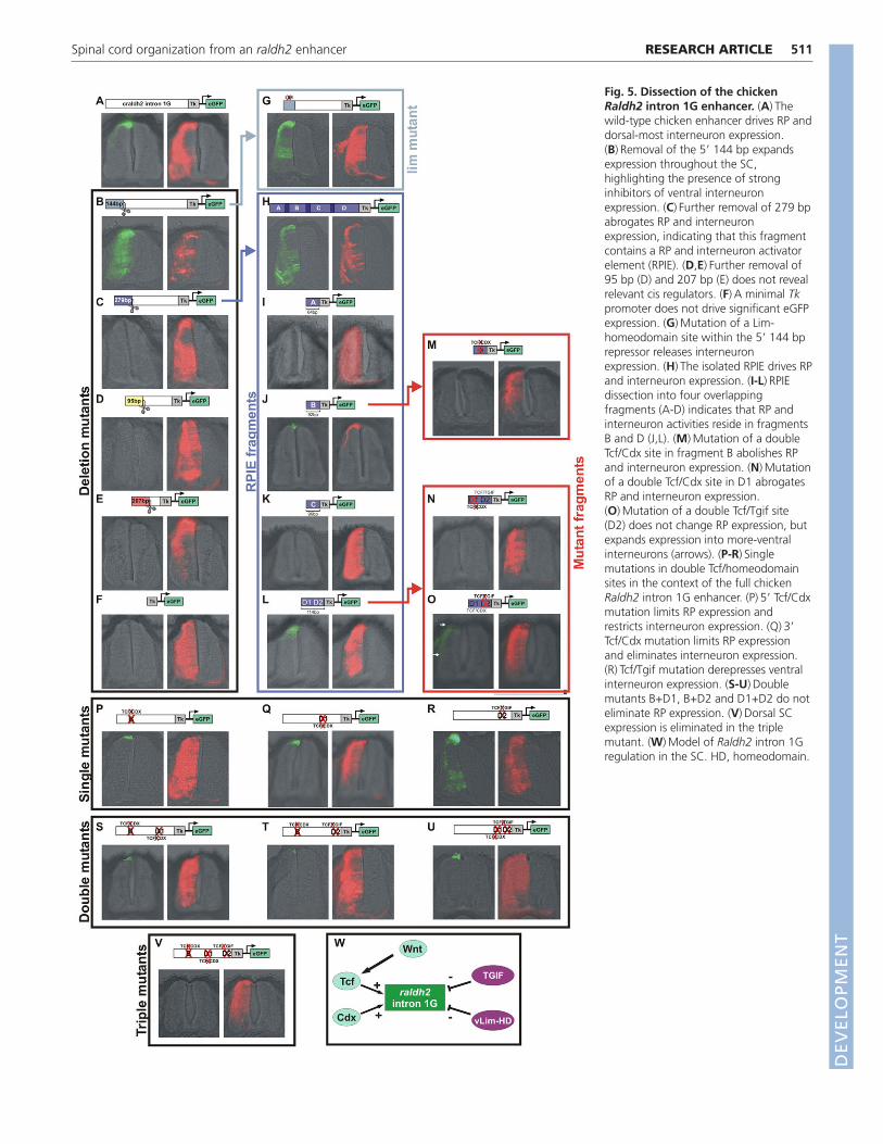
-galactosidase activity in the RP (Fig. 3B,F, arrowhead). From theRP, a second field of -galactosidase was found in dI1 interneuronprogenitors (Fig. 3B,F, arrow). These progenitors migrate ventrally,giving rise to neurons that receive proprioceptive afference and projectto the cerebellum and to commissural interneurons (CIs), which crossthe floor plate and project to the contralateral SC (Bermingham et al.,2001; Helms and Johnson, 1998; Lewis, 2006). A trail of diffuse -galactosidase activity in the mantle region highlighted the ventralmigration of dIs (Fig. 3B-F, asterisk), similar to the expression of aMath1 (Atoh1) transgene and of Dcc and Cntn2 (Tag-1) (Berminghamet al., 2001; Dodd et al., 1988; Furley et al., 1990; Helms and Johnson,1998; Keino-Masu et al., 1996). Ventrally, -galactosidase activitywas found in CI axonal tracts (Fig. 3B-F, white arrows), reminiscentof Math1-expressing CIs (Altman and Bayer, 1997; Helms andJohnson, 1998).
Roof plate and interneuron functions areconserved in tetrapod enhancersTo determine whether RP and dI enhancer functions are conservedin amniotes and amphibians, we cloned chicken and X. laevis raldh2intron 1G CNEs. The chicken CNE drove eGFP expression in theRP and dI1 (Fig. 3J,L). Expression of eGFP was also observed in dIsmigrating towards the ventral thoracic SC (Fig. 3J,L), as well as inventral axonal projections of CIs (Fig. 3L), reminiscent of lacZpatterns in transgenic mice (compare Fig. 3F with 3L). Injection ofthe X. laevis enhancer reporter construct into X. laevis dorsalblastomeres produced restricted eGFP expression in the RP and dIs(Fig. 4A,B), indicating that the dorsal SC function associated withthe enhancer has been conserved for at least 370 million years sincethe divergence of amphibians and amniotes (Hedges and Kumar,2003).
509RESEARCH ARTICLESpinal cord organization from an raldh2 enhancer
Fig. 1. Evolutionary conservation of raldh2. Five raldh2 orthologs(fugu, zebrafish, frog, chick and mouse) were aligned using Homosapiens as baseline. Vertical bars, conserved non-coding elements(CNEs). The red box highlights raldh2 intron 1G conservation.Arrowheads indicate the three CNEs chosen for transient transgenesis.Exons, blue bars. Introns, gray bars. 5� and 3� regions, violet bars. Theasterisk indicates the CNE conserved between chicken and human, butlost from mouse.
Fig. 2. Mouse Raldh2 intron 1G CNE activity in transienttransgenic mice. (A-D)The CNE drives lacZ expression (blue) at theposterior dorsum in all (4/4) transient transgenic mice harvested at 10.5dpc. (E)This -galactosidase field is a subset of the endogenous Raldh2expression domain, as indicated by Raldh2 in situ hybridization (ISH).(F)Thoracic transverse section depicts dorsal spinal cord (SC) and dorsalroot ganglia enhancer activation. (G)Brachial transverse section showsRaldh2 expression in the roof plate (RP) and motoneurons (mn). Dashedlines in A and E indicate the plane of the sections in F and G,respectively.
DEVELO
PMENT
510
In heterologous assays in chicken embryos, the mouse and X.laevis enhancers activated reporter transcription in the RP of the SC(Fig. 4E-H), albeit with differences in domain extension whencompared with the chicken enhancer (Fig. 4C,D). Neither the mousenor the X. laevis enhancer was activated in chicken dIs (Fig. 4E-H),suggesting that the RP function, rather than the dI role, has beenconserved across tetrapod enhancers.
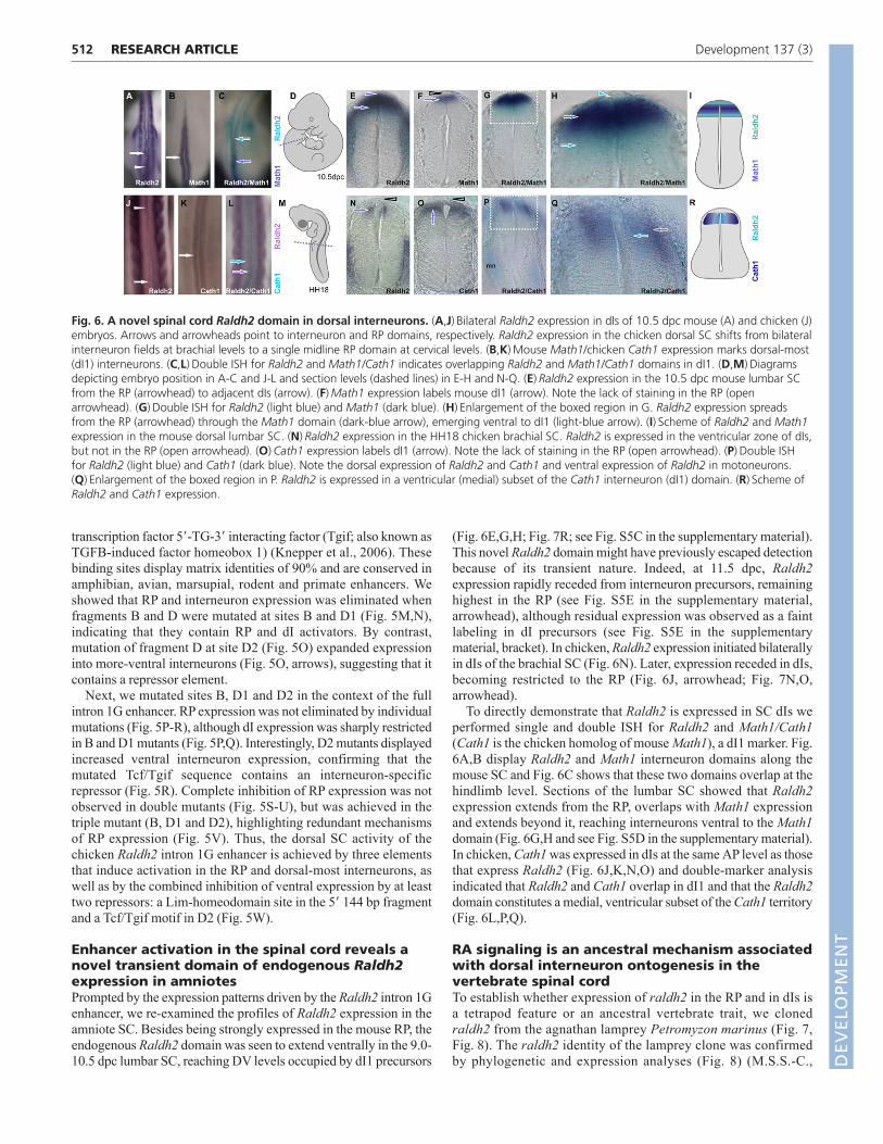
The enhancer is activated by threeTcf-homeodomain sites and is repressed byLim-homeodomain and Tgif sitesWe generated deletion mutants of the chicken Raldh2 intron 1Genhancer and electroporated constructs into the chicken SC (Fig.5A). Removal of the 5� 144 bp did not interfere with RP or dIexpression, but led to eGFP expression throughout the SC,indicating that repression of ventral interneuron expression was lostin the 734 bp construct (Fig. 5B). Because this 5� 144 bp regioncontains a predicted Lim-homeodomain binding site that isconserved from amphibians to primates, we used site-specificmutagenesis to modify this sequence in the context of the full intron1G enhancer. Electroporation of this Lim mutant resulted in eGFPexpression in SC domains ventral to the RP and dI1, indicating thatthis sequence is one of the repressors in the 5� 144 bp fragment (Fig.5G).
Further removal of 279 bp from the 734 bp sequence abrogatedRP and interneuron expression in the 450 bp construct (Fig. 5C),indicating that the cis-regulatory elements directing RP andinterneuron expression are contained in a 279 bp fragment. Thisfragment, termed the RP/interneuron activator element (RPIE), wasisolated and shown to drive RP and interneuron expressionthroughout the SC (Fig. 5H). The RPIE was divided into fourconsecutive overlapping fragments, A-D, and we determined thatRP and interneuron activities reside in fragments B and D (Fig.5J,L). To identify the cis-elements controlling RP and interneuronexpression, we aligned chicken and mouse B and D regions to detectsequences displaying full conservation. These were then altered bysite-directed mutagenesis, creating one mutant for fragment B andtwo mutants for fragment D (D1 and D2). Sites B and D1 containoverlapping nested binding sites for the Wnt pathway transcriptionfactor Tcf and for the Cdx-homeodomain protein. These sites displaymatrix identities to the matrix core that range between 86 and 90%and are conserved across amphibian, avian, marsupial, rodent andprimate enhancers. Site D2 contains another, overlapping Tcf-homeodomain binding site, this time for the repressor homeodomain
RESEARCH ARTICLE Development 137 (3)
Fig. 3. The Raldh2 intron 1G enhancer is a roof plate anddorsal interneuron enhancer in amniotes. (A,E) Dorsal views ofstable transgenic mice. (B,F)Transverse sections at the brachial SC ofthe stable transgenic mice shown in A and E, respectively. Theenhancer is active in the RP (arrowhead), in dorsal interneurons (DI)(black arrow) and in ventrally migrating dorsal interneurons (DIm)(asterisk). -galactosidase expression is also observed in axons ofcommissural interneurons (CI ax) (white arrow). (C,D,G,H) ISH showsRaldh2 expression in the 10.5-11.5 dpc mouse brachial RP.(C,G)Dorsal views. (D,H)Transverse sections. (I-O)Enhancer activity isconserved in chicken. (I)Chicken thoracic SC 48 hours after Raldh2intron 1G electroporation. (J)Section of the embryo in I showing RP(arrowhead), dorsal interneuron (black arrow) and migrating dorsalinterneuron expression (asterisk). (L)Chicken thoracic SC 72 hoursafter Raldh2 intron 1G electroporation showing enhancer activationin the RP (arrowhead), dorsal interneuron (black arrow) andmigrating dorsal interneurons (asterisk), as well as reporterexpression in CI ax (white arrow). (K,O)RFP expression (red) driven bythe chicken beta-actin promoter (positive control). (M,N)eGFPexpression driven by the minimal Tk promoter. Dashed linesindicate plane of section in adjacent panels. (P)Scheme of enhancer-driven expression in mouse and chicken embryonic SC.mn, motoneurons.
Fig. 4. The raldh2 intron 1G enhancer is a tetrapod dorsal spinalcord enhancer. (A,B)Injection of the Xenopus raldh2 intron 1G CNEinto Xenopus laevis blastomeres activates eGFP expression in the RP(arrowhead) and in dIs (arrow) at NF25. (C-H)Electroporation of chickenembryos. Frog (E,F) and mouse (G,H) enhancers retain RP, but notinterneuron, activity in the electroporated chicken thoracic SC at HH25,contrasting with robust activity of the chicken enhancer in the chickenRP (arrowhead) and dIs (arrow) (C,D). The neural tube boundary isoutlined.
DEVELO
PMENT
511RESEARCH ARTICLESpinal cord organization from an raldh2 enhancer
Fig. 5. Dissection of the chickenRaldh2 intron 1G enhancer. (A)Thewild-type chicken enhancer drives RP anddorsal-most interneuron expression.(B)Removal of the 5� 144 bp expandsexpression throughout the SC,highlighting the presence of stronginhibitors of ventral interneuronexpression. (C)Further removal of 279 bpabrogates RP and interneuronexpression, indicating that this fragmentcontains a RP and interneuron activatorelement (RPIE). (D,E)Further removal of95 bp (D) and 207 bp (E) does not revealrelevant cis regulators. (F)A minimal Tkpromoter does not drive significant eGFPexpression. (G)Mutation of a Lim-homeodomain site within the 5� 144 bprepressor releases interneuronexpression. (H)The isolated RPIE drives RPand interneuron expression. (I-L)RPIEdissection into four overlappingfragments (A-D) indicates that RP andinterneuron activities reside in fragmentsB and D (J,L). (M)Mutation of a doubleTcf/Cdx site in fragment B abolishes RPand interneuron expression. (N)Mutationof a double Tcf/Cdx site in D1 abrogatesRP and interneuron expression.(O)Mutation of a double Tcf/Tgif site(D2) does not change RP expression, butexpands expression into more-ventralinterneurons (arrows). (P-R)Singlemutations in double Tcf/homeodomainsites in the context of the full chickenRaldh2 intron 1G enhancer. (P)5� Tcf/Cdxmutation limits RP expression andrestricts interneuron expression. (Q)3�Tcf/Cdx mutation limits RP expressionand eliminates interneuron expression.(R)Tcf/Tgif mutation derepresses ventralinterneuron expression. (S-U)Doublemutants B+D1, B+D2 and D1+D2 do noteliminate RP expression. (V)Dorsal SCexpression is eliminated in the triplemutant. (W)Model of Raldh2 intron 1Gregulation in the SC. HD, homeodomain.
DEVELO
PMENT
512
transcription factor 5�-TG-3� interacting factor (Tgif; also known asTGFB-induced factor homeobox 1) (Knepper et al., 2006). Thesebinding sites display matrix identities of 90% and are conserved inamphibian, avian, marsupial, rodent and primate enhancers. Weshowed that RP and interneuron expression was eliminated whenfragments B and D were mutated at sites B and D1 (Fig. 5M,N),indicating that they contain RP and dI activators. By contrast,mutation of fragment D at site D2 (Fig. 5O) expanded expressioninto more-ventral interneurons (Fig. 5O, arrows), suggesting that itcontains a repressor element.
Next, we mutated sites B, D1 and D2 in the context of the fullintron 1G enhancer. RP expression was not eliminated by individualmutations (Fig. 5P-R), although dI expression was sharply restrictedin B and D1 mutants (Fig. 5P,Q). Interestingly, D2 mutants displayedincreased ventral interneuron expression, confirming that themutated Tcf/Tgif sequence contains an interneuron-specificrepressor (Fig. 5R). Complete inhibition of RP expression was notobserved in double mutants (Fig. 5S-U), but was achieved in thetriple mutant (B, D1 and D2), highlighting redundant mechanismsof RP expression (Fig. 5V). Thus, the dorsal SC activity of thechicken Raldh2 intron 1G enhancer is achieved by three elementsthat induce activation in the RP and dorsal-most interneurons, aswell as by the combined inhibition of ventral expression by at leasttwo repressors: a Lim-homeodomain site in the 5� 144 bp fragmentand a Tcf/Tgif motif in D2 (Fig. 5W).
Enhancer activation in the spinal cord reveals anovel transient domain of endogenous Raldh2expression in amniotesPrompted by the expression patterns driven by the Raldh2 intron 1Genhancer, we re-examined the profiles of Raldh2 expression in theamniote SC. Besides being strongly expressed in the mouse RP, theendogenous Raldh2 domain was seen to extend ventrally in the 9.0-10.5 dpc lumbar SC, reaching DV levels occupied by dI1 precursors
(Fig. 6E,G,H; Fig. 7R; see Fig. S5C in the supplementary material).This novel Raldh2 domain might have previously escaped detectionbecause of its transient nature. Indeed, at 11.5 dpc, Raldh2expression rapidly receded from interneuron precursors, remaininghighest in the RP (see Fig. S5E in the supplementary material,arrowhead), although residual expression was observed as a faintlabeling in dI precursors (see Fig. S5E in the supplementarymaterial, bracket). In chicken, Raldh2 expression initiated bilaterallyin dIs of the brachial SC (Fig. 6N). Later, expression receded in dIs,becoming restricted to the RP (Fig. 6J, arrowhead; Fig. 7N,O,arrowhead).
To directly demonstrate that Raldh2 is expressed in SC dIs weperformed single and double ISH for Raldh2 and Math1/Cath1(Cath1 is the chicken homolog of mouse Math1), a dI1 marker. Fig.6A,B display Raldh2 and Math1 interneuron domains along themouse SC and Fig. 6C shows that these two domains overlap at thehindlimb level. Sections of the lumbar SC showed that Raldh2expression extends from the RP, overlaps with Math1 expressionand extends beyond it, reaching interneurons ventral to the Math1domain (Fig. 6G,H and see Fig. S5D in the supplementary material).In chicken, Cath1 was expressed in dIs at the same AP level as thosethat express Raldh2 (Fig. 6J,K,N,O) and double-marker analysisindicated that Raldh2 and Cath1 overlap in dI1 and that the Raldh2domain constitutes a medial, ventricular subset of the Cath1 territory(Fig. 6L,P,Q).
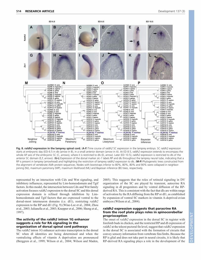
RA signaling is an ancestral mechanism associatedwith dorsal interneuron ontogenesis in thevertebrate spinal cordTo establish whether expression of raldh2 in the RP and in dIs isa tetrapod feature or an ancestral vertebrate trait, we clonedraldh2 from the agnathan lamprey Petromyzon marinus (Fig. 7,Fig. 8). The raldh2 identity of the lamprey clone was confirmedby phylogenetic and expression analyses (Fig. 8) (M.S.S.-C.,
RESEARCH ARTICLE Development 137 (3)
Fig. 6. A novel spinal cord Raldh2 domain in dorsal interneurons. (A,J)Bilateral Raldh2 expression in dIs of 10.5 dpc mouse (A) and chicken (J)embryos. Arrows and arrowheads point to interneuron and RP domains, respectively. Raldh2 expression in the chicken dorsal SC shifts from bilateralinterneuron fields at brachial levels to a single midline RP domain at cervical levels. (B,K)Mouse Math1/chicken Cath1 expression marks dorsal-most(dI1) interneurons. (C,L)Double ISH for Raldh2 and Math1/Cath1 indicates overlapping Raldh2 and Math1/Cath1 domains in dI1. (D,M)Diagramsdepicting embryo position in A-C and J-L and section levels (dashed lines) in E-H and N-Q. (E)Raldh2 expression in the 10.5 dpc mouse lumbar SCfrom the RP (arrowhead) to adjacent dIs (arrow). (F)Math1 expression labels mouse dI1 (arrow). Note the lack of staining in the RP (openarrowhead). (G)Double ISH for Raldh2 (light blue) and Math1 (dark blue). (H)Enlargement of the boxed region in G. Raldh2 expression spreadsfrom the RP (arrowhead) through the Math1 domain (dark-blue arrow), emerging ventral to dI1 (light-blue arrow). (I)Scheme of Raldh2 and Math1expression in the mouse dorsal lumbar SC. (N)Raldh2 expression in the HH18 chicken brachial SC. Raldh2 is expressed in the ventricular zone of dIs,but not in the RP (open arrowhead). (O)Cath1 expression labels dI1 (arrow). Note the lack of staining in the RP (open arrowhead). (P)Double ISHfor Raldh2 (light blue) and Cath1 (dark blue). Note the dorsal expression of Raldh2 and Cath1 and ventral expression of Raldh2 in motoneurons.(Q)Enlargement of the boxed region in P. Raldh2 is expressed in a ventricular (medial) subset of the Cath1 interneuron (dI1) domain. (R)Scheme ofRaldh2 and Cath1 expression.
DEVELO
PMENT
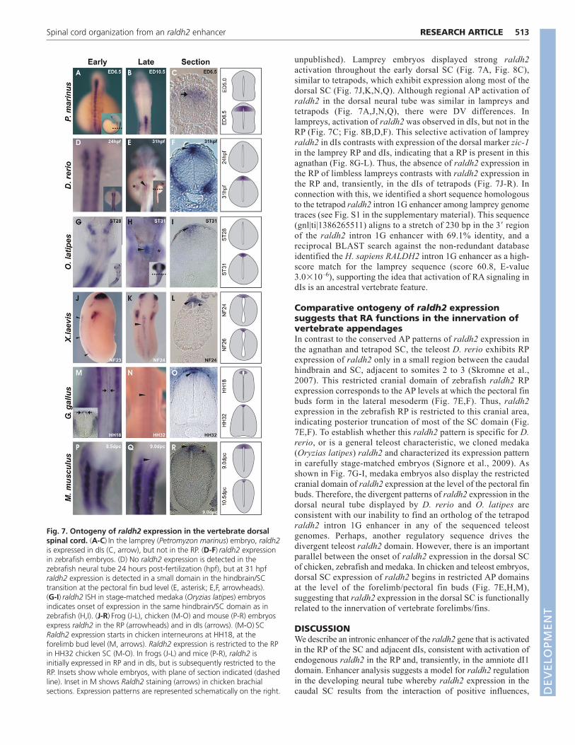
unpublished). Lamprey embryos displayed strong raldh2activation throughout the early dorsal SC (Fig. 7A, Fig. 8C),similar to tetrapods, which exhibit expression along most of thedorsal SC (Fig. 7J,K,N,Q). Although regional AP activation ofraldh2 in the dorsal neural tube was similar in lampreys andtetrapods (Fig. 7A,J,N,Q), there were DV differences. Inlampreys, activation of raldh2 was observed in dIs, but not in theRP (Fig. 7C; Fig. 8B,D,F). This selective activation of lampreyraldh2 in dIs contrasts with expression of the dorsal marker zic-1in the lamprey RP and dIs, indicating that a RP is present in thisagnathan (Fig. 8G-L). Thus, the absence of raldh2 expression inthe RP of limbless lampreys contrasts with raldh2 expression inthe RP and, transiently, in the dIs of tetrapods (Fig. 7J-R). Inconnection with this, we identified a short sequence homologousto the tetrapod raldh2 intron 1G enhancer among lamprey genometraces (see Fig. S1 in the supplementary material). This sequence(gnl|ti|1386265511) aligns to a stretch of 230 bp in the 3� regionof the raldh2 intron 1G enhancer with 69.1% identity, and areciprocal BLAST search against the non-redundant databaseidentified the H. sapiens RALDH2 intron 1G enhancer as a high-score match for the lamprey sequence (score 60.8, E-value3.0�10–6), supporting the idea that activation of RA signaling indIs is an ancestral vertebrate feature.
Comparative ontogeny of raldh2 expressionsuggests that RA functions in the innervation ofvertebrate appendagesIn contrast to the conserved AP patterns of raldh2 expression inthe agnathan and tetrapod SC, the teleost D. rerio exhibits RPexpression of raldh2 only in a small region between the caudalhindbrain and SC, adjacent to somites 2 to 3 (Skromne et al.,2007). This restricted cranial domain of zebrafish raldh2 RPexpression corresponds to the AP levels at which the pectoral finbuds form in the lateral mesoderm (Fig. 7E,F). Thus, raldh2expression in the zebrafish RP is restricted to this cranial area,indicating posterior truncation of most of the SC domain (Fig.7E,F). To establish whether this raldh2 pattern is specific for D.rerio, or is a general teleost characteristic, we cloned medaka(Oryzias latipes) raldh2 and characterized its expression patternin carefully stage-matched embryos (Signore et al., 2009). Asshown in Fig. 7G-I, medaka embryos also display the restrictedcranial domain of raldh2 expression at the level of the pectoral finbuds. Therefore, the divergent patterns of raldh2 expression in thedorsal neural tube displayed by D. rerio and O. latipes areconsistent with our inability to find an ortholog of the tetrapodraldh2 intron 1G enhancer in any of the sequenced teleostgenomes. Perhaps, another regulatory sequence drives thedivergent teleost raldh2 domain. However, there is an importantparallel between the onset of raldh2 expression in the dorsal SCof chicken, zebrafish and medaka. In chicken and teleost embryos,dorsal SC expression of raldh2 begins in restricted AP domainsat the level of the forelimb/pectoral fin buds (Fig. 7E,H,M),suggesting that raldh2 expression in the dorsal SC is functionallyrelated to the innervation of vertebrate forelimbs/fins.
DISCUSSIONWe describe an intronic enhancer of the raldh2 gene that is activatedin the RP of the SC and adjacent dIs, consistent with activation ofendogenous raldh2 in the RP and, transiently, in the amniote dI1domain. Enhancer analysis suggests a model for raldh2 regulationin the developing neural tube whereby raldh2 expression in thecaudal SC results from the interaction of positive influences,
513RESEARCH ARTICLESpinal cord organization from an raldh2 enhancer
Fig. 7. Ontogeny of raldh2 expression in the vertebrate dorsalspinal cord. (A-C)In the lamprey (Petromyzon marinus) embryo, raldh2is expressed in dIs (C, arrow), but not in the RP. (D-F)raldh2 expressionin zebrafish embryos. (D) No raldh2 expression is detected in thezebrafish neural tube 24 hours post-fertilization (hpf), but at 31 hpfraldh2 expression is detected in a small domain in the hindbrain/SCtransition at the pectoral fin bud level (E, asterisk; E,F, arrowheads).(G-I)raldh2 ISH in stage-matched medaka (Oryzias latipes) embryosindicates onset of expression in the same hindbrain/SC domain as inzebrafish (H,I). (J-R)Frog (J-L), chicken (M-O) and mouse (P-R) embryosexpress raldh2 in the RP (arrowheads) and in dIs (arrows). (M-O)SCRaldh2 expression starts in chicken interneurons at HH18, at theforelimb bud level (M, arrows). Raldh2 expression is restricted to the RPin HH32 chicken SC (M-O). In frogs (J-L) and mice (P-R), raldh2 isinitially expressed in RP and in dIs, but is subsequently restricted to theRP. Insets show whole embryos, with plane of section indicated (dashedline). Inset in M shows Raldh2 staining (arrows) in chicken brachialsections. Expression patterns are represented schematically on the right. D
EVELO
PMENT
514
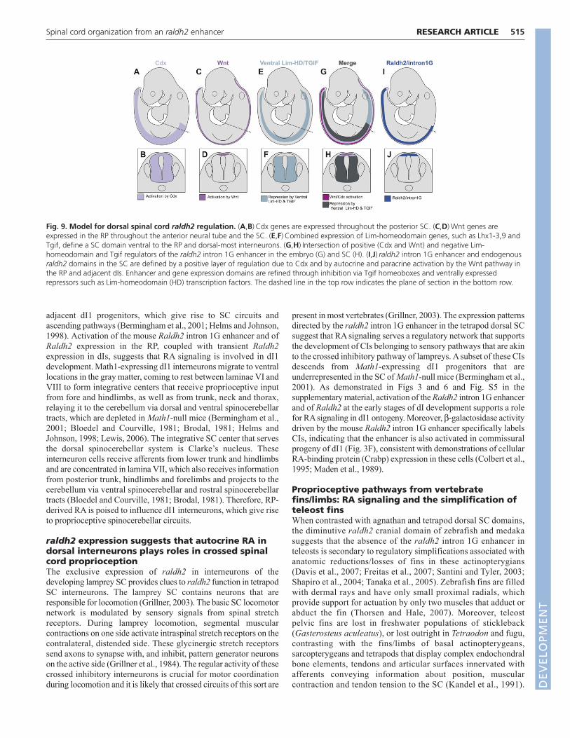
represented by an interaction with Cdx and Wnt signaling, andinhibitory influences, represented by Lim-homeodomain and Tgiffactors. In this model, the intersection between Cdx and Wnt familyactivation focuses raldh2 expression to the dorsal SC and this dorsalexpression domain is refined through inhibition by Lim-homeodomain and Tgif factors that are expressed ventral to thedorsal-most interneuron domains (i.e. dI1), restricting raldh2expression to the RP and dI1 (Fig. 9) (Wine-Lee et al., 2004; Zhouet al., 2003; Iulianella et al., 2003; Knepper et al., 2006; Sheng et al.,1997).
The activity of the raldh2 intron 1G enhancersuggests a role for RA signaling in theorganization of dorsal spinal cord pathwaysThe raldh2 intron 1G enhancer activates transcription in the dorsalSC when dI identities are being determined and when theventralizing effects of vitamin A deprivation are pronounced(Berggren et al., 1999; Wilson et al., 2004; Wilson and Maden,
2005). This suggests that the roles of retinoid signaling in DVorganization of the SC are played by transient, autocrine RAsignaling in dI progenitors and by ventral diffusion of the RP-derived RA. This is consistent with the fact that dIs are within rangeof activation by the RA diffusing from the RP or dI1, as establishedby expansion of ventral SC markers in vitamin A-deprived avianembryos (Wilson et al., 2004).
raldh2 expression suggests that paracrine RAfrom the roof plate plays roles in spinocerebellarproprioceptionThe onset of raldh2 expression in the dorsal SC in register withforelimb buds in chicken, and the restricted RP and dI expression ofraldh2 at the teleost pectoral fin level, suggest that raldh2 expressionin the dorsal SC is associated with the formation of circuits thatconvey sensory information from vertebrate fins/limbs. Because theRP is glial and does not take part in neural circuits, it is likely thatRP-derived RA signaling plays a role in the development of the
RESEARCH ARTICLE Development 137 (3)
Fig. 8. raldh2 expression in the lamprey spinal cord. (A-F)Time course of raldh2 SC expression in the lamprey embryo. SC raldh2 expressionstarts at embryonic day (ED) 6.5 in dIs (arrow in B), in a small anterior domain (arrow in A). At ED 8.5, raldh2 expression extends to encompass thewhole AP axis of the embryonic SC (C, arrows), where it is restricted to dIs (D, arrow). Later (ED 10.5), raldh2 expression is restricted to dIs of theanterior SC domain (E,F, arrows). (G-L)Expression of the dorsal marker zic-1 labels RP and dIs throughout the lamprey neural tube, indicating that aRP is present in lamprey (arrowhead) and highlighting the restriction of lamprey raldh2 expression to dIs. (M-P)Phylogenetic trees constructed fromthe alignment of vertebrate Aldh protein sequences. Nodes with bootstraps inferior to 80%, 80%, 80% and 90% were collapsed in neighbor-joining (NJ), maximum parsimony (MP), maximum likelihood (ML) and Bayesian inference (BI) trees, respectively.
DEVELO
PMENT
adjacent dI1 progenitors, which give rise to SC circuits andascending pathways (Bermingham et al., 2001; Helms and Johnson,1998). Activation of the mouse Raldh2 intron 1G enhancer and ofRaldh2 expression in the RP, coupled with transient Raldh2expression in dIs, suggests that RA signaling is involved in dI1development. Math1-expressing dI1 interneurons migrate to ventrallocations in the gray matter, coming to rest between laminae VI andVIII to form integrative centers that receive proprioceptive inputfrom fore and hindlimbs, as well as from trunk, neck and thorax,relaying it to the cerebellum via dorsal and ventral spinocerebellartracts, which are depleted in Math1-null mice (Bermingham et al.,2001; Bloedel and Courville, 1981; Brodal, 1981; Helms andJohnson, 1998; Lewis, 2006). The integrative SC center that servesthe dorsal spinocerebellar system is Clarke’s nucleus. Theseinterneuron cells receive afferents from lower trunk and hindlimbsand are concentrated in lamina VII, which also receives informationfrom posterior trunk, hindlimbs and forelimbs and projects to thecerebellum via ventral spinocerebellar and rostral spinocerebellartracts (Bloedel and Courville, 1981; Brodal, 1981). Therefore, RP-derived RA is poised to influence dI1 interneurons, which give riseto proprioceptive spinocerebellar circuits.
raldh2 expression suggests that autocrine RA indorsal interneurons plays roles in crossed spinalcord proprioceptionThe exclusive expression of raldh2 in interneurons of thedeveloping lamprey SC provides clues to raldh2 function in tetrapodSC interneurons. The lamprey SC contains neurons that areresponsible for locomotion (Grillner, 2003). The basic SC locomotornetwork is modulated by sensory signals from spinal stretchreceptors. During lamprey locomotion, segmental muscularcontractions on one side activate intraspinal stretch receptors on thecontralateral, distended side. These glycinergic stretch receptorssend axons to synapse with, and inhibit, pattern generator neuronson the active side (Grillner et al., 1984). The regular activity of thesecrossed inhibitory interneurons is crucial for motor coordinationduring locomotion and it is likely that crossed circuits of this sort are
present in most vertebrates (Grillner, 2003). The expression patternsdirected by the raldh2 intron 1G enhancer in the tetrapod dorsal SCsuggest that RA signaling serves a regulatory network that supportsthe development of CIs belonging to sensory pathways that are akinto the crossed inhibitory pathway of lampreys. A subset of these CIsdescends from Math1-expressing dI1 progenitors that areunderrepresented in the SC of Math1-null mice (Bermingham et al.,2001). As demonstrated in Figs 3 and 6 and Fig. S5 in thesupplementary material, activation of the Raldh2 intron 1G enhancerand of Raldh2 at the early stages of dI development supports a rolefor RA signaling in dI1 ontogeny. Moreover, -galactosidase activitydriven by the mouse Raldh2 intron 1G enhancer specifically labelsCIs, indicating that the enhancer is also activated in commissuralprogeny of dI1 (Fig. 3F), consistent with demonstrations of cellularRA-binding protein (Crabp) expression in these cells (Colbert et al.,1995; Maden et al., 1989).
Proprioceptive pathways from vertebratefins/limbs: RA signaling and the simplification ofteleost finsWhen contrasted with agnathan and tetrapod dorsal SC domains,the diminutive raldh2 cranial domain of zebrafish and medakasuggests that the absence of the raldh2 intron 1G enhancer inteleosts is secondary to regulatory simplifications associated withanatomic reductions/losses of fins in these actinopterygians(Davis et al., 2007; Freitas et al., 2007; Santini and Tyler, 2003;Shapiro et al., 2004; Tanaka et al., 2005). Zebrafish fins are filledwith dermal rays and have only small proximal radials, whichprovide support for actuation by only two muscles that adduct orabduct the fin (Thorsen and Hale, 2007). Moreover, teleostpelvic fins are lost in freshwater populations of stickleback(Gasterosteus aculeatus), or lost outright in Tetraodon and fugu,contrasting with the fins/limbs of basal actinopterygeans,sarcopterygeans and tetrapods that display complex endochondralbone elements, tendons and articular surfaces innervated withafferents conveying information about position, muscularcontraction and tendon tension to the SC (Kandel et al., 1991).
515RESEARCH ARTICLESpinal cord organization from an raldh2 enhancer
Fig. 9. Model for dorsal spinal cord raldh2 regulation. (A,B)Cdx genes are expressed throughout the posterior SC. (C,D)Wnt genes areexpressed in the RP throughout the anterior neural tube and the SC. (E,F)Combined expression of Lim-homeodomain genes, such as Lhx1-3,9 andTgif, define a SC domain ventral to the RP and dorsal-most interneurons. (G,H)Intersection of positive (Cdx and Wnt) and negative Lim-homeodomain and Tgif regulators of the raldh2 intron 1G enhancer in the embryo (G) and SC (H). (I,J)raldh2 intron 1G enhancer and endogenousraldh2 domains in the SC are defined by a positive layer of regulation due to Cdx and by autocrine and paracrine activation by the Wnt pathway inthe RP and adjacent dIs. Enhancer and gene expression domains are refined through inhibition via Tgif homeoboxes and ventrally expressedrepressors such as Lim-homeodomain (HD) transcription factors. The dashed line in the top row indicates the plane of section in the bottom row.
DEVELO
PMENT
516
The complex traffic of motor and sensory information to and fromthe muscular limbs of cartilaginous fish, basal actinopterygeansand sarcopterygeans is associated with a higher number of nervesservicing these appendages than in the simpler fins of D. rerio(Thorsen and Hale, 2007). Therefore, it is possible that thevolume of information from sarcopterygean limbs requires adenser network of SC interneuron circuits and ascending fibersthan is required for the comparatively simpler teleost fins.
Afferent information from amniote limbs is routed to SCsegments level with the emergence of fore/hindlimbs, where theyare grouped as Clarke’s nucleus. However, a great deal of limbafferent information is also distributed to SC segments above orbelow the limb buds (Brodal, 1981). As such, amniotes displayan extensive SC interneuron column, termed Clarke’s column,which is distributed along neck, trunk and lumbar segments. Therestriction of RP raldh2 expression to a cranial SC domain thatis level with teleost pectoral fin buds brings into question theexistence of a teleost homolog of the amniote Clarke’s column.It is possible that the relatively small amount of afferentinformation from teleost fins is handled exclusively by SCsegments in register with, or adjacent to, the fins, and for thisreason no structure homologous to the Clarke’s column has beenreported in teleosts. The distribution of SC nuclei is indeedplastic in vertebrates and new spinal nuclei/columns are formedwhen sensory input from peripheral receptors increases after theemergence of complex peripheral structures, or novel sensorycapabilities, as indicated by the development of specific SCnuclei/columns associated with the evolution of chemosensationin the pectoral fins of the teleost northern sea robin (Prionotuscarolinus) (Finger, 2000). In summary, the absence of an raldh2intron 1G enhancer and of raldh2 expression throughout most ofthe SC in zebrafish and medaka are consistent with the loss ofcis and trans factor components of regulatory networksassociated with the simplification of teleost fins (Hildebrand andGoslow, 1998; Shapiro et al., 2004; Tanaka et al., 2005). This isfurther supported by the absence of expression of raldh paralogsin the embryonic teleost SC (Liang et al., 2008; Pittlik et al.,2008).
The ontogeny and phylogeny of RA signaling inthe dorsal spinal cordThe raldh2 intron 1G enhancer is a module that controls RAsignaling in dIs and RP. This module is ontogenetically andphylogenetically associated with the development of twoproprioception modes: one launched by the early activation ofautocrine RA signaling in dI progenitors and linked to theemergence of intraspinal proprioceptive circuits responsible formotor coordination across both sides of the SC during locomotion(Fetcho, 1992); and another represented by a later paracrinesignaling from the RP to a subset of interneuron progenitors thatis linked to the development of spinocerebellar neural circuitsconveying fin/limb proprioception. The first mode is probablyolder and might trace back to the ancestral chordate, presumablya finless cephalochordate-like animal capable of bending its bodyto swim, whereas the second mode probably evolved with theappearance of vertebrate paired appendages. Thus, RA signalingis an ancestral mechanism of DV organization of the SC thatseems to be plastic, allowing for changes in genetic regulationassociated with the evolution of the diverse locomotor patternsand morphologies of vertebrate paired appendages (Goulding,2009). In this sense, it is likely that the lack of raldh2 expressionin the lamprey RP is a derived feature. Although lampreys
display ancestral vertebrate characteristics, Haikouichthys,Myllokunmingia and other agnathan fossils indicate that primitivevertebrates already had prototypes of bilateral fins represented bya pair of continuous mediolateral fin-folds spanning the AP axis,implying that fin absence in extant agnathans is a derived featureof lampreys (Forey, 1995). Thus, it is possible that raldh2expression was lost from the RP owing to a secondary loss ofpaired fins in lampreys (Forey, 1995).
A comparative approach to understandingsignaling in spinal cord development andevolutionCombining comparative genomic and developmental methods is afruitful approach to investigating the ontogeny/phylogeny ofdevelopmental mechanisms that are controlled by signalingsystems in the vertebrate CNS. A considerable amount of non-coding sequence conservation between distantly relatedvertebrates is represented around genes that play essential roles inCNS and heart development (Woolfe et al., 2005), organs thatharbor key vertebrate-specific innovations (Gans and Northcutt,1983; Simoes-Costa et al., 2005; Xavier-Neto et al., 2007). Thus,it is feasible to search for core, conserved vertebratedevelopmental gene regulatory networks that can be used, inselected cases, to infer how specific vertebrate adaptions haveemerged and to understand how class-specific vertebrate bodyplans differ from each other.
AcknowledgementsWe are indebted to Ursula Dräger, Peter McCaffery and Marcus Vinicius Baldofor comments and suggestions; to Richard Behringer and Wellington Cardosofor comments on the manuscript; to Masanori Uchikawa and Jane Johnson forreagents; and to the Faculty of Medicine of the University of São Paulo foraccess to its high-performance computing cluster. This work was supported bygrants from FAPESP (02/11340-2; 04/11569-5; 04/15704-4; 05/60637-6;06/50843-0; 06/61317-8), CNPq 305260/2007-3 and by a DevelopmentTravelling Fellowship from The Company of Biologists.
Competing interests statementThe authors declare no competing financial interests.
Supplementary materialSupplementary material for this article is available athttp://dev.biologists.org/lookup/suppl/doi:10.1242/dev.043257/-/DC1
ReferencesAbascal, F., Zardoya, R. and Posada, D. (2005). ProtTest: selection of best-fit
models of protein evolution. Bioinformatics 21, 2104-2105.Altman, J. and Bayer, S. A. (1997). Development of the Cerebellar System In
Relation to Its Evolution, Structure, and Functions. New York: CRC Press.Berggren, K., McCaffery, P., Drager, U. and Forehand, C. J. (1999).
Differential distribution of retinoic acid synthesis in the chicken embryo asdetermined by immunolocalization of the retinoic acid synthetic enzyme,RALDH-2. Dev. Biol. 210, 288-304.
Bermingham, N. A., Hassan, B. A., Wang, V. Y., Fernandez, M., Banfi, S.,Bellen, H. J., Fritzsch, B. and Zoghbi, H. Y. (2001). Proprioceptor pathwaydevelopment is dependent on Math1. Neuron 30, 411-422.
Blentic, A., Gale, E. and Maden, M. (2003). Retinoic acid signalling centres inthe avian embryo identified by sites of expression of synthesising andcatabolising enzymes. Dev. Dyn. 227, 114-127.
Bloedel, J. R. and Courville, J. (1981). Cerebellar afferent systems. InHandbook of Physiology, vol. 2. Baltimore: Waverly Press.
Brodal, A. (1981). Neurological Anatomy in Relation to Clinical Medicine. NewYork: Oxford University Press.
Chen, Y., Pollet, N., Niehrs, C. and Pieler, T. (2001). Increased XRALDH2activity has a posteriorizing effect on the central nervous system of Xenopusembryos. Mech. Dev. 101, 91-103.
Chizhikov, V. V. and Millen, K. J. (2004). Mechanisms of roof plate formationin the vertebrate CNS. Nat. Rev. Neurosci. 5, 808-812.
Colbert, M. C., Rubin, W. W., Linney, E. and LaMantia, A. S. (1995). Retinoidsignaling and the generation of regional and cellular diversity in the embryonicmouse spinal cord. Dev. Dyn. 204, 1-12.
RESEARCH ARTICLE Development 137 (3)
DEVELO
PMENT
Davis, M. C., Dahn, R. D. and Shubin, N. H. (2007). An autopodial-like patternof Hox expression in the fins of a basal actinopterygian fish. Nature 447, 473-476.
Diez del Corral, R., Olivera-Martinez, I., Goriely, A., Gale, E., Maden, M.and Storey, K. (2003). Opposing FGF and retinoid pathways control ventralneural pattern, neuronal differentiation, and segmentation during body axisextension. Neuron 40, 65-79.
Dodd, J., Morton, S. B., Karagogeos, D., Yamamoto, M. and Jessell, T. M.(1988). Spatial regulation of axonal glycoprotein expression on subsets ofembryonic spinal neurons. Neuron 1, 105-116.
Edgar, R. C. (2004). MUSCLE: a multiple sequence alignment method withreduced time and space complexity. BMC Bioinformatics 5, 113.
Fetcho, J. R. (1992). The spinal motor system in early vertebrates and some ofits evolutionary changes. Brain Behav. Evol. 40, 82-97.
Finger, T. E. (2000). Ascending spinal systems in the fish, Prionotus carolinus. J.Comp. Neurol. 422, 106-122.
Forey, P. L. (1995). Agnathans recent and fossil, and the origin of jawedvertebrates. Rev. Fish Biol. Fisheries 5, 267-303.
Freitas, R., Zhang, G. and Cohn, M. J. (2007). Biphasic Hoxd gene expressionin shark paired fins reveals an ancient origin of the distal limb domain. PLoSONE 2, e754.
Furley, A. J., Morton, S. B., Manalo, D., Karagogeos, D., Dodd, J. andJessell, T. M. (1990). The axonal glycoprotein TAG-1 is an immunoglobulinsuperfamily member with neurite outgrowth-promoting activity. Cell 61, 157-170.
Gans, C. and Northcutt, R. G. (1983). Neural crest and the origin ofvertebrates: a new head. Science 220, 268-273.
Goulding, M. (2009). Circuits controlling vertebrate locomotion: moving in anew direction. Nat. Rev. Neurosci. 10, 507-518.
Grillner, S. (2003). The motor infrastructure: from ion channels to neuronalnetworks. Nat. Rev. Neurosci. 4, 573-586.
Grillner, S., Williams, T. and Lagerback, P. A. (1984). The edge cell, a possibleintraspinal mechanoreceptor. Science 223, 500-503.
Hedges, S. B. and Kumar, S. (2003). Genomic clocks and evolutionarytimescales. Trends Genet. 19, 200-206.
Helms, A. W. and Johnson, J. E. (1998). Progenitors of dorsal commissuralinterneurons are defined by MATH1 expression. Development 125, 919-928.
Hildebrand, M. and Goslow, G. (1998). Analysis of Vertebrate Structure. NewYork: Wiley.
Iulianella, A., Vanden Heuvel, G. and Trainor, P. (2003). Dynamic expressionof murine Cux2 in craniofacial, limb, urogenital and neuronal primordia. GeneExpr. Patterns 3, 571-577.
Kandel, E. R., Schwartz, J. H. and Jessell, T. M. (1991). Principles of NeuralScience. New York: Elsevier.
Keino-Masu, K., Masu, M., Hinck, L., Leonardo, E. D., Chan, S. S., Culotti, J.G. and Tessier-Lavigne, M. (1996). Deleted in Colorectal Cancer (DCC)encodes a netrin receptor. Cell 87, 175-185.
Knepper, J. L., James, A. C. and Ming, J. E. (2006). TGIF, a gene associatedwith human brain defects, regulates neuronal development. Dev. Dyn. 235,1482-1490.
Kothary, R., Clapoff, S., Darling, S., Perry, M. D., Moran, L. A. and Rossant,J. (1989). Inducible expression of an hsp68-lacZ hybrid gene in transgenicmice. Development 105, 707-714.
Lewis, K. E. (2006). How do genes regulate simple behaviours? Understandinghow different neurons in the vertebrate spinal cord are genetically specified.Philos. Trans. R. Soc. Lond. B Biol. Sci. 361, 45-66.
Liang, D., Zhang, M., Bao, J., Zhang, L., Xu, X., Gao, X. and Zhao, Q.(2008). Expressions of Raldh3 and Raldh4 during zebrafish early development.Gene Expr. Patterns 8, 248-253.
Lin, J. T., Wu, M. S., Wang, W. S., Yen, C. C., Chiou, T. J., Liu, J. H., Yang, M.H., Chao, T. C., Chou, S. C. and Chen, P. M. (2003). All-trans retinoid acidincreases Notch1 transcript expression in acute promyelocytic leukemia. Adv.Ther. 20, 337-343.
Maden, M., Ong, D. E., Summerbell, D., Chytil, F. and Hirst, E. A. (1989).Cellular retinoic acid-binding protein and the role of retinoic acid in thedevelopment of the chick embryo. Dev. Biol. 135, 124-132.
Moss, J. B., Xavier-Neto, J., Shapiro, M. D., Nayeem, S. M., McCaffery, P.,Drager, U. C. and Rosenthal, N. (1998). Dynamic patterns of retinoic acidsynthesis and response in the developing mammalian heart. Dev. Biol. 199,55-71.
Muhr, J., Graziano, E., Wilson, S., Jessell, T. M. and Edlund, T. (1999).Convergent inductive signals specify midbrain, hindbrain, and spinal cordidentity in gastrula stage chick embryos. Neuron 23, 689-702.
Niederreither, K., McCaffery, P., Drager, U. C., Chambon, P. and Dolle, P.(1997). Restricted expression and retinoic acid-induced downregulation of theretinaldehyde dehydrogenase type 2 (RALDH-2) gene during mousedevelopment. Mech. Dev. 62, 67-78.
Nieuwkoop, P. D. (1952). Activation and organization of the central nervoussystem in amphibians. Part III. Synthesis of a new working hypothesis. J. Exp.Zool. 120, 83-108.
Novitch, B. G., Wichterle, H., Jessell, T. M. and Sockanathan, S. (2003). Arequirement for retinoic acid-mediated transcriptional activation in ventralneural patterning and motor neuron specification. Neuron 40, 81-95.
Pittlik, S., Domingues, S., Meyer, A. and Begemann, G. (2008). Expression ofzebrafish aldh1a3 (raldh3) and absence of aldh1a1 in teleosts. Gene Expr.Patterns 8, 141-147.
Quandt, K., Frech, K., Karas, H., Wingender, E. and Werner, T. (1995).MatInd and MatInspector: new fast and versatile tools for detection ofconsensus matches in nucleotide sequence data. Nucleic Acids Res. 23, 4878-4884.
Ronquist, F. and Huelsenbeck, J. P. (2003). MrBayes 3, Bayesian phylogeneticinference under mixed models. Bioinformatics 19, 1572-1574.
Rossant, J., Zirngibl, R., Cado, D., Shago, M. and Giguere, V. (1991).Expression of a retinoic acid response element-hsplacZ transgene definesspecific domains of transcriptional activity during mouse embryogenesis.Genes Dev. 5, 1333-1344.
Saitou, N. and Nei, M. (1987). The neighbor-joining method: a new method forreconstructing phylogenetic trees. Mol. Biol. Evol. 4, 406-425.
Santini, F. and Tyler, J. C. (2003). A phylogeny of the families of fossil andextant tetraodontiform fishes (Acanthomorpha, Tetraodontiformes), uppercretaceous to recent. Zool. J. Linn. Soc. 139, 565-617.
Sauka-Spengler, T., Meulemans, D., Jones, M. and Bronner-Fraser, M.(2007). Ancient evolutionary origin of the neural crest gene regulatorynetwork. Dev. Cell 13, 405-420.
Schmidt, H. A., Strimmer, K., Vingron, M. and von Haeseler, A. (2002).TREE-PUZZLE: maximum likelihood phylogenetic analysis using quartets andparallel computing. Bioinformatics 18, 502-504.
Shapiro, M. D., Marks, M. E., Peichel, C. L., Blackman, B. K., Nereng, K. S.,Jonsson, B., Schluter, D. and Kingsley, D. M. (2004). Genetic anddevelopmental basis of evolutionary pelvic reduction in threespinesticklebacks. Nature 428, 717-723.
Sheng, H. Z., Bertuzzi, S., Chiang, C., Shawlot, W., Taira, M., Dawid, I. andWestphal, H. (1997). Expression of murine Lhx5 suggests a role in specifyingthe forebrain. Dev. Dyn. 208, 266-277.
Signore, I. A., Guerrero, N., Loosli, F., Colombo, A., Villalon, A., Wittbrodt,J. and Concha, M. L. (2009). Zebrafish and medaka: model organisms for acomparative developmental approach of brain asymmetry. Philos. Trans. R.Soc. Lond. B Biol. Sci. 364, 991-1003.
Simoes-Costa, M. S., Vasconcelos, M., Sampaio, A. C., Cravo, R. M.,Linhares, V. L., Hochgreb, T., Yan, C. Y., Davidson, B. and Xavier-Neto, J.(2005). The evolutionary origin of cardiac chambers. Dev. Biol. 277, 1-15.
Simoes-Costa, M. S., Azambuja, A. P. and Xavier-Neto, J. (2008). The searchfor non-chordate retinoic acid signaling: lessons from chordates. J. Exp.Zoolog. B Mol. Dev. Evol. 310, 54-72.
Skromne, I., Thorsen, D., Hale, M., Prince, V. E. and Ho, R. K. (2007).Repression of the hindbrain developmental program by Cdx factors is requiredfor the specification of the vertebrate spinal cord. Development 134, 2147-2158.
Sobreira, T. J. and Gruber, A. (2008). Sequence-specific reconstruction fromfragmentary databases using seed sequences: implementation and validationon SAGE, proteome and generic sequencing data. Bioinformatics 24, 1676-1680.
Sockanathan, S. and Jessell, T. M. (1998). Motor neuron-derived retinoidsignaling specifies the subtype identity of spinal motor neurons. Cell 94, 503-514.
Stern, C. D. (1998). Detection of multiple gene products simultaneously by insitu hybridization and immunohistochemistry in whole mounts of avianembryos. Curr. Top. Dev. Biol. 36, 223-243.
Swofford, D. L. (2000). PAUP: Phylogenetic Analyses Using Parsimony andOther Methods. Sunderland, MA: Sinauer.
Tanaka, M., Hale, L. A., Amores, A., Yan, Y. L., Cresko, W. A., Suzuki, T. andPostlethwait, J. H. (2005). Developmental genetic basis for the evolution ofpelvic fin loss in the pufferfish Takifugu rubripes. Dev. Biol. 281, 227-239.
Thorsen, D. H. and Hale, M. E. (2007). Neural development of the zebrafish(Danio rerio) pectoral fin. J. Comp. Neurol. 504, 168-184.
Ulven, S. M., Gundersen, T. E., Weedon, M. S., Landaas, V. O., Sakhi, A. K.,Fromm, S. H., Geronimo, B. A., Moskaug, J. O. and Blomhoff, R. (2000).Identification of endogenous retinoids, enzymes, binding proteins, andreceptors during early postimplantation development in mouse: important roleof retinal dehydrogenase type 2 in synthesis of all-trans-retinoic acid. Dev. Biol.220, 379-391.
Vermot, J., Schuhbaur, B., Le Mouellic, H., McCaffery, P., Garnier, J. M.,Hentsch, D., Brulet, P., Niederreither, K., Chambon, P., Dolle, P. et al.(2005). Retinaldehyde dehydrogenase 2 and Hoxc8 are required in the murinebrachial spinal cord for the specification of Lim1+ motoneurons and thecorrect distribution of Islet1+ motoneurons. Development 132, 1611-1621.
Wang, X., Penzes, P. and Napoli, J. L. (1996). Cloning of a cDNA encoding analdehyde dehydrogenase and its expression in Escherichia coli. Recognition ofretinal as substrate. J. Biol. Chem. 271, 16288-16293.
517RESEARCH ARTICLESpinal cord organization from an raldh2 enhancer
DEVELO
PMENT
518
Wilkinson, D. (1992). Whole Mount In Situ Hybridization: A Practical Approach.Oxford: IRL Press.
Wilson, L. and Maden, M. (2005). The mechanisms of dorsoventral patterningin the vertebrate neural tube. Dev. Biol. 282, 1-13.
Wilson, L. J. and Wingate, R. J. (2006). Temporal identity transition in theavian cerebellar rhombic lip. Dev. Biol. 297, 508-521.
Wilson, L., Gale, E., Chambers, D. and Maden, M. (2004). Retinoic acid andthe control of dorsoventral patterning in the avian spinal cord. Dev. Biol. 269,433-446.
Wine-Lee, L., Ahn, K. J., Richardson, R. D., Mishina, Y., Lyons, K. M. andCrenshaw, E. B., 3rd (2004). Signaling through BMP type 1 receptors isrequired for development of interneuron cell types in the dorsal spinal cord.Development 131, 5393-5403.
Wingender, E., Dietze, P., Karas, H. and Knuppel, R. (1996). TRANSFAC: adatabase on transcription factors and their DNA binding sites. Nucleic AcidsRes. 24, 238-241.
Woolfe, A., Goodson, M., Goode, D. K., Snell, P., McEwen, G. K., Vavouri, T.,Smith, S. F., North, P., Callaway, H., Kelly, K. et al. (2005). Highly conservednon-coding sequences are associated with vertebrate development. PLoS Biol. 3,e7.
Xavier-Neto, J., Neville, C. M., Shapiro, M. D., Houghton, L., Wang, G. F.,Nikovits, W., Jr, Stockdale, F. E. and Rosenthal, N. (1999). A retinoic acid-inducible transgenic marker of sino-atrial development in the mouse heart.Development 126, 2677-2687.
Xavier-Neto, J., Shapiro, M. D., Houghton, L. and Rosenthal, N. (2000).Sequential programs of retinoic acid synthesis in the myocardial and epicardiallayers of the developing avian heart. Dev. Biol. 219, 129-141.
Xavier-Neto, J., Castro, R. A., Sampaio, A. C., Azambuja, A. P., Castillo, H. A.,Cravo, R. M. and Simoes-Costa, M. S. (2007). Parallel avenues in theevolution of hearts and pumping organs. Cell Mol. Life Sci. 64, 719-734.
Zhang, J., Smith, D., Yamamoto, M., Ma, L. and McCaffery, P. (2003). Themeninges is a source of retinoic acid for the late-developing hindbrain. J.Neurosci. 23, 7610-7620.
Zhao, D., McCaffery, P., Ivins, K. J., Neve, R. L., Hogan, P., Chin, W. W. andDrager, U. C. (1996). Molecular identification of a major retinoic-acid-synthesizingenzyme, a retinaldehyde-specific dehydrogenase. Eur. J. Biochem. 240, 15-22.
Zhou, X. H., Brandau, O., Feng, K., Oohashi, T., Ninomiya, Y., Rauch, U. andFassler, R. (2003). The murine Ten-m/Odz genes show distinct but overlappingexpression patterns during development and in adult brain. Gene Expr. Patterns3, 397-405.
RESEARCH ARTICLE Development 137 (3)
DEVELO
PMENT