Embed Size (px)
Citation preview
JOURNAL OF BACTERIOLOGY, June 2008, p. 4001–4016 Vol. 190, No. 110021-9193/08/$08.00�0 doi:10.1128/JB.00135-08Copyright © 2008, American Society for Microbiology. All Rights Reserved.
Insight from TonB Hybrid Proteins into the Mechanism of IronTransport through the Outer Membrane�
Wallace A. Kaserer, Xiaoxu Jiang, Qiaobin Xiao, Daniel C. Scott, Matthew Bauler, Daniel Copeland,Salete M. C. Newton, and Phillip E. Klebba*
Department of Chemistry and Biochemistry, University of Oklahoma, 620 Parrington Oval, Norman, Oklahoma 73019
Received 25 January 2008/Accepted 24 March 2008
We created hybrid proteins to study the functions of TonB. We first fused the portion of Escherichia coli tonBthat encodes the C-terminal 69 amino acids (amino acids 170 to 239) of TonB downstream from E. coli malE(MalE-TonB69C). Production of MalE-TonB69C in tonB� bacteria inhibited siderophore transport. Afteroverexpression and purification of the fusion protein on an amylose column, we proteolytically released theTonB C terminus and characterized it. Fluorescence spectra positioned its sole tryptophan (W213) in a weaklypolar site in the protein interior, shielded from quenchers. Affinity chromatography showed the binding of theTonB C-domain to other proteins: immobilized TonB-dependent (FepA and colicin B) and TonB-independent(FepA�3-17, OmpA, and lysozyme) proteins adsorbed MalE-TonB69C, revealing a general affinity of the Cterminus for other proteins. Additional constructions fused full-length TonB upstream or downstream of greenfluorescent protein (GFP). TonB-GFP constructs had partial functionality but no fluorescence; GFP-TonBfusion proteins were functional and fluorescent. The activity of the latter constructs, which localized GFP inthe cytoplasm and TonB in the cell envelope, indicate that the TonB N terminus remains in the innermembrane during its biological function. Finally, sequence analyses revealed homology in the TonB C terminusto E. coli YcfS, a proline-rich protein that contains the lysin (LysM) peptidoglycan-binding motif. LysMstructural mimicry occurs in two positions of the dimeric TonB C-domain, and experiments confirmed that itphysically binds to the murein sacculus. Together, these findings infer that the TonB N terminus remainsassociated with the inner membrane, while the downstream region bridges the cell envelope from the affinityof the C terminus for peptidoglycan. This architecture suggests a membrane surveillance model of action, inwhich TonB finds occupied receptor proteins by surveying the underside of peptidoglycan-associated outermembrane proteins.
Iron is one target of gram-negative bacterial cell envelopetransport systems, and microbes elaborate high-affinity sid-erophores that complex extracellular iron (70). However, ferricsiderophores, like ferric enterobactin (FeEnt), are too large(716 Da) to pass through general porins in the outer mem-brane (OM), necessitating a different type of transporter toacquire them. On the basis of their 22-stranded transmem-brane �-barrels, OM metal transporters like FepA belong tothe porin superfamily (89). Ligand binding to such receptorsinitiates the transport reaction through their transmembranechannels, which led to their designation as ligand-gated porins(LGP) (88), by analogy to the family of eukaryotic ligand-gatedion channels. It is noteworthy that LGP are mechanisticallydistinct from general, diffusive porins because they bind metalcomplexes with high affinity and actively transport them againsta concentration gradient into the cell. Once in the periplasm,binding proteins adsorb ferric siderophores and deliver themto inner membrane (IM) permeases that actively transporteither the metal complex or free iron into the cytoplasm. Atthe binding stage of the uptake process, ferric siderophorescompete with noxious agents like bacteriophages (H8 [85a] and T5and �80 [18, 103]) and colicins (B, D [37], and M [18]) for entry into
the cell. OM transport of metal complexes and susceptibility tophages and colicins require metabolic energy (13, 25, 85) and usuallyinvolve participation of an additional cell envelope protein, TonB.
Bioinformatic and biophysical data suggest that TonB com-prises three parts that reside in different regions of the cellenvelope: a hydrophobic N-terminal sequence in the IM (27),a central section that spans the periplasm (30, 39), and aC-terminal ���� domain, also in the periplasm, that periph-erally associates with the OM (26, 52, 76, 78, 95). Sequenceanalyses suggested that the upstream, N-terminal portion ofTonB contains one or more membrane-embedded regions(49), including a hydrophobic helix that anchors the protein inthe IM. Gene fusion experiments supported this notion (86),but no structural data are available on the N terminus. Nuclearmagnetic resonance data on synthetic peptides suggested thatmultiply repeated Glu-Pro and Lys-Pro sequences in the cen-tral portion behave like two long rods, joined by a short se-quence with some flexibility, that span over 100 Š(20, 30). TheC terminus of TonB, which is important to its function (4),forms a ���� motif that may dimerize (26, 52) and recruitother polypeptides into its �-sheet (76, 95). There is evidencethat TonB dimerizes in vivo (90), but whether the dimer is itsnormal state or whether TonB fluctuates between monomericand dimeric forms in vivo is not known. The physiology andbiochemistry of TonB action remain obscure, but recent dataconfirm the idea (36, 46) that TonB binds to sites on theperiplasmic surface of TonB-dependent OM proteins. Thisprotein-protein interaction may facilitate LGP transport.
* Corresponding author. Mailing address: Department of Chemistryand Biochemistry, University of Oklahoma, 620 Parrington Oval, Nor-man, OK 73019. Phone: (405) 325-4969. Fax: (405) 325-6111. E-mail:[email protected].
� Published ahead of print on 4 April 2008.
4001
Dow
nloa
ded
from
http
s://j
ourn
als.
asm
.org
/jour
nal/j
b on
03
Janu
ary
2022
by
168.
205.
109.
127.
We biochemically characterized TonB hybrid proteins andits isolated C-terminal domain to gain insight into the func-tions of TonB in OM iron transport. Overexpression of thefusion polypeptides in a tonB� background inhibited sid-erophore uptake, but not colicin susceptibility. The C-terminalpolypeptide adsorbed to FepA-agarose and also to OmpA-agarose and to agarose bearing FepA�13-22, which lacks theTonB box. Thus, the TonB C terminus showed a general affinityfor OM proteins that does not involve interactions with the TonBbox sequence. Green fluorescent protein (GFP)-TonB hybridproteins were fluorescent and functional in TonB-dependentprocesses, refuting the idea that the TonB N terminus leavesthe IM during performance of its biochemical activity (55, 57).Last, the C terminus of TonB contains previously unrecognizedregions that are structurally related to the lysin (LysM) domain(7) that confers peptidoglycan (PG) binding to Escherichia coliYcfS and its homologs.
MATERIALS AND METHODS
Bacterial strains and plasmids. Most strains (Table 1) originated from E. coliBN1071 (F� entA pro trpB1) (48), including KDO23 (entA tonB), a spontaneoustonB mutant from simultaneous selection against colicins B, M, and Ia, andOKN1 (�tonB) (60), which contains a genetically engineered, precise deletion ofthe structural gene. Our motivation for constructing the latter strain was thatchromosomal fragments of siderophore receptor genes may unexpectedly com-plement exogenous genes on plasmids (101). To avoid such phenomena in thetonB background, we completely deleted (28) tonB and then used the strain as ahost for plasmids bearing tonB� or its derivatives. After isolation of strainsKDO23 and OKN1, we screened them for susceptibility to colicins and ability totransport ferric siderophores; we confirmed their mutations by PCR analysis. Inaddition, we performed Western immunoblotting with polyclonal rabbit anti-TonB sera and monoclonal mouse anti-FepA sera to verify the absence of TonBand presence of FepA. In both strains, the tonB� phenotype was reinstated, asevidenced by colicin sensitivity and siderophore nutrition, upon transformationwith pRZ540 (tonB�) (81). Strain ER2507 (malE; New England Biolabs) was thehost strain for pMal fusion plasmids.
pMalp2 (New England Biolabs) (29) contains a Ptac promoter that regulatestranscription of its MalE-LacZ fusion. Using the multiple cloning site of thisvector, we replaced the lacZ moiety with other genes or gene fragments ofinterest. We amplified the DNA encoding the 69 C-terminal residues of E. colitonB from pRZ540 (81, 83), purified the 500-bp product from agarose withGeneclean (Bio 101, Vista, CA), and digested it with XmnI and HindIII. ThePCR band was ligated into pMalp2, and the resulting transformant clones werescreened by restriction analysis with EcoRV and HindIII. Candidate plasmidswith the appropriately sized restriction fragment were sequenced with an ALF-Express automated DNA sequencer (Pharmacia). We designated the productpSTonB69C; similarly constructed variants that fused the region that encodes theC-terminal 135 residues or the intact tonB gene to MalE were pSTonB135C andpSTonB239, respectively. We regulated expression of the fusion proteins byvarying the isopropyl-�-D-thiogalactopyranoside (IPTG) concentration in culturemedia and purified the resulting hybrid proteins by affinity chromatography onamylose resin (32).
We also fused the structural gene for GFP to tonB in several ways. pT23, aderivative of the low-copy vector pHSG575, carries a wild-type chromosomaltonB structural gene (from E. coli BN1071) under control of its natural promoter.pQbioT7-GFP (QbioGene, Irvine, CA) carries a mutant version of the Aequoreavictoria GFP structural gene (sggfp) that contains the substitutions F64L, S65C,and I167T. The first mutation confers greater solubility, stability, and fluoro-phore formation at 37°C, whereas the latter two optimize the protein’s singleexcitation peak at 474 nm. Using PCR, we separately amplified the E. coli tonBpromoter region and the tonB structural gene from pT23 and the sggfp gene frompQbioT7-GFP. We first fused ssgfp in frame with and downstream from tonB onpT23 (pTG and derivatives). Second, we cloned ssgfp (with its stop codon)immediately downstream from the tonB promoter (pTpG), and in another clone,we introduced ssgfp (without a stop codon) in frame between the tonB promoterand the tonB structural gene (pGT). In the latter case, primers introducedHindIII and XbaI cleavage sites at the 5� and 3� ends, respectively, of the tonBpromoter region, XbaI and BamHI sites at the 5� and 3� ends, respectively, ofsggfp, and BamHI and EcoRI sites at the 5� and 3� ends of tonB. After digestionwith the appropriate restriction enzymes, we joined the three fragments with T4ligase to produce tonB promoter-sggfp-tonB. The final 2,117-bp product andpHSG575 were digested with HindIII and EcoRI, agarose purified, and joinedwith T4 ligase to produce pGT. In pGLT, the 5-amino-acid linker sequenceEAAAK was inserted between GFP and TonB by QuikChange mutagenesis atthe 5� end of the tonB moiety in pGT. The result altered the N-terminal sequence
TABLE 1. Bacterial strains and plasmids
Strain or plasmid Relevant genotype or genea Source or reference
E. coli strainsBN1071 entA fepA� tonB� rpsL 48KDO23 BN1071 tonB This studyOKN1 BN1071 �tonB 60OKN3 BN1071 �fepA 60ER2507 (malB) zjb::Tn5 rpsL supE44 fhuA New England BiolabsBL21(DE3) � lysogen, lon ompT Novagen
PlasmidspRZ540 tonB� (TonB) 81pMalp2 malE fusion vector (MalE) New England BiolabspSTonB239 pMalp2 malE-tonB1-239 (MalE-TonB) This studypSTonB135C pMalp2 malE-tonB104-239 (MalE-TonB135C) This studypSTonB69C pMalp2 malE-tonB100-239 (MalE-TonB69C) This studypHSG575 Low-copy-number cloning vector 40pT23 pHSG575 tonB� (TonB) This studypTG pHSG575 tonB-ssgfp (TonB-GFP) This studypTatTG pHSG575 tatss-tonB-ssgfp (TatTonB-GFP) This studyp�ssTG pHSG575 tonB 4-32-ssgfp (4-32TonB-GFP) This studypTpG pHSG575 tonB promoter-ssgfp (GFP)pGT pHSG575 tonB promoter-ssgfp-tonB (GFP-TonB) This studypGLT pHSG575 tonB promoter-ssgfp-tonB (GFP-L-TonB) This studypGT�69C pHSG575 tonB promoter-ssgfp-tonB170-239 (GFP-TonB69C) This studypCol1 pUC18 cba cbi (colicin B) This studypETTonB pET28 6H-tonB (6H-TonB) This study
a For plasmids, the expressed protein is shown in parentheses.
4002 KASERER ET AL. J. BACTERIOL.
Dow
nloa
ded
from
http
s://j
ourn
als.
asm
.org
/jour
nal/j
b on
03
Janu
ary
2022
by
168.
205.
109.
127.
of TonB from MTLDLPRR to MAEAAAKA. This construct is predicted (5) toform a small helical peptide between GFP and TonB.
Isolation of the TonB C terminus. E. coli ER2507/pSTonB69C was grownovernight in LB broth (68) with ampicillin and streptomycin at 100 g/ml andsubcultured at 1% into LB broth containing 0.2% glucose. When the culturereached an optical density at 600 nm of 0.5, we added 0.5 mM IPTG to theculture; after the culture was subjected to shaking for 3 h at 37°C, we collectedthe cells by centrifugation at 3,000 g for 20 min and resuspended them in bufferA (10 mM Tris-HCl, 0.4 M NaCl, 1 mM EDTA, and 10 mM �-mercaptoethanol).After lysis by passage through a French pressure cell (three times at 14,000lb/in2), we clarified the lysate by ultracentrifugation at 100,000 g for 30 min andchromatographed the supernatant on an amylose column (approximately 15 ml)equilibrated with buffer A (32). After the column was washed with 10 bedvolumes of buffer A, we eluted adsorbed proteins with 10 mM maltose in bufferA. The eluted fractions were pooled and dialyzed against Tris-buffered saline(TBS) (50 mM Tris chloride [pH 7.5] and 0.5 M NaCl), sodium dodecyl sulfate(SDS) was added to 0.05%, and TonB69C was cleaved by digestion with factorXa. The digestion products were resolved on Sephacryl S100HR (Pharmacia) inTBS plus 0.05% SDS, and the SDS was removed from pooled fractions ofTonB69C by acetone precipitation.
Fractionation of cell envelopes. Bacteria were cultured in L broth (67), sub-cultured in morpholinepropanesulfonic acid (MOPS) minimal medium (69),harvested in mid-exponential phase of growth, washed with and resuspended inTBS, and chilled on ice. OM and IM fragments were formed by lysis of bacteriain a French pressure cell (96) and purified by sucrose gradient centrifugation(72).
Proteins and antisera. Hen egg white lysozyme was purchased from Sigma. E.coli OM proteins OmpA, FepA, and FepA�13-17 were purified by differentialextraction of cell envelopes with Triton X-100 (77, 91). Colicin B (ColB) wasderived from supernatants of E. coli DM1187/pCol1, and factor X was frombovine blood; we purified both the toxin (77) and the protease (105) by conven-tional chromatographic methods and activated factor X to factor Xa with snakevenom (45). E. coli MalE was purified from E. coli ER2507/pMalp2 after IPTGinduction by French pressure cell lysis and subsequent amylose chromatography(32). E. coli FepB was purified as previously described (97). Colicin titer wasexpressed as the reciprocal of the highest dilution that resulted in a clearing ofa lawn of a sensitive bacterial strain.
Full-length TonB was purified as a His-tagged soluble protein. The tonBstructural gene on pRZ540 was amplified by PCR and cloned in pET28 (EMDBiosciences) to create an N-terminal six-histidine tag. The resulting plasmid(pETTonB) was transformed into E. coli BL21(DE3) (Invitrogen) and inducedto high levels of 6H-TonB synthesis by growth in LB broth with IPTG (0.5 mM).6H-TonB was purified in a single step by passage of crude cell lysates overNi2�-nitrilotriacetic acid agarose (Qiagen). Rabbits were intramuscularly immu-nized with purified 6H-TonB (50 to 100 g) six times over a 6-week period, withFreund’s complete adjuvant in the first injection and with Freund’s incompleteadjuvant or alum in subsequent injections. Rabbits were bled by cardiac punc-ture, and anti-TonB sera was prepared and diluted 105-fold for use in Westernimmunoblots.
Electrophoresis and Western immunoblots. For SDS-polyacrylamide gel elec-trophoresis (PAGE) (60), samples were suspended in SDS sample buffer plus 3%�-mercaptoethanol, heated at 100°C for 5 min, and electrophoresed on 10%polyacrylamide slabs. For Western immunoblots, proteins were transferred tonitrocellulose paper, blocked in TBS plus 1% gelatin, and incubated with murineascitic fluid (0.5%) or polyclonal rabbit antisera (0.01%) in TBS plus 1% gelatinfor 3 h. The nitrocellulose paper was washed in TBS plus 0.05% Tween 20,incubated with alkaline phosphatase-conjugated goat anti-mouse immunoglob-ulin or alkaline phosphatase-conjugated goat anti-rabbit immunoglobulin, re-spectively, for 2 h, washed, and developed with bromochloroindolyl phosphateand nitroblue tetrazolium (88).
Conjugation of proteins to Sepharose 6B. Aliquots (25 ml) of Sepharose 6Bwere activated by the dropwise addition of 6 g CNBr in distilled water, whilemaintaining the pH between 11.0 and 12.0 (62). The resin was washed withdistilled water and suspended in the appropriate conjugation buffer. For OMproteins, 50 mg of purified material was dissolved in 0.2 M NaHCO3 containing2% Triton X-100, added to the activated resin, and incubated overnight at 4°Cwith gentle stirring (6, 74, 80). After conjugation, the resin was washed with 2liters of distilled water, 2 liters of 1 M acetic acid, and 2 liters of distilled water.Aliquots of the first wash were saved to determine the efficiency of proteinconjugation, which was usually �95%. For non-OM proteins, Triton X-100 wasomitted from the conjugation buffer. After modification, we equilibrated theresins in TBS, pH 7.5.
Spectroscopic measurements. UV-visible spectra were acquired on a BeckmanDU 640 spectrophotometer in 1.0-cm cuvettes. Circular dichroism (CD) spectrawere obtained using an AVIV 60DS CD spectrometer. Either 0.1- or 1.0-cmcuvettes were used to measure the CD spectra of TonB6C9. For fluorescencespectroscopy, excitation and emission spectra were recorded with an SLM8000fluorimeter upgraded to 8100 functionality (SLM Instruments, Rochester, NY),equipped with a 450-watt xenon light source and a cooled photomultipler tubehousing, operated in the photon-counting mode.
Sequence and structural analyses. Protein sequences were obtained fromNCBI and initially studied by BLASTP to identify their homologous relatives inthe NCBI database. Orthologs and paralogs were then compared by CLUSTALWto discern regions of identity and similarity. When crystallographic data wereavailable, related polypeptides and structural motifs were aligned and superim-posed by LSQMAN (50, 53). The overlay was performed using 1IHR, the 1.55-Åstructure of the dimeric TonB C terminus, and the first model in 1E0G, the LysMdomain from E. coli MltD. The first model was chosen because the PDB file gaveno information on the best representative conformer in the ensemble. LSQMANused brute-force alignment, with a search fragment length of 5 amino acids anda step size of 2, searching for the maximum number of matched residues. Theprogram produced graphic output of the structural relationships.
PG binding experiments. PG was purified from exponential-phase E. coliBN1071 by standard procedures (34, 35, 64). Bacteria from 500 ml of LB broth,growing exponentially with aeration (to an optical density at 600 nm of �0.7 to0.9), were rapidly chilled in an ice-salt-water bath, harvested by centrifugation at4°C, and resuspended in 5 ml of ice-cold water. The cell suspension was addeddropwise to 10 ml of boiling 4% SDS solution in a 50-ml Erlenmeyer flask on ahot plate over a period of about 10 min. After the solution was boiled for 30 min,it was allowed to cool to room temperature and stand overnight. The crudesacculi were collected by ultracentrifugation (100,000 g, 1.5 h) and washed freeof SDS by repeated resuspensions in 15 ml of water and ultracentrifugations(four times). �-Amylase (100 g/ml in 10 mM Tris-HCl [pH 7.0]) was added andincubated for 2 h at 37°C to degrade high-molecular-weight glycogen; pronase(preincubated at 60°C for 2 h to inactivate any contaminating lysozyme) wasadded (200 g/ml) and incubated for 2 h at 37°C to digest bound lipoprotein andany other protein contaminations. SDS was added to a concentration of 1%, andthe sacculi were boiled for 15 min. The SDS was removed by repeated centrif-ugations and resuspensions in water (four times). The purified sacculi werestored in water containing 0.05% sodium azide at 4°C at a concentration of about1 mg/ml.
For binding studies, aliquots of purified PG were mixed with 30-g aliquots ofpurified proteins (MalE, MalE-TonB69C, and FepB) in 20 mM Tris-Cl, pH 8(final volume of 100 l) at room temperature for 30 min and then centrifuged at100,000 g for 45 min in a Beckman Optima TL ultracentrifuge. Both thesupernatants and the resuspended pellets were subjected to SDS-PAGE. Thegels were stained with Coomassie blue, photographed, and analyzed by Image-Quant (Molecular Dynamics) to determine the amount of protein in the pelletsand supernatants.
RESULTS
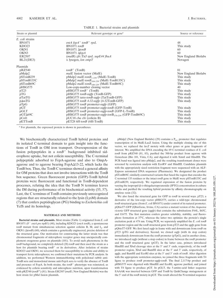
Cloning, expression, and purification of TonB69C. We ge-netically fused DNA encoding the C-terminal 69 residues ofTonB (amino acids 170 to 239) downstream from malE onpMalp2 and purified the resulting hybrid protein (MalE-TonB69C) by affinity chromatography on an amylose resin (29,32, 100). The electrophoretic mobility of MalE-TonB69C onSDS-polyacrylamide gels was slightly less than that expectedfor a protein of its predicted size, 49.5 kDa (Fig. 1A). However,TonB itself has aberrant, decreased mobility on SDS-poly-acrylamide gels. After IPTG induction to high levels of expres-sion, we isolated the fusion protein in high yield (50 mg/liter ofculture media) and 95% homogeneity (Fig. 1).
MalE-TonB69C contains a factor Xa cleavage site betweenthe two polypeptides, but it was susceptible to the proteaseonly in the presence of 0.05% SDS, suggesting that in thenative state the cleavage site is buried in protein structure. Thefactor Xa digestion products were resolved on a SephacrylS100HR gel filtration column in TBS containing 0.05% SDS,
VOL. 190, 2008 TonB AND IRON TRANSPORT THROUGH THE OUTER MEMBRANE 4003
Dow
nloa
ded
from
http
s://j
ourn
als.
asm
.org
/jour
nal/j
b on
03
Janu
ary
2022
by
168.
205.
109.
127.
resulting in �95% purity of the 8-kDa TonB69C polypeptide(Fig. 1). Purified TonB69C was concentrated by acetone pre-cipitation and suspended in 20 mM Tris-Cl, pH 7.5.
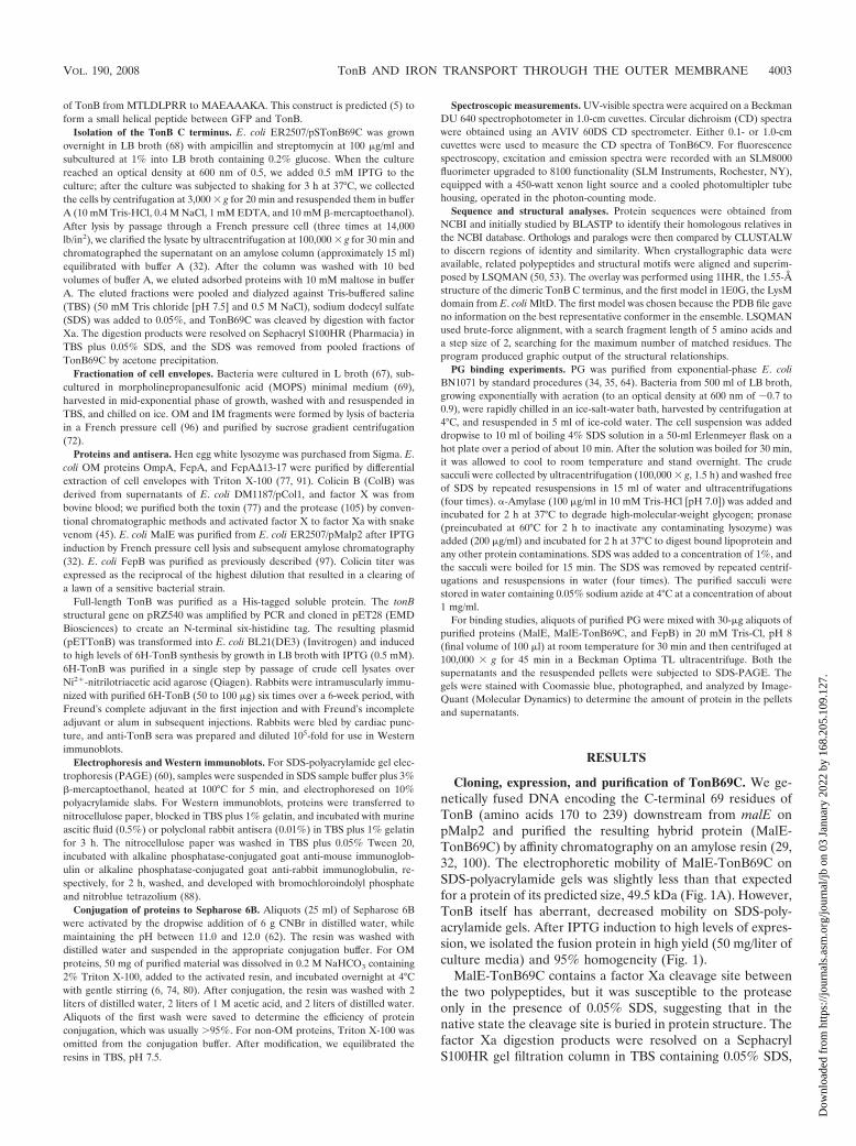
Spectroscopic characterizations. The C terminus of TonBcontains a single tryptophan residue at position 213 within the�-helix of the domain. Measurements of its intrinsic fluores-cence revealed a maximum at 338 nm (Fig. 2). Stepwise addi-tions of the denaturant guanidinium hydrochloride (ultimatelyto 4 M) sequentially shifted the emission maximum to 352 nmand decreased fluorescence intensity 45%. The emission max-imum of the denatured polypeptide was similar to that of freetryptophan in aqueous solution. These data portrayed the so-lution structure of TonB69C as consistent with the crystalstructure (26) folded into a form that ensconces W213 in aninternal, slightly polar environment. CD spectra (Fig. 2) wereless informative but showed that the domain contains second-ary structure and a defined three-dimensional structure.The presence of significant near-UV signals indicated thatTonB69C folded into a well-defined structure with �-helicalcontent and other secondary structure typical of an alpha-betaprotein.
Effects of quenchers. We evaluated the susceptibility ofW213 to quenching by acrylamide, iodide, and cesium. Stern-Volmer analysis of fluorescence in the presence of acrylamideyielded Stern-Volmer constants (Ksvs) of 5.9 0.4 for nativeTonB69C and 8.4 0.4 for denatured TonB69C (Fig. 2). Thecharged species iodide and cesium less effectively quenchedW213 fluorescence: the Ksv values were 2.6 0.2 and 4.4 0.1when the native and denatured polypeptides, respectively,were exposed to iodide, and 0.37 0.005 and 1.6 0.001,respectively, when they were exposed to cesium. Increasedionic strength (0.4 M NaCl) did not perturb the fluorescenceemission spectrum (data not shown). The overall linearity of
the Stern-Volmer plots from iodide and cesium (Fig. 2) indi-cated collisional quenching of W213 in both native and dena-tured TonB69C. For acrylamide, the Stern-Volmer plots werelinear to 0.3 M but deviated from linearity at higher concen-
FIG. 1. Expression and purification of MalE-TonB69C. E. coliKDO23/pSTonB69C was grown to mid-log in LB broth, IPTG wasadded to 0.5 mM, and the cells were shaken at 37°C for 3 more hours.(A) Aliquots of 108 cells were collected before (lane 1) and 1 h (lane2) and 2 hours (lane 3) after the addition of IPTG. The bacteria werelysed and subjected to SDS-PAGE on a 10% gel. The overexpressionof MalE-TonB69C (arrow) appears in lane 3. The positions of molec-ular size markers (m) (in kilodaltons), Bio-Rad Precision Plus proteinstandards, are shown to the left of the gel. (B) After lysis by passagethrough a French pressure cell, the lysate was clarified by centrifuga-tion at 100,000 g for 30 min and applied to an amylose column.MalE-TonB69C was eluted with maltose (lane 1), and TonB69C(arrow) was released by digestion with factor Xa (lane 2) (16% gel).(C) The digestion mixture was chromatographed on Sephacryl S-100 inTBS plus 0.05% SDS and analyzed on a 16% gel. The arrow indicatesthe position of TonB69C, which eluted last from the column (lanes 12to 16).
FIG. 2. Spectroscopic properties of TonB69C. (A) Guanidine HCl titra-tion. Fluorescence emission spectra of a single tryptophan (W213) was mon-itored in the presence of the denaturant guanidine hydrochloride. The redshift of its emission maximum and decrease in fluorescence with increasingconcentrations of guanidinium infer the unfolding of the domain and theincreasing exposure of the residue to the solvent. The inset plots show emis-sion maximum (Emax) (E) and F/F0 (‚) versus the concentration of guani-dinium-HCl. (B to D) Susceptibility of TonB69C to quenching. PurifiedTonB69C, either native (F) or denatured (E), was incubated with increasingconcentrations of acrylamide (B), KI (C), and CsCl (D), and its fluorescenceemissions were recorded and then analyzed by the Stern-Volmer equation,assuming collisional quenching by the following equation: Fo/F � 1 � Ksv [Q],where Fo and F are the fluorescence intensities in the absence or presence,respectively, of the quencher, Ksv is the Stern-Volmer constant, and [Q] is theconcentration of the quencher. The data were analyzed by linear regression todetermine Ksv values for each quencher (see the text). (E) Circular dichroism.The CD spectrum of purified TonB69C was recorded in TBS and showedextrema characteristic of �-helical content at 209 nm and other secondarystructure at higher wavelengths. The CD data excluded the possibility thatTonB69C exists in solution as a random coil form and were consistent withthe form of a folded alpha-beta protein.
4004 KASERER ET AL. J. BACTERIOL.
Dow
nloa
ded
from
http
s://j
ourn
als.
asm
.org
/jour
nal/j
b on
03
Janu
ary
2022
by
168.
205.
109.
127.
trations. Again, the data inferred that W213 inhabits an envi-ronment of TonB69C where it is more susceptible to the hy-drophobic quencher acrylamide and partially shielded from thehydrated charged species iodide and cesium.
Functionality of the isolated TonB C terminus. In agree-ment with previous reports (90, 93), in tonB strains, overex-pression of MalE-TonB69C did not restore TonB-dependentprocesses (Table 2), suggesting that for activity the TonB Cterminus must remain connected to the N-terminal domain,presumably anchored in the IM (see below). Overexpression ofMalE-TonB69C in wild-type strains inhibited the transport ofboth FeEnt and ferrichrome (Table 2). The plasmid-derivedC-terminal fragment competed with native TonB in a way thatimpaired the overall ferric siderophore uptake reaction. How-ever, sensitivity to the TonB-dependent colicin B was unaf-fected by overexpression of MalE-TonB69C. The general per-meability of the OM was not compromised by MalE-TonB69C,as shown by the identical antibiotic sensitivity (to chloram-phenicol, erythromycin, rifampin, bacitracin, tetracycline, no-vobiocin, and neomycin [88]) of strains expressing wild-typeTonB or its MalE fusion proteins (data not shown).
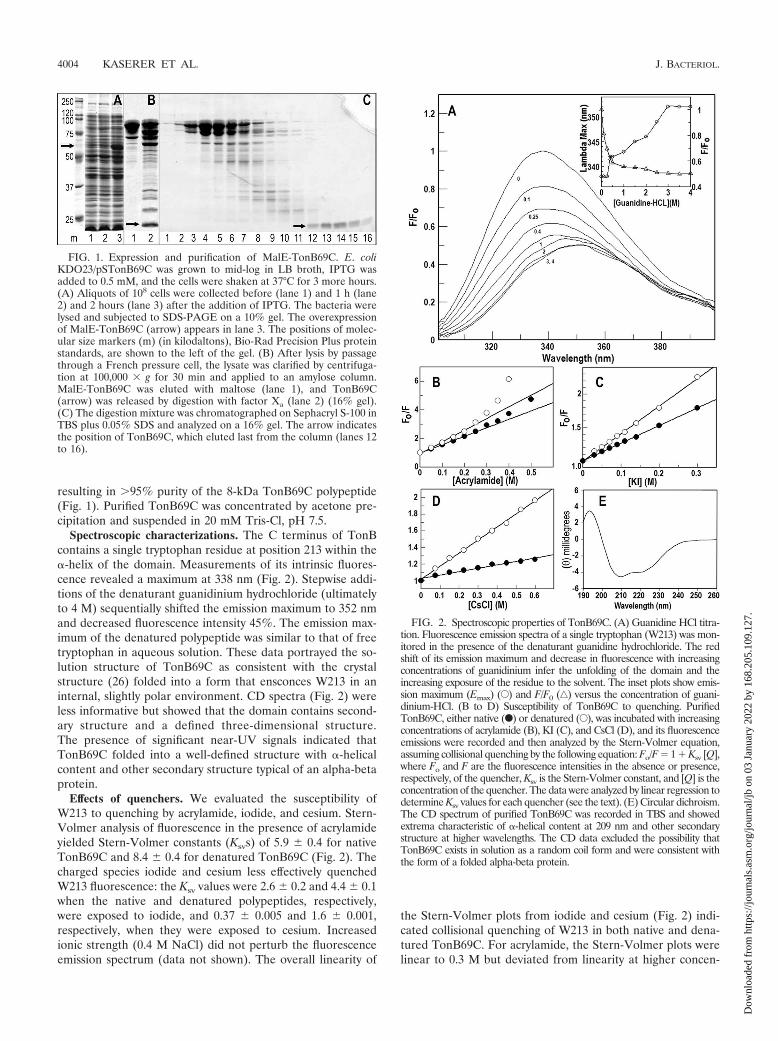
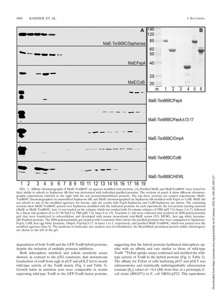
Adsorption of MalE-TonB69C to proteins. We immobilizedFepA, FepA�13-17 (which lacks the TonB box), colicin B,OmpA, or lysozyme on cyanogen bromide-activated Sepharoseand chromatographed MalE-TonB69C on the derivatized aga-roses. We equilibrated the resins in TBS, passed purified frac-tions of MalE-TonB69C over columns containing the resins,and analyzed the effluents from a salt gradient (0 to 0.5 MNaCl) by immunoblotting with mouse monoclonal anti-MalE(Fig. 3). Neither purified MalE nor MalE-TonB-69C bound to
the unconjugated agarose, and MalE alone also passedthrough resins containing immobilized proteins. Conversely,MalE-TonB69C adsorbed to the protein-modified agarosesand remained bound until it was eluted by the salt gradient.The adsorption of MalE-TonB69C, but not MalE, to the resinsindicated that the TonB C terminus was responsible for thebinding activity. The relative elution position of MalE-TonB69C in the course of the salt gradient (Fig. 3) suggestedthe order of its affinity for the conjugated proteins: lysozyme �ColB � OmpA � FepA�12-17 � FepA. That is, the leastaffinity was for lysozyme-Sepharose (MalE-TonB69C elutedearliest in the salt gradient), and the highest affinity was forFepA-Sepharose (MalE-TonB69C eluted last in the salt gra-dient). The association of TonB69C with FepA�13-17 indi-cated that the interaction did not require binding to the recep-tor’s TonB box. Adsorption to immobilized OmpA andlysozyme demonstrated that the TonB C terminus has generalaffinity for other proteins. TonB69C had higher affinity for OMproteins, because it required higher salt concentrations to elutefrom OmpA-Sepharose than from lysozyme-Sepharose: about50% of the applied MalE-TonB69C passed unabsorbedthrough the lysozyme-Sepharose column.
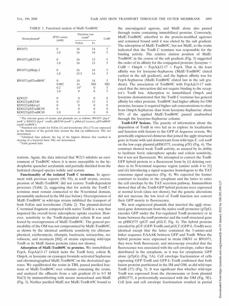
TonB-GFP fusions. The paucity of information about thedisposition of TonB in vivo led us to explore its localizationand function with fusions to the GFP of Aequorea victoria. Wegenetically engineered chimeras that joined the sggfp structuralgene in frame with and downstream from wild-type E. coli tonBon the low-copy plasmid pHSG575, creating pTG (Fig. 4). Theconstruct showed weak TonB activity, as assayed by its abilityto facilitate ferric siderophore uptake and colicin sensitivity,but it was not fluorescent. We attempted to convert the TonB-GFP hybrid protein to a fluorescent form by (i) deleting resi-dues in its N-terminal sequence region (amino acids 4 to 32)and (ii) introducing a signal sequence homologous to the TATconsensus signal sequence (Fig. 4). We expected the formerconstruct to localize to the cytoplasm and the latter to enterthe cell envelope by the TAT secretion pathway. Immunoblotsshowed that all the TonB-GFP hybrid proteins were expressedat normal levels (data not shown), but the genetic alterationsdid not increase the low level of TonB function nor converttheir GFP moiety to fluorescence.
We next engineered plasmids that inserted the sggfp struc-tural gene downstream from the native tonB promoter (pTpG;encodes GFP under the Fur-regulated TonB promoter) or inframe between the tonB promoter and the tonB structural geneon pHSG575 (pGT and pGLT; Fig. 4). The hybrid proteinsencoded by pGT (GFP-TonB) and pGLT (GFP-L-TonB) wereidentical except that the latter contained the 5-amino-acidlinker sequence EAAAK between GFP and TonB. When thehybrid proteins were expressed in strain OKN1 or BN1071,they were both fluorescent, and microscopy revealed that thefluorescence was associated with the cell envelope, rather thandistributed in the cytoplasm, as it was for cytoplasmic GFPalone (pTpG) (Fig. 5A). Cell envelope fractionation of cellsexpressing GFP-TonB and GFP-L-TonB confirmed that bothfusion proteins partitioned in the IM and OM, as did wild-typeTonB (57) (Fig. 5). It was significant that whether wild-typeTonB was expressed from the chromosome or from plasmidpHSG575, it preferentially associated with the OM (Fig. 5E).Cell lysis and cell envelope fractionation resulted in partial
TABLE 2. Functional analysis of MalE-TonB69C
Strain/plasmida IPTG concn(mM)
Nutrition testresultb
ColBc
FeEnt Fc
BN1071 0 16 14 51.0 16 14 5
BN1071/pRZ540 0 16 13 51.0 16 13 5
BN1071/pMalp-2 0 16 14 51.0 15.5 14 5
BN1071/pSTonB69C 0 14 14 50.002 14 ND ND0.2 14d 0 51.0 0 0 5
KDO23 0 0 0 0KDO23/pRZ540 0 15 13 5KDO23/pMal-p2 0 0 0 0KDO23/pSTonBC69 0 0 0 0KDO23/pSTonBC69 1.0 0 0 0
a The relevant genes of strains and plasmids are as follows: BN1071 (fepA�
tonB�); KDO23 (fepA� tonB); pRZ540 (tonB�), pMal-p2 (vector), pSTonB69C(malE-tonB69C).
b Nutrition test results for FeEnt (3) and ferrichrome (Fc) (71) are expressedas the diameter of the growth halo around the disk (in millimeters). ND, notdetermined.
c Tabulated data indicate the log of the highest dilution that resulted inclearing of a bacterial lawn. ND, not determined.
d Faint growth halo.
VOL. 190, 2008 TonB AND IRON TRANSPORT THROUGH THE OUTER MEMBRANE 4005
Dow
nloa
ded
from
http
s://j
ourn
als.
asm
.org
/jour
nal/j
b on
03
Janu
ary
2022
by
168.
205.
109.
127.
degradation of both TonB and the GFP-TonB hybrid proteins,despite the inclusion of multiple protease inhibitors.
Both siderophore nutrition and colicin sensitivity assaysshowed, in contrast to the pTG constructs, that downstreamlocalization of tonB from ssgfp in pGT and pGLT led to nearlywild-type activity of the TonB moiety (Fig. 6 and Table 3).Growth halos in nutrition tests were comparable in strainsexpressing wild-type TonB or the GFP-TonB fusion proteins,
suggesting that the hybrid proteins facilitated siderophore up-take with an affinity and rate similar to those of wild-typeTonB. 59FeEnt uptake assays confirmed and clarified the wild-type activity of TonB in the hybrid proteins (Fig. 6; Table 3).The affinity for FeEnt of cells harboring pGT and pGLT wassubnanomolar and statistically indistinguishable (dissociationconstant [Kd] values of �0.4 nM) from that of a prototypic E.coli strain (BN1071) or E. coli OKN1/pT23. This equivalence
FIG. 3. Affinity chromatography of MalE-TonB69C on agarose modified with proteins. (A) Purified MalE and MalETonB69C were tested fortheir ability to adsorb to Sepharose 6B that was derivatized with individual purified proteins. The sections of panel A show different chromato-graphic experiments (labeled to the right with the test protein/immobilized protein). The top three sections are control experiments: MalE-TonB69C chromatographed on unmodified Sepharose 6B, and MalE chromatographed on Sepharose 6B modified with FepA or ColB. MalE didnot adsorb to any of the modified agaroses; for brevity, only the results with FepA-Sepharose and ColB-Sepharose are shown. The remainingsections show MalE-TonB69C passed over Sepharose modified with the indicated proteins. In each experiment, the test protein starting material(MalE or MalE-TonB69C; lane 1) was loaded on the column, which was washed with 10 column volumes of TBS (pH 7.5) (lanes 2 to 7), followedby a linear salt gradient (0 to 0.5 M NaCl in TBS [pH 7.5]; lanes 8 to 19). Fractions (1 ml) were collected and resolved on SDS-polyacrylamidegels that were transferred to nitrocellulose and developed with mouse monoclonal anti-MalE serum (93). HEWL, hen egg white lysozyme.(B) Purified proteins. The SDS-polyacrylamide gel (stained with Coomassie blue) shows the purified proteins that were conjugated to Sepharose:FepA, ColB, hen egg white lysozyme, OmpA, FepA�13-17, in lanes 1 to 5, respectively, and purified MalE-TonB69C, which was passed over themodified agaroses (lane 6). The positions of molecular size markers (m) (in kilodaltons), the BenchMark prestained protein ladder (Invitrogen),are shown to the left of the gel.
4006 KASERER ET AL. J. BACTERIOL.
Dow
nloa
ded
from
http
s://j
ourn
als.
asm
.org
/jour
nal/j
b on
03
Janu
ary
2022
by
168.
205.
109.
127.
also appeared in the overall transport affinity for FeEnt, whichwas the same for all the strains (Km values of �1 to 2 nM). Themost noticeable difference between strain OKN1 harboring pT23,pGT, or pGLT was a 25% decrease in Vmax in the latter twostrains, which was consistent with their 25% lower FeEnt-bindingcapacity (Table 3). This reduction in FeEnt uptake rate accountsfor the slightly larger growth halos in nutrition tests (3, 24). Thedecreased binding capacity translates into decreased Vmax; thelower transport rate likely relates to slightly lower levels of GFP-TonB and GFP-L-TonB relative to that of TonB from pT23 (Fig.5E and G). Still, the turnover numbers of FepA proteins in thesestrains were equivalent, indicating that the activities of GFP-TonB and GFP-L-TonB were indistinguishable from that of wild-type TonB itself. Thus, the presence of GFP at the N terminus ofTonB had no measurable effect on the ability of the bacteria totransport iron.
Sequence relationship between TonB and YcsF. Analysis ofthe sequence of the TonB C terminus revealed homology to E.coli ycfS, which encodes a 320-amino-acid, proline-rich (8.4%)protein with a calculated mass of 34.6 kDa. YcfS is a memberof a family of putative periplasmic proteins (7) of unknownfunction that contains a signal peptide (residues 1 to 23) fol-lowed by a hydrophobic, potential IM anchor, a lysin (LysM)motif (residues 45 to 91) that confers affinity for PG (7, 98),and a central proline-rich sequence. These attributes bear sim-ilarities to those of TonB, another proline-rich (16.7%)periplasmic protein that contains a hydrophobic N-terminalsequence, postulated to act as an IM anchor. CLUSTALWcomparison of the E. coli LysM motif and the primary structureof TonB mapped a homologous region in the C terminus ofTonB (residues 175 to 231; Fig. 7). Although the sequences ofthe 48-residue LysM motif and the C-terminal 69 amino acidsof TonB are not highly conserved (19% identity, 77% homol-ogy), low overall identity is typical among LysM-containing,
PG-binding cell envelope proteins (75, 98). In this alignment,it was noteworthy that D11 in LysM, at the center of thePG-binding surface (7), corresponded with E205, on the exte-rior surface of the TonB C terminus. Moreover, an LSQMANcomparison of the solved structure of the LysM motif from E.coli MltD (7) (RCSB 1e0g) and the C-domain of TonB (26)(RCSB 1Ihr) identified a second region of similarity on theexternal surface of TonB’s dimeric ���� domain (Fig. 7).Superposition of the LysM crystal structure onto this secondsite revealed a nearly identical region of polypeptide folding inTonB. LSQMAN found 17 C� positions (17 of 48; 35% of C�)that aligned over 21 atoms with an overall root mean squaredeviation of 1.58 Å (Fig. 7). The most significant alignments ofside chain residues were Arg 6, Arg 8, Asp 11, Lys 18, Arg 19,and His 20 on LysM, which superposed on Arg 171, Glu 173,Asp 189, Arg 211, Arg 212, and Trp 213, respectively, on TonB.In summary, the in silico analyses identified two regions in theTonB C terminus with an equivalent fold and aligned residuesto LysM, suggesting that TonB has affinity for PG.
Binding of MalE-TonB69C to PG. To test this prediction, wepurified sacculi from E. coli by SDS extraction and evaluatedtheir ability to adsorb MalE-TonB69C. The purified PG frac-tion was free of cell envelope proteins, including the majorproteins and iron-regulated LGP, as indicated by SDS-PAGE(Fig. 8) and by its transparency at 280 nm (data not shown).The purified sacculi precipitated MalE-TonB69C from solu-tion, but not MalE nor FepB (Fig. 8). The binding reactionmanifested saturation behavior: increasing amounts of PGbound and precipitated increasing amounts of MalE-TonB69Cto a plateau value. The control proteins were themselves bio-logically active: maltose-binding protein was purified by amy-lose affinity columns, and chromatographically purified FepB(97) bound FeEnt (data not shown). Therefore, the affinity of
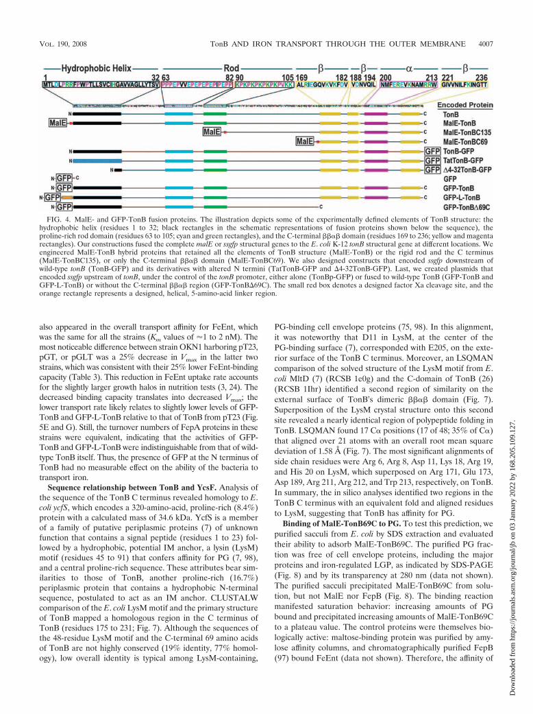
FIG. 4. MalE- and GFP-TonB fusion proteins. The illustration depicts some of the experimentally defined elements of TonB structure: thehydrophobic helix (residues 1 to 32; black rectangles in the schematic representations of fusion proteins shown below the sequence), theproline-rich rod domain (residues 63 to 105; cyan and green rectangles), and the C-terminal ���� domain (residues 169 to 236; yellow and magentarectangles). Our constructions fused the complete malE or ssgfp structural genes to the E. coli K-12 tonB structural gene at different locations. Weengineered MalE-TonB hybrid proteins that retained all the elements of TonB structure (MalE-TonB) or the rigid rod and the C terminus(MalE-TonBC135), or only the C-terminal ���� domain (MalE-TonBC69). We also designed constructs that encoded ssgfp downstream ofwild-type tonB (TonB-GFP) and its derivatives with altered N termini (TatTonB-GFP and �4-32TonB-GFP). Last, we created plasmids thatencoded ssgfp upstream of tonB, under the control of the tonB promoter, either alone (TonBp-GFP) or fused to wild-type TonB (GFP-TonB andGFP-L-TonB) or without the C-terminal ���� region (GFP-TonB�69C). The small red box denotes a designed factor Xa cleavage site, and theorange rectangle represents a designed, helical, 5-amino-acid linker region.
VOL. 190, 2008 TonB AND IRON TRANSPORT THROUGH THE OUTER MEMBRANE 4007
Dow
nloa
ded
from
http
s://j
ourn
als.
asm
.org
/jour
nal/j
b on
03
Janu
ary
2022
by
168.
205.
109.
127.

FIG. 5. (A to C) Fluorescence microscopic and electrophoretic analyses of GFP-TonB expression and localization. E. coli OKN3/pTpG (A),OKN3/pGT (B), and OKN3/pGLT (C) were grown in MOPS minimal medium and observed by fluorescence microscopy. Note that E. coli OKN3/pTpG,which expresses cytoplasmic GFP, showed diffuse, uniformly distributed fluorescence throughout the cells, whereas when GFP was fused upstream ofTonB in E. coli OKN3/pGT and OKN3/pGLT, its fluorescence associated with the cell envelope, presumably from the association of TonB with the IM.(D and E) Expression of GFP-TonB hybrid proteins. A total of 108 cells of E. coli strains OKN1 (�tonB), BN1071 (tonB�), OKN3 (�fepA), OKN1/pT23(tonB�), OKN1/pGT (ssgfp-tonB), and OKN1/pGLT (ssgfp-L-tonB), shown in lanes 1 to 6, respectively, were grown in MOPS minimal medium, lysed insample buffer, and subjected to SDS-PAGE and Western immunoblotting with mouse anti-FepA monoclonal antibody 45 (�-FepA) (88) (D) orpolyclonal rabbit anti-TonB (�-TonB) (E). The molecular size markers (m) (sizes in kilodaltons) were the BenchMark prestained protein ladder(Invitrogen). Panel D shows comparable expression of FepA in all fepA� strains; panel E shows the expression of wild-type TonB (lanes 2 to 4),GFP-TonB (lane 5), and GFP-L-TonB (lane 6) at comparable levels and the expected molecular sizes. (F and G) Fractionation of cell envelopes byFrench pressure cell lysis and sucrose gradient centrifugation. Bacteria were grown in MOPS minimal medium, collected by centrifugation, and lysed ina French pressure cell at 14,000 lb/in2, and their membranes were resolved on sucrose step gradients and subjected to SDS-PAGE (F). IM samples fromstrains BN1071, OKN1, OKN1/pT23, OKN1/pGT, and OKN1/pGLT appear in lanes 1, 3, 5, 7, and 9, respectively; OM samples from the same strainsappear in lanes 2, 4, 6, 8 and 10, respectively. In panel G, an identical gel was transferred to nitrocellulose and subjected to Western immunoblotting withrabbit anti-TonB sera.
4008 KASERER ET AL. J. BACTERIOL.
Dow
nloa
ded
from
http
s://j
ourn
als.
asm
.org
/jour
nal/j
b on
03
Janu
ary
2022
by
168.
205.
109.
127.
TonB for PG was not a general characteristic of periplasmicproteins, but a specific attribute of TonB itself.
DISCUSSION
The relationship between iron acquisition and microbialpathogenesis (1, 38, 51, 84, 104) underscores the importance ofthe role of TonB in cell envelope physiology. Passage of ferriccomplexes through the OM requires TonB activity, and onetheory of this requirement is that TonB participates in trans-port energetics by capturing proton motive force from the IM(where its N terminus resides) and distributing it to the OMtransporters (14, 23, 76, 82, 95). According to the “shuttle”model of TonB action, it associates with the IM proteins ExbBand ExbD (42, 56), acquires proton motive force-generatedenergy by an unknown structural transition, and transmits (15)or physically transports (55, 57) the energy across theperiplasm to the OM. The proposed interaction of “energized”TonB with OM proteins entails recognition of ligand-boundreceptors and release of the stored force to them by protein-protein interactions between the C-terminal residues of TonBand the TonB box sequence of the LGP (76, 95). The trans-ferred energy presumably facilitates internalization of boundferric siderophores and vitamin B12 (22), as well as bacterialsusceptibility to B-group colicins and certain bacteriophages.Although the requirements for both energy and TonB in OMmetal transport reactions are established, the participation ofTonB in energy transduction or the bioenergetics of iron up-take remains undemonstrated.
Our experimental data relate to TonB function in severalways and suggest a model for its role in cell envelope biochem-istry. We found that (i) TonB has a affinity for OM proteins,regardless of whether they contain a TonB box; (ii) GFP-TonBfusion proteins, which localize the hydrophilic GFP moiety inthe cytoplasm, retain TonB function; and (iii) the C terminusof TonB binds PG, as a result of its comparable fold to the lysinstructural motif.
It was unexpected that expression of MalE-TonB69Cblocked FeEnt and ferrichrome uptake, but not colicin B kill-
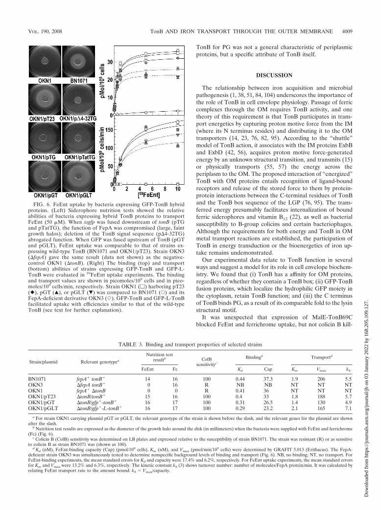
FIG. 6. FeEnt uptake by bacteria expressing GFP-TonB hybridproteins. (Left) Siderophore nutrition tests showed the relativeabilities of bacteria expressing hybrid TonB proteins to transportFeEnt (50 M). When ssgfp was fused downstream of tonB (pTGand pTatTG), the function of FepA was compromised (large, faintgrowth halos); deletion of the TonB signal sequence (p�4-32TG)abrogated function. When GFP was fused upstream of TonB (pGTand pGLT), FeEnt uptake was comparable to that of strains ex-pressing wild-type TonB (BN1071 and OKN1/pT23). Strain OKN3(�fepA) gave the same result (data not shown) as the negative-control OKN1 (�tonB). (Right) The binding (top) and transport(bottom) abilities of strains expressing GFP-TonB and GFP-L-TonB were evaluated in 59FeEnt uptake experiments. The bindingand transport values are shown in picomoles/109 cells and in pico-moles/109 cells/min, respectively. Strain OKN1 (�) harboring pT23(�), pGT (Œ), or pGLT (�) was compared to BN1071 (E) and itsFepA-deficient derivative OKN3 (�). GFP-TonB and GFP-L-TonBfacilitated uptake with efficiencies similar to that of the wild-typeTonB (see text for further explanation).
TABLE 3. Binding and transport properties of selected strains
Strain/plasmid Relevant genotypea
Nutrition testresultb ColB
sensitivityc
Bindingd Transportd
FeEnt Fc Kd Cap Km Vmax k8
BN1071 fepA� tonB� 14 16 100 0.44 37.3 1.9 206 5.5OKN3 �fepA tonB� 0 16 R NB NB NT NT NTOKN1 fepA� �tonB 0 0 R 0.41 36 NT NT NTOKN1/pT23 �tonB/tonB� 15 16 100 0.4 33 1.8 188 5.7OKN1/pGT �tonB/gfp�-tonB� 16 17 100 0.31 26.5 1.4 130 4.9OKN1/pGLT �tonB/gfp�-L-tonB� 16 17 100 0.29 23.2 2.1 165 7.1
a For strain OKN1 carrying plasmid pGT or pGLT, the relevant genotype of the strain is shown before the slash, and the relevant genes for the plasmid are shownafter the slash.
b Nutrition test results are expressed as the diameter of the growth halo around the disk (in millimeters) when the bacteria were supplied with FeEnt and ferrichrome(Fc) (Fig. 6).
c Colicin B (ColB) sensitivity was determined on LB plates and expressed relative to the susceptibility of strain BN1071. The strain was resistant (R) or as sensitiveto colicin B as strain BN1071 was (shown as 100).
d Kd (nM), FeEnt-binding capacity (Cap) (pmol/109 cells), Km (nM), and Vmax (pmol/min/109 cells) were determined by GRAFIT 5.013 (Erithacus). The FepA-deficient strain OKN3 was simultaneously tested to determine nonspecific background levels of binding and transport (Fig. 6). NB, no binding; NT, no transport. ForFeEnt-binding experiments, the mean standard errors for Kd and capacity were 17.4% and 6.2%, respectively. For FeEnt uptake experiments, the mean standard errorsfor Km and Vmax were 13.2% and 6.3%, respectively. The kinetic constant k8 (3) shows turnover number: number of molecules/FepA protein/min. It was calculated byrelating FeEnt transport rate to the amount bound: k8 � Vmax/capacity.
VOL. 190, 2008 TonB AND IRON TRANSPORT THROUGH THE OUTER MEMBRANE 4009
Dow
nloa
ded
from
http
s://j
ourn
als.
asm
.org
/jour
nal/j
b on
03
Janu
ary
2022
by
168.
205.
109.
127.
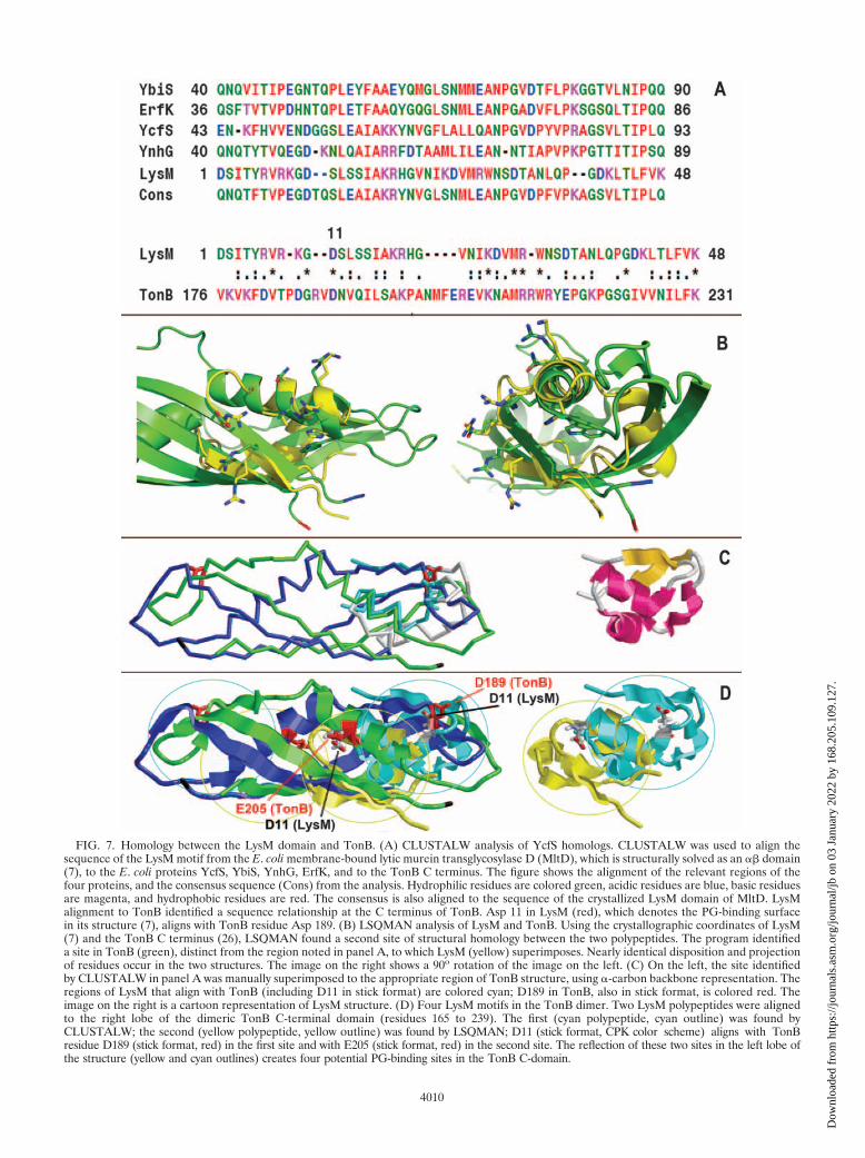
FIG. 7. Homology between the LysM domain and TonB. (A) CLUSTALW analysis of YcfS homologs. CLUSTALW was used to align thesequence of the LysM motif from the E. coli membrane-bound lytic murein transglycosylase D (MltD), which is structurally solved as an �� domain(7), to the E. coli proteins YcfS, YbiS, YnhG, ErfK, and to the TonB C terminus. The figure shows the alignment of the relevant regions of thefour proteins, and the consensus sequence (Cons) from the analysis. Hydrophilic residues are colored green, acidic residues are blue, basic residuesare magenta, and hydrophobic residues are red. The consensus is also aligned to the sequence of the crystallized LysM domain of MltD. LysMalignment to TonB identified a sequence relationship at the C terminus of TonB. Asp 11 in LysM (red), which denotes the PG-binding surfacein its structure (7), aligns with TonB residue Asp 189. (B) LSQMAN analysis of LysM and TonB. Using the crystallographic coordinates of LysM(7) and the TonB C terminus (26), LSQMAN found a second site of structural homology between the two polypeptides. The program identifieda site in TonB (green), distinct from the region noted in panel A, to which LysM (yellow) superimposes. Nearly identical disposition and projectionof residues occur in the two structures. The image on the right shows a 90o rotation of the image on the left. (C) On the left, the site identifiedby CLUSTALW in panel A was manually superimposed to the appropriate region of TonB structure, using �-carbon backbone representation. Theregions of LysM that align with TonB (including D11 in stick format) are colored cyan; D189 in TonB, also in stick format, is colored red. Theimage on the right is a cartoon representation of LysM structure. (D) Four LysM motifs in the TonB dimer. Two LysM polypeptides were alignedto the right lobe of the dimeric TonB C-terminal domain (residues 165 to 239). The first (cyan polypeptide, cyan outline) was found byCLUSTALW; the second (yellow polypeptide, yellow outline) was found by LSQMAN; D11 (stick format, CPK color scheme) aligns with TonBresidue D189 (stick format, red) in the first site and with E205 (stick format, red) in the second site. The reflection of these two sites in the left lobe ofthe structure (yellow and cyan outlines) creates four potential PG-binding sites in the TonB C-domain.
4010
Dow
nloa
ded
from
http
s://j
ourn
als.
asm
.org
/jour
nal/j
b on
03
Janu
ary
2022
by
168.
205.
109.
127.
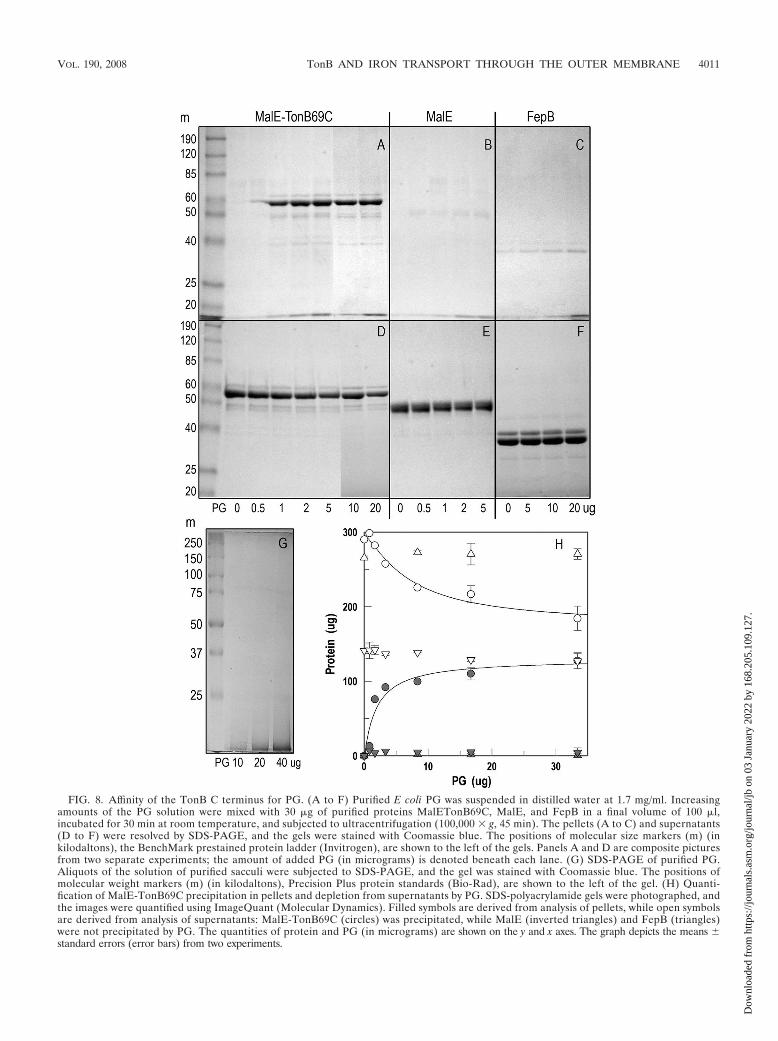
FIG. 8. Affinity of the TonB C terminus for PG. (A to F) Purified E coli PG was suspended in distilled water at 1.7 mg/ml. Increasingamounts of the PG solution were mixed with 30 g of purified proteins MalETonB69C, MalE, and FepB in a final volume of 100 l,incubated for 30 min at room temperature, and subjected to ultracentrifugation (100,000 g, 45 min). The pellets (A to C) and supernatants(D to F) were resolved by SDS-PAGE, and the gels were stained with Coomassie blue. The positions of molecular size markers (m) (inkilodaltons), the BenchMark prestained protein ladder (Invitrogen), are shown to the left of the gels. Panels A and D are composite picturesfrom two separate experiments; the amount of added PG (in micrograms) is denoted beneath each lane. (G) SDS-PAGE of purified PG.Aliquots of the solution of purified sacculi were subjected to SDS-PAGE, and the gel was stained with Coomassie blue. The positions ofmolecular weight markers (m) (in kilodaltons), Precision Plus protein standards (Bio-Rad), are shown to the left of the gel. (H) Quanti-fication of MalE-TonB69C precipitation in pellets and depletion from supernatants by PG. SDS-polyacrylamide gels were photographed, andthe images were quantified using ImageQuant (Molecular Dynamics). Filled symbols are derived from analysis of pellets, while open symbolsare derived from analysis of supernatants: MalE-TonB69C (circles) was precipitated, while MalE (inverted triangles) and FepB (triangles)were not precipitated by PG. The quantities of protein and PG (in micrograms) are shown on the y and x axes. The graph depicts the means standard errors (error bars) from two experiments.
VOL. 190, 2008 TonB AND IRON TRANSPORT THROUGH THE OUTER MEMBRANE 4011
Dow
nloa
ded
from
http
s://j
ourn
als.
asm
.org
/jour
nal/j
b on
03
Janu
ary
2022
by
168.
205.
109.
127.
ing. These data indicate that the ferric siderophores and thetoxin diverge in their transport pathways at the stage inhibitedby the isolated TonB C terminus. The inhibition of a TonB-dependent process by MalE-TonB69C in the periplasm dem-onstrated that the TonB C terminus folded properly in thechimeric protein, as was observed for other constructs thatexpressed it in the periplasm (43), and for TolA (12). Fluores-cence spectra of the purified fusion protein concurred with thisconclusion: W213 in TonB69C was preferentially quenched byacrylamide and quenched less by iodide and cesium, suggest-ing, as seen in crystal structures (26, 76, 95), that it localizes ina weakly polar environment (21, 102) where it is more acces-sible to the hydrophobic quencher than to the hydrated ions.The positive ion likely quenched most poorly as a result ofrepulsion from basic residues (arginines 187, 211, 212, and 214)in the proximity of W213, as seen in the crystal structure.Characterizations by CD were less definitive but reflected thepresence of � and � structure. Quenching deviated from lin-earity at higher acrylamide concentrations, implying structuralchanges in the C terminus that result in noncollisional quench-ing effects. Together, the results suggested that in solutionTonB69C assumed a biologically relevant tertiary structure: itfolded into a globular domain with properties expected fromthe TonB crystal structure. Once purified, MalE-TonB69C ad-sorbed to TonB-dependent and -independent proteins, show-ing that besides its affinity for the TonB box of siderophorereceptors, the TonB C terminus nonspecifically adsorbs toother proteins, including OmpA. This finding, which concurswith previous reports (41), is mechanistically important be-cause of the abundance of OmpA in the OM (�105 copies/cell)(92). The globular periplasmic domain of OmpA (99) mayconstitute another target for recruitment into the �-sheetwithin the C terminus of TonB, and it is known to exist in closeproximity to FepA (94) in the OM bilayer. When viewed inlight of IM localization of its N terminus, these data suggestthat TonB may specifically or nonspecifically bridge the cellenvelope by adsorbing to the periplasmic domains of TonB-dependent or TonB-independent OM proteins, respectively.Previous experiments support this notion: elimination of itsterminal 8 residues did not impair TonB function, whereasdeletion of the last 15 amino acids blocked activity (4). Atruncated form of TonB lacking residues 174 to 239 did notcross-link to OM proteins or associate with the OM (57).
Letain and Postle (57) proposed that TonB shuttles betweenthe membranes of the cell envelope during its facilitation ofmetal transport, and Larsen et al. (55) reiterated this mecha-nism. The fluorescent, biologically active GFP-TonB hybridproteins (expressed from pGT and pGLT) are relevant to thisidea, because the residence of their GFP moiety in the cyto-plasm prohibits their transposition from IM to OM. We found,as did others (31, 61, 66), that GFP does not fluoresce in theperiplasm. The fluorescence of GFP-TonB and GFP-L-TonBimplies cytoplasmic localization of the upstream GFP moiety,and their biological activity indicates cell envelope localizationof the downstream TonB protein. These results argue againstthe shuttle model. Letain and Postle (57) studied the partition-ing of TonB between the IM and OM and cited their results asevidence that it may physically depart the IM and specificallyassociate with LGP in the OM. Yet, the separation of cellenvelope fractions by any existing methodology is imperfect
and incomplete. The French pressure cell lysis/sucrose gradi-ent centrifugation method depends on brute-force separationof cell envelope components: a pressure differential explodescells and presumably peels their IM and OM apart. In even thebest experiments, the IM contains OM proteins because thelatter traverse the former during biogenesis, and IM proteinscontaminate the OM fraction, at least in part from the ineffi-ciency of the membrane separation, and potentially from thearchitecture and physiology of the cell envelope (see below).Therefore, the observation of IM proteins in the OM fractionsand vice versa is difficult to interpret in a mechanistic sense.Furthermore, other models may explain the cell envelope frac-tionation results. If TonB bridges the periplasm by virtue of itsN terminus in the IM and its C terminus is associated with PGor OM proteins, then this structural organization alone ac-counts for its appearance in both fractions when the cell en-velope pulls apart. The membranes are broken during theFrench pressure cell lysis procedure, and it is pertinent thatneither the precise architecture of the periplasm nor the natureof its physical disruption (i.e., the fracture planes) are known.Finally, whereas the IM is a fluid mosaic bilayer, the outerlipopolysaccharide leaflet confers asymmetry and reduced flu-idity to the OM, creating a quasifrozen state in the presence ofdivalent cations (73). In addition, OM proteins associate withthe PG polymer (2, 59, 75, 87). In the absence of adhesions orcontinuities, it is conceivable that the IM and OM might effi-ciently separate during French pressure cell-mediated lysis, butif proteins or PG conjoin the two membranes in specializedregions, as was postulated (44, 54), observed (8, 63), and jus-tified on the basis of Omp, PG, and lipopolysaccharide biosyn-thesis pathways (10, 47), then the isolation of pure OM and IMfractions is unlikely. Hence, immunoblot characterizations offractionated cell envelopes are potentially ambiguous and sub-ject to interpretation. Despite these reservations, the fusion ofGFP upstream of TonB significantly decreased associationwith the OM without compromising TonB function. Whilewild-type TonB, whether chromosome or plasmid mediated,preferentially fractionated with the OM (three- to fourfold),both GFP-TonB and GFP-L-TonB distributed approximatelyequally, demonstrating that the GFP moiety enhanced associ-ation with the IM. The distribution was not absolute, in thatthe fusion protein was also seen in the OM fraction. Thepresence of GFP-TonB fusions in the OM fraction underscoresthe point that French pressure cell-mediated lysis does notpreserve the normal distribution of proteins in the IM andOM. The upshot of these considerations is that characteriza-tions of fractionated cell envelopes are fraught with uncer-tainty.
The potential flaws in such fractionation experiments sug-gest that in vivo evidence provides more accurate informationabout the localization of TonB or its various domains in thecell envelope. The extrinsic fluorophore labeling study ofLarsen et al. (55) adopted such an approach, but it lacked adefinitive control and structural information about the target,N-terminal domain of TonB. Its conclusions are subject toquestion in that (i) the experiments did not exclude the possi-bility that the Oregon green fluorophore penetrated the IMbilayer, and (ii) it is not known where the target residue L3Cnormally resides (in the IM bilayer or on its inner or outersurface). Therefore, although the authors concluded that the
4012 KASERER ET AL. J. BACTERIOL.
Dow
nloa
ded
from
http
s://j
ourn
als.
asm
.org
/jour
nal/j
b on
03
Janu
ary
2022
by
168.
205.
109.
127.
TonB N terminus departed the IM bilayer in vivo, objectionsexist to this conclusion. The data that we report showing thefluorescence and functionality of GFP-TonB fusions undercutthe shuttle model. The surface of A. victoria GFP contains 71charged acidic or basic side chains, effectively precluding itsmovement through a membrane bilayer in vivo. Therefore, thetransport ability of strains producing these fluorescent con-structs demonstrates that TonB still functions when its N ter-minus remains anchored in the IM.
YcsF and its E. coli paralogs YnhG, YbiS, and ErfK form afamily of cell envelope proteins. Each one, composed of ap-proximately 320 amino acids, has an N-terminal hydrophobicregion (putative transmembrane helix), a PG-binding domain(LysM) in the first third of primary structure, and a high per-centage of proline residues (8 to 10%, roughly twice that ofmost proteins and half as much as TonB). The structural ho-mology between the LysM motif and regions of the TonB Cterminus raised the possibility of an affiliation between TonBand the murein sacculus. Experiments confirmed this infer-
ence: purified sacculi precipitated microgram quantities of pu-rified MalE-TonB69C, but not MalE alone. Thus, in additionto its other binding functions, the TonB C-domain manifestssignificant affinity for PG. Although the in vivo disposition ofTonB as a monomer or dimer remains uncertain, both may bephysiologically rationalized, and there is experimental evi-dence for both the monomer in solution (78) and the dimer inlive cells (90). However, LysM homology in TonB occurs onlyin the context of a dimeric C-domain, because the proposedbinding clefts derive from � and � elements of differentpolypeptides; they do not involve the portion of the �-sheetthat recruits heterologous polypeptides. The evidence that theLysM regions of TonB are suited for PG binding is strong. ItsC-domain folds such that the backbone of region 2 has a rootmean square deviation of 1.58 Å from that of LysM. Second,numerous residues relevant to binding are comparably distrib-uted across the surfaces of both proteins (Fig. 7), includingD189 and E205 in TonB, that project like D11, the residue thatdefines the PG-binding surface of LysM (7). The presence of
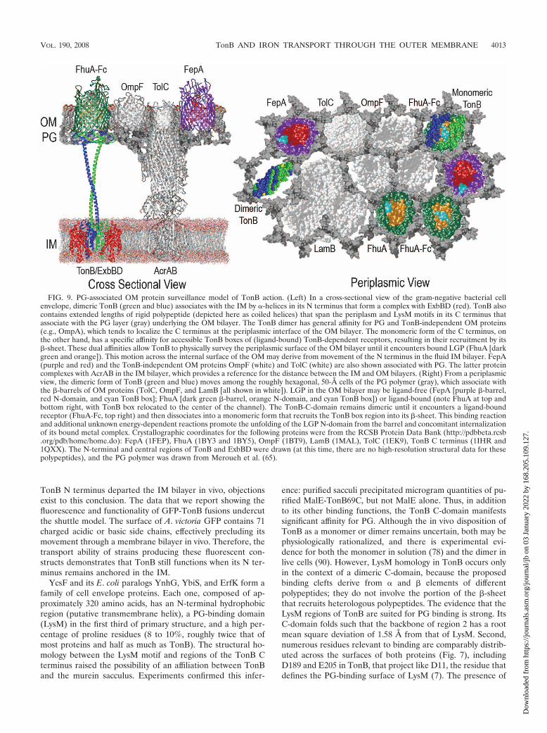
FIG. 9. PG-associated OM protein surveillance model of TonB action. (Left) In a cross-sectional view of the gram-negative bacterial cellenvelope, dimeric TonB (green and blue) associates with the IM by �-helices in its N terminus that form a complex with ExbBD (red). TonB alsocontains extended lengths of rigid polypeptide (depicted here as coiled helices) that span the periplasm and LysM motifs in its C terminus thatassociate with the PG layer (gray) underlying the OM bilayer. The TonB dimer has general affinity for PG and TonB-independent OM proteins(e.g., OmpA), which tends to localize the C terminus at the periplasmic interface of the OM bilayer. The monomeric form of the C terminus, onthe other hand, has a specific affinity for accessible TonB boxes of (ligand-bound) TonB-dependent receptors, resulting in their recruitment by its�-sheet. These dual affinities allow TonB to physically survey the periplasmic surface of the OM bilayer until it encounters bound LGP (FhuA [darkgreen and orange]). This motion across the internal surface of the OM may derive from movement of the N terminus in the fluid IM bilayer. FepA(purple and red) and the TonB-independent OM proteins OmpF (white) and TolC (white) are also shown associated with PG. The latter proteincomplexes with AcrAB in the IM bilayer, which provides a reference for the distance between the IM and OM bilayers. (Right) From a periplasmicview, the dimeric form of TonB (green and blue) moves among the roughly hexagonal, 50-Å cells of the PG polymer (gray), which associate withthe �-barrels of OM proteins (TolC, OmpF, and LamB [all shown in white]). LGP in the OM bilayer may be ligand-free (FepA [purple �-barrel,red N-domain, and cyan TonB box]; FhuA [dark green �-barrel, orange N-domain, and cyan TonB box]) or ligand-bound (note FhuA at top andbottom right, with TonB box relocated to the center of the channel). The TonB-C-domain remains dimeric until it encounters a ligand-boundreceptor (FhuA-Fc, top right) and then dissociates into a monomeric form that recruits the TonB box region into its �-sheet. This binding reactionand additional unknown energy-dependent reactions promote the unfolding of the LGP N-domain from the barrel and concomitant internalizationof its bound metal complex. Crystallographic coordinates for the following proteins were from the RCSB Protein Data Bank (http://pdbbeta.rcsb.org/pdb/home/home.do): FepA (1FEP), FhuA (1BY3 and 1BY5), OmpF (1BT9), LamB (1MAL), TolC (1EK9), TonB C terminus (1IHR and1QXX). The N-terminal and central regions of TonB and ExbBD were drawn (at this time, there are no high-resolution structural data for thesepolypeptides), and the PG polymer was drawn from Meroueh et al. (65).
VOL. 190, 2008 TonB AND IRON TRANSPORT THROUGH THE OUTER MEMBRANE 4013
Dow
nloa
ded
from
http
s://j
ourn
als.
asm
.org
/jour
nal/j
b on
03
Janu
ary
2022
by
168.
205.
109.
127.
the lysin folds does not necessarily implicate them as respon-sible for the observed PG binding, and further experiments arewarranted to confirm that conclusion. However, the lysin do-mains present potential PG-binding regions on the TonB Cterminus; in the dimeric state, it contains four distinct sites thatmay interact with PG.
It is germane that the tripartite structural organization ofTonB resembles the architecture of TolA, a component of theTolQRAB system that functions in maintenance of the struc-tural integrity of the OM. The Tol system is required forinfection by some bacteriophages (f1, M13, fd, and Ike) andgroup A colicins (E1, E2, E3, A, K, L, and N). Analogous towhat we and others (43) found for the TonB C terminus,overexpression of the TolA C terminus in tolA� cells decreasedTolA-dependent functions. These and other data illustrate theneed for full-length TolA and TonB and the ability of theirisolated C termini to interfere with OM transport systems.TolA spans the periplasm and associates with Pal in the OM byan interaction between two short sequences within the proteins(11, 79). Pal is a PG-binding lipoprotein that also contains thelysin motif. Finally, components of the Tol and Ton systemsmay substitute for each other, as illustrated by the complemen-tation of exbB and exbD strains by homologous proteins of theTol system, TolQ and TolR (16, 17). In summary, the homol-ogous sequences and analogous results for the components ofthe TolA- and TonB-dependent transport systems imply simi-lar mechanisms of action, which in both cases involve associa-tion with PG.
The persistent association of the N terminus of TonB withthe IM and the general affinity of its C terminus for PG andOM proteins clarify the disposition of TonB in the cell enve-lope. Combined with a body of genetic, biochemical, and struc-tural data, our results portray a cell envelope (Fig. 9) in whichTonB normally spans the periplasm, temporarily or perma-nently adhering the IM and OM together at certain locations(i.e., adhesion zones [9, 54]). OM proteins may covalently ornoncovalently associate with PG in vivo, including lipoprotein(19), Pal (75), OmpF (2, 59, 87), and FepA (data not shown).Neither OmpF nor FepA has periplasmic domains, so theirassociation with PG must occur at the internal surface of theOM, and high-resolution microscopic studies show the PGpolymer in contact with the OM bilayer (63). Recent structuraldata indicate that PG forms a honeycomb of 50-Šcells be-neath the OM, creating a framework for association with OMproteins (65). The approximate 50-Šdiameter of both OM�-barrels and the dimeric TonB C terminus, which are notlikely coincidental, raise the possibility that the latter movesacross the cells in this matrix until it encounters ligand-boundsiderophore receptors. Bound LGP are distinguished by therepositioning of their TonB box to the center of their pores(33, 58), and in monomeric form, TonB may intercalate thesepolypeptides into its C-terminal ��� domain (76, 95).
These points suggest a mechanism that we have designated“membrane surveillance,” in which the interconvertible di-meric and monomeric forms of TonB display two distinct af-finities. The nonspecific affinity of the dimeric C terminus forPG and TonB-independent OM proteins (e.g., OmpA) local-izes it at the periplasmic interface of the OM bilayer. When itencounters a ligand-bound receptor, the affinity of the mono-mer for an accessible TonB box polypeptide supersedes the
general affinity of the dimer, promoting dissociation and bind-ing of the monomer to the LGP. This affiliation begins theprocess of ligand transport through the channel, which re-quires further energized reactions, and may occur by pulling,unfolding, or expelling the N terminus into the periplasm (60).
ACKNOWLEDGMENTS
The research was supported by NIH grant GM53836 and NSF grantMCB0417694 to P.E.K.
Thanks go to Amparo G. Marcos and Chuck Smallwood for theircomments on the manuscript.
REFERENCES
1. Abergel, R. J., M. K. Wilson, J. E. Arceneaux, T. M. Hoette, R. K. Strong,B. R. Byers, and K. N. Raymond. 2006. Anthrax pathogen evades themammalian immune system through stealth siderophore production. Proc.Natl. Acad. Sci. USA 103:18499–18503.
2. Alphen, W. V., and B. Lugtenberg. 1977. Influence of osmolarity of thegrowth medium on the outer membrane protein pattern of Escherichia coli.J. Bacteriol. 131:623–630.
3. Annamalai, R., B. Jin, Z. Cao, S. M. Newton, and P. E. Klebba. 2004.Recognition of ferric catecholates by FepA. J. Bacteriol. 186:3578–3589.
4. Anton, M., and K. J. Heller. 1991. Functional analysis of a C-terminallyaltered TonB protein of Escherichia coli. Gene 105:23–29.
5. Arai, R., H. Ueda, A. Kitayama, N. Kamiya, and T. Nagamune. 2001.Design of the linkers which effectively separate domains of a bifunctionalfusion protein. Protein Eng. 14:529–532.
6. Axen, R., J. Porath, and S. Ernback. 1967. Chemical coupling of peptidesand proteins to polysaccharides by means of cyanogen halides. Nature214:1302–1304.
7. Bateman, A., and M. Bycroft. 2000. The structure of a LysM domain fromE. coli membrane-bound lytic murein transglycosylase D (MltD). J. Mol.Biol. 299:1113–1119.
8. Bayer, M. E. 1991. Zones of membrane adhesion in the cryofixed envelopeof Escherichia coli. J. Struct. Biol. 107:268–280.
9. Bayer, M. H., W. Keck, and M. E. Bayer. 1990. Localization of penicillin-binding protein 1b in Escherichia coli: immunoelectron microscopy andimmunotransfer studies. J. Bacteriol. 172:125–135.
10. Bos, M. P., V. Robert, and J. Tommassen. 2007. Biogenesis of the gram-negative bacterial outer membrane. Annu. Rev. Microbiol. 61:191–214.
11. Bouveret, E., R. Derouiche, A. Rigal, R. Lloubes, C. Lazdunski, and H.Benedetti. 1995. Peptidoglycan-associated lipoprotein-TolB interaction. Apossible key to explaining the formation of contact sites between the innerand outer membranes of Escherichia coli. J. Biol. Chem. 270:11071–11077.
12. Bouveret, E., L. Journet, A. Walburger, E. Cascales, H. Benedetti, and R.Lloubes. 2002. Analysis of the Escherichia coli Tol-Pal and TonB systems byperiplasmic production of Tol, TonB, colicin, or phage capsid soluble do-mains. Biochimie 84:413–421.
13. Bradbeer, C. 1993. The proton motive force drives the outer membranetransport of cobalamin in Escherichia coli. J. Bacteriol. 175:3146–3150.
14. Braun, V. 2006. Energy transfer between biological membranes. ACSChem. Biol. 1:352–354.
15. Braun, V. 1995. Energy-coupled transport and signal transduction throughthe gram-negative outer membrane via TonB-ExbB-ExbD-dependent re-ceptor proteins. FEMS Microbiol. Rev. 16:295–307.
16. Braun, V. 1989. The structurally related exbB and tolQ genes are inter-changeable in conferring tonB-dependent colicin, bacteriophage, and albo-mycin sensitivity. J. Bacteriol. 171:6387–6390.
17. Braun, V., and C. Herrmann. 1993. Evolutionary relationship of uptakesystems for biopolymers in Escherichia coli: cross-complementation be-tween the TonB-ExbB-ExbD and the TolA-TolQ-TolR proteins. Mol. Mi-crobiol. 8:261–268.
18. Braun, V., and H. Wolff. 1973. Characterization of the receptor protein forphage T5 and colicin M in the outer membrane of E. coli B. FEBS Lett.34:77–80.
19. Braun, V., and H. Wolff. 1970. The murein-lipoprotein linkage in the cellwall of Escherichia coli. Eur. J. Biochem. 14:387–391.
20. Brewer, S., M. Tolley, I. P. Trayer, G. C. Barr, C. J. Dorman, K. Hannavy,C. F. Higgins, J. S. Evans, B. A. Levine, and M. R. Wormald. 1990. Struc-ture and function of X-Pro dipeptide repeats in the TonB proteins ofSalmonella typhimurium and Escherichia coli. J. Mol. Biol. 216:883–895.
21. Burstein, E. A., N. S. Vedenkina, and M. N. Ivkova. 1973. Fluorescence andthe location of tryptophan residues in protein molecules. Photochem.Photobiol. 18:263–279.
22. Cadieux, N., N. Barekzi, and C. Bradbeer. 2007. Observations on thecalcium dependence and reversibility of cobalamin transport across theouter membrane of Escherichia coli. J. Biol. Chem. 282:34921–34928.
23. Cadieux, N., P. G. Phan, D. S. Cafiso, and R. J. Kadner. 2003. Differential
4014 KASERER ET AL. J. BACTERIOL.
Dow
nloa
ded
from
http
s://j
ourn
als.
asm
.org
/jour
nal/j
b on
03
Janu
ary
2022
by
168.
205.
109.
127.
substrate-induced signaling through the TonB-dependent transporter BtuB.Proc. Natl. Acad. Sci. USA 100:10688–10693.
24. Cao, Z., Z. Qi, C. Sprencel, S. M. Newton, and P. E. Klebba. 2000. Aromaticcomponents of two ferric enterobactin binding sites in Escherichia colifepA. Mol. Microbiol. 37:1306–1317.
25. Cao, Z., P. Warfel, S. M. Newton, and P. E. Klebba. 2003. Spectroscopicobservations of ferric enterobactin transport. J. Biol. Chem. 278:1022–1028.
26. Chang, C., A. Mooser, A. Pluckthun, and A. Wlodawer. 2001. Crystal struc-ture of the dimeric C-terminal domain of TonB reveals a novel fold. J. Biol.Chem. 276:27535–27540.
27. Chu, B. C., R. S. Peacock, and H. J. Vogel. 2007. Bioinformatic analysis ofthe TonB protein family. Biometals 16:16.
28. Datsenko, K. A., and B. L. Wanner. 2000. One-step inactivation of chro-mosomal genes in Escherichia coli K-12 using PCR products. Proc. Natl.Acad. Sci. USA 97:6640–6645.
29. di Guan, C., P. Li, P. D. Riggs, and H. Inouye. 1988. Vectors that facilitatethe expression and purification of foreign peptides in Escherichia coli byfusion to maltose-binding protein. Gene 67:21–30.
30. Evans, J. S., B. A. Levine, I. P. Trayer, C. J. Dorman, and C. F. Hig-gins.1986. Sequence-imposed structural constraints in the TonB protein ofE. coli. FEBS Lett. 208:211–216.
31. Feilmeier, B. J., G. Iseminger, D. Schroeder, H. Webber, and G. J. Phillips.2000. Green fluorescent protein functions as a reporter for protein local-ization in Escherichia coli. J. Bacteriol. 182:4068–4076.
32. Ferenci, T., and U. Klotz. 1978. Affinity chromatographic isolation of theperiplasmic maltose binding protein of Escherichia coli. FEBS Lett. 94:213–217.
33. Ferguson, A. D., E. Hofmann, J. W. Coulton, K. Diederichs, and W. Welte.1998. Siderophore-mediated iron transport: crystal structure of FhuA withbound lipopolysaccharide. Science 282:2215–2220.
34. Glauner, B. 1988. Separation and quantification of muropeptides with high-performance liquid chromatography. Anal. Biochem. 172:451–464.
35. Glauner, B., J. V. Holtje, and U. Schwarz. 1988. The composition of themurein of Escherichia coli. J. Biol. Chem. 263:10088–10095.
36. Gudmundsdottir, A., P. E. Bell, M. D. Lundrigan, C. Bradbeer, and R. J.Kadner. 1989. Point mutations in a conserved region (TonB box) of Esch-erichia coli outer membrane protein BtuB affect vitamin B12 transport. J.Bacteriol. 171:6526–6533.
37. Guterman, S. K. 1971. Inhibition of colicin B by enterochelin. Biochem.Biophys. Res. Commun. 44:1149–1155.
38. Hagan, E. C., and H. L. Mobley. 2007. Uropathogenic Escherichia coli outermembrane antigens expressed during urinary tract infection. Infect. Im-mun. 75:3941–3949.
39. Hannavy, K., G. C. Barr, C. J. Dorman, J. Adamson, L. R. Mazengera,M. P. Gallagher, J. S. Evans, B. A. Levine, I. P. Trayer, and C. F. Higgins.1990. TonB protein of Salmonella typhimurium. A model for signal trans-duction between membranes. J. Mol. Biol. 216:897–910.
40. Hashimoto-Gotoh, T., F. C. Franklin, A. Nordheim, and K. N. Timmis.1981. Specific-purpose plasmid cloning vectors. I. Low copy number, tem-perature-sensitive, mobilization-defective pSC101-derived containmentvectors. Gene 16:227–235.
41. Higgs, P. I., T. E. Letain, K. K. Merriam, N. S. Burke, H. Park, C. Kang,and K. Postle. 2002. TonB interacts with nonreceptor proteins in the outermembrane of Escherichia coli. J. Bacteriol. 184:1640–1648.
42. Higgs, P. I., P. S. Myers, and K. Postle. 1998. Interactions in the TonB-dependent energy transduction complex: ExbB and ExbD form homomul-timers. J. Bacteriol. 180:6031–6038.
43. Howard, S. P., C. Herrmann, C. W. Stratilo, and V. Braun. 2001. In vivosynthesis of the periplasmic domain of TonB inhibits transport through theFecA and FhuA iron siderophore transporters of Escherichia coli. J. Bac-teriol. 183:5885–5895.
44. Ishidate, K., E. S. Creeger, J. Zrike, S. Deb, B. Glauner, T. J. MacAlister,and L. I. Rothfield. 1986. Isolation of differentiated membrane domainsfrom Escherichia coli and Salmonella typhimurium, including a fractioncontaining attachment sites between the inner and outer membranes andthe murein skeleton of the cell envelope. J. Biol. Chem. 261:428–443.
45. Jesty, J., and M. P. Esnouf. 1973. The preparation of activated factor X andits action on prothrombin. Biochem. J. 131:791–799.
46. Kadner, R. J. 1990. Vitamin B12 transport in Escherichia coli: energycoupling between membranes. Mol. Microbiol. 4:2027–2033.
47. Kim, S., J. C. Malinverni, P. Sliz, T. J. Silhavy, S. C. Harrison, and D.Kahne. 2007. Structure and function of an essential component of the outermembrane protein assembly machine. Science 317:961–964.
48. Klebba, P. E., M. A. McIntosh, and J. B. Neilands. 1982. Kinetics ofbiosynthesis of iron-regulated membrane proteins in Escherichia coli. J.Bacteriol. 149:880–888.
49. Klebba, P. E., J. M. Rutz, J. Liu, and C. K. Murphy. 1993. Mechanisms ofTonB-catalyzed iron transport through the enteric bacterial cell envelope.J. Bioenerg. Biomembr. 25:603–611.
50. Kleywegt, G. J. 1997. Detecting folding motifs and similarities in proteinstructure. Methods Enzymol. 277:525–545.
51. Koczura, R., and A. Kaznowski. 2003. Occurrence of the Yersinia high-
pathogenicity island and iron uptake systems in clinical isolates of Klebsiellapneumoniae. Microb. Pathog. 35:197–202.
52. Kodding, J., F. Killig, P. Polzer, S. P. Howard, K. Diederichs, and W. Welte.2005. Crystal structure of a 92-residue C-terminal fragment of TonB fromEscherichia coli reveals significant conformational changes compared tostructures of smaller TonB fragments. J. Biol. Chem. 280:3022–3028.
53. Kolodny, R., P. Koehl, and M. Levitt. 2005. Comprehensive evaluation ofprotein structure alignment methods: scoring by geometric measures. J.Mol. Biol. 346:1173–1188.
54. Konisky, J. 1979. Specific transport systems and receptors for colicins andphages. John Wiley and Sons, New York, NY.
55. Larsen, R. A., T. E. Letain, and K. Postle. 2003. In vivo evidence of TonBshuttling between the cytoplasmic and outer membrane in Escherichia coli.Mol. Microbiol. 49:211–218.
56. Larsen, R. A., M. G. Thomas, and K. Postle. 1999. Proton motive force,ExbB and ligand-bound FepA drive conformational changes in TonB. Mol.Microbiol. 31:1809–1824.
57. Letain, T. E., and K. Postle. 1997. TonB protein appears to transduceenergy by shuttling between the cytoplasmic membrane and the outermembrane in Escherichia coli. Mol. Microbiol. 24:271–283. (Erratum, 25:617.)
58. Locher, K. P., B. Rees, R. Koebnik, A. Mitschler, L. Moulinier, J. P.Rosenbusch, and D. Moras. 1998. Transmembrane signaling across theligand-gated FhuA receptor: crystal structures of free and ferrichrome-bound states reveal allosteric changes. Cell 95:771–778.
59. Lugtenberg, B., R. Peters, H. Bernheimer, and W. Berendsen. 1976. Influ-ence of cultural conditions and mutations on the composition of the outermembrane proteins of Escherichia coli. Mol. Gen. Genet. 147:251–262.
60. Ma, L., W. Kaserer, R. Annamalai, D. C. Scott, B. Jin, X. Jiang, Q. Xiao, H.Maymani, L. M. Massis, L. C. Ferreira, S. M. Newton, and P. E. Klebba.2007. Evidence of ball-and-chain transport of ferric enterobactin throughFepA. J. Biol. Chem. 282:397–406.
61. Ma, X., D. W. Ehrhardt, and W. Margolin. 1996. Colocalization of celldivision proteins FtsZ and FtsA to cytoskeletal structures in living Esche-richia coli cells by using green fluorescent protein. Proc. Natl. Acad. Sci.USA 93:12998–13003.
62. March, S. C., I. Parikh, and P. Cuatrecasas. 1974. A simplified method forcyanogen bromide activation of agarose for affinity chromatography. Anal.Biochem. 60:149–152.
63. Matias, V. R. F., A. Al-Amoudi, J. Dubochet, and T. J. Beveridge. 2003.Cryo-transmission electron microscopy of frozen-hydrated sections of Esch-erichia coli and Pseudomonas aeruginosa. J. Bacteriol. 185:6112–6118.
64. Mengin-Lecreulx, D., and J. van Heijenoort. 1985. Effect of growth condi-tions on peptidoglycan content and cytoplasmic steps of its biosynthesis inEscherichia coli. J. Bacteriol. 163:208–212.
65. Meroueh, S. O., K. Z. Bencze, D. Hesek, M. Lee, J. F. Fisher, T. L.Stemmler, and S. Mobashery. 2006. Three-dimensional structure of thebacterial cell wall peptidoglycan. Proc. Natl. Acad. Sci. USA 103:4404–4409.
66. Mileykovskaya, E., Q. Sun, W. Margolin, and W. Dowhan. 1998. Localiza-tion and function of early cell division proteins in filamentous Escherichiacoli cells lacking phosphatidylethanolamine. J. Bacteriol. 180:4252–4257.
67. Miller, I., D. Maskell, C. Hormaeche, K. Johnson, D. Pickard, and G.Dougan. 1989. Isolation of orally attenuated Salmonella typhimurium fol-lowing TnphoA mutagenesis. Infect. Immun. 57:2758–2763.
68. Miller, J. H. 1972. Experiments in molecular genetics. Cold Spring HarborLaboratory, Cold Spring Harbor, NY.
69. Neidhardt, F. C., P. L. Bloch, and D. F. Smith. 1974. Culture medium forenterobacteria. J. Bacteriol. 119:736–747.
70. Neilands, J. B., T. Peterson, and S. A. Leong. 1980. High affinity irontransport in microorganisms. ACS Symp. Ser. 140:263–278.
71. Newton, S. M., J. D. Igo, D. C. Scott, and P. E. Klebba. 1999. Effect of loopdeletions on the binding and transport of ferric enterobactin by FepA. Mol.Microbiol. 32:1153–1165.
72. Nikaido, H., and E. Y. Rosenberg. 1983. Porin channels in Escherichia coli:studies with liposomes reconstituted from purified proteins. J. Bacteriol.153:241–252.
73. Nikaido, H., and M. Vaara. 1985. Molecular basis of bacterial outer mem-brane permeability. Microbiol. Rev. 49:1–32.
74. Oudega, B., W. J. Oldenziel-Werner, P. Klaasen-Boor, A. Rezee, J. Glas,and F. K. de Graaf. 1979. Purification and characterization of cloacin DF13receptor from Enterobacter cloacae and its interaction with cloacin DF13 invitro. J. Bacteriol. 138:7–16.
75. Parsons, L. M., F. Lin, and J. Orban. 2006. Peptidoglycan recognition byPal, an outer membrane lipoprotein. Biochemistry 45:2122–2128.
76. Pawelek, P. D., N. Croteau, C. Ng-Thow-Hing, C. M. Khursigara, N. Moi-seeva, M. Allaire, and J. W. Coulton. 2006. Structure of TonB in complexwith FhuA, E. coli outer membrane receptor. Science 312:1399–1402.
77. Payne, M. A., J. D. Igo, Z. Cao, S. B. Foster, S. M. Newton, and P. E.Klebba. 1997. Biphasic binding kinetics between FepA and its ligands.J. Biol. Chem. 272:21950–21955.
78. Peacock, R. S., A. M. Weljie, S. P. Howard, F. D. Price, and H. J. Vogel.
VOL. 190, 2008 TonB AND IRON TRANSPORT THROUGH THE OUTER MEMBRANE 4015
Dow
nloa
ded
from
http
s://j
ourn
als.
asm
.org
/jour
nal/j
b on
03
Janu
ary
2022
by
168.
205.
109.
127.
2005. The solution structure of the C-terminal domain of TonB and inter-action studies with TonB box peptides. J. Mol. Biol. 345:1185–1197.
79. Pommier, S., M. Gavioli, E. Cascales, and R. Lloubes. 2005. Tol-dependentmacromolecule import through the Escherichia coli cell envelope requiresthe presence of an exposed TolA binding motif. J. Bacteriol. 187:7526–7534.
80. Porath, J., R. Axen, and S. Ernback. 1967. Chemical coupling of proteins toagarose. Nature 215:1491–1492.
81. Postle, K., and R. F. Good. 1983. DNA sequence of the Escherichia colitonB gene. Proc. Natl. Acad. Sci. USA 80:5235–5239.
82. Postle, K., and R. A. Larsen. 2007. TonB-dependent energy transductionbetween outer and cytoplasmic membranes. Biometals 20:453–465.
83. Postle, K., and W. S. Reznikoff. 1978. HindII and HindIII restriction mapsof the att�80-tonB-trp region of the Escherichia coli genome, and location ofthe tonB gene. J. Bacteriol. 136:1165–1173.
84. Prentice, A. M., H. Ghattas, and S. E. Cox. 2007. Host-pathogen interac-tions: can micronutrients tip the balance? J. Nutr. 137:1334–1337.
85. Pugsley, A. P., and P. Reeves. 1977. Uptake of ferrienterochelin by Esche-richia coli: energy-dependent stage of uptake. J. Bacteriol. 130:26–36.
85a.Rabsch, W., L. Ma, G. Wiley, F. Z. Najar, W. Kaserer, D. W. Schuerch, J. E.Klebba, B. A. Roe, J. A. Laverde-Gomez, M. Schallmey, S. M. Newton, andP. E. Klebba. 2007. FepA- and TonB-dependent bacteriophage H8: recep-tor binding and genomic sequence. J. Bacteriol. 189:5658–5674.
86. Roof, S. K., J. D. Allard, K. P. Bertrand, and K. Postle. 1991. Analysis ofEscherichia coli TonB membrane topology by use of PhoA fusions. J. Bac-teriol. 173:5554–5557.
87. Rosenbusch, J. P. 1974. Characterization of the major envelope proteinfrom Escherichia coli. Regular arrangement on the peptidoglycan and un-usual dodecyl sulfate binding. J. Biol. Chem. 249:8019–8029.
88. Rutz, J. M., J. Liu, J. A. Lyons, J. Goranson, S. K. Armstrong, M. A.McIntosh, J. B. Feix, and P. E. Klebba. 1992. Formation of a gated channelby a ligand-specific transport protein in the bacterial outer membrane.Science 258:471–475.
89. Saier, M. H., Jr. 2000. Families of proteins forming transmembrane chan-nels. J. Membr. Biol. 175:165–180.
90. Sauter, A., S. P. Howard, and V. Braun. 2003. In vivo evidence for TonBdimerization. J. Bacteriol. 185:5747–5754.
91. Schnaitman, C. A. 1973. Outer membrane proteins of Escherichia coli. I.Effect of preparative conditions on the migration of protein in polyacryl-amide gels. Arch. Biochem. Biophys. 157:541–552.
92. Schweizer, M., H. Schwarz, I. Sonntag, and U. Henning. 1976. Mutationalchange of membrane architecture. Mutants of Escherichia coli K12 missingmajor proteins of the outer cell envelope membrane. Biochim. Biophys.Acta 448:474–491.
93. Scott, D. C. 2000. Ph.D. dissertation. University of Oklahoma, Norman.94. Scott, D. C., S. M. Newton, and P. E. Klebba. 2002. Surface loop motion in
FepA. J. Bacteriol. 184:4906–4911.95. Shultis, D. D., M. D. Purdy, C. N. Banchs, and M. C. Wiener. 2006. Outer
membrane active transport: structure of the BtuB:TonB complex. Science312:1396–1399.
96. Smit, J., Y. Kamio, and H. Nikaido. 1975. Outer membrane of Salmonellatyphimurium: chemical analysis and freeze-fracture studies with lipopoly-saccharide mutants. J. Bacteriol. 124:942–958.
97. Sprencel, C., Z. Cao, Z. Qi, D. C. Scott, M. A. Montague, N. Ivanoff, J. Xu,K. M. Raymond, S. M. Newton, and P. E. Klebba. 2000. Binding of ferricenterobactin by the Escherichia coli periplasmic protein FepB. J. Bacteriol.182:5359–5364.
98. Steen, A., G. Buist, K. J. Leenhouts, M. El Khattabi, F. Grijpstra, A. L.Zomer, G. Venema, O. P. Kuipers, and J. Kok. 2003. Cell wall attachmentof a widely distributed peptidoglycan binding domain is hindered by cellwall constituents. J. Biol. Chem. 278:23874–23881.
99. Sugawara, E., M. Steiert, S. Rouhani, and H. Nikaido. 1996. Secondarystructure of the outer membrane proteins OmpA of Escherichia coli andOprF of Pseudomonas aeruginosa. J. Bacteriol. 178:6067–6069.
100. Szmelcman, S., J. M. Clement, M. Jehanno, O. Schwartz, L. Montagnier,and M. Hofnung. 1990. Export and one-step purification from Escherichiacoli of a MalE-CD4 hybrid protein that neutralizes HIV in vitro. J. Acquir.Immune Defic. Syndr. 3:859–872.
101. Vakharia, H. L., and K. Postle. 2002. FepA with globular domain deletionslacks activity. J. Bacteriol. 184:5508–5512.
102. Vivian, J. T., and P. R. Callis. 2001. Mechanisms of tryptophan fluorescenceshifts in proteins. Biophys. J. 80:2093–2109.
103. Wayne, R., and J. B. Neilands. 1975. Evidence for common binding sites forferrichrome compounds and bacteriophage �80 in the cell envelope ofEscherichia coli. J. Bacteriol. 121:497–503.
104. Wyckoff, E. E., A. R. Mey, A. Leimbach, C. F. Fisher, and S. M. Payne. 2006.Characterization of ferric and ferrous iron transport systems in Vibriocholerae. J. Bacteriol. 188:6515–6523.
105. Yin, E. T., and S. Wessler. 1968. Bovine thrombin and activated factor X.Separation and purification. J. Biol. Chem. 243:112–117.
4016 KASERER ET AL. J. BACTERIOL.
Dow
nloa
ded
from
http
s://j
ourn
als.
asm
.org
/jour
nal/j
b on
03
Janu
ary
2022
by
168.
205.
109.
127.