Embed Size (px)
Citation preview
Inside the brainOur brains contain some 86 billion neurons, or nerve cells, which communicate via junctions called synapses. At these junctions, neurons are just 20 nm apart. Inside each of our heads you’ll fi nd one brain, billions of neurons and trillions of synapses. We’ve shown one of each in detail below.
KNOW YOUR NEUROTRANSMITTERSKNOW YOUR LOBES
BRAIN
NEURON
SYNAPSE
Stroke
Big Picture is a free post-16 resource for teachers that explores issues around biology and medicine. www.wellcome.ac.uk/bigpicture/brain/posterThe Wellcome Trust is a charity registered in England and Wales, no. 210183. Its sole trustee is The Wellcome Trust Limited, a company registered in England and Wales, no. 2711000 (whose registered o� ce is at 215 Euston Road, London NW1 2BE, UK). PU-5045.2/12K/12–2011/JS
Synapse: The junction between neurons. When a nerve signal travelling along an axon reaches a synapse, it triggers the release of a neurotransmitter that diff uses across the synaptic gap and binds to receptor proteins on the surface of the receiving neuron. This binding causes an infl ux of ions, changing the membrane voltage and initiating an electrical signal in the second neuron.
Neurotransmitter: A chemical that neurons use to communicate with one another. There are many kinds, including serotonin, dopamine and noradrenaline.
Receptor: A protein molecule embedded in the cell membrane, which binds neurotransmitter molecules released by other neurons.
Axon: The long projection that carries signals away from the cell body towards other neurons. The membrane voltage change from an incoming signal here triggers the opening of channels that allow ions (charged atoms) to fl ow into the cell from outside. This causes more channels further along the axon to open, creating an electrical signal (action potential) that propagates along it.
Cerebral hemispheres: The two halves of the brain, each of which controls and receives information from the opposite side of the body.
Cortex: The thin, folded structure on the outside surface of the brain.
Cerebellum: The ‘little brain’ that controls balance and coordinates movements. It’s normally required for learning motor skills, such as riding a bike, and is involved in thought processes.
Cranial nerve nuclei: Clusters of neurons in the brain stem, the axons of which form the cranial nerves.
Substantia nigra: The ‘black substance’ contains cells that produce the neurotransmitter dopamine and the pigment melatonin, giving it a black appearance.
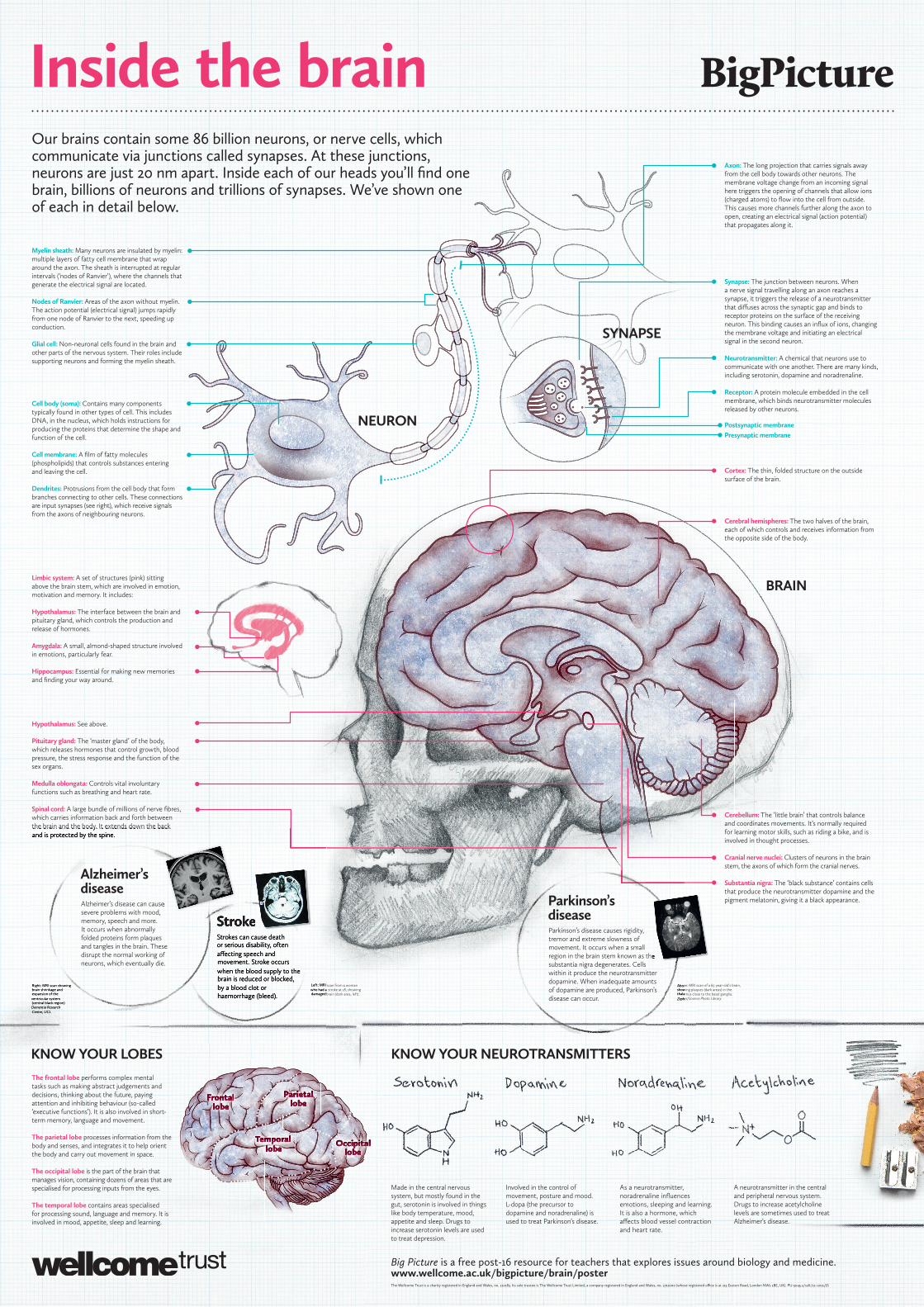
The frontal lobe performs complex mental tasks such as making abstract judgements and decisions, thinking about the future, paying attention and inhibiting behaviour (so-called ‘executive functions’). It is also involved in short-term memory, language and movement.
The parietal lobe processes information from the body and senses, and integrates it to help orient the body and carry out movement in space.
The occipital lobe is the part of the brain that manages vision, containing dozens of areas that are specialised for processing inputs from the eyes.
The temporal lobe contains areas specialised for processing sound, language and memory. It is involved in mood, appetite, sleep and learning.
FrontalFrontalFrontallobelobelobe
ParietalParietalParietallobelobelobe
OccipitalOccipitalOccipitallobelobelobe
TemporalTemporalTemporallobelobelobe
Made in the central nervous system, but mostly found in the gut, serotonin is involved in things like body temperature, mood, appetite and sleep. Drugs to increase serotonin levels are used to treat depression.
Involved in the control of movement, posture and mood. L-dopa (the precursor to dopamine and noradrenaline) is used to treat Parkinson’s disease.
As a neurotransmitter, noradrenaline infl uences emotions, sleeping and learning. It is also a hormone, which aff ects blood vessel contraction and heart rate.
A neurotransmitter in the central and peripheral nervous system. Drugs to increase acetylcholine levels are sometimes used to treat Alzheimer’s disease.
Right: MRI scan showing brain shrinkage and expansion of the ventricular system (central black region).Dementia Research Centre, UCL
Left: MRI scan from a woman who had a stroke at 18, showing damaged brain (dark area, left).
Above: MRI scan of a 65-year-old’s brain, showing plaques (dark areas) in the thalamus close to the basal ganglia.Zephyr/Science Photo Library
Strokes can cause death or serious disability, often aff ecting speech and movement. Stroke occurs when the blood supply to the brain is reduced or blocked, by a blood clot or haemorrhage (bleed).
Postsynaptic membranePresynaptic membrane
Myelin sheath: Many neurons are insulated by myelin: multiple layers of fatty cell membrane that wrap around the axon. The sheath is interrupted at regular intervals (‘nodes of Ranvier’), where the channels that generate the electrical signal are located.
Nodes of Ranvier: Areas of the axon without myelin. The action potential (electrical signal) jumps rapidly from one node of Ranvier to the next, speeding up conduction.
Glial cell: Non-neuronal cells found in the brain and other parts of the nervous system. Their roles include supporting neurons and forming the myelin sheath.
Cell body (soma): Contains many components typically found in other types of cell. This includes DNA, in the nucleus, which holds instructions for producing the proteins that determine the shape and function of the cell.
Cell membrane: A fi lm of fatty molecules (phospholipids) that controls substances entering and leaving the cell.
Dendrites: Protrusions from the cell body that form branches connecting to other cells. These connections are input synapses (see right), which receive signals from the axons of neighbouring neurons.
Hypothalamus: See above.
Pituitary gland: The ‘master gland’ of the body, which releases hormones that control growth, blood pressure, the stress response and the function of the sex organs.
Medulla oblongata: Controls vital involuntary functions such as breathing and heart rate.
Spinal cord: A large bundle of millions of nerve fi bres, which carries information back and forth between the brain and the body. It extends down the back and is protected by the spine.
Limbic system: A set of structures (pink) sitting above the brain stem, which are involved in emotion, motivation and memory. It includes:
Hypothalamus: The interface between the brain and pituitary gland, which controls the production and release of hormones.
Amygdala: A small, almond-shaped structure involved in emotions, particularly fear.
Hippocampus: Essential for making new memories and fi nding your way around.
Stroke
Right: MRI scan showing brain shrinkage and expansion of the ventricular system (central black region).Dementia Research Centre, UCL
Left: MRI scan from a woman who had a stroke at 18, showing damaged brain (dark area, left).
Strokes can cause death or serious disability, often aff ecting speech and movement. Stroke occurs when the blood supply to the brain is reduced or blocked, by a blood clot or haemorrhage (bleed).
the brain and the body. It extends down the back and is protected by the spine.
Above: MRI scan of a 65-year-old’s brain, showing plaques (dark areas) in the thalamus close to the basal ganglia.Zephyr/Science Photo Library
Parkinson’sdiseaseParkinson’s disease causes rigidity, tremor and extreme slowness of movement. It occurs when a small region in the brain stem known as the substantia nigra degenerates. Cells within it produce the neurotransmitter dopamine. When inadequate amounts of dopamine are produced, Parkinson’s disease can occur.
Alzheimer’sdiseaseAlzheimer’s disease can cause severe problems with mood, memory, speech and more. It occurs when abnormally folded proteins form plaques and tangles in the brain. These disrupt the normal working of neurons, which eventually die.
Stroke
region in the brain stem known as the
Alzheimer’s disease can cause