Embed Size (px)
Citation preview
Inorganic mercury in human astrocytes, oligodendrocytes,corticomotoneurons and the locus ceruleus: implicationsfor multiple sclerosis, neurodegenerative disordersand gliomas
Roger Pamphlett . Stephen Kum Jew
Received: 8 May 2018 / Accepted: 21 June 2018 / Published online: 29 June 2018
� The Author(s) 2018
Abstract Neurotoxic metals have been implicated in
the pathogenesis of multiple sclerosis, neurodegener-
ative disorders and brain tumours but studies of the
location of heavy metals in human brains are rare. In a
man who injected himself with metallic mercury the
cellular location of mercury in his brain was studied
after 5 months of continuous exposure to inorganic
mercury arising from metallic mercury deposits in his
organs. Paraffin sections from the primary motor and
sensory cortices and the locus ceruleus in the pons
were stained with autometallography to detect inor-
ganic mercury and combined with glial fibrillary
acidic protein immunohistochemistry to identify
astrocytes. Inorganic mercury was found in grey
matter subpial, interlaminar, protoplasmic and vari-
cose astrocytes, white matter fibrous astrocytes, grey
but not white matter oligodendrocytes, corticomo-
toneurons and some locus ceruleus neurons. In sum-
mary, inorganic mercury is taken up by five types of
human brain astrocytes, as well as by cortical oligo-
dendrocytes, corticomotoneurons and locus ceruleus
neurons. Mercury can induce oxidative stress, stimu-
late autoimmunity and damage DNA, mitochondria
and lipid membranes, so its location in these CNS cells
suggests it could play a role in the pathogenesis of
multiple sclerosis, neurodegenerative conditions such
as Alzheimer’s disease and amyotrophic lateral scle-
rosis, and glial tumours.
Keywords Human brain � Inorganic mercury �Astrocyte � Oligodendrocyte � Corticomotoneuron �Locus ceruleus � Multiple sclerosis � Alzheimer’s
disease � Amyotrophic lateral sclerosis (ALS) � Braintumour
Introduction
Neurotoxic metals have been implicated in the patho-
genesis of a number of human nervous system
disorders (Caito and Aschner 2015). Clinical, exper-
imental and epidemiological studies suggest that
mercury could play a part in the pathogenesis of
multiple sclerosis (Aminzadeh and Etminan 2007),
Alzheimer’s disease (Mutter et al. 2010) and amy-
otrophic lateral sclerosis (ALS) (Pamphlett and Kum
Jew 2013). A number of pathogenetic mechanisms
have been linked to mercury, all of which are
suspected to operate in neurodegenerative diseases.
These include the production of reactive oxygen
R. Pamphlett � S. Kum Jew
Discipline of Pathology, The University of Sydney,
Camperdown, Australia
R. Pamphlett (&)
Discipline of Pathology, Brain and Mind Centre, The
University of Sydney and Department of Neuropathology,
Royal Prince Alfred Hospital, 94 Mallett St,
Camperdown, NSW 2050, Australia
e-mail: [email protected]
123
Biometals (2018) 31:807–819
https://doi.org/10.1007/s10534-018-0124-4(0123456789().,-volV)(0123456789().,-volV)
species (Lund et al. 1993), apoptosis (Ceccatelli et al.
2010), DNA damage (Crespo-Lopez et al. 2009),
RNA damage (Chang 1977), epigenetic changes (-
Basu et al. 2014) and autoimmunity (Vas and Mon-
estier 2008). However, no convincing link between
mercury and neurological disorders has been estab-
lished. One problem in detecting neurotoxic metals in
diseased brains is that the cells that originally
contained the metals are likely to have been destroyed
by the pathological process by the time the brain is
available for examination after autopsy. Furthermore,
most studies of mercury in the human nervous system
have relied on studying the brains of people years after
exposure by which time much of the metal is likely to
have been cleared from the brain (Tiffany-Castiglion
and Qian 2001).
Inorganic mercury (iHg) appears to be the proxi-
mate toxic form of CNS mercury (Charleston et al.
1996) but the location of iHg in the human brain is
poorly understood (Clarkson and Magos 2006). We
were able to study the cellular location of iHg in a man
who had been exposed continuously to mercury for
5 months. Using the histochemical technique of silver
nitrate autometallography, iHg had previously been
detected in his corticomotoneurons and locus ceruleus
neurons, as well as in undefined glial cells in the
brain (Pamphlett and Waley 1996). Autometallogra-
phy can now be combined with immunohistochem-
istry to detect which classes of cells contain
iHg (Pamphlett and Kum Jew 2015) so this combina-
tion of methods was used in an attempt to detect more
precisely which cells contain iHg in an undamaged
human brain that had been exposed to mercury at the
time of death.
Methods
Clinical details
A 24 year-old man had injected himself intravenously
with metallic mercury taken from thermome-
ters (Kedziora and Duflou 1995). X-rays showed
collections of mercury in his right ventricle, through-
out both lung fields and in the pelvic venous plexuses.
He remained asymptomatic but died 5 months later
after lacerating his wrists. At autopsymetallic mercury
collections were seen on the cut surfaces of his right
ventricular myocardium, lungs and pelvic veins.
Staining inorganic mercury and astrocytes
7 lm sections of formalin-fixed paraffin-embedded
blocks taken from the cerebral cortex, which included
the primary sensory and motor cortices and underlying
white matter, as well as sections from the pons
containing the locus ceruleus, were stained with silver
nitrate autometallography to detect iHg (Danscher and
Stoltenberg 2006). Briefly, sections were placed in
physical developer containing gum arabic, citrate
buffer, hydroquinone and silver nitrate at 26 �C for
85 min in the dark, then washed in sodium thiosul-
phate to remove unbound silver, lightly counterstained
with hematoxylin and viewed under bright-field
microscopy. A positive control section was of mouse
spinal cord where spinal motor neurons contained iHg
following an intraperitoneal injection of mercuric
chloride (Pamphlett and Png 1998). The silver-coated
deposits of iHg in cells are seen microscopically as
black grains and referred to as AMGHg (autometal-
lography-demonstrable mercury). To identify iHg in
astrocytes AMGHg-stained sections were immunos-
tained with polyclonal rabbit-anti-human glial fibril-
lary acidic protein (GFAP, DAKO Z0334) at 1:2000
for 60 min at 37 �C and visualised with diaminoben-
zidine tetrahydrochloride. Oligodendrocytes were
identified by their characteristically cleared cytoplasm
and contrast-enhanced nuclei. To assess general
pathology sections were stained with hematoxylin
and eosin.
Ethics
The project was carried out in accordance with the
ethical standards of the Human Ethics Review Com-
mittee of the Sydney Local Health District (Royal
Prince Alfred Hospital Zone project X14-0029) with
approval from the Coroner’s Office, Department of
Forensic Medicine, Glebe, New South Wales and in
accordance with the Declaration of Helsinki as revised
in 2000. The institutional review board waived the
need for written informed consent from relatives of the
participant since this was a de-identified retrospective
study of autopsy-obtained tissue.
123
808 Biometals (2018) 31:807–819
Results
General histology
No abnormities were seen on hematoxylin and eosin
sections of the cerebral cortex or white matter. In
particular, there was no evidence of neuronal or
oligodendrocyte cell loss, astrocytic hypertrophy (i.e.,
no visible eccentric perinuclear eosinophilic astrocytic
cytoplasm) or destructive tissue damage leading to
microglial activation (i.e., no foamy macrophages).
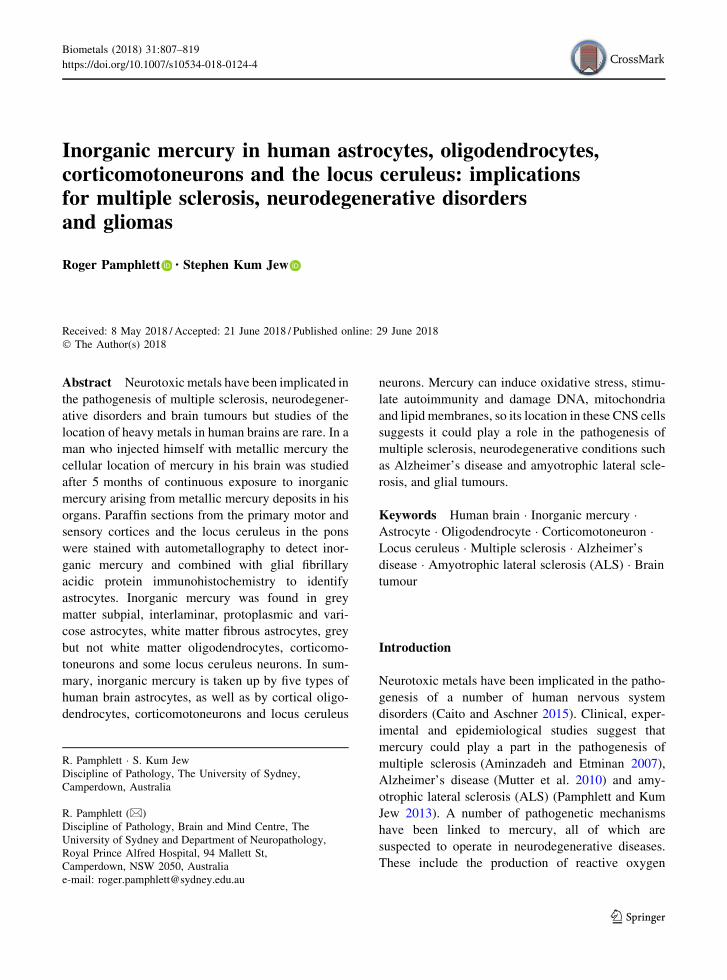
Astrocyte architecture
GFAP immunostaining allowed visualisation of all
astrocyte subtypes in the cortex and white matter
(Fig. 1). The cell bodies of subpial and interlaminar
astrocytes were in cortical layers I, protoplasmic
astrocytes in layers III to VI, varicose astrocytes
mostly in layer VI and fibrous astrocytes in the white
matter. The long undulating processes of interlaminar
astrocytes and the long beaded processes of varicose
astrocytes were noted.
Cortical grey matter
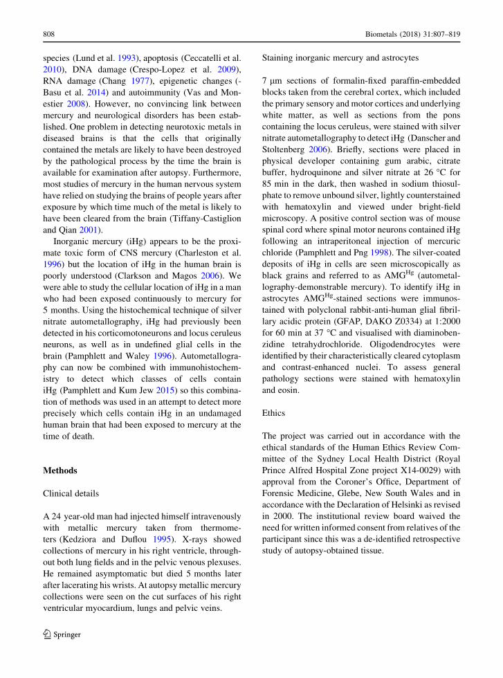
Subpial astrocytes
iHg was prominent in the glia limitans and the
immediate subpial regions of cortical layer I where
the cell bodies and processes of subpial astrocytes
contained dense iHg (Fig. 2).
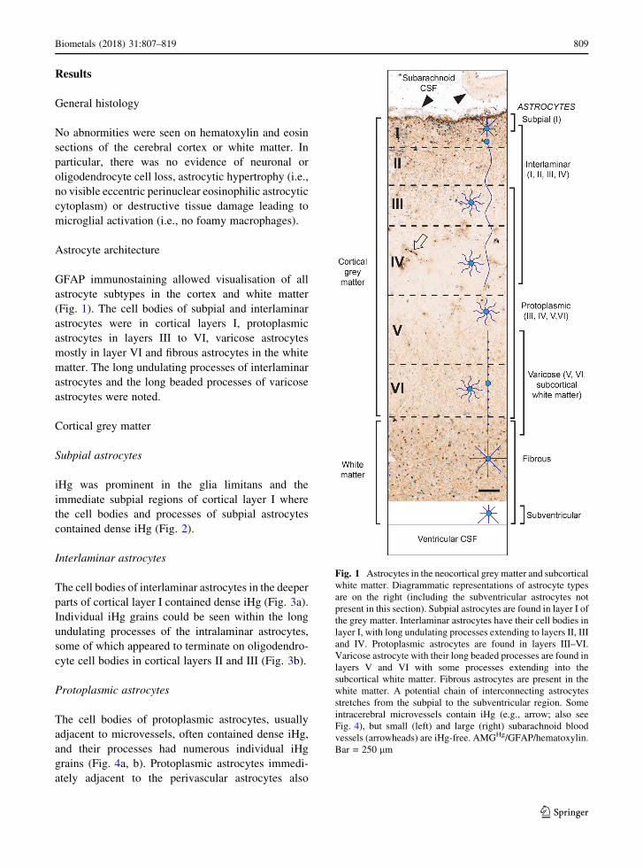
Interlaminar astrocytes
The cell bodies of interlaminar astrocytes in the deeper
parts of cortical layer I contained dense iHg (Fig. 3a).
Individual iHg grains could be seen within the long
undulating processes of the intralaminar astrocytes,
some of which appeared to terminate on oligodendro-
cyte cell bodies in cortical layers II and III (Fig. 3b).
Protoplasmic astrocytes
The cell bodies of protoplasmic astrocytes, usually
adjacent to microvessels, often contained dense iHg,
and their processes had numerous individual iHg
grains (Fig. 4a, b). Protoplasmic astrocytes immedi-
ately adjacent to the perivascular astrocytes also
Fig. 1 Astrocytes in the neocortical grey matter and subcortical
white matter. Diagrammatic representations of astrocyte types
are on the right (including the subventricular astrocytes not
present in this section). Subpial astrocytes are found in layer I of
the grey matter. Interlaminar astrocytes have their cell bodies in
layer I, with long undulating processes extending to layers II, III
and IV. Protoplasmic astrocytes are found in layers III–VI.
Varicose astrocyte with their long beaded processes are found in
layers V and VI with some processes extending into the
subcortical white matter. Fibrous astrocytes are present in the
white matter. A potential chain of interconnecting astrocytes
stretches from the subpial to the subventricular region. Some
intracerebral microvessels contain iHg (e.g., arrow; also see
Fig. 4), but small (left) and large (right) subarachnoid blood
vessels (arrowheads) are iHg-free. AMGHg/GFAP/hematoxylin.
Bar = 250 lm
123
Biometals (2018) 31:807–819 809
contained iHg. Cortical layers III and IV had the
greatest concentration of iHg-containing protoplasmic
astrocytes.
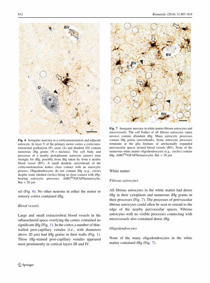
Varicose astrocytes
The cell bodies of varicose astrocytes in cortical layers
V and VI contained large amounts of iHg (Fig. 5). The
varicosities on the processes of these astrocytes
appeared to have a predilection for iHg, with only a
few grains in the processes between the varicosities.
Varicose astrocytic cell processes were often seen in
close proximity to oligodendrocytes.
Fig. 2 Inorganic mercury in grey matter subpial astrocytes.
a The subpial glia limitans stains strongly for iHg (filled arrow).
Cell bodies of subpial astrocytes in layer I contain iHg (e.g.,
open arrows). Small iHg grains can be seen in astrocytic
processes (arrowheads) belonging to either subpial or interlam-
inar astrocytes. The wall of a penetrating blood vessel (BV)
contains iHg. The pia mater (asterisk, artefactually separated
from the underlying cortex) contains no significant iHg.
AMGHg/GFAP/hematoxylin. b A control section without
AMGHg staining from the same region shows no black deposits
within the brown subpial astrocyte cell bodies or processes.
GFAP/hematoxylin. Bars = 20 lm
Fig. 3 Inorganic mercury in grey matter interlaminar astro-
cytes and oligodendrocytes. a The cell bodies of interlaminar
astrocytes in layer I (open arrows) stain strongly for iHg. iHg
grains (arrowheads) are seen in their long undulating processes.
A group of oligodendrocytes have iHg in their cytoplasm (e.g.,
dashed circles) whereas others are iHg-free (e.g., circles).
Bar = 20 lm. b A high power view shows the cell body of one
oligodendrocyte with a mostly empty white perikaryon. iHg has
been concentrated into its artefactually shrunken cytoplasm
(arrowhead) to the right of a pale-staining nucleus. The
descending process of an interlaminar astrocyte appears to
terminate in an end bulb (open arrow) on the surface of the
oligodendrocyte. AMGHg/GFAP/hematoxylin. Bar = 10 lm
123
810 Biometals (2018) 31:807–819
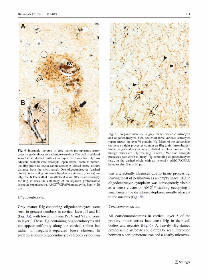
Oligodendrocytes
Grey matter iHg-containing oligodendrocytes were
seen in greatest numbers in cortical layers II and III
(Fig. 3a), with fewer in layers IV, V and VI and none
in layer I. These iHg-containing oligodendrocytes did
not appear uniformly along the cortical ribbon but
rather in irregularly-separated loose clusters. In
paraffin sections oligodendrocyte cell body cytoplasm
was artefactually shrunken due to tissue processing,
leaving most of perikaryon as an empty space. iHg in
oligodendrocyte cytoplasm was consequently visible
as a dense cluster of AMGHg staining occupying a
small area of the shrunken cytoplasm, usually adjacent
to the nucleus (Fig. 3b).
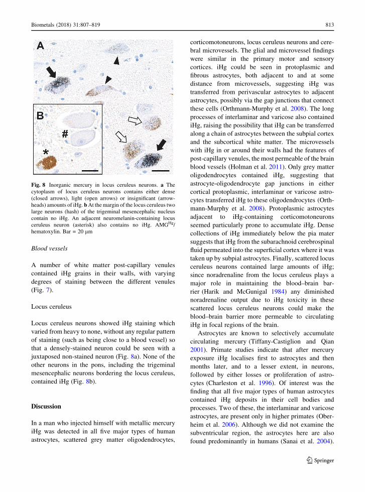
Corticomotoneurons
All corticomotoneurons in cortical layer 5 of the
primary motor cortex had dense iHg in their cell
bodies and neurites (Fig. 6). A heavily iHg-stained
protoplasmic astrocyte could often be seen interposed
between a corticomotoneuron and a nearby microves-
Fig. 4 Inorganic mercury in grey matter protoplasmic astro-
cytes, oligodendrocytes and microvessels. a The wall of a bloodvessel (BV, dashed outline) in layer III stains for iHg. An
adjacent protoplasmic astrocyte (open arrow) contains numer-
ous iHg grains as does a second astrocyte (closed arrow) a short
distance from the microvessel. One oligodendrocyte (dashed
circle) contains iHg but most oligodendrocytes (e.g., circles) are
iHg-free. b The wall of a small blood vessel (BV) stains strongly
for iHg as does the cell body of an adjacent protoplasmic
astrocyte (open arrow). AMGHg/GFAP/hematoxylin. Bars = 20
lm
Fig. 5 Inorganic mercury in grey matter varicose astrocytes
and oligodendrocytes. Cell bodies of three varicose astrocytes
(open arrows) in layer VI contain iHg. Many of the varicosities
on these straight processes contain an iHg grain (arrowheads).
Some oligodendrocytes (e.g., dashed circles) contain iHg
though others are iHg-free (e.g., circles). Varicose astrocyte
processes pass close to many iHg-containing oligodendrocytes
(e.g., in the dashed circle with an asterisk). AMGHg/GFAP/
hematoxylin. Bar = 20 lm
123
Biometals (2018) 31:807–819 811
sel (Fig. 6). No other neurons in either the motor or
sensory cortex contained iHg.
Blood vessels
Large and small extracerebral blood vessels in the
subarachnoid space overlying the cortex contained no
significant iHg (Fig. 1). In the cortex a number of thin-
walled post-capillary venules (i.e., with diameters
above 20 lm) had iHg grains in their walls (Fig. 1).
These iHg-stained post-capillary venules appeared
most prominently in cortical layers III and IV.
White matter
Fibrous astrocytes
All fibrous astrocytes in the white matter had dense
iHg in their cytoplasm and numerous iHg grains in
their processes (Fig. 7). The processes of perivascular
fibrous astrocytes could often be seen to extend to the
edge of the nearby perivascular spaces. Fibrous
astrocytes with no visible processes connecting with
microvessels also contained dense iHg.
Oligodendrocytes
None of the many oligodendrocytes in the white
matter contained iHg (Fig. 7).
Fig. 6 Inorganic mercury in a corticomotoneuron and adjacent
astrocyte. In layer V of the primary motor cortex a corticomo-
toneuronal perikaryon (P), axon (A) and dendrite (D) contain
numerous iHg grains (N = nucleus). The cell body and
processes of a nearby protoplasmic astrocyte (arrow) stain
strongly for iHg, possibly from iHg taken by from a nearby
blood vessel (BV). A small dendrite (arrowhead) of the
corticomotoneuron makes close contact with an astrocytic
process. Oligodendrocytes do not contain iHg (e.g., circle)
despite some (dashed circles) being in close contact with iHg-
bearing astrocytic processes. AMGHg/GFAP/hematoxylin.
Bar = 20 lm
Fig. 7 Inorganic mercury in white matter fibrous astrocytes and
microvessels. The cell bodies of all fibrous astrocytes (open
arrows) contain abundant iHg. Many astrocytic processes
contain iHg grains (arrowheads). Some astrocytic processes
terminate at the glia limitans of artefactually expanded
perivascular spaces around blood vessels (BV). None of the
numerous white matter oligodendrocytes (e.g., circles) contain
iHg. AMGHg/GFAP/hematoxylin. Bar = 20 lm
123
812 Biometals (2018) 31:807–819
Blood vessels
A number of white matter post-capillary venules
contained iHg grains in their walls, with varying
degrees of staining between the different venules
(Fig. 7).
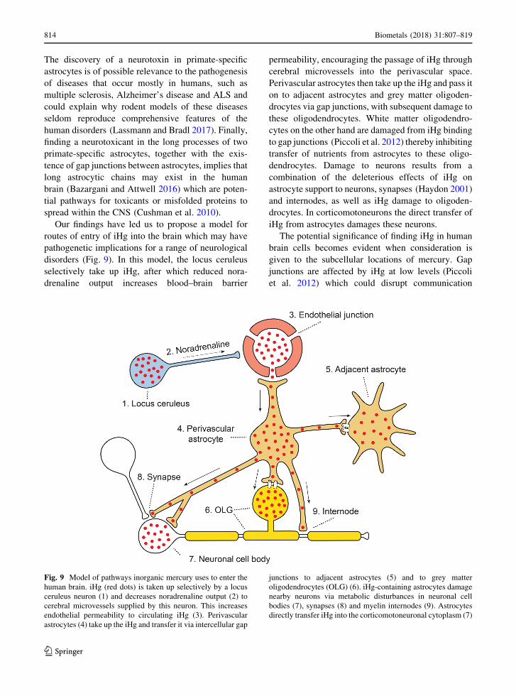
Locus ceruleus
Locus ceruleus neurons showed iHg staining which
varied from heavy to none, without any regular pattern
of staining (such as being close to a blood vessel) so
that a densely-stained neuron could be seen with a
juxtaposed non-stained neuron (Fig. 8a). None of the
other neurons in the pons, including the trigeminal
mesencephalic neurons bordering the locus ceruleus,
contained iHg (Fig. 8b).
Discussion
In a man who injected himself with metallic mercury
iHg was detected in all five major types of human
astrocytes, scattered grey matter oligodendrocytes,
corticomotoneurons, locus ceruleus neurons and cere-
bral microvessels. The glial and microvessel findings
were similar in the primary motor and sensory
cortices. iHg could be seen in protoplasmic and
fibrous astrocytes, both adjacent to and at some
distance from microvessels, suggesting iHg was
transferred from perivascular astrocytes to adjacent
astrocytes, possibly via the gap junctions that connect
these cells (Orthmann-Murphy et al. 2008). The long
processes of interlaminar and varicose also contained
iHg, raising the possibility that iHg can be transferred
along a chain of astrocytes between the subpial cortex
and the subcortical white matter. The microvessels
with iHg in or around their walls had the features of
post-capillary venules, the most permeable of the brain
blood vessels (Holman et al. 2011). Only grey matter
oligodendrocytes contained iHg, suggesting that
astrocyte-oligodendrocyte gap junctions in either
cortical protoplasmic, interlaminar or varicose astro-
cytes transferred iHg to these oligodendrocytes (Orth-
mann-Murphy et al. 2008). Protoplasmic astrocytes
adjacent to iHg-containing corticomotoneurons
seemed particularly prone to accumulate iHg. Dense
collections of iHg immediately below the pia mater
suggests that iHg from the subarachnoid cerebrospinal
fluid permeated into the superficial cortex where it was
taken up by subpial astrocytes. Finally, scattered locus
ceruleus neurons contained large amounts of iHg;
since noradrenaline from the locus ceruleus plays a
major role in maintaining the blood–brain bar-
rier (Harik and McGunigal 1984) any diminished
noradrenaline output due to iHg toxicity in these
scattered locus ceruleus neurons could make the
blood–brain barrier more permeable to circulating
iHg in focal regions of the brain.
Astrocytes are known to selectively accumulate
circulating mercury (Tiffany-Castiglion and Qian
2001). Primate studies indicate that after mercury
exposure iHg localises first to astrocytes and then
months later, and to a lesser extent, in neurons,
followed by either losses or proliferation of astro-
cytes (Charleston et al. 1996). Of interest was the
finding that all five major types of human astrocytes
contained iHg deposits in their cell bodies and
processes. Two of these, the interlaminar and varicose
astrocytes, are present only in higher primates (Ober-
heim et al. 2006). Although we did not examine the
subventricular region, the astrocytes here are also
found predominantly in humans (Sanai et al. 2004).
Fig. 8 Inorganic mercury in locus ceruleus neurons. a The
cytoplasm of locus ceruleus neurons contains either dense
(closed arrows), light (open arrows) or insignificant (arrow-
heads) amounts of iHg. bAt the margin of the locus ceruleus two
large neurons (hash) of the trigeminal mesencephalic nucleus
contain no iHg. An adjacent neuromelanin-containing locus
ceruleus neuron (asterisk) also contains no iHg. AMGHg/
hematoxylin. Bar = 20 lm
123
Biometals (2018) 31:807–819 813
The discovery of a neurotoxin in primate-specific
astrocytes is of possible relevance to the pathogenesis
of diseases that occur mostly in humans, such as
multiple sclerosis, Alzheimer’s disease and ALS and
could explain why rodent models of these diseases
seldom reproduce comprehensive features of the
human disorders (Lassmann and Bradl 2017). Finally,
finding a neurotoxicant in the long processes of two
primate-specific astrocytes, together with the exis-
tence of gap junctions between astrocytes, implies that
long astrocytic chains may exist in the human
brain (Bazargani and Attwell 2016) which are poten-
tial pathways for toxicants or misfolded proteins to
spread within the CNS (Cushman et al. 2010).
Our findings have led us to propose a model for
routes of entry of iHg into the brain which may have
pathogenetic implications for a range of neurological
disorders (Fig. 9). In this model, the locus ceruleus
selectively take up iHg, after which reduced nora-
drenaline output increases blood–brain barrier
permeability, encouraging the passage of iHg through
cerebral microvessels into the perivascular space.
Perivascular astrocytes then take up the iHg and pass it
on to adjacent astrocytes and grey matter oligoden-
drocytes via gap junctions, with subsequent damage to
these oligodendrocytes. White matter oligodendro-
cytes on the other hand are damaged from iHg binding
to gap junctions (Piccoli et al. 2012) thereby inhibiting
transfer of nutrients from astrocytes to these oligo-
dendrocytes. Damage to neurons results from a
combination of the deleterious effects of iHg on
astrocyte support to neurons, synapses (Haydon 2001)
and internodes, as well as iHg damage to oligoden-
drocytes. In corticomotoneurons the direct transfer of
iHg from astrocytes damages these neurons.
The potential significance of finding iHg in human
brain cells becomes evident when consideration is
given to the subcellular locations of mercury. Gap
junctions are affected by iHg at low levels (Piccoli
et al. 2012) which could disrupt communication
Fig. 9 Model of pathways inorganic mercury uses to enter the
human brain. iHg (red dots) is taken up selectively by a locus
ceruleus neuron (1) and decreases noradrenaline output (2) to
cerebral microvessels supplied by this neuron. This increases
endothelial permeability to circulating iHg (3). Perivascular
astrocytes (4) take up the iHg and transfer it via intercellular gap
junctions to adjacent astrocytes (5) and to grey matter
oligodendrocytes (OLG) (6). iHg-containing astrocytes damage
nearby neurons via metabolic disturbances in neuronal cell
bodies (7), synapses (8) and myelin internodes (9). Astrocytes
directly transfer iHg into the corticomotoneuronal cytoplasm (7)
123
814 Biometals (2018) 31:807–819
between astrocytes and oligodendrocytes. Mitochon-
dria are implicated in a number of neurodegenerative
diseases and iHg localises to mitochondria (Chang
and Hartmann 1972). Mercury has a particular affinity
for the sulfhydryl groups of cysteine-containing
proteins (Clarkson and Magos 2006) found in lipid
membranes of the Golgi apparatus, the endoplasmic
reticulum and lysosomes, all of which have been
linked to neurodegenerative diseases (Farina and
Aschner 2017). Furthermore, ultrastructural studies
have shown iHg binds to all these lipid-rich sites in the
nervous system (Chang and Hartmann 1972).
In multiple sclerosis a primary role for astrocytes
has been postulated (Brosnan and Raine 2013) and
astrocytes proliferate markedly in otherwise normal
multiple sclerosis white matter (Allen 1981). The
demyelination in neuromyelitis optica, a disorder
related to multiple sclerosis, results from an autoim-
mune attack on aquaporin-4 in astrocytic foot pro-
cesses (Lucchinetti et al. 2014); this may be relevant
to other demyelinating conditions that could be
triggered by mercury, since aquaporins are inhibited
by mercury (Ximenes-da-Silva 2016). iHg-induced
damage to astrocytes could augment the neuroinflam-
mation seen in multiple sclerosis since astrocytes are
key regulators of brain immune responses (Colombo
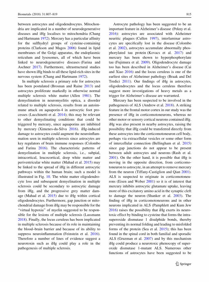
and Farina 2016). The characteristic patterns of
demyelination in multiple sclerosis, i.e., subpial,
intracortical, leucocortical, deep white matter and
periventricular white matter (Mahad et al. 2015) may
be linked to the spread of iHg in different astrocytic
pathways within the human brain; such a model is
illustrated in Fig. 10. The white matter oligodendro-
cyte loss and subsequent demyelination in multiple
sclerosis could be secondary to astrocytic damage
from iHg, and the progressive grey matter dam-
age (Mahad et al. 2015) due to iHg within cortical
oligodendrocytes. Furthermore, gap junction or mito-
chondrial damage from iHg may be responsible for the
‘‘virtual hypoxia’’ of myelin suggested to be respon-
sible for the lesions of multiple sclerosis (Lassmann
2018). Finally, the locus ceruleus has been implicated
in multiple sclerosis because of its role in maintaining
the blood–brain barrier and because of its ability to
suppress neuroinflammation (Feinstein et al. 2016).
Therefore a number of lines of evidence suggest a
neurotoxin such as iHg could play a role in the
pathogenesis of multiple sclerosis.
Astrocyte pathology has been suggested to be an
important feature in Alzheimer’s disease (Pekny et al.
2016): astrocytes are associated with Alzheimer
neuritic plaques (Cullen 1997), interlaminar astro-
cytes are specifically lost in the disease (Colombo
et al. 2002), astrocytes accumulate abnormally phos-
phorylated tau protein (Kovacs et al. 2017) and
mercury has been shown to hyperphosphorylate
tau (Fujimura et al. 2009). Oligodendrocyte damage
too has been described in Alzheimer’s disease (Cai
and Xiao 2016) and the locus ceruleus is one of the
earliest sites of Alzheimer pathology (Braak and Del
Tredici 2011). Our findings of iHg in astrocytes,
oligodendrocytes and the locus ceruleus therefore
suggest more investigations of heavy metals as a
trigger for Alzheimer’s disease are warranted.
Mercury has been suspected to be involved in the
pathogenesis of ALS (Andrew et al. 2018). A striking
feature in the frontal motor cortex in our study was the
presence of iHg in corticomotoneurons, whereas no
other motor or sensory cortical neurons contained iHg.
iHg was also present in nearby astrocytes, raising the
possibility that iHg could be transferred directly from
these astrocytes into the corticomotoneuron cell body,
perhaps via extracellular vesicles or some other form
of intercellular connection (Bellingham et al. 2015)
since gap junctions do not appear to be present
between adult astrocytes and neurons (Rash et al.
2001). On the other hand, it is possible that iHg is
moving in the opposite direction, from corticomo-
toneuron to astrocyte, in an attempt to remove mercury
from the neuron (Tiffany-Castiglion and Qian 2001).
ALS is suspected to originate in corticomotoneu-
rons (Eisen and Weber 2001) so it is of interest that
mercury inhibits astrocytic glutamate uptake, leaving
more of this excitatory amino acid in the synaptic cleft
to damage the neuron (Shanker et al. 2003). The
finding of iHg in corticomotoneurons and in other
neurons implicated in ALS (Pamphlett and Kum Jew
2016) raises the possibility that iHg exerts its neuro-
toxic effect by binding to cysteine that forms the intra-
superoxide dismutase 1 disulphide bonds, thereby
preventing its normal folding and leading to misfolded
forms of the protein (Sea et al. 2015); this has been
found in the spinal cord in both familial and sporadic
ALS (Gruzman et al. 2007) and by this mechanism
iHg could produce a neurotoxic phenocopy of super-
oxide dismutase 1-mutant ALS. Numerous other
functions of astrocytes have been suggested to be
123
Biometals (2018) 31:807–819 815
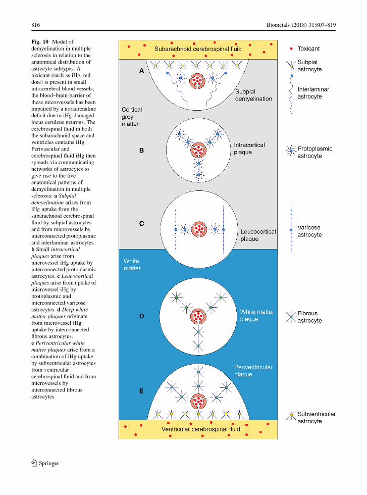
Fig. 10 Model of
demyelination in multiple
sclerosis in relation to the
anatomical distribution of
astrocyte subtypes. A
toxicant (such as iHg, red
dots) is present in small
intracerebral blood vessels;
the blood–brain-barrier of
these microvessels has been
impaired by a noradrenaline
deficit due to iHg-damaged
locus ceruleus neurons. The
cerebrospinal fluid in both
the subarachnoid space and
ventricles contains iHg.
Perivascular and
cerebrospinal fluid iHg then
spreads via communicating
networks of astrocytes to
give rise to the five
anatomical patterns of
demyelination in multiple
sclerosis: a Subpial
demyelination arises from
iHg uptake from the
subarachnoid cerebrospinal
fluid by subpial astrocytes
and from microvessels by
interconnected protoplasmic
and interlaminar astrocytes.
b Small intracortical
plaques arise from
microvessel iHg uptake by
interconnected protoplasmic
astrocytes. c Leucocorticalplaques arise from uptake of
microvessel iHg by
protoplasmic and
interconnected varicose
astrocytes. d Deep white
matter plaques originate
from microvessel iHg
uptake by interconnected
fibrous astrocytes.
e Periventricular whitematter plaques arise from a
combination of iHg uptake
by subventricular astrocytes
from ventricular
cerebrospinal fluid and from
microvessels by
interconnected fibrous
astrocytes
123
816 Biometals (2018) 31:807–819
disturbed in ALS (Yamanaka and Komine 2018) and
motor neuron death can be triggered by astrocytes (Re
et al. 2014). Oligodendrocyte too appear to play a role
in ALS (Philips et al. 2013). Therefore the presence of
iHg in astrocytes, oligodendrocytes and corticomo-
toneurons may have relevance to the pathogenesis of
ALS.
Brain cells appear to be particularly sensitive to the
genotoxic effects of mercury (Crespo-Lopez et al.
2009) and mercury can cause epigenetic modifica-
tions (Ray et al. 2014) which together with DNA
mutations may be responsible for the development of
gliomas (Caffo et al. 2014). The possibility that
mercury could be a trigger for glioblastomas, the most
malignant form of astrocytoma, was initially raised
because of a report that dentists and dental nurses had
an increased risk for these tumours (Ahlbom et al.
1986), though these results were not repeated in other
studies (Boffetta et al. 1993). Oligodendrogliomas
have been reported to be common in the cortical-
subcortical region of the brain (Smits 2016), a region
in our study where cortical oligodendrocytes often
contained iHg. So it is of interest that these two
common primary brain tumours, glioblastoma and
oligodendroglioma, arise from astrocytes and oligo-
dendrocytes, which contain large amounts of poten-
tially-mutagenic iHg after human exposure to
mercury.
In conclusion, in a man who injected himself with
metallic mercury we found inorganic mercury in cells
that are involved in the pathogenesis of multiple
sclerosis, Alzheimer’s disease, ALS and brain
tumours. Many of the pathophysiological mechanisms
thought to underlie these disorders are mirrored by the
multiple deleterious effects of intracellular mercury.
Elemental analysis studies of mercury and other
neurotoxicants within the cells of the human brain
are warranted to further investigate the contributions
of neurotoxins to this range of disorders.
Acknowledgements Supported by the Aimee Stacey
Memorial and Ignacy Burnett Bequests.
Open Access This article is distributed under the terms of the
Creative Commons Attribution 4.0 International License (http://
creativecommons.org/licenses/by/4.0/), which permits unre-
stricted use, distribution, and reproduction in any medium,
provided you give appropriate credit to the original
author(s) and the source, provide a link to the Creative Com-
mons license, and indicate if changes were made.
References
Ahlbom A, Norell S, Rodvall Y, Nylander M (1986) Dentists,
dental nurses, and brain tumours. Br Med J 292:662
Allen IV (1981) The pathology of multiple sclerosis–fact, fiction
and hypothesis. Neuropathol Appl Neurobiol 7:169–182
Aminzadeh KK, Etminan M (2007) Dental amalgam and mul-
tiple sclerosis: a systematic review and meta-analysis.
J Public Health Dent 67:64–66
Andrew AS, Chen CY, Caller TA, Tandan R, Henegan PL,
Jackson BP, Hall BP, Bradley WG, Stommel EW (2018)
Toenail mercury levels are associated with amyotrophic
lateral sclerosis risk. Muscle Nerve. https://doi.org/10.
1002/mus.26055
Basu N, Goodrich JM, Head J (2014) Ecogenetics of mercury:
from genetic polymorphisms and epigenetics to risk
assessment and decision-making. Environ Toxicol Chem
33:1248–1258
Bazargani N, Attwell D (2016) Astrocyte calcium signaling: the
third wave. Nat Neurosci 19:182–189
Bellingham SA, Guo B, Hill AF (2015) The secret life of
extracellular vesicles in metal homeostasis and neurode-
generation. Biol Cell 107:389–418
Boffetta P, Merler E, Vainio H (1993) Carcinogenicity of
mercury and mercury compounds. Scand J Work Environ
Health 19:1–7
Braak H, Del Tredici K (2011) The pathological process
underlying Alzheimer’s disease in individuals under thirty.
Acta Neuropathol 121:171–181
Brosnan CF, Raine CS (2013) The astrocyte in multiple sclerosis
revisited. Glia 61:453–465
Caffo M, Caruso G, Fata GL, Barresi V, Visalli M, Venza M,
Venza I (2014) Heavy metals and epigenetic alterations in
brain tumors. Curr Genom 15:457–463
Cai Z, Xiao M (2016) Oligodendrocytes and Alzheimer’s dis-
ease. Int J Neurosci 126:97–104
Caito S, Aschner M (2015) Neurotoxicity of metals. Handb Clin
Neurol 131:169–189
Ceccatelli S, Dare E, Moors M (2010) Methylmercury-induced
neurotoxicity and apoptosis. Chem Biol Interact
188:301–308
Chang LW (1977) Neurotoxic effects of mercury—a review.
Environ Res 14:329–373
Chang LW, Hartmann HA (1972) Electron microscopic histo-
chemical study on the localization and distribution of
mercury in the nervous system after mercury intoxication.
Exp Neurol 35:122–137
Charleston JS, Body RL, Bolender RP, Mottet NK, Vahter ME,
Burbacher TM (1996) Changes in the number of astrocytes
and microglia in the thalamus of the monkey Macaca fas-
cicularis following long-term subclinical methylmercury
exposure. Neurotoxicology 17:127–138
Clarkson TW, Magos L (2006) The toxicology of mercury and
its chemical compounds. Crit Rev Toxicol 36:609–662
Colombo E, Farina C (2016) Astrocytes: key regulators of
neuroinflammation. Trends Immunol 37:608–620
Colombo JA, Quinn B, Puissant V (2002) Disruption of astro-
glial interlaminar processes in Alzheimer’s disease. Brain
Res Bull 58:235–242
123
Biometals (2018) 31:807–819 817
Crespo-Lopez ME, Macedo GL, Pereira SI, Arrifano GP,
Picanco-Diniz DL, do Nascimento JL, Herculano AM
(2009) Mercury and human genotoxicity: critical consid-
erations and possible molecular mechanisms. Pharmacol
Res 60:212–220
Cullen KM (1997) Perivascular astrocytes within Alzheimer’s
disease plaques. NeuroReport 8:1961–1966
Cushman M, Johnson BS, King OD, Gitler AD, Shorter J (2010)
Prion-like disorders: blurring the divide between trans-
missibility and infectivity. J Cell Sci 123:1191–1201
Danscher G, Stoltenberg M (2006) Silver enhancement of
quantum dots resulting from (1) metabolism of toxic metals
in animals and humans, (2) in vivo, in vitro and immersion
created zinc-sulphur/zinc-selenium nanocrystals, (3) metal
ions liberated from metal implants and particles. Prog
Histochem Cytochem 41:57–139
Eisen A, Weber M (2001) The motor cortex and amyotrophic
lateral sclerosis. Muscle Nerve 24:564–573
Farina M, Aschner M (2017) Methylmercury-induced neuro-
toxicity: focus on pro-oxidative events and related conse-
quences. Adv Neurobiol 18:267–286
Feinstein DL, Kalinin S, Braun D (2016) Causes, consequences,
and cures for neuroinflammation mediated via the locus
coeruleus: noradrenergic signaling system. J Neurochem
139(Suppl 2):154–178
Fujimura M, Usuki F, Sawada M, Takashima A (2009)
Methylmercury induces neuropathological changes with
tau hyperphosphorylation mainly through the activation of
the c-jun-N-terminal kinase pathway in the cerebral cortex,
but not in the hippocampus of the mouse brain. Neuro-
toxicology 30:1000–1007
Gruzman A, Wood WL, Alpert E, Prasad MD, Miller RG,
Rothstein JD, Bowser R, Hamilton R,Wood TD, Cleveland
DW, Lingappa VR, Liu J (2007) Common molecular sig-
nature in SOD1 for both sporadic and familial amyotrophic
lateral sclerosis. Proc Natl Acad Sci USA
104:12524–12529
Harik SI, McGunigal T Jr (1984) The protective influence of the
locus ceruleus on the blood-brain barrier. Ann Neurol
15:568–574
Haydon PG (2001) GLIA: listening and talking to the synapse.
Nat Rev Neurosci 2:185–193
Holman DW, Klein RS, Ransohoff RM (2011) The blood-brain
barrier, chemokines and multiple sclerosis. Biochim Bio-
phys Acta 1812:220–230
Kedziora A, Duflou J (1995) Attempted suicide by intravenous
injection of mercury: a rare cause of cardiac granulomas. A
case report. Am J Forensic Med Pathol 16:172–176
Kovacs GG, Robinson JL, Xie SX, Lee EB, Grossman M, Wolk
DA, Irwin DJ, Weintraub D, Kim CF, Schuck T, Yousef A,
Wagner ST, Suh E, Van Deerlin VM, Lee VM, Tro-
janowski JQ (2017) Evaluating the patterns of aging-re-
lated tau astrogliopathy unravels novel insights into brain
aging and neurodegenerative diseases. J Neuropathol Exp
Neurol 76:270–288
Lassmann H (2018) Multiple sclerosis pathology. Cold Spring
Harb Perspect Med 8:a028936
Lassmann H, Bradl M (2017) Multiple sclerosis: experimental
models and reality. Acta Neuropathol 133:223–244
Lucchinetti CF, Guo Y, Popescu BF, Fujihara K, Itoyama Y,
Misu T (2014) The pathology of an autoimmune
astrocytopathy: lessons learned from neuromyelitis optica.
Brain Pathol 24:83–97
Lund BO, Miller DM, Woods JS (1993) Studies on Hg(II)-in-
duced H2O2 formation and oxidative stress in vivo and
in vitro in rat kidney mitochondria. Biochem Pharmacol
45:2017–2024
Mahad DH, Trapp BD, Lassmann H (2015) Pathological
mechanisms in progressive multiple sclerosis. Lancet
Neurol 14:183–193
Mutter J, Curth A, Naumann J, Deth R, Walach H (2010) Does
inorganic mercury play a role in Alzheimer’s disease? A
systematic review and an integrated molecular mechanism.
J Alzheimers Dis 22:357–374
Oberheim NA, Wang X, Goldman S, Nedergaard M (2006)
Astrocytic complexity distinguishes the human brain.
Trends Neurosci 29:547–553
Orthmann-Murphy JL, Abrams CK, Scherer SS (2008) Gap
junctions couple astrocytes and oligodendrocytes. J Mol
Neurosci 35:101–116
Pamphlett R, Kum Jew S (2013) Uptake of inorganic mercury by
human locus ceruleus and corticomotor neurons: implica-
tions for amyotrophic lateral sclerosis. Acta Neuropathol
Commun 1:13
Pamphlett R, Kum Jew S (2015) Different populations of human
locus ceruleus neurons contain heavy metals or hyper-
phosphorylated tau: implications for amyloid-beta and tau
pathology in Alzheimer’s disease. J Alzheimers Dis
45:437–447
Pamphlett R, Kum Jew S (2016) Age-related uptake of heavy
metals in human spinal interneurons. PLoS ONE
11:e0162260
Pamphlett R, Png FY (1998) Shrinkage of motor axons fol-
lowing systemic exposure to inorganic mercury. J Neu-
ropathol Exp Neurol 57:360–366
Pamphlett R, Waley P (1996) Uptake of inorganic mercury by
the human brain. Acta Neuropathol 92:525–527
Pekny M, Pekna M, Messing A, Steinhauser C, Lee JM, Parpura
V, Hol EM, Sofroniew MV, Verkhratsky A (2016) Astro-
cytes: a central element in neurological diseases. Acta
Neuropathol 131:323–345
Philips T, Bento-Abreu A, Nonneman A, Haeck W, Staats K,
Geelen V, Hersmus N, Kusters B, Van Den Bosch L, Van
Damme P, Richardson WD, Robberecht W (2013) Oligo-
dendrocyte dysfunction in the pathogenesis of amyotrophic
lateral sclerosis. Brain 136:471–482
Piccoli C, D’Aprile A, Scrima R, Ambrosi L, Zefferino R,
Capitanio N (2012) Subcytotoxic mercury chloride inhibits
gap junction intercellular communication by a redox- and
phosphorylation-mediated mechanism. Free Radic Biol
Med 52:916–927
Rash JE, Yasumura T, Dudek FE, Nagy JI (2001) Cell-specific
expression of connexins and evidence of restricted gap
junctional coupling between glial cells and between neu-
rons. J Neurosci 21:1983–2000
Ray PD, Yosim A, Fry RC (2014) Incorporating epigenetic data
into the risk assessment process for the toxic metals
arsenic, cadmium, chromium, lead, and mercury: strategies
and challenges. Front Genet 5:201
Re DB, Le Verche V, Yu C, Amoroso MW, Politi KA, Phani S,
Ikiz B, Hoffmann L, Koolen M, Nagata T, Papadimitriou
D, Nagy P, Mitsumoto H, Kariya S, Wichterle H,
123
818 Biometals (2018) 31:807–819
Henderson CE, Przedborski S (2014) Necroptosis drives
motor neuron death in models of both sporadic and familial
ALS. Neuron 81:1001–1008
Sanai N, Tramontin AD, Quinones-Hinojosa A, Barbaro NM,
Gupta N, Kunwar S, Lawton MT, McDermott MW, Parsa
AT, Manuel-Garcia Verdugo J, Berger MS, Alvarez-
Buylla A (2004) Unique astrocyte ribbon in adult human
brain contains neural stem cells but lacks chain migration.
Nature 427:740–744
Sea K, Sohn SH, Durazo A, Sheng Y, Shaw BF, Cao X, Taylor
AB, Whitson LJ, Holloway SP, Hart PJ, Cabelli DE, Gralla
EB, Valentine JS (2015) Insights into the role of the unu-
sual disulfide bond in copper-zinc superoxide dismutase.
J Biol Chem 290:2405–2418
Shanker G, Syversen T, Aschner M (2003) Astrocyte-mediated
methylmercury neurotoxicity. Biol Trace Elem Res
95:1–10
Smits M (2016) Imaging of oligodendroglioma. Br J Radiol
89:20150857
Tiffany-Castiglion E, Qian Y (2001) Astroglia as metal depots:
molecular mechanisms for metal accumulation, storage
and release. Neurotoxicology 22:577–592
Vas J, Monestier M (2008) Immunology of mercury. Ann N Y
Acad Sci 1143:240–267
Ximenes-da-Silva A (2016) Metal ion toxins and brain aqua-
porin-4 expression: an overview. Front Neurosci 10:233
Yamanaka K, Komine O (2018) The multi-dimensional roles of
astrocytes in ALS. Neurosci Res 126:31–38
123
Biometals (2018) 31:807–819 819