Embed Size (px)
Citation preview

Innate Immunity (Part 1)
Yasmin Thanavala Department of Immunology
X 8536

Kuby Immunology SEVENTH EDITION
CHAPTER 5 Innate Immunity
Copyright © 2013 by W. H. Freeman and Company
Judy Owen • Jenni Punt • Sharon Stranford.

Innate immunity: most ancient line of defense, some form found in all multi cellular plants and animals.
Adaptive immunity : more recent evolutionary and evolved in jawed vertebrates. It complements innate immunity.

Key Elements of Innate Immunity


Innate and adaptive immune systems have co-evolved and show a high degree of interaction and interdependence.
If innate immune response is poor, the adaptive immune response will be feeble. In other words, recognition by the innate sets the stage for an effective immune response.
Innate system includes: physical/anatomical, chemical and cellular barriers.

Skin and other Epithelial Barriers
Despite these surface barriers and molecules some pathogens have evolved ways to evade immune defenses.
Via fimbriae or pili (made up of pilin protein) the bacteria interact with glycoproteins or glyolipids only expressed by epithelial cells of the mucous membrane of particular organs.
eg. Influenza virus attaches firmly to respiratory tract cells expressing sialic acid residues of glycosylated receptor proteins via its hemagglutinin; N. gonorrhorae attaches to urogenital tract via its pili but also OPA protein which helps the bacteria to adhere within colonies but also adhere to host cells especially those that express CEA.


Effectors of Innate Responses to infection

The immune system:
1) Senses/detects the presence of a pathogen
2) Mounts a response

Sensors: Soluble or membrane bound molecules (receptors) that recognize molecular patterns or motifs absent in the host but present in the pathogen.
Pattern Recognition Receptors (PRRs) on host cell recognize Pathogen Associated Molecular Patterns ( PAMPs).
PAMPs: combination of sugars, lipoproteins and some nucleic acid motifs.


Steps in the phagocytosis of a bacterium

The activation of phagocytosis can also occur indirectly by the phagocyte recognizing soluble proteins (called opsonins) that have bound to the microbes surface thus enhancing phagocytosis. This process is called opsonization (to make tasty). Once opsonins are bound to the surface of the microbe the are recognized by opsonin receptors on the phagocyte activating phagocytosis.


Leukocyte Extravasation
• Rigorously controlled migration of leukocytes from the blood into the tissue.
• Regulated by small molecular mediators, including chemokines and complement proteins, and by cell adhesion molecules.
. This process will be covered in depth in a later chapter.


Psoriasin (an anti-microbial protein) prevents colonization of skin by E.coli.

• Human Defensins: Cationic peptide, 29-35 residues, with 6 invariant cysteines that form disulphide bonds stabilizing the peptide into a relatively rigid three dimensional structure.
• Human defensins kill a variety of bacteria (E.coli, Streptococcus pneumoniae, Pseudomonas aeruginosa, and Hemophilus influenzae), and also attack the envelope of viruses like some herpes viruses and influenza virus.
• Made by paneth cells of the intestine, epithelial cells of the pancreas and kidney.
• Neutrophils are also a rich source of these peptides. Stored in granules where they kill phagocytosed microbes
• Defensins kill rapidly, within minutes.
• Even slowest acting anti-microbial peptide will kill within 90 mins.

Severe fungal infection in a fruit fly: unable to synthesize antifungal peptide drosomycin

Components of Innate Immunity Soluble molecules
• Antimicrobial peptides • Cytokines • Complement proteins • Acute phase response proteins Membrane receptors • TLR • NOD • SR

Cytokines:
Cyto= cell
Kinein= to move
Cytokines bind to specific receptors on the membrane of target cells triggering signal transduction pathways


Cytokines properties: pleiotropy, redundancy, synergy, antagonism and cascade induction


• Tumor – Swelling
• Rubor – Redness
• Calor – Heat
• Dolar – Pain
• Functio laesa
– Loss of function
Described by the Romans >2000 years ago
Added by Galen
Hallmarks of Acute Local Inflammation

• Swelling – Caused by increased vascular
permeability, accumulation of fluid (edema) and extravasation of leukocytes into the area
• Redness – Caused by increased blood volume
(vasodilation) and platelet leaking into the area
• Heat – Caused by increased blood volume
Pain and loss of function
Hallmarks of Acute Local Inflammation

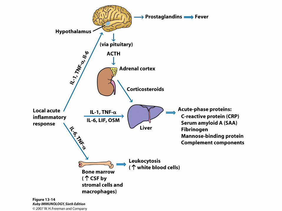
Local inflammatory response is accompanied by a systemic response known as the Acute Phase response. This response is marked by the induction of fever, increased synthesis of hormones ACTH and hydrocortisone and increased production of leukocytes and the production of a large number of proteins by the liver called Acute Phase Proteins
Acute Phase Proteins made during the acute phase of the response to infection (preceding recovery or death).
eg. C-reactive protein levels increase 1000 fold during an acute phase response.
complement components (C3, factor B, factor D and properdin), mannose-binding lectin (MBL binds to mannose residues on the surface of bacteria, fungi and some viruses) are also acute phase proteins.

A connection between inflammation, the immune system and artery disease was first suggested from studies in animals fed an artherosclerosis-inducing diet; lots of leukocytes found firmly attached to the arterial walls.
Now it is know that leukocytes are important in the development of artherosclerotic plaques.
Examination of blood levels of inflammatory markers IL-6, TNFα, CRP and the traditional risk markers (cholesterol, LDL, HDL) followed for 6-8 years in men and women.
Of the inflammatory markers only CRP levels were found to be associated with higher risk of coronary disease.
Statins which lower cholesterol levels also lower inflammation. Statins result in lowering levels of CRP.

Cell Types of Innate Immunity

Neutrophils • First line of defense—first cell type that migrates from the blood to the site of
infection. • Essential for innate immunity against bacteria and fungi. • Anti-microbial activity:
– Phagocytosis; direct or by opsonization – Oxidative and nonoxidate killing – Oxidative mechanism : reactive oxygen species (ROS) and reactive
nitrogen species (RNS) ROS include—superoxide ion •O2
- , hydrogen peroxide(H2O2), hypochlorus acid (HOCL). ROS generated by the NADPH phagosome oxidase (phox) enzyme complex
– Reaction of nitric oxide with superoxide generates RNS – Nonoxidative mechanism: neutrophil granules fuse with phagosome
releasing their anti-microbial peptides/ proteins (bactericidial permeability-increasing protein BPI), enzymes (proteases, lysozymes) that help to destroy the pathogen
• Increased expression of inducible nitric oxide synthetase (iNOS)


Macrophages • Activated by TLR binding to their ligand • Activated macrophages exibit:
– Increased phagocytosis – Increased respiratory burst – Increased expression of inducible nitric oxide
synthetase (iNOS). iNOS oxidizes L-arginine to L-citrulline and nitric oxide (NO)
– Secrete cytokines IL-1, IL-6, and TNF-α – Produce complement proteins – Express higher levels of MHC class 11 and thereby
present antigens to T cells

NK Cell • Critical first line of defense against viral
infections. • Can distinguish between an infected and un-
infected host cell. By killing the virally infected host cell they eliminate source of additional virus.
• Secrete cytokines IFNγ and TNFα - these cytokines stimulate maturation of
dendritic cells. - IFNγ mediates macrophage activation and
regulates TH cell development.

Dendritic Cells • Critical cells for transition from innate to adaptive
immunity. • Binding of TLRs to recognize pathogens - this stimulates DC activation and maturation
(increased production and surface expression of MHC Class 11 and co-stimulatory molecules).
-activated DCs migrate to lymphoid tissue and present antigen to TH and TC cells
• DCs can generate ROS and RNS. • Plasmacytoid DCs are potent producers of type 1
interferons that block viral replication. • Myeloid DCs produce IL12, IL6 and TNF-α, all potent
inducers of inflammation.

The immune system:
1) Senses/detects the presence of a pathogen
2) Mounts a response

Sensors: Soluble or membrane bound molecules (receptors) that recognize molecular patterns or motifs absent in the host but present in the pathogen.
Pattern Recognition Receptors (PRRs) on host cell recognize Pathogen Associated Molecular Patterns ( PAMPs).
PAMPs: combination of sugars, lipoproteins and some nucleic acid motifs.

1996: Jules Hoffman and Bruno Lamaitre Cell 86: 973 reported that mutations in Toll made the fruit fly highly susceptible to lethal fungal infection.
1997: Ruslan Medzhitov and Charles Janeway showed that this pathway conserved between fruit flies and humans. Showed that a human protein ( TLR4) that they identified by homology of its cytoplasmic domain and that of Toll when transfected into a cell line activated the expression of immune response genes.
1998: Bruce Beutler mutant mice (lps ) gene encoded a mutant form of TLR4, resistant to fatal doses of LPS.


TLRs • Membrane bound receptors • Structurally Conserved
– Multiple leucine-rich regions in extra cellular region
– Contain conserved TIR (Toll/IL1 receptor) in intercellular domain for signaling.

TLR structure


Function as either hetero or homodimers Pairing affects specificity
TLR1/2 TLR2/6
Location reflects ligands: TLRs that recognize extracellular ligands are found on the cell surface TLRs that recognize intracellular ligands are found on the endosome