Embed Size (px)
Citation preview
Injury Mechanisms and Classifications
Core Concepts in Athletic Training and TherapySusan Kay Hillman

Objectives Describe the anatomical reference position.
Use appropriate anatomical terminology to describe the location and position of a structure relative to the rest of the body.
Identify characteristics relating to the various stages of physical maturity.
Explain distinctiveness of the various types of musculoskeletal tissue.
Differentiate between elastic and plastic tissue properties.
Classify injuries as either acute or chronic based on the onset and duration of symp toms.
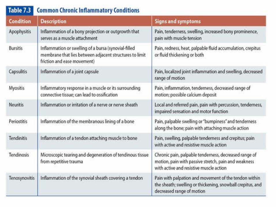
Define the common chronic inflammatory conditions, including signs and symptoms.
Define the various classifications of closed soft tissue wounds, including degrees of severity.
Define and classify closed and open wounds of the bone and joint articulations.
Classify nerve injuries according to mechanism, severity, and signs and symptoms.
Identify the classifications of open (exposed) wounds.
Introduction Proper reference to anatomical positions,
knowledge of injury terminology, and mechanisms essential for communicating effectively with other health care professionals
Assist you in documenting findings, convey history information during medical referrals, and collaborate with other healthcare professionals regarding care of your athlete
Anatomical Reference Terminology
All anatomical descriptions and references are based on standardized position of the body Anatomical Position
Allows us to reference specific body regions in relation to the body as a whole and one anatomical landmark to another Avoid confusion and misinterpretation of
your findings Can be standing or supine (on the spine)
Standing most common and easiest to visualize
Anatomical Reference Terminology
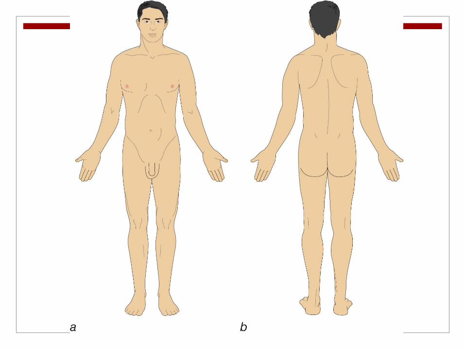
Anatomical position
Feet together, flat on the ground, toes facing forward
Legs and knees straight and in line with hip, torso and head, which are also straight and facing forward
Upper limbs positioned at persons side, with elbows straight
Shoulders rotated so palms are facing forward
Anatomical Reference Terminology
Once in anatomical position one can begin to refer to specific structure using various anatomical terms
Describe position of body parts with reference to other body parts or the body as a whole
Also synonyms reserved for particular body regions For example anterior=structure near front of
body, and anterior surface of hands is referred to as palmar or ventral

Anatomical Reference Terminology
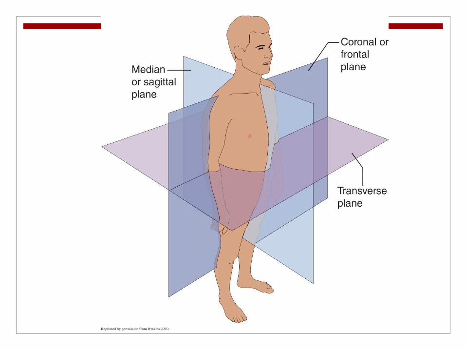
From anatomical position can also define three anatomical planes of movement useful in describing postural positions, motions, and function of various muscles and joints Imaginary planes that separate the body into left and
right (sagittal or median), top and bottom (Transverse), and front and back (frontal or coronal) For example when nodding your head or flexing your
elbow this occurs in the sagittal plane Shaking your head no or rotating your palms so it is
facing backwards takes plane in the transverse Lifting arms out to the side occurs in the frontal
plane
Anatomical Reference Terminology
Patient positioning terminology important and helpful for understanding starting positions for various medical testing Supine (face up) and prone (face down)
refers to patient laying down Short sitting-patient sitting on edge of table
with legs hanging off the edge Long sitting patient sitting with legs out in
front of them with legs on the table or floor

Physical Maturity Classifications
Allows us to define stages of physical growth Normal anatomic and physiologic development from
infancy to older adulthood Infancy: (0-12 months) physical changes occur most
rapidly. Dependent neonate to a child learning motor skills such
as turning, sitting, crawling and walking Gain 3 x birth weight in this time
Childhood: (1-11 years) infancy to onset of puberty Steady growth and development Skeleton is immature with epiphyseal plates open to
allow bones to elongate 1-5 early childhood, 6-11 middle childhood
Physical Maturity Classifications
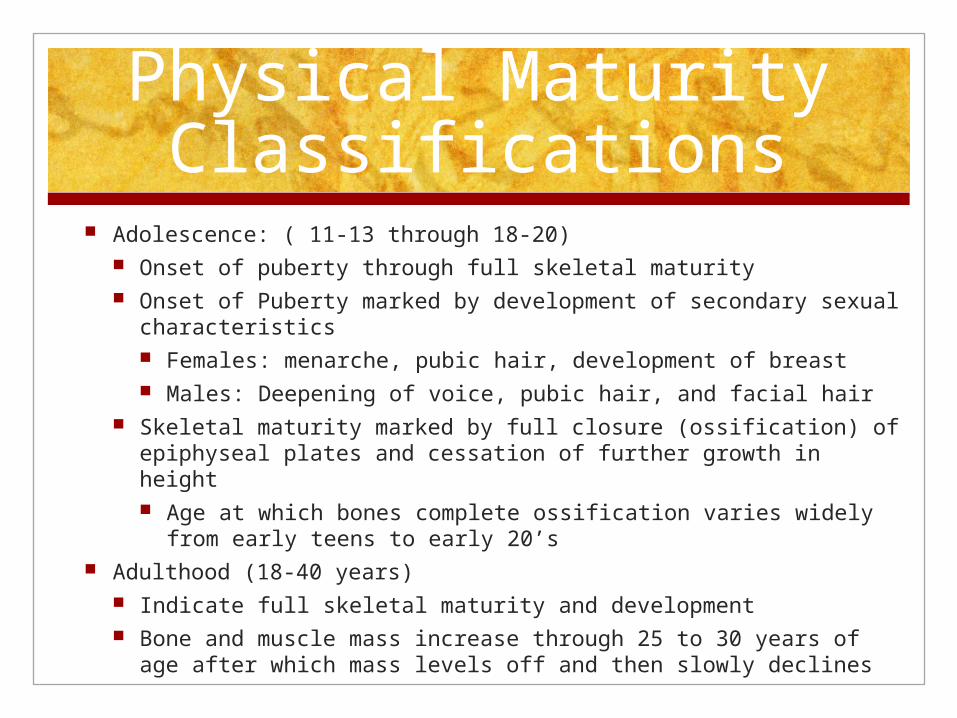
Adolescence: ( 11-13 through 18-20) Onset of puberty through full skeletal maturity Onset of Puberty marked by development of secondary sexual
characteristics Females: menarche, pubic hair, development of breast Males: Deepening of voice, pubic hair, and facial hair
Skeletal maturity marked by full closure (ossification) of epiphyseal plates and cessation of further growth in height Age at which bones complete ossification varies widely
from early teens to early 20’s Adulthood (18-40 years)
Indicate full skeletal maturity and development Bone and muscle mass increase through 25 to 30 years of age
after which mass levels off and then slowly declines

Middle adulthood (40-60 years) Gradual decline in strength coordination and
balance
Older adulthood (> 60 years) Spans rest of human beings life Accelerating decline in strength, coordination
and balance Highly individual depending on lifestyle,
activity, nutrition and disease
Injury Mechanisms
Foundation of body movements made up of several simple machines Levers, pulleys, and wedges among other more
complex systems Bodies capable of performing very intricate and
detailed work along with incredible feats of strength, power and endurance
However, body influenced by internal and external mechanical forces that can negatively affect performance
Important to understand musculoskeletal system, physical properties of the musculoskeletal tissue, internal and external mechanical forces that can cause injury
Injury Mechanisms
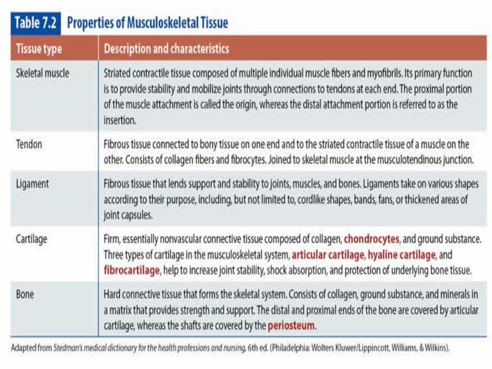
Musculoskeletal Tissue
Five tissue types Categorized by Soft and Skeletal Tissue
Soft tissue Muscles, tendons, ligaments, and
cartilage
Skeletal Tissue Bone
Injury Mechanisms
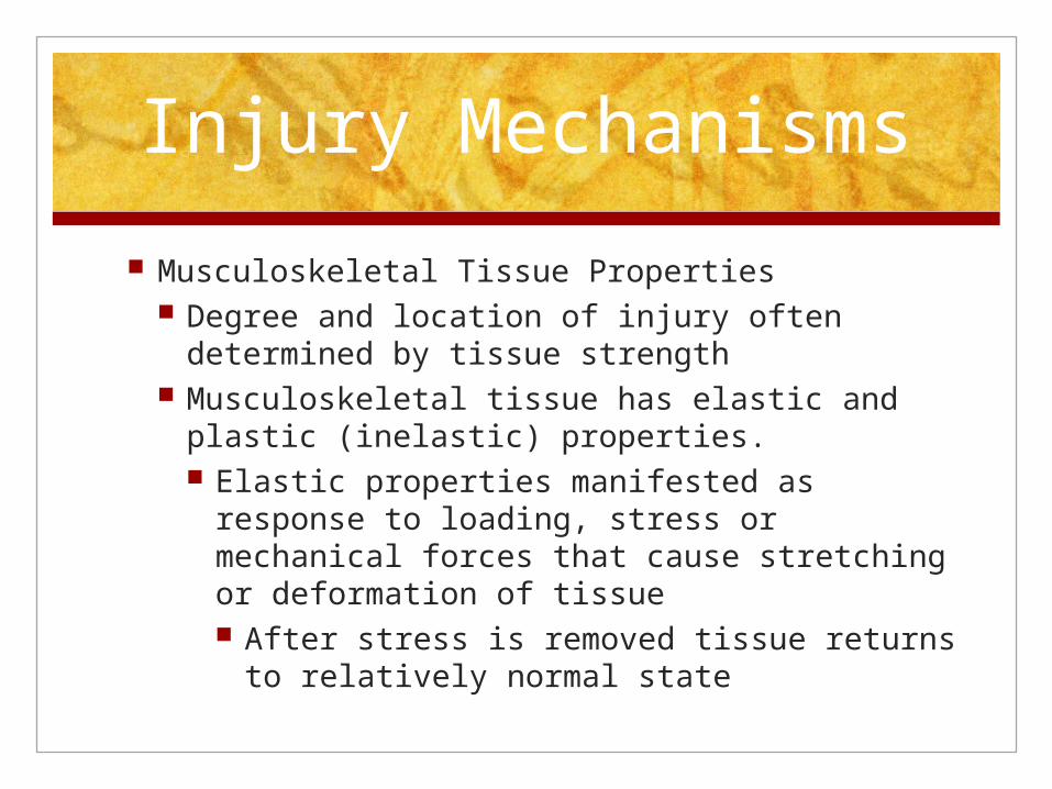
Musculoskeletal Tissue Properties Degree and location of injury often
determined by tissue strength Musculoskeletal tissue has elastic and plastic
(inelastic) properties. Elastic properties manifested as response
to loading, stress or mechanical forces that cause stretching or deformation of tissue After stress is removed tissue returns to
relatively normal state

Injury Mechanisms
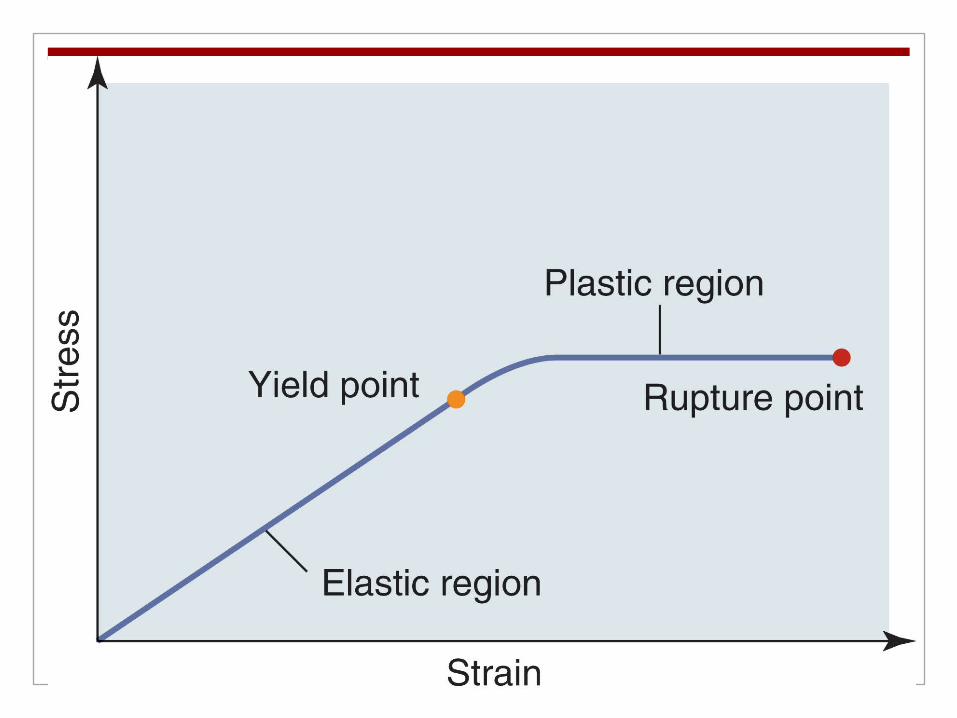
Plastic Properties manifested at end range of elastic properties rendering tissue unable to return to normal
state
Tissue retains some amount of deformation due to structural injury
• Yield point: determined by specific amount or level of stress
• Example: stretching a rubber band. Point at which it breaks is considered yield point
• Enough force to eliminate elastic property recovery and cause rubber band to undergo
plastic deformation
Injury Mechanisms
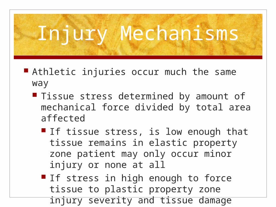
Athletic injuries occur much the same way Tissue stress determined by amount of
mechanical force divided by total area affected If tissue stress, is low enough that tissue
remains in elastic property zone patient may only occur minor injury or none at all
If stress in high enough to force tissue to plastic property zone injury severity and tissue damage more significant
Injury Mechanisms
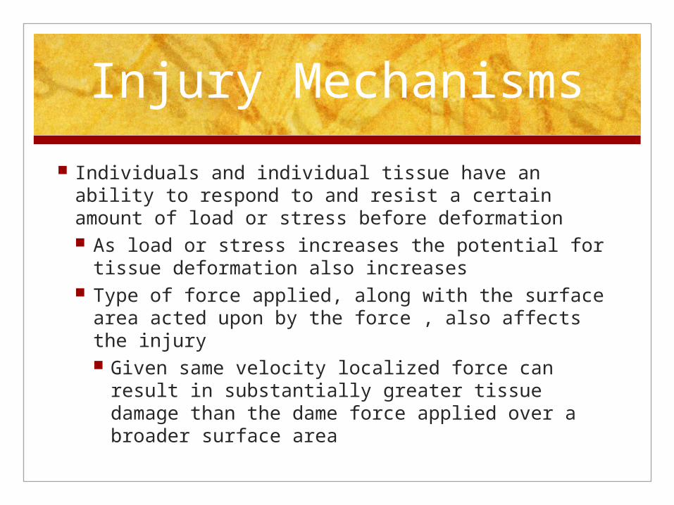
Individuals and individual tissue have an ability to respond to and resist a certain amount of load or stress before deformation As load or stress increases the potential for tissue
deformation also increases Type of force applied, along with the surface area
acted upon by the force , also affects the injury Given same velocity localized force can result in
substantially greater tissue damage than the dame force applied over a broader surface area
Injury Mechanisms
Tissue damage may be the result: unpredictable accident or injury Overuse Overload Poor posture Skeletal immaturity Lack of conditioning Improper mechanics Fatigue Inflexibility Muscle imbalance Genetics
Mechanical Forces
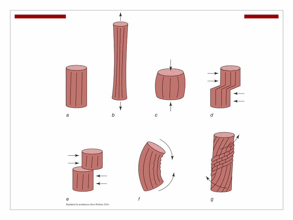
Stress or load applied to the body to cause injury or tissue deformation is a result of 1 of 5 types of mechanical force Excessive compression
Squeezing or condensing of tissue due to external forces applied directly opposite of each other Bruises (contusions) Crushing injuries (compression fractures) Pinching Injuries due to direct impact
Mechanical Forces
Shear Forces that cause tissue to “slide” over
adjoining surfaces or structures in a parallel fashion Brain injuries Tibiofemoral translation injuries such as
ACL and PCL injuries Blisters Lumbar spine injuries
Mechanical Forces
Torsion Twisting mechanism that causes rotation
along long axis or fixed point Opposite ends of tissue are rotated in
opposite directions Example: Body rotating over Foot fixed or
lower leg Occurs to bones and ligaments

Tension Stretching or lengthening of musculoskeletal
tissue due to stress or strain Caused by pulling or drawing apart Pull of tissue in opposite direction causes
tissue in between to stretch Muscle strains or ruptures commonly
caused by tension within the musculotendinous unit Where muscle makes transition into
tendon
• Weak part of muscle
Mechanical Forces
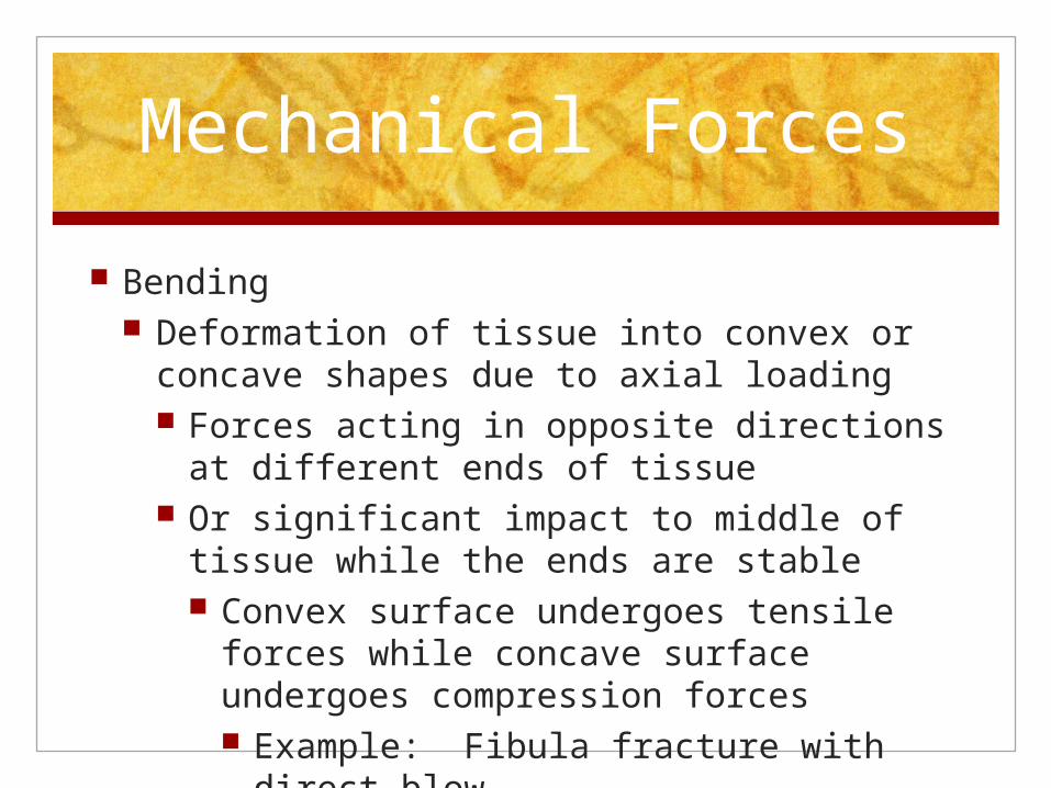
Bending Deformation of tissue into convex or concave
shapes due to axial loading Forces acting in opposite directions at
different ends of tissue Or significant impact to middle of tissue
while the ends are stable Convex surface undergoes tensile forces
while concave surface undergoes compression forces Example: Fibula fracture with direct
blow
Mechanical Forces
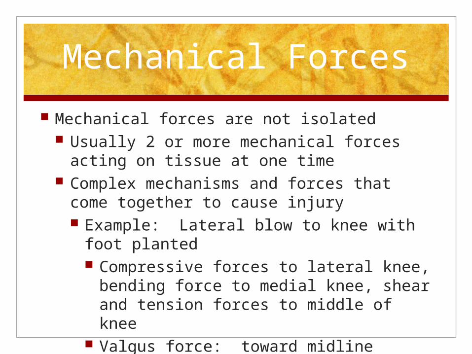
Mechanical forces are not isolated Usually 2 or more mechanical forces acting
on tissue at one time Complex mechanisms and forces that come
together to cause injury Example: Lateral blow to knee with foot
planted Compressive forces to lateral knee,
bending force to medial knee, shear and tension forces to middle of knee
Valgus force: toward midline Varrus Force: Away from midline
Time Classification Relating to Mechanism of Injury
Acute Injuries Conditions that have sudden onset, short
duration, and occur via mechanical forces that exceed elastic properties causing tissue deformation
Single traumatic event: blunt force trauma, dynamic overload of muscle, tendon, joint capsule or ligamentous tissue

Time Classification Relating to Mechanism
of Injury Chronic Injuries
Gradual onset, prolonged duration, and occur as a result of accumulation of minor insults or repetitive stresses
Exact mechanism not often known Overuse, accumulative microtrauma, repetitive
overloading, abnormal friction that is greater than body's ability to heal and recover before additional stress is added
“too much, too soon, too often” Often more difficult to treat overuse (chronic)
injuries than acute injuries
Injury Classifications
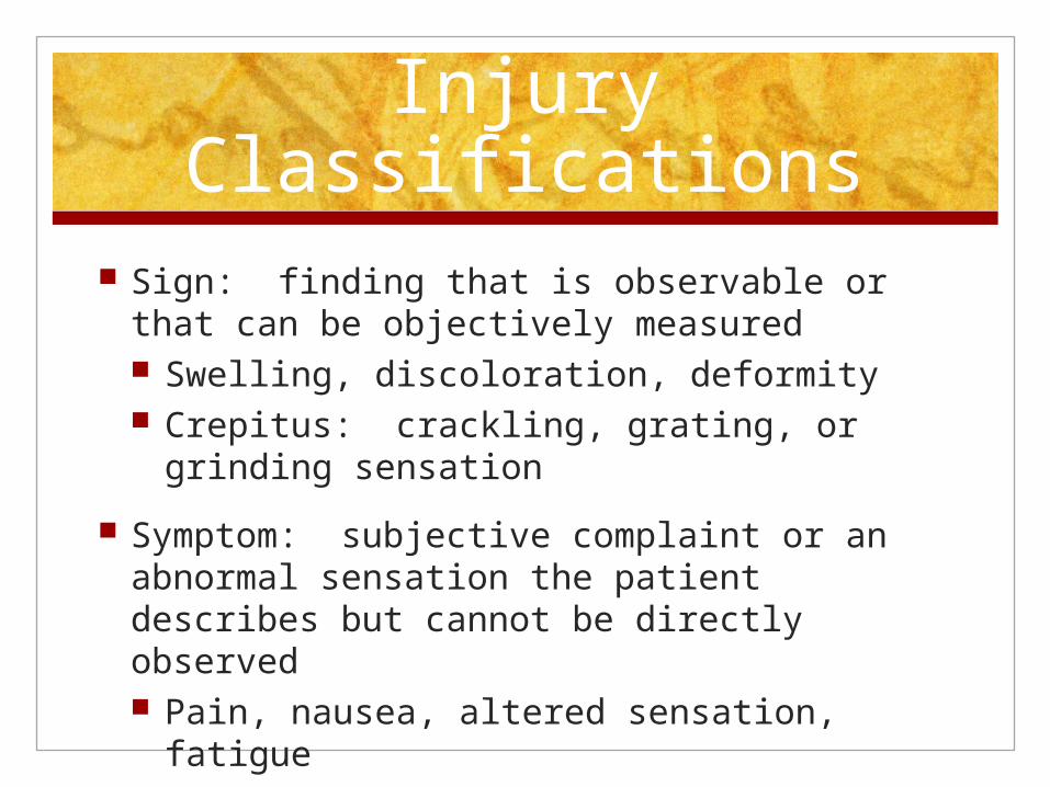
Sign: finding that is observable or that can be objectively measured Swelling, discoloration, deformity Crepitus: crackling, grating, or grinding
sensation
Symptom: subjective complaint or an abnormal sensation the patient describes but cannot be directly observed Pain, nausea, altered sensation, fatigue
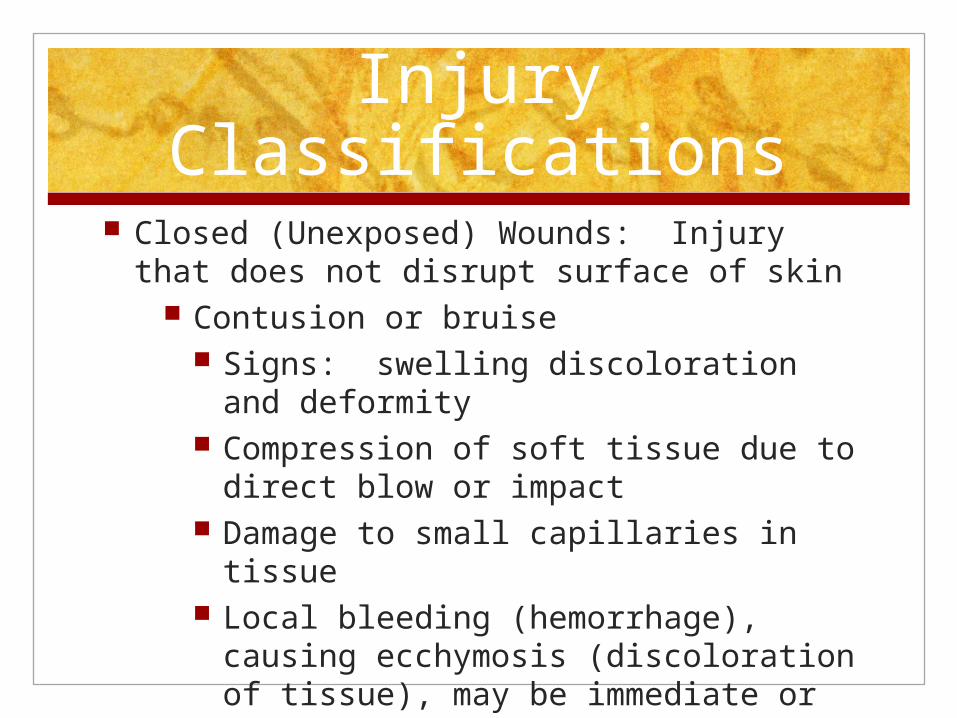
Injury Classifications Closed (Unexposed) Wounds: Injury that
does not disrupt surface of skin Contusion or bruise
Signs: swelling discoloration and deformity
Compression of soft tissue due to direct blow or impact
Damage to small capillaries in tissue Local bleeding (hemorrhage), causing
ecchymosis (discoloration of tissue), may be immediate or delayed

Contusion Severity
• First degree: superficial damage, minimal swelling, localized tenderness, no limitations to strength or ROM
• Second Degree: Increased pain and hemorrhage, increased area and depth of tissue damage, mild to moderate limitation sin ROM and muscle function or both
• Third degree: severe tissue compression, severe pain, significant hemorrhage and development of hematoma• Significant limitations in ROM and muscle function• Suspect damage to deeper structures such as none

Closed Wounds
Sprains: injury to ligaments or capsular structure Ligaments attach bone to bone
Injury occurs when 2 bones separate or go beyond normal ROM
First Degree: mild overstretching Mild pain and tenderness, little or no disability AROM and PROM not limited but some pain at
end range Firm definitive end point (feel) Degree of swelling and discoloration not great
indicator of severity
Sprain Severity
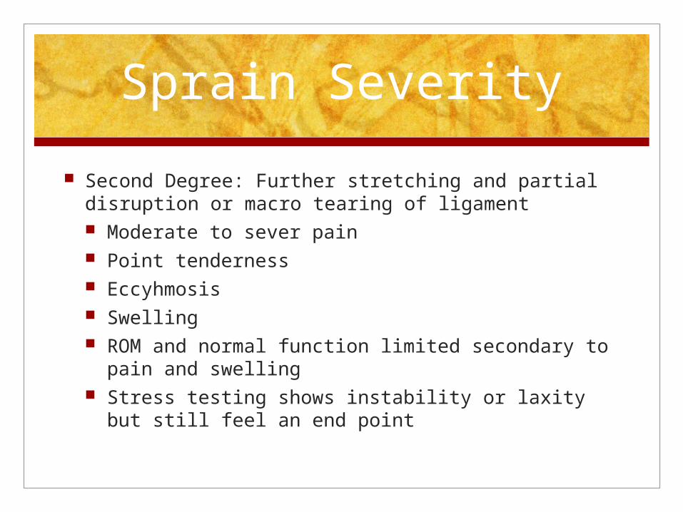
Second Degree: Further stretching and partial disruption or macro tearing of ligament Moderate to sever pain Point tenderness Eccyhmosis Swelling ROM and normal function limited secondary to pain
and swelling Stress testing shows instability or laxity but still feel
an end point
Sprain severity
Third Degree: Complete disruption or rupture or loss of ligament integrity Associated with feeling or sound of a pop Immediate pain and disability Rapid swelling. Eccyhmosis and loss of function Stress test shows moderate to severe instability with
no firm end point “ soft or mushy” Can be deceiving because Rom and stress testing less
painful because ligament not intact
Strains
Stretching or tearing of muscle or tendon Violent, forceful contraction or
overstretching Fatigue, lack of warm up muscle strength
imbalance, and dyssynchrony
Strain Severity
First Degree: overstretching and micro tearing of muscle or tendon. No gross fiber disruption Mild pain and tenderness Typically full AROM and PROM Pain with resisted muscle contraction

Strain Severity
Second Degree: further stretching or partial tearing of muscle or tendon fibers Immediate pain, localized tenderness and
disability Varying degrees of swelling, eccyhmosis, and
decreased ROM and strength Pain with active muscle contraction and
passive muscle stretch May have palpable defect
Strain Severity
Third Degree: Muscle or tendon completely ruptured Audible pop Immediate pain and loss of function Palpable defect on superficial muscles Muscle hemorrhage and diffuse swelling ROM and strength may or may not be
affected or painful
Injury Classifications Open (Exposed) Wounds
Injuries that involve disruption of the skin Caused by friction or blunt or sharp trauma Susceptible to infection
Monitor for pus increased pain, redness, swelling, heat and red streaks running from wound to trunk If signs of infection are present refer to
medical professional

Injury Classifications
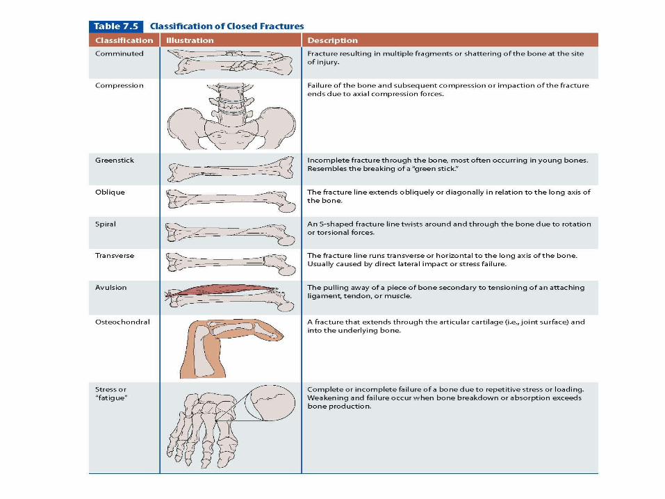
Bone and Joint Injuries Closed Fractures: disruption in continuity of bone without disruption
of skin surface Traumatic (Acute): immediate pain, rapid swelling, bony
tenderness, false joint, crepitus, deformity Displaced fracture concern with secondary injury
• Evaluate neurovascular status distal to fracture site
Stress Fracture: S & S not always as obvious Onset of pain is gradual Pain or deep ache may be first noticeable during activity and
subside with rest, • progresses to more constant pain if offending
activity continues Swelling is minimal and localized tenderness over fracture site

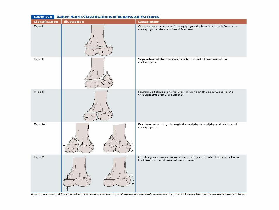
Bone and Joint Injuries Epiphyseal Injury
Disruption of epiphysis or epiphyseal plate (growth plate) Can cause premature closing and growth abnormalities
Dislocation Complete disassociation of 2 joint surfaces Forces cause joint to exceed passed its normal ROM Severe Stretching or complete disruption of joint capsule
and supporting ligaments Pain swelling, loss of function, deformity
Subluxation Incomplete disassociation of 2 joint surfaces Disability, pain, selling and joint instability varies Often history of sensation of joint slipping or giving out
Injury ClassificationsNerve Injuries
Compression or tensioning of neural structure
Laceration of nerve can occur secondary to fracture, dislocation, penetrating trauma
Anesthesia: no sensation
Parathesia: tingling, burning, numbness
Hyperesthesia: hypersensitivity
Paralysis: complete loss of muscle function
Nerve Injuries Neuropraxia: transient and reversible
loss of nerve function
Axontmesis: partial disruption of nerve Considerable atrophy and
weakness due to prolonged healing 2 weeks to a year
Neurotmesis: most severe nerve injury, complete severance of the nerve
Neuralgia: achiness or pain along distribution of nerve secondary to irritation or inflammation
Neuroma: thickening of a nerve or “nerve tumor”, secondary to chronic irritation or inflammation