Embed Size (px)
Citation preview
Initial Diagnostic Workup of Acute Leukemia Guideline from the College of American Pathologists (CAP) and the American Society of Hematology (ASH)
Early Online Release Publication: Archives of Pathology and Laboratory Medicine
2/22/2017
Background
The diagnosis and prognostic determination of acute leukemia currently incorporate • Clinical information
• Morphology
• Immunophenotyping
• Cytogenetics
• Molecular genetics
© 2017 College of American Pathologists. All rights reserved.
Background continued
Why have guidelines for the work-up of acute leukemia?
Clinical practice guidelines have the potential to: • Standardize care across all settings
• Reduce duplication of testing
• Support the use of specific tests
• Improve patient care and outcomes
© 2017 College of American Pathologists. All rights reserved.
Introduction
• The College of American Pathologists (CAP) and the American Society for Hematology (ASH) formed an expert panel to review the relevant literature and to establish a guideline for appropriate laboratory testing as well as for the clinical information necessary for the initial diagnosis of acute leukemia, including AML, ALL, and acute leukemias of ambiguous lineage.
© 2017 College of American Pathologists. All rights reserved.
Introduction continued
• The panel closely followed the Institute of Medicine Clinical Practice Guidelines We Can Trust standards for guideline development
1. Establish transparency
2. Manage conflicts of interest
3. Establish a multi-disciplinary panel
4. Perform systematic review
5. Rate strength of recommendations
6. Articulate the recommendations
7. Include external review
© 2017 College of American Pathologists. All rights reserved.
Guideline expert panel members
• Daniel A. Arber, MD, CAP co-chair
• James W. Vardiman, MD, ASH co-chair
• Melissa Cessna, MD
• Joan Etzell, MD
• Sa Wang, MD
• Karl Theil, MD, PhD
• Michael J. Borowitz, MD, PhD
• Robert Hasserjian, MD
• Kathryn Foucar, MD
• J. Douglas Rizzo, MD © 2017 College of American Pathologists. All rights reserved.
Guideline staff
• Nicole E. Thomas, MPH, CT(ASCP)cm, CAP Senior Manager, Center Guideline Development
• Robert Plovnick, MD, ASH Director of Quality Improvement
• Robert Kunkle, ASH Senior Manager, Practice Guidelines
• Kendall Alexander, MPH, ASH Clinical Quality Projects Specialist
• R. Bryan Rumble, MSc, Methodology Consultant
• Tony Smith, MLS, ECMS (AIIM), Medical Librarian © 2017 College of American Pathologists. All rights reserved.
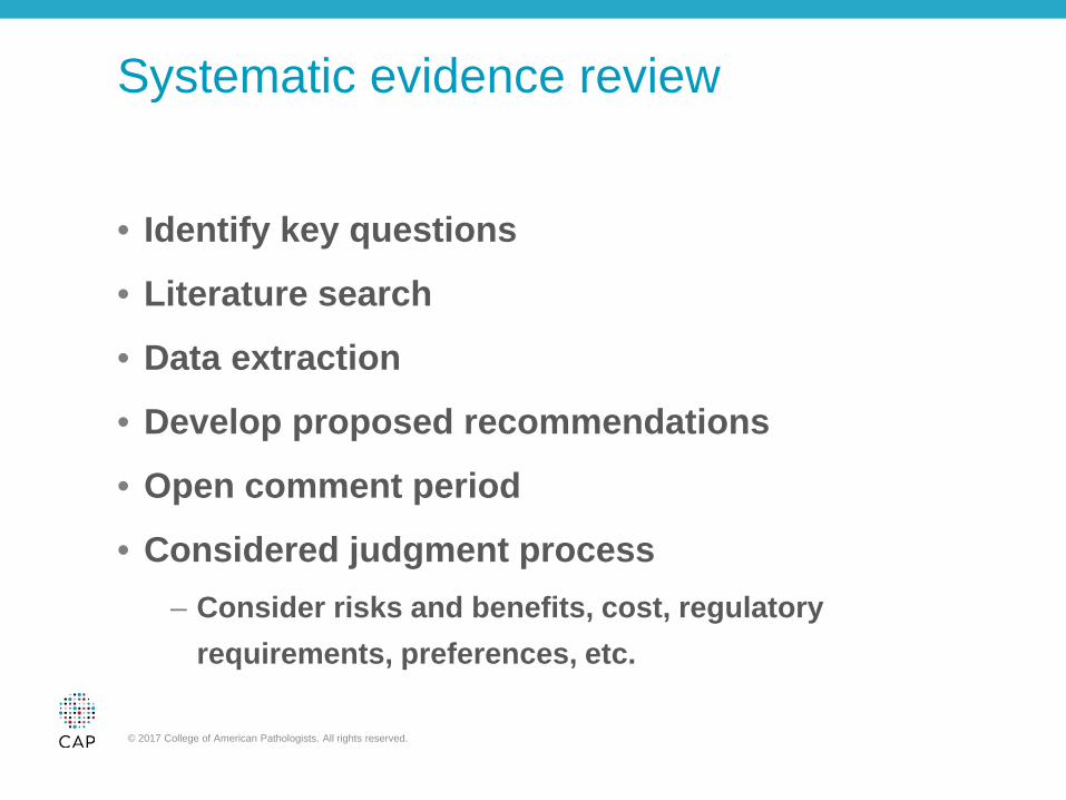
Systematic evidence review
• Identify key questions
• Literature search
• Data extraction
• Develop proposed recommendations
• Open comment period
• Considered judgment process – Consider risks and benefits, cost, regulatory
requirements, preferences, etc.
© 2017 College of American Pathologists. All rights reserved.
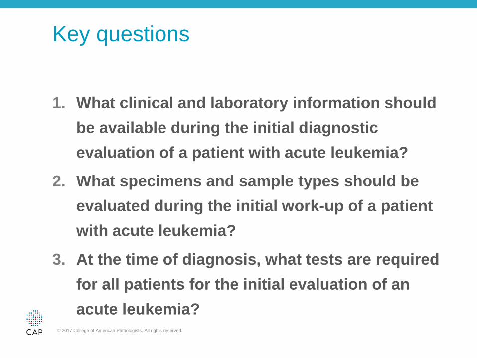
Key questions
1. What clinical and laboratory information should be available during the initial diagnostic evaluation of a patient with acute leukemia?
2. What specimens and sample types should be evaluated during the initial work-up of a patient with acute leukemia?
3. At the time of diagnosis, what tests are required for all patients for the initial evaluation of an acute leukemia?
© 2017 College of American Pathologists. All rights reserved.
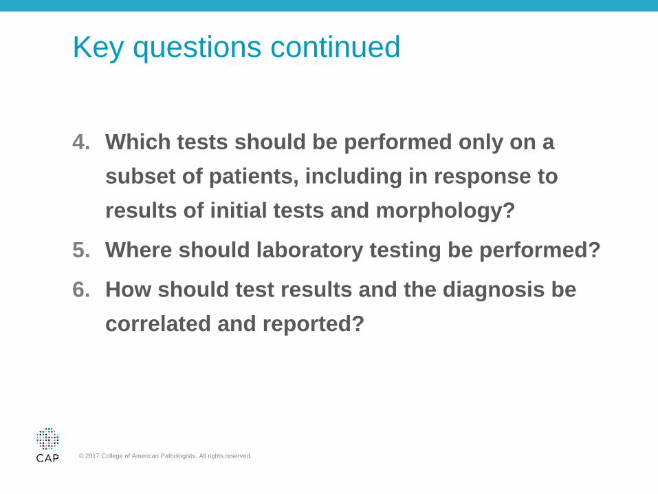
Key questions continued
4. Which tests should be performed only on a subset of patients, including in response to results of initial tests and morphology?
5. Where should laboratory testing be performed?
6. How should test results and the diagnosis be correlated and reported?
© 2017 College of American Pathologists. All rights reserved.
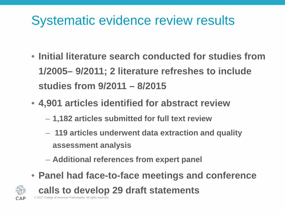
Systematic evidence review results
• Initial literature search conducted for studies from 1/2005– 9/2011; 2 literature refreshes to include studies from 9/2011 – 8/2015
• 4,901 articles identified for abstract review – 1,182 articles submitted for full text review
– 119 articles underwent data extraction and quality assessment analysis
– Additional references from expert panel
• Panel had face-to-face meetings and conference calls to develop 29 draft statements
© 2017 College of American Pathologists. All rights reserved.
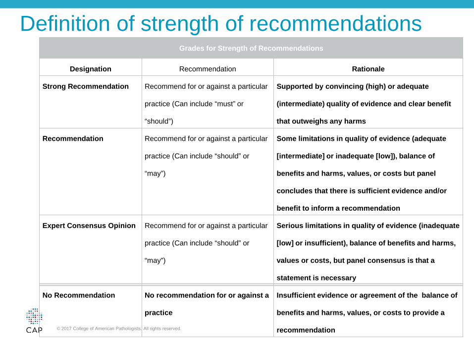
Definition of strength of recommendations Grades for Strength of Recommendations
Designation Recommendation Rationale
Strong Recommendation Recommend for or against a particular
practice (Can include “must” or
“should”)
Supported by convincing (high) or adequate
(intermediate) quality of evidence and clear benefit
that outweighs any harms
Recommendation Recommend for or against a particular
practice (Can include “should” or
“may”)
Some limitations in quality of evidence (adequate
[intermediate] or inadequate [low]), balance of
benefits and harms, values, or costs but panel
concludes that there is sufficient evidence and/or
benefit to inform a recommendation
Expert Consensus Opinion Recommend for or against a particular
practice (Can include “should” or
“may”)
Serious limitations in quality of evidence (inadequate
[low] or insufficient), balance of benefits and harms,
values or costs, but panel consensus is that a
statement is necessary
No Recommendation No recommendation for or against a
practice
Insufficient evidence or agreement of the balance of
benefits and harms, values, or costs to provide a
recommendation © 2017 College of American Pathologists. All rights reserved.
Systematic evidence review results continued
• Results from the open comment period – August 10-31, 2015: 29 draft statements were presented
– 162-200 respondents for each draft statement
– 789 written comments
– 26 draft statements achieved more than 90% agreement, two achieved 80 - 90% agreement, and one received 70-80% agreement
• Final guideline includes 27 guideline statements (recommendations)
© 2017 College of American Pathologists. All rights reserved.
Guideline Statement 1
Strong Recommendation - The treating clinician should provide relevant clinical data or ensure that this is readily accessible by the pathologist.
Note: These data include, but is not limited to the patient's age, gender, ethnicity, history of any hematologic disorder or known predisposing conditions or syndromes, any prior malignancy, exposure to cytotoxic therapy, immunotherapy, radiotherapy, or other possibly toxic substances, and any additional clinical findings of diagnostic or prognostic importance. The treating clinician should also include any history of possibly confounding factors, such as recent growth factor therapy, transfusions or other medications that might obscure or mimic the features of acute leukemia. The treating clinician should also obtain and provide information regarding any family history of any hematologic disorder or other malignancies.
© 2017 College of American Pathologists. All rights reserved.
Guideline Statement 1 rationale
• Clinical information is essential in AL diagnosis
• Detection of germline predisposition disorders is critical for treatment and prognosis
• Pathologist must know about confounding factors such as G-CSF therapy or other prior therapy
© 2017 College of American Pathologists. All rights reserved.
Guideline Statement 2
Recommendation - The treating clinician should provide relevant physical examination and imaging findings or ensure that these are readily accessible by the pathologist.
Note: This includes, but is not limited to, neurologic exam findings and the presence of tumor masses (e.g., mediastinal), other tissue lesions (e.g., cutaneous), and/or organomegaly.
© 2017 College of American Pathologists. All rights reserved.
Guideline Statement 2 rationale
• Extramedullary sites of disease could be biopsied, if necessary
• Diagnostic evaluation could be performed on an extramedullary specimen
• Certain type of tumor masses (e.g. mediastinal mass) could provide clues as to a specific AL subtype
© 2017 College of American Pathologists. All rights reserved.
Guideline Statement 3
Strong Recommendation - The pathologist should review recent or concurrent complete blood counts (CBCs) and leukocyte differentials and evaluate a peripheral blood smear.
© 2017 College of American Pathologists. All rights reserved.
Guideline Statement 4 Strong Recommendation - The treating clinician or pathologist should obtain a fresh bone marrow aspirate for all patients suspected of acute leukemia, a portion of which should be used to make bone marrow aspirate smears for morphologic evaluation. If performed, the pathologist should evaluate an adequate bone marrow trephine core biopsy, bone marrow trephine touch preparations, and/or marrow clots, in conjunction with the bone marrow aspirates. Note: If bone marrow aspirate material is inadequate or if there is compelling clinical reason to avoid bone marrow examination, peripheral blood may be used for diagnosis and ancillary studies if sufficient numbers of blasts are present. If a bone marrow aspirate is unobtainable, touch imprint preparations of a core biopsy should be prepared and evaluated, and an additional core biopsy may be submitted unfixed in tissue culture medium for disaggregation for flow and genetic studies. Optimally, the same physician should interpret the BM aspirate smears and the core biopsy specimens, or the interpretations of these specimens should be correlated if performed by different physicians.
© 2017 College of American Pathologists. All rights reserved.
Guideline Statement 4 rationale
• Specialized testing requires viable cells so adequate specimens for both routine assessment and specialized testing are essential
© 2017 College of American Pathologists. All rights reserved.
Guideline Statement 5 Strong Recommendation - In addition to morphologic assessment (blood and bone marrow), the pathologist or treating clinician should obtain sufficient samples and perform conventional cytogenetic analysis (i.e., karyotype), appropriate molecular genetic and/or fluorescent in-situ hybridization (FISH) testing, and flow cytometric immunophenotyping (FCI). The flow cytometry panel should be sufficient to distinguish acute myeloid leukemia (including acute promyelocytic leukemia), T-cell acute lymphoblastic leukemia (T-ALL) (including early T-cell precursor leukemias), B-cell precursor ALL (B-ALL), and acute leukemia of ambiguous lineage on all patients diagnosed with acute leukemia. Molecular genetic and/or FISH testing does not, however, replace conventional cytogenetic analysis. Note: If sufficient bone marrow aspirate or peripheral blood material is not available for FCI, immunohistochemical studies may be used as an alternative method for performing limited immunophenotyping. In addition, a second bone marrow core biopsy can be obtained and submitted unfixed in tissue culture media for disaggregation for genetic studies and flow cytometry.
© 2017 College of American Pathologists. All rights reserved.

Guideline Statement 6
Expert Consensus Opinion - For patients with suspected or confirmed acute leukemia, the pathologist may request and evaluate cytochemical studies to assist in the diagnosis and classification of acute myeloid leukemia (AML).
© 2017 College of American Pathologists. All rights reserved.
Guideline Statement 6 rationale
• Cytoechemical MPO staining is a rapid and sensitive method to confirm myeloid blast lineage
• Cytochemical MPO is especially valuable in the rapid diagnosis of acute promyelocytic leukemia
© 2017 College of American Pathologists. All rights reserved.
Guideline Statement 7
Recommendation - The treating clinician or pathologist may use cryopreserved cells or nucleic acid, formalin fixed, non-decalcified paraffin-embedded (FFPE) tissue, or unstained marrow aspirate or peripheral blood smears obtained and prepared from peripheral blood, bone marrow aspirate or other involved tissues for molecular or genetic studies in which the use of such material has been validated. Such specimens must be properly identified and stored under appropriate conditions in a laboratory that is in compliance with regulatory and/or accreditation requirements.
© 2017 College of American Pathologists. All rights reserved.
Guideline Statement 7 rationale
• Both banked, viable cell specimens and formalin-fixed, paraffin-embedded specimens are useful for various types of testing if fresh tissue is not available
© 2017 College of American Pathologists. All rights reserved.
Guideline Statement 8
Strong Recommendation - For patients with acute lymphoblastic leukemia (ALL) receiving intrathecal therapy, the treating clinician should obtain a cerebrospinal fluid (CSF) sample. The treating clinician or pathologist should ensure that a cell count is performed and that examination/enumeration of blasts on a cytocentrifuge preparation is performed and is reviewed by the pathologist.
© 2017 College of American Pathologists. All rights reserved.
Guideline Statement 8 rationale
• Monitoring of CSF is a standard practice in patients with AL, especially acute lymphoblastic leukemias
• The pathologist must assess these CSF specimens for recognition and enumeration of blasts
© 2017 College of American Pathologists. All rights reserved.
Guideline Statement 9
Expert Consensus Opinion - For patients with acute leukemia other than those with ALL receiving intrathecal therapy, the treating clinician may, under certain circumstances, obtain a cerebrospinal fluid (CSF) sample when there is no clinical contraindication. The treating clinician or pathologist should ensure that a cell count is performed and that examination/enumeration of blasts on a cytocentrifuge preparation is performed and is reviewed by the pathologist.
© 2017 College of American Pathologists. All rights reserved.
Guideline Statement 10
Recommendation - For patients with suspected or confirmed acute leukemia, the pathologist may use flow cytometry in the evaluation of CSF.
© 2017 College of American Pathologists. All rights reserved.
Guideline Statement 11
Strong Recommendation - For patients who present with extramedullary disease without bone marrow or blood involvement, the pathologist should evaluate a tissue biopsy and process it for morphologic, immunophenotypic, cytogenetic, and molecular genetic studies, as recommended for the bone marrow. Note: Additional biopsies may be indicated to obtain fresh material for ancillary testing.
© 2017 College of American Pathologists. All rights reserved.
Guideline Statement 12
Strong Recommendation - For patients with suspected or confirmed acute leukemia, the pathologist or treating clinician should ensure that flow cytometry analysis or molecular characterization is comprehensive enough to allow subsequent detection of minimal residual disease (MRD).
© 2017 College of American Pathologists. All rights reserved.
Guideline Statement 12 rationale
• Flow cytometric immunophenotyping is required for lineage confirmation in all cases of acute leukemia.
• Since persistent leukemia after therapy is a key prognostic factor for some types of AL, minimal residual disease monitoring by either flow cytometry or genetic testing is often required.
© 2017 College of American Pathologists. All rights reserved.
Guideline Statement 13
Strong Recommendation - For pediatric patients with suspected or confirmed B-ALL, the pathologist or treating clinician should ensure that testing for t(12;21)(p13.2;q22.1); ETV6-RUNX1, t(9;22)(q34.1;q11.2); BCR-ABL1, KMT2A(MLL) translocation, iAMP 21, and trisomy 4 and 10 is performed.
© 2017 College of American Pathologists. All rights reserved.
Guideline Statement 14
Strong Recommendation - For testing for t(9;22)(q34.1;q11.2) and BCR-ABL1; Recommendation for testing for KMT2A (MLL) translocations For adult patients with suspected or confirmed B-ALL, the pathologist or treating clinician should ensure that testing for t(9;22)(q34.1;q11.2) ; BCR-ABL1 is performed. In addition, testing for KMT2A (MLL) translocations may be performed.
© 2017 College of American Pathologists. All rights reserved.
Guideline Statement 15
Recommendation - For patients with suspected or confirmed ALL, the pathologist or treating clinician may order appropriate mutational analysis for selected genes that influence diagnosis, prognosis, and/or therapeutic management that includes, but is not limited to: PAX5, JAK1, JAK2, and/or IKZF1 for B-ALL and NOTCH1 and/or FBXW7 for T-ALL. Testing for overexpression of CRLF2 may also be performed for B-ALL.
© 2017 College of American Pathologists. All rights reserved.
Guideline Statements 13-17 rationale
• Genetic testing provides both diagnostic and prognostic information in many types of AL including precursor B-ALL and all AML subtypes.
© 2017 College of American Pathologists. All rights reserved.
Guideline Statement 16
Strong recommendation - for testing for FLT3-ITD; Recommendation for testing for other mutational analysis – For pediatric and adult patients with suspected or confirmed acute myeloid leukemia (AML) of any type, the pathologist or treating clinician should ensure that testing for FLT3-ITD is performed. The pathologist or treating clinician may order mutational analysis that includes but is not limited to: IDH1, IDH2, TET2, WT1, DNMT3A, and/or TP53 for prognostic and/or therapeutic purposes.
© 2017 College of American Pathologists. All rights reserved.
Guideline Statement 17
Strong Recommendation - for testing for KIT mutation in adult patients with CBF AML; Expert Consensus Opinion for testing for KIT mutation in pediatric patients with CBF AML – For adult patients with confirmed core binding factor (CBF) AML (AML with t(8;21)(q22;q22.1); RUNX1-RUNX1T1 or inv(16)(p13.1q22) /t(16;16)(p13.1;q22); CBFB-MYH11), the pathologist or treating clinician should ensure that appropriate mutational analysis for KIT is performed. For pediatric patients with confirmed core binding factor AML (AML with t(8;21)(q22;q22.1); RUNX1-RUNX1T1 or inv(16)(p13.1q22) /t(16;16)(p13.1;q22); CBFB-MYH11), the pathologist or treating clinician may ensure that appropriate mutational analysis for KIT is performed.
© 2017 College of American Pathologists. All rights reserved.
Guideline Statement 18
Strong Recommendation - For patients with suspected acute promyelocytic leukemia (APL), the pathologist or treating physician should also ensure that rapid detection of PML-RARA is performed. The treating physician should also order appropriate coagulation studies to evaluate for disseminated intravascular coagulation (DIC).
© 2017 College of American Pathologists. All rights reserved.
Guideline Statement 18 rationale
• Because of imminent risk of life-threatening hemorrhage, the rapid diagnosis of acute promyelocytic leukemia is essential.
• Implementation of ATRA therapy as early as possible is critical.
© 2017 College of American Pathologists. All rights reserved.
Guideline Statement 19
Strong Recommendation - For patients other than those with confirmed core binding factor AML, APL, or AML with myelodysplasia-related cytogenetic abnormalities, the pathologist or treating clinician should also ensure that mutational analysis for NPM1, CEBPA, and RUNX1 is also performed.
© 2017 College of American Pathologists. All rights reserved.
Guideline Statement 20
No Recommendation - For patients with confirmed acute leukemia, no recommendation is made for or against the use of global/gene specific methylation, micro RNA (miRNA) expression, or gene expression analysis for diagnosis or prognosis.
© 2017 College of American Pathologists. All rights reserved.
Guideline Statement 21
Strong Recommendation - For patients with confirmed mixed phenotype acute leukemia (MPAL), the pathologist or treating clinician should ensure that testing for t(9;22)(q34.1;q11.2); BCR-ABL1, and KMT2A (MLL) translocations is performed.
© 2017 College of American Pathologists. All rights reserved.

Guideline Statement 22
Strong Recommendation - All laboratory testing performed for the initial work-up and diagnosis of a patient with acute leukemia must be performed in a laboratory that is in compliance with regulatory and/or accreditation requirements.
© 2017 College of American Pathologists. All rights reserved.
Guideline Statement 23
Strong Recommendation - If after examination of a peripheral blood smear, it is determined that the patient will require immediate referral to another institution with expertise in the management of acute leukemia for treatment, the initial institution should, whenever possible, defer invasive procedures including bone marrow aspiration and biopsies to the treatment center to avoid duplicate procedures, associated patient discomfort, and additional costs.
© 2017 College of American Pathologists. All rights reserved.

Guideline Statement 24
Strong Recommendation - If a patient is referred to another institution for treatment, the primary institution should provide the treatment center with all laboratory results, pathology slides, flow cytometry data, cytogenetic information, and a list of pending tests at the time of the referral. Pending test results should be forwarded as they become available.
© 2017 College of American Pathologists. All rights reserved.
Guideline Statement 25
Strong Recommendation - If a patient is referred to another institution for treatment, the primary institution should provide the treatment center with all laboratory results, pathology slides, flow cytometry data, cytogenetic information, and a list of pending tests at the time of the referral. Pending test results should be forwarded as they become available.
© 2017 College of American Pathologists. All rights reserved.
Guideline Statement 26
Strong Recommendation - The pathologist and treating clinician should coordinate and ensure that all tests performed for classification, management, predicting prognosis and disease monitoring are entered into the patient’s medical records.
Note: This information should include the sample source, adequacy, and collection information as applicable.
© 2017 College of American Pathologists. All rights reserved.
Guideline Statement 27
Strong Recommendation – Treating physicians and pathologists should use the current World Health Organization (WHO) terminology for the final diagnosis and classification of acute leukemia.
© 2017 College of American Pathologists. All rights reserved.
Conclusion
Clinical Practice Guidelines: Here to Stay • Provide significant positive impact on patient care
• Increases standardization across practice settings
• Continuous updating is key
© 2017 College of American Pathologists. All rights reserved.
Link to guideline
• http://www.archivesofpathology.org/doi/pdf/10.5858/arpa.2016-0504-CP
© 2017 College of American Pathologists. All rights reserved.