Embed Size (px)
Citation preview

1
Inhibition of spleen tyrosine kinase as treatment of postoperative ileus
Sjoerd H.W. van Bree1, Pedro J. Gomez-Pinilla2, Fleur S. van de Bovenkamp1, Martina Di Giovangiulio2, Giovanna Farro2, Andrea Nemethova2, Cathy Cailotto1, Wouter J. de Jonge1, Kevin Lee3, Cesar Ramirez-Molina3, Dave Lugo3, Michael Skynner3, Guy E.E. Boeckxstaens1,2, Gianluca Matteoli2.
1Tytgat institute of Liver and Intestinal Research, Department of Gastroenterology&Hepatology, Academic Medical Center, Amsterdam, the Netherlands. 2 Department of Clinical and Experimental Medicine, University Hospital Leuven, University of Leuven, Leuven, Belgium. 3GlaxoSmithKline, Gunnel's wood road, Stevenage, United Kingdom.
Key words: spleen tyrosine kinase, inflammation, leukocytes, mast cells, postoperative ileus
Corresponding author: Dr. Gianluca Matteoli, DVM, PhD Department of Clinical and Experimental Medicine Translational Research Center for Gastrointestinal Disorders (TARGID) University of Leuven Herestraat 49, O&N1 bus 701 3000 Leuven, Belgium email: [email protected] Tel.: +32 16 330238/ +32-16-330671 (secr)
Disclosure: All authors concur with the submission. The authors state that there is no conflicting financial interest. The Corresponding Author has the right to grant on behalf of all authors and does grant on behalf of all authors, an exclusive licence (or non exclusive for government employees) on a worldwide basis to the BMJ Publishing Group Ltd and its Licensees to permit this article to be published in Gut editions and any other BMJPGL products to exploit all subsidiary rights, as set out in our licence. Word count: 4678 Grant support: G. Boeckxstaens and S. van Bree (VICI) and W. de Jonge (VIDI) were supported by governmental grants from the Netherlands Organization for Scientific Research (NWO). G. Matteoli and P.J. Gomez-Pinilla were supported by governmental fellowships of the Flemish “Fonds Wetenschappelijk Onderzoek” (FWO). G. Boeckxstaens was supported by a governmental grant (Odysseus program, G.0905.07) of the FWO.
Abbreviations: POI – postoperative ileus SYK – spleen tyrosine kinase GI – gastrointestinal IM – intestinal manipulation GC – geometric center L – laparotomy GSK143 – GSK compound143 SP – substance P TNP – trinitrophenyl DOX – doxantrazole PMC – peritoneal mast cell SCF – Stem cell factor PBS – phosphate buffered saline i.p. – intraperitoneal FITC – fluorescein isothiocyanate 4-MUG – 4-methylumbelliferyl glucosaminide HPF – high power field DMSO – dimethyl sulphoxide

2
ABSTRACT Objective Intestinal inflammation resulting from manipulation-induced mast cell activation is a crucial
mechanism in the pathophysiology of postoperative ileus (POI). Recently it has been shown that
spleen tyrosine kinase (Syk) is involved in mast cell degranulation. Therefore, we have evaluated the
effect of the Syk-inhibitor GSKcompound143 (GSK143) as potential treatment to shorten POI.
Design In vivo: In a mouse model of POI, the effect of the Syk inhibitor (GSK143) was evaluated on
gastrointestinal transit, muscular inflammation and cytokine production.
In vitro: The effect of GSK143 and doxantrazole were evaluated on cultured peritoneal mast cells and
bone marrow derived macrophages.
Results In vivo: Intestinal manipulation resulted in a delay of gastrointestinal transit at t=24h
(Geometric Center (GC): 4.4±0.3). Doxantrazole and GSK143 significantly increased gastrointestinal
transit (GC doxantrazole (10 mg/kg): 7.2±0.7, GSK143 (1mg/Kg): 7.6±0.6), reduced inflammation and
prevented recruitment of immune cells in the intestinal muscularis.
In vitro: In peritoneal mast cells, SP (0-90 μM) and trinitrophenyl (0-4 μg/mL) induced a concentration-
dependent release of β-hexosaminidase. Pretreatment with doxantrazole and GSK143 (0.03-10 μM)
concentration-dependently blocked SP and trinitrophenyl induced β-hexosaminidase release. In
addition, GSK143 was able to reduce cytokine expression in endotoxin treated bone marrow derived
macrophages in a concentration dependent manner.
Conclusion The Syk-inhibitor GSK143 reduces macrophage activation and mast cell degranulation in
vitro. In addition, it inhibits manipulation-induced intestinal muscular inflammation and restores
intestinal transit in mice. These findings suggest that Syk inhibition may be a new tool to shorten POI.
3
SUMMARY BOX
What is already known about this subject?
- Postoperative ileus is mediated by intestinal inflammation resulting from manipulation-induced
mast cell and macrophages activation.
- Spleen tyrosine kinase (Syk) is one of the critical tyrosine kinases involved in mast cell
degranulation and macrophage activation.
- GSK143 is a potent and highly selective Syk inhibitor with efficacy in a range of inflammatory
models.
What are the new findings?
- GSK143 significantly reduces the inflammatory response to intestinal manipulation thereby
preventing POI.
- The Syk inhibitor GSK143 inhibits SP and TNP induced peritoneal mast cell degranulation.
- The Syk inhibitor GSK143 reduces cytokine expression in LPS treated macrophages.
How might it impact on clinical practice in the foreseeable future?
- These data indicate that GSK143 inhibits mast cell degranulation and macrophage activation, and
prevents the inflammatory cascade leading to POI.
- This suggests that Syk can be a potential target for intestinal inflammatory diseases and may be a
new tool to shorten POI.
4
INTRODUCTION
Postoperative ileus (POI) is characterized by generalized hypomotility of the gastrointestinal
tract after abdominal surgery and leads to increased morbidity and prolonged hospitalization. Recent
evidence shows that POI is mediated by infiltration of leukocytes in the intestinal muscle layer in
response to surgical handling of the gut.(1-3) The importance of this inflammatory response in POI is
underscored by the beneficial effect of pharmacological interventions blocking the influx of
leukocytes.(1;2;4) We previously demonstrated that mast cells are involved in this inflammatory
response: leukocyte recruitment was reduced in mast cell deficient mice whereas the mast cell
stabilizers ketotifen and doxantrazole reduced muscular inflammation and shortened POI.(5) Based on
these results, we performed a pilot study with ketotifen (4 or 12 mg for 6 days) in patients undergoing
abdominal surgery.(6) Although gastric emptying was significantly improved by ketotifen, this
compound has central side effects such as sleepiness and dizziness and has anticholinergic properties
potentially counteracting the beneficial effect of mast cell stabilization on gastrointestinal motility.(7-
9) A potential alternative approach to stabilize mast cells is blockade of intracellular spleen tyrosine
kinase (Syk). Syk is one of the critical tyrosine kinases involved in mast cell degranulation induced by
IgE crosslinking. Crosslinking of the FcεRI receptor causes phosphorylation of Syk subsequently
activating intracellular pro-inflammatory pathways.(10) Therefore, Syk inhibitors suppress the
signaling cascades that normally lead to degranulation of mast cells.(11) In addition to mast cells,
activation of macrophages residing in the muscularis externa has been correlated with the intestinal
inflammatory response resulting in POI.(12) Interestingly, Syk is also playing a crucial role in
macrophage activation.(13-15) Indeed, inhibition of Syk signalling resulted in inhibition of the NF-
kappaB (NF-kB) pathway and reduced cytokine production in lipopolysaccharide (LPS)-treated
macrophages. Therefore, we hypothesized that modulation of the Syk pathway may be a potential
new therapeutic strategy for POI.
5
GSK143 is a potent and highly selective Syk inhibitor with efficacy in a range of inflammatory
models.(16) The aim of this study was to investigate the effect of GSK143 in preventing manipulation-
induced intestinal inflammation thereby shortening POI.
6
MATERIAL AND METHODS
Animals
Laboratory animals were kept under environmentally controlled conditions (light on from 8:00
AM to 8:00 PM with standard mouse chow and water ad libitum; 20°C–22°C, 55% humidity). Ten to
twelve weeks old wild type C57NL/BL6 mice were purchased from Charles River Laboratories
(Maastricht, The Netherlands). Studies were performed according to the guidelines of the Dutch
Central Committee for Animal Experiments. All experiments were approved by the Animal Care and
Use Committee of the University of Amsterdam (Amsterdam, The Netherlands) and the Animal
Experiments Committee of the Medical Faculty of the Catholic University of Leuven (Leuven, Belgium).
Primary culture of peritoneal mast cells
Peritoneal cells were harvested by abdominal lavage with phosphate buffered saline (PBS) in
C57NL/BL6 mice. After centrifugation, peritoneal cells were resuspended in peritoneal mast cell (PMC)
culture medium (RPMI 1640 (GIBCO) containing 10% fetal calf serum, 1% penicillin/streptomycin and
6% bone marrow mast cell supplement (containing 20% MEM non-essential amino acids, 1% L-
glutamine, 0.22% Sodium Pyruvate, 0.005% β-mercapto-ethanol). Stem cell factor (SCF) 100 ng/mL
was added to achieve enrichment of PMC. Cells were incubated in a 5% CO2 humidified atmosphere at
37°C in 75 cm2 tissue culture flasks for a minimum of 3 weeks. PMC cultured for 4-7 weeks were used
for the in vitro experiments. FcεRI and CD117 expression was assessed by direct immunofluorescence.
Cells were harvested and washed using ice-cold staining buffer (PBS supplemented with 0.5% BSA,
0.3mM EDTA and 0.01% NaN3). Next, cells were incubated at 4°C with FITC-labeled anti-mouse CD117
(c-kit; 1:150) and PE labelled FcεRI-α (1:800) or corresponding isotype control and subsequently
washed with staining buffer. Fluorescence was analyzed by flow cytometry using a FACSCalibur (BD
Biosciences, San Jose, CA) equipped with CellQuest software.
Activation of peritoneal mast cells
Cells were collected and centrifuged for 5 minutes at 370 g and resuspended in Tyrode’s buffer
supplemented with 0.1% BSA at a density of 1 x 10⁶ cells/mL. Cells (50 μL/well; 5x10⁴ cells/well) were

7
seeded in 96-well plates and activated by SP or TNP-Ovalbumin. To this end, cells were incubated with
Tyrode’s buffer at 37 °C for 30 minutes and challenged with Tyrode’s buffer (control), SP (0-90 μM) or
TNP-Ovalbumin (0-4 μg/mL) at 37°C for 10 or 30 minutes. Cells challenged with TNP-Ovalbumin were
incubated overnight with mouse IgE anti-TNP (0.5 μg/μL) in PMC culture medium at a density of 2x10⁶
cells/mL. To stop this reaction, plates were centrifuged at 390 g at 4 °C for 5 minutes and supernatant
was collected. In a separate series of experiments, the effect of GSK143 and doxantrazole on SP and
IgE crosslinking (TNP-Ovalbumin induced) mediated mast cell activation was evaluated. Cells were
pretreated with 10 μL Tyrode’s buffer (placebo), GSK143 (0.03-10 μM) or the classic mast cell stabilizer
doxantrazole (227 μM) at 37°C for 30 minutes. Subsequently, cells were activated with 50 μL SP (90
µM) or TNP (40 ng/mL) at 37°C for 10 minutes.
Primary Bone Marrow-Derived Macrophages
Bone marrow cells were isolated from C57NL/BL6 mice. One hundred and fifty thousand bone
marrow cells were plated in 10 cm plates in 2 ml of BM-medium (DMEM supplemented with 20% low-
endotoxin fetal bovine serum, 30% L929-cell conditioned medium, 1% L-glutamine, 1% Pen/Strep,
0.5% Na Pyruvate, 0.1% β-mercaptoethanol) and cultured for 10 days (17). At day 10, macrophages
were incubated with different concentration of GSK143 or with doxantrazole as shown in figure 4 for
15 minutes before stimulation with LPS (100ng/ml, E. coli 055:B5) for 2 h. To assess purity at day 10,
cells were harvested and washed using ice-cold staining buffer (PBS supplemented with 0.5% BSA,
0.3mM EDTA and 0.01% NaN3). Next, cells were incubated at 4 °C with Pe-Cy7-labeled anti-mouse
CD11b (1:200) and APC labelled F4/80 (1:100) or corresponding isotype control for 40 minutes and
subsequently washed with staining buffer. Fluorescence was analyzed by flow cytometry using a
FACSCanto (BD Biosciences, San Jose, CA). Analysis was performed using FlowJo (version 4.6.2,
Treestar).
Measurement of degranulation of peritoneal mast cells
To quantify mast cell activation, we measured the release of β-hexosaminidase in the
supernatant. Supernatant was incubated during 2 h with a 4-methylumbelliferyl glucosaminide (4-
8
MUG) substrate solution (3.79 mg MUG/mL DMSO) in 0.1 M citrate buffer (pH 4.5) at 37°C in a 5% CO2
humidified atmosphere. The reaction was stopped by adding 0.2 M glycine buffer (pH 10.7).
Fluorescence was measured using a multiwell plate reader at an emission wavelength (λ) of 360 nm
and excitation wavelength (λ) of 460 nm. The percentage of degranulation was calculated as follows:
((a – b)/(t – b)) x 100, where a is the amount of β-hexosaminidase released from stimulated cells, b is
the amount released from unstimulated cells (basal release by cells incubated with Tyrode’s buffer
only), and t is total cellular β-hexosaminidase cellular content, determined by total lysis of cells by 1%
Triton X-100.
Reagents
RPMI 1640 (containing 10% fetal calf serum, 1% penicillin/streptomycin and 6% bone marrow
mast cell supplement (containing 20% MEM non-essential amino acids, 1% L-glutamine, 0.22% Sodium
Pyruvate, 0.005% ß-mercapto-ethanol)) was purchased from GIBCO. Tyrode’s buffer (5.6 mM glucose,
10 mM HEPES, 137 mM NaCl, 2.7 mM KCl, 1 mM CaCl2, 0.4 mM NaH2PO4, pH 7.2) supplemented with
bovine serum albumin (BSA) was used as stimulation medium. Recombinant Mouse SCF⁄C-KIT Ligand
(1.0 mg Recombinant Mouse SCF in 1.8 mL 40 mM Tris Buffer) was purchased from Invitrogen.
Doxantrazole (a kind gift of Agne’s François, Institut Gustave Roussy, Villejuif, France) was dissolved in
dimethyl sulphoxide (DMSO). GSK143 (kindly provided by GlaxoSmithKline, Gunnels Wood Road,
Stevenage, United Kingdom) was dissolved in DMSO at 10 mM and serial dilutions were prepared in
DMSO. The final concentration of DMSO in cell suspensions was 0.5% and did not evoke more β-
hexosaminidase release compared to basal release by cells incubated with Tyrode’s buffer only. Mouse
IgE anti-TNP was obtained from BD Pharmingen. SP was purchased from Sigma-Aldrich and TNP-
Ovalbumin from Biosearch Technologies. Lipopolysaccharide from Escherichia coli 055:B5 was
obtained from Sigma-Aldrich.
Surgical procedure
Mice were anesthetized by intraperitoneal (i.p.) injection of a mixture of Ketamine (Ketalar
100 mg/kg) and Xylazine (Rompun 10 mg/kg). Anesthetized mice underwent a laparotomy alone or a
laparotomy followed by small intestinal manipulation (IM).(18) Surgery was performed under sterile

9
conditions and performed as follows: a midline abdominal incision was made, and the peritoneum was
opened over the linea alba. The small bowel was carefully externalized and layered on a moist gauze
pad. Contact with or stretch on stomach or colon was strictly avoided. The small intestine was
manipulated from the cecum to the distal duodenum and back for a total of three times, using a sterile
moist cotton applicator attached to a device enabling the application of a constant pressure with 9
grams of weight. After the surgical procedure, the abdomen was closed by a continuous 2-layer suture
(Mersilene, 6-0 silk). After closure, mice were allowed to recover for 3 h in a heated (32°C) recovery
cage without administration of analgesic agents as these can interact with intestinal motility and the
postoperative inflammatory process.
Drugs administration
Mice received doxantrazole (10 mg/kg in 5% NaHCO3, pH 7.4) 16 h and 1 h before and 1 h after
IM by i.p. injection. The other groups of animals received orally GSK143 (0.1 - 10 mg/kg in 0.5%
methylcellulose solution in water) or vehicle (0.5% methylcellulose solution in water) also at 3 time
points; 1.5 h before, and 1.5 and 6 h after IM (n= 5-8 per group). In another group of mice the effect of
a single oral administration of GSK143 (1, 3 or 10 mg/kg in 0.5% methylcellulose solution in water) was
given 1.5 h before intestinal manipulation. The researcher performing the operations was blinded for
the type of pharmacological treatment.
Gastrointestinal transit measurements
Gastrointestinal function 24 h postoperatively was determined in vivo by measurement of
gastrointestinal transit of liquid non-absorbable fluorescein labeled dextran (FITC-dextran). Ten
microliters of FITC-dextran (70,000 Da; Invitrogen, Paisley, UK) dissolved in 0.9% saline (6,25 mg/mL)
were administered via oral gavage. Ninety minutes later, animals were killed by cervical dislocation
and the entire bowel from stomach to distal colon was collected. The contents of the stomach, small
bowel (divided into 10 segments of equal length), the cecum, and colon (3 segments of equal length)
were collected and the amount of FITC in each bowel segment was determined in duplicate using a
fluorimeter (Synergy HT, BioTek Instruments Inc., VT, USA) with a excitation wavelength (λ) of 485 nm
and emission wavelength (λ) of 528 nm. The distribution of the fluorescent dextran along the
10
gastrointestinal tract was determined by calculating the geometric center (GC): Σ (percent of total
fluorescent signal in each segment × the segment number)/100 for quantitative statistical comparison
among experimental groups.(19)
Whole mount preparation and histochemistry
To quantify the degree of inflammation in whole mounts of the intestinal muscularis, segment
number 7 of the small bowel was cut open, fecal content was washed, and segments were fixed with
100% ethanol for 10 minutes, transferred to ice cold modified Krebs solution and pinned flat in a glass-
dish. Mucosa and submucosa were removed and the remaining full-thickness sheets of muscularis
externa were stained for polymorphonuclear neutrophils with Hanker Yates reagent (Sigma-Aldrich,
Zwijndrecht, The Netherlands) for 10 minutes. To quantify the extent of intestinal muscle
inflammation, the number of myeloperoxidase (MPO) positive cells in 10 randomly chosen
representative high-power fields (HPF, 668.4 µm x 891.2 µm) was counted and the average was
calculated. Tissue sections were coded so that the researchers were unaware of the surgical and
pharmacological treatment of the specimens when the number of MPO positive cells was determined.
Cytokine measurements
For cytokine measurements, 2 jejunal muscularis segments (segments 4 and 5) were added to
500 μL lysis buffer containing 300 mM NaCl, 30 mM Tris, 2 mM MgCl2, 2 mM CaCl2, 1% Triton X-100,
pepstatin A, leupeptin and aprotinin (all 20 ng/mL; pH 7.4), homogenized, and incubated at 4°C for 30
minutes. Homogenates were centrifuged at 1500 g at 4°C for 15 minutes and supernatants were
stored at -20°C until assays were performed. Ccl2, IL-6, IL-1β and TNF-α in supernatants were analyzed
by mouse ELISA (R&D Systems, Abingdon, England) according to the manufacturer’s instructions.
RNA extraction and inflammatory gene expression
Total RNA was extracted from bone marrow derived macrophages stimulated with LPS
(100ng/ml, E. coli 055:B5) for 2 h. RNA extraction was performed using RNeasy Mini Kit (Qiagen)
following the manufacturer’s instructions. Total RNA was transcribed into complementary cDNA by
qScript cDNA SuperMix (Quanta Biosciences) according to the manufacturer’s instructions.
Quantitative real-time transcription polymerase chain reactions (RT-PCR) were performed with the

11
LightCycler 480 SYBR Green I Master (Roche) on the Light Cycler 480, (Roche). Results were quantified
using the 2-CT method (20). The expression levels of the genes of interest were normalized to the
expression levels of the reference gene rpl32. PCR experiments were performed in triplicate. Primer
sequences used are listed in supplementary table 1.
Cell isolation from the intestinal muscularis for flow cytometry
24 h after the surgery, muscularis externa from the small intestine was isolated and
enzymatically digested in MEMα medium (Lonza) containing 100 µg/ml of Penicillin, 100 µg/ml of
Streptomycin, 50 µM beta-mercaptoethanol, 5% FCS, 5mg/ml protease type I (Sigma-Aldrich), 20
mg/ml collagenase type II (Sigma-Aldrich) and 5U/ml DNase I for 15 minutes at 37 °C. Cell suspensions
were filtered through a nylon mesh. Before staining, cells were pre-incubated with an anti-FcR
antibody (clone 24G2; BD Biosciences). Cells were then stained with the following antibodies: CD45.2-
FITC (104, BD Biosciences), CD11b-PeCy7 (M1/70, BD Biosciences), F4/80-APC (BM8, eBioscience),
Ly6G-PercPCy5.5 (IA8, BD Biosciences), Ly6C-PE (AL-21, BD Biosciences) and analyzed by flow
cytometry using a FACSCanto (BD Biosciences). Analysis was performed using FlowJo (version 4.6.2,
Treestar). Immune cells were gated for their surface expression of CD45.2. Subsequently monocytes
were identified for their surface expression of CD45.2, CD11b, F4/80 and Ly6C, while neutrophils were
identified as CD45.2, CD11b and Ly6G positive but F4/80 negative cells (21).
Blood quantification of GSK143
GSK143 blood concentration levels were assessed in mice treated with a single oral dose of
GSK143 (prepared in 0.5% Methylcellulose) at 1, 3 and 10 mg/kg or vehicle alone. Blood samples were
taken at 1.5 h post dose (prior to surgery) and at 6 h and 25.5 h post dose. The blood concentration
levels were determined by reverse phase liquid chromatography coupled to tandem mass
spectrometry (LC-MS/MS) at the laboratories of the Quantitative Pharmacology group at
GlaxoSmithKline, Stevenage, United Kingdom. In brief, 40 µL of the diluted samples (blood:water, 1:1,
v/v) were assayed against matrix matched calibration standards prepared over the range 1 to 2000
ng/mL. Samples and calibration standards were extracted by protein precipitation with 200 µL of

12
acetonitrile containing 50 ng/mL of a GSK proprietary internal standard and centrifuged at 3600 g. All
prepared samples and standards were analyses on a LC gradient using a Phenomenex Kinetex C18, 2.6
µm, 50 x 2.1 mm column. GSK143 concentration data was generated and processed using Analyst 1.4.2
software.
Statistical analysis
The data on muscularis cytokine production were not normally distributed. The Kruskal–Wallis
test was performed to assess whether the cohort of data was statistically different. When variance of
medians was statistically significant, the Mann–Whitney U test was used to identify the statistical
differences within the cohort. All other data were statistically analyzed by use of one-way analysis of
variance (ANOVA) followed by Dunnett’s Multiple Comparison test (for in vitro data) or Bonferroni’s
multiple comparison test (for in vivo data). A probability level of P less than 0.05 was considered
statistically significant and results are shown as means ± SEM. Graph Pad Prism version 5.01 software
was used to perform statistical analysis and create graphs.
13
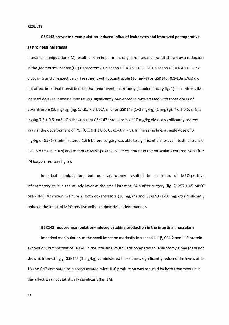
RESULTS
GSK143 prevented manipulation-induced influx of leukocytes and improved postoperative
gastrointestinal transit
Intestinal manipulation (IM) resulted in an impairment of gastrointestinal transit shown by a reduction
in the geometrical center (GC) (laparotomy + placebo GC = 9.5 ± 0.3, IM + placebo GC = 4.4 ± 0.3, P <
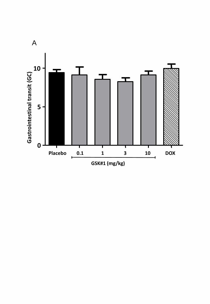
0.05, n= 5 and 7 respectively). Treatment with doxantrazole (10mg/kg) or GSK143 (0.1-10mg/kg) did
not affect intestinal transit in mice that underwent laparotomy (supplementary fig. 1). In contrast, IM-
induced delay in intestinal transit was significantly prevented in mice treated with three doses of
doxantrazole (10 mg/kg) (fig. 1: GC: 7.2 ± 0.7, n=6) or GSK143 (1–3 mg/kg) (1 mg/kg): 7.6 ± 0.6, n=8; 3
mg/kg 7.3 ± 0.5, n=8). On the contrary GSK143 three doses of 10 mg/kg did not significantly protect
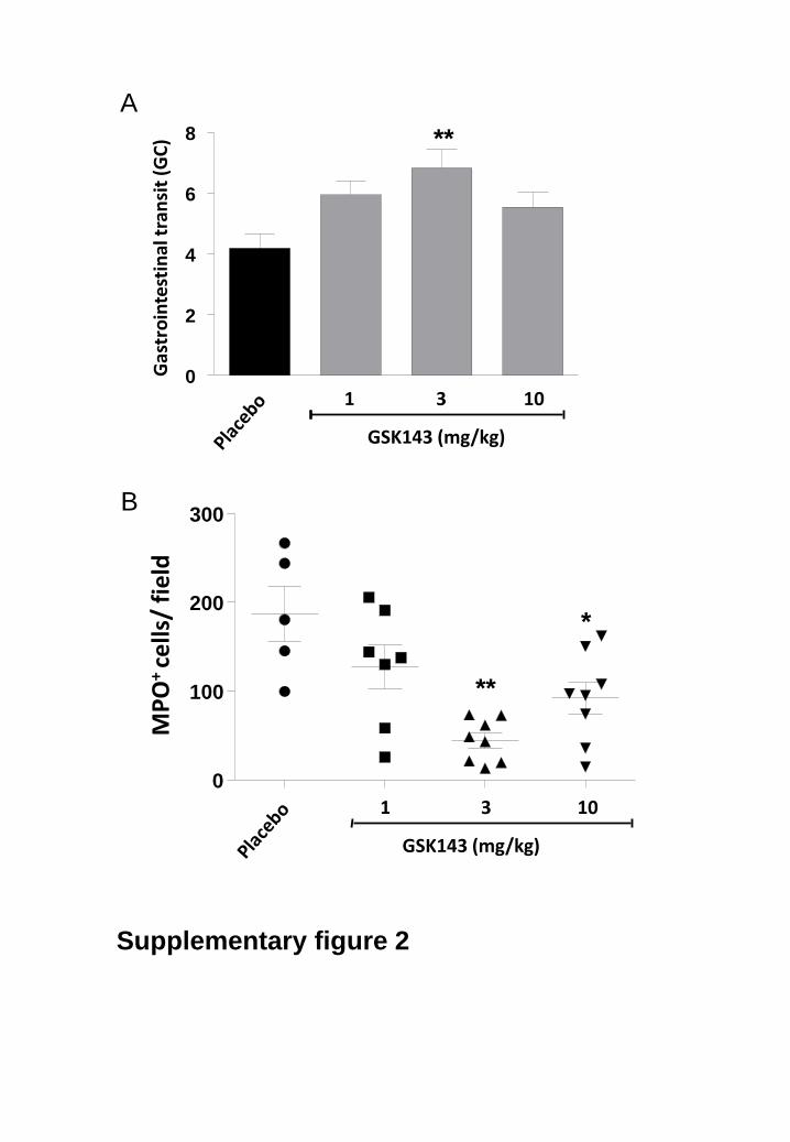
against the development of POI (GC: 6.1 ± 0.6; GSK143: n = 9). In the same line, a single dose of 3
mg/kg of GSK143 administered 1.5 h before surgery was able to significantly improve intestinal transit
(GC: 6.83 ± 0.6, n = 8) and to reduce MPO-positive cell recruitment in the muscularis externa 24 h after
IM (supplementary fig. 2).
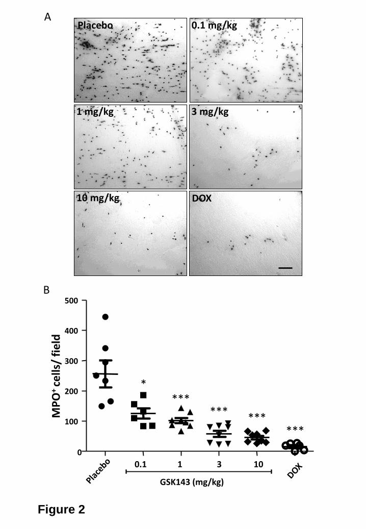
Intestinal manipulation, but not laparotomy resulted in an influx of MPO-positive
inflammatory cells in the muscle layer of the small intestine 24 h after surgery (fig. 2: 257 ± 45 MPO+
cells/HPF). As shown in figure 2, both doxantrazole (10 mg/kg) and GSK143 (1-10 mg/kg) significantly
reduced the influx of MPO positive cells in a dose dependent manner.
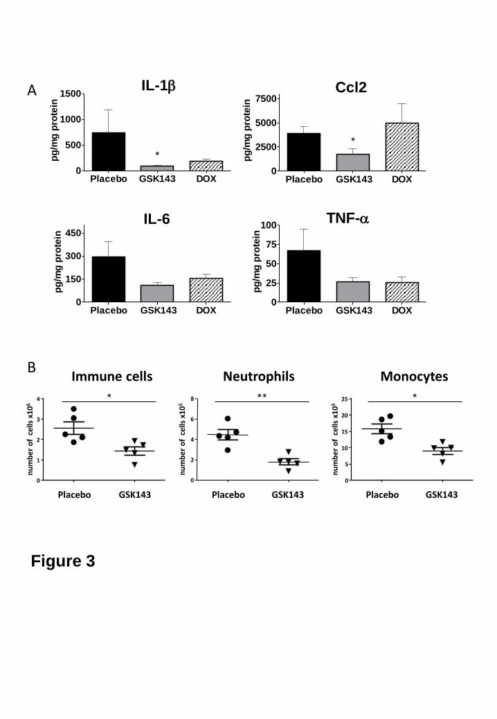
GSK143 reduced manipulation-induced cytokine production in the intestinal muscularis
Intestinal manipulation of the small intestine markedly increased IL-1β, CCL-2 and IL-6 protein
expression, but not that of TNF-α, in the intestinal muscularis compared to laparotomy alone (data not
shown). Interestingly, GSK143 (1 mg/kg) administered three times significantly reduced the levels of IL-
1β and Ccl2 compared to placebo treated mice. IL-6 production was reduced by both treatments but
this effect was not statistically significant (fig. 3A).

14
As Syk inhibition resulted in a reduced number of MPO-positive cells and a lower amount of
cytokine secretion in the muscularis layer of manipulated mice, we addressed whether GSK143 may
also affect recruitment of specific subsets of inflammatory cells. As shown in figure 3B, treatment with
GSK143 (1mg/kg) administered three times resulted in a significant reduction of immune cell
recruitment in the muscularis externa. Syk inhibition affected recruitment of both neutrophils and
monocytes suggesting a potent and broad anti-inflammatory effect of this treatment (fig. 3B).
GSK143 inhibited SP and TNP induced peritoneal mast cell degranulation and LPS-induced
activation in macrophages
To define the concentration range of GSK143 to be tested in vitro, blood samples were
collected from mice treated with a single dose of 1, 3 or 10 mg/kg GSK143 (Supplementary figure 3).
Based on these data, the effect of GSK143 in the concentration range of 0.03-10 uM GSK143 on
isolated mast cells and macrophages was further studied.
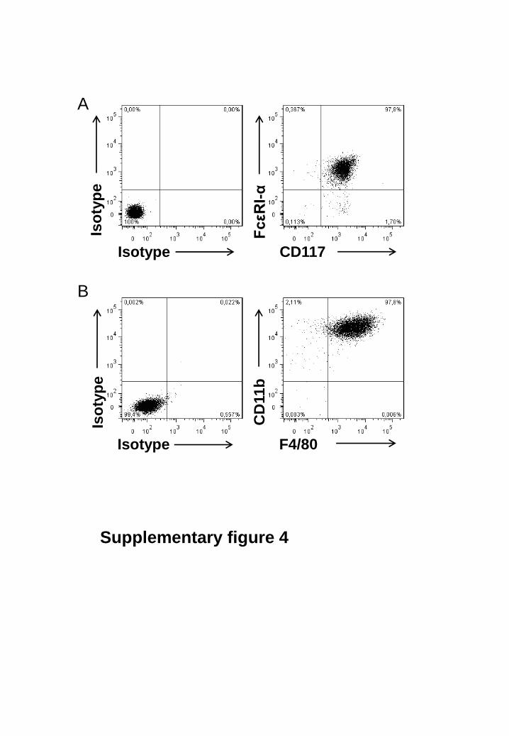
Freshly isolated peritoneal cells from C57NL/BL6 mice were cultured for 4-7 weeks yielding a >
94% (FcεRI+, CD117+) pure PMC population resulting from an expansion of differentiated PMC in the
presence of 100 ng/mL SCF (supplementary fig. 4A). Basal release of β-hexosaminidase by PMC
incubated without stimulus (control) was below 7% of total cellular β-hexosaminidase content.
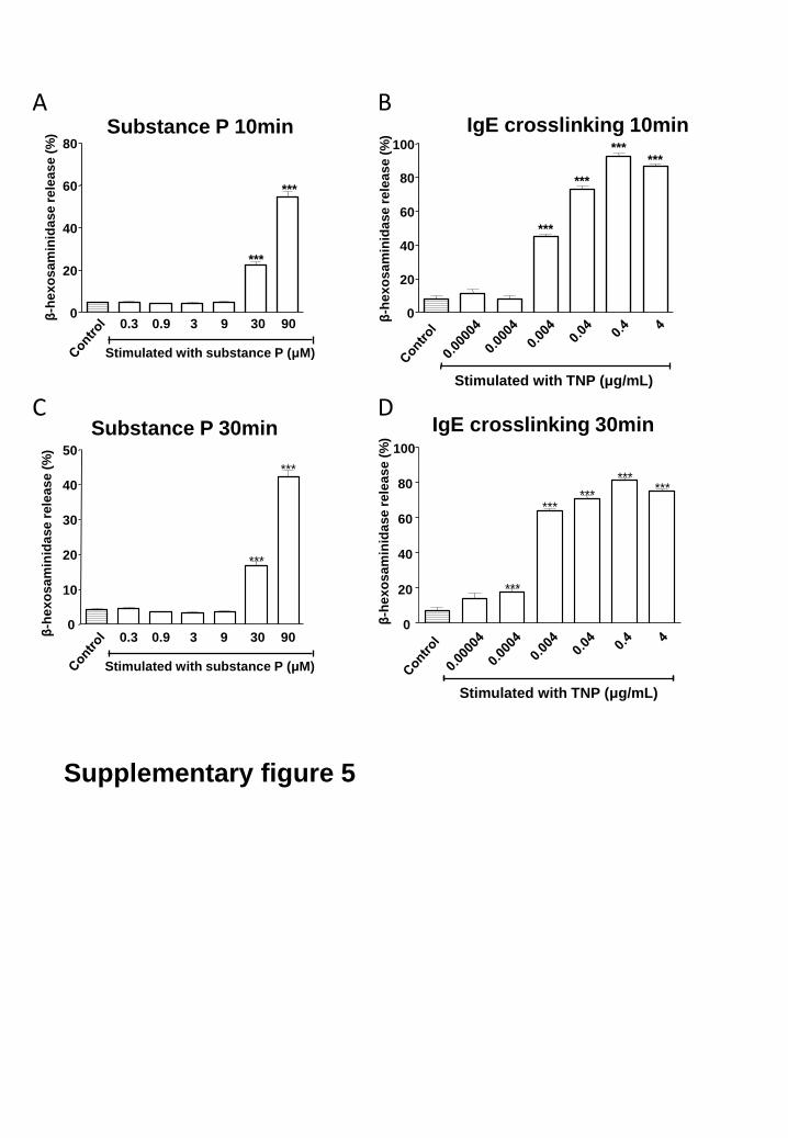
Stimulation with SP (0-90 μM) or TNP (0-4 μg/mL) for 10 minutes resulted in a concentration-
dependent response of β-hexosaminidase release with maximal release 54.7 ± 2.6% and 92.6 ± 2.2% of
total cellular β-hexosaminidase content for SP and TNP respectively (supplementary fig. 5). Stimulation
with SP or TNP for 30 minutes showed similar results (supplementary fig. 5).
Subsequently, the effect of GSK143, vehicle or doxantrazole was studied on PMC stimulated
with 90 μM SP or 0.04 μg/mL TNP. Pretreatment with doxantrazole (227 µM) significantly reduced SP
induced β-hexosaminidase release from 43.5 ± 1.0% of total cellular content to 2.9 ± 0.2%. β-
hexosaminidase release by SP stimulated PMC was also inhibited significantly by ≥ 0.3 μM GSK143, but
to a lesser extent than TNP-induced mast cell activation. (fig. 4A). The TNP induced β-hexosaminidase
release was 70.3 ± 4.3% of total cellular content. Pretreatment with doxantrazole (227 µM) and
15
GSK143 (≥ 0.3 μM) significantly reduced this release to a maximum of 2.8 and 8.7% respectively (fig.
4B).
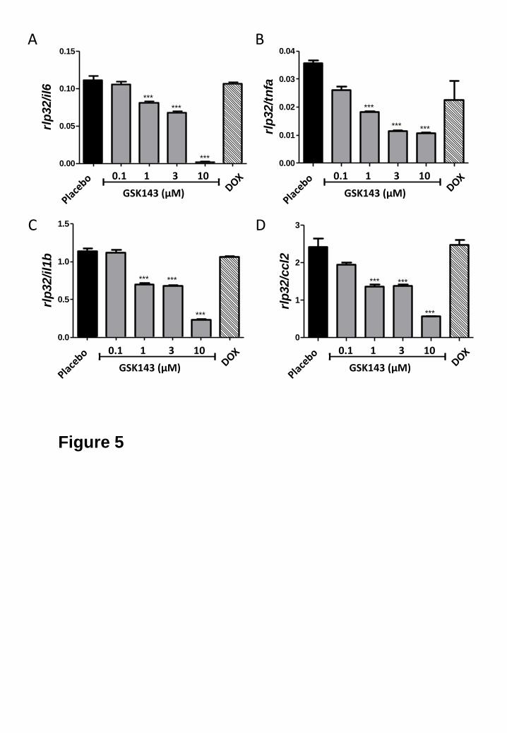
Cultured bone marrow derived macrophages (purity > 95% (CD11b, F4/80; supplementary fig.
4B) from C57NL/BL6 mice were pretreated with GSK143 (0.1-10 μM) or doxantrazole (227 μM) for 30
minutes prior LPS stimulation (100 ng/ml). Two hours after stimulation, macrophages were harvested
and cytokine expression was assessed by quantitative PCR. GSK143 significantly reduced expression of
cytokines such as IL-6, TNF-a, IL-1β and CCL2 (fig. 5). Interestingly, pretreatment with doxantrazole
(227 µM) did not affect macrophage activation.
16
DISCUSSION
Postoperative ileus is mediated by intestinal inflammation resulting from manipulation-
induced mast cell and macrophage activation. In the present study, we investigated the anti-
inflammatory effect of a new Syk inhibitor and its ability to reduce POI. The Syk inhibitor GSK143
significantly reduced the inflammatory response to intestinal manipulation thereby preventing POI. In
addition, we demonstrated that GSK143 inhibited both FcεRI and SP mediated degranulation of PMC
and endotoxin-induced macrophage activation. Taken together, these data strongly suggest that Syk
inhibition may represent a new therapeutic approach for POI.
Syk is required for FcεRI signalling in mast cells and activates intracellular signaling cascades
involved in the transcription and translation of inflammatory mediators.(22;23) Syk inhibitors suppress
the signaling cascades that normally lead to degranulation of mast cells.(11;22-24) In animal models
Syk inhibitors have successfully prevented mast cell mediated inflammatory diseases such as
rheumatoid arthritis and allergic rhinitis. (25;26) Moreover, Syk has been reported to play a crucial role
in macrophage activation.(13-15) As both mast cells and macrophages have been implicated in the
pathophysiology of POI, the present study was designed to establish whether blockade of intracellular
Syk could represent an alternative approach to inhibit immune cell activation evoked by intestinal
manipulation and thus represent a new tool to shorten POI.
At first, we evaluated the effect of GSK143 on primary cultured PMC. We have chosen to study
this subset of mast cells as they functionally resemble connective tissue mast cells,(27) the
subpopulation of mast cells most likely involved in POI.(1;27) In addition to IgE crosslinking, mast cells
can also be activated by SP released by visceral afferent nerves with subsequent activation of
inflammatory cells, a mechanism referred to as neurogenic inflammation.(5;27;27;28) In our in vitro
experiments, we indeed showed concentration-dependent activation of PMC by SP and IgE
crosslinking. In addition, we demonstrated that GSK143 significantly blocked the FcεRI and SP
mediated degranulation of PMC. Of note, the concentrations of GSK143 inhibiting the activation of
mast cells and macrophages in vitro were in the range of serum levels obtained following single
17
administration of the compound. Interestingly, the inhibitory effect of GSK143 was more potent for β-
hexosaminidase release induced by IgE crosslinking compared with SP. This finding suggests that the
signaling pathways involved in SP-induced mast cell activation are less dependent on spleen tyrosine
phosphorylation compared to the FcεRI induced degranulation. Several lines of evidence indicate that
SP can stimulate mast cells not only via its NK1 receptor, but also by NK1 receptor-independent
pathways. Notably, at high concentrations SP induces mast cell degranulation by receptor-
independent pathways,(29) which is mediated by G protein(s), protein kinase C, calcium, and
phospholipase C.(29-35) These data would suggest that in addition to NK1 receptor mediated
activation of Syk, other mechanisms may be involved explaining why GSK143 markedly inhibited FcεRI
mediated and, to a lesser degree at higher drug concentrations, SP mediated degranulation.
Alternatively, if SP-induced mast cell activation does not involve Syk, our data would indicate that
GSK143 interacts with different intracellular signaling pathways. Potential mechanism could be
competitive inhibition at the SP receptor level by membrane incorporation of GSK143 resulting in the
competition of SP binding to the cell membrane surface proteoglycans.(29;36;37) Secondly, GSK143
may interfere with intracellular inositol triphosphate and subsequent increase intracellular calcium
ions ultimately inhibiting degranulation.(38;39) An in-depth analysis of these signaling pathways was
beyond the scope of this article, and further study is needed to fully understand the mechanisms
underlying Syk-inhibition of SP induced mast cell degranulation.
Syk inhibition has been proposed as an important therapeutic strategy for the treatment of
mast cell mediated upper airway diseases, such as allergic rhinitis.(40) Furthermore, Weinblatt et al.
detected mast cells and Syk expression in the synovium of rheumatoid arthritis patients and
demonstrated a significant clinical improvement after treatment with an oral Syk inhibitor.(41) As a
consequence, inhibition of Syk has received increasing attention as new therapeutic approach for a
variety of disorders. Previously, we have shown in mice that mast cells are important players in
triggering the local intestinal inflammatory response leading to POI.(42-44) In patients undergoing
surgery, even gentle inspection of the intestine at the very beginning of the surgical procedure triggers
the release of mast cell mediators.(43) In accordance with these data, recent in vitro work
18
demonstrated that Syk-deficient mast cells fail to release mast cell mediators.(10) In the study
reported here, GSK143 significantly reduced the upregulation of pro-inflammatory cytokines in the
intestinal muscularis 24 h after intestinal manipulation. Moreover, GSK143 and the mast cell stabilizer
doxantrazole attenuated the leukocytic influx and in addition improved gastrointestinal transit. This
confirms the beneficial effect of Syk inhibition on the postoperative inflammatory phase within the
intestine.(24)
Syk has been recently reported to also represent a key player in the regulation of the NF-kB
pathway in LPS-treated macrophages. Indeed, inhibition of Syk signalling in rat alveolar as well as in
peritoneal macrophages and in the monocytic cell line THP-1 resulted in reduction of pro-
inflammatory cytokine secretion.(14) In the present study, we evaluated the effect of GSK143 on
cultured macrophages treated with endotoxin. In line with previous reports, we showed that GSK143
reduced the expression of IL-6, TNF-a, IL-1β and CCL2 in the intestinal muscularis from intestinal
manipulated mice. As macrophages are key players in orchestrating the leukocytic influx in the
manipulated intestine (12), these data suggest that interaction of GSK143 with macrophages in the
intestinal muscularis also contributes to the beneficial effect of GSK143 reducing muscular
inflammation. Similar findings were previously reported by Moore et al. as treatment of mice with the
inhibitor of protein tyrosine kinase tyrphostin reduced inflammatory influx and upregulation of pro-
inflammatory cytokines, and significantly inhibited activation of NF-kB.(3) Taken together, our in vitro
data indicate that GSK143 may inhibit at the same time mast cells and macrophages exerting a broad
anti-inflammatory effect in vivo. It should be emphasized though that Syk also affects the adaptive
immune system.(45) Hence, we cannot exclude that the beneficial effects of GSK143 on POI reported
here may extend beyond its interaction with mast cells and macrophages.(24)
To date, the exact triggers activating mast cells and macrophages during abdominal surgery
are still unclear but besides neuropeptides (such as SP, VIP and CGRP) and specific antigens (via IgE
crosslinking)(28) a variety of stimuli, including bacterial components and several physical stimuli could
be involved. Physical stimuli may be perioperative temperature changes, or the inevitable surgical
induced tissue damage that may activate mast cells via the local release of Damage Associated
19
Molecular Pattern Molecules, IL-1, reactive oxygen species and complement fragments such as C3a
and C5a.(46) Interestingly, Pamuk et al. recently investigated the ability of a Syk inhibitor to protect
mice against mesenteric ischemia-reperfusion induced injury and found that both local and remote
lung injury were reduced with a significant reduction of leukocytic infiltration,(47) suggesting the use
of Syk inhibitors in the suppression of tissue damage evoked inflammatory response. The effect of
GSK143 on resident macrophages or peritoneal mast cells should be further evaluated by studying the
levels of Syk phosphorylation. Nonetheless, our data indicate that GSK143 inhibits mast cell and
macrophage activation, and consequently prevents the inflammatory cascade leading to POI.(12;42-
44)
Clinical studies have shown that Syk inhibitors are efficient in allergy,(48) immune
thrombocytopenic purpura,(49) B-cell lineage malignancies and autoimmune diseases like rheumatoid
arthritis.(41;50) Importantly, these compounds were mostly well tolerated,(48) and not associated
with serious side effects.(41;47;49-51) Syk inhibition in patients with rheumatoid arthritis for 6 months
was reported to be associated with the development of elevated blood pressure, mild neutropenia
and gastrointestinal adverse effects such as gastritis and nausea.(41) Although these side effects may
develop after prolonged use of Syk inhibitors, it is suspected that even milder or no side effects will
develop in postoperative patients who require shorter treatment. This is in line with our experimental
model where one single dose of GSK143 prior surgery was able to prevent delay in gastrointestinal
transit and to significantly reduce intestinal inflammation.
In conclusion, our study showed that GSK143 inhibited degranulation of peritoneal mast cells
and reduced the expression of inflammatory cytokines by macrophage. In addition, Syk inhibition
improved intestinal inflammation and intestinal transit, suggesting that GSK143 can be a useful tool to
treat POI.
20
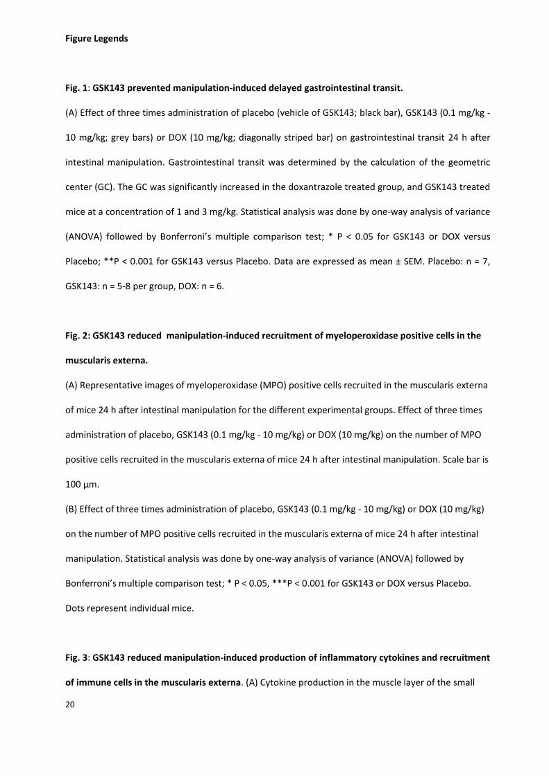
Figure Legends
Fig. 1: GSK143 prevented manipulation-induced delayed gastrointestinal transit.
(A) Effect of three times administration of placebo (vehicle of GSK143; black bar), GSK143 (0.1 mg/kg -
10 mg/kg; grey bars) or DOX (10 mg/kg; diagonally striped bar) on gastrointestinal transit 24 h after
intestinal manipulation. Gastrointestinal transit was determined by the calculation of the geometric
center (GC). The GC was significantly increased in the doxantrazole treated group, and GSK143 treated
mice at a concentration of 1 and 3 mg/kg. Statistical analysis was done by one-way analysis of variance
(ANOVA) followed by Bonferroni’s multiple comparison test; * P < 0.05 for GSK143 or DOX versus
Placebo; **P < 0.001 for GSK143 versus Placebo. Data are expressed as mean ± SEM. Placebo: n = 7,
GSK143: n = 5-8 per group, DOX: n = 6.
Fig. 2: GSK143 reduced manipulation-induced recruitment of myeloperoxidase positive cells in the
muscularis externa.
(A) Representative images of myeloperoxidase (MPO) positive cells recruited in the muscularis externa
of mice 24 h after intestinal manipulation for the different experimental groups. Effect of three times
administration of placebo, GSK143 (0.1 mg/kg - 10 mg/kg) or DOX (10 mg/kg) on the number of MPO
positive cells recruited in the muscularis externa of mice 24 h after intestinal manipulation. Scale bar is
100 μm.
(B) Effect of three times administration of placebo, GSK143 (0.1 mg/kg - 10 mg/kg) or DOX (10 mg/kg)
on the number of MPO positive cells recruited in the muscularis externa of mice 24 h after intestinal
manipulation. Statistical analysis was done by one-way analysis of variance (ANOVA) followed by
Bonferroni’s multiple comparison test; * P < 0.05, ***P < 0.001 for GSK143 or DOX versus Placebo.
Dots represent individual mice.
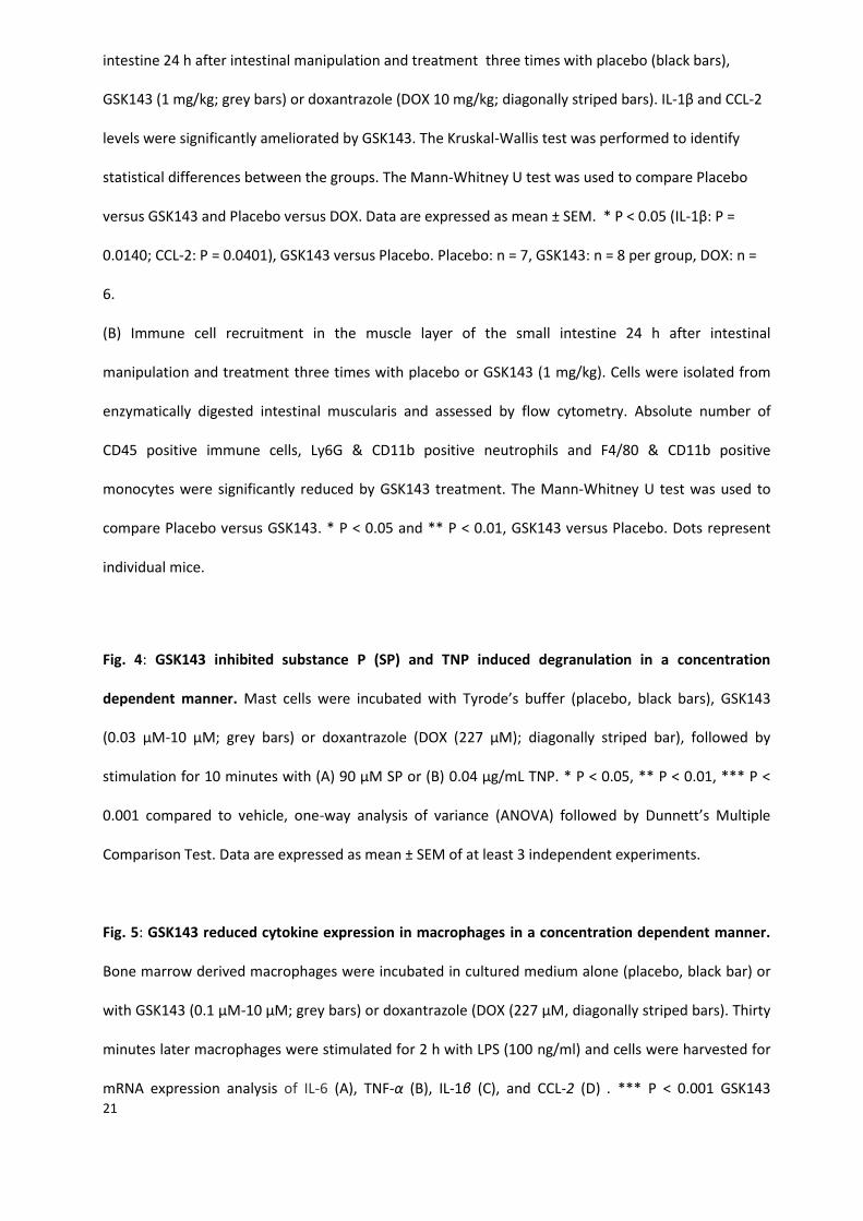
Fig. 3: GSK143 reduced manipulation-induced production of inflammatory cytokines and recruitment
of immune cells in the muscularis externa. (A) Cytokine production in the muscle layer of the small
21
intestine 24 h after intestinal manipulation and treatment three times with placebo (black bars),
GSK143 (1 mg/kg; grey bars) or doxantrazole (DOX 10 mg/kg; diagonally striped bars). IL-1β and CCL-2
levels were significantly ameliorated by GSK143. The Kruskal-Wallis test was performed to identify
statistical differences between the groups. The Mann-Whitney U test was used to compare Placebo
versus GSK143 and Placebo versus DOX. Data are expressed as mean ± SEM. * P < 0.05 (IL-1β: P =
0.0140; CCL-2: P = 0.0401), GSK143 versus Placebo. Placebo: n = 7, GSK143: n = 8 per group, DOX: n =
6.
(B) Immune cell recruitment in the muscle layer of the small intestine 24 h after intestinal
manipulation and treatment three times with placebo or GSK143 (1 mg/kg). Cells were isolated from
enzymatically digested intestinal muscularis and assessed by flow cytometry. Absolute number of
CD45 positive immune cells, Ly6G & CD11b positive neutrophils and F4/80 & CD11b positive
monocytes were significantly reduced by GSK143 treatment. The Mann-Whitney U test was used to
compare Placebo versus GSK143. * P < 0.05 and ** P < 0.01, GSK143 versus Placebo. Dots represent
individual mice.
Fig. 4: GSK143 inhibited substance P (SP) and TNP induced degranulation in a concentration
dependent manner. Mast cells were incubated with Tyrode’s buffer (placebo, black bars), GSK143
(0.03 μM-10 μM; grey bars) or doxantrazole (DOX (227 μM); diagonally striped bar), followed by
stimulation for 10 minutes with (A) 90 μM SP or (B) 0.04 μg/mL TNP. * P < 0.05, ** P < 0.01, *** P <
0.001 compared to vehicle, one-way analysis of variance (ANOVA) followed by Dunnett’s Multiple
Comparison Test. Data are expressed as mean ± SEM of at least 3 independent experiments.
Fig. 5: GSK143 reduced cytokine expression in macrophages in a concentration dependent manner.
Bone marrow derived macrophages were incubated in cultured medium alone (placebo, black bar) or
with GSK143 (0.1 μM-10 μM; grey bars) or doxantrazole (DOX (227 μM, diagonally striped bars). Thirty
minutes later macrophages were stimulated for 2 h with LPS (100 ng/ml) and cells were harvested for
mRNA expression analysis of IL-6 (A), TNF-α (B), IL-1β (C), and CCL-2 (D) . *** P < 0.001 GSK143
22
compared to vehicle, one-way analysis of variance (ANOVA) followed by Dunnett’s Multiple
Comparison Test. Data are expressed as mean ± SEM of at least 3 independent experiments.
23
Supplementary figure legends.
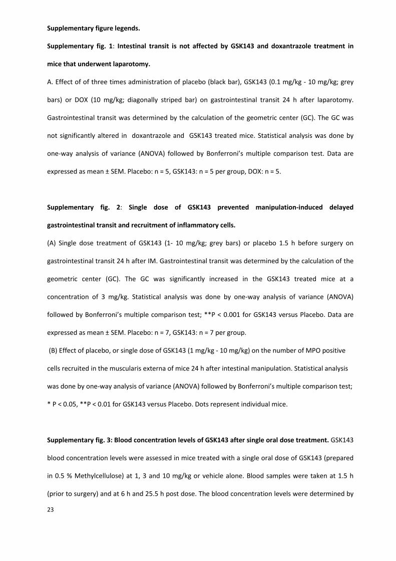
Supplementary fig. 1: Intestinal transit is not affected by GSK143 and doxantrazole treatment in
mice that underwent laparotomy.
A. Effect of of three times administration of placebo (black bar), GSK143 (0.1 mg/kg - 10 mg/kg; grey
bars) or DOX (10 mg/kg; diagonally striped bar) on gastrointestinal transit 24 h after laparotomy.
Gastrointestinal transit was determined by the calculation of the geometric center (GC). The GC was
not significantly altered in doxantrazole and GSK143 treated mice. Statistical analysis was done by
one-way analysis of variance (ANOVA) followed by Bonferroni’s multiple comparison test. Data are
expressed as mean ± SEM. Placebo: n = 5, GSK143: n = 5 per group, DOX: n = 5.
Supplementary fig. 2: Single dose of GSK143 prevented manipulation-induced delayed
gastrointestinal transit and recruitment of inflammatory cells.
(A) Single dose treatment of GSK143 (1- 10 mg/kg; grey bars) or placebo 1.5 h before surgery on
gastrointestinal transit 24 h after IM. Gastrointestinal transit was determined by the calculation of the
geometric center (GC). The GC was significantly increased in the GSK143 treated mice at a
concentration of 3 mg/kg. Statistical analysis was done by one-way analysis of variance (ANOVA)
followed by Bonferroni’s multiple comparison test; **P < 0.001 for GSK143 versus Placebo. Data are
expressed as mean ± SEM. Placebo: n = 7, GSK143: n = 7 per group.
(B) Effect of placebo, or single dose of GSK143 (1 mg/kg - 10 mg/kg) on the number of MPO positive
cells recruited in the muscularis externa of mice 24 h after intestinal manipulation. Statistical analysis
was done by one-way analysis of variance (ANOVA) followed by Bonferroni’s multiple comparison test;
* P < 0.05, **P < 0.01 for GSK143 versus Placebo. Dots represent individual mice.
Supplementary fig. 3: Blood concentration levels of GSK143 after single oral dose treatment. GSK143
blood concentration levels were assessed in mice treated with a single oral dose of GSK143 (prepared
in 0.5 % Methylcellulose) at 1, 3 and 10 mg/kg or vehicle alone. Blood samples were taken at 1.5 h
(prior to surgery) and at 6 h and 25.5 h post dose. The blood concentration levels were determined by
24
reverse phase liquid chromatography coupled to tandem mass spectrometry (LC-MS/MS). (A) Graph
presenting the blood concentration levels obtained after a single oral dose of GSK143. (B) Data (mean
± sd) from each group of animals surgically treated during this study at 1.5, 6 and 25.5 h post-dose.
Supplementary fig. 4: FACS plot of cultured mouse peritoneal mast cells and bone marrow derived
macrophages. (A) FcεRI and CD117 expression was assessed by flow cytometry in cultured mouse
peritoneal mast cells. Only more than 94% pure PMC (FcεRI+, CD117+) populations were used for the
experiments. Data are representative of 3 experiments. (B) CD11b and F4/80 expression was assessed
by flow cytometry mouse bone marrow derived macrophages (BMDMs) after 10 days of culture. Only
more than 94% pure BMDMs (CD11b+, F4/80+) populations were used for the experiments. Data are
representative of 3 independent experiments.
Supplementary fig. 5: Substance P and TNP induced concentration-dependent peritoneal mast cell
degranulation. Mast cells were incubated with (A, C) substance P; 0-90 μM and (B, D) trinitrophenyl
(TNP); 0-4 μg/mL). The β-hexosaminidase release was significantly increased by SP at a concentration
of ≥ 30 μM and by TNP at a concentration of ≥ 0.004 μg/mL. Cells incubated with vehicle (Control)
reflect the basal release. *** P < 0.001 compared to control, one-way analysis of variance (ANOVA)
followed by Dunnett’s Multiple Comparison Test. Data are expressed as mean ± SEM of at least 3
independent experiments.
Supplementary table S1. Primer sequences used for qRT-PCR.
Gene Sense Antisense
Rpl32 5’-AAGCGAAACTGGCGGAAAC-3’ 5’-TAACCGATGTTGGGCATCAG-3’
Tnfa 5’- TCTTCTCATTCCTGCTTGTGG-3’ 5’- CACTTGGTGGTTTGCTACGA-3’
Il1β 5’-GACCTTCCAGGATGAGGACA-3’ 5’-TCCATTGAGGTGGAGAGCTT-3’
Il6 5’-CCATAGCTACCTGGAGTACATG-3’ 5’-TGGAAATTGGGGTAGGAAGGAC-3’
Ccl2 5’-CACGTGTTGGCTCAGCCAGATGC-3’ 5’-CCTTCTTGGGGTCAGCACAGACC-3’
25
Reference List
(1) Boeckxstaens GE, de Jonge WJ. Neuroimmune mechanisms in postoperative ileus. Gut 2009;58(9):1300-11.
(2) Kalff JC, Carlos TM, Schraut WH et al. Surgically induced leukocytic infiltrates within the rat intestinal muscularis mediate postoperative ileus. Gastroenterology 1999;117(2):378-87.
(3) Moore BA, Turler A, Pezzone MA et al. Tyrphostin AG 126 inhibits development of postoperative ileus induced by surgical manipulation of murine colon. Am J Physiol Gastrointest Liver Physiol 2004;286(2):G214-G224.
(4) The FO, de Jonge WJ, Bennink RJ et al. The ICAM-1 antisense oligonucleotide ISIS-3082 prevents the development of postoperative ileus in mice. Br J Pharmacol 2005;146(2):252-8.
(5) de Jonge WJ, van den Wijngaard RM, The FO et al. Postoperative ileus is maintained by intestinal immune infiltrates that activate inhibitory neural pathways in mice. Gastroenterology 2003;125(4):1137-47.
(6) The FO, Buist MR, Lei A et al. The role of mast cell stabilization in treatment of postoperative ileus: a pilot study. Am J Gastroenterol 2009;104(9):2257-66.
(7) Abu-Dalu R, Zhang JM, Hanani M. The actions of ketotifen on intestinal smooth muscles. Eur J Pharmacol 1996;309(2):189-93.
(8) Dyson AJ, Mackay AD. Ketotifen in adult asthma. Br Med J 1980;280(6211):360-1.
(9) Schwarzer G, Bassler D, Mitra A et al. Ketotifen alone or as additional medication for long-term control of asthma and wheeze in children. Cochrane Database Syst Rev 2004;(1):CD001384.
(10) Rivera J, Olivera A. A current understanding of Fc epsilon RI-dependent mast cell activation. Curr Allergy Asthma Rep 2008;8(1):14-20.
(11) Rossi AB, Herlaar E, Braselmann S et al. Identification of the Syk kinase inhibitor R112 by a human mast cell screen. J Allergy Clin Immunol 2006;118(3):749-55.
(12) Wehner S, Behrendt FF, Lyutenski BN et al. Inhibition of macrophage function prevents intestinal inflammation and postoperative ileus in rodents. Gut 2007;56(2):176-85.
(13) Lee YG, Chain BM, Cho JY. Distinct role of spleen tyrosine kinase in the early phosphorylation of inhibitor of kappaB alpha via activation of the phosphoinositide-3-kinase and Akt pathways. Int J Biochem Cell Biol 2009;41(4):811-21.
(14) Ulanova M, Asfaha S, Stenton G et al. Involvement of Syk protein tyrosine kinase in LPS-induced responses in macrophages. J Endotoxin Res 2007;13(2):117-25.
(15) Stenton GR, Ulanova M, Dery RE et al. Inhibition of allergic inflammation in the airways using aerosolized antisense to Syk kinase. J Immunol 2002;169(2):1028-36.

26
(16) Liddle J, Atkinson FL, Barker MD et al. Discovery of GSK143, a highly potent, selective and orally efficacious spleen tyrosine kinase inhibitor. Bioorg Med Chem Lett 2011;21(20):6188-94.
(17) The F, Cailotto C, van d, V et al. Central activation of the cholinergic anti-inflammatory pathway reduces surgical inflammation in experimental post-operative ileus. Br J Pharmacol 2011;163(5):1007-16.
(18) Schmidt J, Stoffels B, Moore BA et al. Proinflammatory role of leukocyte-derived Egr-1 in the development of murine postoperative ileus. Gastroenterology 2008;135(3):926-36, 936.
(19) Livak KJ, Schmittgen TD. Analysis of relative gene expression data using real-time quantitative PCR and the 2(-Delta Delta C(T)) Method. Methods 2001;25(4):402-8.
(20) Costello PS, Turner M, Walters AE et al. Critical role for the tyrosine kinase Syk in signalling through the high affinity IgE receptor of mast cells. Oncogene 1996;13(12):2595-605.
(21) Matsubara S, Li G, Takeda K et al. Inhibition of spleen tyrosine kinase prevents mast cell activation and airway hyperresponsiveness. Am J Respir Crit Care Med 2006;173(1):56-63.
(22) Mocsai A, Ruland J, Tybulewicz VL. The SYK tyrosine kinase: a crucial player in diverse biological functions. Nat Rev Immunol 2010;10(6):387-402.
(23) Cohen S, Fleischmann R. Kinase inhibitors: a new approach to rheumatoid arthritis treatment. Curr Opin Rheumatol 2010;22(3):330-5.
(24) Denyer J, Patel V. Syk kinase inhibitors in allergic diseases. Drug News Perspect 2009;22(3):146-50.
(25) Malbec O, Roget K, Schiffer C et al. Peritoneal cell-derived mast cells: an in vitro model of mature serosal-type mouse mast cells. J Immunol 2007;178(10):6465-75.
(26) Bueno L, Fioramonti J, Delvaux M et al. Mediators and pharmacology of visceral sensitivity: from basic to clinical investigations. Gastroenterology 1997;112(5):1714-43.
(27) van der Kleij HP, Ma D, Redegeld FA et al. Functional expression of neurokinin 1 receptors on mast cells induced by IL-4 and stem cell factor. J Immunol 2003;171(4):2074-9.
(28) Bueb JL, Mousli M, Landry Y et al. A pertussis toxin-sensitive G protein is required to induce histamine release from rat peritoneal mast cells by bradykinin. Agents Actions 1990;30(1-2):98-101.
(29) Mousli M, Hugli TE, Landry Y et al. Peptidergic pathway in human skin and rat peritoneal mast cell activation. Immunopharmacology 1994;27(1):1-11.
(30) Mousli M, Bronner C, Landry Y et al. Direct activation of GTP-binding regulatory proteins (G-proteins) by substance P and compound 48/80. FEBS Lett 1990;259(2):260-2.
(31) Chahdi A, Mousli M, Landry Y. Substance P-related inhibitors of mast cell exocytosis act on G-proteins or on the cell surface. Eur J Pharmacol 1998;341(2-3):329-35.
(32) Lorenz D, Wiesner B, Zipper J et al. Mechanism of peptide-induced mast cell degranulation. Translocation and patch-clamp studies. J Gen Physiol 1998;112(5):577-91.
(33) Bueb JL, Mousli M, Bronner C et al. Activation of Gi-like proteins, a receptor-independent effect of kinins in mast cells. Mol Pharmacol 1990;38(6):816-22.
(34) Moon KD, Post CB, Durden DL et al. Molecular basis for a direct interaction between the Syk protein-tyrosine kinase and phosphoinositide 3-kinase. J Biol Chem 2005;280(2):1543-51.

27
(35) Mousli M, Bronner C, Bueb JL et al. Activation of rat peritoneal mast cells by substance P and mastoparan. J Pharmacol Exp Ther 1989;250(1):329-35.
(36) Kobayashi S, Somlyo AV, Somlyo AP. Heparin inhibits the inositol 1,4,5-trisphosphate-dependent, but not the independent, calcium release induced by guanine nucleotide in vascular smooth muscle. Biochem Biophys Res Commun 1988;153(2):625-31.
(37) Ahmed T, Syriste T, Mendelssohn R et al. Heparin prevents antigen-induced airway hyperresponsiveness: interference with IP3-mediated mast cell degranulation? J Appl Physiol 1994;76(2):893-901.
(38) Patou J, Holtappels G, Affleck K et al. Syk-kinase inhibition prevents mast cell activation in nasal polyps. Rhinology 2011;49(1):100-6.
(39) Weinblatt ME, Kavanaugh A, Burgos-Vargas R et al. Treatment of rheumatoid arthritis with a Syk kinase inhibitor: a twelve-week, randomized, placebo-controlled trial. Arthritis Rheum 2008;58(11):3309-18.
(40) de Jonge WJ, The FO, van der Coelen D et al. Mast cell degranulation during abdominal surgery initiates postoperative ileus in mice. Gastroenterology 2004;127(2):535-45.
(41) The FO, Bennink RJ, Ankum WM et al. Intestinal handling-induced mast cell activation and inflammation in human postoperative ileus. Gut 2008;57(1):33-40.
(42) Snoek SA, Dhawan S, van Bree SH et al. Mast cells trigger epithelial barrier dysfunction, bacterial translocation and postoperative ileus in a mouse model. Neurogastroenterol Motil 2011.
(43) Mocsai A, Ruland J, Tybulewicz VL. The SYK tyrosine kinase: a crucial player in diverse biological functions. Nat Rev Immunol 2010;10(6):387-402.
(44) Mecheri S, David B. Unravelling the mast cell dilemma: culprit or victim of its generosity? Immunol Today 1997;18(5):212-5.
(45) Pamuk ON, Tsokos GC. Spleen tyrosine kinase inhibition in the treatment of autoimmune, allergic and autoinflammatory diseases. Arthritis Res Ther 2010;12(6):222.
(46) Meltzer EO, Berkowitz RB, Grossbard EB. An intranasal Syk-kinase inhibitor (R112) improves the symptoms of seasonal allergic rhinitis in a park environment. J Allergy Clin Immunol 2005;115(4):791-6.
(47) Podolanczuk A, Lazarus AH, Crow AR et al. Of mice and men: an open-label pilot study for treatment of immune thrombocytopenic purpura by an inhibitor of Syk. Blood 2009;113(14):3154-60.
(48) Friedberg JW, Sharman J, Sweetenham J et al. Inhibition of Syk with fostamatinib disodium has significant clinical activity in non-Hodgkin lymphoma and chronic lymphocytic leukemia. Blood 2010;115(13):2578-85.
(49) Rivera J, Colbert RA. Healing the Syk through kinase inhibitors. N Engl J Med 2010;363(14):1362-4.
0
2
4
6
8
10
0.1 1 3 10
GSK143 (mg/kg)
** * *
Gas
tro
inte
stin
al t
ran
sit
(GC
)
A
Figure 1
A
Figure 2
0
100
200
300
400
500
0.1 1 3 10
GSK143 (mg/kg)
***
MP
O+
cells
/ fi
eld
B
*** ***
***
*
Placebo
1 mg/kg
10 mg/kg
0.1 mg/kg
3 mg/kg
DOX
Placebo GSK143
0
1
2
3
4
nu
mb
er
of
ce
lls x
10
6
Neutrophils
Placebo GSK143
0
2
4
6
8
nu
mb
er
of
ce
lls x
10
5
Placebo GSK143
0
5
10
15
20
25
nu
mb
er
of
ce
lls x
10
5
Monocytes Immune cells
* ** *
A
B
Figure 3
Placebo GSK143 DOX0
500
1000
1500IL-1
pg
/mg
pro
tein
Placebo GSK143 DOX0
150
300
450
IL-6
pg
/mg
pro
tein
Placebo GSK143 DOX0
25
50
75
100TNF-
pg
/mg
pro
tein
Placebo GSK143 DOX0
2500
5000
7500Ccl2
pg
/mg
pro
tein
*
*
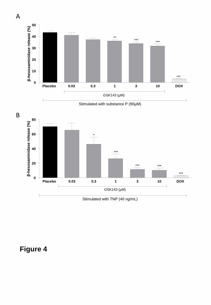
Figure 4
Placebo 0.03 0.3 1 3 10 DOX 0
10
20
30
40
50
GSK143 (μM)
Stimulated with substance P (90μM)
β-h
exo
sam
inid
ase r
ele
ase (
%)
A
***
*** ***
**
Placebo 0.03 0.3 1 3 10 DOX 0
20
40
60
80
Stimulated with TNP (40 ng/mL)
GSK143 (μM)
β-h
exo
sam
inid
ase r
ele
ase (
%)
B
***
***
***
*
***
0.1 1 3 10
GSK143 (μM)
0.00
0.05
0.10
0.15rl
p32/il6
0.00
0.01
0.02
0.03
0.04
rlp
32/t
nfa
0.1 1 3 10
GSK143 (μM)
0.0
0.5
1.0
1.5
rlp
32/il1
b
0.1 1 3 10
GSK143 (μM)
0
1
2
3
rlp
32/c
cl2
0.1 1 3 10
GSK143 (μM)
A
*** ***
***
*** ***
***
***
*** ***
***
***
***
Figure 5
B
C D
Placebo 0.1 1 3 10 DOX
GSK#1 (mg/kg)
0
5
10
Gas
tro
inte
stin
al t
ran
sit
(GC
)
A
Supplementary figure 2
0
2
4
6
8
1 3 10
GSK143 (mg/kg)
Gas
tro
inte
stin
al t
ran
sit
(GC
) **
1 3 10
GSK143 (mg/kg)
0
100
200
300
MP
O+
cells
/ fi
eld
**
*
A
B

A
B
Supplementary figure 3
GSK143 Blood Concentration (uM)
Dose (mg/kg)
Time (h) 1 3 10
1.5 0.15 ± 0.05 0.38 ± 0.24 1.84 ± 1.03
6 0.04 ± 0.02 0.14 ± 0.03 0.68 ± 0.44
25.5 0.03 ± 0.06 0.03 ± 0.01 0.15 ± 0.10
0,0001
0,001
0,01
0,1
1
0 5 10 15 20 25
Blo
od
Co
nce
ntr
atio
n (
uM
)
Time (h)
Mean Profile 1 mg/kg
Mean Profile 3 mg/kg
Mean Profile 10 mg/kg
CD
11b
F4/80
Iso
typ
e
Isotype
FcεR
I-α
CD117
Iso
typ
e
Isotype
A
B
Supplementary figure 4
Supplementary figure 5
0
20
40
60
80
100
***
***
*** ***
IgE crosslinking 10min
β-h
ex
os
am
inid
ase
re
lea
se
(%
)
B
0
20
40
60
80
100
***
*** ***
*** ***
Stimulated with TNP (μg/mL)
β-h
ex
os
am
inid
ase
rele
as
e (
%)
IgE crosslinking 30min D
Stimulated with TNP (μg/mL)
0
10
20
30
40
50
***
***
β-h
ex
os
am
inid
ase
rele
as
e (
%)
Substance P 30min C
0.3 0.9 3 9 30 90
Stimulated with substance P (μM)
0
20
40
60
80
***
***
Substance P 10min
β-h
ex
os
am
inid
ase
re
lea
se
(%
)
A
0.3 0.9 3 9 30 90
Stimulated with substance P (μM)