Embed Size (px)
Citation preview
Inhibition of Rho-Kinase Attenuates Hypoxia-InducedAngiogenesis in the Pulmonary Circulation
Jean-Marc Hyvelin,* Katherine Howell,* Alistair Nichol, Christine M. Costello,Robert J. Preston, Paul McLoughlin
Abstract—Pulmonary hypertension (PH) is a common complication of chronic hypoxic lung diseases, which increasemorbidity and mortality. Hypoxic PH has previously been attributed to structural changes in the pulmonary vasculatureincluding narrowing of the vascular lumen and loss of vessels, which produce a fixed increase in resistance. Usingquantitative stereology, we now show that chronic hypoxia caused PH and remodeling of the blood vessel walls in ratsbut that this remodeling did not lead to structural narrowing of the vascular lumen. Sustained inhibition of theRhoA/Rho-kinase pathway throughout the period of hypoxic exposure attenuated PH and prevented remodeling inintra-acinar vessels without enlarging the structurally determined lumen diameter. In chronically hypoxic lungs, acuteRho kinase inhibition markedly decreased PVR but did not alter the alveolar to arterial oxygen gap. In addition toincreased vascular resistance, chronic hypoxia induced Rho kinase–dependent capillary angiogenesis. Thus, hypoxic PHwas not caused by fixed structural changes in the vasculature but by sustained vasoconstriction, which was largely Rhokinase dependent. Importantly, this vasoconstriction had no role in ventilation-perfusion matching and optimization ofgas exchange. Rho kinase also mediated hypoxia-induced capillary angiogenesis, a previously unrecognized butpotentially important adaptive response. (Circ Res. 2005;97:185-191.)
Key Words: pulmonary hypertension � angiogenesis � RhoA � Rho-kinase � Y-27632
Sustained pulmonary hypertension (PH) is a common com-plication of chronic hypoxic lung diseases that is strongly
associated with increased morbidity and reduced survival. More-over, the presence of cor pulmonale is an independent predictorof increased mortality, suggesting that PH contributes directly tomortality (reviewed in Hopkins et al1). The increase in pulmo-nary vascular resistance (PVR) caused by chronic hypoxia haspreviously been attributed to structural changes in the vascula-ture, in particular encroachment of the remodeled arteriolar wallsinto the vascular lumen and loss of blood vessels, althoughrecent reports have cast doubt on this paradigm.1–3 In particular,we have recently shown for the first time that hypoxia inducesangiogenesis in the adult pulmonary circulation, a potentiallybeneficial adaptation, and does not cause vessel loss as previ-ously believed.2
The small G-protein RhoA and its downstream effectorRho-kinase (ROCK) play a central role in diverse cellularfunctions including smooth muscle contraction, cytoskeletalrearrangement, cell migration, cell proliferation, and geneexpression.4–8 Given these important functions, it is notsurprising that disturbances of this pathway have been iden-tified as important pathogenetic mechanisms in many dis-
eases of the cardiovascular system, including systemic hyper-tension, arteriosclerosis, and ischemic heart disease.9 –11
Blockade of the RhoA/ROCK pathway effectively correctsblood pressure in a number of animal models of systemichypertension8,11 and is a key regulator of vascular smoothmuscle proliferation and migration in disease-induced sys-temic vascular remodeling.9,10 This pathway also regulatesendothelial cell proliferation and migration. In vitro modelsand in vivo Matrigel implants show that inhibition of RhoAor ROCK prevents growth factor–induced endothelial cellmigration and organization into capillary-like structures12,13
and vessel formation.14 Moreover, ROCK inhibitors attenuatetumor growth in vivo in a manner that is compatible with anantiangiogenic effect.15 Recognition of the therapeutic poten-tial of inhibiting this pathway has lead to the development ofspecific small molecule inhibitors of ROCK.8,11,15
Taken together, these reports suggest that the RhoA/ROCKpathway might contribute to the development of hypoxic PHand the associated pulmonary vascular remodeling. Thepurpose of this study was to assess the potential role of thispathway in the development of chronic hypoxia-induced PHand the associated structural changes in the pulmonary
Original received December 16, 2004; resubmission received April 12, 2005; revised resubmission received June 6, 2005; accepted June 8, 2005.From the Departments of Physiology (J.-M.H., K.H., A.N., R.J.P., P.M.) and Pharmacology (C.M.C.), Conway Institute of Biomolecular and
Biomedical Research and the Dublin Molecular Medicine Centre (P.M.), University College, Dublin, Ireland.Dr P. McLoughlin has received an unrestricted research grant from Actelion, a company that manufactures an endothelin receptor antagonist used in
the treatment of primary pulmonary arterial hypertension.*Both authors contributed equally to this work.Correspondence to Dr Paul McLoughlin, Department of Physiology, University College, Earlsfort Terrace, Dublin 2, Ireland. E-mail [email protected]© 2005 American Heart Association, Inc.
Circulation Research is available at http://circres.ahajournals.org DOI: 10.1161/01.RES.0000174287.17953.83
185
by guest on July 16, 2018http://circres.ahajournals.org/
Dow
nloaded from
by guest on July 16, 2018http://circres.ahajournals.org/
Dow
nloaded from
by guest on July 16, 2018http://circres.ahajournals.org/
Dow
nloaded from
by guest on July 16, 2018http://circres.ahajournals.org/
Dow
nloaded from
by guest on July 16, 2018http://circres.ahajournals.org/
Dow
nloaded from
by guest on July 16, 2018http://circres.ahajournals.org/
Dow
nloaded from
by guest on July 16, 2018http://circres.ahajournals.org/
Dow
nloaded from
by guest on July 16, 2018http://circres.ahajournals.org/
Dow
nloaded from
by guest on July 16, 2018http://circres.ahajournals.org/
Dow
nloaded from
by guest on July 16, 2018http://circres.ahajournals.org/
Dow
nloaded from
by guest on July 16, 2018http://circres.ahajournals.org/
Dow
nloaded from
by guest on July 16, 2018http://circres.ahajournals.org/
Dow
nloaded from
by guest on July 16, 2018http://circres.ahajournals.org/
Dow
nloaded from
by guest on July 16, 2018http://circres.ahajournals.org/
Dow
nloaded from
by guest on July 16, 2018http://circres.ahajournals.org/
Dow
nloaded from
by guest on July 16, 2018http://circres.ahajournals.org/
Dow
nloaded from
by guest on July 16, 2018http://circres.ahajournals.org/
Dow
nloaded from
by guest on July 16, 2018http://circres.ahajournals.org/
Dow
nloaded from
by guest on July 16, 2018http://circres.ahajournals.org/
Dow
nloaded from
vasculature. In particular, we wished to test the hypothesisthat, although chronic inhibition of ROCK would inhibit thedevelopment of hypoxic PH, it would simultaneously inhibithypoxia-induced pulmonary angiogenesis.
Materials and MethodsAn expanded Materials and Methods section can be found in theonline supplement available at http://circres.ahajournals.org.
Hypoxic Pulmonary HypertensionChronic hypoxic PH was induced by housing adult male rats in anormobaric hypoxic chamber (FiO2 0.10) for 1 or 3 weeks aspreviously described.16 Control rats were housed in normoxic con-ditions (FiO2 0.21) in the same room. To assess the effects of chronicROCK inhibition, rats were randomized into 3 groups; 1 group wasmaintained in normoxic conditions (control group), while 2 groupswere housed in hypoxic conditions for 1 week. Y-27632 was used toinhibit the phosphorylation activity of ROCK.4–8,17 One hypoxicgroup (CH-Y27632) received Y-27632 (30 mg � kg�1 � day�1, orally)while the other did not receive Y-27632 (CH group).
Measurement of Vascular Pressures and GasExchange In VivoRats were anesthetized, tracheostomised, paralyzed, and mechani-cally ventilated. Venous and arterial cannulae were placed for drugadministration, blood gas sampling, and blood pressure measure-ment. Right ventricular pressure was measured by introduction of aneedle via a subdiaphragmatic approach just before euthanasia.
Isolated Perfused Lung PreparationLungs were isolated under anesthesia, mechanically ventilated, andperfused at constant low flow with a mixture of blood and physio-logical saline of constant hematocrit so that pulmonary arterialpressure (PAP) was a direct index of PVR.17 Capillary pressure wasassessed by the double occlusion technique.17
Tissue CollectionAt the end of each isolated lung experiment, the left lung (LL) wasfixed under standard conditions for stereological quantification ofstructural vascular changes.2 The right lungs were snap frozen forsubsequent protein and mRNA analysis.
Data AnalysisFor stereological analyses, total volume of specific compartments,vessel and capillary lengths, and alveolar epithelial and capillaryendothelial surface areas were reported per left lung. For normallydistributed data, responses were reported as mean�SEM, n refers tothe number of rats from which tissue was obtained. For multiplecomparisons of means across experimental groups, analysis ofvariance was performed followed by Student–Newman Keuls post-hoc test for pair-wise comparisons. For non-normally distributeddata, multiple comparisons of medians across experimental groupswere performed using the Kruskal–Wallis test followed by Mann–Whitney U with the Bonferonni post-hoc correction; values ofP�0.05 were accepted as statistically significant.
ResultsEffect of Acute ROCK Inhibition on EstablishedHypoxic Pulmonary HypertensionMean peak right ventricular systolic pressure (RVSPpeak) measuredduring ventilation with 30% oxygen (53.5�3.5 mm Hg) in rats(n�5) maintained chronically hypoxic for 3 weeks was signifi-cantly (P�0.01) greater than that in control nonhypoxic rats(28.8�1.4 mm Hg), demonstrating the presence of fixed PH; ie,increased PVR that was not attributable to acute hypoxic pulmonaryvasoconstriction (HPV). After administration of the potent ROCK
inhibitor Y27632 (15 mg � kg�1),11 mean RVSPPeak was reducedsignificantly (P�0.02) to a value of 34.6 (�5.6) mm Hg.
Changes in RVSPPeak in response to Y27632 in vivo maynot have resulted from changes in PVR but may have resultedfrom changes in venous return, cardiac output, left atrialpressure, neurogenic control, and other factors. To examinethe direct effect of ROCK inhibition on PVR in lungschronically hypoxic for 3 weeks, we used an isolated lungpreparation. Mean PAP in chronically hypoxic lungs (n�5)was 15.1 (�0.6) mm Hg and was significantly reduced(P�0.01) to 13.0 (�0.9) mm Hg by Y27632 (3 �m.l�1). Atthe highest Y27632 concentration (100 �m.l�1) mean PAPwas further reduced (P�0.01) to 10.7 (�0.4) mm Hg, a valuethat was similar to that in control nonhypoxic lungs (seebelow and Figure 1).
Thus the dominant mechanism underlying chronic hypoxicPH was sustained vasoconstriction and not structural changesin the pulmonary vasculature. These results suggested thatsustained ROCK inhibition in vivo would prevent the devel-opment of chronic hypoxic PH.
Chronic ROCK Inhibition In VivoTo confirm that chronic exposure to a hypoxic environment for1 week caused fixed PH, we measured RVSPPeak in anesthetizedrats (n�5) during ventilation with 30% oxygen, and found thatafter such exposure RVSPPeak was significantly (P�0.01) ele-vated (42.5�3.0 mm Hg) when compared with control nonhy-poxic rats (28.8�1.4 mm Hg). For the reasons outlined above,further investigation of the role of ROCK in the development ofPH was undertaken using the isolated lung preparation.
Mean right ventricular (RV) weight (see supplementalTable I) in a group maintained hypoxic for 1 week (CHgroup) and in a hypoxic group chronically treated withY-27632 (CH-Y27632) for 1 week were both significantlyhigher than that of the control group. The sustained treatmentwith ROCK inhibitor significantly reduced the RV hypertro-phy compared with hypoxia alone although it did not alter thepolycythemic response to hypoxia (supplemental Table I).
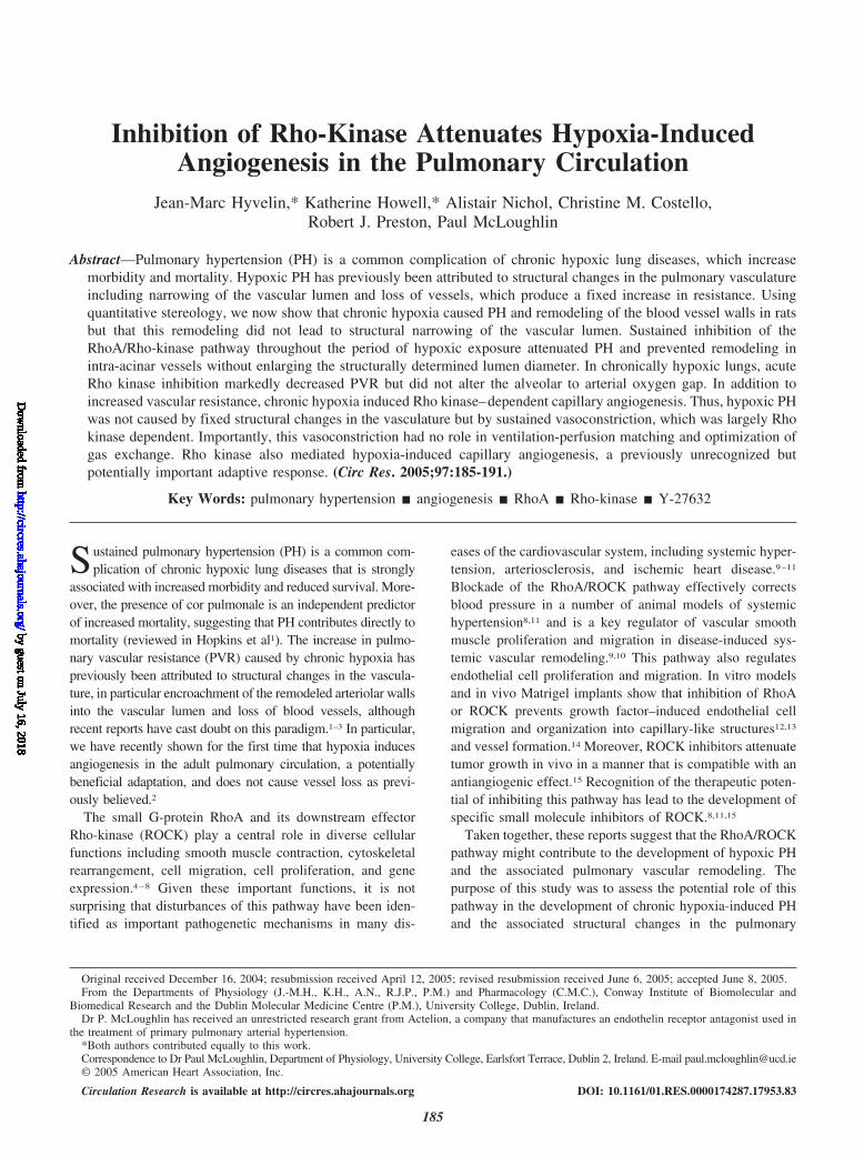
We assessed the total PVR in isolated lungs under 3different conditions, ie, lungs ventilated with 16%, 7%, or 3%O2 (Figure 1). These concentrations give alveolar values thatapproximate normal sea level alveolar pO2, the alveolar pO2
experienced by the rats during hypoxic exposure in the
Figure 1. PAP in isolated control, CH, and CH-Y-27632 lungsventilated with 16%O2, 7%O2, and 3%O2. #significantly differentfrom the 2 other groups at the same inspired oxygen concentra-tion (P�0.001); *significantly different from the other inspiredoxygen concentrations within the same group (P�0.001). N�7in all groups; bars indicate SEM.
186 Circulation Research July 22, 2005
by guest on July 16, 2018http://circres.ahajournals.org/
Dow
nloaded from
chamber, and an alveolar pO2 that produces a maximalhypoxic vasoconstrictor response. Lungs from the controlgroup showed normal low PVR when ventilated with 16% O2
and significant increases in resistance in response to reduc-tion in alveolar O2 indicating normal HPV (Figure 1).Chronically hypoxic lungs ventilated with 7% O2 had signif-icantly elevated vascular resistance compared with controllungs ventilated with the same inspired oxygen, and thisresistance was not reduced by acute return to normal alveolaroxygen concentration (16% O2). Reduction to 3% O2 in thechronically hypoxic lungs caused a further small increase inresistance to a value similar to that seen in control lungs at thesame inspired oxygen (Figure 1). Chronic administration of theROCK inhibitor Y-27632 prevented the development of PH inresponse to sustained exposure to hypoxia and also abolished theacute hypoxic vasoconstrictor response (Figure 1).
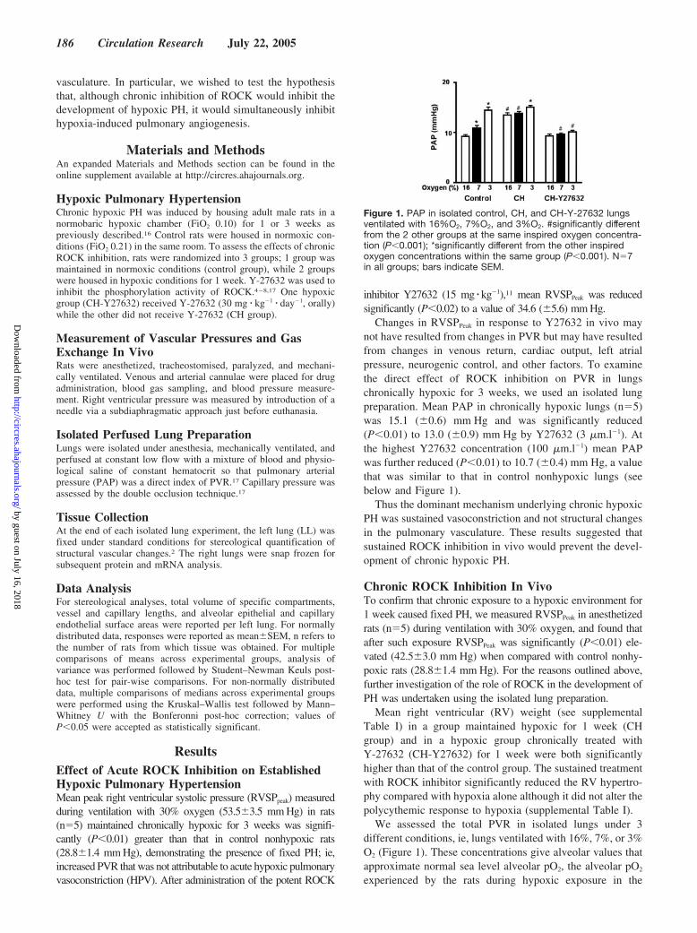
To evaluate the contribution of ROCK-dependent vasocon-striction to the control of PVR in these chronically hypoxicand control lungs, Y-27632 was added to the perfusate up to3 �mol/L, a concentration that selectively inhibitedROCK.8,11 In normoxic lungs, ventilated with 16%O2,Y-27632 up to 3 �mol � l�1 did not change the PAP (Figure2A), demonstrating that ROCK activity did not play a role inthe control of vascular resistance in the normal lung.
In the CH group, acute administration of Y-27632 mark-edly decreased the PAP in a dose-dependent fashion (Figure2A). This concentration of ROCK inhibitor abrogated ap-proximately two-thirds of the increase induced by chronichypoxia through an effect that was mainly localized to thearterial (precapillary) segment (Figure 2B). This demon-
strated that a vasoconstrictor activity of ROCK played a keyrole in maintaining chronic hypoxic PH.
Finally, in the CH-Y27632 group, in which the baselinePAP was similar to that in the control group, acute addition ofY-27632 had no significant effect on PA pressure in theisolated lungs, demonstrating that the chronic treatment withY-27632 had maximally inhibited the effect of ROCK activ-ity on PVR (Figure 2A).
In a separate series of experiments, we assessed the effectof higher, potentially nonselective8,11 concentrations ofY-27632 (up to 100 �mol/L) on the PAP in lungs maintainedchronically hypoxic for 1 week and found that PAP wasdecreased to 10.1 (�0.2) mm Hg (n�3), a value similar tothat in control lungs (9.6�0.3 mm Hg). These results weresimilar to those obtained in the 3-week hypoxic group andsupported the conclusion that the dominant mechanism un-derlying chronic hypoxic PH is sustained vasoconstriction.
In the presence of Y-27632 (3 �mol � l�1), reducing theoxygen level to 3% did not produce significant pulmonaryvasoconstriction in any group, indicating that ROCK is a keymediator of acute HPV (data not shown).
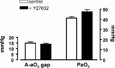
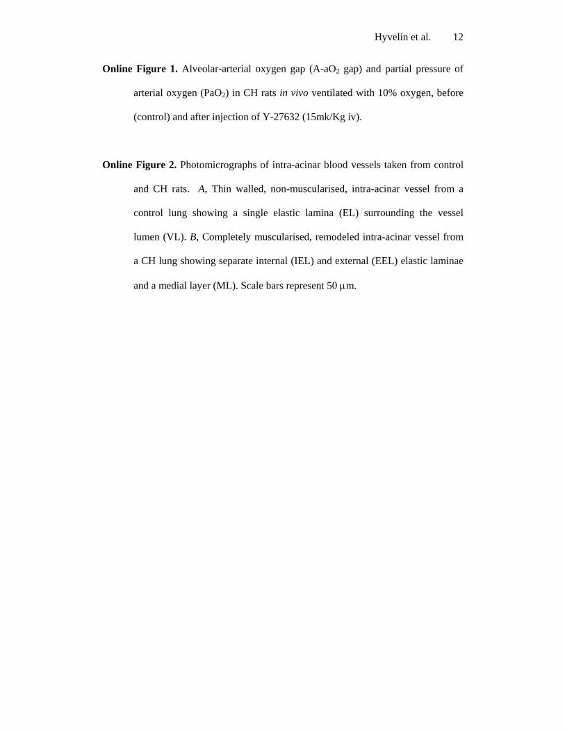
Effect of Acute ROCK Inhibition on PulmonaryOxygen Uptake in Hypoxic RatsIn chronically hypoxic rats ventilated with a hypoxic inspirate(FiO2 0.10), there was no change in either the A-aO2 gap orthe partial pressure of oxygen in arterial blood (PaO2) afteracute administration of Y-27632 (see online supplement).The A-aO2 gap and PaO2 were unchanged after injection ofvehicle (data not shown).
Effect of Chronic Inhibition of ROCK onHypoxia-Induced Angiogenesis andVascular RemodelingThe mean left lung volume was significantly increased in theCH group (3.28�0.15 mL) when compared with both thecontrol group (2.54�0.12 mL, P�0.001) and the CH-Y-27632 group (2.98�0.05 mL, P�0.05).
In the control lungs, the majority of intra-acinar vesselswere thin-walled, with a single elastic lamina and no discern-able tunica media (see online supplement results). In the CHgroup remodeling of the wall of intra-acinar vessels wasevident, as shown by the development of separate internal andexternal elastic laminae and an intervening layer of media(see online supplement results). After chronic inhibition ofROCK, the structure of vessel walls was similar to thatobserved in the control group suggesting inhibition of hyp-oxia-induced vascular remodeling.
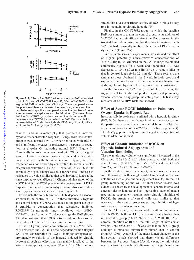
In the CH group, the mean total length of intra-acinarvessels (9236�630 cm � LL�1) was significantly higher thanin the control group (5327�392 cm � LL�1, P�0.001). Afterchronic inhibition of ROCK, the total length of intra-acinarvessels (7864�508 cm � LL�1) was less than in the CH groupalthough it remained significantly higher than in controlgroup (P�0.01). Analysis of the mean lumen diameter of theintra-acinar vessels showed that there was no differencebetween the 3 groups (Figure 3A). However, the ratio of thewall thickness to the lumen diameter was significantly in-
Figure 2. A, Effect of Y-27632 added acutely on PAP in isolatedcontrol, CH, and CH-Y-27632 lungs. B, Effect of Y-27632 on thesegmental PVR in control and CH lungs. The upper panel showsthe pressure difference between the pulmonary artery and thecapillaries (Art-cap); the lower panel shows the gradient of pres-sure between the capillaries and the left atrium (Cap-ven). Notethat the CH-Y27632 group has been omitted from panel Bbecause acute Y27632 had no effect on PAP. Each symbol isrepresentative of 7 rats, bars indicate SEM. #significantly differ-ent from the 2 other groups (P�0.05).
Hyvelin et al Y-27632 Prevents Hypoxic Pulmonary Angiogenesis 187
by guest on July 16, 2018http://circres.ahajournals.org/
Dow
nloaded from
creased in the CH-group when compared with control andCH-Y27632 lungs (Figure 3B).
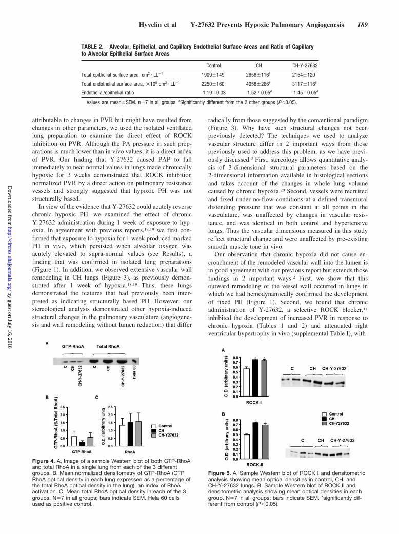
The total capillary volume and total capillary length (Table 1)were significantly increased in the CH group when comparedwith the control group, whereas after chronic treatment withY-27632 these hypoxia-induced increases were significantlyreduced and not significantly different from those in controlgroup. The total capillary endothelial surface area was alsosignificantly increased in the CH lungs when compared with thecontrol group (Table 2). In the CH-Y-27632 group, capillaryendothelial surface area was significantly less than that in the CHgroup although it remained elevated above the control value(Table 2). Chronic hypoxia also caused a significant increase inthe total alveolar epithelial surface area when compared with thecontrol group (Table 2), although in the CH-Y-27632 group thisvalue was significantly less that in the CH group and similar tothat in the control group. In both hypoxic groups, the increase incapillary endothelial surface area was proportionately greaterthan that in the epithelial surface area as indicated by theincrease in the mean ratio of capillary endothelium to alveolarepithelium (Table 2).
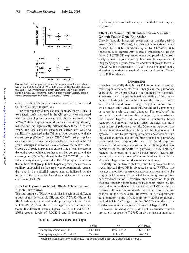
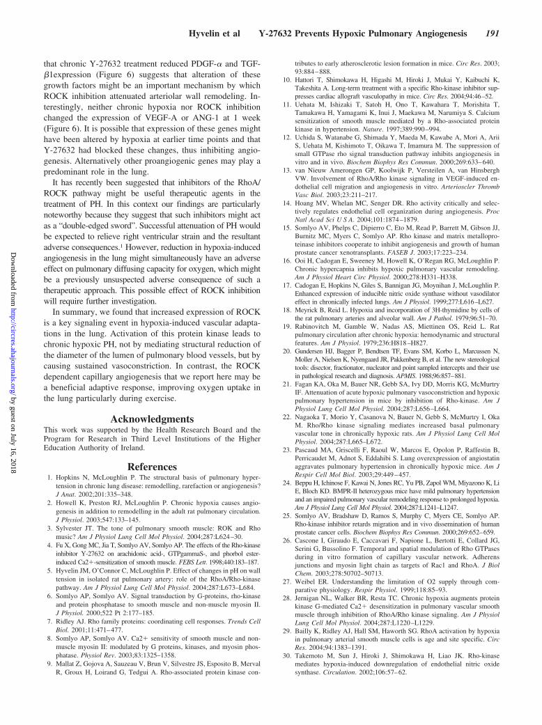
Effect of Hypoxia on RhoA, RhoA Activation, andROCK ExpressionThe total amount of RhoA was similar in each of the differentgroups of rats; ie, control, CH, and CH-Y-27632 (Figure 4).RhoA activation, expressed as the percentage of total RhoAin GTP-RhoA form, showed no significant difference be-tween the different groups (Figure 4). In CH and CH-Y-27632 groups levels of ROCK I and II isoforms were
significantly increased when compared with the control group(Figure 5).
Effect of Chronic ROCK Inhibition on VascularGrowth Factor Gene ExpressionChronic hypoxia increased expression of platelet-derivedgrowth factor-� (PDGF-�), and this effect was significantlyreduced by ROCK inhibition (Figure 6). Chronic ROCKinhibition also significantly reduced transforming growthfactor �-1 (TGF-�1) expression when compared with chron-ically hypoxic lungs (Figure 6). Interestingly, expression ofthe proangiogenic genes vascular endothelial growth factor A(VEGF-A) and angiopoeitin-1 (ANG-1) was not significantlyaltered at the end of one week of hypoxia and was unaffectedby ROCK inhibition.
DiscussionIt has been generally thought that PH predominantly resultedfrom hypoxia-induced structural changes in the pulmonaryvasculature, which produced a fixed increase in resistance.These structural changes included remodeling of the arterio-lar walls leading to encroachment into the vascular lumen,and loss of blood vessels, suggesting that interventions,which successfully ameliorated PH, would act by preventingor reversing such structural changes. The results of thepresent study cast doubt on this paradigm by demonstratingthat chronic hypoxia did not cause a structurally basedreduction of pulmonary vascular lumen diameter, nor did itcause a loss of pulmonary vessels. Moreover, we report thatchronic inhibition of ROCK abrogated the development ofhypoxic PH, not by preventing structural encroachment intothe vascular lumen, but by inhibiting sustained pulmonaryvasoconstriction. Importantly, we also found hypoxia-induced capillary angiogenesis in the adult lung that wasdependent on the RhoA/ROCK pathway. ROCK inhibitionreduced the expression of key vascular growth factors sug-gesting that this was one of the mechanisms by which itattenuated hypoxia-induced vascular remodeling.
Initially, we confirmed that exposure to hypoxia for threeweeks induced fixed PH in vivo; ie, increased RVSPPeak thatwas not immediately reversed on exposure to normal alveolaroxygen and thus was not mediated by acute hypoxic pulmo-nary vasoconstriction. Previously, this observation, togetherwith the extensive remodeling of pulmonary arterioles, hasbeen taken as evidence that the increased PVR in chronichypoxic PH was predominantly attributable to structuralchanges in the vasculature. However, in our experimentsadministration of the ROCK inhibitor Y-27632 produced amarked fall in PAP suggesting that ROCK-dependent vaso-constriction was the major determinant of hypoxic PH.
Because the changes in peak right ventricular systolicpressure in response to Y-27632 in vivo might not have been
TABLE 1. Capillary Volume and Length
Control CH CH-Y-27632
Total capillary volume, cm3 � LL�1 0.158�0.004 0.217�0.015# 0.177�0.008
Total capillary length, �105 cm � LL�1 7.4�0.6 11.9�0.8# 8.6�0.6
Values are mean�SEM. n�7 in all groups. #Significantly different from the 2 other groups (P�0.05).
Figure 3. A, Scatter plot showing intra-acinar vessel lumen diame-ters in control, CH and CH-Y-27632 lungs. B, Scatter plot showingthe ratio of wall thickness to lumen diameter. Each point repre-sents a single rat. Horizontal bars indicate median values. #signifi-cantly different from the other 2 groups (P�0.05).
188 Circulation Research July 22, 2005
by guest on July 16, 2018http://circres.ahajournals.org/
Dow
nloaded from
attributable to changes in PVR but might have resulted fromchanges in other parameters, we used the isolated ventilatedlung preparation to examine the direct effect of ROCKinhibition on PVR. Although the PA pressure in such prep-arations is much lower than in vivo values, it is a direct indexof PVR. Our finding that Y-27632 caused PAP to fallimmediately to near normal values in lungs made chronicallyhypoxic for 3 weeks demonstrated that ROCK inhibitionnormalized PVR by a direct action on pulmonary resistancevessels and strongly suggested that hypoxic PH was notstructurally based.
In view of the evidence that Y-27632 could acutely reversechronic hypoxic PH, we examined the effect of chronicY-27632 administration during 1 week of exposure to hyp-oxia. In agreement with previous reports,18,19 we first con-firmed that exposure to hypoxia for 1 week produced markedPH in vivo, which persisted when alveolar oxygen wasacutely elevated to supra-normal values (see Results), afinding that was confirmed in isolated lung preparations(Figure 1). In addition, we observed extensive vascular wallremodeling in CH lungs (Figure 3), as previously demon-strated after 1 week of hypoxia.18,19 Thus, these lungsdemonstrated the features that had previously been inter-preted as indicating structurally based PH. However, ourstereological analysis demonstrated other hypoxia-inducedstructural changes in the pulmonary vasculature (angiogene-sis and wall remodeling without lumen reduction) that differ
radically from those suggested by the conventional paradigm(Figure 3). Why have such structural changes not beenpreviously detected? The techniques we used to analyzevascular structure differ in 2 important ways from thosepreviously used to address this problem, as we have previ-ously discussed.2 First, stereology allows quantitative analy-sis of 3-dimensional structural parameters based on the2-dimensional information available in histological sectionsand takes account of the changes in whole lung volumecaused by chronic hypoxia.20 Second, vessels were recruitedand fixed under no-flow conditions at a defined transmuraldistending pressure that was constant at all points in thevasculature, was unaffected by changes in vascular resis-tance, and was identical in both control and hypertensivelungs. Thus the vascular dimensions measured in this studyreflect structural change and were unaffected by pre-existingsmooth muscle tone in vivo.
Our observation that chronic hypoxia did not cause en-croachment of the remodeled vascular wall into the lumen isin good agreement with our previous report but extends thosefindings in 2 important ways.2 First, we show that thisoutward remodeling of the vessel wall occurred in lungs inwhich we had hemodynamically confirmed the developmentof fixed PH (Figure 1). Second, we found that chronicadministration of Y-27632, a selective ROCK blocker,11
inhibited the development of increased PVR in response tochronic hypoxia (Tables 1 and 2) and attenuated rightventricular hypertrophy in vivo (supplemental Table I), with-
Figure 4. A, Image of a sample Western blot of both GTP-RhoAand total RhoA in a single lung from each of the 3 differentgroups. B, Mean normalized densitometry of GTP-RhoA (GTPRhoA optical density in each lung expressed as a percentage ofthe total RhoA optical density in the lung), an index of RhoAactivation. C, Mean total RhoA optical density in each of the 3groups. N�7 in all groups; bars indicate SEM. Hela 60 cellsused as positive control.
Figure 5. A, Sample Western blot of ROCK � and densitometricanalysis showing mean optical densities in control, CH, andCH-Y-27632 lungs. B, Sample Western blot of ROCK II anddensitometric analysis showing mean optical densities in eachgroup. N�7 in all groups; bars indicate SEM. *significantly dif-ferent from control (P�0.05).
TABLE 2. Alveolar, Epithelial, and Capillary Endothelial Surface Areas and Ratio of Capillaryto Alveolar Epithelial Surface Areas
Control CH CH-Y-27632
Total epithelial surface area, cm2 � LL�1 1909�149 2658�116# 2154�120
Total endothelial surface area, �105 cm2 � LL�1 2250�160 4058�266# 3117�116#
Endothelial/epithelial ratio 1.19�0.03 1.52�0.05# 1.45�0.05#
Values are mean�SEM. n�7 in all groups. #Significantly different from the 2 other groups (P�0.05).
Hyvelin et al Y-27632 Prevents Hypoxic Pulmonary Angiogenesis 189
by guest on July 16, 2018http://circres.ahajournals.org/
Dow
nloaded from

out causing structural enlargement of the lumen (Figure 3).We confirmed that the dose used chronically in vivo waseffective in blocking the contractile activity of the ROCKpathway by demonstrating that after isolation, these lungsshowed no additional vasodilator response to acute Y-27632administration (Figure 2). The failure of chronic ROCKinhibition to significantly enlarge mean-maximal-lumen di-ameter was observed, despite the fact that it successfullyattenuated wall remodeling, a finding that is supported by therecent report of Fagan et al.21 Taken together, our resultsshow that chronic hypoxic PH develops without structuralchange in the mean-maximal-vascular-lumen diameter and,furthermore, PH can be prevented without enlarging thestructurally determined lumen diameter. This suggests thatchronic hypoxic PH is not caused by structurally imposedchanges in vascular resistance.
This conclusion is further reinforced by the demonstrationthat acute administration of a high concentration of Y-27632normalized PVR in isolated lungs made chronically hyperten-sive by exposure to 1 and 3 weeks of hypoxia, both in vivo andin isolated lungs (Figure 2 and see Results), results that confirmthe recent report of Nagaoka et al.22 Evidence supporting theimportant role of RhoA/ROCK is provided by the demonstrationof a marked reduction in PVR (Figure 2) at concentrations ofY-27632 that are selective for ROCK (3 micromolar or less),8,11
although these concentrations did not return pulmonary arterialpressure to normal values. It has been previously shown thatY-27632 relaxes vascular smooth muscle, preconstricted byeither agonists or hypoxia, by blocking ROCK-mediated phos-phorylation of the regulatory protein CPI-17 and the MYPT-1 ofmyosin light chain phosphatase (MLCP), both of which effectsactivate MLCP, thus reducing myosin light chain phosphoryla-tion and relaxing smooth muscle.15,21 Administration of higherconcentrations of Y-27632 returned PVR to control values,although this vasodilator action might not have resulted fromspecific ROCK inhibition.5,8 Clearly, acute normalization couldnot occur if vascular resistance was increased by a fixedstructural mechanism.
The RhoA/ROCK-dependent sustained vasoconstrictionobserved in chronically hypoxic lungs is distinct from thatmediating acute HPV because acute normalization of alveolaroxygen did not reduce PVR after either 1 or 3 weeks ofhypoxic exposure. This suggested that the sustained vasocon-striction of chronic hypoxia did not have a role in ventilation-perfusion matching. To test this directly, we examined theeffect of acute ROCK inhibition on the alveolar-arterial
oxygen gap, an index of physiological shunting, and foundthat this was unaltered (see online supplement). This suggeststhat chronic ROCK-mediated hypoxic PH is a maladaptiveresponse, because it overloads the right ventricle withoutimproving gas exchange and is in marked contrast to thebeneficial optimization of ventilation-perfusion matchingproduced by acute HPV.
In the present study, we found that chronic hypoxia inducedcapillary angiogenesis in the adult lung in vivo (Tables 1 and 2),confirming the results of our previous report.2 In support of thisPascaud et al reported that angiostatin, an inhibitor of angiogen-esis, aggravated PH in the hypoxic lung, an observation thatsuggests new vessel formation in hypoxic lungs.23 More re-cently, Beppu and colleagues demonstrated hypoxia-inducedcapillary angiogenesis in the adult mouse lung.24 We now extendthose findings by reporting that chronic ROCK inhibition sig-nificantly attenuated this hypoxia-induced angiogenesis (Tables1 and 2). Inhibition of RhoA or ROCK has previously beenshown to prevent growth factor–induced endothelial cell migra-tion and organization into capillary-like structures in vitro andvessel formation in Matrigel implant.12,13,15,25,26 Our finding thatY-27632 attenuated hypoxia-induced angiogenesis in the lung is,to our knowledge, the first report directly demonstrating that theRhoA/ROCK pathway has a central role in hypoxic angiogen-esis within adult tissues in vivo. In the lung, this may beparticularly important as hypoxic angiogenesis increases thediffusing capacity for oxygen and may therefore be an importantadaptive response to hypoxia.2 The possibility that hypoxia-induced angiogenesis might improve oxygen uptake in the lungawaits direct functional evidence. However, studies in highaltitude and burrowing animals adapted to hypoxic environ-ments show that the enlarged pulmonary capillary bed found insuch species enhances oxygen uptake during exercise in hypoxiawhen oxygen uptake becomes diffusion limited, but not at restwhen uptake is perfusion limited.27
The action of ROCK in the chronically hypoxic lung wasnot the result of increased expression of RhoA, a finding ingood agreement with the recent report of Jernigan et al.28
Moreover, ROCK activity could not have resulted fromincreased RhoA activation because GTP-RhoA was reduced(although not significantly) and not increased (Figure 4). Thisresult is similar to that of Bailly et al who showed reducedRhoA activation in chronically hypoxic pulmonary vascularsmooth muscle,29 although increased RhoA activation hasbeen reported in chronically hypoxic lungs after 4 weeks.28
The ROCK effects that we observed may have been mediatedthrough the increased expression of both its isoforms (Figure5), a finding in good agreement with the demonstration thathypoxia augments expression of this enzyme in culturedpulmonary smooth muscle cells.30
ROCK activity is an important regulator of cytoskeletalrearrangement in endothelial cells during migration, andblockade of this action inhibits in vitro organization ofendothelial cells into capillary structures.14,26 Such inhibitionof cytoskeletal reorganization could have contributed to theattenuated angiogenesis that we observed in the CH-Y-27632group. We explored the possibility that inhibition of remod-eling and angiogenesis might also have been mediated byaltered expression of key vascular growth factors. Our finding
Figure 6. Quantitative real-time transcript levels in control (n�4),CH (n�6), and CH-Y27632 (n�7) rats. Data are presented asthe ratio of target gene to ribosomal RNA control. *significantlydifferent from the other 2 groups (P�0.01); †significantly differ-ent from CH group (P�0.01).
190 Circulation Research July 22, 2005
by guest on July 16, 2018http://circres.ahajournals.org/
Dow
nloaded from
that chronic Y-27632 treatment reduced PDGF-� and TGF-�1expression (Figure 6) suggests that alteration of thesegrowth factors might be an important mechanism by whichROCK inhibition attenuated arteriolar wall remodeling. In-terestingly, neither chronic hypoxia nor ROCK inhibitionchanged the expression of VEGF-A or ANG-1 at 1 week(Figure 6). It is possible that expression of these genes mighthave been altered by hypoxia at earlier time points and thatY-27632 had blocked these changes, thus inhibiting angio-genesis. Alternatively other proangiogenic genes may play apredominant role in the lung.
It has recently been suggested that inhibitors of the RhoA/ROCK pathway might be useful therapeutic agents in thetreatment of PH. In this context our findings are particularlynoteworthy because they suggest that such inhibitors might actas a “double-edged sword”. Successful attenuation of PH wouldbe expected to relieve right ventricular strain and the resultantadverse consequences.1 However, reduction in hypoxia-inducedangiogenesis in the lung might simultaneously have an adverseeffect on pulmonary diffusing capacity for oxygen, which mightbe a previously unsuspected adverse consequence of such atherapeutic approach. This possible effect of ROCK inhibitionwill require further investigation.
In summary, we found that increased expression of ROCKis a key signaling event in hypoxia-induced vascular adapta-tions in the lung. Activation of this protein kinase leads tochronic hypoxic PH, not by mediating structural reduction ofthe diameter of the lumen of pulmonary blood vessels, but bycausing sustained vasoconstriction. In contrast, the ROCKdependent capillary angiogenesis that we report here may bea beneficial adaptive response, improving oxygen uptake inthe lung particularly during exercise.
AcknowledgmentsThis work was supported by the Health Research Board and theProgram for Research in Third Level Institutions of the HigherEducation Authority of Ireland.
References1. Hopkins N, McLoughlin P. The structural basis of pulmonary hyper-
tension in chronic lung disease: remodelling, rarefaction or angiogenesis?J Anat. 2002;201:335–348.
2. Howell K, Preston RJ, McLoughlin P. Chronic hypoxia causes angio-genesis in addition to remodelling in the adult rat pulmonary circulation.J Physiol. 2003;547:133–145.
3. Sylvester JT. The tone of pulmonary smooth muscle: ROK and Rhomusic? Am J Physiol Lung Cell Mol Physiol. 2004;287:L624–30.
4. Fu X, Gong MC, Jia T, Somlyo AV, Somlyo AP. The effects of the Rho-kinaseinhibitor Y-27632 on arachidonic acid-, GTPgammaS-, and phorbol ester-induced Ca2�-sensitization of smooth muscle. FEBS Lett. 1998;440:183–187.
5. Hyvelin JM, O’Connor C, McLoughlin P. Effect of changes in pH on walltension in isolated rat pulmonary artery: role of the RhoA/Rho-kinasepathway. Am J Physiol Lung Cell Mol Physiol. 2004;287:L673–L684.
6. Somlyo AP, Somlyo AV. Signal transduction by G-proteins, rho-kinaseand protein phosphatase to smooth muscle and non-muscle myosin II.J Physiol. 2000;522 Pt 2:177–185.
7. Ridley AJ. Rho family proteins: coordinating cell responses. Trends CellBiol. 2001;11:471–477.
8. Somlyo AP, Somlyo AV. Ca2� sensitivity of smooth muscle and non-muscle myosin II: modulated by G proteins, kinases, and myosin phos-phatase. Physiol Rev. 2003;83:1325–1358.
9. Mallat Z, Gojova A, Sauzeau V, Brun V, Silvestre JS, Esposito B, MervalR, Groux H, Loirand G, Tedgui A. Rho-associated protein kinase con-
tributes to early atherosclerotic lesion formation in mice. Circ Res. 2003;93:884–888.
10. Hattori T, Shimokawa H, Higashi M, Hiroki J, Mukai Y, Kaibuchi K,Takeshita A. Long-term treatment with a specific Rho-kinase inhibitor sup-presses cardiac allograft vasculopathy in mice. Circ Res. 2004;94:46–52.
11. Uehata M, Ishizaki T, Satoh H, Ono T, Kawahara T, Morishita T,Tamakawa H, Yamagami K, Inui J, Maekawa M, Narumiya S. Calciumsensitization of smooth muscle mediated by a Rho-associated proteinkinase in hypertension. Nature. 1997;389:990–994.
12. Uchida S, Watanabe G, Shimada Y, Maeda M, Kawabe A, Mori A, AriiS, Uehata M, Kishimoto T, Oikawa T, Imamura M. The suppression ofsmall GTPase rho signal transduction pathway inhibits angiogenesis invitro and in vivo. Biochem Biophys Res Commun. 2000;269:633–640.
13. van Nieuw Amerongen GP, Koolwijk P, Versteilen A, van HinsberghVW. Involvement of RhoA/Rho kinase signaling in VEGF-induced en-dothelial cell migration and angiogenesis in vitro. Arterioscler ThrombVasc Biol. 2003;23:211–217.
14. Hoang MV, Whelan MC, Senger DR. Rho activity critically and selec-tively regulates endothelial cell organization during angiogenesis. ProcNatl Acad Sci U S A. 2004;101:1874–1879.
15. Somlyo AV, Phelps C, Dipierro C, Eto M, Read P, Barrett M, Gibson JJ,Burnitz MC, Myers C, Somlyo AP. Rho kinase and matrix metallopro-teinase inhibitors cooperate to inhibit angiogenesis and growth of humanprostate cancer xenotransplants. FASEB J. 2003;17:223–234.
16. Ooi H, Cadogan E, Sweeney M, Howell K, O’Regan RG, McLoughlin P.Chronic hypercapnia inhibits hypoxic pulmonary vascular remodeling.Am J Physiol Heart Circ Physiol. 2000;278:H331–H338.
17. Cadogan E, Hopkins N, Giles S, Bannigan JG, Moynihan J, McLoughlin P.Enhanced expression of inducible nitric oxide synthase without vasodilatoreffect in chronically infected lungs. Am J Physiol. 1999;277:L616–L627.
18. Meyrick B, Reid L. Hypoxia and incorporation of 3H-thymidine by cells ofthe rat pulmonary arteries and alveolar wall. Am J Pathol. 1979;96:51–70.
19. Rabinovitch M, Gamble W, Nadas AS, Miettinen OS, Reid L. Ratpulmonary circulation after chronic hypoxia: hemodynamic and structuralfeatures. Am J Physiol. 1979;236:H818–H827.
20. Gundersen HJ, Bagger P, Bendtsen TF, Evans SM, Korbo L, Marcussen N,Moller A, Nielsen K, Nyengaard JR, Pakkenberg B, et al. The new stereologicaltools: disector, fractionator, nucleator and point sampled intercepts and their usein pathological research and diagnosis. APMIS. 1988;96:857–881.
21. Fagan KA, Oka M, Bauer NR, Gebb SA, Ivy DD, Morris KG, McMurtryIF. Attenuation of acute hypoxic pulmonary vasoconstriction and hypoxicpulmonary hypertension in mice by inhibition of Rho-kinase. Am JPhysiol Lung Cell Mol Physiol. 2004;287:L656–L664.
22. Nagaoka T, Morio Y, Casanova N, Bauer N, Gebb S, McMurtry I, OkaM. Rho/Rho kinase signaling mediates increased basal pulmonaryvascular tone in chronically hypoxic rats. Am J Physiol Lung Cell MolPhysiol. 2004;287:L665–L672.
23. Pascaud MA, Griscelli F, Raoul W, Marcos E, Opolon P, Raffestin B,Perricaudet M, Adnot S, Eddahibi S. Lung overexpression of angiostatinaggravates pulmonary hypertension in chronically hypoxic mice. Am JRespir Cell Mol Biol. 2003;29:449–457.
24. Beppu H, Ichinose F, Kawai N, Jones RC, Yu PB, Zapol WM, Miyazono K, LiE, Bloch KD. BMPR-II heterozygous mice have mild pulmonary hypertensionand an impaired pulmonary vascular remodeling response to prolonged hypoxia.Am J Physiol Lung Cell Mol Physiol. 2004;287:L1241–L1247.
25. Somlyo AV, Bradshaw D, Ramos S, Murphy C, Myers CE, Somlyo AP.Rho-kinase inhibitor retards migration and in vivo dissemination of humanprostate cancer cells. Biochem Biophys Res Commun. 2000;269:652–659.
26. Cascone I, Giraudo E, Caccavari F, Napione L, Bertotti E, Collard JG,Serini G, Bussolino F. Temporal and spatial modulation of Rho GTPasesduring in vitro formation of capillary vascular network. Adherensjunctions and myosin light chain as targets of Rac1 and RhoA. J BiolChem. 2003;278:50702–50713.
27. Weibel ER. Understanding the limitation of O2 supply through com-parative physiology. Respir Physiol. 1999;118:85–93.
28. Jernigan NL, Walker BR, Resta TC. Chronic hypoxia augments proteinkinase G-mediated Ca2� desensitization in pulmonary vascular smoothmuscle through inhibition of RhoA/Rho kinase signaling. Am J PhysiolLung Cell Mol Physiol. 2004;287:L1220–L1229.
29. Bailly K, Ridley AJ, Hall SM, Haworth SG. RhoA activation by hypoxiain pulmonary arterial smooth muscle cells is age and site specific. CircRes. 2004;94:1383–1391.
30. Takemoto M, Sun J, Hiroki J, Shimokawa H, Liao JK. Rho-kinasemediates hypoxia-induced downregulation of endothelial nitric oxidesynthase. Circulation. 2002;106:57–62.
Hyvelin et al Y-27632 Prevents Hypoxic Pulmonary Angiogenesis 191
by guest on July 16, 2018http://circres.ahajournals.org/
Dow
nloaded from
and Paul McLoughlinJean-Marc Hyvelin, Katherine Howell, Alistair Nichol, Christine M. Costello, Robert J. Preston
CirculationInhibition of Rho-Kinase Attenuates Hypoxia-Induced Angiogenesis in the Pulmonary
Print ISSN: 0009-7330. Online ISSN: 1524-4571 Copyright © 2005 American Heart Association, Inc. All rights reserved.is published by the American Heart Association, 7272 Greenville Avenue, Dallas, TX 75231Circulation Research
doi: 10.1161/01.RES.0000174287.17953.832005;97:185-191; originally published online June 16, 2005;Circ Res.
http://circres.ahajournals.org/content/97/2/185World Wide Web at:
The online version of this article, along with updated information and services, is located on the
http://circres.ahajournals.org/content/suppl/2005/06/16/01.RES.0000174287.17953.83.DC1Data Supplement (unedited) at:
http://circres.ahajournals.org//subscriptions/
is online at: Circulation Research Information about subscribing to Subscriptions:
http://www.lww.com/reprints Information about reprints can be found online at: Reprints:
document. Permissions and Rights Question and Answer about this process is available in the
located, click Request Permissions in the middle column of the Web page under Services. Further informationEditorial Office. Once the online version of the published article for which permission is being requested is
can be obtained via RightsLink, a service of the Copyright Clearance Center, not theCirculation Researchin Requests for permissions to reproduce figures, tables, or portions of articles originally publishedPermissions:
by guest on July 16, 2018http://circres.ahajournals.org/
Dow
nloaded from
Hyvelin et al. 1
ONLINE SUPPLEMENT
Expanded Materials and Methods All study protocols were approved by the University Ethics Committee and conducted
under license from the Department of Health.
Hypoxic pulmonary hypertension
Chronic hypoxic pulmonary hypertension was induced by housing adult male rats in a
normobaric hypoxic chamber (FiO2 0.10) for one or three weeks as previously
described 1. Control rats were housed in normoxic conditions (FiO2 0.21) in the same
room. To assess the effects of chronic ROCK inhibition, rats were randomized into
three groups, one of which was maintained in normoxic conditions (control group)
while two groups were housed in hypoxic conditions for one week. One hypoxic
group (CH-Y27632) received Y-27632 (30mg.kg-1.day-1, orally) while the other did
not receive Y-27632 (CH group). Y–27632 was purchased from Tocris (UK).
Measurement of vascular pressures and gas exchange in vivo.
After induction of anesthesia (60mg.kg-1 sodium pentobarbital intra-peritoneally), a
tracheostomy was performed, the lung were mechanically ventilated (FiO2, 0.3; rate,
90 breaths.min-1; tidal volume, 4.5 ml.Kg-1; positive end-expiratory pressure, 2.5 cm
H2O; 15-minute recruitments with positive end-expiratory pressure of 15 cm H2O for
20 breaths), and carotid arterial and dorsal penile vein camulas were inserted.
Anesthesia and muscle relaxation were maintained with intravenous infusions of
alphaxalone-alphadolone (Saffan) and panucorium, respectively. Depth of anesthesia
Hyvelin et al. 2
was assessed by monitoring the hemodynamic response to paw-clamp. Stable
physiological conditions were obtained before entering the animal into the protocol,
and animals were excluded when baseline inclusion criteria (i.e., normal oxygenation,
acid-base status, hemodynamic status and temperature) were not met. After 30
minutes stabilization, animals were ventilated with hypoxic gas mixture (10% O2,
90%N2) for 15 minutes then Y-27632 (15mg.Kg-1) or vehicle were injected
intravenously and animal kept under hypoxic condition for a further 15minutes. This
was the maximum dose of Y–27632 that could be tolerated because of the reduction
in systemic arterial pressure that it caused. Systemic mean arterial pressure, peak
airway pressure, and rectal temperature were recorded throughout. Arterial blood
gases and exhaust gases were determined every 15 minutes. Alveolar-arterial O2 gap
calculation were made using the complete alveolar equation2. In some experiments,
right ventricular pressure was measured at end expiration by introduction of a needle
(23g) via a sub-diaphragmatic approach just prior to euthanasia.
Lung isolation and perfusion.
An isolated ventilated perfused lung preparation was used to assess the effect of
experimental interventions on total pulmonary vascular resistance, as previously
described 3. In brief, rats were anaesthetized (60mg.kg-1 sodium pentobarbital intra-
peritoneally), and mechanically ventilated (SAR-830P small-animal ventilator, CWE,
Ardmore, PA) at a tidal volume of 1.8 ml and a respiratory frequency of 80
breathsmin-1 as previously described. The animals were then anti-coagulated
(1000 IU.Kg-1 heparin intra-venously) and killed by exsanguination. The thoracic
contents were exposed through a midline sternotomy, and cannulas were inserted into
the main pulmonary artery and left atrium and tied in place. The thoracic contents
Hyvelin et al. 3
were removed en bloc and suspended in a chamber maintained at 37°C, while
ventilation continued with a warmed and humidified gas mixture of 5% CO2, 21% O2,
balance N2 for the normoxic rats and 5% CO2, 7% O2, balance N2 for the hypoxic rats.
Airway pressure was continuously monitored, and a positive end-expiratory pressure
of 2.0 cmH2O was maintained. The lungs were briefly hyperinflated to an airway
pressure of 16 cmH20 every 5 minutes to prevent development of progressive
atelectasis.
Lungs were perfused with a mixture of blood and physiological saline
solution. The blood was collected from normoxic rats and mixed with PSS (in
mmol/L: 121 NaCl, 21 NaHCO3, 5.4 KCl, 4 MgSO4, 1 NaH2PO4, 1.8 CaCl2, 5.6
glucose plus 4% Ficoll) in a ration of 2:1. Perfusion was maintained at a constant flow
(0.04 ml.min-1.Kg-1) so that changes in arterial perfusion pressure reflected changes in
total pulmonary vascular resistance. Venous outflow pressure was maintained
constant at 3.5 mmHg to ensure zone 3 conditions at the end of expiration. All
measurements of arterial perfusion pressure were made at end of expiration. Arterial,
venous and airway pressure were continuously recorded with an analog-to-digital
system (Biopac MP100 WS, Linton Instrumentation, Norfolk, UK).
The baseline capillary pressure (Pc) was assessed by the double occlusion
technique. Briefly inflow and venous outflow lines were simultaneously occluded and
vascular pressure rapidly equilibrated to a new pressure representative of Pc 4. This
allowed pre-capillary and post-capillary resistances to be calculated separately.
Hyvelin et al. 4
ROCK inhibition
Y–27632 was used to inhibit ROCK phosphorylation activity both in vivo and in
isolated lung experiments5-7. At concentrations less than 10 µm.l–1 this agent is a
relatively selective ROCK inhibitor. Higher concentrations approach or exceed its Ki
for other kinases that regulate smooth muscle tone including protein kinase A, protein
kinase C especially novel isoforms, citron kinase and protein kinase N5-7. We used
high concentrations to determine whether pulmonary hypertension was structurally
based or due to vascular smooth muscle contraction. However, it is important to note
that the actions of this compound may then be due to effects additional to ROCK
inhibition5-7.
Tissue collection
At the end of each isolated lung experiment, the lung was ventilated with a gas
mixture 5%CO2, 16%O2, balance N2, and the pulmonary artery was perfused with
normal saline (37°C) until the effluent was clear of blood. Calcium-free physiological
saline solution was introduced into the pulmonary circulation to induce complete
relaxation of the pulmonary vessels. A ligature was subsequently tied around the
right main bronchus and the right branch of the pulmonary artery and the right lobes
were removed and quickly snap frozen for further analysis. The left lung was fully
inflated at a pressure of 25 cmH20 with fixative (4% wt.vol-1 paraformaldehyde),
followed by simultaneous infusion of this solution through the pulmonary artery (62.5
cmH2O). The cannula in the left atrium was clamped to obstruct the outflow from the
pulmonary veins, so that the pulmonary vasculature was fixed under ‘no flow’
conditions. This maneuver produced a constant capillary transmural distending
Hyvelin et al. 5
pressure (37.5 cmH2O) at all locations along the vascular bed. After one hour, the
main-stem bronchi and pulmonary artery were tied off at the level of the hilum, and
the left lung volume determined by water displacement, as previously described 8.
Measurement of Right Ventricular weights
The right ventricular free wall (RV) was dissected from the left ventricle and septum
(LV + S), each ventricle weighed separately and the ratio of RV to LV + S weight
calculated. Right ventricular weight was expressed in grams per 100 grams of body
weight.
Assessment of RhoA activation and ROCK level.
Frozen lung tissue was homogenized as previously described 9. Briefly lung tissue
was homogenized using a polytron in 5 volumes of ice-cold homogenization buffer
comprising (in mmol/L) 50 Tris-HCl (pH 7.2), 500 NaCl, 10 MgCl2, 1
ethylenediaminetetraacetic acid (EDTA), 1%Triton X-100, 0.5% deoxycholic acid,
0.1% sodium dodecyl sulfate (SDS), 20 µg.ml-1 each leupeptinin and aprotinin and 1
mM phenylmethyl sulfonyl fluoride (PMSF). Homogenates were clarified by
centrifugation at 5000g at 4°C for 10 min. Protein concentration was determined
using a commercial protein assay kit (BCA Protein Assay, Pierce Biotechnology,
Rokford, USA). A small amount of supernatant (7.5 µg of protein) was taken to
determine the amount of total RhoA, ROCK-I and ROCK-II by western blot analysis.
Equal amounts of protein (500 µg) were used to determine the amount of GTP-bound
RhoA using Rhotekin immobilized onto glutathione-agarose beads (Upstate, UK).
Each sample was then analyzed by SDS-15% polyacrylamide gels. Resolved proteins

Hyvelin et al. 6
were transferred onto nitrocellulose. Primary monoclonal antibody 26C4 (SantaCruz
Biotechnology Inc.) was used at 1:6000 dilution for detection of RhoA, primary
monoclonal antibodies anti ROCK-α or anti ROCK-β (BD Biosciences, UK) were
used at 1:1000. Horseradish peroxidase-conjugated secondary goat antibody anti-
mouse IgG (SantaCruz Biotechnology Inc.) was used at 1:8000. Membranes were
incubated for 1h each with primary and secondary antibody diluted in TBS-Tween
with 5% non-fat dry milk. The immunoreactive bands were detected by ECL
(Amersham Biosciences, UK) and quantified by densitometric analysis (NIH Image
1.63, National Institutes of Health, USA). The extent of RhoA activation (GTP-
RhoA) was expressed as the ratio of the density of the GTP-RhoA band to that of the
total RhoA in each sample.
Stereological assessment of vascular structural changes
We examined the structural changes induced by hypoxia in arterial, capillary and
venous vessels as previously described using isotropic uniform random sections
obtained from each left lung 8. Briefly, the left lung was systematically cut into 4 mm
thick slices starting from a randomly chosen position within the first 4 mm. The slices
were embedded in resin and cut into bars at 2 mm intervals, and every third bar
selected from a randomly chosen start point between one and three. Sections (1-2
µm) were taken from a random position within each tissue block and stained with
Toluidine Blue. Random fields of view from each section were examined by light
microscopy, captured using video camera (JVC KY-F55B Eurotec, Ireland) and
imported into Stereology Toolbox (Morphometrix, USA) for analysis.
Volume densities of specific tissues in the lung (volume per unit volume of
lung) were estimated by point counting, and the absolute volumes were calculated
Hyvelin et al. 7
using the previously measured lung volumes. The volume densities of the extra-acinar
tissues (defined as large vessels and airways, down to and including terminal
bronchioles, and their associated connective tissue, vasa vasorum and nerves) and the
volume of intra-acinar tissues (defined as respiratory bronchioles, alveolar tissue and
the associated vessels) were estimated by point counting. The intra-acinar gas
exchange region of the lung was next considered as three sub-compartments: the
intra-acinar blood vessels (that is all blood vessels excluding capillaries), the intra-
acinar airspaces and, finally, the alveolar walls including capillaries. To estimate the
volume densities of the vessel lumen and the vessel wall including tunica intima and
tunica media, images of randomly selected intra-acinar vessels were captured and
placed randomly within point counting grids. Capillary lumen volume density was
estimated by point counting on random fields of view obtained at high magnification.
To estimate the length of intra-acinar vessels per unit lung volume (length
density) the number of intra-acinar vessels that transected counting frames randomly
superimposed over images taken from IUR sections was counted as previously
described. The vessels were considered to be cylindrical in shape, thus the average
radius of the intra-acinar vessels was calculated from the measured length and volume
densities using standard formulae. Capillary length and volume density were
calculated in the same manner as intra-acinar vessels.
Extraction of RNA and real-time PCR (TaqMan) quantification
Total RNA was extracted from whole lung samples, from control (n=4), CH (n=6)
and CH-Y27632 (n=7) animals using RNeasy Mini columns (Qiagen; Germany) and
reverse transcribed (RT) to cDNA using Superscript II RNase H-Reverse
Hyvelin et al. 8
Transcriptase kit (Invitrogen, USA). Probe and primer sequences were designed to
non-redundant sequences (transforming growth factor, beta 1 (Tgfb1):
F-primer: 5’-GTCCCAAACGTCGAGGTGA-3’;
R-primer: 5’-CCATGAGGAGCAGGAAGGG-3’;
probe: 5’-TGGGCACCATCCATGACATGC-3’),
or ordered from ABI as Assays-on-Demand Gene Expression Assays: (i) platelet
derived growth factor, alpha (Pdgfa; Assay ID: Rn00709363_m1), (ii) vascular
endothelial growth factor A (Vegfa; Assay ID: Rn00582935_m1), and (iii)
angiopoietin 1 (Angpt1; Assay ID: Rn00585552_m1). Probes were labeled with
FAM. The Eukaryotic 18S rRNA (VIC) pre-developed assay reagent kit was used as
the endogenous control. Reactions were carried out on the ABI PRISM 7900
Sequence Detection System, according to the TaqMan PCR protocol, and mRNA
transcript levels were determined using the comparative Ct method (ABI Prism 7700
Sequence Detection System User Bulletin #2).
Chemicals
All salts and drugs were supplied by Sigma-Aldrich except where otherwise indicated.
Data Analysis
For stereological analyses, total volume of specific compartments, vessel and
capillary lengths and alveolar epithelial and capillary endothelial surface areas were
reported per left lung. For normally distributed data, responses were reported as
Hyvelin et al. 9
means ± standard error of the mean (SEM), n refers to the number of rats from which
tissue was obtained. For multiple comparisons of means across experimental groups,
analysis of variance was carried out followed by Student-Newman Keuls post hoc test
for pair-wise comparisons. For non-normally distributed data, multiple comparisons
of medians across experimental groups were carried out using the Kruskal-Wallis test
followed by Mann-Whitney-U with the Bonferonni post hoc correction; values of
P<0.05 were accepted as statistically significant.
Hyvelin et al. 10
Online Supplement Results
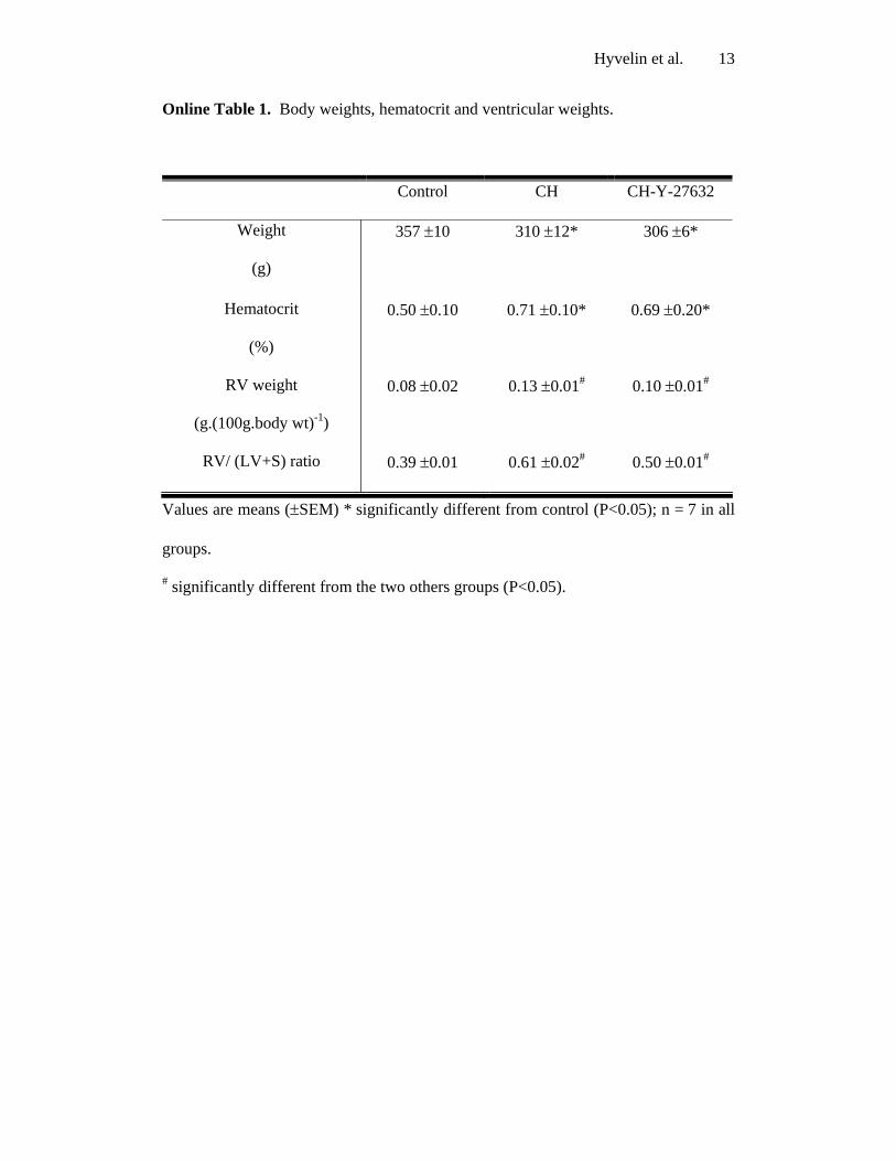
Effects of one week hypoxic exposure on body weight, right ventricle and hematocrit
Mean body weights (Online Table 1) in a group maintained hypoxic for one week
(CH group) and in a hypoxic group chronically treated with Y-27632, (CH-Y27632),
were significantly lower than that of the control group, a well-established effect of
chronic hypoxia. Chronic hypoxia also caused a significant increase in hematocrit
(Online Table 1). In both hypoxic groups, right ventricular hypertrophy was evident,
as demonstrated by the increased mean weight of the right ventricle (RV) and the
increased ratio of right to left ventricular weight compared to the control group
(Online Table 1). ROCK inhibitor treatment significantly reduced the hypoxia-
induced RV hypertrophy although it did not alter the polycythemic response to
hypoxia (Online Table 1).
Effect of acute ROCK inhibition on pulmonary oxygen uptake in hypoxic rats.
In chronically hypoxic rats ventilated with a hypoxic inspirate (FiO2 0.10) there was
no change in either the A-aO2 gap or the partial pressure of oxygen in arterial blood
(PaO2) following acute administration of Y-27632 (Online Figure 1). These results
suggest that ROCK-dependent vasoconstriction in chronically hypoxic hypertensive
lungs does not contribute to optimizing ventilation perfusion matching. The A-aO2
gap and PaO2 were unchanged after injection of vehicle (data not shown).
Effect of chronic inhibition of ROCK on hypoxia-induced angiogenesis and vascular
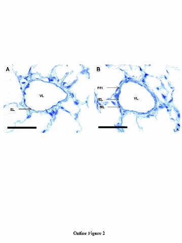
remodeling.In the control lungs, the majority of intra-acinar vessels were thin-walled,
with a single elastic lamina and no discernable tunica media (Online Figure 2A),
findings typical of normal lungs with normal low pulmonary vascular resistance. In
Hyvelin et al. 11
the CH group remodeling of the wall of intra-acinar vessels was evident, as shown by
the development of separate internal and external elastic laminae and an intervening
layer of media (Online Figure 2B), a finding that is characteristic of the vascular
remodeling of chronic hypoxic pulmonary hypertension. Following chronic inhibition
of ROCK, the structure of vessel walls was similar to that observed in the control
group suggesting inhibition of hypoxia-induced vascular remodeling.
Hyvelin et al. 14
References for online supplement
1. Ooi H, Cadogan E, Sweeney M, Howell K, O'Regan RG, McLoughlin P.
Chronic hypercapnia inhibits hypoxic pulmonary vascular remodeling. Am J
Physiol Heart Circ Physiol. 2000;278:H331-8.
2. Swenson ER, Robertson HT, Hlastala MP. Effects of inspired carbon dioxide
on ventilation-perfusion matching in normoxia, hypoxia, and hyperoxia. In:
Am J Respir Crit Care Med; 1994:1563-9.
3. Cadogan E, Hopkins N, Giles S, Bannigan JG, Moynihan J, McLoughlin P.
Enhanced expression of inducible nitric oxide synthase without vasodilator
effect in chronically infected lungs. In: Am J Physiol; 1999:L616-27.
4. Cadogan E, Hopkins N, Giles S, Bannigan JG, Moynihan J, McLoughlin P.
Enhanced expression of inducible nitric oxide synthase without vasodilator
effect in chronically infected lungs. Am J Physiol. 1999;277:L616-27.
5. Uehata M, Ishizaki T, Satoh H, Ono T, Kawahara T, Morishita T, Tamakawa
H, Yamagami K, Inui J, Maekawa M, Narumiya S. Calcium sensitization of
smooth muscle mediated by a Rho-associated protein kinase in hypertension.
Nature. 1997;389:990-994.
6. Somlyo AP, Somlyo AV. Ca2+ sensitivity of smooth muscle and nonmuscle
myosin II: modulated by G proteins, kinases, and myosin phosphatase. Physiol
Rev. 2003;83:1325-58.
7. Ishizaki T, Uehata M, Tamechika I, Keel J, Nonomura K, Maekawa M,
Narumiya S. Pharmacological properties of Y-27632, a specific inhibitor of
rho-associated kinases. Mol Pharmacol. 2000;57:976-83.
Hyvelin et al. 15
8. Howell K, Preston RJ, McLoughlin P. Chronic hypoxia causes angiogenesis in
addition to remodelling in the adult rat pulmonary circulation. In:
J.Physiol.2003.Feb.15.;547.(Pt.1.):133.-45.; 2003:133-145.
9. Hyvelin JM, O'Connor C, McLoughlin P. Effect of changes in pH on wall
tension in isolated rat pulmonary artery: role of the RhoA/Rho-kinase
pathway. In: Am J Physiol Lung Cell Mol Physiol; 2004:L673-84.
Hyvelin et al. 12
Online Figure 1. Alveolar-arterial oxygen gap (A-aO2 gap) and partial pressure of
arterial oxygen (PaO2) in CH rats in vivo ventilated with 10% oxygen, before
(control) and after injection of Y-27632 (15mk/Kg iv).
Online Figure 2. Photomicrographs of intra-acinar blood vessels taken from control
and CH rats. A, Thin walled, non-muscularised, intra-acinar vessel from a
control lung showing a single elastic lamina (EL) surrounding the vessel
lumen (VL). B, Completely muscularised, remodeled intra-acinar vessel from
a CH lung showing separate internal (IEL) and external (EEL) elastic laminae
and a medial layer (ML). Scale bars represent 50 µm.
Hyvelin et al. 13
Online Table 1. Body weights, hematocrit and ventricular weights.
Control CH CH-Y-27632
Weight
(g)
357 ±10
310 ±12*
306 ±6*
Hematocrit
(%)
0.50 ±0.10 0.71 ±0.10* 0.69 ±0.20*
RV weight
(g.(100g.body wt)-1)
0.08 ±0.02 0.13 ±0.01# 0.10 ±0.01#
RV/ (LV+S) ratio 0.39 ±0.01 0.61 ±0.02# 0.50 ±0.01#
Values are means (±SEM) * significantly different from control (P<0.05); n = 7 in all
groups.
# significantly different from the two others groups (P<0.05).