Embed Size (px)
Citation preview
RESEARCH ARTICLE Open Access
Salidroside attenuates hypoxia-inducedpulmonary arterial smooth muscle cellproliferation and apoptosis resistance byupregulating autophagy through theAMPK-mTOR-ULK1 pathwayDi Gui1, Zhimin Cui1, Lin Zhang1, Chang Yu2, Dan Yao1, Min Xu1, Mayun Chen1, Peiliang Wu1, Guoping Li3,Liangxing Wang1* and Xiaoying Huang1*
Abstract
Background: Recent studies have shown that both adenosine monophosphate activated protein kinase (AMPK)and the mammalian target of rapamycin (mTOR) are energy sensors and are related to autophagy. Our recentreports have shown that salidroside can exert protective effects against hypoxia-induced pulmonary arterial smoothmuscle cell (PASMC) proliferation and apoptosis resistance through the AMPK pathway. This study aims to explorethe relationship among AMPK, mTOR and ULK1 in PASMCs under hypoxic conditions and to investigate whetherthe protective effects of salidroside are related to the autophagic cell death pathway.
Methods: Rat PASMCs were cultured and divided into five groups: the normoxia, hypoxia, hypoxia + MHY1485(mTOR agonist), hypoxia + rapamycin (mTOR inhibitor) and hypoxia + salidroside groups. Hypoxic cells were treatedas indicated for 24 h. Cell viability was evaluated by the CCK-8 assay. Cell apoptosis was measured by the TUNELassay. The autophagy flux of PASMCs was evaluated with tandem mRFP-GFP fluorescence microscopy. Autophagosomeswere detected by electron microscopy. Protein expression of LC3, p62, AMPK, P-AMPK (Thr 172), P-ULK1 (Ser 555 and Ser317), mTOR, P-mTOR (Ser 2448), ULK1 and P-ULK1 (Ser 757) was detected by western blot assay.(Continued on next page)
* Correspondence: [email protected]; [email protected] of Pulmonary Medicine, The First Affiliated Hospital of WenzhouMedical University, Key Laboratory of Heart and Lung, Wenzhou, Zhejiang325000, People’s Republic of ChinaFull list of author information is available at the end of the article
© The Author(s). 2017 Open Access This article is distributed under the terms of the Creative Commons Attribution 4.0International License (http://creativecommons.org/licenses/by/4.0/), which permits unrestricted use, distribution, andreproduction in any medium, provided you give appropriate credit to the original author(s) and the source, provide a link tothe Creative Commons license, and indicate if changes were made. The Creative Commons Public Domain Dedication waiver(http://creativecommons.org/publicdomain/zero/1.0/) applies to the data made available in this article, unless otherwise stated.
Gui et al. BMC Pulmonary Medicine (2017) 17:191 DOI 10.1186/s12890-017-0477-4

(Continued from previous page)
Results: PASMC proliferation and apoptosis resistance were observed under hypoxic conditions. Autophagy flux, thenumber of autophagosomes and the LC3II/LC3I ratio were increased in the hypoxia group compared with thenormoxia group, whereas p62 expression was decreased. Treatment with rapamycin or salidroside reversedhypoxia-induced PASMC proliferation and apoptosis resistance and further increased autophagy flux, autophagosomelevels and the LC3II/LC3I ratio but decreased p62 expression. Treatment with MHY1485 reversed hypoxia-inducedPASMC apoptosis resistance and decreased autophagy flux as well as increased autophagosome levels, the LC3II/LC3Iratio and p62 expression. P-AMPK (Thr 172) and P-ULK1 (Ser 555) of the AMPK-ULK1 pathway were increased in thehypoxia group and were further increased in the salidroside group. Rapamycin and MHY1485 had no effect on eitherP-AMPK (Thr 172) or P-ULK1 (Ser 555). Phosphorylation of ULK1 at serine 317 did not significantly affect the five groups.Furthermore, P-mTOR (Ser 2448) and P-ULK1 (Ser 757) of the AMPK-mTOR-ULK1 pathway were decreased in thehypoxia group and were further decreased in the salidroside group. MHY1485 increased the expression of bothP-mTOR(Ser 2448) and P-ULK1(Ser 757), whereas rapamycin had the opposite effect.
Conclusions: Salidroside might inhibit hypoxia-induced PASMC proliferation and reverse apoptosis resistance viathe upregulation of autophagy through both the AMPKα1-ULK1 and AMPKα1-mTOR-ULK1 pathways.
Keywords: PASMCs, Hypoxia, Ampk, mTOR, ULK1, Autophagy
BackgroundThe pathogenesis of pulmonary hypertension (PH) consistsof intima lesions, medial vascular remodeling and adventi-tial remodeling [1]. Pulmonary arterial smooth muscle cells(PASMCs) are the main constituents of medial layer of ves-sels. Recent studies have revealed that in patients with PH,PASMC hyperplasia plays a key role in medial vascularthickening [2], and their resistance to apoptosis also deter-mines the ultimate fate of PASMCs in PH [1].Adenosine monophosphate activated protein kinase
(AMPK) is an enzyme that can be activated by changesin the AMP/ATP ratio. It can maintain the balance ofenergy metabolism [3]. AMPK is a heterotrimer thatconsists of α, β and γ subunits. Both β and γ subunitsare regulatory subunits, but the α subunit is a catalyticsubunit. The phosphorylation of α subunit at the threo-nine 172 residue is an essential step for the activation ofAMPK [4]. In a variety of diseases, AMPK plays a pro-tective role. It has been reported that metformin inhibitsendothelin-1-induced PASMC proliferation via AMPKactivation [5]. Additionally, our previous studies haveshown that the selective AMPK activator 5′-ami-noimi-dazole-4-carboxamide ribonucleoside (AICAR) can exertprotective effects against hypoxia-induced PASMC pro-liferation and apoptosis resistance [6, 7]. Therefore,AMPK plays an important role in inhibiting hypoxic pul-monary hypertension (HPH).Mammalian target of rapamycin (mTOR) is a serine/
threonine protein kinase and is also an energy sensor.It is important in the proliferation of PASMCs. XuepingLiu et al. [8] have revealed that mTOR siRNA couldinhibit the proliferation of PASMCs under hypoxic con-ditions. Additionally, Vera P. Krymskaya et al. [9] haverevealed that rapamycin, which inhibits mTORC1
signaling, could inhibit chronic hypoxia-induced ratPASMC proliferation.As energy sensors, AMPK and mTOR have a close re-
lationship. AMPK can downregulate mTOR expressionthrough different pathways [10, 11]. It has been reportedthat aspirin can help protect against the development ofcolorectal cancer through the activation of autophagy viaupregulating AMPK activity and downregulating mTORactivity [12]. It was reported that the activation of au-tophagy through the modulation of mTOR-related sig-naling could result in pro-apoptotic effects on gastriccancer cells [13]. Additionally, in the research of idio-pathic pulmonary fibrosis (IPF), Yair Romero et al. [14]found that the persistent activation of mTOR-relatedpathways decreased the activation of autophagy, whichcontributed to apoptosis resistance in the IPF fibroblasts.Hypoxia-induced PASMC proliferation possesses severalcharacteristics that are similar to tumor cells [15]. Thepresent study aimed to identify if autophagy might beregulated by the AMPK-mTOR pathway and to confirmif the stimulation of autophagy might exert a protectiveeffect on HPH.Salidroside has many biological properties, such as
cardioprotective, anti-cancer, anti-fatigue and antiviraleffects [16–19]. Our previous reports have suggestedthat salidroside can exert protective effects againsthypoxia-induced PASMC hyperproliferation and apop-tosis resistance through an AMPKα1-dependent path-way [7]. However, the downstream signaling pathwayremains unclear. In this study, we aimed to investigatethe downstream signaling pathway of AMPK and revealwhether autophagy plays an essential role in hypoxia-induced PASMC proliferation and apoptosis resistance;additionally, we sought to investigate whether salidroside
Gui et al. BMC Pulmonary Medicine (2017) 17:191 Page 2 of 12

could attenuate HPH by regulating autophagy throughthe AMPK-mTOR pathway.
MethodsReagentsSalidroside, 4,6-dimorpholino-N-(4-nitrophenyl)-1,3,5-tria-zin-2-amine (MHY1485) and collagenase type I wereobtained from Sigma (St Louis, MO, USA). Rapamycin wasobtained from LC laboratories (Woburn, MA, USA).Dulbecco’s modified Eagle medium (DMEM, high glucose),streptomycin, penicillin G and fetal bovine serums (FBS)were obtained from Gibco BRL (Gaithersburg, MD, USA).The rabbit antibodies against LC3B, GAPDH, P-ULK1 (Ser757 and Ser 317), mTOR, P-mTOR (Ser 2448) and p62were obtained from Cell Signaling Technology (Beverly,MA, USA). The rabbit antibody against P-ULK1 (Ser 555)was obtained from US Biological (Swampscott, MA, USA).The rabbit antibody against P-AMPK (Thr 172) was ob-tained from Abways Technology (Shanghai, China). Therabbit antibody against AMPK was obtained from Abcam(Cambridge, UK). Cell counting kit-8 (CCK-8) was pur-chased from Dojindo Laboratories (Kumamoto, Japan). Thein-situ Cell Death Detection Kit was purchased from RocheDiagnostics (Penzberg, Germany). Tandem monomericRFP-GFP-tagged LC3 (tfLC3) was purchased from Gene-chem (Shanghai, China).
Cell culture and treatmentRat PASMCs were derived from pulmonary arteries asdescribed previously [6, 7] and were cultured in DMEMsupplemented with 100 μg/ml streptomycin, 100 IU/mlpenicillin and 10% FBS. Then, cells were divided into fivegroups: the normoxia (N), hypoxia (H), hypoxia +MHY1485(mTOR agonist, 2 μmol/L), hypoxia + rapamycin (mTORinhibitor, 0.5 μmol/L), hypoxia + salidroside (499.5 μmol/L)groups. Hypoxia-treated PASMCs were treated as indicatedfor 24 h. All hypoxia groups were kept for 24 h in the hyp-oxia incubator at 37 °C with 5% CO2, 5% O2 and 90% N2,whereas the normoxia group was kept in a normal incuba-tor at 37 °C with 21% O2, 5% CO2 and 74% N2.
Cell viability assayCell viability was determined by the CCK-8 assay.PASMCs were seeded in 96-well microplates at a con-centration of 1 × 10⁴ cells/well. After they were prein-cubated in complete medium at 37 °C in 21% O2 and5%CO2 for 24 h, PASMCs were pretreated with salidro-side, rapamycin or MHY1485 before exposure to hyp-oxia. Cell growth was observed under a microscopebefore the CCK-8 assay. After 24 h of hypoxia, CCK-8was added to the cells at a concentration of 10 μl/wellfor 2 h. A microplate reader was used to determine theabsorbance at 450 nm.
Cell apoptosis detectionAfter pretreatment, cells that adhered to the cover slipswere fixed with fresh 4% paraformaldehyde for 1 h at15 °C-25 °C. Cells were incubated with 3% H2O2 for10 min at 15 °C-25 °C. Then, 0.1% Triton X-100 was in-cubated with the cells for 15 min at room temperature.Both anti-goat serum and 5% BSA were used to blocknon-specific binding. An in-situ Cell Death DetectionKit was used according to the manufacturer’s instruc-tions. DAB and hematoxylin were used for cell staining.Light microscopy was used to observe the cover slips.Five randomly selected fields from each cover slip wereanalyzed to determine the percentage of terminal deoxy-nucleotidyl (TUNEL)-positive cells.
Autophagy flux detected by tandem mRFP-GFPfluorescence microscopyCells were seeded onto 6-well microplates at a con-centration of 5 × 10⁴ cells/well. After preincubationin complete medium at 37 °C in 21% O2 and 5%CO2
for 24 h, the cells were transfected with tfLC3 ac-cording to the manufacturer’s instructions to monitorautophagy flux. Eight hours after transfection, thecells were washed with PBS; next, complete culturemedium was added to the cells. At 48 h after trans-fection, the cells were treated with salidroside, rapa-mycin or MHY1485. Cells were incubated in thehypoxia chamber for another 24 h. Finally, the sam-ples were examined under a fluorescence microscope(Nikon, Tokyo, Japan).
Autophagosome detection of rat PASMCsCells were fixed with both 2.5% glutaraldehyde and 1%osmic acid in sequence and then stained with 1% uranylacetate. Acetone was used to dehydrate the cells, andthen the cells were embedded in epoxy resin 812. Ultra-microtome slices V (Sweden) were used to cut the fixedcells into ultrathin sections. The number of autophago-somes in the cells was evaluated with Hitachi H-600transmission electron microscopy (Hitachi, Japan).
Detection of autophagy-related proteins LC3B and p62 bywestern blotting analysisCells were harvested and incubated with ice-cold RIPAbuffer containing PMSF. Then, the samples werecentrifuged at 4 °C at 12000 rpm for 15 min. TheBradford method was used to quantify the protein con-centrations. A total of 40 μg of protein from each groupwas separated using 12% SDS-PAGE. The proteins weretransferred to PVDF membranes and blocked with 5%skimmed milk. Specific primary antibodies againstLC3B (1:1000, 2775S) and p62 (1:1000, 5114S) were usedto detect the proteins. GAPDH (1:1000, 5174S) was usedas an internal control. Horseradish peroxidase-conjugated
Gui et al. BMC Pulmonary Medicine (2017) 17:191 Page 3 of 12

secondary antibodies were incubated with the proteins ata 1:10,000 dilution. Quantity one-4.6.2 software (Bio-Rad Laboratories, Hercules, CA, USA) was used toquantify the density of immunoblots after the detec-tion of immunoreactive bands with BeyoECL Plusreagents (Beyotime, China).
The AMPK-ULK1 pathway-related proteins AMPKα1,P-AMPKα1 (Thr 172), P-ULK1 (ser 555) and P-ULK1(ser 317) were detected by western blotting analysisA total of 40 μg of protein from each group wasseparated using 8% SDS-PAGE. Specific primary anti-bodies against AMPKα1 (1:2000, ab32047), phosphor-ylated AMPKα1 (1:2000, CY5556), phosphorylatedULK1 (Ser 555, 1:1000, U1500-70F) and phosphory-lated ULK1 (Ser 317, 1:1000, 12753S) were used todetect the proteins. The experimental procedures forwestern blotting analysis were performed as describedabove.
The mTOR-ULK1 pathway-related proteins mTOR,P-mTOR, ULK1 and P-ULK1 (ser 757) were detectedby western blotting analysisA total of 40 μg of protein from each group was sep-arated using 8% SDS-PAGE. Specific primary anti-bodies against mTOR (1:1000, 2983S), phosphorylatedmTOR (1:1000, 5536S), ULK1 (1:1000, 8054 s) andphosphorylated ULK1 (Ser 757, 1:1000, 14202S) wereused to detect the proteins. The experimental proce-dures for western blotting analysis were performed asdescribed above.
Statistical analysisThe results were expressed as the mean ± standard devi-ation (SD). Statistical significance was determined withone-way ANOVA followed by the least significant differ-ence (LSD) test. A value of P < 0.05 was considered tobe statistically significant. All calculations were per-formed by SPSS version 21.0.
Fig. 1 Autophagy is involved in hypoxia-induced PASMC proliferation. a Cell density in each group as viewed under a microscope. b CCK-8 valuesin each group. *p < 0.05 vs. the N group; and #p < 0.05 vs. the H group
Gui et al. BMC Pulmonary Medicine (2017) 17:191 Page 4 of 12

ResultsAutophagy was involved in hypoxia-induced PASMCproliferation and apoptosis resistancePASMCs were treated with hypoxia with or without rapa-mycin or MHY1485 to demonstrate the role of autophagyin hypoxia-induced PASMC proliferation and apoptosis re-sistance. As shown in Fig. 1a and b, the density of PASMCsunder the microscope in the H group was greater than thatin the N group. Additionally, the viability of hypoxia-treated PASMCs was obviously increased (P < 0.05). Thedensity and viability of cells treated with rapamycin werereduced (P < 0.05), whereas MHY1485 had no effect on celldensity and viability (P > 0.05). As shown in Fig. 2a and b,the apoptosis index of PASMCs under hypoxic conditionswas obviously decreased (P < 0.05). The apoptosis index ofPASMCs treated with rapamycin or MHY1485 was in-creased (P < 0.05). Meanwhile, the number of autophago-somes, the ratio of LC3II to LC3I and autophagic flux wereincreased and the expression of p62 was decreased in cellstreated with hypoxia. These changes were further enhancedin PASMCs treated with rapamycin (Figs. 3, 4 and 5).
Therefore, we infer that hypoxia could enhance autophagy,which can further be enhanced by rapamycin. In PASMCstreated with MHY1485, although the number of autopha-gosomes and the ratio of LC3II to LC3I were increased, theautophagic flux of PASMCs was decreased, and the expres-sion of p62 was increased (Figs. 3, 4 and 5). Therefore, weconcluded that MHY1485 could inhibit autophagy. Theseresults indicate that the increase in autophagy under hyp-oxic conditions might compensatory, and further enhancedautophagy may decrease PASMC proliferation and increasePASMCs apoptosis. The detailed mechanisms underlyingthe decrease in autophagy, increase in the apoptosis index,and lack of effects on proliferation following MHY1485treatment in hypoxic PASMCs were investigated next.
Enhanced autophagy flux may underlie the effects ofsalidroside on PASMC proliferation and apoptosisresistance under hypoxic conditionsTo confirm autophagy activation following differentinterventions, we transfected tfLC3 into PASMCs(Fig. 3). Since the GFP signal is sensitive to acidic
Fig. 2 Autophagy is involved in hypoxia-induced PASMC apoptosis resistance. a Images of cell apoptosis in each group as determined by theTUNEL assay. b Quantitative analysis of the cell apoptosis index by TUNEL assay. *p < 0.05 vs. the N group; and #p < 0.05 vs. the H group
Gui et al. BMC Pulmonary Medicine (2017) 17:191 Page 5 of 12

conditions, this form of LC3 displays only red fluor-escence when located in autolysosomes. Increasedred spots were observed in hypoxic cells. Further-more, an increased number of red spots were ob-served in cells treated with rapamycin andsalidroside. However, both green and red spots wereobserved in cells treated with MHY1485. These datarevealed that hypoxia enhanced the autophagy fluxof cells, which was further enhanced by rapamycinand salidroside. However, MHY1485 weakened theautophagy flux of PASMCs.
Increased numbers of autophagosomes may be relatedto the salidroside-induced decrease in the proliferationand increase in the apoptosis of PASMCs under hypoxicconditionsTo confirm the number of autophagosomes in PASMCsafter different interventions, transmission electron mi-croscopy was performed (Fig. 4). The number of autop-hagosomes were increased in cells under hypoxicconditions. The number of autophagosomes were fur-ther increased in cells treated with salidroside, rapamy-cin and MHY1485. These results together with the
Fig. 3 Enhanced autophagy flux may be involved in the effects of salidroside on PASMC proliferation and apoptosis resistance under hypoxicconditions. After transfection with tfLC3, PASMCs were exposed to hypoxia and treated with MHY1485, rapamycin or salidroside; next, the cellswere observed under a fluorescence microscope. *p < 0.05 vs. the N group; and #p < 0.05 vs. the H group
Gui et al. BMC Pulmonary Medicine (2017) 17:191 Page 6 of 12

autophagy flux analysis indicated that salidroside in-creased the number of autophagosomes by activating au-tophagy. However, MHY1485 increased the number ofautophagosomes through suppressing lysosomal fusion.
Increased autophagy may be related to thesalidroside-induced attenuation of PASMC proliferationand apoptosis resistance under hypoxic conditionsTo confirm autophagy activation following different inter-ventions, protein levels of LC3 and p62 were detected bywestern blotting analysis (Fig. 5). The ratio of LC3II toLC3I was increased and p62 expression was diminished inthe hypoxia group (P < 0.05). In both the rapamycin andsalidroside groups, changes in the ratio of LC3II to LC3Iand p62 expression were further enhanced (P < 0.05).However, both the ratio of LC3II to LC3I and p62
expression were increased in the MHY1485 group com-pared to the hypoxia group (P < 0.05). These data togetherwith autophagy flux and autophagosome detection re-vealed that rapamycin and salidroside further increasesautophagy compared to hypoxia, which reverses PASMCproliferation and apoptosis resistance (Figs. 1a, b and 2a,b). However, increased levels of autophagosomes in theMHY1485 group resulted in increased LC3II expression,which may be related to the reversal of PASMC prolifera-tion and apoptosis resistance.
The AMPK-ULK1 (ser 555) pathway, which regulatesautophagy, is upregulated by salidroside treatmentin PASMCsTo confirm whether the increase in autophagy inducedby salidroside is dependent on the regulation of the
Fig. 4 Increased numbers of autophagosomes may be related to salidroside-induced decreases in the proliferation and increases in the apoptosisof PASMCs under hypoxic conditions. Images of autophagosomes in PASMCs of each group acquired by Hitachi H-600 transmission electronmicroscopy. The control group exhibited less autophagosomes than the hypoxia group. The salidroside group exhibit increased numbers ofautophagosomes than the hypoxia group. Red arrow: autophagosomes
Gui et al. BMC Pulmonary Medicine (2017) 17:191 Page 7 of 12

AMPK-ULK1 pathways, cells were treated with hypoxiaand salidroside. The levels of AMPK, phosphorylatedAMPK and phosphorylated ULK1 (Ser 555) were upreg-ulated in the hypoxia group. The changes in AMPK,phosphorylated AMPK and phosphorylated ULK1 (Ser555) expression were further enhanced in the salidrosidegroup compared to the hypoxia group (P < 0.05). Tofurther confirm whether the role of salidroside is mTORdependent, cells were treated with the mTOR inhibitorrapamycin and the mTOR agonist MHY1485. Theexpression of these proteins in cells treated byMHY1485 and rapamycin was similar to that in the hyp-oxia group (P > 0.05). The phosphorylation of ULK1 atserine 317 had no significant effect on the five groups(P > 0.05) (Fig. 6a). These data revealed that salidrosidecan exert a protective effect through the AMPK-ULK1(Ser 555) pathway (Fig. 7).
The mTOR-ULK1 (ser 757) pathway, which regulatesautophagy, was downregulated by salidroside in PASMCsTo confirm whether the increase in autophagy inducedby salidroside was also dependent on the regulation ofthe mTOR-ULK1 pathway, mTOR, P-mTOR, ULK1 andP-ULK1 (S757) protein levels were detected (Fig. 6b).Protein expression of mTOR, phosphorylated mTORand phosphorylated ULK1 (Ser 757) was decreased in
the hypoxia group (P < 0.05). Changes in mTOR, phos-phorylated mTOR and phosphorylated ULK1 (Ser 757)expression were further enhanced in the salidroside andrapamycin groups compared to the hypoxia group(P < 0.05). Protein expression levels of mTOR, phos-phorylated mTOR and phosphorylated ULK1 (Ser 757)were increased in the MHY1485 group compared to thehypoxia group (P < 0.05). The expression of ULK1 con-trasts that of P-ULK1 (Ser 757). These data revealed thatsalidroside can exert a protective effect through themTOR-ULK1 (Ser 757) pathway (Fig. 7).
DiscussionAutophagy is the process by which cellular componentsare self-degraded. Portions of cytosol or organelles aresequestered by double-membrane autophagosomes,which fuse with lysosomes and are degraded by residenthydrolases [20]. Autophagy can be divided into threemain types according to the different delivery pathwaysof cargo to the lysosome: macroautophagy, microauto-phagy and chaperone-mediated autophagy (CMA) [21].Macroautophagy is the most widely studied processamong the three main forms of autophagy, and thepresent study focuses on it. The process of autophagyconsists of induction, vesicle nucleation, membraneelongation, autophagosome formation, autophagosome
Fig. 5 Increased autophagy may be related to the salidroside-induced attenuation of PASMC proliferation and apoptosis resistance under hypoxicconditions. Images and quantitative analysis of p62 expression and the LC3II/LC3I ratio in PASMCs by western blot assay. GAPDH was used as aninternal control. *p < 0.05 vs. the N group; and #p < 0.05 vs. the H group
Gui et al. BMC Pulmonary Medicine (2017) 17:191 Page 8 of 12

maturation/cargo assimilation, autophagosome lysosomefusion, autolysosomal acidification, substrate degradationand recycling [22]. Induction is initiated by the activation
of the ATG1 complex [23]. UNC-51-like kinase 1 (ULK1)and 2 are the mammalian homologs of ATG1 [24]. Mostimportantly, ULK1 constantly associates with ATG13, and
Fig. 6 The AMPK-ULK1 (Ser555) pathway is upregulated and The mTOR-ULK1 (Ser757) pathway is downregulated by salidroside in PASMCs. aImages and quantitative analysis of AMPKα1, phosphorylated AMPKα1, and phosphorylated ULK1 (Ser555, 317) in PASMCs by western blot assay.b Images and quantitative analysis of mTOR, phosphorylated mTOR, ULK1 and phosphorylated ULK1 (Ser 757) in PASMCs by western blot assay.GAPDH was used as an internal control. *p < 0.05 vs. the N group; and #p < 0.05 vs. the H group
Gui et al. BMC Pulmonary Medicine (2017) 17:191 Page 9 of 12

thus, ULK1 is the key regulatory protein for mammalianautophagy initiation [25].HPH is one of the main complications of chronic ob-
structive pulmonary diseases, which is associated withpulmonary vascular remodeling caused by chronic hyp-oxia. Endothelial cell dysfunction, excessive contractionof blood vessels and smooth muscle cell hyperplasia areall associated with pulmonary vascular remodeling [26].Therefore, inhibition of PASMC hyperproliferation andapoptosis resistance may be an efficient therapeuticstrategy for HPH.In a previous study, we revealed that 5-Aminoimidazole-
4-carboxamide ribonucleotide (AICAR), an agonist ofAMPK, decreases proliferation and increases the apoptosisindex of hypoxic PASMCs [6]. Additional studies haverevealed that salidroside has the same effect as AICAR [7].However, its molecular mechanism has not been fullyinvestigated.The result of the CCK-8 assay showed that hypoxia in-
creases the viability of PASMCs, and salidroside reversesthe effect of hypoxia. Rapamycin, which enhances au-tophagy in cells, obviously inhibited the viability ofPASMCs. As reported by Juliana Navarro-Yepes et al.[27], the autophagy-induced excessive degradation of es-sential cellular components may cause autophagic cell
death. In the present study, MHY1485, an agonist ofmTOR, slightly decreased the viability of PASMCs. YeonJa Choi et al. [28] revealed that MHY1485 not only in-hibits autophagy by activating mTOR but also sup-presses lysosomal fusion. In addition, Daniel J. Klionskyet al. [29] revealed that the reasons for the accumulationof autophagosomes include upregulating autophagy in-duction and downregulating autophagosome degradation.In our study, although MHY1485 increased autophago-some and LC3BII levels, it inhibited autophagy flux, whichconcurred with the findings of Yeon Ja Choi et al. [28]. Al-though decreased autophagy flux may be related to an in-crease in the proliferation of PASMCs, increased LC3Bcould exert opposite effects [30]. This explains whyMHY1485 did not have an effect on PASMC viabilityunder hypoxic conditions. Altogether, we concluded thatautophagy may be increased as a compensatory responseto hypoxia and that further enhancements in autophagymay lead to a decrease in cell proliferation.Using the TUNEL assay, the present study showed that
hypoxia decreased the apoptosis index of PASMCs.Salidroside obviously reversed the effect of hypoxia.Rapamycin, which induces autophagy, also increased theapoptosis index of PASMCs under hypoxic conditions.As reported by Jacob M. Gump et al. [31], enhancedautophagy might selectively promote Fas-mediatedapoptotic cell death. MHY1485 also increased the apop-tosis index of PASMCs under hypoxic conditions, whichmay also be attributed to its pharmacological action.MHY1485 increased autophagosome and LC3B levels ina manner that is similar to the induction of autophagy.Furthermore, Shi Chen [32] reported that the accumula-tion of autophagosomes may aggravate alveolar macro-phage apoptosis. Therefore, we infer that enhancedautophagy may increase PASMC apoptosis in HPH.In a previous study [7], we demonstrated that salidro-
side can exert protective effects on PASMCs under hyp-oxic conditions through an AMPK-dependent pathway.Dmitry A.Goncharov et al. [33] revealed that AMPKcould reverse PASMC proliferation by downregulatingmTORC1 signaling. mTOR is an autophagy-associatedprotein, and it can inhibit autophagy through the phos-phorylation of ULK1 [25, 34]. Moreover, previous studieshave revealed that AMPK can enhance autophagy by dir-ectly phosphorylating the serine sites of ULK1, which isthe protein responsible for autophagy induction [34, 35].Therefore, AMPK can phosphorylate ULK1 throughboth direct and indirect pathways. Altogether, we inferthat salidroside may exert protective effects on PASMCsunder hypoxic conditions through both the AMPK-ULK1 and AMPK-mTOR-ULK1 pathways.In this study, we first investigated the AMPK-ULK1
pathway. We applied MHY1485, an mTOR activator, orrapamycin, an mTOR inhibitor. Then, we determined if
Fig. 7 The signaling pathways of this experiment. Salidroside exertedprotective effects against hypoxic PASMCs via the upregulation ofautophagy through both the AMPK-ULK1 (Ser 555) and AMPK-mTOR-ULK1 (Ser 757) pathways
Gui et al. BMC Pulmonary Medicine (2017) 17:191 Page 10 of 12
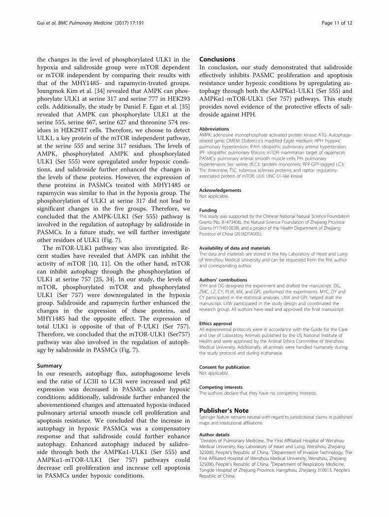
the changes in the level of phosphorylated ULK1 in thehypoxia and salidroside group were mTOR dependentor mTOR independent by comparing their results withthat of the MHY1485- and rapamycin-treated groups.Joungmok Kim et al. [34] revealed that AMPK can phos-phorylate ULK1 at serine 317 and serine 777 in HEK293cells. Additionally, the study by Daniel F. Egan et al. [35]revealed that AMPK can phosphorylate ULK1 at theserine 555, serine 467, serine 627 and threonine 574 res-idues in HEK293T cells. Therefore, we choose to detectULK1, a key protein of the mTOR independent pathway,at the serine 555 and serine 317 residues. The levels ofAMPK, phosphorylated AMPK and phosphorylatedULK1 (Ser 555) were upregulated under hypoxic condi-tions, and salidroside further enhanced the changes inthe levels of these proteins. However, the expression ofthese proteins in PASMCs treated with MHY1485 orrapamycin was similar to that in the hypoxia group. Thephosphorylation of ULK1 at serine 317 did not lead tosignificant changes in the five groups. Therefore, weconcluded that the AMPK-ULK1 (Ser 555) pathway isinvolved in the regulation of autophagy by salidroside inPASMCs. In a future study, we will further investigateother residues of ULK1 (Fig. 7).The mTOR-ULK1 pathway was also investigated. Re-
cent studies have revealed that AMPK can inhibit theactivity of mTOR [10, 11]. On the other hand, mTORcan inhibit autophagy through the phosphorylation ofULK1 at serine 757 [25, 34]. In our study, the levels ofmTOR, phosphorylated mTOR and phosphorylatedULK1 (Ser 757) were downregulated in the hypoxiagroup. Salidroside and rapamycin further enhanced thechanges in the expression of these proteins, andMHY1485 had the opposite effect. The expression oftotal ULK1 is opposite of that of P-ULK1 (Ser 757).Therefore, we concluded that the mTOR-ULK1 (Ser757)pathway was also involved in the regulation of autoph-agy by salidroside in PASMCs (Fig. 7).
SummaryIn our research, autophagy flux, autophagosome levelsand the ratio of LC3II to LC3I were increased and p62expression was decreased in PASMCs under hypoxicconditions; additionally, salidroside further enhanced theabovementioned changes and attenuated hypoxia-inducedpulmonary arterial smooth muscle cell proliferation andapoptosis resistance. We concluded that the increase inautophagy in hypoxic PASMCs was a compensatoryresponse and that salidroside could further enhanceautophagy. Enhanced autophagy induced by salidro-side through both the AMPKα1-ULK1 (Ser 555) andAMPKα1-mTOR-ULK1 (Ser 757) pathways coulddecrease cell proliferation and increase cell apoptosisin PASMCs under hypoxic conditions.
ConclusionsIn conclusion, our study demonstrated that salidrosideeffectively inhibits PASMC proliferation and apoptosisresistance under hypoxic conditions by upregulating au-tophagy through both the AMPKα1-ULK1 (Ser 555) andAMPKα1-mTOR-ULK1 (Ser 757) pathways. This studyprovides novel evidence of the protective effects of sali-droside against HPH.
AbbreviationsAMPK: adenosine monophosphate activated protein kinase; ATG: Autophagy-related gene; DMEM: Dulbecco’s modified Eagle medium; HPH: hypoxicpulmonary hypertension; IPAH: idiopathic pulmonary arterial hypertension;IPF: idiopathic pulmonary fibrosis; mTOR: mammalian target of rapamycin;PASMCs: pulmonary arterial smooth muscle cells; PH: pulmonaryhypertension; Ser: serine; tfLC3: tandem monomeric RFP-GFP-tagged LC3;Thr: threonine; TSC: tuberous sclerosis proteins; and raptor: regulatory-associated protein of mTOR; ULK: UNC-51-like kinase
AcknowledgementsNot applicable.
FundingThis study was supported by the Chinese National Natural Science FoundationGrants (No. 81473406), the Natural Science Foundation of Zhejiang ProvinceGrants (Y17H010028), and a project of the Health Department of ZhejiangProvince of China (2016DTA005).
Availability of data and materialsThe data and materials are stored in the Key Laboratory of Heart and Lungof Wenzhou Medical University and can be requested from the first authorand corresponding author.
Authors’ contributionsXYH and DG designed the experiment and drafted the manuscript. DG,ZMC, LZ, CY, PLW, MX, and GPL performed the experiments. MYC, DY andCY participated in the statistical analyses. LXW and GPL helped draft themanuscript. LXW participated in the study design and coordinated theresearch group. All authors have read and approved the final manuscript.
Ethics approvalAll experimental protocols were in accordance with the Guide for the Careand Use of Laboratory Animals published by the US National Institute ofHealth and were approved by the Animal Ethics Committee of WenzhouMedical University. Additionally, all animals were handled humanely duringthe study protocol and during euthanasia.
Consent for publicationNot applicable.
Competing interestsThe authors declare that they have no competing interests.
Publisher’s NoteSpringer Nature remains neutral with regard to jurisdictional claims in publishedmaps and institutional affiliations.
Author details1Division of Pulmonary Medicine, The First Affiliated Hospital of WenzhouMedical University, Key Laboratory of Heart and Lung, Wenzhou, Zhejiang325000, People’s Republic of China. 2Department of Invasive Technology, TheFirst Affiliated Hospital of Wenzhou Medical University, Wenzhou, Zhejiang325000, People’s Republic of China. 3Department of Respiratory Medicine,Tongde Hospital of Zhejiang Province, Hangzhou, Zhejiang 310013, People’sRepublic of China.
Gui et al. BMC Pulmonary Medicine (2017) 17:191 Page 11 of 12

Received: 16 May 2017 Accepted: 6 October 2017
References1. Tuder RM, Marecki JC, Richter A, Fijalkowska I, Flores S, et al. Pathology of
pulmonary hypertension. Clin Chest Med. 2007;28(1):23–vii.2. Dorfmuller P. Pulmonary hypertension: pathology. Handb Exp Pharmacol.
2013;218:59–75.3. Ruderman NB, Carling D, Prentki M, Cacicedo JM. AMPK, insulin resistance,
and the metabolic syndrome. J Clin Invest. 2013;123(7):2764–72.4. Viollet B, Horman S, Leclerc J, Lantier L, Foretz M, Billaud M, et al. AMPK
inhibition in health and disease. Crit Rev Biochem Mol. 2010;45(4):276–95.5. Wu YY, Liu L, Zhang YH, Wang GZ, Han D, Ke R, et al. Activation of AMPK
inhibits pulmonary arterial smooth muscle cells proliferation. Exp Lung Res.2014;40(5):251–8.
6. Huang XY, Fan R, Lu YY, Yu C, Xu XM, Zhang X, et al. Regulatory effect ofAMP-activated protein kinase on pulmonary hypertension induced bychronic hypoxia in rats: in vivo and in vitro studies. Mol Biol Rep. 2014;41(6):4031–41.
7. Chen MY, Cai H, Yu C, Wu PL, Xu XM, Fan R, et al. Salidroside exerts protectiveeffects against chronic hypoxia-induced pulmonary arterial hypertension viaAMPKα1-dependent pathways. Am J Transl Res. 2016;8(1):12–27.
8. Liu XP, Wang GS, You ZC, Qian P, Chen HP, Dou Y, et al. Inhibition of hypoxia-induced proliferation of pulmonary arterial smooth muscle cells by a mTORsiRNA-loaded cyclodextrin nanovector. Biomaterials. 2014;35:4401–16.
9. Krymskaya VP, Snow J, Cesarone G, Khavin I, Goncharov DA, Lim PN, et al.mTOR is required for pulmonary arterial vascular smooth muscle cellproliferation under chronic hypoxia. FASEB J. 2011;25(6):1922–33.
10. Lnoki K, Zhu TQ, Guan KL. TSC2 mediates cellular energy response tocontrol cell growth and survival. Cell. 2003;115(5):577–90.
11. Gwinn DM, Shackelford DB, Egan DF, Mihaylova MM, Mery A, Vasquez DS,et al. AMPK phosphorylation of raptor mediates a metabolic checkpoint.Mol Cell. 2008;30(2):214–26.
12. Din FVN, Valanciute A, Houde VP, Zibrova D, Green KA, Sakamoto K,et al. Aspirin inhibits mTOR signaling, activates AMP-activated proteinkinase, and induces autophagy in colorectal cancer cells.Gastroenterology. 2012;142(7):26–33.
13. Wang K, Liu R, Li JY, Mao JL, Lei YL, Wu JH, et al. Quercetin inducesprotective autophagy in gastric cancer cells: involvement of Akt-mTOR-andhypoxia-induced factor 1α-mediated signaling. Autophagy. 2011;7(9):966–78.
14. Romero Y, Bueno M, Ramirez R, Alvarez D, Sembrat JC, Goncharova EA, et al.mTORC1 activation decreases autophagy in aging and idiopathicpulmonary fibrosis and contributes to apoptosis resistance in IPF fibroblasts.Aging Cell. 2016;15(6):1103–12.
15. Sutendra G, Michelakis ED. The metabolic basis of pulmonary arterialhypertension. Cell Metab. 2014;19(4):558–73.
16. Wu TJ, Zhou HP, Jin ZX, Bi SH, Yang XL, Yi DH, et al. Cardioprotection ofsalidroside from ischemia/reperfusion injury by increasing N-acetylglucosaminelinkage to celluar proteins. Eur J Pharmacol. 2009;613(1-3):93–9.
17. Hu XL, Lin SX, Yu DH, Qiu SF, Zhang XQ, Mei RH. A preliminary study: theanti-proliferation effect of salidroside on different human cancer cell lines.Cell Biol Toxicol. 2010;26(6):499–507.
18. Wiegant FAC, Surinova S, Ytsma E, Makkinje ML, Wikman G, Post JA. Plantadaptogens increase lifespan and stress resistance in C. Elegans.Biogerontology. 2009;10(1):27–42.
19. Wang HB, Ding YY, Zhou J, Sun XL, Wang SW. The in vitro and in vivoantiviral effects of salidroside from Rhodiola Rosea L. against coxsackievirusB3. Phytomedicine. 2009;16(2-3):146–55.
20. He C, Klionsky DJ. Regulation mechanisms and signaling pathways ofAutophagy. Annu Rev Genet. 2009;43(1):67–93.
21. Yang ZF, Klionsky DJ. Eaten alive: a history of macroautophagy. Nat Cell Biol.2010;12(9):814–22.
22. Klionsky DJ. The molecular machinery of autophagy: unanswered questions.J Cell Sci. 2005;118(Pt 1):7–18.
23. Chen YQ, Klionsky DJ. The regulation of autophagy-unanswered questions. JCell Sci. 2011;124(Pt 2):161–70.
24. Mizushima N. The role of the Atg1/ULK1 complex in autophagy regulation.Curr Opin Cell Biol. 2010;22(2):132–9.
25. Shang LB, Chen S, Du FH, Li S, Zhao LP, Wang XD. Nutrient starvation elicitsan acute autophagic response mediated by Ulk1 dephosphorylation and its
subsequent dissociation from AMPK. P Natl Acad Sci USA. 2011;108(12):4788–93.
26. Budhiraja R, Tuder RM, Hassoun PM. Endothelial dysfunction in pulmonaryhypertension. Circulation. 2004;109(2):159–65.
27. Yepes JN, Burns M, Anadhan A, Khalimonchuk O, Razo LM, Vega BQ, et al.Oxidative stress, redox signaling, and autophagy: cell death versus survival.Antioxid Redox Signal. 2014;21(1):66–85.
28. Choi YJ, Park YJ, Park JY, Jeong HO, Kim DH, Ha YM, et al. Inhibitory effect ofmTOR activator MHY 1485 on autophagy: suppression of lysosomal fusion.PLoS One. 2012;7(8):e43418.
29. Klionsky DJ, Abdalla FC, Abeliovich H, Abraham RT, Arozena AA, Adeli K,et al. Guidelines for the use and interpretation of assays for monitoringautophagy. Autophagy. 2012;8(4):445–544.
30. Lee SJ, Smith A, Guo LP, Alastalo TP, Li ML, Sawada H, et al. Autophagicprotein LC3B confers resistance against hypoxia-induced pulmonaryhypertension. Am J Respir Crit Care Med. 2011;183(5):649–58.
31. Gump JM, Staskiewicz L, Morgan MJ, Bamberg A, Riches DWH, Thorburn A.Autophagy variation within a cell population determines cell fate viaselective degradation of Fap-1. Nat Cell Biol. 2014;16(1):47–54.
32. Chen S, Yuan JX, Yao SQ, Jin YL, Chen G, Tian W, et al. Lipopolysaccharidesmay aggravate apoptosis through accumulation of autophagosomes inalveolar macrophages of human silicosis. Autophagy 2015; 11(12): 2346-2357.
33. Goncharov DA, Kudryashova TV, Ziai H, Stansbury KI, Delisser H, KrymskayaVP, et al. mTORC2 coordinates pulmonary artery smooth muscle cellmetabolism, proliferation and survival in pulmonary arterial hypertension.Circulation. 2014;129(8):864–74.
34. Kim J, Kundu M, Viollet B, Guan KL. AMPK and mTOR regulate autophagythrough direct phosphorylation of Ulk1. Nat Cell Biol. 2011;13(2):132–41.
35. Egan DF, Shackelford DB, Mihaylova MM, Gelino SR, Kohnz RA, Mair W, et al.Phosphorylation of ULK1 (hATG1) by AMP-activated protein kinase connectsenergy sensing to mitophagy. Science. 2011;331(6010):456–61.
• We accept pre-submission inquiries
• Our selector tool helps you to find the most relevant journal
• We provide round the clock customer support
• Convenient online submission
• Thorough peer review
• Inclusion in PubMed and all major indexing services
• Maximum visibility for your research
Submit your manuscript atwww.biomedcentral.com/submit
Submit your next manuscript to BioMed Central and we will help you at every step:
Gui et al. BMC Pulmonary Medicine (2017) 17:191 Page 12 of 12