Embed Size (px)
Citation preview
Inhibition of neural crest migration underliescraniofacial dysmorphology and Hirschsprung’sdisease in Bardet–Biedl syndromeJonathan L. Tobin*, Matt Di Franco†, Erica Eichers‡, Helen May-Simera*, Monica Garcia‡, Jiong Yan‡, Robyn Quinlan*,Monica J. Justice‡, Raoul C. Hennekam§¶, James Briscoe�, Masazumi Tada**, Roberto Mayor**, Alan J. Burns††,James R. Lupski‡, Peter Hammond*†, and Philip L. Beales*‡‡
*Molecular Medicine Unit, UCL Institute of Child Health, 30 Guilford Street, London WC1N 1EH, United Kingdom; †Biomedical Informatics Unit, UCLEastman Dental Institute for Oral Health Care Sciences, 256 Gray’s Inn Road, London WC1X 8LD, United Kingdom; ‡Department of Molecular and HumanGenetics, Baylor College of Medicine, One Baylor Plaza, Houston, TX 77030; �Developmental Neurobiology, National Institute for Medical Research,London NW7 1AA, United Kingdom; **Department of Anatomy and Developmental Biology, University College London, London WC1E 6BT,United Kingdom; ††Neural Development Unit, UCL Institute of Child Health, 30 Guilford Street, London WC1N 1EH, United Kingdom; §Clinicaland Molecular Genetics Unit, UCL Institute of Child Health, 30 Guilford Street, London WC1N 1EH, United Kingdom; and ¶Departmentof Pediatrics, Academic Medical Centre, University of Amsterdam, 1105 AZ Amsterdam, The Netherlands
Edited by Kathryn V. Anderson, Sloan–Kettering Institute, New York, NY, and approved March 6, 2008 (received for review July 30, 2007)
Facial recognition is central to the diagnosis of many syndromes,and craniofacial patterns may reflect common etiologies. In thepleiotropic Bardet–Biedl syndrome (BBS), a primary ciliopathy withintraflagellar transport dysfunction, patients have a characteristicfacial ‘‘gestalt’’ that dysmorphologists have found difficult tocharacterize. Here, we use dense surface modeling (DSM) to revealthat BBS patients and mouse mutants have mid-facial defectsinvolving homologous neural crest-derived structures shared byzebrafish morphants. These defects of the craniofacial (CF) skele-ton arise from aberrant cranial neural crest cell (NCC) migration.These effects are not confined to the craniofacial region, butvagal-derived NCCs fail to populate the enteric nervous system,culminating in disordered gut motility. Furthermore, morphantsdisplay hallmarks of disrupted Sonic Hedgehog (Shh) signalingfrom which NCCs take positional cues. We propose a modelwhereby Bbs proteins modulate NCC migration, contributing tocraniofacial morphogenesis and development of the enteric ner-vous system. These migration defects also explain the associationof Hirschsprung’s disease (HD) with BBS. Moreover, this is apreviously undescribed method of using characterization of facialdysmorphology as a basis for investigating the pathomechanism ofCF development in dysmorphic syndromes.
sonic hedgehog � Wnt � cilia � cell migration
Recognition of the facial ‘‘gestalt’’ is central to diagnosis of manygenetic disorders, but the great variability of features often
hinders successful classification (1). Recently, noninvasive 3D sur-face imaging has characterized dysmorphology in syndromes (2, 3).None, however, has been used to either define subtle facial dys-morphism or aid investigation of mechanisms for craniofacialdysmorphology.
Bardet–Biedl syndrome (BBS) causes retinal degeneration, post-axial polydactyly, obesity, renal dysfunction, and cognitive impair-ment. Twelve BBS genes (BBS1–BBS12) have been discovered, andpathogenesis lies in primary cilia dysfunction (4). BBS4, BBS6, andBBS8 (investigated in this study) are expressed in ciliated epitheliaand localize to the centrosome and basal bodies of ciliated cells(5–7). Subtle craniofacial abnormalities in patients have beenreported (8–10). Among the many additional features of BBS isHirschsprung’s disease (HD), a disorder of the enteric nervoussystem (ENS) (11).
Streams of neural crest cells (NCCs) from the caudal brain formmost of the craniofacial (CF) skeleton (see ref. 12 for review).Cranial NCCs (CNCC) follow defined paths to populate thefrontonasal prominence and branchial arch mesenchyme. Here,they proliferate and differentiate into structures of the face andcranium. Sonic Hedgehog (Shh) expressed in the ventral brain and
oral ectoderm is essential for the formation of most facial structures(12). Shh-deficient mice have severe loss of craniofacial bones, and,in humans, SHH mutations cause midline CF defects with holo-prosencephaly (HPE) (12).
The ENS regulates gastrointestinal motility and secretion. Itderives from vagal and lumbosacral NCCs (13). In HD, character-ized by megacolon, there are reduced or absent enteric ganglia inthe distal colon.
Despite the plethora of recent evidence pointing to ciliarydysfunction in BBS, both craniofacial and gastrointestinal defectsare difficult to reconcile on this basis alone. In this study, we showthat BBS proteins are required for NCC migration, culminating inShh-dependent craniofacial defects, and gut motility disorder, likelyunderlying the HD association with BBS.
ResultsThree-Dimensional Dense Surface Modeling (DSM) Reveals Compara-ble Craniofacial Defects in Man and Mouse. To assess CF defects inBBS, we performed 3D DSM face analysis on 83 patients (irre-spective of mutations) and 230 controls. To account for growthdifferences, analyses were performed separately for subjects �20years old. Facial scans were annotated with key landmarks [sup-porting information (SI) Fig. S1], DSMs of facial and nasal regionswere computed for subsets of faces, and differences betweencontrol and BBS landmarks were measured. Of several inter-landmark measurements, the most significant were anomalouslength and width of the face (Fig. S2). To avoid the effects ofexcessive soft tissue in patient faces, we first restricted shape analysisto the nasal region where obesity has least effect. Color-distance-coded comparisons of mean BBS and control noses for children andadults showed nasal bridge hypoplasia and nasal shortening/reduced bulbosity at the nasal tip (red-yellow; Fig. 1 A and B).Dynamic morphs between BBS and control mean noses demon-strate these and other shape differences in the nasal alae and
Author contributions: J.L.T., J.B., R.M., A.J.B., J.R.L., P.H., and P.L.B. designed research; J.L.T.,M.D.F., E.E., H.M.-S., M.G., J.Y., R.Q., M.J.J., R.C.H., M.T., A.J.B., and P.H. performed research;H.M.-S., J.B., M.T., R.M., and A.J.B. contributed new reagents/analytic tools; J.L.T., M.D.F.,M.J.J., A.J.B., J.R.L., P.H., and P.L.B. analyzed data; and J.L.T., P.H., and P.L.B. wrote thepaper.
The authors declare no conflict of interest.
This article is a PNAS Direct Submission.
Freely available online through the PNAS open access option.
‡‡To whom correspondence should be addressed. E-mail: [email protected].
This article contains supporting information online at www.pnas.org/cgi/content/full/0707057105/DCSupplemental.
© 2008 by The National Academy of Sciences of the USA
6714–6719 � PNAS � May 6, 2008 � vol. 105 � no. 18 www.pnas.org�cgi�doi�10.1073�pnas.0707057105
Dow
nloa
ded
by g
uest
on
Mar
ch 2
2, 2
020
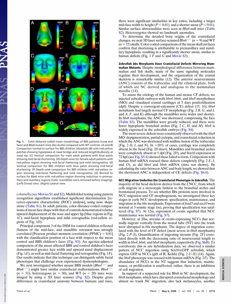
columella (see Movies S1 and S2). Multifolded testing using patternrecognition algorithms established significant discrimination [re-ceiver-operator characteristic (ROC) analysis], using nose shapealone (Table S1). In adult patients, color-distance-coded compar-isons of mean face shape with that of controls demonstrated relativeupward displacement of the nose and upper lip (blue regions in Fig.1C), mid-facial hypoplasia and mild retrognathia (red-yellow re-gions of Fig. 1D).
Principal component 3 of the DSM for control children reflectedflatness of the mid-face, and mandible retrusion was stronglycorrelated [Pearson product moment correlation (PPMC) � 0.81]with the classification position of a face with respect to the meancontrol and BBS children’s faces (Fig. S3). An age/size-adjustedcomparison of the mean affected BBS and control children’s facesdemonstrated greater face width and upward nasal displacement(Fig. 1E), as well as mid-facial flattening and retrognathia (Fig. 1F).Our results indicate that this technique can distinguish subtle facialphenotypes that challenge even experienced dysmorphologists.
We next investigated whether mouse BBS models (Bbs4�/� andBbs6�/�) might have similar craniofacial malformations. Bbs4�/�
(n � 31), heterozygous (n � 30), and WT (n � 20) mice wereimaged by using a 3D laser scanner (Fig. S2). Despite grossdifferences in craniofacial anatomy between humans and mice,
there were significant similarities in key ratios, including a largermid-face width to height (P � 0.01), and a shorter snout (P � 0.01).Similar surface abnormalities were seen in Bbs6-null mice (TableS2). Heterozygotes showed no landmark anomalies.
To determine the detailed bony origins of the craniofacialchanges, we next 3D laser surface-scanned Bbs6�/� (n � 9) and WT(n � 15) skulls. Color-coded comparisons of the mean skull surfacesconfirm that shortening is attributable to premaxillary and maxil-lary hypoplasia, resulting in a significantly shorter snout, similar tohuman deficits (Fig. 1 F and G and Movie S3).
Zebrafish bbs Morphants Have Craniofacial Defects Mirroring Mam-malian Mutants. Despite morphological differences between mam-malian and fish skulls, many of the same signaling moleculesregulate their development, and the organization of the cranialskeleton is remarkably similar (12). The anterior neurocranium(ANC) consists of the trabeculae and the ethmoid plate, bothof which are NC derived and analogous to the mammalianmaxilla (14).
To assess the etiology of the human and mouse CF defects, weinjected zebrafish embryos with bbs4, bbs6, and bbs8 morpholinos(MO) and visualized cranial cartilages at 5 days postfertilization(dpf). Despite a convergent-extension (CE) defect (15, 16), bbs4morphants had largely normal CF morphology (Fig. 2 B, G, and Land A, F, and K) although the mandibles were wider and shorter.In bbs6 morphants, the ANC was shortened, compressing the face(Table S3). The mandibles were greatly reduced, and there werefewer hypoplastic branchial arches (Fig. 2 C, H, and M). bbs8 iswidely expressed in the zebrafish embryo (Fig. S4).
The most severe defects were consistently observed with the bbs8MO: depigmentation, partial cyclopia, and craniofacial reduction inwhich the ANC was shortened with midline fusion of the trabeculae(Fig. 2 D, I, and N). In �20% of cases, cartilage was completelyabsent in the head (Fig. 2D Inset). Mandibles and branchial archeswere completely absent at 5 dpf (Fig. 2D), and Sox10 expression at72 hpf (see Fig. S5A) showed these failed to form. Coinjections withhuman bbs8 mRNA rescued these defects completely (Fig. 2 E, J,and O), as did bbs4 and bbs6 mRNA (data not shown). Bycalculating the ratio between ANC and body length, we showed thatthe shortened ANC is independent of CE defects (Fig. S6A).
NCC Migration Underlies the Craniofacial Phenotype in Zebrafish. Themajority of the head skeleton derives from the CNCC, from whichcells migrate in a stereotypic fashion to the branchial arches andfrontonasal process. To see whether Bbs proteins were involved inNCC migration and CF morphogenesis, we investigated three keystages in early NCC development: specification, maintenance, andmigration in the bbs morphants. Expression of foxd3 and sox10 werenormal at 5-somite stage (ss), proving that specification was unal-tered (Fig. S7). At 12ss, expression of crestin signified that NCCmaintenance was normal (Fig. S7).
However, at 20ss, streams of crestin-expressing NCCs that nor-mally migrate ventrally from the neural tube (NT) into the trunkwere disrupted in bbs morphants. The degree of migration corre-lated with the level of CF defect (most severe in bbs8 morphants)(Fig. 2 P–S). Quantification of migrating streams (Fig. 2Y) corre-lated directly with the decreasing ratio of neurocranial length towidth in bbs4, bbs6, and bbs8 morphants, respectively (Fig. S6B). Tocorroborate this in situ hybridization data, we observed a similarpattern of NCC migration using Sox10:eGFP zebrafish, whichexpress GFP in NCCs (gift of R. Kelsh) (Fig. 2 U–X) (14). Again,the bbs8 phenotype was rescued with human mRNA (Fig. 2T). Theabundance of NCCs in the NT suggests that induction, mainte-nance, or proliferation is unaffected; rather, there is an inhibitionof cell migration.
In support of a suspected role for Bbs8 in NC development, thebbs8 morphants, which have disrupted craniofacial morphology andalmost no trunk NC migration, also lack melanocytes, another
Fig. 1. Color-distance-coded mean morphology of BBS patients (nose andface) and Bbs6 mutant mice (dry skulls) compared with WT controls. (A and B)Comparison normal to surface for BBS children (A)/adults (B) with red-yellowpatches showing hypoplasia at nasal bridge and reduced length/bulbosity atnasal tip. (C) Vertical comparison for male adult patients with blue patchshowing mid-facial shortening. (D) Depth-wise for female adult patients withred-yellow region showing mid-facial flattening and mild retrognathia. (E)Vertical comparison for BBS children with blue patch showing mid-facialshortening. (F) Depth-wise comparison for BBS children with red-yellow re-gion showing mid-facial flattening and mild retrognathia. (G) Normal tosurface for Bbs6 mice with red-yellow region showing reduction in premax-illary and maxillary regions (note: mandibles were removed before imaging).(Left) Dorasl view. (Right) Lateral view.
Tobin et al. PNAS � May 6, 2008 � vol. 105 � no. 18 � 6715
MED
ICA
LSC
IEN
CES
Dow
nloa
ded
by g
uest
on
Mar
ch 2
2, 2
020
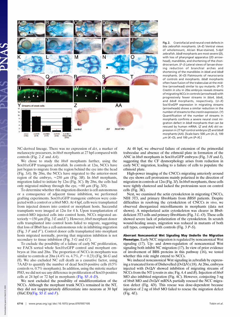
NC-derived lineage. There was no expression of dct, a marker ofmelanocyte precursors, in bbs8 morphants at 27 hpf compared withcontrols (Fig. 2 Z and AA).
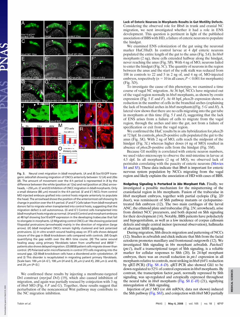
We chose to study the bbs8 morphants further, using theSox10:eGFP transgenic zebrafish. In controls at 12ss, NCCs havejust begun to migrate from the region behind the eye into the head(Fig. 3A). By 20ss, the NCCs have migrated to the anterior-mostregion of the embryo, �250 �m (Fig. 3B). In bbs8 morphants,migration failed to initiate by 12ss (Fig. 3C). By 20ss, the cells hadonly migrated midway through the eye, �80 �m (Fig. 3D).
To determine whether this migration disorder is cell autonomousor a consequence of adjacent tissue inhibition, we performedgrafting experiments. Sox10:eGFP transgenic embryos were coin-jected with a control or a bbs8 MO. At 4 hpf, cells were transplantedfrom injected donors into control or morphant hosts. Successfultransplants were imaged at 12ss for 4 h. Upon transplantation ofcontrol-MO injected cells into control hosts, NCCs migrated an-teriorly �150 �m (Fig. 3 E and E�). However, bbs8 morphant donorcells transplanted into control hosts failed to migrate, suggestingthat loss of Bbs8 has a cell-autonomous role in inhibiting migration(Fig. 3 F and F�). Control donor cells transplanted into morphanthosts migrated normally, proving that migration inhibition is notsecondary to tissue inhibition (Fig. 3 G and G�).
To exclude the possibility of a failure of early NC proliferation,we FACS sorted whole Sox10:eGFP control and morphant em-bryos at 16ss and 20ss. The proportion of NCCs in morphants wassimilar to controls at 20ss (4.4% vs. 4.7%, P � 0.23) (Fig. S6 G andH). We also excluded NC cell death as a causative factor, using7AAD to quantify the number of dead Sox10-positive cells (0.5%controls vs. 0.7% morphants). In addition, using the mitotic markerPH3, we did not see any difference in proliferation of Sox10-positivecells at 24 hpf or 72 hpf in morphants (Fig. S5 A–D).
We next excluded the possibility of misdifferentiation of theNCCs. Although the morphant trunk NCCs remained in the NT,they did not inappropriately differentiate into neurons at 30 hpf(HuC/D)(Fig. S5 E and F).
At 48 hpf, we observed failure of extension of the primordialtrabeculae and absence of the ethmoid plate in formation of theANC in bbs8 morphants in Sox10:eGFP embryos (Fig. 3 H and I),suggesting that the CF dysmorphology arises from reduction inearly NCC migration, leading to a failure of cells to populate theethmoid plate.
High-power imaging of the CNCCs migrating anteriorly aroundthe eye shows cell protrusions mainly polarized in the direction ofmigration in controls at 12ss (Fig. 3J). In bbs8 morphants, these cellswere tightly clustered and lacked the protrusions seen on controlcells (Fig. 3K).
Next, we examined the actin cytoskeleton in migrating CNCCs,NIH 3T3, and primary fibroblasts from BBS8 patients. Despitedifficulties in resolving the cytoskeleton of CNCCs in vivo, weobserved disorganized microfilaments in morphants (data notshown). A mispolarized actin cytoskeleton was clearer in Bbs8-deficient 3T3 cells and primary fibroblasts (Fig. 3 L–O). These cellsshowed severe lack of polarization of the cytoskeleton. In scratchwound-healing assays, migration was reduced in mutants of bothcell types, compared with controls (Fig. 3 P–S).
Aberrant Noncanonical Wnt Signaling May Underlie the MigrationPhenotype. Early NCC migration is regulated by noncanonical Wntsignaling (17). Up- and down-regulation of noncanonical Wntsignaling both inhibit NC migration (17). In view of prior evidenceof involvement of BBS proteins in this pathway (16), we testedwhether this role might extend to NCCs.
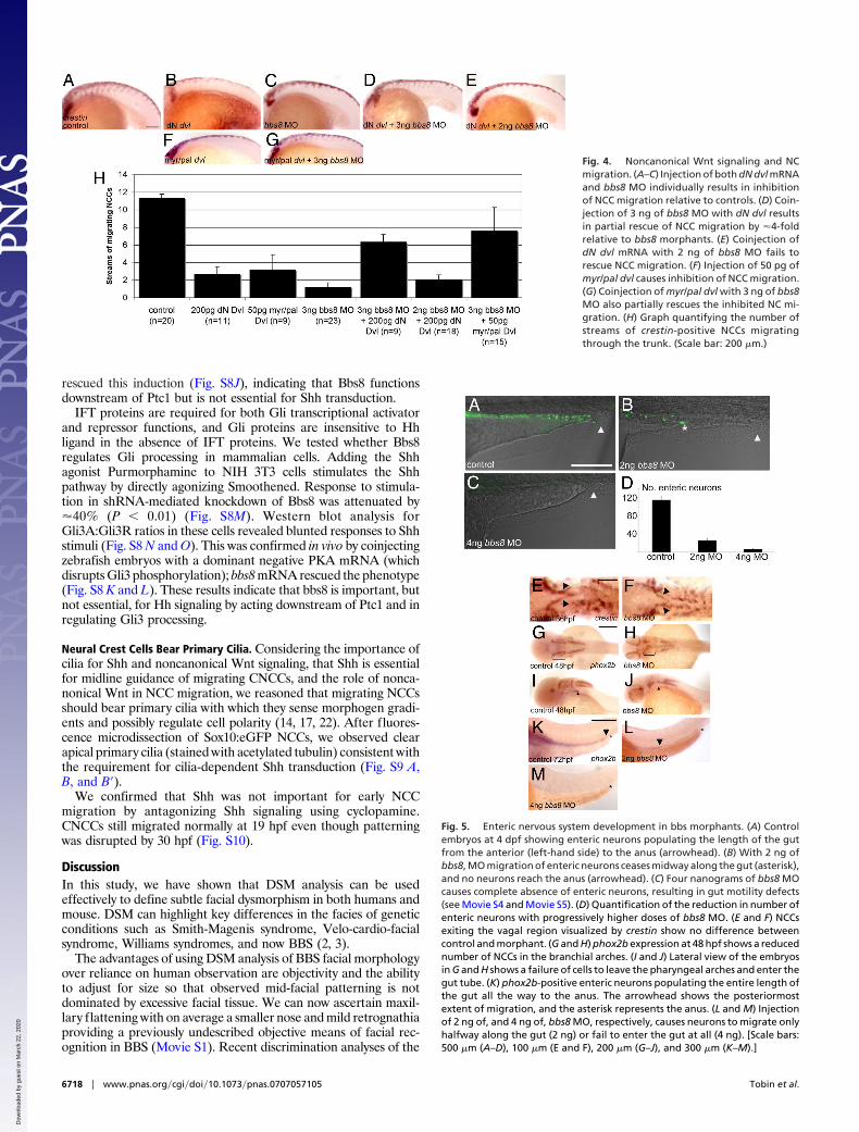
We induced noncanonical Wnt signaling in zebrafish by express-ing a truncated form of Dishevelled (Dvl�N) (18). At 20ss, embryosinjected with Dvl�N showed inhibition of migrating streams ofNCCs from the NT (crestin in situ; Fig. 4 A and B). Injection of bbs8MO also inhibited migration (Fig. 4C). However, coinjecting 3 ngof bbs8 MO and Dvl�N mRNA partially rescued the NCC migra-tion defect (Fig. 4D). This rescue was dose-dependent becauseinjection of 2 ng of bbs8 MO failed to rescue the migration defect(Fig. 4E).
Fig. 2. Craniofacial and neural crest defects inbbs zebrafish morphants. (A–E) Ventral viewsof wholemount, Alcian Blue-stained, 5-dpfzebrafish. bbs8 morphants are most severe (D),with loss of pharyngeal apparatus (2D arrow-head), mandibles, and shortening of the chon-drocranium. (F–J) Lateral views of larvae show-ing reduction of branchial arches andshortening of the mandibles in bbs6 and bbs8morphants. (K–O) Flatmounts of neurocraniaof controls and morphants. bbs8 morphantsoften have fusion of the trabeculae at the mid-line (arrowhead) similar to syu mutants. (P–T)Crestin in situ in 20ss embryos reveals streamsof migrating NCCs in controls (arrowhead) withprogressively fewer streams in bbs4, bbs6,and bbs8 morphants, respectively. (U–X)Sox10:eGFP expression in migrating streams(arrowheads) shows a similar reduction in thenumber of streams to the crestin expression. (Y)Quantification of the number of streams inmorphants confirms a severe neural crest mi-gration defect in bbs8 morphants that can berescued by human mRNA. (Z and AA) dct ex-pression in 27 hpf control embryos (Z) and bbs8morphants (AA). [Scale bars: 500 �m (A–J), 100�m (K–O), and 100 �m (P–X).]
6716 � www.pnas.org�cgi�doi�10.1073�pnas.0707057105 Tobin et al.
Dow
nloa
ded
by g
uest
on
Mar
ch 2
2, 2
020
We confirmed these results by injecting a membrane-targetedDvl construct (myr/pal Dvl) (19), which also caused inhibition ofmigration, and again was partially rescued by coinjection with 3 ngof bbs8 MO (Fig. 4 F and G). Together, these results suggest thatperturbation of the noncanonical Wnt pathway may contribute tothe NC migration inhibition.
Lack of Enteric Neurons in Morphants Results in Gut Motility Defects.Considering the observed role for Bbs8 in trunk and cranial NCmigration, we next investigated whether it had a role in ENSdevelopment. This question is pertinent in light of the publishedassociation of BBS with HD, a failure of enteric neurons to populatethe hindgut.
We examined ENS colonization of the gut using the neuronalmarker HuC/HuD. In control larvae at 4 dpf enteric neuronspopulated the entire length of the gut to the anus (Fig. 5A). In bbs8morphants (2 ng), these cells extended halfway along the hindgut,never reaching the anus (Fig. 5B). With 4 ng of MO, neurons failedto enter the hindgut (Fig. 5C). The quantity of neurons in the regionbetween the anus and the start of the yolk stalk was reduced from108 in controls to 22 and 5 in 2 ng of, and 4 ng of, MO-injectedembryos, respectively (n � 10 in all cases; P � 0.001 for morphants)(Fig. 5D).
To investigate the cause of this phenotype, we examined a timecourse of vagal NC migration. At 36 hpf, NCCs have migrated outof the vagal region normally in bbs8 morphants, as shown by crestinexpression (Fig. 5 E and F). At 48 hpf, phox2b expression shows areduction in the number of cells in the branchial arches (explainingthe lack of branchial arches in bbs8 morphants)(Fig. 5 G and H). Alateral view shows that there are no cells migrating into the gut tubein morphants at this time (Fig. 5 I and J), suggesting that the lackof ENS arises from a failure of cells to migrate from the vagalregion, through the arches and into the gut, not from a failure ofspecification or exit from the vagal region.
We confirmed the HuC results by in situ hybridization for phox2bat 72 hpf. In controls, phox2b-positive cells populated the gut to theanus (Fig. 5K). With 2 ng of MO, cells reach the midpoint of thehindgut (Fig. 5L) whereas higher doses (4 ng of MO) resulted inabsence of phox2b-positive cells from the hindgut (Fig. 5M).
Because GI motility is correlated with enteric neuron numbers,we used video microscopy to observe the mid-intestine in larvae at4.5 dpf. In all morphants (2 ng of MO), we observed lack ofperistalsis correlating with the paucity of enteric neurons (MoviesS4 and S5). These data indicate that Bbs8 is important for entericnervous system population by NCCs migrating from the vagalregion and likely explains the association of HD with cases of BBS.
Hedgehog Signaling Is Perturbed in bbs Morphant Fish. Finally, weinvestigated a possible mechanism for the mispatterning of thecraniofacial region in bbs morphants. Fusion of the trabeculae inbbs8 morphant embryos, together with partial cyclopia (Fig. 2DInset), was reminiscent of Shh pathway mutants or cyclopamine-treated fish embryos (12). The two main cartilages of the larvalANC, the ethmoid plate and trabeculae (Fig. 2N; arrowhead), arisefrom distinct NCC precursors, and both depend on Shh signalingfor their development (14). Notably, BBS patients have polydactylyand hypogenitalism, as well as a low incidence of corpus callosumdefects and single central incisor (personal observation), hallmarksof aberrant SHH signaling.
During migration, Shh directs migration and patterning of NCCs(12). Studies in zebrafish and chick indicate that Shh from the facialectoderm promotes maxillary and frontonasal outgrowth (12). Weinvestigated Shh signaling in bbs morphant zebrafish. Patched1(ptc1), itself a transcriptional target of Shh signaling, is a reliablemarker for cellular responses to Shh (20). In 24-hpf morphantembryos, there was an overall reduction in ptc1 expression in allmorphants relative to controls, most striking in bbs8 (64% reductionby qRT-PCR) (Fig. S8 A–D). qRT-PCR also showed Gli1 to bedown-regulated to 52% of control expression in bbs8 morphants. Bycontrast, the transcription factor pax6, normally repressed by Shhsignaling, was up-regulated and ectopically expressed throughoutthe neural tube in bbs8 morphants (Fig. S8 E–H) (21), signifyingmisregulation of Shh signaling.
Injection of ptc1 MO (or shh mRNA; data not shown) inducedthe Shh pathway (Fig. S8I), and coinjection with bbs8 MO partially
Fig. 3. Neural crest migration in bbs8 morphants. (A and B) Sox10:GFP trans-genic zebrafish showing migration of CNCCs anteriorly between 12 (A) and 20ss(B). The amount of movement over the 4-h period is represented in B by thedifference between the white (position at 12ss) and red (position at 20ss) arrow-heads, �250 �m. (C and D) Inhibition of CNCC migration in bbs8 morphants. Onlya small distance (80 �m) moved in the 4-h period. (E and E�) NCCs from controlMO-injected embryos grafted into control hosts migrate anteriorly to populatethe head. The arrowhead shows the position of the anteriormost cell showing itschange in position over the 4-h period. (F and F�) Cells taken from bbs8 morphantdonors fail to migrate when transplanted into control hosts, suggesting that themigration defect is cell autonomous. (G and G�) Control cells transplanted intobbs8morphanthostsmigrateasnormal. (Hand I)Controlandmorphantembryosat 48 hpf showing Sox10:eGFP expression in the developing trabeculae that failto elongate in morphants. (J) Migrating control CNCCs at 14ss showing polarizedfilopodial protrusions (arrowheads) mainly in the direction of migration (largearrow). (K) bbs8 morphant CNCCs remain tightly clustered and lack polarizedprotrusions. (L) In vitro scratch wound healing assay on 3T3 cells shows delayedclosure of the gap in Bbs8 knockdown cells compared with controls. (M) Graphquantifying the gap width over the 48-h time course. (N) The same woundhealing assay using primary fibroblasts taken from unaffected and BBS8�/�
patients also shows delayed migration. (O) BBS8 patient cells migrate slower thancontrol. (P) Polarized actin microfilaments in control 3T3 cells migrating into thewound gap. (Q) Bbs8 knockdown cells have a disordered actin cytoskeleton. (Rand S) This disorder is recapitulated in migrating patient primary fibroblasts.[Scale bars: 100 �m (A–G�), 100 �m (H and I), 20 �m (J and K), 200 �m (L and N),and 20 �m (P–S).]
Tobin et al. PNAS � May 6, 2008 � vol. 105 � no. 18 � 6717
MED
ICA
LSC
IEN
CES
Dow
nloa
ded
by g
uest
on
Mar
ch 2
2, 2
020
rescued this induction (Fig. S8J), indicating that Bbs8 functionsdownstream of Ptc1 but is not essential for Shh transduction.
IFT proteins are required for both Gli transcriptional activatorand repressor functions, and Gli proteins are insensitive to Hhligand in the absence of IFT proteins. We tested whether Bbs8regulates Gli processing in mammalian cells. Adding the Shhagonist Purmorphamine to NIH 3T3 cells stimulates the Shhpathway by directly agonizing Smoothened. Response to stimula-tion in shRNA-mediated knockdown of Bbs8 was attenuated by�40% (P � 0.01) (Fig. S8M). Western blot analysis forGli3A:Gli3R ratios in these cells revealed blunted responses to Shhstimuli (Fig. S8 N and O). This was confirmed in vivo by coinjectingzebrafish embryos with a dominant negative PKA mRNA (whichdisrupts Gli3 phosphorylation); bbs8 mRNA rescued the phenotype(Fig. S8 K and L). These results indicate that bbs8 is important, butnot essential, for Hh signaling by acting downstream of Ptc1 and inregulating Gli3 processing.
Neural Crest Cells Bear Primary Cilia. Considering the importance ofcilia for Shh and noncanonical Wnt signaling, that Shh is essentialfor midline guidance of migrating CNCCs, and the role of nonca-nonical Wnt in NCC migration, we reasoned that migrating NCCsshould bear primary cilia with which they sense morphogen gradi-ents and possibly regulate cell polarity (14, 17, 22). After fluores-cence microdissection of Sox10:eGFP NCCs, we observed clearapical primary cilia (stained with acetylated tubulin) consistent withthe requirement for cilia-dependent Shh transduction (Fig. S9 A,B, and B�).
We confirmed that Shh was not important for early NCCmigration by antagonizing Shh signaling using cyclopamine.CNCCs still migrated normally at 19 hpf even though patterningwas disrupted by 30 hpf (Fig. S10).
DiscussionIn this study, we have shown that DSM analysis can be usedeffectively to define subtle facial dysmorphism in both humans andmouse. DSM can highlight key differences in the facies of geneticconditions such as Smith-Magenis syndrome, Velo-cardio-facialsyndrome, Williams syndromes, and now BBS (2, 3).
The advantages of using DSM analysis of BBS facial morphologyover reliance on human observation are objectivity and the abilityto adjust for size so that observed mid-facial patterning is notdominated by excessive facial tissue. We can now ascertain maxil-lary flattening with on average a smaller nose and mild retrognathiaproviding a previously undescribed objective means of facial rec-ognition in BBS (Movie S1). Recent discrimination analyses of the
Fig. 4. Noncanonical Wnt signaling and NCmigration. (A–C) Injection of both dN dvl mRNAand bbs8 MO individually results in inhibitionof NCC migration relative to controls. (D) Coin-jection of 3 ng of bbs8 MO with dN dvl resultsin partial rescue of NCC migration by �4-foldrelative to bbs8 morphants. (E) Coinjection ofdN dvl mRNA with 2 ng of bbs8 MO fails torescue NCC migration. (F) Injection of 50 pg ofmyr/pal dvl causes inhibition of NCC migration.(G) Coinjection of myr/pal dvl with 3 ng of bbs8MO also partially rescues the inhibited NC mi-gration. (H) Graph quantifying the number ofstreams of crestin-positive NCCs migratingthrough the trunk. (Scale bar: 200 �m.)
Fig. 5. Enteric nervous system development in bbs morphants. (A) Controlembryos at 4 dpf showing enteric neurons populating the length of the gutfrom the anterior (left-hand side) to the anus (arrowhead). (B) With 2 ng ofbbs8, MO migration of enteric neurons ceases midway along the gut (asterisk),and no neurons reach the anus (arrowhead). (C) Four nanograms of bbs8 MOcauses complete absence of enteric neurons, resulting in gut motility defects(see Movie S4 and Movie S5). (D) Quantification of the reduction in number ofenteric neurons with progressively higher doses of bbs8 MO. (E and F) NCCsexiting the vagal region visualized by crestin show no difference betweencontrol and morphant. (G and H) phox2b expression at 48 hpf shows a reducednumber of NCCs in the branchial arches. (I and J) Lateral view of the embryosin G and H shows a failure of cells to leave the pharyngeal arches and enter thegut tube. (K) phox2b-positive enteric neurons populating the entire length ofthe gut all the way to the anus. The arrowhead shows the posteriormostextent of migration, and the asterisk represents the anus. (L and M) Injectionof 2 ng of, and 4 ng of, bbs8 MO, respectively, causes neurons to migrate onlyhalfway along the gut (2 ng) or fail to enter the gut at all (4 ng). [Scale bars:500 �m (A–D), 100 �m (E and F), 200 �m (G–J), and 300 �m (K–M).]
6718 � www.pnas.org�cgi�doi�10.1073�pnas.0707057105 Tobin et al.
Dow
nloa
ded
by g
uest
on
Mar
ch 2
2, 2
020
faces of controls and matched individuals with genetic conditionshave performed at accuracy levels of between 85% and 95% (2),and now the nasal region alone in BBS offers similar accuracy. Wealso extended DSM-based analysis to mice and demonstratedcomparable craniofacial abnormalities in mouse, as well as fish,models of BBS. These results suggest that BBS genes have a crucialrole in CF development.
Despite morphological differences between human, mouse, andfish skulls, many of the same signaling pathways and embryologicalsequences regulate their CF development (12). The zebrafish hasbecome a valid model for the study of CF development (14). Similarto previously reported zebrafish models of human genetic diseases,observed phenotypes are often more pronounced likely because ofthe rapid development and associated sensitivity to genetic pertur-bation (23, 24).
We have observed defective development in comparable skeletalregions of the mid-face in both mouse and humans mutated for BBSgenes. In fish, we identified a requirement of Bbs8 for CNCCmigration in to the anterior section of the head. Shh-like cranio-facial defects in bbs8 morphants have led us to demonstrate that Bbsproteins are also required for efficient Shh signaling. In light ofrecent studies indicating the importance of cilia and IFT in suc-cessful Shh transduction (22, 25, 26) and knowledge that Bbsproteins regulate IFT processes (5, 27), BBS deficiency can explainthe craniofacial dysmorphology.
These migratory defects were not confined to CNCCs: weshowed a lack of ENS development and loss of gut motility in Bbsmorphants probably explaining the association of HD with BBS (8,28, 29). HD arises as a consequence of NCC migration defects andhas been linked to Hh deregulation. Shh mutant mice developaganglionic colons and anorectal malformations (30, 31), and,moreover, HD has been associated with distal limb defects, ano-rectal malformations, and HPE (11).
Although early NC migration is Shh-independent, we show thatit may depend, at least in part, on noncanonical Wnt signaling.Therefore, we propose a hypothesis for the development of CFdysmorphology and HD whereby a lack of BBS proteins perturbsnoncanonical Wnt signaling essential for early NCC migration.Because Shh signaling in CNCCs is essential for normal patterning(12), we speculate that NCCs arriving in the head have a depressedresponse to Shh secreted by the facial ectoderm, culminating in
poor maxillary and frontonasal outgrowth. The combination ofinhibition of migration and Shh insensitivity in Bbs-deficient NCCsprovides a mechanism for development of aganglionic segments inBBS-associated HD.
MethodsHuman and Mouse Craniofacial Study. BBS patients and unaffected controls weresubjected to 3D facial scanning, and the ‘‘mean’’ face was calculated. Bbs4�/� andWT mouse fur was painted with cornstarch and scanned for gross facial morphol-ogy. Skulls were prepared by using potassium hydroxide and scanned on abenchtop laser scanner. Dense surface morphometry was compiled by usingin-house custom software.
Zebrafish. Morpholinos against bbs4, bbs6, and bbs8 were injected into zebrafishembryos. For wholemount in situ hybridization, standard protocols were usedwith the following probes: ptc1, pax6, phox2b, foxd3, sox10, and crestin. Immu-nohistochemistry was performed by using standard protocols. Embryos wereimaged on a Leica SPUV confocal microscope. For FACS analysis, embryos weredissociated with trypsin and triturated before sorting with a Beckton DickinsonFACS machine.
Cell Culture. NIH 3T3 cells stably expressing an shRNA against bbs8 or controlsequence were stimulated with purmorphamine (Calbiochem) or recombinantShh (R & D Systems) and subjected to a luciferase assay (Promega). Alternatively,cells were lysed, and the proteins were electrophoresed and blotted with anti-Gli3 antibody (Santa Cruz). For wound healing assays, a scratch was made oncultured NIH 3T3 or primary human fibroblasts. The actin cytoskeleton wasstained with Phalloidin-594 (Invitrogen) and imaged on a Zeiss AxioImager. Fullmaterials and methods can be found in SI Materials and Methods.
ACKNOWLEDGMENTS. We thank all the patients and members of the Laurence-Moon-Bardet-Biedl Society who took part in the facial scanning study; MasatakeKai (University College London) and Anita Mynett (National Institute for MedicalResearch, London) for technical support and reagents; Dan Jagger for help withconfocal imaging; Robert Kelsh (University of Bath, Bath, U.K.) for the gift ofSox10:GFP fish; Peter Scambler for helpful commentary; and Nico Katsanis (TheJohns Hopkins University, Baltimore) for the gift of bbs4 and -6 morpholinos. Thiswork was supported by the National Institutes of Health (NIH) (M.D.F.); waspartially supported by NewLife (Birth Defects Foundation) and by NIH Grants P50DE 016215-01 and Fogarty/NIH R21TW06761-01 (to P.H.); was supported in partby the National Institute of Dental and Craniofacial Research, NIH Grant RO1DE015210 (to J.R.L.); was supported by NIH Grants P01 ES11253 and R01 HD39372(to M.J.J.); and was supported by the Medical Research Council (J.L.T., J.B., andH.M.-S.). P.L.B. is a Wellcome Trust Senior Research Fellow.
1. Winter RM (1996) Analysing human developmental abnormalities. Bioessays 18:965–971.
2. Hammond P, et al. (2005) Discriminating power of localized three-dimensional facialmorphology. Am J Hum Genet 77:999–1010.
3. Tassabehji M, et al. (2005) GTF2IRD1 in craniofacial development of humans and mice.Science 310:1184–1187.
4. Tobin JL, Beales PL (2007) Bardet-Biedl syndrome: Beyond the cilium. Pediatr Nephrol22:926–936.
5. Ansley SJ, et al. (2003) Basal body dysfunction is a likely cause of pleiotropic Bardet-Biedl syndrome. Nature 425:628–633.
6. Kim JC, et al. (2004) The Bardet-Biedl protein BBS4 targets cargo to the pericentriolarregion and is required for microtubule anchoring and cell cycle progression. Nat Genet36:462–470.
7. Kim JC, et al. (2005) MKKS/BBS6, a divergent chaperonin-like protein linked to theobesity disorder Bardet-Biedl syndrome, is a novel centrosomal component requiredfor cytokinesis. J Cell Sci 118:1007–1020.
8. Beales PL, Elcioglu N, Woolf AS, Parker D, Flinter FA (1999) New criteria for improveddiagnosis of Bardet-Biedl syndrome: Results of a population survey. J Med Genet36:437–446.
9. Lorda-Sanchez I, Ayuso C, Sanz R, Ibanez A (2001) Does Bardet-Biedl syndrome have acharacteristic face? J Med Genet 38:E14.
10. Moore SJ, et al. (2005) Clinical and genetic epidemiology of Bardet-Biedl syndrome inNewfoundland: A 22-year prospective, population-based, cohort study. Am J MedGenet A 132:352–360.
11. Amiel J, Lyonnet S (2001) Hirschsprung disease, associated syndromes, and genetics: Areview. J Med Genet 38:729–739.
12. Tapadia MD, Cordero DR, Helms JA (2005) It’s all in your head: New insights intocraniofacial development and deformation. J Anat 207:461–477.
13. Burns AJ, Thapar N (2006) Advances in ontogeny of the enteric nervous system.Neurogastroenterol Motil 18:876–887.
14. Wada N, et al. (2005) Hedgehog signaling is required for cranial neural crest morpho-genesis and chondrogenesis at the midline in the zebrafish skull. Development132:3977–3988.
15. Badano JL, et al. (2006) Dissection of epistasis in oligogenic Bardet-Biedl syndrome.Nature 439:326–330.
16. Ross AJ, et al. (2005) Disruption of Bardet-Biedl syndrome ciliary proteins perturbsplanar cell polarity in vertebrates. Nat Genet 37:1135–1140.
17. De Calisto J, Araya C, Marchant L, Riaz CF, Mayor R (2005) Essential role of noncanonicalWnt signalling in neural crest migration. Development 132:2587–2597.
18. Tada M, Smith JC (2000) Xwnt11 is a target of Xenopus Brachyury: Regulation ofgastrulation movements via Dishevelled, but not through the canonical Wnt pathway.Development 127:2227–2238.
19. Simons M, et al. (2005) Inversin, the gene product mutated in nephronophthisis typeII, functions as a molecular switch between Wnt signaling pathways. Nat Genet37:537–543.
20. Goodrich LV, Milenkovic L, Higgins KM, Scott MP (1997) Altered neural cell fates andmedulloblastoma in mouse patched mutants. Science 277:1109–1113.
21. Briscoe J, et al. (1999) Homeobox gene Nkx2.2 and specification of neuronal identityby graded Sonic hedgehog signalling. Nature 398:622–627.
22. Liu A, Wang B, Niswander LA (2005) Mouse intraflagellar transport proteins regulateboth the activator and repressor functions of Gli transcription factors. Development132:3103–3111.
23. Jopling C, van Geemen D, den Hertog J (2007) Shp2 knockdown and Noonan/LEOPARDmutant Shp2-induced gastrulation defects. PLoS Genet 3:e225.
24. Li YX, et al. (2007) Fetal alcohol exposure impairs Hedgehog cholesterol modificationand signaling. Lab Invest 87:231–240.
25. Corbit KC, et al. (2005) Vertebrate Smoothened functions at the primary cilium. Nature437:1018–1021.
26. Huangfu D, Anderson KV (2005) Cilia and Hedgehog responsiveness in the mouse. ProcNatl Acad Sci USA 102:11325–11330.
27. Blacque OE, et al. (2004) Loss of C. elegans BBS-7 and BBS-8 protein function results incilia defects and compromised intraflagellar transport. Genes Dev 18:1630–1642.
28. de Pontual L, et al. (2007) Epistatic interactions with a common hypomorphic Ret allelein syndromic Hirschsprung disease. Hum Mutat 28:790–796.
29. Lorda-Sanchez I, Ayuso C, Ibanez A (2000) Situs inversus and hirschsprung disease: Twouncommon manifestations in Bardet-Biedl syndrome. Am J Med Genet 90:80–81.
30. Mo R, Kim JH, Zhang J, Chiang C, Hui CC, Kim PC (2001) Anorectal malformations causedby defects in sonic hedgehog signaling. Am J Pathol 159:765–774.
31. Ramalho-Santos M, Melton DA, McMahon AP (2000) Hedgehog signals regulatemultiple aspects of gastrointestinal development. Development 127:2763–2772.
Tobin et al. PNAS � May 6, 2008 � vol. 105 � no. 18 � 6719
MED
ICA
LSC
IEN
CES
Dow
nloa
ded
by g
uest
on
Mar
ch 2
2, 2
020