Embed Size (px)
Citation preview
[CANCER RESEARCH 55, 2548-2555, June 15, 1995]
Activation of Progelatinase B (MMP-9) by Gelatinase A (MMP-2)1
Rafael Fridman,2 Marta Toth, Daniel Peña,and Shahriar Mobashery
Departments of Pathologv [R. F., M. T, D. P.I and Chemistry IS. M.}, Wayne State University, Detroit, Michigan 48201
ABSTRACT
The Mr 72,000 (MMP-2; gelatinase A) and Mr 92,000 (MMP-9; gela-tinase B) gelatinases are two members of the family of matrix metallo-
proteinases (MMPs). These proteinases are thought to play a critical rolein tumor cell invasion and are frequently coexpressed in human cancers.Gelatinases are secreted in a latent inactive form, and their conversion tothe active species can be accomplished by other proteolytic enzymes,including other MMPs. We report herein that organomercurial or plasmamembrane-activated M, 72,000 gelatinase A activates progelatinase B to
an M,. 82,000 active form in a process inhibited by tissue inhibitor ofmetalloproteinase (TIMP)-l and TIMP-2. Progelatinase B activation was
accomplished by the two active species of gelatinase A, the M, 62,000 and,Vi,.45,000 forms, generated after plasma membrane or organomercurialactivation of TIMP-2-free progelatinase A. The MT 45,000 species ofgelatinase A lacks both the NH2-terminal profragment and the COOH-
terminal domain known to play a role in plasma membrane activation andthe regulation of TIMP-2 inhibition. These results suggest a novel mech
anism of activation of progelatinase B mediated by gelatinase A speciesthat may be localized in the surface of tumor cells and enhance matrixdegradation during cancer metastasis.
INTRODUCTION
Tumor metastasis requires proteolytic degradation of ECM3 com
ponents to facilitate the invasion of basement membranes and connective tissue matrices by the malignant cancer cells. Several groupsof proteases have been implicated in tumor cell invasion includingMMPs (1, 2), serine proteases (3), and cysteine proteases (4). The Mr72,000 gelatinase A/type IV collagenase (MMP-2; Ref. 5) and the MT92,000 gelatinase B/type IV collagenase (MMP-9; Ref. 6) are two
members of the MMP family postulated to play a critical role in tumorinvasion and angiogenesis (1, 7). Elevated levels of these enzymeswere reported in human cancers (7-14), and the metastatic potential of
tumor cells in experimental models of metastasis has been correlatedwith the expression and activity of these proteinases (reviewed in Ref.7). The association of the gelatinases with malignancy may be relatedto their ability to degrade basement membrane collagen IV to yield1/4 NH2-terminal and 3/4 COOH-terminal fragments (5, 6, 15). For
that reason, these proteinases are also known as type IV collagenases(5, 6). In addition, they degrade gelatin and thus are named gelatinases(2, 16). Gelatinases degrade other ECM components in vitro, including collagens V, VIIs and XI; fibronectin; laminin (2,5, 6,16); elastin(17, 18); proteoglycans (18-20); and entactin (21), although with
different efficiencies and probably involving different cleavage sites.They may also attack other biologically relevant molecules. Forexample, gelatinase A hydrolyzes the Lys16-Leu17 peptide bond of a
synthetic decapeptide representing the soluble ß-amyloidsequence ofamino-acid residues 10-20 (22) and the Lys16-Leu17 and Met35-Val36
Received 1/13/95; accepted 4/17/95.The costs of publication of this article were defrayed in part by the payment of page
charges. This article must therefore be hereby marked advertisement in accordance with18 U.S.C. Section 1734 solely to indicate this fact.
1This work was supported in part by Department of Defense Grant DAMD17-94-J-
4356 (to R. F.).2 To whom requests for reprints should be addressed, at Department of Pathology,
School of Medicine, Wayne State University, 540 East Canfield Avenue, Detroit, MI48201.
3 The abbreviations used are: ECM, extracellular matrix; MMP, matrix metallopro
teinase; MT-MMP, membrane-type MMP; TIMP, tissue inhibitor of metalioproteinase;APMA, /j-aminophenylmercuric acetate.
peptide bonds of ß-amyloidpeptides 1-40 and 1-42, respectively,isolated from brains of Alzheimer's disease patients (23). We have
shown recently that the gelatinases can hydrolyze galectin-3, a cellsurface lectin involved in cell-cell and cell-matrix interactions and
metastasis (24). Thus, the spectrum of proteins that can be potentiallycleaved by these MMPs suggests an important role for these proteinases in the regulation of various biological processes.
The progelatinases, like other members of the MMP family, aresecreted in a latent form that requires activation (1, 2, 16). Thus,proenzyme activation is a critical event in the regulation of gelatinaseactivity and may be essential for ECM degradation during tumor cellinvasion (1, 7). The physiological mechanisms responsible for activation of the progelatinases are not completely understood but mayinvolve the action of other proteases, including other MMPs. Previousstudies (25-27) have shown a plasma membrane-dependent activation
specific for progelatinase A, possibly mediated by a recently identified MT-MMP (28). The plasma membrane-dependent activation of
progelatinase A is induced in cultured cells by treatment with phorbolester (26, 27), concanavalin A (25, 26, 29, 30), transforming growthfactor ß(26), or a collagen substrate (31). Plasma membrane activation of progelatinase A was shown to generate the reported M, 62,000active species with the NH2-terminal sequence starting at Tyr81 but
also to generate an additional active species of M, 41,000-45,000
(27) with high specific activity (29, 30). A similar pattern of activationwas observed after organomercurial activation of progelatinase A freeof TIMP-2 (32, 33), a specific inhibitor of the Mr 72,000 enzyme
known to form a noncovalent complex with the proenzyme form (34,35). We (32) have reported previously that the Mr 45,000 speciesshowed an electrophoretic mobility similar to a recombinant truncatedgelatinase A lacking the COOH-terminal domain, suggesting that
formation of the Mr 45,000 form also involved a cleavage at theCOOH-terminal region. Interestingly, the COOH-terminal domain ofprogelatinase A is the TIMP-2 binding domain (32, 36-38) in the
proenzyme form. Removal of this domain does not impair catalyticactivity but reduces the rate of TIMP-2 inhibition (32, 37, 38). Thus,
the nature of the active species of gelatinase A, formed after plasmamembrane activation, may regulate enzyme activity and inhibition byTIMP-2.
Progelatinase B, in contrast to the Mr 72,000 enzyme, appears notto be activated by a plasma membrane-dependent mechanism and is
usually detected in the culture media of normal and tumor cells in alatent form (25-27). Studies with purified enzymes, however, have
shown the ability of several proteases to activate progelatinase B.These include stromelysin-1 (MMP-3; Refs. 39 and 40), plasmin (41),
and tissue kallikrein (42). Activation of progelatinase B with stromelysin-1, which is most efficient (40), generates an Mr 82,000 active
species with enzymatic activity (39, 40). The coordinated regulationof progelatinase B and stromelysin-1 expression by cytokines in
certain cells has been suggested to facilitate progelatinase B activation(39). However, in some tumors, these enzymes are not always coexpressed, in contrast to gelatinase A and B. For example, high levels ofgelatinase A and B were detected in breast tumors (43-45), whereasstromelysin-1 mRNA was undetectable (43). We have also reportedenhanced expression of both gelatinases in breast tumors by immu-
nohistochemistry (46). High levels of both gelatinases were alsoreported in colon (10) and bladder (14) cancers. Due to the frequent
2548
on March 24, 2019. © 1995 American Association for Cancer Research.cancerres.aacrjournals.org Downloaded from
PROGELATINASE B ACTIVATION
coexpression of the Mr 72,000 and Mr 92,000 gelatinases in humantumors, we chose to investigate the ability of these enzymes toactivate each other. Here we show that gelatinase A can activateprogelatinase B to generate an MT 82,000 active form. This processwas observed after plasma membrane activation of progelatinase Aand could be mediated by either of the active species of gelatinase A.In addition, progelatinase B activation by gelatinase A was inhibitedby TIMP-1 and TIMP-2. These results suggest a novel, but not
exclusive, mechanism for progelatinase B activation that may enhanceECM degradation in certain tumors and possibly contribute to tumorcell invasion.
MATERIALS AND METHODS
Expression and Purification of Recombinant Enzymes and Inhibitors.Human recombinant progelatinase A, progelatinase B, TIMP-2, and TIMP-1
were all expressed in mammalian cells using a recombinant vaccinia virusexpression system (Vac/T7), as described previously (32, 47). The cDNA forhuman progelatinase B was a generous gift from Drs. K. Tryggvason and A.Tuuttila (University of Oulu, Oulu, Finland). TIMP-1 cDNA was kindlyprovided by Dr. Stetler-Stevenson (NIH, Bethesda, MD). Recombinant vac
cinia viruses containing either the progelatinases or TIMP cDNAs wereobtained by homologous recombination as described previously (32, 47).
Progelatinase A and B were purified from the media (Opti-MEM I; GIBCO,
Grand Island, NY) of HeLa cells infected with the appropriate recombinantviruses, as described previously (32, 33). TIMP-2 was purified by affinitychromatography using a mAb (CA-101) against human TIMP-2, as describedpreviously (32, 33). TIMP-1 was purified from the media of infected HeLacells using a lentil lectin-Sepharose 4B (Sigma Chemical Co.); the column was
equilibrated with 20 mM HEPES (pH 7.5), 500 mM NaCl, 1 mM CaCl2, 10%glycerol, 0.05% Brij-35, and 0.02% NaN3 (48). TIMP-1 was eluted with 500mM methyl a-D-mannopyranoside (Sigma) diluted in the same buffer, dialyzed
against 50 mM Tris/HCl (pH 7.5), 150 mM NaCl, 5 mM CaCl2, and 0.02%Brij-35 (collagenase buffer), concentrated in a Centricon-10 concentrator(Amicon, Beverly, MA), and purified by reverse phase-HPLC (48). The
concentrations of the purified enzymes and TIMPs were determined by aminoacid analysis (49).
Purification of Gelatinase A Active Species. To isolate the active formsof gelatinase A, 1-2 mg of progelatinase A in 20 mM Tris/HCl (pH 7.5), 5 mMCaCl2, and 0.02% Brij-35 were activated with 1 mM APMA for l h at 37°C.
The activated gelatinase A was applied to a red-agarose (Sigma) column (10 x0.5 cm) equilibrated with the same buffer. A gradient of 0.05-3 M NaCl in
equilibrating buffer was then applied to the column. The A/r 45,000 species wasrecovered in the void volume, whereas the M, 62,000 species eluted with 3 MNaCl. To separate other activation fragments from the M, 62,000 species, thesample was dialyzed against collagenase buffer and was subjected to gelatinaffinity chromatography. Three fragments of M, 32,000, 26,000, and 12,000were recovered in the flow through, whereas the M, 62,000 species was elutedfrom the gelatin column with 10% DMSO in the collagenase buffer. Thepurified Mr 62,000 and A/r 45,000 species were concentrated with a Centricon-10 concentrator and analyzed by zymography and SDS-PAGE. Protein
concentrations of the purified active species were determined by amino acidanalysis (49).
Microsequence Analysis. The activation species of either gelatinase A orgelatinase B were separated by SDS-PAGE under reducing conditions and
transferred to an Immobilen membrane (Millipore, Marlboro, MA). The transferred proteins were stained with 1% amido black in 20% isopropanol and 10%acetic acid, and the appropriate stained bands were cut out of the membrane.The NH2-terminal sequence of the immobilized proteins was determined on anApplied Biosystems 475A gas-phase protein sequencer.
Enzyme Assays. Zymography was performed in 10% SDS-polyacrylamidegels containing 0.1% gelatin using precast mini-gels from Novex (Encinitas,
CA), as described earlier (32, 33). Samples of the purified enzymes wereresuspended in the Laemmli sample buffer without reducing agents and werenot subsequently heated. Gelatinase activity was determined using heat-denatured rat [3H]collagen type I (18,000 cpm//xg; Dupont NEN, Wilmington, DE)
as the substrate (—60,000 cpm/reaction) as described previously (32, 33).
Immunoblots. Purified samples were subjected to SDS-PAGE under re
ducing conditions. The separated proteins were transferred to nitrocellulosepaper. After blocking with 3% BSA and 3% nonfat dry milk in 50 mM Tris/HCl(pH 7.5), the blots were incubated with the corresponding primary antibodydiluted in 50 mM Tris/HCl (pH 7.5), 150 mM NaCl, and 0.1% Tween 20. Theimmunodetection of the antigen was performed using the immunoperoxidaseABC kit (Vector Laboratories). The characterization of the mAbs to thegelatinases and TIMP-2 was described previously (46, 50). A rabbit polyclonal
antibody against a synthetic peptide comprising residues 513 to 530 of humanprogelatinase A was a generous gift from Dr. Steven Ledbetter (Upjohn Co.,Kalamazoo, MI).
Activation of Progelatinases. Progelatinase A was activated with eitherAPMA or plasma membrane. For APMA activation, progelatinase A in collagenase buffer was incubated (30 min at 37°C)with 1 mM APMA (final
concentration) prepared from a 10X APMA solution in 50 mM NaOH. Toremove APMA, the active gelatinase A was applied to a Quick-Spin column
(Boehringer Mannheim, Indianapolis, IN) that was equilibrated previouslywith collagenase buffer. The concentration of active gelatinase A was thendetermined by titration with a known amount of TIMP-2, assuming a 1:1
stoichiometry for complete inhibition (32, 33, 51). Plasma membrane activation of progelatinase A (27) was performed using plasma membranes isolatedfrom HT1080 cells treated with phorbol ester (a generous gift from Dr. GregGoldberg, Washington University, St. Louis, MO). Briefly, progelatinase A(50-200 ng) diluted in 25 mM Hepes/KOH (pH 7.5) and 0.1 mM CaCl2 wasincubated (1-3 h) with 10 \i.g of isolated plasma membrane, as described
previously (27). The presence of active gelatinase A species was determined byzymography or immunoblot analysis.
To activate progelatinase B with gelatinase A, progelatinase B was dilutedin collagenase buffer, and each solution was individually incubated (37°Cfor
various periods of time) with either activated gelatinase A (by APMA orplasma membranes) or purified Mr 62,000 or M, 45,000 species. The reactionwas then stopped by the addition of the Laemmli sample buffer containing 1%ß-mercaptoethanol. Conversion to the active forms was determined afterseparation of the samples by SDS-PAGE and staining of the gels with 0.25%Coomassie Brilliant Blue R-250 in 10% methanol/5% acetic acid or byimmunoblot analysis using a mAb (CA-209) to human progelatinase B. In
some experiments, progelatinase B was incubated with stoichiometric amountsof TIMP-1 or TIMP-2 for 20 min at 22°Cbefore activation with gelatinase A.
Progelatinase B was also activated in concentrated conditioned media ofHT1080 cells by adding various concentrations of exogenous recombinantgelatinase A species. To obtain conditioned medium, a confluent T75 tissueculture flask was incubated (16 h at 37°C)with 7 ml of serum-free DMEM in
a CO2 incubator. The medium was then collected, clarified by centrifugation(10 min at 2000 rpm), and concentrated (10-fold) using a Centricon-10
concentrator.
RESULTS
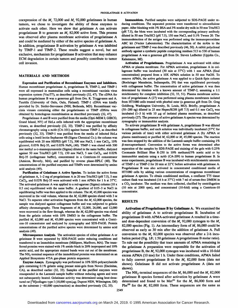
Activation of Progelatinase B by Gelatinase A. We examined theability of gelatinase A to activate progelatinase B. Incubation ofprogelatinase B with APMA-activated gelatinase A resulted in a time-and dose-dependent conversion of the M, 92,000 zymogen to the M,
82,000 species (Fig. 1). An intermediary form of M, 86,000 could beobserved as early as 30 min after the addition of gelatinase A. Fullconversion to the M, 82,000 species was observed after a 2-h incu
bation period (Fig. Iß;1:50 gelatinase A:progelatinase B molar ratio).To rule out the possibility that trace amounts of APMA remaining inthe gelatinase A preparation were responsible for the activation ofprogelatinase B, the Mr 92,000 zymogen was incubated with a 10-fold
excess APMA (10 HIM)for 1 h. Under these conditions, APMA failedto fully convert progelatinase B to the M, 82,000 form (data notshown). Gelatinase B did not activate progelatinase A (data notshown).
The NH2-terminal sequences of the MT86,000 and the M, 82,000
gelatinase B species formed after activation by gelatinase A weredetermined and found to be Met41 for the M, 86,000 form andPhe88 for the Mt 82,000 form. These sequences are the same as
2549
on March 24, 2019. © 1995 American Association for Cancer Research.cancerres.aacrjournals.org Downloaded from

Fig. 1. SDS-PAGE analysis of the activation of progelatinase B by gelatinase A. Human recombinant progelatinase A was activated with 1 ITIMAPMA (l h at 37°C)
and applied to a Quick Spin column to remove the APMAas described in "Materials and Methods." The activatedgelatinase A was incubated (37°C)with human recombi
nant progelatinase B for various time periods at a 1:100(A) or 1:50 (ß)gelatinase A:progelatinase B molar ratio.At the end of the indicated incubation times, the samples(1 /j.g/lane of progelatinase B) were analyzed in 10%SDS-polyacrylamide gel under reducing conditions, followed by staining of the gel with 0.25% Coomassie Brilliant Blue. Left lane, molecular weight standards (Bio-
Rad). Arrow on the righi, the activated gelatinase A usedin the experiment.
kDa
97_
66
45
PROGELATINASE B ACTIVATION
A
¡* »'***
B
O 0.5 l 2 3 4 O 0.5 1 2 3 4
Time (hrs)
those reported previously for the activation of progelatinase B bystromelysin-1 (39).
Plasma Membrane-activated Gelatinase A Activates Progelati
nase B. Progelatinase A can be activated by APMA (5) or by plasmamembrane containing MT-MMP (25-28). We reported previously thatAPMA activation of TIMP-2-free progelatinase A generated two
active species of Mr 62,000 and Mr 45,000 (32, 33). We compared thepattern of activation of progelatinase A by APMA and plasma membrane of HT1080 cells treated with phorbol ester. As shown in Fig. 2,
72-kDa —¿�62-kDa —¿�
45-kDa —¿�
Fig. 2. Activation of progelatinase A by plasma membranes or APMA. ProgelatinaseA (200 ng/lane) in 25 HIMHEPES/KOH (pH 7.5)-0.1 mm CaCl2 was incubated (37°C)
with either 10 fig of HT1080 plasma membranes (Lane 1, 1 h; Lane 2, 2 h) or with 1 mMAPMA for 1 h (Lane 3) or with buffer alone (Lane 4). The samples were then separatedby electrophoresis in a 10% SDS-polyacrylamide gel under reducing conditions, followed
by transfer to nitrocellulose paper. The blot was developed using a mAb to progelatinaseA as described in "Materials and Methods." Molecular weights on the left indicate the
relative mass of the gelatinase A species.
plasma membrane- (Fig. 2, Lanes I and 2) or APMA- (Fig. 2, Lane
3) activated progelatinase A into a M, 62,000 and a Mr 45,000 species.An intermediary form of M, 64,000 to 66,000 could also be observed(not visible in Fig. 2). Thus, both APMA and plasma membraneactivation of progelatinase A generate the Mr 62,000 and Mr 45,000species.
Since APMA activated-gelatinase A caused activation of progelatinase B, we wished to examine the effect of plasma membrane-
activated gelatinase A on progelatinase B activation. To this end, theplasma membranes from phorbol ester treated-HTlOSO cells wereincubated (3 h at 37°C)with progelatinase A, and then progelatinase
B was added to the reaction mixture for an additional 3-h incubation
period. The reaction mixture was then analyzed by immunoblot usinga mAb (CA-209) against progelatinase B and a mAb (CA-801) against
progelatinase A. These studies (Fig. 3) showed that plasma membraneactivated-gelatinase A caused the conversion of progelatinase B to the
Mr 86,000 and M, 82,000 species (Fig. 3, Lanes 1 and 2). Theimmunoblot also showed the bands corresponding to the plasmamembrane-activated gelatinase A, including a Mr 64,000, 62,000, anda 45,000 form. When progelatinase A was incubated (3 h at 37°C)
with plasma membranes in the presence of recombinant TIMP-2, there
was a significant reduction in the formation of the Mr 62,000 and Mr45,000 species, and most of the M, 72,000 enzyme remained in thelatent form (Fig. 3, Lanes 3 and 4). Consistently, the addition ofprogelatinase B to the mixture of plasma membranes, progelatinase A,and TIMP-2, followed by another 3-h incubation period, had no effect
on the activation of progelatinase B (Fig. 3, Lanes 3 and 4). Also,progelatinase B incubated with plasma membranes in the absence ofprogelatinase A was not activated (data not shown), as reportedpreviously (25-27). Taken together, these studies demonstrate that
Fig. 3. Progelatinase B activation by plasmamembrane-activated gelatinase A and inhibition byTIMP-2. Plasma membranes (10 fig/reaction) wereincubated (3 h at 37°C)with progelatinase A (200
ng/reaction) in 25 mM HEPES/KOH (pH 7.5}-0.1
mM CaCl2 in the absence (Lanes 1 and 2) or presence (Lanes 3 and 4) of stoichiometric amounts (60ng/reaction) of TIMP-2 to progelatinase A. An ali
quot of the reaction mixture was then incubated withprogelatinase B (200 ng/lane) at 1:10 (Lanes 1 and3) and 1:50 (lanes 2 and 4) molar ratios (gelatinaseA:progelatinase B) for another 3 h at 37°C.The
mixtures were then separated by electrophoresis in a10% SDS-polyacrylamide gel under reducing con
ditions, followed by transfer to nitrocellulose paper.The blot was developed using a mixture of mAbs toprogelatinase A and B, as described in "Materialsand Methods." Molecular weights on the left repre
sent the prestained molecular weight standards (lowrange; Bio-Rad).
Progelatinase B
Progelatinase A
49.5-
TIMP-22550
on March 24, 2019. © 1995 American Association for Cancer Research.cancerres.aacrjournals.org Downloaded from
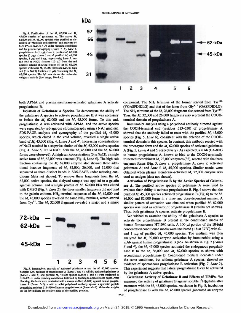
PROGELATINASE B ACTIVATION
Fig. 4. Purification of the Mt 62,000 and Mr45,000 species of gelatinase A. The active A/r62,000 and MT45,000 species were purified as described in "Materials and Methods" and analyzed by
SDS-PAGE (Lanes 1-3) under reducing conditionsand by gelatin-zymography (Lanes 4-6), Lane I,
progelatinase A (1 fig); Lane 2, purified Afr 62,000species (1 fig); Lanes 3 and 4, purified Mr 45,000species, 1 fig and 1 ng, respectively; Lane 5, lowsalt (0.5 M NaCi) fraction (10 ¿il)from the redagarose column showing elution of the Mt 62,000species with some A/r 45,000 form; and Lane 6, highsalt (3 M NaCl) fraction (10 fil) containing the Mr62,000 species. The left lane shows the molecularweight standards (low range; Bio-Rad).
kDa
97
66
45
31 •¿�1 2 3
both APMA and plasma membrane-activated gelatinase A activate
progelatinase B.Isolation of Gelatinase A Species. To demonstrate the ability of
the gelatinase A species to activate progelatinase B, it was necessaryto isolate the Mr 62,000 and the Mr 45,000 forms. To this end,progelatinase A was activated with APMA, and the active specieswere separated by red-agarose chromatography using a NaCl gradient.SDS-PAGE analysis and zymography of the purified Mr 45,000
species, which eluted in the void volume, revealed a single activeband of Mr 45,000 (Fig. 4, Lanes 3 and 4). Increasing concentrationsof NaCl resulted in a stepwise elution of the Mr 62,000 active species(Fig. 4, Lane 5, 0.5 M NaCl; both the M, 45,000 and the M, 62,000forms were observed). At high salt concentrations (3 MNaCl), a singleactive form of M, 62,000 was detected (Fig. 4, Lane 6). The high-salt
fraction containing the Mr 62,000 enzyme also showed three additional inactive fragments of M, 32,000, 26,000, and 12,000 thatseparated as three distinct bands in SDS-PAGE under reducing con
ditions (data not shown). To remove these fragments from the M,62,000 active species, the dialyzed sample was applied to a gelatinagarose column, and a single protein of Mr 62,000 kDa was elutedwith DMSO (Fig. 4, Lane 2); the three smaller fragments did not bindto the gelatin column. NH2-terminal sequence of the Mr 62,000 and
the Mr 45,000 species revealed the same NH2 terminus, which startedfrom Tyr81. The Mr 32,000 fragment revealed a major and a minor
72-kDa —¿�62-kDa —¿�
45-kDa —¿�
1 2 3 456Fig. 5. Immunoblot analysis of activated gelatinase A and the M, 45,000 species.
Samples (200 ng/lane) of progelatinase A (Lanes I and 4), APMA-activated gelatinase A(Lanes 2 and 3) and purified M, 45,000 species (Lanes 3 and 6) were subjected toSDS-PAGE under reducing conditions, followed by blotting to nitrocellulose paper. Afterblocking, the blots were incubated with a mouse mAb (CA-801) against human progelatinase A (Lanes 1-3) or with a rabbit polyclonal antibody against a synthetic peptidecomprising residues 513—530of human progelatinase A (Lanes 4-6). Molecular weightson the left indicate the relative mass of the purified proteins.
component. The NH2 terminus of the former started from Tyr4lc(YGASPDIDLG) and that of the latter from Gly417 (GASPD1DLG).The NH2 terminus of the Mr 26,000 fragment also started from Tyr416.
Thus, the M, 32,000 and 26,000 fragments may represent the COOH-
terminal domain of progelatinase A.Immunoblot analysis using a polyclonal antibody directed against
the COOH-terminal end (residues 513-530) of progelatinase A
showed that the antibody failed to react with the purified MT 45,000species (Fig. 5, Lane 6), consistent with the deletion of the COOH-
terminal domain in this species. In contrast, this antibody reacted withthe proenzyme form and the Mr 62,000 species of activated gelatinaseA (Fig. 5, Lanes 4 and 5, respectively). As expected, a mAb (CA-801)to human progelatinase A, known to bind to the COOH-terminally
truncated recombinant Mr 72,000 enzymes (32), reacted with the threeenzymes forms (Fig. 5, Lane I, progelatinase A; Lane 2, activatedgelatinase A; and Lane 3, Mr 45,000 species). Similar results wereobtained when plasma membrane-activated Mr 72,000 enzyme was
used as antigen (data not shown).Activation of Progelatinase B by the Active Species of Gelatin
ase A. The purified active species of gelatinase A were used toevaluate their ability to activate progelatinase B. Fig. 6 shows that thepurified M, 45,000 species activated progelatinase B (Fig. 6) to the Mr86,000 and 82,000 forms in a time- and dose-dependent manner. A
similar pattern of activation was obtained when purified Mr 62,000species was used as activator of progelatinase B (results not shown).Thus, both gelatinase A species activate progelatinase B.
We wished to examine the ability of the gelatinase A species toactivate the progelatinase B present in the conditioned media ofhuman fibrosarcoma HT1080 cells. A 100-^,1 portion of the 10-foldconcentrated conditioned media were incubated (l h at 37°C)with 0.1
and 1 fAg of purified Mr 45,000 species. The medium was thenanalyzed for M, 92,000 enzyme activation by immunoblot using amAb against human progelatinase B (46). As shown in Fig. 7 (Lanes3 and 4), the Mr 45,000 species activated the endogenous progelatinase B to the Mr 86,000 and Mr 82,000 species as shown withrecombinant progelatinase B. Conditioned medium incubated underthe same conditions, but without gelatinase A species, showed noevidence of spontaneous progelatinase B activation (Fig. 7, Lane 2).This experiment suggests that natural progelatinase B can be activatedby the gelatinase A active species.
Gelatinase Activity of Gelatinase B and Effects of TIMPs. Wemeasured the activity of gelatinase B against soluble [3H]gelatin after
treatment with the Mr 45,000 species. As shown in Fig. 8, incubationof progelatinase B with the Mr 45,000 species generated an enzyme
2551
on March 24, 2019. © 1995 American Association for Cancer Research.cancerres.aacrjournals.org Downloaded from
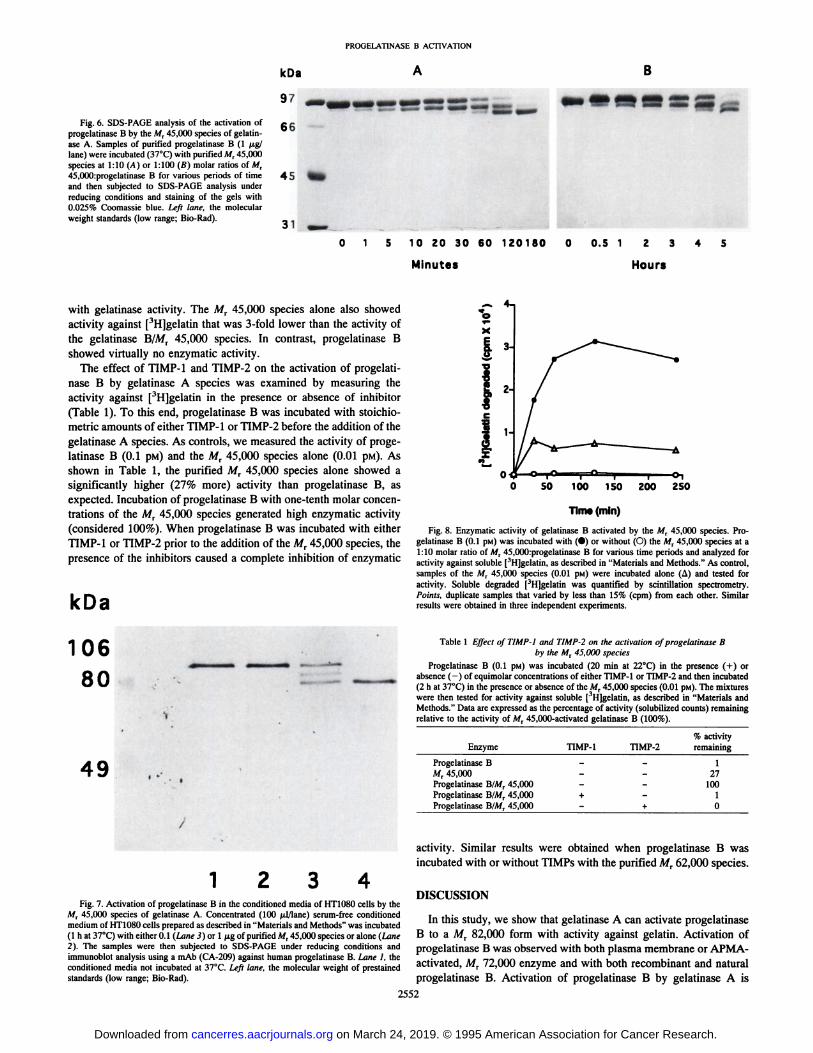
PROOELATINASE B ACTIVATION
Fig. 6. SDS-PAGE analysis of the activation ofprogelatinase B by the M, 45,000 species of gelatin-
ase A. Samples of purified progelatinase B (l ^tg/lane) were incubated (37°C)with purified M, 45,000
species at 1:10 (A) or 1:100 (B) molar ratios of M,45,000:progelatinase B for various periods of timeand then subjected to SDS-PAGE analysis under
reducing conditions and staining of the gels with0.025% Coomassie blue. Left lane, the molecularweight standards (low range; Bio-Rad).
kOa
97 ,
66
45
31
B
1 5 10 ZO 30 60 120180
Minutes
0.5 1 Z 3
Hours
with gelatinase activity. The Mr 45,000 species alone also showedactivity against [^Hjgelatin that was 3-fold lower than the activity of
the gelatinase B/Mr 45,000 species. In contrast, progelatinase Bshowed virtually no enzymatic activity.
The effect of TIMP-1 and TIMP-2 on the activation of progelati
nase B by gelatinase A species was examined by measuring theactivity against [^HJgelatin in the presence or absence of inhibitor
(Table 1). To this end, progelatinase B was incubated with stoichio-metric amounts of either TIMP-1 or TIMP-2 before the addition of the
gelatinase A species. As controls, we measured the activity of progelatinase B (0.1 pin) and the M, 45,000 species alone (0.01 pM). Asshown in Table 1, the purified M, 45,000 species alone showed asignificantly higher (27% more) activity than progelatinase B, asexpected. Incubation of progelatinase B with one-tenth molar concen
trations of the Mr 45,000 species generated high enzymatic activity(considered 100%). When progelatinase B was incubated with eitherTIMP-1 or TIMP-2 prior to the addition of the Mr 45,000 species, the
presence of the inhibitors caused a complete inhibition of enzymatic
kDa
2-
1-
O 50 100 150 200 250
Time (min)
Fig. 8. Enzymatic activity of gelatinase B activated by the M, 45,000 species. Progelatinase B (0.1 pM) was incubated with (•)or without (O) the M, 45,000 species at a1:10 molar ratio of M, 45,000:progelatinase B for various time periods and analyzed foractivity against soluble [3H]gelatin, as described in "Materials and Methods." As control,
samples of the M, 45,000 species (0.01 pm) were incubated alone (A) and tested foractivity. Soluble degraded [^HJgelatin was quantified by scintillation spectrometry.
Points, duplicate samples that varied by less than 15% (cpm) from each other. Similarresults were obtained in three independent experiments.
10680
49
Table 1 Effect ofTIMP-I ami TIMP-2 on the activation of progelatinase B
by the M, 45,000 speciesProgelatinase B (0.1 pM) was incubated (20 min at 22°C)in the presence (+) or
absence (-) of equimolar concentrations of either TIMP-1 or TIMP-2 and then incubated(2 h at 37°C)in the presence or absence of the MT45,000 species (0.01 pM). The mixtureswere then tested for activity against soluble [~H]gelatin, as described in "Materials andMethods." Data are expressed as the percentage of activity (solubilized counts) remaining
relative to the activity of Afr 45,000-activated gelatinase B (100%).
Enzyme TIMP-1TIMP-2Progelatinase
B - -
M, 45,000Progelatinase B/M, 45,000Progelatinase B/M, 45,000 +Progelatinase B/M, 45,000 - +%
activityremaining1
27100
10
activity. Similar results were obtained when progelatinase B wasincubated with or without TIMPs with the purified Mr 62,000 species.
Fig. 7. Activation of progelatinase B in the conditioned media of HT1080 cells by theM, 45,000 species of gelatinase A. Concentrated (100 /J/Iane) serum-free conditionedmedium of HT1080 cells prepared as described in "Materials and Methods" was incubated(l h at 37°C)with either 0.1 (Lane 3) or 1 fig of purified MT45,000 species or alone (Lane
2). The samples were then subjected to SDS-PAGE under reducing conditions andimmunoblot analysis using a mAb (CA-209) against human progelatinase B. Lane 1, theconditioned media not incubated at 37°C.Left lane, the molecular weight of prestained
standards (low range; Bio-Rad).
DISCUSSION
In this study, we show that gelatinase A can activate progelatinaseB to a MT 82,000 form with activity against gelatin. Activation ofprogelatinase B was observed with both plasma membrane or APMA-
activated, Mr 72,000 enzyme and with both recombinant and naturalprogelatinase B. Activation of progelatinase B by gelatinase A is
2552
on March 24, 2019. © 1995 American Association for Cancer Research.cancerres.aacrjournals.org Downloaded from
I'KCH.I l ATINASK B ACTIVATION
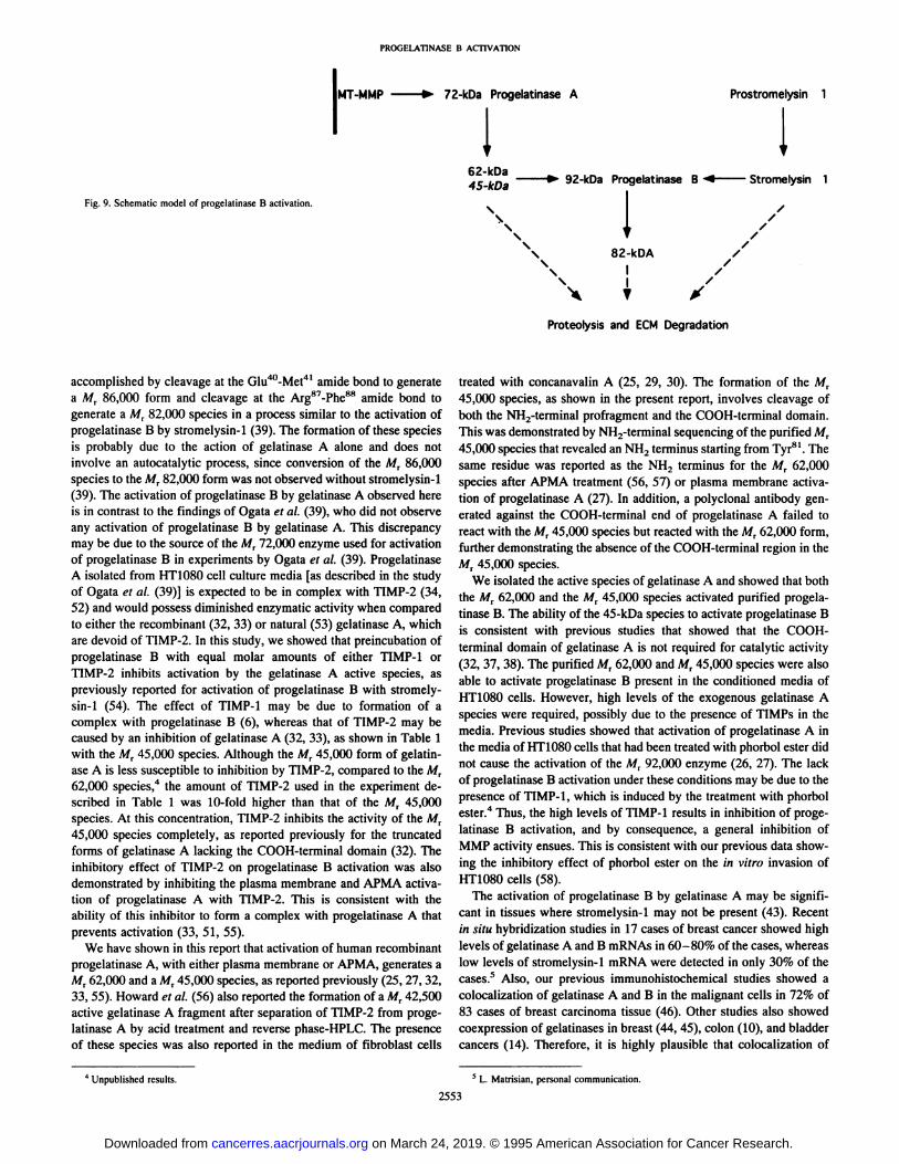
IMT-MMP 72-kDa Progelatinase A Prostromelysin 1
Fig. 9. Schematic model of progelatinase B activation.
•¿�5, » ^ 92-kDa Progelatinase B •¿�+ Stromelysin 1
I\ 82-kDA
II
X ? t
Proteolysis and ECM Degradation
accomplished by cleavage at the Glu40-Met4' amide bond to generatea Mr 86,000 form and cleavage at the Arg87-Phe88 amide bond to
generate a M, 82,000 species in a process similar to the activation ofprogelatinase B by stromelysin-1 (39). The formation of these species
is probably due to the action of gelatinase A alone and does notinvolve an autocatalytic process, since conversion of the M, 86,000species to the M, 82,000 form was not observed without stromelysin-1
(39). The activation of progelatinase B by gelatinase A observed hereis in contrast to the findings of Ogata et al. (39), who did not observeany activation of progelatinase B by gelatinase A. This discrepancymay be due to the source of the M, 72,000 enzyme used for activationof progelatinase B in experiments by Ogata et al. (39). ProgelatinaseA isolated from HT1080 cell culture media [as described in the studyof Ogata et al. (39)] is expected to be in complex with TIMP-2 (34,
52) and would possess diminished enzymatic activity when comparedto either the recombinant (32, 33) or natural (53) gelatinase A, whichare devoid of TIMP-2. In this study, we showed that preincubation ofprogelatinase B with equal molar amounts of either TIMP-1 orTIMP-2 inhibits activation by the gelatinase A active species, as
previously reported for activation of progelatinase B with stromelysin-1 (54). The effect of TIMP-1 may be due to formation of acomplex with progelatinase B (6), whereas that of TIMP-2 may be
caused by an inhibition of gelatinase A (32, 33), as shown in Table 1with the M, 45,000 species. Although the M, 45,000 form of gelatinase A is less susceptible to inhibition by TIMP-2, compared to the Mr62,000 species,4 the amount of TIMP-2 used in the experiment de
scribed in Table 1 was 10-fold higher than that of the Mr 45,000species. At this concentration, TIMP-2 inhibits the activity of the MT
45,000 species completely, as reported previously for the truncatedforms of gelatinase A lacking the COOH-terminal domain (32). Theinhibitory effect of TIMP-2 on progelatinase B activation was also
demonstrated by inhibiting the plasma membrane and APMA activation of progelatinase A with TIMP-2. This is consistent with the
ability of this inhibitor to form a complex with progelatinase A thatprevents activation (33, 51, 55).
We have shown in this report that activation of human recombinantprogelatinase A, with either plasma membrane or APMA, generates aMr 62,000 and a Mr 45,000 species, as reported previously (25, 27, 32,33, 55). Howard et al. (56) also reported the formation of a Mr 42,500active gelatinase A fragment after separation of TIMP-2 from progelatinase A by acid treatment and reverse phase-HPLC. The presence
of these species was also reported in the medium of fibroblast cells
treated with concanavalin A (25, 29, 30). The formation of the M,45,000 species, as shown in the present report, involves cleavage ofboth the NH2-terminal profragment and the COOH-terminal domain.This was demonstrated by NH2-terminal sequencing of the purified Mr45,000 species that revealed an NH2 terminus starting from Tyrsl. The
same residue was reported as the NH, terminus for the M, 62,000species after APMA treatment (56, 57) or plasma membrane activation of progelatinase A (27). In addition, a polyclonal antibody generated against the COOH-terminal end of progelatinase A failed to
react with the M, 45,000 species but reacted with the Mr 62,000 form,further demonstrating the absence of the COOH-terminal region in the
M, 45,000 species.We isolated the active species of gelatinase A and showed that both
the M, 62,000 and the Mr 45,000 species activated purified progelatinase B. The ability of the 45-kDa species to activate progelatinase Bis consistent with previous studies that showed that the COOH-
terminal domain of gelatinase A is not required for catalytic activity(32, 37, 38). The purified M, 62,000 and M, 45,000 species were alsoable to activate progelatinase B present in the conditioned media ofHT1080 cells. However, high levels of the exogenous gelatinase Aspecies were required, possibly due to the presence of TIMPs in themedia. Previous studies showed that activation of progelatinase A inthe media of HT108Ãœcells that had been treated with phorbol ester didnot cause the activation of the Mr 92,000 enzyme (26, 27). The lackof progelatinase B activation under these conditions may be due to thepresence of TIMP-1, which is induced by the treatment with phorbolester.4 Thus, the high levels of TIMP-1 results in inhibition of proge
latinase B activation, and by consequence, a general inhibition ofMMP activity ensues. This is consistent with our previous data showing the inhibitory effect of phorbol ester on the in vitro invasion ofHT 1080 cells (58).
The activation of progelatinase B by gelatinase A may be significant in tissues where stromelysin-1 may not be present (43). Recent
in situ hybridization studies in 17 cases of breast cancer showed highlevels of gelatinase A and B mRNAs in 60-80% of the cases, whereaslow levels of stromelysin-1 mRNA were detected in only 30% of thecases.5 Also, our previous immunohistochemical studies showed a
colocalization of gelatinase A and B in the malignant cells in 72% of83 cases of breast carcinoma tissue (46). Other studies also showedcoexpression of gelatinases in breast (44, 45), colon (10), and bladdercancers (14). Therefore, it is highly plausible that colocalization of
4 Unpublished results. 5 L. Matrisian, personal communication.
2553
on March 24, 2019. © 1995 American Association for Cancer Research.cancerres.aacrjournals.org Downloaded from
PROGELATINASE B ACTIVATION
these enzymes in tumor tissue would facilitate their interaction foractivation, leading to degradation of ECM.
Fig. 9 depicts a model for the interaction of several members of theMMP family, leading to the activation of progelatinase B. The membrane enzyme, MT-MMP, of tumor cells activates progelatinase A to
generate the M, 62,000 and M, 45,000 species. This process has beenrecently shown to involve the binding of TIMP-2 to MT-MMP,followed by binding of progelatinase A to the TIMP-2-MT-MMP
complex (55). The activation process and substrate specificity ofMT-MMP remain to be defined. The active gelatinase A species may
in turn activate progelatinase B, as shown in this study, and degradeECM. Although the physiological significance of the A/r 45,000species of gelatinase A (Fig. 9, italics) remains presently obscure, it isinteresting that the rate of TIMP-2 inhibition of active gelatinase Aspecies lacking the COOH-terminal domain is significantly lower than
that of the Mr 62,000 form (32, 37, 38, 59). Thus, cleavage of theCOOH-terminal domain, should it occur in vivo, would result in theformation of gelatinase A species that are less susceptible to TIMP-2
inhibition but remain catalytically competent and capable of activation of progelatinase B. Recently, activation of progelatinase B infibroblast monolayers supplemented with plasminogen or prostrome-lysin-1 was described (40). Thus, stromelysin-1 would efficiently
activate progelatinase B (39, 40, 54), if it were available; however, itsexpression in breast cancer remains unclear at the present (43). Plasmin, on the other hand, is a poor activator of progelatinase B (40, 41,54). Nevertheless, the role of each proteinase in activation of progelatinase B would be determined by its abundance and accessibility, thestate of activation, the rate of formation of the active species, and thepresence of specific inhibitors (29). Although the role of TIMP-1 andTIMP-2 is not depicted in this model (Fig. 9), these inhibitors are
critical regulators of the activation process and modulators of theenzymatic activity of the MMPs. As such, they play a crucial role indetermining the extent of ECM degradation during tumor cell invasion. This and previous studies have shown that both TIMP-1 andTIMP-2 can inhibit the activation of progelatinase A and B and the
respective proteolytic activity of the resultant enzymes by forming acomplex with the latent enzymes and/or binding to the active enzymeforms. Further studies on the relationship between these MMPs andTIMPs will provide a clearer understanding of the regulation of MMPactivity and the role of these proteases in tumor cell invasion.
ACKNOWLEDGMENTS
We are indebted to Dr. Greg Goldberg for providing us with the plasmamembrane preparation and for his useful comments on the manuscript and Dr.Scott Wilhelm for his valuable suggestions.
REFERENCES
1. Licita, L. A., Steeg, P. A., and Sieller-Stevenson, W. G. Cancer metastasis andangiogenesis: an imbalance of positive and negative regulation. Cell, 64: 327-336,1991.
2. Matrisian, L. M. Metalloproteinases and their inhibitors in tissue remodelling. TrendsGenet., 6: 121-125, 1990.
3. Testa, J. E., and Quigley, J. P. The role of urokinase-type plasminogen activator inaggressive tumor cell behavior. Cancer Metastasis Rev., 9: 353—367,1990.
4. Sloane, B. F., Moin, K., and Lah, T. T. Regulation of lysosomal endopeptidases inmalignant neoplasia. In: T. G. Pretlow and T. P. Pretlow (eds.), Biochemical andMolecular Aspects of Selected Cancers, Vol. 2, pp. 411-466. New York: AcademicPress, 1994.
5. Collier, I. E., Wilhelm, S. M., Eisen, A. Z., Marmer, B. L., Grant, G. A., Seltzer, J. L.,Kronberger, A., He, C., Bauer, E. A., and Goldberg, G. I. H-ras oncogene-trans-formed human bronchial epithelial cells (TBE-1) secrete a single metalloproteasecapable of degrading basement membrane collagen. J. Biol. Chem., 263: 6579-6587,1988.
6. Wilhelm, S. M., Collier, I. E., Marmer, B. L., Eisen, A. Z., Grant, G. A., andGoldberg, G. I. SV40-transformed human lung fibroblasts secrete a 92 kDa type IV
collagenase which is identical to that secreted by normal human macrophages. J. Biol.Chem., 264: 17213-17221, 1989.
10.
12.
14.
15.
16.
17.
18.
19.
20.
21.
22.
23.
24.
25.
26.
27.
28.
29.
30.
31.
32.
33.
Stetler-Stevenson, W. G. Type IV collagenases in tumor invasion and metastasis.Cancer Metastasis Rev., 9: 289-303, 1990.D'Errico, A., Garbisa, S., Liotta, L. A., Castranovo, V., Stetler-Stevenson, W. G., and
Grigioni, W. F. Augmentation of type IV collagenase, laminili receptor and KÌ62proliferation antigen associated with human colon, gastric and breast carcinomaprogression. Mod. Pathol., 4: 239-246, 1991.
Poulsom, R., Hanby, A. M., Pignatelli, M., Jeffrey, R. E., Longcroft, J. M., Rogers,L., and Stamp, G. W. Expression of gelatinase A and TIMP-2 mRNAs in desmo-
plastic fibroblasts in both mammary carcinomas and basal cell carcinomas. J. Clin.Pathol., 46: 429-436, 1993.
Pyke, C., Ralfkiaer, E., Tryggvason, K., and Daño,K. Messenger RNA for two typeIV collagenases is located in stromal cells in human colon cancer. Am. J. Pathol., 142:359-365, 1993.Poulsom, R., Pignatelli, M., Stetler-Stevenson, W. G., Liotta, L. A., Wright, P. A.,
Jeffrey, R. E., Longcroft, J. A., Rogers, L., and Stamp, G. W. H. Stromal expressionof 72 kDa type IV collagenase (MMP-2) and TIMP-2 mRNAs in colorectal neoplasia.Am. J. Pathol., 141: 389-396, 1992.Autio-Harmainen, H., Karttunen, T., Hurskainen, T., Hoyhtya, M., Kauppila, A., andTryggvason, K. Expression of 72 kDa type IV collagenase (gelatinase A) in benignand malignant ovarian tumors. Lab. Invest., 69: 312—321,1993.
Zucker, S., Lysik, R. M., Zarrabi, M. H., and Moll, U. M, 92,000 type IV collagenaseis increased in plasma of patients with colon cancer and breast cancer. Cancer Res.,53: 140-146, 1993.
Davies, B., Waxman, J., Wasan, H., Abel, P., Williams, G., Krausz, T., Neal, D.,Thomas, D., Hanby, A., and Balkwill, F. Levels of matrix metalloproteases in bladdercancer correlate with tumor grade and invasion. Cancer Res., 53: 5365-5369, 1993.
Fessier, L., Duncan, K., and Tryggvason, K. Identification of the procollagen IVcleavage products produced by a specific tumor collagenase. J. Biol. Chem., 259:9783-9789, 1984.
Woessner, J. F. Matrix metalloproteinases and their inhibitors in connective tissueremodelling. FASEB J., 5: 2145-2154, 1991.
Senior, R. M., Griffin, G. L., Fliszar, C. J., Shapiro, S. D., Goldberg, G. I., andWelgus, H. G. Human 92- and 72 kilodalton type IV collagenases are elastases.J. Biol. Chem., 266: 7870-7875, 1991.Murphy, G., Cockett, M. I., Ward, R. V., and Docherty, A. J. Matrix metalloprotein-ase degradation of elastin, type IV collagen and proteoglycan. A quantitative comparison of the activities of 95 kDa and 72 kDa gelatinases, stromelysins-1 and -2 andpunctuated metalloproteinase (PUMP). Biochem. J., 277: 277-279, 1991.Nguyen, Q., Murphy, G., Hughes, C. E., Mort, J. S., and Roughley, P. J. Matrixmetalloproteinases cleave at two distinct sites on human cartilage link protein.Biochem. J., 295: 595-598, 1993.
Fosang, A. J., Last, K.. Knauper, V., Neame, P. J., Murphy, G., Hardingham, T. E.,Tschesche, H., and Hamilton, J. A. Fibroblast and neutrophil collagenases cleave attwo distinct sites in the cartilage aggrecan interglobular domain. Biochem. J., 295:273-276, 1993.
Sires, U. I., Griffin, G. L., Broekelman, T. J., Mecham, R. P., Murphy, G., Chung,A. E., Welgus, H. G., and Senior, R. M. Degradation of entactin by matrix metalloproteinases: susceptibility to matrilysin and identification of cleavage sites. J. Biol.Chem., 268: 2069-2074, 1993.
Miyazaki, K., Hasegawa, M., Funahashi, K., and Umeda, M. A metalloproteinaseinhibitor domain in Alzheimer amyloid protein precursor. Nature (Lond.), 362:839-841, 1993.
Roher, A. E., Kasunic, T. C., Woods, A. S., Cotter, R. J., Ball, M. J., and Fridman,R. Proteolysis of A/3 peptide from Alzheimer disease brain by gelatinase A. Biochem.Biophys. Res. Commun., 205: 1755-1761, 1994.Ochieng, J., Fridman, R., Nangia-Makker, P., Liotta, L. A., Stetler-Stevenson, W. G.,and Raz, A. Galectin-3 is a novel substrate for human matrix metalloproteinases 2 and9. Biochemistry, 33: 14109-14114, 1994.Ward, R. V., Atkinson, S. J., Slocombe, P. M., Docherty, A. J. P., Reynolds, J. J., andMurphy, G. Tissue inhibitor of metalloproteinases-2 inhibits the activation of 72 kDaprogelatinase by fibroblasts membranes. Biochim. Biophys. Acta, 1079: 242-246,1991.Brown, P. D., Kleiner, D. E., Unsworth, E. J., and Stetler-Stevenson, W. G. Cellularactivation of the 72 kDa type IV procollagenase/TIMP-2 complex. Kidney Int., 43:163-170, 1993.Strongin, A. Y., Marmer, B. L., Grant, G. A., and Goldberg, G. I. Plasma membrane-dependent activation of the 72 kDa type IV collagenase is prevented by complexformation with TIMP-2. J. Biol. Chem., 268: 14033-14039, 1993.
Sato, H., Takino, T., Okada, Y., Cao, J., Shinagawa, A., Yamamoto, E., and Seiki, M.A matrix metalloproteinase expressed in the surface of tumor cells. Nature (Lond.),370: 61-65, 1994.Overall, C. M., and Dodek, J. Concanavalin-A produces a matrix-degradative phe-notype in human fibroblasts. Induction/endogenous activation of collagenase, 72 kDagelatinase and pump-1 is accompanied by suppression of tissue inhibitor of metalloproteinases. J. Biol. Chem., 265: 21141-21151, 1990.Overall, C. M. Regulation of tissue inhibitor of matrix metalloproteinase expression.Ann. NY Acad. Sci., 732: 51-64, 1994.Azzam, H. S., and Thompson, E. W. Collagen-induced activation of 72,000 type IVcollagenase in normal and malignant fibroblastoid cells. Cancer Res., 52: 4540-4544, 1992.Fridman, R., Fuerst, T. R., Bird, R. E., Hoyhtya, M., Oelkuct, M., Kraus, S.,Komarek, D., Liotta, L. A., Berman, M. L., and Stetler-Stevenson, W. G. Domainstructure of human 72-kDa gelatinase/type IV collagenase. J. Biol. Chem., 267:15398-15405, 1992.Fridman, R., Bird, R., Hoyhtya, M., Oelkuct, M., Komarek, D., Liang, C-M., Berman,M. L., Liotta, L. A., Stetler-Stevenson, W. G., and Fuerst, T. R. Expression of human
2554
on March 24, 2019. © 1995 American Association for Cancer Research.cancerres.aacrjournals.org Downloaded from
PROGELATINASE B ACTIVATION
recombinant 72 kDa gelatinase/type IV collagenase and TIMP-2: characterization ofcomplex and free enzyme. Biochem. J., 289: 411-416, 1993.
34. Goldberg, G. I., Manner, B. L., Grant, G. A., Eisen, A. Z., Wilhelm, S. M., and He,C. Human 72-kilodalton type IV collagenase forms a complex with tissue inhibitor ofmetalloproteinase designated TIMP-2. Proc. Nati. Acad. Sci. USA, 86: 8207-8211,
1989.35. Stetler-Stevenson, W. G., Krutzsch, H. C., and Liotta, L. A. Tissue inhibitor of
metalloproteinase-2 (TIMP-2). J. Biol. Chem., 264: 17374-17378, 1989.36. Howard, E. W., and Banda, M. J. Binding of tissue inhibitor of metalloproteinases-2
to two distinct sites on human 72 kDa gelatinase. J. Biol. Chem., 266: 17972-17977,
1991.37. Murphy, G., Willenbrock, F., Ward, R. V., Cockett, M. I., Eaton, D„and Docherty,
A. J. P. The C-terminal domain of 72 kDa gelatinase is not required for catalysis, butis essential for membrane activation and modulates interactions with tissue inhibitorsof metalloproteinases. Biochem. J., 283: 637-641, 1992.
38. Willenbrock, F., Crabbe, T., Slocombe, P. M., Sutton, C. W., Docherty, A. J. P,Cockett, M. I., O'Shea, M., Brocklehurst, K., Phillips, I. R., and Murphy, G. The
activity of the tissue inhibitors of metalloproteinases is regulated by C-terminal
domain interactions: a kinetic analysis of the inhibition of gelatinase A. Biochemistry,32: 433Ü-4337, 1993.
39. Ogata, Y., Enghild, J. J., and Nagase, H. Matrix metalloproteinase 3 (stromelysin)activates the precursor for the human matrix metalloproteinase 9. J. Biol. Chem., 267:3581-3584, 1992.
40. O'Connell, J. P., Willenbrock, F., Docherty, A. J. P., Eaton, D., and Murphy, G.
Analysis of the role of the COOH-terminal domain in the activation, proteolyticactivity and tissue inhibitor of metalloproteinase interactions of gelatinase B. J. Biol.Chem., 269: 14967-14973, 1994.
41. Okada, Y., Gonoji, Y., Naka, K., Tornita, K., Nakanishi, I., Iwata, K., Yamashita, K.,and Hayakawa, T. Matrix metalloproteinase 9 (92-kDa gelatinase/type IV collagenase) from HT1080 human fibrosarcoma cells. J. Biol. Chem., 267: 21712-21719,
1992.42. Menashi, S., Fridman, R., Desrevieres, S., Lu, H., Legrand, Y., and Soria, C.
Regulation of 92-kDa gelatinase B activity in the extracellular matrix by tissuekallikrein. Ann. NY Acad. Sci., 732: 466-468, 1994.
43. Polene, M., Clavel, C., Cockett, M., Bentzmann, S. G., Murphy, G., andBirembaut, P. Detection and localization of mRNAs encoding matrix metalloproteinases and their inhibitors in human breast pathology. Invasion Metastasis, 13:31-37, 1993.
44. Brown, P. D., Bloxidge, R. E., Anderson, E., And Howell, A. Expression of activatedgelatinase in human invasive breast carcinoma. Clin. Exp. Metastasis, //: 183-189,
1993.45. Davies, B., Miles, D. W., Happerfield, L. C., Naylor, M. S., Bobrow, L. G., Rubens,
R. D., and Balkwill, F. R. Activity of type IV collagenases in benign and malignantbreast disease. Br. J. Cancer, 67: 1126-1131, 1993.
46. Visscher, D. W., Hoyhtya, M., Ottosen, S. K., Liang, C-M., Sarkar, F. H., Crissman,J. D., and Fridman, R. Enhanced expression of tissue inhibitor of metalloproteinase-2
(TIMP-2) in the stroma of breast carcinomas correlates with tumor recurrence. Int. J.Cancer, 59: 339-344, 1994.
47. Fuerst, T. M., Earl, P. L., and Moss, B. Use of a hybrid vaccinia virus-TV RNApolymerasc system for expression of target genes. Mol. Cell. Biol., 7: 2538-2544, 1987.
48. Howard, E. W., Bullen, E. C., and Banda, M. J. Preferential inhibition of 72- and92-kDa gelatinases by tissue inhibitor of metalloproteinases-2. J. Biol. Chem., 266:13070-13075, 1991.
49. Heinrikson, L., and Meredith, S. C. Amino acid analysis by reverse-phase high-performance liquid chromatography: precolumn derivatization with phenylisothio-cyanato. Anal. Biochem., 136: 65-74, 1984.
50. Hoyhtya, M., Fridman, R., Komarek, D., Porter-Jordan, K., Stetler-Stevenson, W. G.,Liotta, L. A., and Liang, C-M. Immunohistochemical localization of matrix metalloproteinase 2 and its specific inhibitor TIMP-2 in neoplastic tissues with monoclonalantibodies. Int. J. Cancer, 56: 500-505, 1994.
51. Kleiner, D. E., Tuuttila, A., Tryggvason, K., and Stetler-Stevenson, W. G. Stabilityanalysis of latent and active 72-kDa type IV collagenase: the role of issue inhibitor ofmetalloproteinases-2 (TIMP-2). Biochemistry, 32: 1583-1592, 1993.
52. Moll, U. M, Youngleib, G. L., Rosinski, K. B., and Quigley, J. P. Tumor promoter-
stimulated M, 92,000 gelatinase secreted by normal and malignant human cells:isolation and characterization of the enzyme from HT1080 tumor cells. Cancer Res.,50: 6162-6170, 1990.
53. Kolkenbrock, H., Orgell, D., Hecker-Kia, A., Noack, W., and Ulbrich, N. Thecomplex between a tissue inhibitor of metalloproteinases-2 (TIMP-2) and 72-kDaprogelatinase is a metalloproteinase inhibitor. Eur. J. Biochem., 198: 775-781, 1991.
54. Goldberg, G. I., Strongin, A., Collier, I. E., Genrich, L. T., and Marmer, B. L.Interaction of 92-kDa type IV collagenase with the tissue inhibitor of metalloproteinases prevents dimerization, complex formation with interstitial collagenase, andactivation of the proenzyme with stromelysin. J. Biol. Chem., 267: 4583-4591,1992.
55. Strongin, A. Y., Collier, I., Bannikov, G., Marmer, B. L., Grant, G. A., and Goldberg,G.I. Mechanism of cell surface activation of 72 kDa type collagenase: isolation of theactivated form of the membrane metalloprotease. J. Biol. Chem., 270: 5331-5338,1995.
56. Howard, E. W., Bullen, E. C., and Banda, M. J. Regulation of the autoactivation ofhuman 72-kDa progelatinase by the tissue inhibitor of metalloproteinases-2. J. Biol.Chem., 266: 13064-13069, 1991.
57. Stetler-Stevenson, W. G., Krutzsch, H. C., Wacher, M. P., Margulies, I. M. K., andLiotta, L. A. The activation of human type IV collagenase proenzyme: sequenceidentification of the major conversion product following organomercurial activation.J. Biol. Chem., 264: 1353-1356, 1989.
58. Fridman, R., Lacal, J. C., Reich, R., Bonfil, R. D., and Ahn, C. Differential effect ofphorbol ester on the in vilro invasiveness of malignant and non-malignant humanfibroblast cells. J. Cell. Physiol., ¡42:55-60, 1990.
59. Nguyen, Q., Willenbrock, F., Cockett, M. I., O'Shea, M., Docherty, A. J., and
Murphy, G. Different domain interactions are involved in the binding of tissueinhibitor of metalloproteinases to stromelysin-1 and gelatinase A. Biochemistry, 33:2089-2095, 1994.
2555
on March 24, 2019. © 1995 American Association for Cancer Research.cancerres.aacrjournals.org Downloaded from
1995;55:2548-2555. Cancer Res Rafael Fridman, Marta Toth, Daniel Peña, et al. Activation of Progelatinase B (MMP-9) by Gelatinase A (MMP-2)
Updated version
http://cancerres.aacrjournals.org/content/55/12/2548
Access the most recent version of this article at:
E-mail alerts related to this article or journal.Sign up to receive free email-alerts
Subscriptions
Reprints and
To order reprints of this article or to subscribe to the journal, contact the AACR Publications
Permissions
Rightslink site. Click on "Request Permissions" which will take you to the Copyright Clearance Center's (CCC)
.http://cancerres.aacrjournals.org/content/55/12/2548To request permission to re-use all or part of this article, use this link
on March 24, 2019. © 1995 American Association for Cancer Research.cancerres.aacrjournals.org Downloaded from