Embed Size (px)
Citation preview

2018
UNIVERSIDADE DE LISBOA
FACULDADE DE CIÊNCIAS
DEPARTAMENTO DE QUÍMICA E BIOQUÍMICA
Influence of histone octamer core distortion
on nucleosome thermal mobility
Alexandre Dias Cirilo
Mestrado em Bioquímica
Especialização em Bioquímica
Dissertação orientada por:
Dra. Silvija Bilokapić-Halić
Prof. Dra. Margarida Gama-Carvalho

Alexandre Dias Cirilo Influence of histone octamer core distortion on nucleosome thermal mobility
ii
Alexandre Dias Cirilo Influence of histone octamer core distortion on nucleosome thermal mobility
iii
Acknowledgements
I would first like to thank my external supervisor Dr. Silvija Bilokapić-Halić from the Ludwig-
Maximilians-Universität München (LMU) in Munich, Germany. I learned a lot throughout this
academic year and am sure that having been driven to think by myself, perform and critically analyse
the experiments was the best way to grow as a scientist. I would also like to thank Prof. Dr. Mario Halić
for accepting me into the Halić lab at the Gene Center of the LMU and allowing me to do my master
thesis project for a whole academic year. Moreover, I would like to thank everybody that I worked with
during my stay, especially the members of the lab. Thank you for helping me whenever I required
assistance and for all the support.
I would also like to acknowledge Prof. Dr. Margarida Gama-Carvalho of the Faculty of Sciences
at the University of Lisbon who gladly accepted to be my internal supervisor and assisted me whenever
needed.
Furthermore, I want to reference the Erasmus+ programme ant its respective grant allocation
without which I would not have been able to pursue my thesis project abroad.
Finally, I am deeply grateful to my parents for allowing me to pursue my objectives and
ambitions, for always being present and for all the support and endless love. This work would not have
been possible without them.
Alexandre Dias Cirilo Influence of histone octamer core distortion on nucleosome thermal mobility
iv

Alexandre Dias Cirilo Influence of histone octamer core distortion on nucleosome thermal mobility
v
Abstract
The nucleosome is the fundamental repeating subunit of eukaryotic chromatin. The histones,
hallmarks of the nucleosome, are small basic proteins subdivided into two super families: linker (H1)
and core histones (H2A, H2B, H3 and H4). Core histones assemble together to form the histone octamer
which then, together with double-stranded DNA, embodies the complex structure of the nucleosome.
Most importantly, the nucleosome allows for the tight packaging of the large genome into the relatively
constricted space available inside the nucleus by folding into a series of higher-order molecular
structures. Furthermore, it allows for the fine-tuned temporal and spatial regulation of several DNA-
templated processes, such as DNA transcription, replication and repair, unequivocally required for the
cells to thrive and exert their functions. Since their discovery in the 1960s, histone post-translational
modifications (e.g. methylation, phosphorylation and acetylation) have been shown to affect gene
expression regulation, with some modifications being associated with gene activation, whilst others have
been associated with gene silencing. Interestingly, several studies have demonstrated the existence of a
“histone code” which states that the presence of post-translational modifications on histones affect
binding affinities of chromatin-binding proteins leading to altered transcriptional states of the underlying
DNA. The epigenetic information encoded via these modifications occurring on histones can be passed
on to the next generation of cells, acting as an additional layer of information that can be stored inside
the cell, further increasing the complexity and versatility of the genetic material.
It has been shown that the nucleosome exhibits shifting when incubated at high temperatures
towards the DNA ends in regard to the nucleosomal DNA. In this context, the current project aims to
observe the influence of histone octamer core distortion on nucleosome thermal mobility. To do so,
several histone mutants were prepared, albeit only one mutant nucleosome was assembled, apart from
the wild-type nucleosome. This work corroborates the observed thermally-driven shifting pattern of
wild-type nucleosomes. The H4_V43C H3_F104C mutant nucleosome assembled in this project has
shown itself to increase nucleosome structural integrity, possibly through the establishment of a
disulphide bridge between L1 (loop 1) of core histone H4 and 2 (-helix 2) of core histone H3,
conversely, hindering nucleosome mobility. This suggests that for nucleosome to slide along DNA in a
noncatalyzed fashion, there must be a requirement of histone octamer plasticity.
Moreover, structural analysis of Fkbp39, a putative histone chaperone in Saccharomyces pombe,
both alone and in complex with (H2A-H2B) and with (H3-H4)2 was attempted with electron
cryomicroscopy, though unsuccessful. Further optimization strategies are likely required to stabilize the
putative complexes.
Lastly, several chromatin-binding proteins were screened for binding to the nucleosome, as a
complementary experiment. Hpf1, Parp2 and Alf from Homo sapiens seem like the best candidates for
more in-depth studies to fully disclose their binding affinities to chromatin.
Keywords: Nucleosome, Histone code, PTMs, Epigenetics, Chromatin

Alexandre Dias Cirilo Influence of histone octamer core distortion on nucleosome thermal mobility
vi

Alexandre Dias Cirilo Influence of histone octamer core distortion on nucleosome thermal mobility
vii
Resumo
O nucleossoma é a subunidade fundamental da cromatina eucariótica. As histonas, proteínas
características do nucleossoma, são pequenas proteínas básicas subdivididas em duas superfamílias:
linker (H1) e core (H2A, H2B, H3 e H4). As histonas core oligomerizam para formar o octamero de
histonas que, conjuntamente com ADN de cadeia dupla, estabelecem a estrutura complexa do
nucleossoma. A estrutura do nucleossoma foi inicialmente identificada em 1975 recorrendo à digestão
da cromatina por uma nuclease micrococal, sendo que apenas em 1997 foi divulgada a estrutura do
nucleossoma obtida por difração de raios-X com uma resolução de 2.8 Å, e mais tarde, em 2002, com
uma resolução de 1.9 Å, o que revela ser mais do que suficiente para as cadeias laterais e a cadeia
principal serem observadas com elevado grau de confiança. As histonas partilham um domínio entre
elas: o histone fold, constituído por cerca de 70 resíduos de aminoácidos, capaz de facilitar a
heterodimerização das histonas e que se encontra localizado na região C-terminal e consiste em três
hélices-α (α1, α2 e α3) ligadas entre si por dois loops (L1 e L2) flanqueando a hélice-α 2. As histonas
são constituídas por um domínio globular e uma região N-terminal flexível rica em resíduos de lisina e
arginina. Estas proteínas heterodimerizam com o auxílio de interações hidrofóbicas, formando o
handshake motif. No contexto celular, o nucleossoma permite o empacotamento do genoma no espaço
relativamente limitado disponível dentro do núcleo, através do folding numa série de estruturas
moleculares de ordem gradualmente superior. Mais ainda, permite a regulação temporal e espacial de
vários processos modelados pelo ADN, como por exemplo a transcrição, a replicação e o reparo de
ADN, necessários para que as células prosperem e exerçam as suas funções.
Desde a sua descoberta na década de 1960, as modificações pós-traducionais de histonas (e.g.
metilação, fosforilação e acetilação) foram associadas à regulação da expressão génica, com algumas
modificações sendo associadas à ativação de genes, enquanto outras foram associadas ao seu
silenciamento. Vários estudos demonstraram a existência de um “código de histonas” que estipula que
diferentes modificações pós-traducionais nas histonas afetam as afinidades de ligação de proteínas
capazes de estabelecerem uma ligação à cromatina, levando a estados transcricionais alterados do ADN
subjacente. A informação epigenética codificada por estas modificações que ocorrem nas histonas pode
ser passada para a próxima geração de células, agindo como uma camada adicional de informação que
pode ser armazenada dentro da célula, aumentando ainda mais a complexidade e a variabilidade do
material genético.
Foi previamente demonstrado que o nucleossoma exibe um deslocamento, quando incubado a
altas temperaturas, para os terminais da molécula de ADN que está enrolada em volta do nucleossoma.
Neste contexto, o presente projeto visa observar a influência da distorção do octamero de histonas na
mobilidade térmica do nucleossoma. Deste modo, vários mutantes de histonas foram preparados,
embora apenas um nucleossoma mutante tenha sido gerado, além do nucleossoma wild-type. Para a
obtenção de histonas recombinantes com elevado grau de pureza, recorreu-se a técnicas cromatográficas.
Em primeiro lugar, foram expressas histonas recombinantes (wild-type e mutantes) em células
Escherichia coli BL21 (DE3) Rosetta competentes a partir de plasmídeos in house. As histonas core
H2A e H2B, bem como H3 e H4, foram coexpressas, uma vez que a coexpressão destes dois pares de
proteínas reduz o tempo necessário para a sua síntese, para além de produzir complexos solúveis
passíveis de serem purificados por sucessivas técnicas experimentais. Posteriormente, executou-se a
purificação das mesmas através do uso de uma cromatografia de afinidade seguida duma cromatografia
de troca iónica.
Alexandre Dias Cirilo Influence of histone octamer core distortion on nucleosome thermal mobility
viii
Após todas as histonas terem sido geradas em grandes quantidades e elevado grau de pureza,
efetuou-se a montagem do octamero. Paralelamente, foi sintetizada a sequência Widom 601,
previamente descrita na literatura como sendo uma sequência de muito alta afinidade para o
nucleossoma, via PCR usando primers apropriados. Neste trabalho foram geradas duas variantes da
sequência supramencionada: curta (147 pb) e longa (227 pb). Uma vez obtidos todos os componentes
necessários para a montagem do nucleossoma, recorreu-se à reconstituição do mesmo através de diálises
(com gradiente de salinidade). Por fim, os nucleossomas gerados foram usados para serem efetuados
thermal shift assays e native gel shift assays. Os thermal shift assays foram usados para se observar o
deslocamento do nucleossoma ao longo do ADN (baseado na sequência Widom 601) e o efeito da
distorção do octamero na mobilidade do nucleossoma induzida pela temperatura. Quanto aos native gel
shift assays, um ensaio experimental rápido e sensível para a deteção de interações proteína-ácido
nucleico, foram usados para observar a afinidade de ligação de diversas proteínas com ligação putativa
à cromatina.
Este trabalho corroborou o padrão observado de deslocamento de nucleossomas wild-type para
as extremidades do ADN associado ao mesmo. O nucleossoma mutante H4_V43C H3_F104C
concebido neste projeto demonstrou estar associado ao aumento da integridade estrutural e rigidez do
nucleossoma, possivelmente através do estabelecimento de uma ponte dissulfeto entre L1 (loop 1) da
histona H4 e 2 (hélice- 2) da histona H3. O estudo do impacto da plasticidade do octamero de
histonas, consequência das diversas modificações que podem ocorrer nos seus resíduos de aminoácidos
e que levam à distorção do core de histonas, é relevante no sentido de tentar entender o seu papel na
regulação da expressão génica. Uma dada mutação irá, hipoteticamente, reforçar a estrutura do
nucleossoma, potencialmente impossibilitando o acesso do complexo basal de transcrição, devido à
alteração da capacidade de mobilidade do nucleossoma em relação ao ADN, e silenciando o(s) gene(s)
codificados no ADN subjacente que se encontra “blindado” pelo nucleossoma, e vice-versa. Também
será possível que alguns complexos capazes de remodelar a cromatina possam tirar partido da alteração
da integridade estrutural conferida pelas diversas mutações ou pela presença de variantes de histonas.
Numa visão geral, os resultados obtidos neste projeto demonstraram que é necessária uma certa
plasticidade do octamero de histonas para se observar o movimento não-catalisado do nucleossoma.
Adicionalmente, foi tentada a análise da Fkbp39, uma peptidilprolil isomerase com um domínio
NPL (nucleoplasmin-like) conservado na região N-terminal, que se pensa ser uma chaperona de histonas
em S. pombe e que é responsável pelo aumento da taxa de isomerização cis-trans das prolinas na cauda
N-terminal (região N-terminal flexível) da histona H3. A análise foi feita por criomicroscopia eletrónica,
tanto com a Fkbp39 não-complexada, como em complexo com (H2A-H2B) e com (H3-H4)2. Nesse
sentido, expressou-se a proteína Fkbp39 em células E. coli BL21 (DE3) Rosetta competentes e foi
executada a sua purificação com elevado grau de pureza através do uso de diferentes técnicas
cromatográficas (cromatografias de afinidade, de troca iónica, e de exclusão molecular). Embora as
purificações tenham sido bem-sucedidas, nem a proteína Fkbp39 isolada, nem em complexo com o
dímero (H2A-H2B) ou com o tetramero (H3-H4)2 foram passíveis de serem observadas por
criomicroscopia electrónica. É possível que, devido à presença de uma região central dinâmica
intrinsecamente desordenada, estratégias sucessivas de otimização sejam necessárias para estabilizar os
complexos putativos.

Alexandre Dias Cirilo Influence of histone octamer core distortion on nucleosome thermal mobility
ix
Por último, várias proteínas putativas de ligação à cromatina foram rastreadas quanto à ligação
aos nucleossomas sintetizados neste projeto recorrendo a native gel shift assays. Hpf1 (Histone
PARylation factor 1), Parp2 (Poly [ADP-ribose] polymerase 2) e Alf (TFIIA-alpha and beta-like factor),
provenientes de Homo sapiens, demonstraram ser os melhores candidatos para estudos estruturais
posteriores mais aprofundados para averiguar com maior confiança as suas afinidades de ligação à
cromatina.
Palavras-chave: Nucleossoma, Código de histonas, Modificaçóes pós-traducionais,
Epigenética, Cromatina

Alexandre Dias Cirilo Influence of histone octamer core distortion on nucleosome thermal mobility
x

Alexandre Dias Cirilo Influence of histone octamer core distortion on nucleosome thermal mobility
xi
Index
Acknowledgements ................................................................................................................................ iii
Abstract ....................................................................................................................................................v
Resumo .................................................................................................................................................. vii
List of Abbreviations ............................................................................................................................ xiv
List of Tables ........................................................................................................................................ xvi
List of Figures ..................................................................................................................................... xvii
1. Introduction ...................................................................................................................................1
1.1. Historical contextualization and overview of histones .............................................................1
1.2. The nucleosome – structural properties and role in genetic material packaging ......................3
1.2.1. Core histones ................................................................................................................... 6
1.2.2. Linker histone H1/H5 ...................................................................................................... 6
1.2.3. Histone variants ............................................................................................................... 6
1.2.4. Nucleosome assembly ..................................................................................................... 6
1.2.5. Histone chaperones .......................................................................................................... 7
1.2.5.1. FK506-binding protein 39 kDa................................................................................ 9
1.2.5.2. Line-of-thought and trends .................................................................................... 11
1.2.6. Outlook .......................................................................................................................... 11
1.3. Nucleosome-interacting proteins ............................................................................................12
1.4. The histone code hypothesis & the role of histone core distortion.........................................13
1.4.1. Histone post-translational modifications ....................................................................... 15
1.4.1.1. Acetylation ............................................................................................................ 15
1.4.1.2. Methylation............................................................................................................ 15
1.4.1.3. Phosphorylation ..................................................................................................... 16
1.4.1.4. Ubiquitination ........................................................................................................ 16
1.5. Nucleosome mobility .............................................................................................................17
1.5.1. Nucleosome phasing ...................................................................................................... 18
1.5.2. Nucleosome stability ..................................................................................................... 18
1.5.3. Effect of temperature on nucleosome mobility ............................................................. 18
1.6. State-of-the-art ........................................................................................................................20
2. Aims and objectives .....................................................................................................................23
Alexandre Dias Cirilo Influence of histone octamer core distortion on nucleosome thermal mobility
xii
3. Materials .......................................................................................................................................24
3.1. Buffers ....................................................................................................................................24
3.2. Antibiotics ..............................................................................................................................25
3.3. Media ......................................................................................................................................25
3.4. Plasmids..................................................................................................................................25
3.5. Primers....................................................................................................................................25
3.6. Gels .........................................................................................................................................26
4. Methods ........................................................................................................................................27
4.1. Bacterial transformation of E. coli cells .................................................................................27
4.2. Plasmid DNA purification ......................................................................................................27
4.3. DNA sequencing ....................................................................................................................27
4.4. Polymerase chain reaction ......................................................................................................27
4.5. DNA precipitation ..................................................................................................................28
4.6. Histone expression .................................................................................................................28
4.7. Recombinant protein purification from heterologous expression in E. coli BL21 (DE3)
Rosetta strain ......................................................................................................................................29
4.7.1. Affinity chromatography: 6xHis-tag purification (Ni-NTA Agarose) .......................... 29
4.7.2. Ion exchange chromatography: protein purification (Sepharose Fast Flow) ................. 29
4.8. Histone octamer assembly ......................................................................................................30
4.9. Nucleosome assembly ............................................................................................................30
4.10. Protein concentration ..............................................................................................................30
4.11. S. pombe Fkbp39 purification and Fkbp39:(H3-H4)2, Fkbp39:(H2A-H2B) complex
formation assessment .........................................................................................................................31
4.12. Chromatin-binding proteins native gel shift assays ................................................................33
4.13. Electrophoresis .......................................................................................................................33
4.13.1. Agarose .......................................................................................................................... 33
4.13.2. Native-PAGE ................................................................................................................. 33
4.13.3. SDS-PAGE .................................................................................................................... 33

Alexandre Dias Cirilo Influence of histone octamer core distortion on nucleosome thermal mobility
xiii
5. Results ...........................................................................................................................................34
5.1. WT and mutant X. laevis histone purification ........................................................................34
5.2. Synthesis and purification of nucleosomal DNA ...................................................................37
5.2.1. Amplification by PCR ................................................................................................... 37
5.2.2. Purification by ethanol precipitation ............................................................................. 38
5.3. WT X. laevis nucleosome assembly .......................................................................................39
5.4. Thermal shifts of WT and mutant X. laevis nucleosomes from X. Laevis ..............................40
5.4.1. Effect of crosslinking WT X. laevis nucleosomes with glutaraldehyde ........................ 42
5.5. Binding of S. pombe Fkbp39 to X. laevis core histones .........................................................43
5.6. Binding affinity of chromatin-binding proteins to WT X. laevis nucleosomes ......................49
6. Discussion .....................................................................................................................................53
6.1. Influence of histone octamer core distortion on nucleosome thermal mobility .....................53
6.2. Fkbp39 structural studies ........................................................................................................55
6.3. Chromatin-binding proteins screening ...................................................................................56
7. Conclusion ....................................................................................................................................57
8. References .....................................................................................................................................58
9. Appendix .......................................................................................................................................62

Alexandre Dias Cirilo Influence of histone octamer core distortion on nucleosome thermal mobility
xiv
List of Abbreviations
Table 0.1 List of abbreviations
Abbreviation Designation
AC Affinity chromatography
ACF ATP-utilizing chromatin assembly and remodelling factor
Amp Ampicillin
Cam Chloramphenicol
ChIP Chromatin immunoprecipitation
c-NCP Centred nucleosome core particle
Cryo-EM Electron cryomicroscopy
D. melanogaster Drosophila melanogaster
DNA
(ADN)
Deoxyribonucleic acid
(Ácido desoxirribonucleico)
dNTP Deoxyribonucleotide triphosphate
DSB Double-strand break
dsDNA Double-stranded deoxyribonucleic acid
E. coli Escherichia coli
EM Electron microscopy
ep-NCP End-positioned nucleosome core particle
FKBD FK506-binding domain
H. sapiens Homo sapiens
HAT Histone acetyltransferase
HDAC Histone deacetylase
His Histidine
IB Inclusion body
IDP Intrinsically disordered proteins
IEX Ion-exchange chromatography
Kan Kanamycin
LB Lysogeny broth
MACS Multiangle light scattering
MMTV Mouse mammary tumour virus
N/A Not available
NCP Nucleosome core particle
NMR Nuclear magnetic resonance
NPL Nucleoplasmin-like
OD600 Optical density at a wavelength of 600 nm
P. falciparum Plasmodium falciparum
PAGE Polyacrylamide gel electrophoresis
PCR Polymerase chain reaction
PPIase Petptidyl-prolyl cis-trans isomerase
PTM Post-translational modification
ref(s). Reference(s)
RSF Remodelling and spacing factor
RT Room temperature

Alexandre Dias Cirilo Influence of histone octamer core distortion on nucleosome thermal mobility
xv
S. pombe Schizosaccharomyces pombe
SANS Small-angle neutron scatterin
SAXS Small-angle X-ray scattering
SE AUC Analytical ultracentrifugation: sedimentation equilibrium
SUMO Small ubiquitin-related modifier
t Time
Tm Melting temperature
WT Wild-type
X. laevis Xenopus laevis

Alexandre Dias Cirilo Influence of histone octamer core distortion on nucleosome thermal mobility
xvi
List of Tables
Table 0.1 List of abbreviations ............................................................................................................. xiv
Table 3.1 List of buffers. ....................................................................................................................... 24
Table 3.2 List of antibiotics. .................................................................................................................. 25
Table 3.3 List of media. ......................................................................................................................... 25
Table 3.4 List of plasmids. .................................................................................................................... 25
Table 3.5 List of primers. ...................................................................................................................... 25
Table 3.6 Composition of Native-PAGE and SDS-PAGE gels. ........................................................... 26
Table 4.1 PCR reaction setup for the long Widom 601 sequence amplification (227 bp). ................... 27
Table 4.2 Components for cell induction. ............................................................................................. 28
Table 4.3 Various devices Amicon® used throughout the project. ........................................................ 30
Table 4.4 Dialysis buffers’ composition for Fkbp39:(H3-H4)2 and FKbp39:(H2A-H2B) assembly. .. 32
Table 5.1 List of purified histones ......................................................................................................... 35
Table 5.2 List of the two versions of the Widom 601 sequence used for nucleosome assembly. ......... 37
Table 5.3 Chromatin-binding proteins checked for putative interactions with WT X. laevis nucleosomes.
............................................................................................................................................................... 49

Alexandre Dias Cirilo Influence of histone octamer core distortion on nucleosome thermal mobility
xvii
List of Figures
Figure 1.1 The histone fold ..................................................................................................................... 1
Figure 1.2 Graphical representation of the nucleosome with its core histones coloured by B-factor ..... 2
Figure 1.3 Overview of the nucleosome and its components in X. laevis............................................... 4
Figure 1.4 2.8 Å solved structure of the nucleosome core particle ......................................................... 5
Figure 1.5 Fkbp39 structural organization .............................................................................................. 9
Figure 1.6 Graphical representation of a nucleosome exhibiting the four canonical core histones and
multiple PTMs on the histones tails in P. falciparum ............................................................................ 13
Figure 1.7 Shifting behaviour of MMTV NCP species ......................................................................... 19
Figure 1.8 Methods for chromatin reconstitution in vitro using purified core histones and DNA ........ 20
Figure 1.9 SDS-PAGE gel exemplifying two methods for histone purification ................................... 21
Figure 1.10 Nucleosome reconstitution experimental workflow .......................................................... 22
Figure 5.1 SimplyBlue™ Safe-stained 17% SDS-PAGE gels of 1101 (see Table 5.1) purification via
affinity and ion-exchange chromatographies ........................................................................................ 34
Figure 5.2 SimplyBlue™ Safe-stained 17% SDS-PAGE gels of “1093” (see Table 7.1) purification via
affinity and ion-exchange chromatographies ........................................................................................ 35
Figure 5.3 SimplyBlue™ Safe-stained 17% SDS-PAGE gel of the histone octamer with different ratios
of (H2A-H2B):(H3-H4)2 ....................................................................................................................... 36
Figure 5.4 1% agarose gel of the purified 227 bp Widom 601 sequence after amplification via PCR . 37
Figure 5.5 1% agarose gel of the purified 227 bp Widom 601 sequence after ethanol precipitation .... 38
Figure 5.6 SYBR Gold-stained 6% Native-PAGE gel of the nucleosome assembly assay using WT X.
laevis core histones and the Widom 601 sequence................................................................................ 39
Figure 5.7 Thermal shift assays of WT and H4_V43C H3_F104C mutant X. laevis nucleosomes ...... 40
Figure 5.8 Designed X. laevis mutant nucleosome exhibiting H4_V43C H3_F104C mutations ......... 41
Figure 5.9 Crosslinking assay of WT X. laevis nucleosomes with 0.1% (v/v) glutaraldehyde at 55 °C 42
Figure 5.10 SimplyBlue™ Safe-stained 17% SDS-PAGE gel of Fkbp39-SUMO-6xHis purification via
affinity chromatography ........................................................................................................................ 43
Figure 5.11 Coomassie-stained 17% SDS-PAGE gels of: (A) Fkbp39-SUMO-6xHis mixing with WT
X. laevis (H3-H4)2; and (B) Removal of the SUMO-6xHis tag from Fkbp39-SUMO-6xHis .............. 44
Figure 5.12 SimplyBlue™ Safe-stained 17% SDS-PAGE gels of: (A) Concentration of Fkbp39+(H3-
H4)2 from X. laevis, and Fkbp39 enriched fractions from IEX; (B) Checking optimal ratio of
Fkbp39:(H3-H4)2 using A8 and A9 fractions of IEX from (A) and X. laevis (H3-H4)2 ...................... 45
Figure 5.13 SimplyBlue™ Safe-stained 17% SDS-PAGE gels of Fkbp39+(H3-H4)2 and Fkbp39+(H2A-
H2B ....................................................................................................................................................... 46
Figure 5.14 Cryo-EM snapshot of Fkbp39:(H3-H4)2 ............................................................................ 46
Figure 5.15 Cryo-EM snapshots of Fkbp39, Fkbp39:(H2A-H2B) and Fkbp39:(H3-H4)2 .................... 48
Figure 5.16 Effect of incubation time and temperature on binding efficiency of different chromatin-
binding proteins ..................................................................................................................................... 50

Alexandre Dias Cirilo Influence of histone octamer core distortion on nucleosome thermal mobility
xviii
Figure 5.17 Effect of glycerol on binding efficiency of different chromatin-binding proteins ............. 51
Figure 5.18 Investigating interactions between the Widom 601 DNA sequence and the chromatin-
binding proteins ..................................................................................................................................... 51
Figure 5.19 Effect of WT X. laevis nucleosomes with two different nucleosomal DNA sequences – long
601 (227 bp) and shorter 601 (149 bp) – on binding efficiency of H. sapiens Alc1 and Alf1 .............. 52
Figure 9.1 Multiple sequence alignment of a histone H3 cluster in eukaryotes .................................... 62
Figure 9.2 PCR amplification schematic of the long version of the strong position Widom 601 sequence
(227 bp) using 797F and 797R primers with #298 plasmid template ................................................... 63

Alexandre Dias Cirilo Influence of histone octamer core distortion on nucleosome thermal mobility
1
1. Introduction
1.1. Historical contextualization and overview of histones
Nearly half a century ago, the foundations for present-day chromatin research were erected when
Kornberg and colleagues proposed that “chromatin structure is based on repeating units of eight histone
molecules and about 200 DNA base pairs”1. Although the existence of histones, as a biological entity,
was revealed in the late 19th century (1884), it wasn’t until 1974, with the advent of modern biochemistry
that brought advanced extraction and purification methods, that the actual stoichiometry and structural
properties of the histone octamer were made public to the scientific community, ultimately leading to
the uncovering of an (H3-H4)2 tetramer and an (H2A-H2B) dimer1. Later on, the structural and spatial
arrangements of these small basic proteins were elucidated through crystallographic analyses of the
octamer, showing that the core histones, albeit sharing low sequence identity, revealed a characteristic
structural motif termed the "histone fold" (approximately 70 amino acid residues) – a domain capable
of facilitating heterodimerization found near the C-terminus of core histones and consisting of three α-
helices (α1, a longer middle α2, and α3) connected by two loops (L1 and L2) in between them like so:
α1-L1-α2-L2-α3 (see Figure 1.1)2. Dimerization of histones occurs in an anti-parallel orientation, and it
has been shown that the histone core binds the nucleosomal DNA at 14 superhelix locations, making up
more than 120 direct atomic interactions3. The histone octamer is formed by a central (H3-H4)2 tetramer
flanked by two (H2A-H2B) dimers. These four dimers are arranged in a two-fold symmetry axis, termed
the dyad, which intersects with the middle of the DNA fragment. These histones, highly conserved
proteins among eukaryotes (see Figure 9.1), consist of a globular domain and a flexible and charged N-
terminal (also termed the "histone tail"; see Figure 1.2) rich in charged lysine and arginine residues, they
assemble into heterodimers via hydrophobic interactions by forming a "handshake motif" (see below in
Figure 1.1) and folding the loops into β-bridges2.
Figure 1.1 The histone fold. Secondary structures are depicted and labelled accordingly. Linear schemes illustrate the presence of
the histone fold in all four core histones. Adapted from ref. 4.

Alexandre Dias Cirilo Influence of histone octamer core distortion on nucleosome thermal mobility
2
Figure 1.2 Graphical representation of the nucleosome with its core histones coloured by B-factor. From the lowest to
the highest order b-factor: blue, red, and yellow. The B-factor (also termed “temperature factor” or “Debye-Waller factor) is a
good indicator of the electron density spreading extent, thus being useful for determining disorder in particular regions of the
molecule (generally above 60 Å2), as well as being useful for the identification of potential errors in model building. Visible
histone tails are annotated accordingly. Representation generated using the UCSF Chimera package from the Resource for
Biocomputing, Visualization, and Informatics at the University of California, San Francisco (supported by NIH P41 RR-01081;
PDB: 1AOI)84.
Alexandre Dias Cirilo Influence of histone octamer core distortion on nucleosome thermal mobility
3
1.2. The nucleosome – structural properties and role in genetic material packaging
Inevitably, when talking about histones, the term “nucleosome” cannot be overlooked. By
definition, the nucleosome refers to the fundamental repeating subunit of eukaryotic chromatin, which
is nowadays considered to be dynamic and polymeric in nature. The nucleosome (see Figure 1.3)
consists of two copies of each core histone, i.e. histone H2A, histone H2B, histone H3, and histone H4,
which form the histone octamer, and has a diameter of approximately 11 nm. DNA is wrapped around
the latter in roughly two turns, forming what came to be known as the "nucleosome core particle" (NCP)
with a molecular weight of around 200 kDa and a diameter of approximately 100 Å. Each NCP is linked
to the adjacent NCP through a linker DNA – ds DNA that holds the two NCPs together and is susceptible
to endonuclease degradation – with a length ranging from 10 to 90 base pairs resulting in the well-known
chromatin polymer. Additionally, a small portion of this linker DNA (approximately 20 base pairs) is
typically bound to the linker histone H1 (which has many reported variants), forming the chromatosome,
i.e. the NCP with the linker histone. Adding the remaining linker DNA to the chromatosome leads to
the formation of the nucleosome. From data collected around the assembly and disassembly of
nucleosomes, several intermediate structures with suboctameric stoichiometries can be inferred: the
hexasome, lacking one (H2A-H2B) dimer, and the tetrasome, lacking two (H2A-H2B) dimers.
Structural analyses have confirmed that the hexasome shows standard nucleosome architecture, albeit
harbouring only a 110 base pairs DNA. In the wake of the discovery of these non-canonical nucleosome
states, an additional complex has been suggested: the hemisome, which consists of one single copy of
each core histone and incorporates other proteins and can accommodate reverse DNA supercoils.
Besides these complexes, several additional variants have been proposed later on, highlighting the idea
that the nucleosome exhibits a dynamic and polymorphic behaviour in vivo4,5.
The structure of the nucleosome was solved swiftly after Kornberg’s proposal, in 1975, resorting
to the micrococcal nuclease digestion of chromatin which cleaved the linker DNA, leaving each single
nucleosome unit with DNA of just about 146 base pairs coiled around it. The ensuing structure, the
nucleosome core particle, was found to be remarkably conserved and could be subjected to
crystallization and subsequent structural biology methods for structural elucidation1. Although higher
resolution structures of the nucleosome core particle were only obtained some years later, with a 7 Å X-
ray structure being solved in 19846, a near-atomic resolution of 2.8 Å X-ray structure (see Figure 1.4)
solved one decade later, in 19977, and a 1.9 Å X-ray structure published in 20028.

Alexandre Dias Cirilo Influence of histone octamer core distortion on nucleosome thermal mobility
4
Figure 1.3 Overview of the nucleosome and its components in X. laevis. Each core histone is depicted accordingly to one
colour (H2A: yellow, H2B: green, H3: blue, H4: magenta). Representations generated using PyMOL (PDB: 1AOI; The PyMOL
Molecular Graphics System, Version 2.0 Schrödinger, LLC).
Histones
Nucleosomal
DNA
H2A (x2)
H2B
H3
H4
90°
90°

Alexandre Dias Cirilo Influence of histone octamer core distortion on nucleosome thermal mobility
5
The relevance of the nucleosome is justified by its swift and efficient control over the structure
and activity of chromatin, which is the primary agent for the plethora of cellular processes involving
DNA that occur in the nucleus9. In addition to the requirement of a tight packaging of the genetic
material inside the nucleus, there is the need for an easily accessible genome by factors that perform the
various DNA-templated processes (e.g. transcription, recombination and repair). This task is attributed
to the histones which bind with DNA very tightly due to the strong electrostatic interactions between
the intrinsic positive charge of histones and the negatively charged DNA. In regard to the accessibility
of DNA within chromatin, multiple entities must work together in order to "mould" chromatin, notably,
histone-modifying enzymes (e.g. histones acetyltransferases and histone methyltransferases),
transcription factors and chromatin remodelling complex (e.g. SWI/SNF). In a broader view, one can
state two main mechanisms by which DNA can be accessed: 1) through the enzymatic modifications of
histones, e.g. methylation and acetylation; 2) by the action of chromatin remodelling complexes capable
of exposing DNA sequences.
In the greater scheme of things, nucleosomes fold up to form the 30 nm diameter chromatin
fibre, commonly known as the "beads on a string" structure, which rearranges itself in higher-order
structured loops of around 300 nm in length. The latter is then compressed to form a 250 nm wide fibre,
which is then coiled into the chromatid of a chromosome. This packaging mechanism allows for an
optimal compaction and decrease in occupied space, which is really useful considering the relatively
small unoccupied space available inside the nucleus10.
Figure 1.4 2.8 Å solved structure of the nucleosome core particle. Core histones H2A in yellow, H2B in green, H3 in blue, and
H4 in magenta. Nucleosomal DNA coloured in beige (helices only). Representation generated using PyMOL (PDB: 1AOI; The
PyMOL Molecular Graphics System, Version 2.0 Schrödinger, LLC).

Alexandre Dias Cirilo Influence of histone octamer core distortion on nucleosome thermal mobility
6
1.2.1. Core histones
The histone octamer core has been shown to exist as an intact biological entity at high ionic
strengths, even without nucleosomal DNA or any crosslinking agents. The latter is not observed at
physiological ionic strengths, instead, the histone octamer dissociates into the (H3-H4)2 tetramer and
two (H2A-H2B) dimers. As said beforehand, the four core histones (H2A, H2B, H3, and H4) share a
common and highly conserved structural motif: "the histone fold". Several additional proteins have been
observed to contain this motif, suggesting it may be hiding other roles. H2A and H3 contain additional
helices at the N-terminus, and H2B contains an additional helix at the C-terminus (see Figure 1.1).
Additionally, core histones also have flexible N-terminal tails that contain a basic region. It is thought
that these tails are disordered in the absence of DNA, since they are not visible in the octamer structure11.
1.2.2. Linker histone H1/H5
The linker histone H1 (H5 is a variant of linker histone H1 in which many lysine residues have
been replaced by arginine residues) binds to the nucleosome and promotes its packaging in a higher-
order structure. Experiments using micrococcal nuclease to digest chromatin have shown that the latter
originated the chromatosome, a metastable intermediate consisting of just about 165 bp DNA coiled
around the octamer, and the linker histone H1. Further digestion by the same agent resulted in the
dissociation of linker histone H1 and the digestion of an additional 20 bp of DNA, originating, as said
beforehand, the nucleosome core particle (with ~146 bp of DNA). When there was no H1, the
chromatosome could not be observed. Restoring H1 reintroduced the chromatosome. Hence, this
experiment suggested that the linker histone H1, in addition to its binding to the nucleosome, could also
protect an additional 20 bp nucleosomal DNA. Moreover, it has also been observed that the tails of the
linker histone span over half the molecule11.
1.2.3. Histone variants
Histone variants have been observed for all histones, except for core histone H4. They usually
differ by a few amino acid residues or by the inclusion of specific domains. The presence of histone
variants introduces flexibility and versatility to chromatin, providing additional means to regulate the
plethora of DNA-templated processes. These variants are classified into two main categories: replicative
(or canonical) histones and replacement histones. And they can affect chromatin organization, histones´
PTMs, as well as the binding to specific partners through the modification of their biochemical
properties. Replicative histones, encoded by multiple gene copies organized in tandem and devoid of
introns, show a peak of expression in the S phase of the cell cycle, and their incorporation into chromatin
is coupled to DNA synthesis. On the other hand, replacement histones are mainly expressed throughout
the cell cycle, sometimes in a tissue-specific manner, and encoded by single genes. Their incorporation
into chromatin can occur throughout the cell cycle and is independent of DNA synthesis. They are also
particularly enriched in highly transcribed regions, centromeres and telomeres4.
1.2.4. Nucleosome assembly
As said earlier, the nucleosome is maintained by the strong attraction between positively charged
histones (due to being enriched in lysine and arginine residues) and the negatively charged DNA (due
to the phosphate groups). In vitro, mixing DNA and histones leads to the formation of insoluble
aggregates instead of nucleosomes, suggesting the presence of additional factors that mediate the
interactions between DNA and histones. Some pioneering studies from the late 1970s presented proteins
that acted as chaperones and were responsible for the prevention of aggregate formation at physiological
salt concentrations, as well as having a role in nucleosome assembly. In vitro, these histone chaperones
can facilitate nucleosome assembly by preventing the formation of aggregates between histones and
DNA. In vivo, they bind histones, prevent non-specific interactions, and promote interactions with

Alexandre Dias Cirilo Influence of histone octamer core distortion on nucleosome thermal mobility
7
specific components such as DNA and other proteins. Today, the consensus asserts that all free histones
are bound to histone chaperones in the cellular context. Generally, histone chaperones exhibit multiple
functions, namely: 1) transport of newly synthesized histones from the cytoplasm to the nucleus; 2)
transport histones to histone-modifying proteins to introduce PTMs; 3) storage of free histones; 4)
deposition of histones on DNA; 5) removal of histones from the nucleosome and/or DNA. These
molecular chaperones specific for histones collaborate with chromatin remodelers to achieve an efficient
and fine-tuned assembly and disassembly of chromatin. In vitro, free (H2A-H2B) and (H3-H4) are
observed as heterodimers at physiological conditions (ionic strength and pH), even though some (H3-
H4)2 tetramers can form. The currently accepted model for nucleosome assembly states that, firstly,
either two dimers or one tetramer of (H3-H4)(2) is deposited onto DNA, forming the tetrasome. As
mentioned earlier, the tetrasome consists of DNA (almost one turn) coiled around one (H3-H4)2
tetramer. Afterwards, two (H2A-H2B) dimers are added to the structure, allowing for the remaining
DNA to coil around the newly formed histone octamer. Nucleosome disassembly is thought to be the
complete reverse process of nucleosome assembly. This stepwise model is thought to be executed by
histone chaperones4.
1.2.5. Histone chaperones
Exhibiting multiple functions, histones chaperones, as suggested by their name, aid histones in
various important processes, such as their transport from the cytoplasm to the nucleus, their storage in
the cellular context, their presentation to histone-modifying enzymes for the introduction of PTMs, and
naturally, their assembly/disassembly. Some histone chaperones are known to be histone-specific, e.g.
chaperones specific for (H2A-H2B) dimers. Additionally, histone chaperones can act at specific cellular
events: during DNA replication or when DNA is not being synthesized, i.e. during transcription or not,
being termed accordingly as "replication dependent" and "replication independent", respectively4. They
can bind histones for a limited time to be modified a posteriori by histone modifying enzymes, or
alternatively, keep them in an inactive state (e.g. to avoid binding of misfolded non-canonical histones
to DNA, a function observed in nucleoplasmin)12.
The first histone chaperone was identified in 1978, purified from X. laevis, it was termed
"nucleoplasmin"13, being extensively characterized over the years as a (H2A-H2B) histone chaperone.
In its particular case, binding to positively charged histones seems to be favoured by the protein’s
abundance of phosphorylated residues and to the presence of acidic loops12. Nowadays, several histone
chaperones have been reported. Namely, for example, the chromatin assembly factor 1 (CAF-1) – a
replication-dependent deposition factor that promotes the assembly – and the histone regulator A
(HIRA) – a protein which is thought to execute the same function as CAF-1, albeit in a replication-
independent fashion. Histone chaperones are thought to have two prominent facets: 1) storage and
specificity; 2) deposition and coupling with DNA-templated processes. They are responsible for
escorting histones from their synthesis to their death, being involved in their provision, localization,
eviction and degradation, and can operate through an "escort network", exhibiting characteristic
structural conformations that allow their recruitment to specific genomic locations and linking to
particular cellular mechanisms. Noteworthily, there seems to be a great interest in studying the role of
histone chaperones in development and disease13.

Alexandre Dias Cirilo Influence of histone octamer core distortion on nucleosome thermal mobility
8
Naturally, before the assembly of histones occurs, their transport from the cytoplasm to the
nucleus must be assured. Regarding (H2A-H2B), this task is most probably performed by Nap1
(Nucleosome assembly protein 1), a cytosolic histone chaperone associated with the shuttling between
the nucleus and the cytoplasm. On the other hand, the transport of (H3-H4) has only been elucidated
more recently. It is likely that the aforementioned shuttling involves a complex framework of
interactions coordinated by multiple histone chaperones in a sequential workflow4.

Alexandre Dias Cirilo Influence of histone octamer core distortion on nucleosome thermal mobility
9
1.2.5.1. FK506-binding protein 39 kDa
As a side project, we wanted to assess if Fkbp39 (FK506-binding protein 39 kDa), also termed
"Ani1"14,15 in S. pombe (commonly known as “fission yeast”), could form a complex in vitro with
heterologously-expressed WT (H2A-H2B) and (H3-H4)2 from X. laevis.
Fkbp39, a PPIase localized in the nucleolus16 responsible for the increase of the rate of cis-trans
isomerization at prolines on the histone H3 N-terminal tail, consists of a NPL core domain conserved
in eukaryotes and fungi located on the N-terminal side of the protein, and a FKBD domain located on
the C-terminal linked to the latter via central dynamic highly charged intrinsically disordered regions
(formed by acidic and basic stretches localized between the two aforementioned domains, NPL and
FKBD)17.
Figure 1.5 Fkbp39 structural organization. (A) Structural organization of FKBP39’s domains obtained by Pfam67 (ref. 18).
N-terminal NPL domain coloured in blue; central highly charged region coloured in yellow for acidic stretches and in red for
basic stretches, and the C-terminal FKBD is coloured in green. (B) Uversky plot of Fkbp39. Globular proteins represented by
white circles and disordered proteins represented by grey circles. Full-length Fkbp39 represented by a black square. The N-
terminal NPL and C-terminal FKBD domains are represented by the blue and green triangles, respectively, and the central
highly charged region is represented by an orange triangle which is clearly shifted towards the region occupied by IDPs. (C)
Structure of NPL domain of Drosophila Fkbp39 (at the N-terminal) generated using PyMOL (PDB: 4CA9; The PyMOL
Molecular Graphics System, Version 2.0 Schrödinger, LLC). (A) and (B) adapted from ref. 17.
Being comprised of 361 amino acid residues and weighing 39.3 kDa, S. pombe Fkbp39 is
thought to be involved in nucleosome assembly, as inferred from sequence orthology (ISO) with S.
cerevisiae Fpr319. This proline isomerization influences H3 methylation, thus indicating a role in gene
expression regulation. In S. cerevisiae, orthologs of the aforementioned PPIase are found, namely, Fpr3
and Fpr420,21. Indeed, Fpr3 and Fpr4, which contain an acidic domain in addition to the conserved PPIase
domain, have been shown to alter chromatin structure in a similar fashion to histone chaperones. Park,
Xiao and Lei suggested that the acidic domain eases histone deposition and may be involved in rDNA
silencing22. Fkbp39, like Fpr4, has a NPL domain, and studies in D. melanogaster have shown that this
NPL domain located on the N-terminal side of the protein most possibly resembles a pentameric
structure highly related to the nucleosplasmin domain, which is settled on a β-propeller architecture
90°

Alexandre Dias Cirilo Influence of histone octamer core distortion on nucleosome thermal mobility
10
consisting of a set of five monomers forming the stable pentameric structure12. Although this pentameric
structure was suggested to occur on the N-terminal domain, a recent study from Koztowska et al., in
opposition to previous declarations mostly based on studies of isolated NPL domains (via SANS, X-ray
diffraction, and NMR), states that the NPL domain of the full-length Fkbp39 is possibly incompatible
with the formation of pentameric complexes. Instead, their work reported that the NPL domain, in the
full-length Fkbp39 in D. melanogaster, may exhibit a quaternary structure instead of the conserved
pentameric structure of nucleoplasmin core domain and other proteins containing the NPL domain in
the N-terminal domain, basing their statement on MALS, SE AUC, and SAXS (which revealed that
oligomerization occurs via the NPL domain) techniques. They also pointed out the dynamic and flexible
nature of Fkbp39, which is thought to be due to the existence of highly flexible disordered regions.
Koztowska et al. Suggested that the tetrameric structure may play an important role in binding to evenly
numbered histones during nucleosome assembly and chromatin structure modification, highlighting
that, despite the high sequence identity between nucleoplasmin core domains, structural architecture can
vary significantly. Nevertheless, they recommend interpreting their results with caution as NPL and
nucleoplasmin core domains might behave differently when explicitly isolated, or in the full-length
protein context. Moreover, they also propose that the differential observation of structural organization
in each of the aforementioned contexts might be associated with the presence of alternative
oligomerization interfaces (e.g. a long disordered linker)17. Previous work by Ramos et al. showed that
the disordered distal region of nucleoplasmin from X. laevis promotes the recognition of distinct
oligomerization states of histones23.Thus, Koztowska et al. advanced that the flexible linker region found
in Fkbp39 might fine-tune the tetramer structure and the oligomerization potential of the NPL domain17.
Furthermore, this NPL domain is also capable of associating with histones, both in vivo and in vitro,
albeit less intensely than in vivo. Edlich-Muth et al. suggested that Fkbp39 would hence interact with
nucleosomes instead of histones. Noteworthily, Fkbp39 has been shown to co-purify with kinetochore
proteins, as well as with microtubules. Moreover, its NPL domain is capable of binding divalent metal
cations, suggesting that Fkbp39 harbours catalytic capabilities. In yeast Fpr4, binding to H2B nuclear
localization sequence and regulation of lysine methylation (and consequently, gene expression), inter
alia, has also been reported. A significant number of Fpr4´s characteristics are effectively shared with
Fkbp39, emphasizing the idea that it may be a functional homologue of Frp4. Edlich-Muth et al. also
showed that Fpr4 could form a pentamer in vivo. This is a valuable discovery, since it would then imply
that binding of Fpr4 (and hypothetically, by analogy, of Fkbp39) to histones would position the five PPI
domains effectively close to histones and neighbouring proteins, which would be reflected in a possibly
bolstered local PPIase activity12. Further research is needed to elucidate Fkbp39’s role and potential
protein-protein interactions. To that intent, deeper insight into each of the composing domains of the
protein is required. A particular focus on the understanding of the pentamer-to-tetramer transition (from
isolated NPL core domains to NPL domains in the full-length Fkbp39) is of utmost importance to gather
additional information in regard to a potential mechanism of action of nuclear Fkbps17. Solving the
structure of the full-length Fkbp39 would be a very good kickstart to attempt to understand how the
protein really works.

Alexandre Dias Cirilo Influence of histone octamer core distortion on nucleosome thermal mobility
11
1.2.5.2. Line-of-thought and trends
Currently, in silico identification of histone chaperones remains challenging due to their diverse
structural nature. It is thought that acidic patches present in many histone chaperones have a
preponderant role in stabilizing the electrostatic interactions with the positively charged histones4.
Current evidence points out to the “shielding” ability of histone chaperones which can protect histones
from unspecific interactions through their binding to charged histone-DNA and histone-histone contact
sites. Moreover, they can also facilitate interactions between histones and DNA by forming less stable
histone-chaperone intermediates, thus playing an important role in chromatin remodelling24.
1.2.6. Outlook
As we are uncovering the existence of various enzymes able to catalyse the modification of
amino acid residues located in the globular region of core histones, the predominant line-of-thought
which was centred on the notion that PTMs occurred almost entirely on the histones tails has been
shifting. Nowadays, the scientific community is looking at PTMs, histone mutations and DNA sequence
variations to study their effect on nucleosome mobility and chromatin structure formation. Although this
has been a tough task for multiple reasons: 1) the highly dynamic and flexible nucleosome can only be
studied through “static snapshots”; 2) there is a lack of structural data for higher-order chromatin
organization; and 3) in vitro models of higher-order chromatin structures are experimentally challenging
to obtain. Taking the aforesaid in consideration, there is still a long way ahead to uncover the totality of
the biological role played by the nucleosome25.

Alexandre Dias Cirilo Influence of histone octamer core distortion on nucleosome thermal mobility
12
1.3. Nucleosome-interacting proteins
As mentioned beforehand, genomic DNA can be accessed through the action of chromatin-
remodelers. There are two major types of chromatin remodelling complexes: 1) covalent histone-
modifying complexes; and 2) ATP-dependent chromatin remodelling complexes. These protein
complexes play crucial roles in chromatin deposition and assembly, chromatin editing, and chromatin
accessibility.
Covalent histone-modifying complexes can catalyse the addition or removal of chemical groups
through various enzymatic modifications (e.g. acetylation, methylation, phosphorylation, and
ubiquitination), these occur in the majority of cases at the N-terminal histone tails (thought to be
unstructured in the context of a single nucleosome). Direct consequences of these modifications are
reflected on the binding affinity between the histones and DNA.
Most importantly, ATP-dependent chromatin remodelling complexes, which utilize the energy
of ATP hydrolysis to function, play a central role in the assembly of chromatin, the accessibility of the
most diverse factors to chromatin, as well as the restructuring of nucleosomes. These protein complexes
do so by packaging and unpackaging chromatin, exposing parts of the genome which must be properly
and tightly regulated in order to have control over cellular processes such as gene transcription, DNA
replication, repair and recombination. ATP-dependent chromatin remodelling complexes are classified
according to their role. Thus, three main types can be described based on ATP-dependent chromatin
remodelling complexes involved in: 1) chromatin assembly (the majority of ISWI- and CHD-family
members); 2) chromatin editing (mainly the INO80/SWR1 family which is involved in histone exchange
through removal or replacement of noncanonical histone variants); and 3) chromatin accessibility
(mainly the SWI/SNF family responsible for the repositioning and ejecting of the histone octamer,
eviction of dimers, and exposure of sites susceptible to be bound by DNA-binding proteins)4.

Alexandre Dias Cirilo Influence of histone octamer core distortion on nucleosome thermal mobility
13
1.4. The histone code hypothesis & the role of histone core distortion
Nowadays, the consensus points to the view that changes in gene expression can occur without
changes in DNA sequence (i.e. epigenetics). Considering the influence of the vast array of PTMs on
histones which had the most diverse and complex consequences over the accessibility of the underlying
DNA, a new concept arose: "the histone code"26–30, which supported the hypothesis that the presence of
different PTMs on histones could lead to varying binding affinities for the various proteins with the
ability to bind to chromatin, subsequently influencing transcriptional states. Notably, this hypothesis can
be seen as incremental in the sense that it can further extend the information potential encoded in the
genome, reminding us of the importance and relevance of the role played by epigenetics in mechanisms
involving chromatin31,32. These modifications (e.g. acetylation and methylation) occur mainly on histone
tails (see Figure 1.6) and are catalysed by enzymes which show a high specificity for distinctive amino
acid positions. Additionally, some modifications have shown to be dependent on others, further
reinforcing the complexity of the histone code. To date, several domains have been shown to selectively
recognize covalent modification marks (e.g. the bromodomain recognizes acetylated lysine residues,
and the chromodomain specifically binds to methylated targets). These effector domains are commonly
found in remodelers and multiple transcription factors. Acetylation and methylation marks can set off
the recruitment of chromatin-modifying proteins. Additionally, the phosphorylation of tyrosine, serine,
and threonine residues, as well as the ubiquitylation of lysine residues, can also be recognized by specific
domains3.
Figure 1.6 Graphical representation of a nucleosome exhibiting the four canonical core histones and multiple PTMs on
the histones tails in P. falciparum. Each histone (H2A, H2B, H3, and H4) is coloured differentially. Amino acid residues
where PTMs occur are represented by their respective single-letter code and linked to their known reported PTMs. Adapted
from ref. 33.

Alexandre Dias Cirilo Influence of histone octamer core distortion on nucleosome thermal mobility
14
More recently, following the discovery of counter examples for most of the histone crosstalks
(e.g. H3K4 and H3K36 methylation which can recruit both histone acetyltransferases – HATs – as well
as histone deacetylases – HDACs) which increases the complexity in attributing exact roles to each
histone modification, chromatin research has shown an increasing focus on the role of the order and
mechanism of addition and removal of these PTMs over the readout of a particular gene34. Furthermore,
a study by Wang and colleagues has shown that recreating histone PTMs in vitro that influence gene
expression, albeit possibly enabling the recruitment of RNA Pol II, was not sufficient for transcription
to occur. Hence, focusing exclusively on mapping histone modification patterns is not enough, there
needs to be an extensive understanding over the recruitment and regulation of the protein complexes
involved in the addition and removal of these epigenetic marks. Only through the knowledge of the
whole can we try to start unravelling the mechanisms that govern gene expression at this level. Also,
with the rise of genome-wide experimental techniques in the 1990s, and the advances over its
optimization in the last decade, it has been made possible to map histone modification marks, the
enzymes which catalyse these PTMs, as well as the various factors involved in these reactions.
Nowadays, it is clear that the regulation of gene expression is not solely governed by transcription
factors, but also by the myriad of biological events that can alter histones´ composition. It appears the
future of research focused on the histone code is shifted towards the uncovering of additional histone
crosstalk networks and the role of order of appearance in each of these complex histone crosstalks34.
Today, the notion that the histone octamer core is somewhat more "plastic" than previously
thought is broadly accepted, providing histone core distortion with new-found roles in chromatin-
templated processes35.

Alexandre Dias Cirilo Influence of histone octamer core distortion on nucleosome thermal mobility
15
1.4.1. Histone post-translational modifications
1.4.1.1. Acetylation
Histone acetylation was the first histone modification to be extensively described, being
discovered in 1961, pioneering studies inferred its association to active transcription states. This was
later in agreement with posterior research which indicated that acetylation neutralizes the positive charge
of lysine residues, which directly conflicted with the electrostatic interactions localized at histone-DNA
contacts through its weakening, leading to a subsequent increase in the accessibility of the underlying
DNA, thus facilitating gene expression.
1.4.1.2. Methylation
Histone methylation, on the other hand, exhibits a lesser direct influence on nucleosome
dynamics when compared to acetylation, which is inferred by the observation that mono-, di-, and
trimethylation of lysine residues in histones have no effect on its positive charge. To date, multiple
histone lysine methylations have been associated with either active (e.g. H3K4me), or repressed (e.g.
H3K9me) transcription states, thus increasing the complexity of the histone code31. Histone methylation
is thought to be the most diverse PTM occurring on histones in matters of the numbers of targeted
residues, its potential for signalling, as well as its role in the regulation of multiple biological processes.
Histone methylation is reversible and occurs on the N-terminal of lysine (K) and arginine (R) residues.
Lysine residues can be mono-, di-, and trimethylated (denoted as "me1", "me2", and "me3",
respectively), and arginine residues can be subjected to mono-, and dimethylation, although the effects
of arginine methylation are less clearly understood. Following the typical nomenclature for histone
methylation, methylated residues in histones are denoted with the respective histone, followed by the
residue single-letter code, its position in the polypeptide chain, and the respective methylation state (e.g.
"H3K79me2" indicates that the lysine residue at position 79 in core histone H3 is dimethylated).
Methylation on the latter amino acid residues, albeit only faintly altering the primary structure,
substantially increases the amount of encoded biological information within the molecule. In vivo,
various canonical evolutionarily conserved lysine and arginine methylation sites are found (e.g. in
humans, H3K4, H3K79, and H4R3). To note, some histidines (H) have been shown to be
monomethylated, albeit being rarely identified. In the cellular context, the direct effect of methylation
of proteins (performed by "writers", which catalyse the addition of a methyl group, and "erasers", which
remove methyl groups) is reflected in the signalling responsible for regulating protein-protein
interactions. To sum it up, methylation events that occur in histones can trigger a myriad of DNA-
templated processes (e.g. activation and repression of transcription) through the action of the so-called
"readers". Loss of control over histone methylation has a drastic impact on the health status of the
affected, being linked to cancer, aging and disorders of varying magnitude4.
Furthermore, it has been shown that histone methylation is present in both eu- and
heterochromatin and can act as a long-term epigenetic mark. In the face of a non-removable histone
lysine methylation imprint, an option for "camouflaging" this mark would be to "dilute" the histone
lysine methylation levels through DNA replication and nucleosome distribution31. Until 2004, histone
methylation was thought to be virtually irreversible. That same year, Shi et al. discovered the first
demethylated histone ("LSD1", nowadays termed "KDM1A")36, reviving interest among the scientific
community. Today, two major classes of histone demethylases have been recorded and classified
according to their mechanism of action.

Alexandre Dias Cirilo Influence of histone octamer core distortion on nucleosome thermal mobility
16
1.4.1.3. Phosphorylation
It is common knowledge that phosphorylation provides a negative charge to the modified
residue. This has direct implications over the strength of interactions between histones and DNA. In
fact, phosphorylation of histones generates electrostatic repulsion towards the negatively charged DNA,
weakening these interactions. In accordance with this, it has been observed that phosphorylation of
H3T118 leads to a more accessible chromatin by interfering with nucleosome wrapping. Additionally,
histone phosphorylation is known to happen in a myriad of biological processes, such as cellular
response to DNA DSBs, role in development and alteration of the affinity of chromatin-binding proteins
to their targets30.
1.4.1.4. Ubiquitination
Although less clearly understood, studies have shown the role of ubiquitination in mediating
gene silencing. In yeast, the ubiquitination of H2B regulates H3 methylation and impacts gene
silencing37. This is a well-fitted example of "histone crosstalk" – also termed "trans-tail regulation of
histone modifications", in the case of PTMs being implemented on the histone tails. Fundamentally,
ubiquitination of histones consists in the attachment of a ubiquitin moiety. If more than one ubiquitin is
added to its substrate, the process is then naturally termed "polyubiquitination", which is strongly
associated with the proteasomal degradation of proteins. On the other hand, monoubiquitination exhibits
different roles, such as the regulation of transcription for example. When comparing the occurrence of
ubiquitination on histones and on the majority of other proteins susceptible to this PTM, histones exhibit
predominantly monoubiquitination rather than polyubiquitination, core histones H2A and H2B being
the most ubiquitinated among all histones4.

Alexandre Dias Cirilo Influence of histone octamer core distortion on nucleosome thermal mobility
17
1.5. Nucleosome mobility
Initially, the pseudo-symmetric nature of the nucleosome was thought to reflect tightly bound
DNA at each histone-minor groove contacts leading to a robust molecular structure. However, ensuing
single molecule experiments observing the energetics of histone-DNA contacts have shown that these
vary extensively in the magnitude of strength between the central dyad, and entry and exit sites. Hence,
nucleosomes are currently known to be dynamic and capable of sliding along DNA in vitro. In vivo, this
sliding is made possible by the action of ATP-dependent nucleosome-remodelling complexes. In a broad
view, these protein complexes are responsible for generating a remodelled nucleosome which is
relatively stable and shows an increased mobility, facilitating the access of the underlying DNA to the
myriad of nucleic acid-interacting proteins3.
To date, several mechanistic models have been proposed in an attempt to expose how
nucleosome mobility works. First and foremost, the "twist diffusion" (or "twisting") model, which states
that single base pairs can be transferred between the linker DNA and the DNA that is coiled around the
histone octamer core, allowing for the twisting or untwisting of this nucleosomal DNA which is then
reflected in the loss or gain of base pairs. If propagation of the twist defect occurs along the DNA coiled
around the nucleosome (i.e. "twist diffusion"), the histone core would then physically shift along the
DNA. Although structural variability around the histone octamer core supports the latter model, the
shifting of DNA outside the boundaries of the histone core would require an alteration in the rotational
phasing of DNA linked to the histone octamer core. Hence, albeit a pertinent mechanism for explaining
the shifting of nucleosomes along DNA in response to thermal fluctuations, it is more likely that
chromatin-remodelers utilize a distinct mechanism for ATP-dependent sliding of nucleosomes38.
The loop (or bulge) propagation model, similar to the twist diffusion model, is based on the
premise that DNA from one linker is capable of transiently shifting onto the nucleosome, thus allowing
for the formation of a loop of DNA which then diffuses around the histone core in a wave-like
propagation. To note, this model differs from the latter in the proposal that DNA is shifted onto the
nucleosome in larger segments (i.e. loop propagation) which, although more expensive in energetical
terms, compensates by allowing for the maintaining of DNA rotational phasing on the histone core,
unlike the twist diffusion model38.
Alternative models (and variations of pre-existing models) for nucleosome mobility have been
proposed. Notably the histone core swivelling model, which proposes that a remodeler-induced change
in the structure of DNA results in the modification of the local landscape of histone-DNA contacts.
These structural changes favour the shifting towards the chromatin-remodeler through the swivelling of
the histone core. This swivelling allows for the shifting of the histone core by one minor groove, thus
physically translocating the nucleosome along DNA while maintaining DNA rotational phasing38.
To note, several studies suggest that histone modifications on the nucleosome surface play a
crucial role in modulating histone-DNA interactions, thus influencing the stability of nucleosome
positioning and the efficiency of nucleosome translocation. Based on the latter premise, an elegant
model has been proposed by Cosgrove and colleagues, the "regulated nucleosome mobility" model3.
This model suggests that histone side chains proximal to histone-DNA contacts can be modified by the
concerted action of chromatin-remodelers and histone-modifying enzymes independently of the
presence or absence of nucleosomal DNA. Through these modifications, histone-DNA interactions are
altered and lead to a modulation in the accessibility of genomic DNA as well as in its higher-order
packaging into chromatin3. To support this model, several unbiased genetic screens have been performed
in yeast. To sum it up, there is a strong consensus that nucleosome mobility, with its roles in nucleosome

Alexandre Dias Cirilo Influence of histone octamer core distortion on nucleosome thermal mobility
18
disruption and repositioning, is a major component of chromatin remodelling processes during
transcription11.
1.5.1. Nucleosome phasing
Naturally, histones are known to bind to a wide variety of DNA sequences. Despite that, past
studies have shown a preferential biding of the histone octamer to a specific sequence, termed "Widom
601"39. This observation has been coined "nucleosome phasing", i.e. the directing of the histone octamer
to bind preferentially to a defined sequence. Later, other strong-positioning DNA sequences have been
identified, e.g. the 3′-LTR of the mouse mammary tumour virus (MMTV-A)40. Nucleosome phasing
was a perplexing concept because histones are viewed as not sequence-specific proteins. But the answer
lies in DNA itself: DNA sequence dictates its structure and bendability. That is, a defined DNA sequence
that can easily bend and mould the nucleosome would lead to a substantial decrease in the free energy
of binding. It is also known that the main parameter of nucleosome positioning is derived from its
rotational setting which is intrinsically related to the ability of a particular DNA sequence to coil around
the histone octamer. Later experiments with designed DNA sequences that would replicate the ideal
sequence for nucleosome phasing have successfully shown that the rotational setting truly played a
major role in nucleosome positioning11. These strong-positioning DNA sequences can be used to
assemble nucleosomes in vitro.
1.5.2. Nucleosome stability
When addressing the concept of nucleosome stability, one must tackle with the following
question: "what determines nucleosome stability?". There are multiple paths one can take to try to
answer this question. Some studies have focused on the analysis of non-nucleosomal states, other
projects discussed DNA accessibility as a determining factor of nucleosome stability. Andrews and
Luger aimed at the thermodynamics of nucleosome (dis)assembly. A direct approach to measure
nucleosome stability would clearly be the dilution of preformed nucleosomes to identify the fraction of
free DNA against nucleosomal DNA. The only problem with this approach resides in the fact that the
reaction is not at equilibrium due to nucleosome assembly being impaired because of high dilution
conditions. And, to acquire any thermodynamic data, equilibrium is a requirement41.
In X. laevis, reconstituted NCPs populations are subdivided into the asy- and symmetrically
positioned histone octamers in regard to the 146 bp DNA. Incubation at higher temperatures yields a
higher amount of the end-positioned NCP – nucleosomes which shift toward the DNA ends. The number
of histone-DNA contacts and the DNA sequence itself influence the temperature and time required for
the complete shifting to end-positioned species25.
1.5.3. Effect of temperature on nucleosome mobility
Nucleosomes tend to be highly resilient to thermal and salt-induced dissociation. This is thought
to be due to the high number of existing histone-DNA contacts which reinforce the overall structure
integrity42. Nevertheless, previous research has shown that nucleosomes containing short nucleosomal
DNA exhibit shifting when incubated at high temperatures for some time. Specifically, nucleosomes
seem to shift towards the DNA ends when submitted to high temperatures42 (see Figure 1.7 A), which
could potentially alter chromatin accessibility and could be explained by the energetic penalty of DNA
crossover at the entry and exit sites of nucleosomes. Flaus and Owen-Hughes showed that when the
linker DNA length increases, the incubation temperature required for observing nucleosome shifting
decreases42 (see Figure 1.7 B). It is not yet fully understood why nucleosomes tend to shift to DNA ends
when incubated at higher temperatures. Flaus and Owen-Hughes argued that it would most possibly not
be so relevant in vivo since only a small number of end-positioned nucleosomes are observed under
those conditions. Additionally, they showed that the nucleosome was more susceptible to shifting when

Alexandre Dias Cirilo Influence of histone octamer core distortion on nucleosome thermal mobility
19
a long DNA arm was present and that a fraction of the nucleosomes could move up to 4 bp beyond the
DNA end42. Furthermore, although no data was shown to back it up, they noticed that nucleosomes
possibly reached equilibrium since they were unable to detect modifications in the nucleosome
positioning when incubating for up to 8 hours.
Noteworthily, Flaus and Owen-Hughes found that nucleosomes were shifted in a stepwise
manner with increments of 40 to 60 bp DNA. Hence, in theory, DNA accessibility to external factors is
altered to a greater extent than it would be in the case of single-base increments in nucleosome shifting.
They proposed that this would allow the amplification of the effects of signalling pathways at promoters,
and that it might influence the dislocation of neighbouring nucleosomes in vivo, affecting the higher-
order chromatin structure42. Also, they noticed that nucleosomes exhibit different preferred positions
when comparing their shifting during chromatin assembly and during thermal-induced repositioning,
which would then support the assumption that the sites preferred by nucleosomes in the chromatin
assembly context are different from those preferred by intact nucleosomes42. It might be that the different
nucleosome states (e.g. tetramer, hexamer, etc.) have different preferred positions.
Figure 1.7 Shifting behaviour of MMTV NCP species. (A) Thermal-induced shifting of nucleosomes. (B) Influence of linker
DNA length on the required incubation temperature for nucleosome shifting. Nucleosomes with asy- and symmetrically
positioned histone octamers in regard to DNA are depicted accordingly. Incubation temperature set at 39, 41, 44, and 47 °C.
“p”: intermediate species. Adapted from ref. 42.
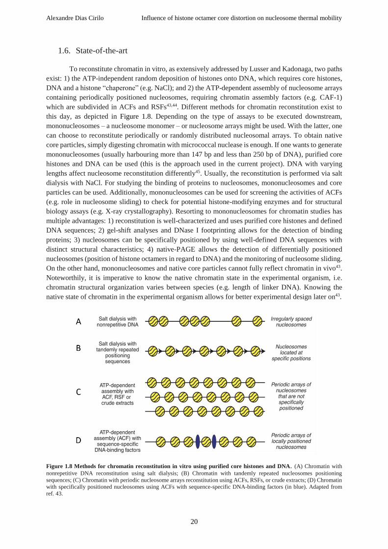
Alexandre Dias Cirilo Influence of histone octamer core distortion on nucleosome thermal mobility
20
1.6. State-of-the-art
To reconstitute chromatin in vitro, as extensively addressed by Lusser and Kadonaga, two paths
exist: 1) the ATP-independent random deposition of histones onto DNA, which requires core histones,
DNA and a histone “chaperone” (e.g. NaCl); and 2) the ATP-dependent assembly of nucleosome arrays
containing periodically positioned nucleosomes, requiring chromatin assembly factors (e.g. CAF-1)
which are subdivided in ACFs and RSFs43,44. Different methods for chromatin reconstitution exist to
this day, as depicted in Figure 1.8. Depending on the type of assays to be executed downstream,
mononucleosomes – a nucleosome monomer – or nucleosome arrays might be used. With the latter, one
can choose to reconstitute periodically or randomly distributed nucleosomal arrays. To obtain native
core particles, simply digesting chromatin with micrococcal nuclease is enough. If one wants to generate
mononucleosomes (usually harbouring more than 147 bp and less than 250 bp of DNA), purified core
histones and DNA can be used (this is the approach used in the current project). DNA with varying
lengths affect nucleosome reconstitution differently45. Usually, the reconstitution is performed via salt
dialysis with NaCl. For studying the binding of proteins to nucleosomes, mononucleosomes and core
particles can be used. Additionally, mononucleosomes can be used for screening the activities of ACFs
(e.g. role in nucleosome sliding) to check for potential histone-modifying enzymes and for structural
biology assays (e.g. X-ray crystallography). Resorting to mononucleosomes for chromatin studies has
multiple advantages: 1) reconstitution is well-characterized and uses purified core histones and defined
DNA sequences; 2) gel-shift analyses and DNase I footprinting allows for the detection of binding
proteins; 3) nucleosomes can be specifically positioned by using well-defined DNA sequences with
distinct structural characteristics; 4) native-PAGE allows the detection of differentially positioned
nucleosomes (position of histone octamers in regard to DNA) and the monitoring of nucleosome sliding.
On the other hand, mononucleosomes and native core particles cannot fully reflect chromatin in vivo43.
Noteworthily, it is imperative to know the native chromatin state in the experimental organism, i.e.
chromatin structural organization varies between species (e.g. length of linker DNA). Knowing the
native state of chromatin in the experimental organism allows for better experimental design later on43.
Figure 1.8 Methods for chromatin reconstitution in vitro using purified core histones and DNA. (A) Chromatin with
nonrepetitive DNA reconstitution using salt dialysis; (B) Chromatin with tandemly repeated nucleosomes positioning
sequences; (C) Chromatin with periodic nucleosome arrays reconstitution using ACFs, RSFs, or crude extracts; (D) Chromatin
with specifically positioned nucleosomes using ACFs with sequence-specific DNA-binding factors (in blue). Adapted from
ref. 43.

Alexandre Dias Cirilo Influence of histone octamer core distortion on nucleosome thermal mobility
21
As mentioned beforehand, histones exhibit attractive hydrophobic and repulsive ionic
interactions. When histones are kept in a high salt (e.g. 2 M NaCl) concentration environment, histone
octamer core formation occurs due to the hindering of the repulsive ionic interactions. Conversely, in
low salt concentration conditions, histones mostly assemble in (H2A-H2B) dimers and (H3-H4)2
tetramers43,46,47. For core histones reconstitution, both with WT and variant species, various experimental
approaches have been previously described43,48,57,49–56. One great advantage of resorting to recombinant
histones for chromatin studies is the absence of eukaryotic factors which usually copurify with histones
during the purification process, the apparent absence of PTMs and the ability to generate diverse mutant
variants43. Furthermore, Lusser and Kadonaga also stated that histones typically stick to plastic and glass
surfaces, thus recommending the use of siliconized materials. When running an SDS-PAGE, from the
slowest to the fastest, core histones migrate in the following order: H3, H2B, H2A, and H4. A good
indicator of the quality of the purification is denoted by the presence of full-length H3. H3 is the most
proteolytically sensitive core histone, hence being more susceptible to degradation. If H3 degrades along
the purification method, a band appears on SDS-PAGE gels between H2A and H443. Core histones can
be purified both from inclusion bodies – intracellular aggregates usually made of proteins – or directly
using whole cell extracts under strong denaturing conditions that allow the solubilization of inclusion
bodies57. An SDS-PAGE gel allowing for the comparison of these two methods is shown below in Figure
1.9.
Figure 1.9 SDS-PAGE gel exemplifying two methods for histone purification. IB: Inclusion bodies purification; RHP:
Rapid histone purification (as proposed in ref. 57); *: Contamination. Each core histone (from Drosophila) is marked
accordingly. Adapted from ref. 57.
For this project, we followed an experimental procedure adapted from the nucleosome
reconstitution methods described in refs. 54 and 57. A step by step diagram of the nucleosome
reconstitution workflow used in this work is shown in Figure 1.10.

Alexandre Dias Cirilo Influence of histone octamer core distortion on nucleosome thermal mobility
22
Figure 1.10 Nucleosome reconstitution experimental workflow. Core histones H2A in yellow, H2B in green, H3 in
blue, and H4 in magenta. Nucleosomal DNA coloured in beige (helices only). Exemplifying for the long version of the
Widom 601 sequence (227bp). Representations generated using PyMOL (PDB: 1AOI; The PyMOL Molecular Graphics
System, Version 2.0 Schrödinger, LLC).

Alexandre Dias Cirilo Influence of histone octamer core distortion on nucleosome thermal mobility
23
2. Aims and objectives
The study of the nucleosome, the fundamental unit of chromatin whose crystal structure at
atomic resolution was solved more than two decades ago, is of vital importance for a better
understanding of the regulation and storage of the extensive genetic information encoded in the genome.
The discovery of its dynamic nature led to the assumption that it could act as an additional layer of gene
expression regulation mechanism. In this work, we aim to observe and corroborate the previously
described thermally-driven mobility of the nucleosome42 using WT nucleosomes assembled from X.
laevis core histones heterologously-expressed in E. coli and an extended version of the strong
positioning Widom 601 sequence (227 bp)39. Furthermore, we also aim to study the mobility of modified
nucleosomes harbouring different histone mutants using native gel shift assays. For each mutant,
specific residues were replaced with cysteine residues. Moreover, for each modification in one specific
histone, a complementary mutation in the other histone from the histone dimer pair will be introduced.
These mutations will be designed using the 2.8 Å solved structure of the nucleosome core particle taking
into account that the designed mutations are structurally close to each other to allow for the formation
of disulphide bonds. The main aim of this project is to describe the influence of histone octamer
distortion on the thermally-driven nucleosome mobility using a designed mutant nucleosome assembled
from scratch.
Additionally, we also aim to observe the ability of Fkbp39, a previously described putative
histone chaperone58, to bind to X. laevis core histone dimer (H2A-H2B), and X. laevis core histone
tetramer (H3-H4)2 and observe the putative complexes via electron microscopy.
Lastly, we aim to screen the binding affinity of several chromatin-modifying enzymes to WT X.
laevis nucleosomes via native gel shift assays.

Alexandre Dias Cirilo Influence of histone octamer core distortion on nucleosome thermal mobility
24
3. Materials
3.1. Buffers
Table 3.1 List of buffers.
Buffer Composition Application
4x SDS-PAGE loading
buffer 200 mM Tris-HCl pH 6.8 8% SDS 40% Glycerol 400 mM DTT
SDS-PAGE
electrophoresis
4x separating gel buffer 1.5M Tris-HCl pH 8.8, 0.4% SDS SDS-PAGE
electrophoresis
4x stacking gel buffer 0.5M Tris-HCl pH 6.8, 0.4% SDS SDS-PAGE
electrophoresis
His buffer A 25 mM Tris-HCl pH 7.5, 1 mM DTT Protein purification
His buffer B 25 mM Tris-HCl pH 7.5, 1 mM DTT, 2 M NaCl Protein purification
His elution buffer 50 mM NaP pH 8.0, 300 mM imidazole pH 8.0, 2 M NaCl Protein purification
His lysis buffer 50 mM NaP pH 8.0, 20 mM imidazole pH 8.0, 2 M NaCl, 3 µM β-
mercaptoethanol Protein purification
His washing buffer 50 mM NaP pH 8.0, 40 mM imidazole pH 8.0, 2 M NaCl Protein purification
Histone dialysis buffer 25 mM Tris-HCl pH 7.5-8.0, 2 M NaCl, 1 mM DTT, 0.1 mM EDTA Protein purification
Inclusion bodies
washing buffer (IBWB) 25 mM Tris-HCl pH 7.5, 200 mM NaCl, 1mM DTT Protein purification
Inclusion bodies
washing buffer with
triton (IBWB+T)
25 mM Tris-HCl pH 7.5, 200 mM NaCl, 1mM DTT, 1% Triton Protein purification
Nucleosome dialysis
buffer 0 M salt 15 mM Tris-HCl pH 7.5, 1 mM DTT
Nucleosome
assembly
Nucleosome dialysis
buffer 1 M salt 1 M NaCl, 15 mM Tris-HCl pH 7.5, 1 mM DTT
Nucleosome
assembly
Protein dialysis buffer
150 30 mM Tris-HCl pH 7.5, 1 mM DTT, 150 mM NaCl Protein purification
Protein dialysis buffer
200 30 mM Tris-HCl pH 7.5, 1 mM DTT, 200 mM NaCl Protein purification
Protein dialysis buffer
200+1G 30 mM Tris-HCl pH 7.5, 1 mM DTT, 200 mM NaCl, 1% glycerol Protein purification
Protein dialysis buffer
200+5G 30 mM Tris-HCl pH 7.5, 1 mM DTT, 200 mM NaCl, 5% glycerol Protein purification
Protein dialysis buffer
200+10G 30 mM Tris-HCl pH 7.5, 1 mM DTT, 200 mM NaCl, 10% glycerol Protein purification
Protein dialysis buffer
300 30 mM Tris-HCl pH 7.5, 1 mM DTT, 300 mM NaCl Protein purification
Protein dialysis buffer
500 30 mM Tris-HCl pH 7.5, 1 mM DTT, 500 mM NaCl Protein purification
S1000 buffer 25 mM Tris-HCl pH 7.5, 1 mM DTT, 2 M NaCl Protein purification
S2000 buffer 25 mM Tris-HCl pH 7.5, 1 mM DTT, 2 M NaCl Protein purification
S500 buffer 25 mM Tris-HCl pH 7.5, 1 mM DTT, 500 mM NaCl Protein purification
S750 buffer 25 mM Tris-HCl pH 7.5, 1 mM DTT, 750 mM NaCl Protein purification
TAE 9 mM Tris pH 7.6, 89 mM boric acid, 2 mM EDTA Agarose gel
electrophoresis
TBE 40 mM Tris pH 7.6, 20 mM acetic acid 20 mM acetic acid SDS-PAGE
electrophoresis
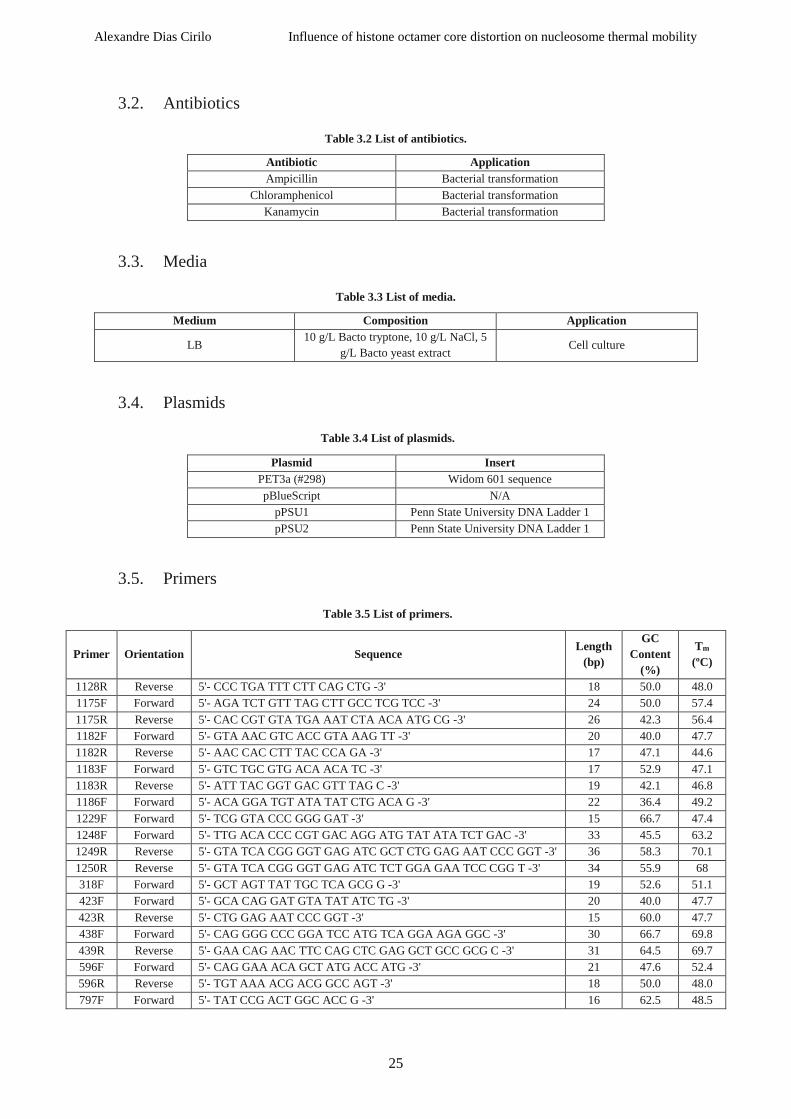
Alexandre Dias Cirilo Influence of histone octamer core distortion on nucleosome thermal mobility
25
3.2. Antibiotics
Table 3.2 List of antibiotics.
Antibiotic Application Ampicillin Bacterial transformation
Chloramphenicol Bacterial transformation
Kanamycin Bacterial transformation
3.3. Media
Table 3.3 List of media.
Medium Composition Application
LB 10 g/L Bacto tryptone, 10 g/L NaCl, 5
g/L Bacto yeast extract Cell culture
3.4. Plasmids
Table 3.4 List of plasmids.
Plasmid Insert PET3a (#298) Widom 601 sequence
pBlueScript N/A
pPSU1 Penn State University DNA Ladder 1
pPSU2 Penn State University DNA Ladder 1
3.5. Primers
Table 3.5 List of primers.
Primer Orientation Sequence Length
(bp)
GC
Content
(%)
Tm
(ºC)
1128R Reverse 5'- CCC TGA TTT CTT CAG CTG -3' 18 50.0 48.0
1175F Forward 5'- AGA TCT GTT TAG CTT GCC TCG TCC -3' 24 50.0 57.4
1175R Reverse 5'- CAC CGT GTA TGA AAT CTA ACA ATG CG -3' 26 42.3 56.4
1182F Forward 5'- GTA AAC GTC ACC GTA AAG TT -3' 20 40.0 47.7
1182R Reverse 5'- AAC CAC CTT TAC CCA GA -3' 17 47.1 44.6
1183F Forward 5'- GTC TGC GTG ACA ACA TC -3' 17 52.9 47.1
1183R Reverse 5'- ATT TAC GGT GAC GTT TAG C -3' 19 42.1 46.8
1186F Forward 5'- ACA GGA TGT ATA TAT CTG ACA G -3' 22 36.4 49.2
1229F Forward 5'- TCG GTA CCC GGG GAT -3' 15 66.7 47.4
1248F Forward 5'- TTG ACA CCC CGT GAC AGG ATG TAT ATA TCT GAC -3' 33 45.5 63.2
1249R Reverse 5'- GTA TCA CGG GGT GAG ATC GCT CTG GAG AAT CCC GGT -3' 36 58.3 70.1
1250R Reverse 5'- GTA TCA CGG GGT GAG ATC TCT GGA GAA TCC CGG T -3' 34 55.9 68
318F Forward 5'- GCT AGT TAT TGC TCA GCG G -3' 19 52.6 51.1
423F Forward 5'- GCA CAG GAT GTA TAT ATC TG -3' 20 40.0 47.7
423R Reverse 5'- CTG GAG AAT CCC GGT -3' 15 60.0 47.7
438F Forward 5'- CAG GGG CCC GGA TCC ATG TCA GGA AGA GGC -3' 30 66.7 69.8
439R Reverse 5'- GAA CAG AAC TTC CAG CTC GAG GCT GCC GCG C -3' 31 64.5 69.7
596F Forward 5'- CAG GAA ACA GCT ATG ACC ATG -3' 21 47.6 52.4
596R Reverse 5'- TGT AAA ACG ACG GCC AGT -3' 18 50.0 48.0
797F Forward 5'- TAT CCG ACT GGC ACC G -3' 16 62.5 48.5

Alexandre Dias Cirilo Influence of histone octamer core distortion on nucleosome thermal mobility
26
797R Reverse 5'- GAG TTC ATC CCT TAT GTG AT -3' 20 40.0 47.7
823R Reverse 5'- GGA TTT GTA GCC TCC GTG -3' 18 55.6 50.3
872F Forward 5'- TAA TAC GAC TCA CTA TAG -3' 18 33.3 41.2
3.6. Gels
Table 3.6 Composition of Native-PAGE and SDS-PAGE gels.
Type Composition Final volume (mL)
Native-PAGE 4 mL TBE 10x, 8 mL Acrylamide/BisAA 30% (37.5:1), 400 µL
APS 10%, 40 µL TEMED, 27.5 mL H2O 40 mL
SDS-PAGE
Separating gel:
7.5 mL 4x separating gel buffer, 17 mL Acrylamide/BisAA 30%
(37.5:1), 300 µL APS 10%, 30 µL TEMED, 2 mL glycerol 25%,
5.5 mL H2O
Stacking gel:
3.75 4x stacking gel buffer, 2 mL Acrylamide/BisAA 30%
(37.5:1), 150 µL APS 10%, 15 µL TEMED, 9.25 mL H2O
30 mL
1
1 β-mercaptoethanol and DTT have to be added to the respective buffers every three days since they tend to degrade with
time.

Alexandre Dias Cirilo Influence of histone octamer core distortion on nucleosome thermal mobility
27
4. Methods
4.1. Bacterial transformation of E. coli cells
Plasmid DNA was transformed into E. coli competent cells resorting to two different strains,
namely, XL-1 Blue and BL21 (DE3) Rosetta. Firstly, 5 µL of plasmid DNA were added to 50 µL of
competent cells in precooled 1.5 mL microcentrifuge tubes and carefully mixed, followed by an
incubation on ice for 15 min. Next, the cells were heat shocked at 42 °C for 45s, and then put on ice for
2 min. 500 µL of LB medium were then added, and cells were put at 37 °C, 160 rpm for 1h in an orbital
shaker to induce cell recovery, followed by plating on LB agar plates with the required antibiotics to
allow for the selection of successfully transformed cells. In the case of XL-1 Blue, Kan was used, and
for BL21 (DE3) Rosetta cells, both Kan and Cam were used. To allow cell growth, plated agar plates
were incubated overnight at 37 °C. For histone expression, only the chemically competent BL21 (DE3)
Rosetta cells were used.
4.2. Plasmid DNA purification
To obtain purified plasmid DNA, pelleted cells obtained from an overnight preculture grown at
37 °C in an orbital shaker (160 rpm) were treated following the NucleoSpin® Plasmid EasyPure protocol
from Macherey-Nagel (http://www.mn-net.com). The washing step was performed twice to ensure
yielding a “cleaner” product.
4.3. DNA sequencing
Firstly, bacterial precultures were prepared from single colonies in 5 mL of LB medium
containing the appropriate antibiotic(s). Precultures were then put to grow overnight in an orbital shaker
at 37 °C. The following day, precultures were centrifuged at 3900 rpm for 15 minutes to harvest cells,
and the supernatant was discarded. Purified plasmid DNA was obtained resorting to the NucleoSpin®
Plasmid EasyPure kit (as described in 4.2). Sequencing of DNA was performed via Sanger sequencing
by GATC Biotech AG. For each sequencing job, a 20 µL aliquot of DNA or primers were sent. Primers
were diluted to a final concentration of 0.1 µM. Sequencing results were analysed via FinchTV software
(Geospiza, Inc.).
4.4. Polymerase chain reaction
The polymerase chain reaction was used for the amplification of the strong-positioning Widom
601 sequence (and variants) used downstream for nucleosome assembly (see subchapter 4.9). The PCR
reaction is described below in Table 4.1.
Table 4.1 PCR reaction setup for the long Widom 601 sequence amplification (227 bp).
Reagent Volume (µL) Source
H2O 33.5 In house
Phusion® HF Buffer (5x) 10 Phusion® HF Buffer Pack (New England Biolabs)
dNTPs (10 mM) 1 Jena Bioscience (Germany)
797F (Forward primer) 2 Metabion (Germany)
797R (Reverse primer) 2 Metabion (Germany)
298 (DNA Template) 1 In house
Phusion polymerase (in house) 0.5 In house

Alexandre Dias Cirilo Influence of histone octamer core distortion on nucleosome thermal mobility
28
For the amplification of the long version of the Widom 601 sequence used in this work, initial
denaturation of DNA was set at 95 °C for 30s, followed by 46 cycles of 10s at 95 °C (denaturation), 10s
at 54 °C (annealing), and 10s at 72 °C (elongation). After the cycles ended, an additional step was set at
72 °C for 30s. Amplified DNA sequences were then analysed on 1% agarose gels (staining with DNA
Gel Loading Dye 6x from Thermo Scientific).
4.5. DNA precipitation
Precipitation of DNA was performed by adding, first and foremost, 2.5 volumes of EtOH 100%
and 10% of 3 M NaOAc pH 5.2. The sample was then let either 1h or overnight at -80 °C, followed by
centrifugation at maximum rcf for 30 min. Supernatant was then removed and 500 µL of EtOH 75%
was added to the DNA pellet. The ensuing solution was centrifuged at maximum rcf for 15 min and the
supernatant was removed. The resulting sample was dried at RT for around 50 min, followed by the
addition of 20 µL of H2O and resuspended. DNA concentration was then assessed via the
NanoPhotometer NP80 (from Implen), and its quality checked via 1% agarose gel (staining with DNA
Gel Loading Dye 6x from Thermo Scientific).
4.6. Histone expression
Protein expression in the chemically competent BL21 (DE3) Rosetta cells was performed in LB
medium. To that intent, when starting from glycerol stocks of the host laboratory stored at -80 °C,
precultures (ranging from 5 mL to 30 mL) were firstly grown overnight in an orbital shaker at 160 rpm
and 37 °C, and the respective antibiotic(s) were also added to the latter, as described in Table 4.2.
Addition of cells from the glycerol stocks to their respective preculture recipient was done by picking
some frozen cells using a 200 µL pipette tip. The following day, the cultured competent cells from the
aforementioned precultures were harvested and used for growing larger cell cultures, with the addition
of glucose 20%, as described in Table 4.2. These were grown at 37 °C, 150 to 200 rpm, until they
reached an optical density at 600 nm (OD600) of 0.6. Afterwards, temperature was lowered to 18 °C for
30 minutes, following the overnight induction of cells with 0.2 mM of IPTG. The following day, cells
were centrifuged at 4000 rpm for 10 minutes and the pelleted cells were collected and either directly
used for downstream applications, or stored at -20 °C.
Table 4.2 Components for cell induction.
Final volume of cell
culture (LB, in L)
Added volume of
glucose 20% (mL)
Added volume of
antibiotic (mL)
Added volume of
preculture (mL)
1 20 1 10
2 40 2 20

Alexandre Dias Cirilo Influence of histone octamer core distortion on nucleosome thermal mobility
29
4.7. Recombinant protein purification from heterologous expression in E. coli BL21
(DE3) Rosetta strain
4.7.1. Affinity chromatography: 6xHis-tag purification (Ni-NTA Agarose)
Cells were resuspended in His lysis buffer using an Omnifix® Luer Solo syringe with Sterican®
single-use hypodermic needles from Braun, this step is very important to avoid clogging the French
press. Following the resuspension step, 200 µL of 0.1 M PMSF and 1 µL of DNAse (Pierce™ Universal
Nuclease for Cell Lysis) were added. The next step consisted in the lysis of the treated cells resorting to
a French press (25 ksi), followed by a centrifugation at 17000 rpm, 4 °C, for 15 minutes. The supernatant
was then incubated with Ni Sepharose 6 Fast Flow histidine-tagged protein purification resin (from GE
Healthcare) for exactly 30 minutes at 4 °C to allow for the binding of the histones to the resin. Next, the
resin was washed 3 to 5 times with the His lysis buffer, being cautious about resuspending the resin each
time, and the supernatant was discarded after each wash. The ensuing washed resin was then loaded
onto a 10 mL disposable polypropylene column (from Thermo Scientific) at 4 °C. The resin was washed
using 10 to 12 mL of His washing buffer, followed by the elution of bound histones using the His elution
buffer. Elution efficiency of the proteins was analysed after running a 17 % SDS-PAGE gel and staining
the gel with SimplyBlue™ Safe-stain. Lastly, the eluted histones were dialyzed overnight at 4 °C in the
histone dialysis buffer.
4.7.2. Ion exchange chromatography: protein purification (Sepharose Fast Flow)
Following the overnight dialysis after the affinity chromatography, ion-exchange
chromatography was performed using 10 mL disposable polypropylene columns from Thermo
Scientific. If reusing columns that were properly washed and stored in EtOH 20%, firstly, the respective
columns would be washed three consecutive times with ddH2O, centrifuging at 3000 rpm, 4 °C, for 3-5
min. The washed resin would then be incubated and mixed with 5x His buffer A and the dialyzed
proteins (the ones previously purified by affinity chromatography) at 4 °C for 30 minutes. After the
binding of proteins to the resin, the sample was centrifuged at 2000-2500 rpm, 4 °C, for 4 minutes
(supernatant, i.e. unbound protein, was collected for subsequent assessment of binding efficiency of the
proteins to the sepharose resin). Remaining beads in the glass bottle were washed with S500 buffer. Next,
at 4 °C, the resin was transferred to a S-column (using the S500 buffer to collect all the remaining beads).
2.5 volumes in column volume were then added for elution using S750, S1000, and S2000 buffers. Each
fraction was collected individually. A 17% SDS-PAGE gel was run to analyse the efficiency of the
purification and check for the enriched fraction(s). Aliquots of 0.5 mL to 1 mL were prepared from the
enriched fractions, frozen in liquid nitrogen, and stored at –80 °C. To note, the used resin was thoroughly
washed with 5 M NaCl, and 2 to 3 rounds of ddH2O, and then stored at 4 °C in EtOH 20%.

Alexandre Dias Cirilo Influence of histone octamer core distortion on nucleosome thermal mobility
30
4.8. Histone octamer assembly
To assemble the histone octamer, stoichiometric amounts of core histones H2A, H2B, H3 and
H4 were mixed and incubated at 4 ºC overnight. Histone ratios were checked on 17% SimplyBlue™
Safe-stained SDS-PAGE gels prior to incubation.
4.9. Nucleosome assembly
As a means to assemble the nucleosome, equimolar mixing of the histone octamer and
nucleosomal DNA must be observed. To that intent, in the current work, the long version of the strong-
positioning Widom 601 sequence (227 bp) was used, as it has been previously shown to be a very strong
and effective histone octamer binding sequence39 (see subchapter 1.5.1). The salt concentration of the
sample containing the nucleosomal DNA was adjusted to 2 M NaCl before mixing it with the histone
octamer. Following the procedure for nucleosome reconstitution as described in ref. 54 (all steps
performed at 4 ºC), each assembly sample was transferred in an Eppendorf lid covered with a pre-wetted
3.5 kDa cut-off standard cellulose regenerated membrane (Spectra/Por® 3 Dialysis Tubing). First and
foremost, the sample was dialysed overnight in nucleosome dialysis buffer 1 M salt, followed by two
rounds of dialyses in 0 M NaCl buffers. The first round of the dialysis in nucleosome dialysis buffer 0
M salt (final volume of 1 L) would take 5 to 6 hours (ideally, 5 hours), followed by the second round of
dialysis in nucleosome dialysis buffer 0 M salt (final volume of 0.5 L), which would take 1 to 1h30
(ideally, 1h30). All three of these dialysis buffers were discarded after three uses. After the sample was
thoroughly dialysed, nucleosome yielding was assessed by direct visualization on a native-PAGE gel
right after staining with Invitrogen™ SYBR™ Gold Nucleic Acid Gel Stain (10000x concentrated in
DMSO, from Fisher Scientific). This step allows to check for the presence of the nucleosome core
particle, as well as the position of the Widom 601 sequence on the latter, and if present, free nucleosomal
DNA. Successfully assembled nucleosomes were used later on for binding assays with multiple
chromatin-binding proteins, as well for thermal shifting assays. Remaining nucleosomes were frozen in
liquid nitrogen and stored at -80 °C.
4.10. Protein concentration
When required, proteins were concentrated to smaller volumes resorting to Amicon®
Centrifugal Filter Units (from Merck), as described in Table 4.3, with varying MW cut-offs and
volumes. Samples were added to the concentrating devices and centrifuged at 3900 rpm for 4 to 6 min.
After each centrifugation, the input volume was pipetted up and down, being cautious not to touch the
cellulose membrane with the pipette tip. Centrifugations were repeated until reaching the desired final
volume, or if precipitation occurred (to avoid any further losses).
Table 4.3 Various devices Amicon® used throughout the project.
Device Volume
(mL) Commercial name
Membrane cut-off
(kDa)
Amicon® Ultra-4 4 Ultracel-30 regenerated cellulose
membrane 30
Amicon® Ultra-
15 15
Ultracel-30 regenerated cellulose
membrane 30
Amicon® Ultra-
15 15
Ultracel-50 regenerated cellulose
membrane 50

Alexandre Dias Cirilo Influence of histone octamer core distortion on nucleosome thermal mobility
31
4.11. S. pombe Fkbp39 purification and Fkbp39:(H3-H4)2, Fkbp39:(H2A-H2B)
complex formation assessment
As mentioned beforehand in subchapter 1.2.5.1, S. pombe Fkbp39’s ability to form a complex
with full-length (H2A-H2B) and (H3-H4)2 from X. laevis was studied as a side project. To that intent,
BL21 (DE3) Rosetta competent cells capable of expressing Fkbp39-SUMO-6xHis (S. pombe) were
firstly grown in a total volume of 2 L (starting from a 30 mL preculture) as described in subchapter 4.6.
Harvested cells were pelleted after centrifugation at 4000 rpm, 4 ºC for 10 min, and then resuspended.
To resuspend pelleted cells, 90 mL of His lysis buffer was added and resuspended cells were transferred
to a clean metallic cup using an Omnifix® Luer Solo syringe with Sterican® single-use hypodermic
needles from Braun. Then, 100 µL of 0.1 M PMSF and 1 µL DNAse were added (for a final volume of
1L). Treated cells were then sonicated using a Branson Sonifier 250 (40% Duty cycle and 4 output
control micro tip limit) for 10 min. Next, sonicated cells were purified by 6xHis-tag protein purification
using Ni-NTA agarose, an affinity chromatography matrix used for purifying recombinant proteins
carrying a 6xHis-tag, as described in subchapter 4.7.1. Elution fractions enriched in Fkbp39 were then
collected, part of it was mixed with WT (H3-H4)2 from X. laevis to check for the optimal ratio of
complex formation, followed by the addition of PreScission enzyme (in house) in order to cut the
SUMO-6xHis tag from Fkbp39. The resulting treated sample was dialysed using a 12-14 kDa cut-off
standard cellulose regenerated membrane (Spectra/Por® 4 Dialysis Tubing) in the histone dialysis buffer
overnight. The next day, the sample was transferred to a protein dialysis buffer 200 and dialysed for 5h.
After the dialysis, the mixture was concentrated (using Amicon® Ultra-15, Ultracel-30 regenerated
cellulose membrane, 30 kDa). The first attempt to concentrate the sample was unsuccessful due to the
occurrence of precipitation and ensuing loss of sample. Hence, 10% glycerol was added to the mixture,
and a new attempt to concentrate the sample was performed. This time, the sample was successfully
concentrated to around 500 µL. The ensuing concentrated sample was run on a 17% SDS-PAGE gel to
check if it yielded enough complex for downstream analyses, followed by size-exclusion
chromatography and further concentration steps until obtaining pure enough complex.
The remaining volume of the elution fractions of Fkbp39 that was not mixed with histones was
subdivided and dialysed overnight, after adding PreScission enzyme (in house), in three buffers with
varying salt (NaCl) concentration (Protein dialysis buffers 150, 300 and 500) to check for optimal salt
concentration in order to avoid Fkbp39 precipitation. 6-8 kDa cut-off standard cellulose regenerated
membranes (Spectra/Por® 1 Dialysis Tubing) were used for these dialyses. The following day, the
dialysed proteins were run on a 17% SDS-PAGE gel with the uncut protein to check for effective
SUMO-tag removal. Fractions dialysed in 150 and 300 mM NaCl containing buffers were further
purified through ion-exchange chromatography, whilst the 500 mM NaCl containing buffer fraction was
stored at -80 °C. Enriched fractions from ion exchange chromatography of Fkbp39 were mixed in
stoichiometric quantities with (H3-H4)2 from the host laboratory after checking for the best ratio on a
17% SDS-PAGE gel. The ensuing mixture was then dialysed overnight in protein dialysis buffer
200+10G to avoid precipitation. Next, the dialysed complex was aliquoted (1 mL of sample in 2 mL
polypropylene screw cap micro tubes, from Sarstedt) and centrifuged at 10k rpm, 4 °C, for 15 min. After
centrifugation, soluble fractions were transferred to new screw cap tubes and the pellets were
resuspended in 4x SDS-PAGE loading buffer. To check for any significant complex loss through
precipitation, an additional 17% SDS-PAGE gel was run. Next, as the final aim of this side project was
getting a stable and pure enough complex of Fkbp39 with (H3-H4)2, and possibly (H2A-H2B), for
downstream cryo-EM applications, a new dialysis was executed overnight in protein dialysis buffer 200,
since it is not recommended to have glycerol in samples for EM analyses59. After mixing (H3-H4)2 and
Fkbp39 in a 1:1 ratio, 7 M guanidine·HCl was added to denature the proteins, and the mixture was

Alexandre Dias Cirilo Influence of histone octamer core distortion on nucleosome thermal mobility
32
incubated at RT for 1h with slow mixing. The use of this strong chaotropic agent is justified by its ability
to solubilize insoluble proteins (such as inclusion bodies), which is the first step to be taken for protein
refolding experiments. After the incubation, the denaturated sample was centrifuged at 15k rpm, 4 °C,
for 15 min. The ensuing soluble fraction was dialysed overnight in protein dialysis buffer 200+10G,
allowing for the overnight refolding of the complex. Remaining purified Fkbp39 from before, as well
as an aliquot of complex, were dialysed protein dialysis buffer 200 (without glycerol to check if Fkbp39
precipitates in buffer without glycerol when not in complex with histones, as well as in complex with
histones). These three dialyses were executed using a 6-8 kDa cut-off standard cellulose regenerated
membrane (Spectra/Por® 1 Dialysis Tubing). Then, ensuing dialysed samples were centrifuged at 15k
rpm for 15 min at 4 °C, and aliquots of the soluble fractions were loaded on a 17% SDS-PAGE gel.
We also tried to check for complex formation of Fkbp39 with WT X. laevis (H2A-H2B) from
the host laboratory. To that intent, (H2A-H2B) samples were loaded on a 17% SDS-PAGE gel with
Fkbp39 to check for optimal ratio.
Due to the high incidence of precipitation either of the complex Fkbp39:(H3-H4)2, either
Fkbp39 alone in protein dialysis buffer 200+10G, new dialyses with varying salt concentration and
glycerol percentage were prepared, as described in Table 4.4.
Table 4.4 Dialysis buffers’ composition for Fkbp39:(H3-H4)2 and FKbp39:(H2A-H2B) assembly.
Dialysed sample(s) [NaCl] (mM) % Glycerol
Fkbp39:(H3-H4)
Fkbp39:(H2A-H2B) 200 1
Fkbp39:(H3-H4)
Fkbp39:(H2A-H2B) 200 5
Fkbp39:(H2A-H2B) 200 10
Fkbp39:(H3-H4)
Fkbp39:(H2A-H2B) 300 0
Fkbp39:(H3-H4)
Fkbp39:(H2A-H2B) 500 0
Complex precipitation issue was ultimately resolved after using the aforementioned buffers.
Hence, the complexes were aliquoted, part of these were frozen in liquid nitrogen and stored at -80 °C
for later downstream applications (i.e. binding screening via gel mobility shift assays), and the remaining
part was used for cryo-EM analyses at the Max Planck Institute of Biochemistry (Munich, Germany).

Alexandre Dias Cirilo Influence of histone octamer core distortion on nucleosome thermal mobility
33
4.12. Chromatin-binding proteins native gel shift assays
Screening of chromatin-binding proteins binding to WT X. laevis nucleosomes was performed
via native gel shift assays. Small volumes of concentrated WT nucleosomes (1 to 10 µL depending on
nucleosome concentration) were mixed with small volumes of concentrated chromatin-binding protein
(0.5 to 5 µL) and incubated at RT for 30 min or 1h at 4 ºC, followed by the addition of 1.5 µL of native-
PAGE loading dye (40% glycerol and 2 µL of bromophenol blue in a final volume of 1 mL) and running
the samples on 6% native-PAGE gels at 4 ºC. Staining of the gels was done with Invitrogen™ SYBR™
Gold Nucleic Acid Gel Stain (incubate twice for 10 min with gentle shaking, wash once with TAE 1x
buffer).
4.13. Electrophoresis
4.13.1. Agarose
The lengths of plasmids, DNA, and the various nucleosomal DNA sequences prepared in this
work were checked by visualization in agarose gels. All agarose gels were prepared the same way: 1%
agarose gels run at 100 to 120 V for 20 to 30 min at RT. The ladder was prepared by mixing 4 µL of
H2O with 1 µL of DNA Gel Loading Dye 6x (Thermo Scientific) and 1 µL of GeneRuler 1kb ladder
(Thermo Scientific). All samples were mixed using 5 µL of sample with 1 µL of DNA Gel Loading Dye
6x (Thermo Scientific). Visualization of the gels was done on a gel documentation system, adjusting the
exposure appropriately. Each figure was printed using Mitsubishi K65HM-CE / KP65HM-CE HD
thermal paper rolls.
4.13.2. Native-PAGE
Analysis of nucleosome assembly assays were done using Native-PAGE gels. For each sample,
10 µL of sample was mixed with 1.5 µL of native-PAGE loading dye, and the ladder was prepared by
mixing 10 µL of H2O, 1.5 µL of native-PAGE loading dye, and 1 µL of the GeneRuler 1kb ladder
(Thermo Scientific). The gels were run at 200 V for 2h, at 4 °C. Afterwards, gels were carefully
disassembled from the cassette and put in a thoroughly cleaned box shielded from light (the lid of the
box was carefully wrapped in aluminium foil) containing 25 mL of 1x TAE buffer. Then, 2.5 µL of
Invitrogen™ SYBR™ Gold Nucleic Acid Gel Stain (10000x concentrated in DMSO, from Fisher
Scientific) was added to the solution contained the immersed gel, being careful not to pipette the stain
directly over the gel but rather on the sides of the box in order to avoid any blob formation which would
impair proper band(s) visualization. The box containing the gel was then put on a shaker at 50 rpm for
10 min at RT. Afterwards, the solution was discarded in the appropriate toxic waste disposal container,
and 25 mL of 1x TAE buffer were added again to the box, putting the box to shake at 50 rpm for 10 min
at RT, as before. The solution was discarded in the same toxic waste disposal container after the previous
step, and the process was repeated once more. Lastly, the gels were analysed on a gel documentation
system, adjusting the exposure appropriately. Each figure was printed using Mitsubishi K65HM-CE /
KP65HM-CE HD thermal paper rolls.
4.13.3. SDS-PAGE
Protein purification was assessed through direct visualization of 17% SDS-PAGE gels after
proper staining using SimplyBlue™ Safe Stain. For each sample, 10 µL of sample were mixed with 5
to 10 µL of 4x SDS-PAGE loading buffer, the ensuing solution was incubated at 90 °C for 3 min, and
then loaded on the gel which was run at 300 V for 35 min. Excess SDS was removed after letting the
gel in boiling deionised water thrice for around 30s, and staining was done by letting the gel stain in
boiling SimplyBlue™ Safe Stain for a short period of about 10 to 20s. If required, destaining of the gel
was done by letting the gel in boiling deionised water for around 30s, and repeated if needed.

Alexandre Dias Cirilo Influence of histone octamer core distortion on nucleosome thermal mobility
34
5. Results
5.1. WT and mutant X. laevis histone purification
To avoid redundancy, this subsection will only cover two examples of histone purification,
namely, X. laevis H4_V22C and WT H3, and H2Aub (ubiquitinated) and WT H2B expressed in BL21
(DE3) Rosetta cells Kan and Cam resistant (sample name: “1101”; see Table 5.1). The remaining histone
purifications executed in the aim of this project are listed in Table 5.1. First and foremost, mutant and
WT histones are purified from the pelleted cells as described in subchapter 4.7.1. All the histones
purified in this project harbour a 6xHis-tag which allows for its purification via affinity chromatography.
A 17% SimplyBlue™ Safe-stained SDS-PAGE gel was run to assess the purified histones yield.
Usually, 10 µL of each fraction were loaded on the gel, using 4x SDS-PAGE loading buffer. 17%
SimplyBlue™ Safe-stained SDS-PAGE gels for X. laevis H4_V22C and WT H3, and H2Aub and WT
H2B purifications via affinity (A) and ion-exchange (B) chromatographies are shown in Figure 5.1 and
Figure 5.2, respectively.
Figure 5.1 SimplyBlue™ Safe-stained 17% SDS-PAGE gels of 1101 (see Table 5.1) purification via affinity and ion-
exchange chromatographies. (A) Affinity chromatography. (B) Ion-exchange chromatography. Ladder: PageRuler
Unstained Protein Ladder (Thermo Scientific), FT: Flow-through; Resin wash: His lysis buffer wash; Column FT: Unbound
protein after loading into the column; Wash: Washing step of the affinity chromatography; Elution: Elution step of the affinity
chromatography; AC: Affinity chromatography; IEX: Ion-exchange chromatography; BD: Before overnight dialysis; AF: After
overnight dialysis; Wash 0.5: Resin wash with 500 mM NaCl buffer; 0.5: 500 mM NaCl buffer; 0.75: 750 mM NaCl buffer; 1:
1 M NaCl buffer; 2 (I) and 2 (II): 2 M NaCl buffer (elution with 2 M NaCl buffer is executed twice). Core histones H3 and H4
are marked accordingly.

Alexandre Dias Cirilo Influence of histone octamer core distortion on nucleosome thermal mobility
35
Figure 5.2 SimplyBlue™ Safe-stained 17% SDS-PAGE gels of “1093” (see Table 7.1) purification via affinity and ion-
exchange chromatographies. (A) Affinity chromatography. (B) Ion-exchange chromatography. Ladder: PageRuler Unstained
Protein Ladder (Thermo Scientific), FT: Flow-through; Resin wash: His lysis buffer wash; Column FT: Unbound protein after
loading into the column; Wash: Washing step of the affinity chromatography; Elution: Elution step of the affinity
chromatography; AC: Affinity chromatography; IEX: Ion-exchange chromatography; BD: Before dialysis; AF: After dialysis;
Wash 0.5: Resin wash with 500 mM NaCl buffer; 0.5: 500 mM NaCl buffer; 0.75: 750 mM NaCl buffer; 1: 1 M NaCl buffer;
2 (I) and 2 (II): 2 M NaCl buffer (elution with 2 M NaCl buffer is executed twice). Core histones H2Aub (ubiquitinated H2A)
and H2B are marked accordingly.
Table 5.1 List of purified histones.
Sample names, histones, source plasmid, expression system, and species, referenced accordingly.
Sample: Protein: Plasmid: Expressed in: Species:
XLgH3+XLgH4 WT H3 and H4 (globular) In house BL21 (DE3) Rosetta X. Laevis
XLH3+XLH4 WT H3 and H4 In house BL21 (DE3) Rosetta X. Laevis
41 H4_I67C In house BL21 (DE3) Rosetta X. Laevis
1102 H4_V43C and H3_F104C In house BL21 (DE3) Rosetta X. Laevis
409B+H3 H4_A16C and WT H3 In house BL21 (DE3) Rosetta X. Laevis
1099 H4_I67C and H3_A76C In house BL21 (DE3) Rosetta X. Laevis
1101 H4_V22C In house BL21 (DE3) Rosetta X. Laevis
1108 H4_T67C, H3_C76A and H3_A110C In house BL21 (DE3) Rosetta X. Laevis
1093 WT ubiquitinated H2A, and WT H2B In house BL21 (DE3) Rosetta X. Laevis
1116 H3_L82C and H4_V81C In house BL21 (DE3) Rosetta X. Laevis
1126 H3_L82C, H3_C110A and H4_V81C In house BL21 (DE3) Rosetta X. Laevis
1022 Ubiquitinated H2A_ E61, H2A_D90A, H2A_E92A, and WT H2B, H3 and
H4 In house BL21 (DE3) Rosetta X. Laevis

Alexandre Dias Cirilo Influence of histone octamer core distortion on nucleosome thermal mobility
36
5.2. Full-length X. laevis histone octamer
To refold the histone octamer, equimolar mixing of each histone core is required. For wild-type
histone octamer refolding, three different ratios of (H2A-H2B):(H3-H4)2 were tested: (9:1), (10:1) and
(11:1). The mixed histones were then briefly mixed and stored overnight at 4 °C. The following day,
histone octamer yielding was assessed by direct visualization on a 17% SDS-PAGE gel, like depicted
in Figure 5.3. Noteworthily, H2A and H2B from X. laevis (13.96 kDa and 13.77 kDa, respectively) are
known to comigrate on SDS-PAGE gels and usually appear as a single stronger band55. Histone octamer
assemblies using the mutant histones prepared in the current project were performed in a similar fashion.
It is fulcral to know the histone:DNA ratio. If there is more histone octamer than DNA, then the excess
of histones will aggregate with DNA. On the other hand, if there is more DNA than histones, then the
generation of nucleosomes will be limited. Thus, titrating the histone:DNA mix is a good approach and,
as suggested by Lusser and Kadonaga, varying each titration reaction by 10% is useful (e.g.
histone:DNA ratios of 9:10, 10:10 and 11:10)43,62.
Figure 5.3 SimplyBlue™ Safe-stained 17% SDS-PAGE gel of the histone octamer with different ratios of (H2A-
H2B):(H3-H4)2. L: PageRuler Unstained Protein Ladder (Thermo Scientific). Three different (v/v) ratios of (H2A-H2B):(H3-
H4)2 are marked accordingly. Core histones (H2A, H2B, H3, and H4) are pointed out.

Alexandre Dias Cirilo Influence of histone octamer core distortion on nucleosome thermal mobility
37
5.3. Synthesis and purification of nucleosomal DNA
5.3.1. Amplification by PCR
Obtaining high amounts of the long and short Widom 601 sequences which will be used in the
nucleosome assembly downstream was done by PCR amplification, following the procedure described
in subchapter 4.4 using the DNA template #298 (see Figure 9.2). For the extended version of the Widom
601 sequence (227 bp; +37 bp on the 5’- terminal and +44 bp on the -3’ terminal), primers 797F and
797R were used. For the shorter version (149 bp) of the Widom 601 sequence, primers 423F and 423R
were used (see Table 5.2 for the DNA sequences of the shorter and extended versions of the Widom 601
sequences). The size of the PCR products was checked on 1% agarose gels (see Figure 5.4).
Table 5.2 List of the two versions of the Widom 601 sequence used for nucleosome assembly.
Widom
601
version
Length
(bp) Primers Amplified sequence via PCR (5’ 3’)
Shorter 149 423F + 423R
GCACAGGATGTATATATCTGACACGTGCCTGGAGACTAGGGAGTAATCCCCTTGGCGGTTAA
AACGCGGGGGACAGCGCGTACGTGCGTTTAAGCGGTGCTAGAGCTGTCTACGACCAATTGAG
CGGCCTCGGCACCGGGATTCTCCAG
Longer 227 797F + 797R
TATCCGACTGGCACCGGCAAGGTCGCTGTTCAATACATGCACAGGATGTATATATCTGACAC
GTGCCTGGAGACTAGGGAGTAATCCCCTTGGCGGTTAAAACGCGGGGGACAGCGCGTACGTG
CGTTTAAGCGGTGCTAGAGCTGTCTACGACCAATTGAGCGGCCTCGGCACCGGGATTCTCCA
GGGCGGCCGCGTATAGGGTCCATCACATAAGGGATGAACTC
Figure 5.4 1% agarose gel of the purified 227 bp Widom 601 sequence after amplification via PCR.
Ladder: GeneRuler 1 kb DNA Ladder (Thermo Scientific).

Alexandre Dias Cirilo Influence of histone octamer core distortion on nucleosome thermal mobility
38
5.3.2. Purification by ethanol precipitation
After obtaining a considerable amount of Widom 601 sequence, purification by ethanol
precipitation was ensued according to the protocol described beforehand in subchapter 4.5. Both the
longer and shorter versions of the Widom 601 sequence were successfully purified (see Figure 5.5 for
the longer Widom 601 sequence ethanol precipitation).
Figure 5.5 1% agarose gel of the purified 227 bp Widom 601 sequence after ethanol precipitation.
Ladder: GeneRuler 1 kb DNA Ladder (Thermo Scientific).

Alexandre Dias Cirilo Influence of histone octamer core distortion on nucleosome thermal mobility
39
5.4. WT X. laevis nucleosome assembly
As stated in subchapter 4.9, nucleosome assembly using wild-type and mutants core histones
from X. laevis, and the strong positioning Widom 601 sequence63 was performed following an adapted
experimental protocol based on the framework described in refs. 54–56.
As seen below in Figure 5.6, successful nucleosome assembly using WT X. laevis core histones
and the Widom 601 sequence yields a considerable amount of centred nucleosomes, i.e. when
nucleosomal DNA is properly wrapped around the histone octamer. It is not unusual to get some end-
positioned nucleosomes, i.e. when the histone octamer is positioned on the edge of the nucleosomal
DNA64. End-positioned nucleosomes migrate further than centred nucleosomes on native-PAGE
electrophoresis. The thick band on top in the “Nucleosome Assembly” well is due to the presence of
aggregates65–67. Reconstituted NCPs exhibit a characteristic slower migration in native-PAGE gels in
regard to free DNA, allowing for the observation of the nucleosome:free DNA ratio43. As described
before in subchapter 5.2, it’s imperative to know the histone:DNA ratio for successful nucleosome
reconstitution.
Downstream, purified nucleosomes can be used for binding and shifting assays. In this project,
assembled nucleosomes were used for screening putative interactions with multiple chromatin-binding
proteins via native gel shift assays and for thermal shift assays.
Figure 5.6 SYBR Gold-stained 6% Native-PAGE gel of the nucleosome assembly assay using WT X. laevis core histones
and the Widom 601 sequence. Ladder: GeneRuler 1 kb DNA Ladder (Thermo Scientific); c-NCP: centred nucleosome core
particle; ep-NCP: end-positioned nucleosome core particle; Free DNA: free Widom 601 sequence.

Alexandre Dias Cirilo Influence of histone octamer core distortion on nucleosome thermal mobility
40
5.5. Thermal shifts of WT and mutant X. laevis nucleosomes from X. Laevis
Thermal shift assays were performed at 62 and 60 °C using WT and mutant X. laevis
nucleosomes (for the mutant, see Figure 5.8), respectively. For each sample in the experiment, a volume
of 0.5 μL of concentrated nucleosome (15 mM Tris pH 7.5, 1 mM DTT) was used. Nucleosome
incubation was done in a FlexCycler2 (Analytik Jena) PCR thermocycler. Samples were stored on ice at
all times, except during incubation. Incubation times were set at 5, 15, 30 and 45 minutes. As a control,
one sample was not incubated, therefore being kept on ice. As shown below in
Figure 5.7 (t = 0 min), nucleosomes tend to shift towards the DNA ends when incubated at high
temperatures. Which confirms previous studies of the effect of temperature on nucleosome
positioning42,68–70. In theory, as reported in ref. 42, this shifting would be gradually visible with time. It
is plausible that, in this experiment, the temperature was high enough to allow for the swift shifting of
the nucleosomes toward the DNA ends. Bands that appear lower than the ep-NCP reference band might
be due to the presence of other nucleosomal states such as, for example, hexamers. The higher amount
of dimer DNA might be due to nucleosome disassembly with time leading to an increase in free DNA
which can then hypothetically form DNA dimers.
Figure 5.7 Thermal shift assays of WT and H4_V43C H3_F104C mutant X. laevis nucleosomes. Performed at 62 and 60
°C, respectively. c-NCP: centred nucleosome core particle; ep-NCP: end-positioned nucleosome core particle; Free DNA: free
Widom 601 sequence; Free DNA dimer: free Widom 601 sequence dimer. Incubation times are marked accordingly in minutes.

Alexandre Dias Cirilo Influence of histone octamer core distortion on nucleosome thermal mobility
41
Figure 5.8 Designed X. laevis mutant nucleosome exhibiting H4_V43C H3_F104C mutations. (A) The nucleosome
exhibiting H4_V43 and H3_F104 residues in green and yellow, respectively. (B) H4_V43C and H3_F104C mutations showing
the potential to form a disulphide linkage. Representations generated using PyMOL (PDB: 1AOI; The PyMOL Molecular
Graphics System, Version 2.0 Schrödinger, LLC).

Alexandre Dias Cirilo Influence of histone octamer core distortion on nucleosome thermal mobility
42
5.5.1. Effect of crosslinking WT X. laevis nucleosomes with glutaraldehyde
Chemical crosslinking using glutaraldehyde (O=CH-CH2-CH2-CH2=O), a potent extensively
reported protein-protein and DNA-protein crosslinker agent (1 DNA-protein crosslink per 2.7 kb of
DNA at 8 μM glutaraldehyde71) fixed the stable nucleosome states72. Crosslinking was done using 0.1%
(v/v) glutaraldehyde. The control was prepared in parallel with the exact same conditions, only omitting
the addition of glutaraldehyde. A SYBR Gold-stained 6% native-PAGE gel referring to the crosslinking
assay is shown in Figure 5.9. As expected, the addition of 0.1% (v/v) glutaraldehyde successfully
crosslinked nucleosomes since no intermediate nucleosome states are observable. The crosslinked
nucleosome acts as a robust biological entity and is apparently unable to go through conformational
changes. Conversely to the thermal shift assay without the addition of glutaraldehyde, the crosslinked
nucleosome is unable to move and stays on the long Widom 601 sequence. This suggests that structural
changes in the histone octamer move DNA.
Figure 5.9 Crosslinking assay of WT X. laevis nucleosomes with 0.1% (v/v) glutaraldehyde at 55 °C. The control was
prepared in parallel with the exact same experimental conditions but omitting the addition of glutaraldehyde. c-NCP: centred
nucleosome core particle; ep-NCP: end-positioned nucleosome core particle; Free DNA: free Widom 601 sequence; Free DNA
dimer: free Widom 601 sequence dimer; GA: Glutaraldehyde. Incubation times are marked accordingly in minutes.
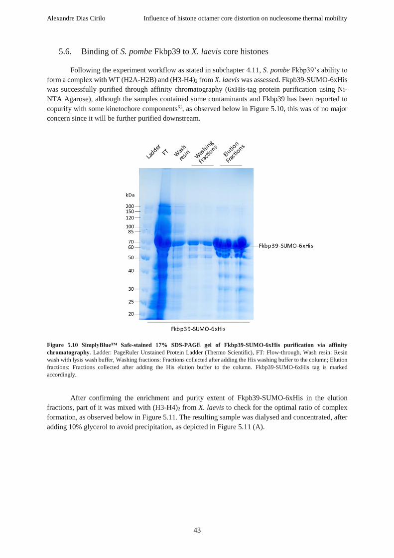
Alexandre Dias Cirilo Influence of histone octamer core distortion on nucleosome thermal mobility
43
5.6. Binding of S. pombe Fkbp39 to X. laevis core histones
Following the experiment workflow as stated in subchapter 4.11, S. pombe Fkbp39’s ability to
form a complex with WT (H2A-H2B) and (H3-H4)2 from X. laevis was assessed. Fkpb39-SUMO-6xHis
was successfully purified through affinity chromatography (6xHis-tag protein purification using Ni-
NTA Agarose), although the samples contained some contaminants and Fkbp39 has been reported to
copurify with some kinetochore components61, as observed below in Figure 5.10, this was of no major
concern since it will be further purified downstream.
Figure 5.10 SimplyBlue™ Safe-stained 17% SDS-PAGE gel of Fkbp39-SUMO-6xHis purification via affinity
chromatography. Ladder: PageRuler Unstained Protein Ladder (Thermo Scientific), FT: Flow-through, Wash resin: Resin
wash with lysis wash buffer, Washing fractions: Fractions collected after adding the His washing buffer to the column; Elution
fractions: Fractions collected after adding the His elution buffer to the column. Fkbp39-SUMO-6xHis tag is marked
accordingly.
After confirming the enrichment and purity extent of Fkpb39-SUMO-6xHis in the elution
fractions, part of it was mixed with (H3-H4)2 from X. laevis to check for the optimal ratio of complex
formation, as observed below in Figure 5.11. The resulting sample was dialysed and concentrated, after
adding 10% glycerol to avoid precipitation, as depicted in Figure 5.11 (A).

Alexandre Dias Cirilo Influence of histone octamer core distortion on nucleosome thermal mobility
44
The addition of PreScission enzyme to Fkbp39-SUMO-6xHis alone successfully allowed the
removal of the SUMO-6xHis-tag, as seen above in Figure 5.11 (B). After adding the aforementioned
enzyme, the sample was subdivided in three equal fractions which were then dialysed in three dialysis
buffers with varying salt concentration (150, 300, and 500 mM NaCl) to determine the optimal
conditions to avoid protein precipitation. From dialyses containing 150, and 300 mM NaCl, dialysed
samples were pulled and further purified trough ion exchange chromatography on an ÄKTA Protein
Purification System. Fractions “A6” to “B1” were analysed on a 17% SDS-PAGE gel and the most
enriched fractions (A8 and A9) were mixed in stochiometric amounts with WT (H3-H4)2 from the host
laboratory and run on a 17% SDS-PAGE gel as well, seen below in Figure 5.12 (A) and (B), respectively.
Figure 5.11 Coomassie-stained 17% SDS-PAGE gels of: (A) Fkbp39-SUMO-6xHis mixing with WT X. laevis (H3-H4)2; and
(B) Removal of the SUMO-6xHis tag from Fkbp39-SUMO-6xHis. Ladder: PageRuler Unstained Protein Ladder (Thermo
Scientific), 2:5 and 5:10 are two different ratios of Fkbp39-SUMO-6xHis:(H3-H4)2, FT: Flow-through after concentrating the
sample, Conc.: Concentrated sample. Fkbp39-SUMO-6xHis, Fkbp39, SUMO-6xHis tag, and X. laevis H3 and H4 are marked
accordingly.

Alexandre Dias Cirilo Influence of histone octamer core distortion on nucleosome thermal mobility
45
Figure 5.12 SimplyBlue™ Safe-stained 17% SDS-PAGE gels of: (A) Concentration of Fkbp39+(H3-H4)2 from X. laevis,
and Fkbp39 enriched fractions from IEX; (B) Checking optimal ratio of Fkbp39:(H3-H4)2 using A8 and A9 fractions
of IEX from (A) and X. laevis (H3-H4)2. Ladder: PageRuler Unstained Protein Ladder (Thermo Scientific), Before conc.:
Before concentrating the sample, FT: Concentration step flow-through, Conc.: Concentrated sample, IEX: Ion-exchange
chromatography. A6, A7, A8, A9, A10, A11 and B1 are IEX collection fractions. Fkbp39, the SUMO-6xHis tag, and the X.
laevis H3 and H4 are marked accordingly. Loaded sample volume for each sample, in µL, is noted above the respective well
in (B).
After Fkbp39 and (H3-H4) were appropriately mixed (i.e. in stoichiometric amounts), 7 M
guanidine hydrochloride was added and the ensuing solution was incubated and gently mixed at room
temperature for 1h to allow the refolding, followed by centrifugation at 15k rpm for 15 min. The solution
was then dialyzed in buffers with varying salt (NaCl) concentration and glycerol content. Dialysis in
Protein dialysis buffer 200 was not successful since a considerable amount of protein precipitated (see
Figure 5.13 A). Dialysis in buffers Protein dialysis buffer 200+1G, Protein dialysis buffer 200+5G,
Protein dialysis buffer 200+10G, Protein dialysis buffer 300, and Protein dialysis buffer 500 yielded a
good amount of complex (see Figure 5.13 B), avoiding any further losses. Noteworthily, one should
avoid having glycerol in the sample if EM analyses are to be executed downstream (generally, avoid
more than 1% glycerol in the sample). Samples which were dialyzed in buffers Protein dialysis buffer
300 and Protein dialysis buffer 200+1G were sent for cryo-EM analysis, and the remaining samples
were frozen in liquid nitrogen and stored at -80 °C.

Alexandre Dias Cirilo Influence of histone octamer core distortion on nucleosome thermal mobility
46
Figure 5.13 SimplyBlue™ Safe-stained 17% SDS-PAGE gels of Fkbp39+(H3-H4)2 and Fkbp39+(H2A-H2B): (A) Before
and after dialysis in 200 mM NaCl buffers (Protein dialysis buffer 200+10G and Protein dialysis buffer 200) of Fkbp39+(H3-
H4)2 from X. laevis; (B) Dialyzed Fkbp39+(H2A-H2B) and Fkbp39+(H3-H4)2 from X. laevis in different dialysis buffers.
Ladder: PageRuler Unstained Protein Ladder (Thermo Scientific), BD: Before dialysis; AD: After dialysis. Fkbp39 and the X.
laevis core histones (H2A, H2B, H3 and H4) are marked accordingly. Dialysis buffer salt (NaCl) molarity (in mM) and glycerol
content (in %) are marked accordingly.
Figure 5.14 Cryo-EM snapshot of Fkbp39:(H3-H4)2. (A) Aggregates seen in EM snapshot. An example is pointed by the
red arrow. (B) Zoom over the pointed area in (A). Aggregates are clearly present in the sample.
Alexandre Dias Cirilo Influence of histone octamer core distortion on nucleosome thermal mobility
47
Cryo-EM analyses showed that aggregation occurred, as observed in Figure 5.14. Further
purifications are likely to be needed to avoid aggregate formation and allow the isolation and observation
of the putative complexes. The area marked by the red arrow in Figure 5.14 A is zoomed in and shown
in Figure 5.14 B to clearly show aggregate presence in the sample. Complementary cryo-EM snapshots
showing the aggregates are depicted in Figure 5.15 for Fkbp39 alone, and complexed with either (H2A-
H2B) or with (H3-H4)2.

Alexandre Dias Cirilo Influence of histone octamer core distortion on nucleosome thermal mobility
48
Figure 5.15 Cryo-EM snapshots of Fkbp39, Fkbp39:(H2A-H2B) and Fkbp39:(H3-H4)2.
Aggregates can be seen in all snapshots. (B) is a small area in (A) zoomed in, as marked by the red arrow.
A large aggregate can be observed within the red circle in (B).

Alexandre Dias Cirilo Influence of histone octamer core distortion on nucleosome thermal mobility
49
5.7. Binding affinity of chromatin-binding proteins to WT X. laevis nucleosomes
Although alternative experimental protocols exist, like the one described in ref. 73 which takes
advantage of chromatin immunoprecipitation using H3-specific antibodies, we decided to take a simpler
approach to observe putative interactions between WT X. laevis nucleosomes containing the Widom
601 sequence as nucleosomal DNA and chromatin-binding proteins provided by the host laboratory
simply by resorting to gel mobility shift assays. The chromatin-binding proteins screened in this project
are listed in Table 5.3.
Table 5.3 Chromatin-binding proteins checked for putative interactions with WT X. laevis nucleosomes.
Chromatin-
binding
protein
Gene Full protein name Species Function(s) References
Alc1 CHDL1
Chromodomain-
helicase-DNA-
binding protein 1-like
(alternative name:
amplified in liver
cancer protein 1)
H. sapiens
• Catalyses nucleosome
sliding in an ATP-dependent
manner;
• Nucleotide binding;
• Helicase activity is strongly
stimulated upon poly(ADP-
ribose)-binding.
74
Alf GTF2A1L TFIIA-alpha and
beta-like factor H. sapiens
• Involved in DNA binding;
• Transcription coactivator
activity.
75
Hpf1 HPF1 Histone PARylation
factor 1 H. sapiens
• Interacts with Parp1 and
Parp2;
• Able to change amino acid
specificity toward serine;
• Promotes histone serine
ADP-ribosylation;
• Involved in histone binding.
76,77
Hpf1 HPF1 Histone PARylation
factor 1 D. melanogaster
Parp1 PARP1 Poly [ADP-ribose]
polymerase 1 H. sapiens • Mediates the poly(ADP-
ribosyl)ation of histones in a
HPF1-dependent manner;
• Histone deacetylase binding.
77
Parp1 PARP1 Poly [ADP-ribose]
polymerase 1 D. melanogaster
Parp2 PARP2 Poly [ADP-ribose]
polymerase 2 H. sapiens
• Mediates serine ADP-
ribosylation of target proteins
following interaction with
HPF1 (HPF1 conferring
serine specificity).
76,78
Different conditions were tested to check for any shifts in the gel mobility shift assays by varying
chromatin-binding protein amount, incubation temperature, incubation time, glycerol content, and using
two different nucleosomal DNAs (short and long version of the Widom 601 sequence – 149 and 227 bp,
respectively – prepared in this project). Furthermore, to exclude that the observed shifts were a
consequence of binding between the chromatin-binding protein and DNA, an assay was executed by
incubating chromatin-binding proteins with DNA only, excluding the nucleosome. To that intent, DNA
was diluted accordingly to have the same concentration as in the nucleosome samples.

Alexandre Dias Cirilo Influence of histone octamer core distortion on nucleosome thermal mobility
50
Figure 5.16 Effect of incubation time and temperature on binding efficiency of different chromatin-binding proteins. ∅: nucleosome only; hs: Homo sapiens; dm: Drosophila melanogaster.
As observed in Figure 5.16, lowering the incubation temperature, even with the increased
incubation time, potentially leads to a less stable complex, since we can observe a considerable increase
in aggregates. From the Figure 5.16, we can appreciate the shifts pointed out by the red arrows,
indicating that H. sapiens Hpf1, Parp2 and Parp1, and D. melanogaster Parp1 might be interacting with
the nucleosome. It would be interesting to see if synergetic effects exist between H. sapiens Hpf1 and
Parp1, and Hfp1 and Parp2.

Alexandre Dias Cirilo Influence of histone octamer core distortion on nucleosome thermal mobility
51
Figure 5.17 Effect of glycerol on binding efficiency of different chromatin-binding proteins.
∅: nucleosome only; hs: Homo sapiens; dm: Drosophila melanogaster.
Adding glycerol to the reaction slightly increased complex stability like depicted in Figure 5.17.
Glycerol was added to avoid possible precipitation of the chromatin-binding proteins.
Figure 5.18 Investigating interactions between the Widom 601 DNA sequence and the chromatin-binding proteins. ∅: DNA only; hs: Homo sapiens; dm: Drosophila melanogaster.
By analysing Figure 5.18, we can assert that Parp1 and Parp2 from H. sapiens can hypothetically
bind to the Widom 601 sequence. Moreover, D. melanogaster Parp1 can also recognize the
aforementioned sequence. H. sapiens Alc1, Hpf1, and D. melanogaster Hpf1 do not seem to bind
nucleosomal DNA alone.

Alexandre Dias Cirilo Influence of histone octamer core distortion on nucleosome thermal mobility
52
Figure 5.19 Effect of WT X. laevis nucleosomes with two different nucleosomal DNA sequences – long 601 (227 bp) and
shorter 601 (149 bp) – on binding efficiency of H. sapiens Alc1 and Alf1. ∅: nucleosome only; hs: Homo sapiens; CBP:
chromatin-binding protein.
A first look at Figure 5.19 leads to the assumption that shifts are only significant at higher
amounts of chromatin-binding proteins, namely, at 2 µL and 5 µL. It is also apparent that H. sapiens
Alc1 interacts with both nucleosomes, i.e. with shorter and longer 601 sequences. In regard to H. sapiens
Alf, it seems as though it only interacts with the nucleosome which harbours the longest version of
nucleosomal DNA (227 bp instead of 149 bp), as marked by the red arrow. This is a clear observation
that this protein interacts with the nucleosome. It might be possible that the overhangs of the extended
version of the nucleosomal DNA are required to allow proper binding of Alf to the nucleosome. Alf
seems like the best candidate for further structural studies aiming at observing its binding affinity to
chromatin, and possibly, understanding the binding mechanism.
Taken together, these native gel shift assays suggest that H. sapiens Hpf1, Parp2 and Alf are the
best candidates for further structural studies aiming at defining their binding affinity the nucleosome. A
good experimental technique that could be used to do so would be the ChIP-based histone association
assay described in ref. 73 using H3-specific antibodies to precipitate chromatin from crosslinked whole
cell extracts. Co-precipitated proteins with chromatin can be detected downstream by SDS-PAGE or
Western blot analysis73.

Alexandre Dias Cirilo Influence of histone octamer core distortion on nucleosome thermal mobility
53
6. Discussion
The identification of the nucleosome, fundamental repeating subunit of chromatin, four decades
ago79 propelled chromatin research to the spotlight. With the advent of modern technologies, highly
reported protocols and the availability of near-atomic resolution structures, our understanding of the
nucleosomes continues to grow. However, many facets remain blurred and require further investigation
to clarify the roles taken by the nucleosome in vivo. In this work, we focused on exploring the influence
of histone octamer core distortion on nucleosome thermal mobility, the putative formation and cryo-EM
analysis of Fkbp39:(H2A-H2B) and Fkbp39:(H3-H4)2 complexes, as well as the screening of multiple
chromatin-binding proteins binding affinity to the nucleosome.
6.1. Influence of histone octamer core distortion on nucleosome thermal mobility
Pioneering studies achieved in 1978 by Beard79 using simian virus 40 minichromosomes were
the first evidence of nucleosome mobility over DNA. Later, work by Flaus and Owen-Hughes 42 went
further ahead and fully characterized the influence of temperature and the role of ATP on nucleosome
mobility, using mouse mammary tumour virus promoter DNA as the nucleosomal DNA template. Their
observations, as well as previous studies in the literature68–70, led to the postulation that nucleosomes
with short DNA fragments tend to migrate towards DNA ends during thermal-induced mobilization.
This observation had obvious repercussions on gene expression, leading to the hypothesis that it would
serve as an additional layer of gene expression regulation mechanism by modulating the ability of
different factors to bind to chromatin.
In this work, WT and mutant nucleosome species were prepared to observe the effect of
temperature on nucleosome mobility as well as the influence of the designed mutations. Therefore,
several X. laevis histones (WT and mutant) were firstly purified after heterologously expressing them in
E. coli BL21 (DE3) Rosetta strain. Core histones H2A and H2B, and core histones H3 and H4 were co-
expressed. Co-expression of histones is very practical and reduces time-consuming individual histone
purification protocols, producing soluble complexes that can be readily purified downstream80, e.g. via
affinity chromatography. With regard to nucleosomal DNA, an extended version (227 bp) of the
previously reported strong-positioning Widom 601 sequence39 was synthesized in high amounts via PCR
and then purified by EtOH precipitation. Nucleosome core particles were assembled using an adapted
protocol based on the ones stated in refs. 54–56 (workflow shown in Figure 1.10).
Firstly, our thermal shift assays at 62 ºC (with incubation times of 5, 15, 30 and 45 minutes)
using WT nucleosomes corroborate the previously reported thermally-driven nucleosome mobility of
WT species (see
Figure 5.7). We observe the same tendency to migrate towards the DNA extremities when
incubated at higher temperatures. Hence, we wanted to access the effect of the designed mutations in
the mutant nucleosome species over its mobility at high temperatures. Our H4_V43C H3_F104C mutant
nucleosome (checked through sequencing) shows the typical mobility pattern at 60 ºC (shifting of the
nucleosome towards the DNA ends, i.e. end-positioned nucleosome). In the case of our mutant, two
different conditions were tested: with DTT and without DTT. As expected, shifting towards the
extremities was more marked when nucleosomes were incubated in reducing conditions (with DTT) as
seen by the presence of stronger bands below the “c-NCP” band, corresponding to the different end-
positioned nucleosomes (“ep-NCP”). DTT, being a reducing agent, will reduce disulphide bonds, and

Alexandre Dias Cirilo Influence of histone octamer core distortion on nucleosome thermal mobility
54
naturally, disulphide bridges present between two cysteine residues will be dismantled. One would then
expect a higher degree of mobility when incubated at high temperatures for some time as the result of
weakened intramolecular and intermolecular forces, as observed in this case. Interestingly, without DTT,
although some shifting is still observable, it is clearly not as marked as in comparison with nucleosomes
incubated in reducing conditions (with DTT). And it seems like longer incubation times do not affect
shifting to a greater extent in both contexts. This suggests that these mutations (H4_V43C, H3_F104C)
significantly affect nucleosome structural integrity, most possibly by strengthening the overall structure
through the increasing of the intermolecular forces most possibly through the establishment of a
disulphide bridge at the interface between L1 (loop 1) of core histone H4 and 2 (-helix 2) of core
histone H3, hence hindering its thermally-driven mobility. This suggests that histone octamer plasticity
is vital for nucleosome to slide along DNA in a noncatalyzed manner.
Chemical crosslinking is a very fit experimental method for protein-protein interaction
identification. In theory, the crosslinking agent leads to the formation of covalent bonds between two
proteins by reacting with the functional groups of the protein. In the cellular context, many transient
interactions between proteins occur. In vitro, when working with purified proteins, this technique is
most useful for validating the presence of protein-protein interactions. Not only can crosslinking be used
to show that two proteins are in close proximity to each other and form interactions, but it can also give
insights on which regions the contacts occur81. Crosslinking of WT X. laevis nucleosomes with 0.1%
(v/v) glutaraldehyde was successful and led to the formation of stable crosslinked nucleosomes with no
observable intermediate nucleosome states and very minimal end-positioned (e-NCP) nucleosomes in
comparison to centred nucleosomes (c-NCP, see Figure 5.9), suggesting that crosslinked histone
octamers are immobilized on DNA, thus indicating that the structural changes in the histone octamer
move the underlying DNA82. Increasing incubation time had no effect on nucleosome mobility since no
variation in end-positioned or intermediate state nucleosomes was observed. Incubation temperature was
set at 55 ºC since it is sufficient to observe nucleosome mobility as seen in the case of non-crosslinked
nucleosomes in Figure 5.9.
The intrinsic plasticity of the histone octamer allows the uncatalyzed DNA translocation and is
a prerequisite for multiple chromatin remodelling complexes. Apart from DNA translocation and
nucleosome flexibility, structural rearrangements of the histone octamer likely influence the stability of
the different conformational states of the nucleosome, which might in turn be used by chromatin
remodelers at their advantage to execute their function(s).

Alexandre Dias Cirilo Influence of histone octamer core distortion on nucleosome thermal mobility
55
6.2. Fkbp39 structural studies
FK506-binding protein 39 kDa, abbreviated “Fkbp39” (and alternatively, “Ani1”), is a PPiase
with a conserved NPL domain that acts as a histone chaperone. This protein is involved in proline
isomerization on core histone H3 N-terminal tail affecting H3 methylation, thus having a role in gene
expression regulation20,58.
In this work, we aimed at purifying S. pombe Fkbp39 for downstream cryo-EM analysis.
Furthermore, we also tried to check for complex formation with (H2A-H2B) and (H3-H4)2, and to
observe the putative complexes under cryo-EM at the Max Planck Institute of Biochemistry (Munich,
Germany).
The purification step of Fkbp39 required a SUMO tag to increase solubility and stability, as well
as a 6xHis tag to allow for affinity chromatography purification. The SUMO-6xHis was successfully
cut out with the addition of in house PreScission enzyme. Initially, purification of Fkbp39:(H3-H4)2 was
not successful since it only yielded significant amounts in buffers with more than 1 % glycerol, which
restricted analysis by cryo-EM. Hence, different dialysis buffers were prepared with varying salt (200,
300 and 500 mM NaCl) molarities and different glycerol content (0, 1, 5 and 10 %), which resulted in
high yields of the putative Fkbp39:(H2A-H2B) and Fkbp39:(H3-H4)2 complexes. The best-looking
samples (dialyzed in buffers Protein dialysis buffer 300 and Protein dialysis buffer 200+1G) were sent
for cryo-EM analysis. Unfortunately, isolation and visualization of either Fkbp39 alone or Fkbp39
complexed with core histones (H2A-H2B) or with (H3-H4)2 was unsuccessful due to aggregate
formation. Further optimization strategies are likely to be required to acquire more stable assemblies.
Fkbp39’s intrinsically disordered regions might be preventing the formation of stable complexes with
core histones.

Alexandre Dias Cirilo Influence of histone octamer core distortion on nucleosome thermal mobility
56
6.3. Chromatin-binding proteins screening
To check for binding of the multiple putative chromatin-binding proteins (listed under Table
5.3) to WT X. laevis nucleosomes, we resorted to native gel shift assays – a “rapid and sensitive method
to detect protein-nucleic acid interactions”83.
Prior to running the electrophoreses, samples containing WT X. laevis nucleosomes mixed with
the chromatin-binding protein(s) were incubated at room temperature for 30 min, or at 4 ºC for 1h.
Furthermore, glycerol content was also varied. Both these settings were used to check for the optimal
reaction conditions to allow proper visualization of shifts, if occurring. Increasing incubation time from
30 min to 1h and reducing incubation temperature from room temperature to 4 ºC mostly led to aggregate
formation (see Figure 5.16). Adding 5% glycerol, on the other hand, led to stronger shift bands (marked
in by red arrows in Figure 5.17), possibly by decreasing the formation of aggregates.
Looking at Figure 5.17, it is clear that Hpf1 (H. sapiens), Parp1 (H. sapiens) and Parp2 (H.
sapiens) interact with the nucleosome. To exclude the possibility of interaction of these proteins with
the Widom 601 sequence alone (as free nucleosomal DNA), an additional native gel shift assay was
done incubating the tested proteins with DNA alone (see Figure 5.18). Interestingly, Parp1 (H. sapiens)
and Parp2 (H. sapiens) seem to bind to DNA alone, as well as Parp1 (D. melanogaster). Further
experiments are required to fully disclose the binding affinities to either free nucleosomal DNA, or the
nucleosome itself.
A first glance at Figure 5.16 provides a snapshot of the binding affinity of the different tested
proteins to the nucleosome. Different combinations of proteins were tested and screened for binding
with the nucleosome in this assay: one chromatin-binding protein alone or a combination of proteins
with varying amounts. As indicated by the red arrows, Hpf1 (H. sapiens) and Parp1 (D. melanogaster)
look like good candidates for further structural studies aiming at their affinity to chromatin. Additionally,
combining Hpf1 (H. sapiens) with increasing amounts of Parp2 or Parp1 (H. sapiens) further increases
shifting, though this experiment alone does not provide enough information, i.e. the interactions between
the aforementioned proteins would have to be checked first, and then synergetic effects might also be
worth considering.
Taking a closer look at Alc1 (H. sapiens) and Alf (H. sapiens) in Figure 5.19 (gel with 5 µL of
chromatin-binding protein, on the far right), using native gel shift assays with the short (149 bp
nucleosomal DNA) and long (227 bp nucleosomal DNA) nucleosomes, Alc1 quite strikingly interacts
with both the long and short nucleosomes and Alf interacts with the long nucleosome only.
Further assays will be required to unambiguously prove the binding of the best chromatin-
binding candidates observed in this project (i.e. H. sapiens Hpf1, Parp2 and Alf), notwithstanding that
these experiments served as a good means to get a glimpse at which protein might be the best contender
to study its interactions with the nucleosome.

Alexandre Dias Cirilo Influence of histone octamer core distortion on nucleosome thermal mobility
57
7. Conclusion
The main aim of this project was to observe the influence of histone octamer core distortion on
nucleosome thermal mobility. Here, we corroborate the shifting of WT X. laevis nucleosomes towards
the extremities (end-positioned) in regards to the nucleosomal DNA (using a 227 bp extended version
of the strong positioning Widom 601 sequence) when incubated at high temperatures (62 ºC)42.
Moreover, this typical shifting pattern of nucleosomes is also reflected in the double mutant nucleosome
(H4_V43C H3_F104C) assembled in this work (incubated at 60 ºC). Interestingly, adding reducing
conditions (via the addition of DTT) when incubating the double mutant yielded a high amount of
intermediate nucleosomal states, conversely to the assay without DTT. Taken together, these
observations suggest that these mutations (H4_V43C and H3_F104C) strengthen the overall nucleosome
structure at the interface between L1 (loop 1) of core histone H4 and 2 (-helix 2) of core histone H3,
hence affecting its thermally-driven mobility by limiting nucleosome sliding along nucleosomal DNA
and suggesting that histone octamer plasticity is a prerequisite for nucleosome to move along DNA in a
noncatalyzed fashion.
Notwithstanding that purification of Fkbp39 (S. Pombe) was successful, Fkbp39:(H2A-H2B)
and Fkbp39:(H3-H4)2 putative complexes analyses by cryo-EM failed due to aggregate formation.
Further protocol optimization strategies are likely to improve complex formation conditions and might
stabilize the putative complexes, thus allowing for downstream structural analysis.
Lastly, the screening of the putative chromatin-binding proteins binding to WT X. laevis
nucleosomes suggested that Alc1, Alf, Hpf1, Parp1 and Parp2 (H. sapiens) might interact with the
nucleosome. Most intriguingly, Hpf1, Parp2 and Alf (H. sapiens) seem like the best candidates for
further studies focused on studying their ability to bind to nucleosomes. A good approach to do so would
be to use the ChIP-based histone association assay described in ref. 73 which uses H3-specific antibodies
to precipitate chromatin from crosslinked whole cell extracts. Crosslinking causes the co-precipitation
of chromatin with the bound proteins which can then be detected by SDS-PAGE or Western blot
analysis73.

Alexandre Dias Cirilo Influence of histone octamer core distortion on nucleosome thermal mobility
58
8. References
1. Kornberg, R. D. & Lorch, Y. Twenty-five years of the nucleosome, fundamental particle of the
eukaryote chromosome. Cell (1999). doi:10.1016/S0092-8674(00)81958-3
2. Alva, V., Ammelburg, M., Söding, J. & Lupas, A. N. On the origin of the histone fold. BMC
Struct. Biol. (2007). doi:10.1186/1472-6807-7-17
3. Cosgrove, M. S., Boeke, J. D. & Wolberger, C. Regulated nucleosome mobility and the histone
code. Nature Structural and Molecular Biology 11, 1037–1043 (Nature Publishing Group,
2004).
4. Workman, J. L. & Abmayr, S. M. Fundamentals of chromatin. Fundamentals of Chromatin
(2014). doi:10.1007/978-1-4614-8624-4
5. Li, B., Carey, M. & Workman, J. L. The Role of Chromatin during Transcription. Cell 128,
707–719 (2007).
6. Richmond, T. J., Finch, J. T., Rushton, B., Rhodes, D. & Klug, A. Structure of the nucleosome
core particle at 7 resolution. Nature (1984). doi:10.1038/311532a0
7. Luger, K., Mäder, A. W., Richmond, R. K., Sargent, D. F. & Richmond, T. J. Crystal structure
of the nucleosome core particle at 2.8 Å resolution. Nature (1997). doi:10.1038/38444
8. Davey, C. A., Sargent, D. F., Luger, K., Maeder, A. W. & Richmond, T. J. Solvent mediated
interactions in the structure of the nucleosome core particle at 1.9 Å resolution. J. Mol. Biol.
(2002). doi:10.1016/S0022-2836(02)00386-8
9. Bartke, T. et al. Nucleosome-interacting proteins regulated by DNA and histone methylation.
Cell 143, 470–484 (2010).
10. Annunziato, A. DNA Packaging: Nucleosomes and Chromatin. Nat. Educ. 1, 1 (2008).
11. Ramakrishnan, V. Histone structure and the organization of the nucleosome. Annu. Rev.
Biophys. Biomol. Struct. 26, 83–112 (1997).
12. Edlich-Muth, C. et al. The pentameric nucleoplasmin fold is present in Drosophila FKBP39
and a large number of chromatin-related proteins. J. Mol. Biol. 427, 1949–1963 (2015).
13. Gurard-Levin, Z. A., Quivy, J.-P. & Almouzni, G. Histone Chaperones: Assisting Histone
Traffic and Nucleosome Dynamics. Annu. Rev. Biochem. 83, 487–517 (2014).
14. PomBase - Gene - ani1 (SPBC1347.02) - CENP-A amino terminus domain (NTD) isomerase
Ani1. Available at: https://www.pombase.org/gene/SPBC1347.02. (Accessed: 20th March
2018)
15. Wood, V. et al. PomBase: A comprehensive online resource for fission yeast. Nucleic Acids
Res. 40, (2012).
16. Matsuyama, A. et al. ORFeome cloning and global analysis of protein localization in the
fission yeast Schizosaccharomyces pombe. Nat. Biotechnol. 24, 841–7 (2006).
17. Koztowska, M. et al. Nucleoplasmin-like domain of FKBP39 from Drosophila melanogaster
forms a tetramer with partly disordered tentacle-like C-terminal segments. Sci. Rep. 7, 1–14
(2017).
18. Pfam: Protein: FKBP4_SCHPO (O74191). Available at: http://pfam.xfam.org/protein/O74191.
(Accessed: 20th March 2018)
19. FPR3 / YML074C Overview. Available at: https://www.yeastgenome.org/locus/S000004539.
(Accessed: 20th March 2018)
20. O74191 (FKBP4_SCHPO). Available at: http://www.uniprot.org/uniprot/O74191. (Accessed:
20th March 2018)
21. Bateman, A. et al. UniProt: the universal protein knowledgebase. Nucleic Acids Res. 45, D158–
D169 (2017).
22. Park, S.-K., Xiao, H. & Lei, M. Nuclear FKBPs, Fpr3 and Fpr4 affect genome-wide genes
transcription. Mol. Genet. Genomics 289, 125–136 (2014).
23. Ramos, I. et al. The intrinsically disordered distal face of nucleoplasmin recognizes distinct
oligomerization states of histones. Nucleic Acids Res. 42, 1311–25 (2014).
24. Hondele, M. & Ladurner, A. G. The chaperone-histone partnership: For the greater good of
histone traffic and chromatin plasticity. Curr. Opin. Struct. Biol. 21, 698–708 (2011).

Alexandre Dias Cirilo Influence of histone octamer core distortion on nucleosome thermal mobility
59
25. Luger, K. Structure and dynamic behavior of nucleosomes. Curr. Opin. Genet. Dev. 13, 127–
135 (2003).
26. Cosgrove, M. S. & Wolberger, C. How does the histone code work? Biochem. Cell Biol. 83,
468–476 (2005).
27. De La Cruz, X., Lois, S., Sánchez-Molina, S. & Martínez-Balbás, M. A. Do protein motifs read
the histone code? BioEssays 27, 164–175 (2005).
28. Kouzarides, T. Chromatin Modifications and Their Function. Cell 128, 693–705 (2007).
29. Bowman, G. D. & Poirier, M. G. Post-translational modifications of histones that influence
nucleosome dynamics. Chemical Reviews 115, 2274–2295 (2015).
30. Zentner, G. E. & Henikoff, S. Regulation of nucleosome dynamics by histone modifications.
Nature Structural and Molecular Biology 20, 259–266 (2013).
31. Thomas Jenuwein, C. D. A. Translating the Histone Code. (2001).
32. Henikoff, S. & Shilatifard, A. Histone modification: Cause or cog? Trends in Genetics 27,
389–396 (2011).
33. Cui, L. & Miao, J. Chromatin-Mediated epigenetic regulation in the malaria parasite
Plasmodium falciparum. Eukaryotic Cell 9, 1138–1149 (2010).
34. Lee, J., Smith, E. & Shilatifard, A. The Language of Histone Crosstalk Jung-Shin. 142, 682–
685 (2013).
35. Sinha, K. K., Gross, J. D. & Narlikar, G. J. Distortion of histone octamer core promotes
nucleosome mobilization by a chromatin remodeler. Science (80-. ). (2017).
doi:10.1126/science.aaa3761
36. Shi, Y. et al. Histone demethylation mediated by the nuclear amine oxidase homolog LSD1.
Cell 119, 941–953 (2004).
37. Sun, Z. W. & Allis, C. D. Ubiquitination of histone H2B regulates H3 methylation and gene
silencing in yeast. Nature 418, 104–108 (2002).
38. Bowman, G. D. Mechanisms of ATP-dependent nucleosome sliding. Curr. Opin. Struct. Biol.
20, 73–81 (2010).
39. Lowary, P. T. & Widom, J. New DNA sequence rules for high affinity binding to histone
octamer and sequence-directed nucleosome positioning. J. Mol. Biol. 276, 19–42 (1998).
40. Frouws, T. D., Duda, S. C. & Richmond, T. J. X-ray structure of the MMTV-A nucleosome
core. Proc. Natl. Acad. Sci. 113, 1214–1219 (2016).
41. Andrews, A. J. & Luger, K. Nucleosome Structure(s) and Stability: Variations on a Theme.
Annu. Rev. Biophys. 40, 99–117 (2011).
42. Flaus, A. & Owen-Hughes, T. Dynamic Properties of Nucleosomes during Thermal and ATP-
Driven Mobilization. Mol. Cell. Cell Biol. 23, 7767–7779 (2003).
43. Lusser, A. & Kadonaga, J. T. Strategies for the reconstitution of chromatin. Nat. Methods 1,
19–26 (2004).
44. Ito, T., Tyler, J. K. & Kadonaga, J. T. Chromatin assembly factors: a dual function in
nucleosome formation and mobilization? Genes Cells 2, 593–600 (1997).
45. Tatchell, K. & Van Holde, K. E. Nucleosome Reconstitution: Effect of DNA Length on
Nucleosome Structure. Biochemistry 18, 2871–2880 (1979).
46. Ruiz-Carrillo, A. & Jorcano, J. L. An Octamer of Core Histones in Solution: Central Role of
the H3·H4 Tetramer in the Self-Assembly1. Biochemistry 18, 760–768 (1979).
47. Kornberg, R. D. & Thonmas, J. O. Chromatin Structure: Oligomers of the Histones. Science
(80-. ). 184, 865–868 (1974).
48. Simon, R. H. & Felsenfeld, G. A new procedure for purifying histone pairs H2A+H2B and
H3+H4 from chromatin using hydroxylapatite. Nucleic Acids Res. 6, 689–696 (1979).
49. Khuong, M. T., Fei, J., Cruz-Becerra, G. & Kadonaga, J. T. A simple and versatile system for
the ATP-dependent assembly of chromatin. J. Biol. Chem. 292, 19478–19490 (2017).
50. Rhodes, D. & Laskey, R. A. Assembly of nucleosomes and chromatin in Vitro. Methods
Enzymol. 170, 575–585 (1989).
51. Huynh, V. A. T., Robinson, P. J. J. & Rhodes, D. A method for the in vitro reconstitution of a
defined ‘30 nm’ chromatin fibre containing stoichiometric amounts of the linker histone. J.
Mol. Biol. 345, 957–968 (2005).
52. Tanaka, Y. et al. Expression and purification of recombinant human histones. Methods 33, 3–

Alexandre Dias Cirilo Influence of histone octamer core distortion on nucleosome thermal mobility
60
11 (2004).
53. Luger, K., Rechsteiner, T. J. & Richmond, T. J. Preparation of nucleosome core particle from
recombinant histones. Methods Enzymol. 304, 3–19 (1999).
54. Ivić, N., Groschup, B., Bilokapić, S. & Halić, M. Simplified Method for Rapid Purification of
Soluble Histones. Croat. Chem. Acta 89, 1–10 (2016).
55. Rogge, R. A. et al. Assembly of Nucleosomal Arrays from Recombinant Core Histones and
Nucleosome Positioning DNA. J. Vis. Exp. 1–10 (2013). doi:10.3791/50354
56. Luger. Methods for Reconstitution of Nucleosome Core Particles from Recombinant Histones
and DNA. 375, 23–44 (2004).
57. Klinker, H., Haas, C., Harrer, N., Becker, P. B. & Mueller-Planitz, F. Rapid purification of
recombinant histones. PLoS One (2014). doi:10.1371/journal.pone.0104029
58. Kuzuhara, T. & Horikoshi, M. A nuclear FK506-binding protein is a histone chaperone
regulating rDNA silencing. Nat. Struct. Mol. Biol. (2004). doi:10.1038/nsmb733
59. Thompson, R. F., Walker, M., Siebert, C. A., Muench, S. P. & Ranson, N. A. An introduction
to sample preparation and imaging by cryo-electron microscopy for structural biology. Methods
100, 3–15 (2016).
60. 4X SDS-PAGE loading buffer. Cold Spring Harb. Protoc. 2006, pdb.rec10588-pdb.rec10588
(2006).
61. Przewloka, M. R. et al. Molecular analysis of core kinetochore composition and assembly in
Drosophila melanogaster. PLoS One 2, (2007).
62. Fyodorov, D. V. & Kadonaga, J. T. Dynamics of ATP-dependent chromatin assembly by ACF.
Nature 418, 896–900 (2002).
63. Vasudevan, D., Chua, E. Y. D. & Davey, C. A. Crystal Structures of Nucleosome Core
Particles Containing the ‘601’ Strong Positioning Sequence. J. Mol. Biol. 403, 1–10 (2010).
64. Isaac, R. S. et al. Nucleosome breathing and remodeling constrain CRISPR-Cas9 function.
Elife 5, (2016).
65. Bertin, A., Mangenot, S., Renouard, M., Durand, D. & Livolant, F. Structure and phase
diagram of nucleosome core particles aggregated by multivalent cations. Biophys. J. 93, 3652–
3663 (2007).
66. De Frutos, M., Raspaud, E., Leforestier, A. & Livolant, F. Aggregation of nucleosomes by
divalent cations. Biophys. J. 81, 1127–1132 (2001).
67. Allahverdi, A., Chen, Q., Korolev, N. & Nordenskiöld, L. Chromatin compaction under mixed
salt conditions: Opposite effects of sodium and potassium ions on nucleosome array folding.
Sci. Rep. 5, (2015).
68. Meersseman, G., Pennings, S. & Bradbury, E. M. Mobile nucleosomes - a general behavior.
EMBO J 11, 2951–2959 (1992).
69. Flaus, A. & Richmond, T. J. Positioning and stability of nucleosomes on MMTV 3’LTR
sequences. J. Mol. Biol. 275, 427–441 (1998).
70. Sakaue, T., Yoshikawa, K., Yoshimura, S. H. & Takeyasu, K. Histone core slips along DNA
and prefers positioning at the chain end. Phys. Rev. Lett. 87, 78105-1-78105–4 (2001).
71. Kuykendall, J. R. & Bogdanffy, M. S. Efficiency of DNA-histone crosslinking induced by
saturated and unsaturated aldehydes in vitro. Mutat. Res. Lett. 283, 131–136 (1992).
72. Czarnota, G. J. & Ottensmeyer, F. P. Structural states of the nucleosome. J. Biol. Chem. 271,
3677–3683 (1996).
73. Ricke, R. M. & Bielinsky, A. K. Easy detection of chromatin binding proteins by the histone
association assay. Biol. Proced. Online 7, 60–69 (2005).
74. Ahel, D. et al. Poly(ADP-ribose)-dependent regulation of DNA repair by the chromatin
remodeling enzyme ALC1. Science (80-. ). (2009). doi:10.1126/science.1177321
75. Upadhyaya, A. B., Lee, S. H. & Dejong, J. Identification of a general transcription factor
TFIIAalpha/beta homolog selectively expressed in testis. J. Biol. Chem. (1999).
doi:10.1074/jbc.274.25.18040
76. Bonfiglio, J. J. et al. Serine ADP-Ribosylation Depends on HPF1. Mol. Cell (2017).
doi:10.1016/j.molcel.2017.01.003
77. Gibbs-Seymour, I., Fontana, P., Rack, J. G. M. & Ahel, I. HPF1/C4orf27 Is a PARP-1-
Interacting Protein that Regulates PARP-1 ADP-Ribosylation Activity. Mol. Cell (2016).

Alexandre Dias Cirilo Influence of histone octamer core distortion on nucleosome thermal mobility
61
doi:10.1016/j.molcel.2016.03.008
78. Amé, J. C. et al. PARP-2, a novel mammalian DNA damage-dependent poly(ADP-ribose)
polymerase. J. Biol. Chem. (1999). doi:10.1074/jbc.274.25.17860
79. Beard, P. Mobility of histones on the chromosome of simian virus 40. Cell (1978).
doi:10.1016/0092-8674(78)90279-9
80. Anderson, M. et al. Co-expression as a convenient method for the production and purification
of core histones in bacteria. Protein Expr. Purif. (2010). doi:10.1016/j.pep.2010.03.013
81. Kapoor, M. How to crosslink proteins. Cell. Mol. Microb. Biol. Div. … 1–6 (1996).
82. Bilokapic, S., Strauss, M. & Halic, M. Structural rearrangements of the histone octamer
translocate DNA. Nat. Commun. (2018). doi:10.1038/s41467-018-03677-z
83. Hellman, L. M. & Fried, M. G. Electrophoretic mobility shift assay (EMSA) for detecting
protein-nucleic acid interactions. Nat. Protoc. (2007). doi:10.1038/nprot.2007.249
84. Pettersen, E. F. et al. UCSF Chimera - A visualization system for exploratory research and
analysis. J. Comput. Chem. 25, 1605–1612 (2004).
85. Waterhouse, A. M., Procter, J. B., Martin, D. M. A., Clamp, M. & Barton, G. J. Jalview
Version 2-A multiple sequence alignment editor and analysis workbench. Bioinformatics 25,
1189–1191 (2009).

Alexandre Dias Cirilo Influence of histone octamer core distortion on nucleosome thermal mobility
62
9. Appendix
Figure 9.1 Multiple sequence alignment of a histone H3 cluster in eukaryotes. The cluster (cluster ID: UniRef90_P08898) was
obtained through UniProt database, using manually refined entries only (Swiss-Prot). (A) Visualization of the alignment was
performed via Jalview 2.10.3b1 using the Clustal color scheme (blue: hydrophobic, red: positive charge, magenta: negative charge,
green: polar, pink: cysteines, orange: glycines, yellow: prolines, cyan: aromatic, white: unconserved). Conservation, quality, consensus
and occupancy bar plots indicate the extent of: conservation, likelihood of observing mutations, percentage of the modal residue and
ungapped positions for each amino acid residue (i.e. per column), respectively. (B) The phylogenetic tree was built using the neighbor
joining method with the BLOSUM62 matrix on Jalview 2.10.3b185.

Alexandre Dias Cirilo Influence of histone octamer core distortion on nucleosome thermal mobility
63
Figure 9.2 PCR amplification schematic of the long version of the strong position Widom
601 sequence (227 bp) using 797F and 797R primers with #298 plasmid template.
Canonical Widom 601 sequence shown in blue. Long Widom 601 sequence (227 bp) shown
in orange. In house plasmid sequence #298 sequence is undisclosed. Representation using
AngularPlasmid (http://angularplasmid.vixis.com/).


![territorium 22, 2015, 77-86 · B = 0,251 com a classe B [4 o-10 ], considerando-se que o grau de afinidade µ varia entre 0,0 e 1,0 (fig. 3). Fig. 3 – Aplicação da função fuzzy](https://img.pdfslide.us/doc/110x75/5f6c789ececd014de643aac8/territorium-22-2015-77-86-b-0251-com-a-classe-b-4-o-10-considerando-se.jpg)