Embed Size (px)
Citation preview

INFLUENCE OF CROSPOVIDONE ON THE ORAL
BIOAVAILABILITY OF A MODEL DRUG AND TOXIN
TAN SIEW SIEW
UNIVERSITI SAINS MALAYSIA
2008

INFLUENCE OF CROSPOVIDONE ON THE ORAL BIOAVAILABILITY OF A
MODEL DRUG AND TOXIN
by
TAN SIEW SIEW
Thesis submitted in fulfillment of the requirements for the degree of
Master of Science
April 2008

ii
To my beloved parents and family members

iii
ACKNOWLEDGEMENTS
First and foremost, I would like to express my deepest gratitude to my supervisor,
Professor Dr. Yuen Kah Hay for his invaluable advice and guidance throughout my
postgraduate study. I am most grateful for his patience, support and encouragement in
supervising my work.
I am also indebted to Dr. Wong Jia Woei for her guidance and assistance throughout
my postgraduate years. My sincere appreciation also goes to Ms. Lim Sin Yee, Ms.
Ang Pay Kim and Mr. Wan Teow Seng who never failed to help and assist in the
animal studies.
I am very grateful to all my colleagues and friends, Dr. Nurzalina, Dr. Ng Bee Hong, Dr.
Yap Siew Ping, Ai Beoy, Wai Peng, Ching Kee, Sheau Chin, Bee Yean, Sandy, Hwee
Ching, Phaik Chin, Mei Mei, Enrico, Gamal, Samer, Belle, Mahmoud, Jiyauddin,
Sabihah, Erena, Grace and Kamarul for providing an enjoyable working environment
and helping out in one way or another.
I would also like to take this opportunity to thank the staffs of School of Pharmaceutical
Sciences, USM, especially Pn. Yong, En. Hassan, Mr. Tan Seow Pheng, Mr. Chuah
Lian Siah, En. Yusof, En. Fizal and En. Adnan for their generous help. Also, my
appreciation goes to Institute of Postgraduate Studies (IPS), ISP Asia Pacific Pte. Ltd.,
Singapore, Hovid Ltd., Ipoh as well as Hovid R&D laboratory in USM, Penang for
providing me an excellent environment and their generous support of materials for the
study.

iv
Last but not least, my deepest appreciation goes to my parents and family members for
believing in me. I am indebted to them for their understanding, constant
encouragement and boundless love that have motivate me in my intellectual spirit.
v
TABLE OF CONTENTS
PAGE
TITLE i
DEDICATION ii
ACKNOWLEDGEMENTS iii
TABLE OF CONTENTS v
LIST OF TABLES xii
LIST OF FIGURES xiv
LIST OF APPENDICES xvi
ABSTRAK xx
ABSTRACT xxii
CHAPTER 1: INTRODUCTION 1
1.1 Bioavailability and absorption of drugs 1
1.1.1 Introduction 1
1.1.2 Oral drug absorption 1
1.1.3 Physiological factors influencing oral drug absorption 3
1.1.3(a) Membrane permeability 3
1.1.3(b) Membrane transport mechanism 3
1.1.3(c) Gastrointestinal pH 5
1.1.3(d) Gastric emptying rate 6
1.1.3(e) Intestinal transit and motility 6
1.1.3(f) Perfusion of the gastrointestinal tract 7
1.1.3(g) Food-drug interaction 7
1.1.3(h) First-pass metabolism 8
1.1.3(i) The intestinal drug efflux 9
1.1.4 Physicochemical factors 9

vi
1.1.4(a) Lipophilicity 9
1.1.4(b) Stability 10
1.1.4(c) Solubility 10
1.1.4(d) Particle size 11
1.1.4(e) Dissociation constant 11
1.1.4(f) Crystal form 12
1.1.5 Dosage form factors 12
1.1.5(a) Type of dosage form 12
1.1.5(b) Influence of excipients 13
1.1.6 Methods to improve oral bioavailability 13
1.2 Adsorption of drugs 15
1.2.1 Introduction 15
1.2.2 Adsorption isotherms 16
1.2.2(a) Langmuir adsorption isotherm 18
1.2.2(b) Freundlich adsorption isotherm 18
1.2.3 Factors affecting adsorption 19
1.2.3(a) Solubility of adsorbate 19
1.2.3(b) pH 19
1.2.3(c) Nature of the adsorbent 20
1.2.3(d) Temperature 20
1.2.4 Medical and pharmaceutical applications of adsorption 21
1.2.4(a) Improving drug dissolution/bioavailability 21
1.2.4(b) Adsorption of noxious substances from the alimentary
tract
21
1.2.5 Adsorption problems in drug formulations 22
1.3 Crospovidone 22
1.3.1 Introduction 22

vii
1.3.2 Physical and chemical properties of crospovidone 23
1.3.3 Toxicological and safety profiles 24
1.3.4 Pharmaceutical applications of crospovidone 25
1.3.4(a) Improvement of the dissolution and bioavailability of drugs
by complex formation
25
1.3.4(b) Adsorption of endotoxins by complexation 26
1.4 Aflatoxin B1 27
1.4.1 Introduction 27
1.4.2 Physico-chemical and biological properties 27
1.4.3 Aflatoxin derivatives 28
1.4.4 Pharmacokinetics 28
1.4.5 Toxicity 30
1.5 Ibuprofen 31
1.5.1 Introduction 31
1.5.2 Physico-chemical and biological properties 32
1.5.3 Pharmacokinetics 32
1.6 Summary and scope of the study 33
CHAPTER 2: DEVELOPMENT OF A HIGH-PERFORMANCE LIQUID
CHROMATOGRAPHIC METHOD FOR THE ANALYSIS OF
AFLATOXIN B1(B2A) FROM ASPERGILLUS FLAVUS IN
PLASMA
34
2.1 Introduction 34
2.2 Materials 36
2.3 Methods 36
2.3.1 Instrumentation 36
2.3.2 Sample preparation 37

viii
2.3.3 Standard solutions and calibration curves 37
2.3.4 Extraction recovery, accuracy and precision 38
2.4 Results 38
2.5 Discussion 41
2.6 Conclusion 45
CHAPTER 3: PREPARATION AND IN VITRO CHARACTERISATION OF
DRUG-CROSPOVIDONE FORMULATIONS
46
3.1 Introduction 46
3.2 Materials 47
3.3 Methods 48
3.3.1 General method for preparation of drug-crospovidone formulations 48
3.3.2 Effect of particle size of crospovidone on drug adsorption 48
3.3.3 Isothermal analysis of drug adsorption onto crospovidone 49
3.3.4 Investigation of various factors affecting drug adsorption 49
3.3.4(a) Concentration of crospovidone 50
3.3.4(b) Optimum hydration time 50
3.3.4(c) Optimum contact time 51
3.3.4(d) pH of the adsorption medium 51
3.3.4(e) Ratio of drug to crospovidone 51
3.3.5 Preparation of aflatoxin B1 loaded crospovidone formulation 52
3.3.5(a) Concentration of crospovidone 52
3.3.5(b) Optimum hydration time 52
3.3.5(c) Optimum contact time 53
3.3.5(d) Isothermal adsorption of aflatoxin B1 onto crospovidone 53
3.3.6 Analysis of drug concentration 54
3.3.7 Calculation of drug adsorption 54

ix
3.4 Results 56
3.4.1 Influence of particle size of crospovidone on drug adsorption 56
3.4.2 Isothermal analysis of drug adsorption onto crospovidone 56
3.4.3 Investigation of factors affecting drug adsorption 60
3.4.3(a) Influence of concentration of crospovidone on drug
adsorption
60
3.4.3(b) Influence of hydration time of crospovidone on drug
adsorption
60
3.4.3(c) Influence of contact time of crospovidone on drug
adsorption
63
3.4.3(d) Influence of pH of the adsorption medium on drug
adsorption
63
3.4.3(e) Influence of drug to polymer ratio on drug adsorption 63
3.4.4 Preparation of aflatoxin B1 loaded crospovidone formulation 67
3.4.4(a) Influence of concentration of crospovidone on aflatoxin B1
adsorption
67
3.4.4(b) Influence of hydration time of crospovidone on aflatoxin B1
adsorption
67
3.4.4(c) Influence of reaction time of crospovidone on aflatoxin B1
adsorption
70
3.4.4(d) Isothermal adsorption of aflatoxin B1 onto crospovidone 70
3.5 Discussion 74
3.6 Conclusion 77
CHAPTER 4: COMPARATIVE ORAL BIOAVAILABILITY OF AFLATOXIN
SOLUTION AND AFLATOXIN-CROSPOVIDONE
PREPARATION
79
x
4.1 Introduction 79
4.2 Resources 79
4.2.1 Materials 79
4.2.2 Animals 80
4.3 Methods 80
4.3.1 Preparation of test formulations 80
4.3.2 Study protocol 81
4.3.3 Analysis of plasma aflatoxin B1 concentration 82
4.3.4 Data and pharmacokinetic analysis 82
4.3.5 Statistical analysis 82
4.4 Results 83
4.5 Discussion 83
4.6 Conclusion 86
CHAPTER 5: IN VIVO STUDY TO COMPARE ORAL BIOAVAILABILITY OF
IBUPROFEN SUSPENSION AND IBUPROFEN-
CROSPOVIDONE PREPARATION
87
5.1 Introduction 87
5.2 Resources 88
5.2.1 Materials 88
5.2.2 Animals 88
5.2.3 Methods 88
5.3.1 Preparation of test formulations 88
5.3.2 Study protocol 89
5.3.3 Analysis of plasma ibuprofen concentration 91
5.3.4 Pharmacokinetic analysis 92
5.3.5 Statistical analysis 92

xi
5.3.6 In vitro evaluation of the drug release from ibuprofen-
crospovidone preparation using dynamic dialysis technique
92
5.4 Results 93
5.5 Discussion 96
5.6 Conclusion 98
CHAPTER 6: SUMMARY AND GENERAL CONCLUSION 100
CHAPTER 7: SUGGESTIONS FOR FURTHER WORK 102
REFERENCES 104
APPENDICES 115
CERTIFICATE OF ACKNOWLEDGEMENT 144

xii
LIST OF TABLES
Page
2.1 Regression parameters and statistics for calibration curves (n=6)
of the plasma AF B1 assay method
42
2.2 Extraction recovery, within- and between-day precision and
accuracy (n=6) of the plasma AF B1 assay method
43
3.1 The charge, log P values and wavelengths used for model drugs
in the analysis
55
3.2(a) Adsorption of diclofenac sodium to 1% w/v crospovidone of three
different particle sizes and a drug to polymer ration of 1:100 (n=3)
57
3.2(b) Adsorption of diclofenac sodium to 1% w/v crospovidone of three
different particle sizes at a drug to polymer ration of 1: 500 (n=3)
58
3.3 The Freundlich crospovidone adsorption isotherm parameters
obtained for diclofenac sodium, cefotaxime sodium, ibuprofen,
metoprolol tartrate and theoplylline at 25°C.
59
3.4 Adsorption of ibuprofen to different concentration of crospovidone
at 5 µg/ml of ibuprofen in distilled water (n=3)
61
3.5 Adsorption of ibuprofen to 1.0% w/v of crospovidone at various
hydration time and 2 hours of reaction time (n=3)
62
3.6 Adsorption of ibuprofen to 1.0% w/v of crospovidone at various
reaction time and 1 hour of hydration time (n=3)
63
3.7 Adsorption of ibuprofen to 1.0% w/v of crospovidone at pH 2.4,
4.4 and 6.4 after 2 hours of reaction time at a drug to
crospovidone ratio of 1:2000 (n=3)
65
3.8 Adsorption of ibuprofen to 0.1% w/v of crospovidone at various
drug to polymer ratios and 2 hours of reaction time (n=3)
66

xiii
3.9 Adsorption of aflatoxin B1 to different concentration of
crospovidone at 500 ng/ml of aflatoxin B1 in distilled water (n=3)
68
3.10 Adsorption of aflatoxin to 0.8% w/v of crospovidone at various
hydration time and 2 hours of reaction time (n=3)
69
3.11 Adsorption of aflatoxin to 0.8% w/v of crospovidone at various
reaction time and 1 hour of hydration time (n=3)
71
4.1 Individual numerical values of AUC0-4t, Cmax and Tmax for aflatoxin
B1 after oral administration of aflatoxin B1-crospovidone
preparation and aflatoxin B1 solution
85
5.1 Sequence of administration of the ibuprofen suspension and
ibuprofen-crospovidone preparation
90
5.2 Individual numerical values of AUC0-8t, Cmax and Tmax for ibuprofen
suspension and ibuprofen-crospovidone preparation
95

xiv
LIST OF FIGURES
Page
1.1 Classification of isotherm shapes 17
1.2 Chemical structure of N-vinyl-2-pyrrolidone 23
1.3 Chemical structure of aflatoxin B1 28
1.4 Chemical structure of ibuprofen 32
2.1 Chemical structure of AF B1 and AF B2a 34
2.2 Chromatograms for the analysis of AF B1 in (a) human blank
plasma and (b) rat blank plasma
39
2.3 Chromatograms for the analysis of AF B1 in (a) human blank
plasma spiked with 80 ng/ml prazosin and 12.5 ng/ml AF B1, and
(b) rat plasma containing 19.0 ng/ml AF B1 obtained from a rat 1
hour after oral administration of 500.0 µg of AF B1 per kg body
weight of the rat
40
3.1 Amount, x, of aflatoxin B1 adsorbed per unit mass, m, of
crospovidone plotted against the equilibrium concentration, C
72
3.2 Log of the amount of aflatoxin B1 adsorbed per unit mass of
crospovidone plotted against the log of the equilibrium
concentration
73
4.1 Mean plasma concentration versus time profiles of AF B1 (mean ±
S.E.M., n = 6) after oral administration of 0.5 mg of AF B1 per kg
of rat in the form of an AF B1 solution and AF B1-crospovidone
preparation
84
5.1 Mean plasma concentration versus time profiles of ibuprofen
(mean ± S.E.M., n = 8) after oral administration of 10 mg of
ibuprofen per kg of rat in the form of ibuprofen suspension and
94

xv
ibuprofen-crospovidone preparation
5.2 Mean concentration versus time profile of ibuprofen (Mean ±
SEM, n=4) released from dialysis sacs with and without
crospovidone
97

xvi
LIST OF APPENDICES
Page
3.1(a) Percentage values of diclofenac sodium adsorbed to 1% w/v
crospovidone of three different particle sizes and a drug to
polymer ratio of 1:100
116
3.1(b) Amount of diclofenac sodium adsorbed to 1% w/v crospovidone
of three different particle sizes and a drug to polymer ration of
1:100
116
3.2(a) Percentage values of diclofenac sodium adsorbed to 1% w/v
crospovidone of three different particle sizes and a drug to
polymer ratio of 1:500
117
3.2(b) Amount of diclofenac sodium adsorbed to 1% w/v crospovidone
of three different particle sizes and a drug to polymer ration of
1:500
117
3.3(a) Isothermal analysis of diclofenac sodium adsorption onto
crospovidone. Crospovidone was dosed at a concentration of
1.0% w/v in distilled water and the concentration of diclofenac
sodium ranged from 10 to 10000 µg/ml
118
3.3(b) Amount, x, of diclofenac sodium adsorbed per unit mass, m, of
crospovidone plotted against the equilibrium concentration, C
119
3.3(c) Log of equilibrium concentration of diclofenac sodium plotted
against log of amount of drug adsorbed per unit mass of
crospovidone
120
3.3(d) Isothermal analysis of metoprolol tartrate adsorption onto
crospovidone. Crospovidone was dosed at a concentration of
1.0% w/v in distilled water and the concentration of metoprolol
tartrate ranged from 10 to 10000 µg/ml
121
3.3(e) Amount, x, of metoprolol tartrate adsorbed per unit mass, m, of
crospovidone plotted against the equilibrium concentration, C
122
3.3(f) Log of equilibrium concentration of metoprolol tartrate plotted
against log of amount of drug adsorbed per unit mass of
crospovidone
123

xvii
3.3(g) Isothermal analysis of theophylline adsorption onto
crospovidone. Crospovidone was dosed at a concentration of
1.0% w/v in distilled water and the concentration of theophylline
ranged from 10 to 1000 µg/ml
124
3.3(h) Amount, x, of theophylline adsorbed per unit mass, m, of
crospovidone plotted against the equilibrium concentration, C
124
3.3(i) Log of equilibrium concentration of theophylline plotted against
log of amount of drug adsorbed per unit mass of crospovidone
125
3.3(j) Isothermal analysis of ibuprofen adsorption onto crospovidone.
Crospovidone was dosed at a concentration of 0.1% w/v in
distilled water and the concentration of ibuprofen ranged from 1
to 100 µg/ml
126
3.3(k) Amount, x, of ibuprofen adsorbed per unit mass, m, of
crospovidone plotted against the equilibrium concentration, C
126
3.3(l) Log of equilibrium concentration of ibuprofen plotted against log
of amount of drug adsorbed per unit mass of crospovidone
127
3.3(m) Isothermal analysis of cefotaxime sodium adsorption onto
crospovidone. Crospovidone was dosed at a concentration of
1.0% w/v in distilled water and the concentration of cefotaxime
sodium ranged from 20 to 100000 µg/ml
128
3.3(n) Amount, x, of cefotaxime sodium adsorbed per unit mass, m, of
crospovidone plotted against the equilibrium concentration, C
128
3.3(o) Log of equilibrium concentration of cefotaxime sodium plotted
against log of amount of drug adsorbed per unit mass of
crospovidone
129
3.4(a) Percentage values of ibuprofen adsorbed to crospovidone at
various concentration of crospovidone after 2 hours of reaction
time at 5 µg/ml of ibuprofen in distilled water
130
3.4(b) Amount of ibuprofen adsorbed to crospovidone at various
concentration of crospovidone after 2 hours of reaction time at 5
µg/ml of ibuprofen in distilled water
130
3.5(a) Percentage values of ibuprofen adsorbed to 1% w/v
crospovidone at various hydration time of crospovidone and 2
hours of reaction time at 5 ug/ml of ibuprofen in distilled water
131

xviii
3.5(b) Amount of ibuprofen adsorbed to 1% w/v crospovidone at various
hydration time of crospovidone and 2 hours of reaction time at 5
ug/ml of ibuprofen in distilled water
131
3.6(a) Percentage values of ibuprofen adsorbed to 1% w/v
crospovidone at various reaction time of crospovidone and 1
hour of hydration time at 5 ug/ml of ibuprofen in distilled water
132
3.6(b) Amount of ibuprofen adsorbed to 1% w/v crospovidone at various
reaction time of crospovidone and 1 hour of hydration time at 5
ug/ml of ibuprofen in distilled water
132
3.7(a) Percentage values of ibuprofen adsorbed to 1% w/v
crospovidone at various pH after 1 hour of hydration time and 2
hours of reaction time with ibuprofen
133
3.7(b) Amount of ibuprofen adsorbed to 1% w/v crospovidone at various
pH after 1 hour of hydration time and 2 hours of reaction time
with ibuprofen
133
3.8(a) Percentage values of ibuprofen adsorbed to 0.1% w/v
crospovidone at various drug to polymer ratios and 2 hours of
reaction time with ibuprofen in distilled water
134
3.8(b) Amount of ibuprofen adsorbed to 0.1% w/v crospovidone at
various drug to polymer ratios and 2 hours of reaction time with
ibuprofen in distilled water
134
3.9(a) Percentage values of aflatoxin adsorbed to crospovidone at
various concentration of crospovidone after 2 hours of reaction
time at 500 ng/ml of aflatoxin in distilled water
135
3.9(b) Amount of aflatoxin adsorbed to crospovidone at various
concentration of crospovidone after 2 hours of reaction time at
500 ng/ml of aflatoxin in distilled water
135
3.10(a) Percentage values of aflatoxin adsorbed to 0.8% w/v
crospovidone at various hydration time of crospovidone and 2
hours of reaction time at 500 ng/ml of aflatoxin in distilled water
136
3.10(b) Amount of aflatoxin adsorbed to 0.8% w/v crospovidone at
various hydration time of crospovidone and 2 hours of reaction
time at 500 ng/ml of aflatoxin in distilled water
136

xix
3.11(a) Percentage values of aflatoxin adsorbed to 0.8% w/v
crospovidone at various reaction time of aflatoxin and
crospovidone and 1 hour of hydration time at a concentration of
500 ng/ml of aflatoxin in distilled water
137
3.11(b) Amount of aflatoxin adsorbed to 0.8% w/v crospovidone at
various reaction time of aflatoxin and crospovidone and 1 hour of
hydration time at a concentration of 500 ng/ml of aflatoxin in
distilled water
137
3.12 Isothermal analysis of aflatoxin B1 adsorption on crospovidone.
Crospovidone was dosed at a concentration of 0.8% w/v in
distilled water and the concentration of aflatoxin B1 ranged from
0.25 to 8.0 ug/ml
138
4.1 Approval letter from the Animal Ethic Committee (AEC),
Universiti Sains Malaysia for the study on influence of
crospovidone on the oral bioavailability of drugs and aflatoxin
139
4.2(a) Plasma AF B1 concentration values of individual rats after oral
administration of 0.5 mg of AF B1 per kg of rat in the form of an
AF B1 solution
140
4.2(b) Plasma AF B1 concentration values of individual rats after oral
administration of 0.5 mg of AF B1 per kg of rat in the form of an
AF B1-crospovidone preparation
140
5.1 Approval letter from the Animal Ethic Committee (AEC),
Universiti Sains Malaysia for the study on influence of
crospovidone on the oral bioavailability of ibuprofen
141
5.2(a) Plasma ibuprofen concentration values of individual rats after
oral administration of 10 mg of ibuprofen per kg of rat in the form
of an ibuprofen suspension
142
5.2(b) Plasma ibuprofen concentration values of individual rats after
oral administration of 10 mg of ibuprofen per kg of rat in the form
of ibuprofen-crospovidone preparation
142
5.3(a) Mean concentration of ibuprofen released from dialysis sacs
containing ibuprofen-crospovidone preparation
143
5.3(b) Mean concentration of ibuprofen released from dialysis sacs
containing ibuprofen suspension
143
xx
PENGARUH CROSPOVIDONE KE ATAS BIOKEPEROLEHAN ORAL DRUG
DAN TOKSIN MODEL
ABSTRAK
Kajian ini dijalankan untuk menyiasat potensi crospovidone dalam pencegahan toksisiti
aflatoxin B1 serta kebolehannya untuk meningkatkan biokeperolehan oral drug yang
mempunyai keterlarutan rendah dalam air, iaitu ibuprofen. Pada peringkat awal kajian
ini, satu kaedah kromatografi cecair prestasi tinggi yang ringkas, spesifik dan sensitive
telah berjaya dibangunkan untuk menentukan kepekatan aflatoxin B1 dalam plasma.
Kaedah ini mempunyai kejituan dan ketepatan yang baik bagi julat kepekatan dari 0.1
ng/ml ke 50.0 ng/ml.
Pelbagai faktor yang mempengaruhi penjerapan aflatoxin B1 terhadap crospovidone,
seperti kepekatan crospovidone, masa hidrasi, masa reaksi, pH and nisbah drug
kepada polimer telah dikaji untuk mendapatkan keadaan optimum bagi penyediaan
formulasi aflatoxin B1-crospovidone. Seterusnya, satu formulasi aflatoxin B1-
crospovidone dengan nisbah drug kepada polimer 1:200 yang disediakan dalam
keadaan optimum telah diberikan kepada tikus Sprague-Dawley dalam satu kajian in
vivo. Kajian biokeperolehan ini telah dijalankan mengikut rekabentuk perbandingan
selari untuk menyelidik biokeperolehan oral aflatoxin B1 dalam bentuk larutan akueus
aflatoxin B1 dan formulasi aflatoxin B1-crospovidone. Walau bagaimanapun, profil
plasma bagi kedua-dua formulasi didapati tidak menunjukkan perbezaan yang ketara.
Ini menunjukkan bahawa crospovidone tidak mengubah biokeperolehan aflatoxin B1
dengan signifikan.
Selain itu, kebolehan crospovidone untuk meningkatkan biokeperolehan oral ibuprofen
juga telah dijalankan dengan menggunakan tikus. Kajian in vivo ini telah dijalankan

xxi
mengikut rekabentuk bersilang dua-hala untuk membanding biokeperolehan oral bagi
formulasi ibuprofen-crospovidone dalam larutan penimbal fosfat pH 2.4 dengan
suspensi ibuprofen dalam larutan penimbal fosfat pH 2.4. Kajian tersebut menunjukkan
bahawa formulasi ibuprofen-crospovidone dapat meningkatkan biokeperolehan oral
ibuprofen sebanyak kira-kira 5 kali berbanding dengan suspensi ibuprofen.
Kesimpulannya, crospovidone didapati dapat membantu meningkatkan biokeperolehan
oral drug yang mempunyai keterlarutan rendah dalam air seperti ibuprofen.

xxii
INFLUENCE OF CROSPOVIDONE ON THE ORAL BIOAVAILABILITY OF A
MODEL DRUG AND TOXIN
ABSTRACT
The present study was conducted to evaluate the potential of crospovidone in
preventing aflatoxin B1 toxicity as well as its ability to enhance the oral bioavailability of
a poorly water-soluble drug, namely ibuprofen. In the first part of the study, a simple,
specific and sensitive high performance liquid chromatographic method was
successfully developed for the analysis of aflatoxin B1 in plasma. The method has good
precision and accuracy over concentrations ranging from 0.1 ng/ml to 50.0 ng/ml.
Various factors affecting the adsorption of aflatoxin B1 to crospovidone, such as
concentration of crospovidone, hydration time, contact time, pH and drug to polymer
ratio were investigated to obtain the optimum conditions for preparation of aflatoxin B1-
crospovidone formulation. Subsequently, an aflatoxin B1-crospovidone formulation with
a drug to polymer ratio of 1:200 was administered to Sprague-Dawley rats in an in vivo
study. This comparative bioavailability study was conducted according to a parallel
study design to evaluate the oral bioavailability of aflatoxin B1 administered as an
aqueous solution and aflatoxin B1-crospovidone formulation. The plasma profiles of the
two preparations were closely similar indicating that crospovidone did not significantly
alter the oral bioavailability of aflatoxin B1.
The ability of crospovidone for enhancing oral bioavailability of ibuprofen was also
evaluated in rats. The in vivo evaluation was conducted according to a crossover study
design to compare the oral bioavailability of ibuprofen-crospovidone formulation in
phosphate buffer pH 2.4 and ibuprofen suspension in phosphate buffer pH 2.4. The
study showed that the ibuprofen-crospovidone formulation could increase the oral

xxiii
availability of ibuprofen by approximately 5.0 times compared to the ibuprofen
suspension. In conclusion, crospovidone was found to be useful in enhancing the oral
bioavailability of a pooly water-soluble drug such as ibuprofen.

1
CHAPTER 1
INTRODUCTION
1.1 Bioavailability and absorption of drugs
1.1.1 Introduction
The terms oral absorption and oral bioavailability are widely used in the
pharmacokinetic and biopharmaceutic literature. They have been treated either
identically or differently in several widely cited books and created confusion in
publications (Chiou, 2001). Oral absorption is defined as the process by which a drug
proceeds from the site of administration (mouth) to the site of measurement (systemic
circulation) within the body (Rowland and Tozer, 1980) whereas oral bioavailability
refers to the rate and extent of absorption of a drug from its dosage form into the
systemic circulation (Wagner, 1975).
1.1.2 Oral drug absorption
There are many routes of drug administration such as oral, intravenous, transdermal,
intramuscular, subcutaneous, buccal, nasal, rectal and inhalation. Of these, the oral
route remains the most preferred because it is the simplest, most convenient and
safest means of drug administration (York, 2002). However, there are some
disadvantages associated with oral dosing. One of the disadvantages is the
interindividual and intraindividual variations in both the rate and extent of oral
absorption (Lin et al., 1999). Other disadvantages, include poor stability of some drugs
in the low pH of the gastric juices, first-pass metabolism effect of the liver and difficulty
in swallowing the dosage form (Barich et al., 2005).
Gastrointestinal tract is a muscular tube of approximately 6 m in length stretching from
the mouth to the anus. The four main anatomical areas are the oesophagus, stomach,
small intestine and large intestine. The mouth is the point of entry for oral drugs. It is
2
linked to the stomach via the oesophagus (Ashford, 2002a). In the stomach, the
ingested solids are reduced to chyme by the action of acid and enzymatic digestion
(Washington et al., 2001a).
The next part of the gastrointestinal tract to be encountered by pharmaceuticals is the
small intestine, which enables efficient absorption of nutrients, fluids, electrolytes, and
drugs, as well as the simultaneous exclusion of antigenic or toxic inflammatory
substances (Knutson et al., 2000). The large intestine is the final part of the
gastrointestinal tract. It is concerned primarily with the absorption of water and
secretion of mucus to aid the unabsorbed intestinal contents to slide down the
intestines (Florence and Attwood, 1988a).
For a drug molecule to be orally bioavailable, it has to traverse the epithelial layer of
the gastrointestinal tract. It needs to remain in solution in order to be absorbed. The
drug then needs to diffuse across the mucous layer, across the unstirred water layer,
and subsequently across the gastrointestinal membrane. After passing through this
cellular barrier the drug encounters the liver before it reaches the systemic circulation
(Ashford, 2002a). Moreover, the drug must overcome the chemical barrier which
consists of metabolizing enzymes that can degrade it. At the same time, it must have
optimal physiochemical properties for its permeation across the biological barriers
(Calcagno and Siahaan, 2005).
Thus, there are many factors that can influence the rate and extent of drug absorption
and these include physiological, physicochemical and formulation factors and they are
briefly discussed in the following sections.

3
1.1.3 Physiological factors influencing oral drug absorption
1.1.3(a) Membrane permeability
The gastrointestinal membrane separates the lumen of the stomach and intestines
from the systemic circulation. It is the main barrier to the absorption of drugs from the
gastrointestinal tract. The membrane is semipermeable in nature. It allows the passage
of lipid-soluble molecules across it and the passage of water and small hydrophilic
molecules through its numerous aqueous pores. Additionally, there are some
transporter proteins that exist in the membrane which transport materials back and
forth across it (Ashford, 2002a).
As most drugs are weak electrolytes, it is expected that the unionised form of either
acidic or basic drugs, being the lipid soluble species, will diffuse across the
gastrointestinal membrane while the ionised forms will be rejected. This is the basis of
the pH-partition hypothesis in which drug absorption and solute transport across
membrane is pH dependent (Florence and Attwood, 1988a). However, absorption is a
dynamic process involving dissolution, ionization, partition and blood flow, and
consequently the correlation of pH-partition predictions with experiments is often poor
(Washington et al., 2001b).
1.1.3(b) Membrane transport mechanism
There are two main mechanisms of drug transport across the gastrointestinal
membrane, the transcellular and paracellular. The transcellular or across the cell
pathway is further divided into simple passive diffusion, carrier-mediated transport and
endocytosis.
Passive diffusion is the major route of transport for relatively small lipophillic molecules
encompassing many drugs. Passive diffusion is a term used to characterize the
movement of drug molecules down a concentration gradient without cellular
4
expenditure of energy. This process is neither saturable nor inhibited by other
molecules (Bourne et al., 1986). The rate of transport is determined by the
physicochemical properties of the drug, the nature of the membrane and the
concentration gradient of the drug across the membrane (Ashford, 2002a). The drug
transport rate of passive diffusion increases in parallel with drug lipophilicity and
inversely with the square root of molecular size (Terasaki and Ohtsuki, 2005).
There are certain compounds that are absorbed transcellularly by a carrier-mediated
transport mechanism, of which there are two main types, active transport and facilitated
diffusion. In active transport, materials can be transported across membrane against a
concentration gradient. This process requires metabolic energy and it can be inhibited
by inhibitors of cellular energy metabolism (Bourne et al., 1986).
There are a large number of carrier-mediated active transport systems in the small
intestine such as the peptide transporters, nucleoside transporters, sugar transporters,
bile acid transporters, organic anion transporters and vitamin transporters. Many
nutrients, such as amino acids, sugars, electrolytes, vitamins and bile salts are actively
transported (Ashford, 2002a). The peptide-like drugs, such as the β-lactam antibiotics,
angiotensin-converting enzyme inhibitors, and bestatin, rely on the intestinal dipeptide
transporter PepT1 for their efficient absorption (Lee, 2000).
On the other hand, facilitated diffusion differs from active transport in that it cannot
transport a substance against a concentration gradient of that substance. Therefore,
facilitated diffusion does not require an energy input but does require a concentration
gradient for its driving force (Ashford, 2002a). Another type of transcellular transport is
by endocytosis. Endocytosis is the uptake of extracellular material, exogenous
molecules, or macromolecules into a cell by invagination of the plasma membrane and
vesicle formation (Ritschel and Kearns, 2004a).
5
The paracellular pathway is the transport of materials in the aqueous pores between
the cells rather than across them. The cells are joined together via closely fitting tight
junctions on their apical side. The paracellular route of absorption is important for the
transport of ions such as calcium and for the transport of sugars, amino acids and
peptides at concentrations above the capacity of their carriers. Small hydrophilic and
charged drugs also cross the gastrointestinal epithelium via the paracellular pathway.
The molecular weight cut-off for the paracellular route is usually considered to be 200
Da (Ashford, 2002a).
1.1.3(c) Gastrointestinal pH
The pH of fluids along the gastrointestinal tract varies considerably. The gastric juice is
highly acidic, with a pH within the range of 1 – 3.5 in the fasted state. However, the
intestinal pH values are higher than gastric pH values with pH ranges between 4.4 –
7.4 (Ashford, 2002a). Many drugs are ionisable within the physiological pH range
(Ozturk et al., 1988), which impacts their aqueous solubility (TenHoor et al., 1991). For
drugs absorbed by passive diffusion, those exhibiting low aqueous solubility tend to
have a slower oral absorption rate than those exhibiting high aqueous solubility (Kwan
et al., 1986).
The pH of the gastrointestinal tract may also affect the rate of dissolution of a drug from
a formulation, or drug stability. The effect of pH on drug stability can be exemplified by
the acid-labile penicillin antibiotics and certain polypeptides such as insulin (Bourne et
al., 1986). The result of this instability is incomplete and poor bioavailability, as only a
fraction of the administered dose reaches the systemic circulation in the form of intact
drug (Ashford, 2002a).

6
1.1.3(d) Gastric emptying rate
Stomach acts as a reservoir, and regulates the passage of materials to the duodenum
and small intestine where absorption takes place. As the absorptive capacity of the
small intestine is far larger than that of the stomach, the gastric emptying rate may
profoundly influence the rate and extent of absorption (Bourne et al., 1986, Ritschel
and Kearns, 2004b). Gastric emptying of pharmaceuticals is highly variable and is
dependent on the dosage form and the fed or fasted state of the stomach. Normal
gastric residence time usually ranges between 5 minutes and 2 hours (Ashford, 2002a).
A number of factors can influence the gastric emptying rate. These include the amount
and composition of food (Palin et al., 1982), viscosity of gastrointestinal content (Smart
and Kellaway, 1989) and drug given concomitantly (Levy et al., 1972). Other
physiological factors such as position of body and exercise were reported to have a
limited effect. However, penicillins are unstable in acid and decompose if stomach
emptying is delayed. Other drugs, such as aspirin, may irritate the gastric mucosa
during prolonged contact (Shargel and Yu, 1993).
1.1.3(e) Intestinal transit and motility
The intestinal transit time is an important factor in determining efficient drug absorption
because it determines the residence time of the drug in the absorption site (Kimura and
Higaki, 2002). Mean small intestinal transit time has been found consistent, between 3
and 4 hours, and is independent of fed or fasted state conditions (Yu et al., 1996).
However, some drugs (Greiff and Rowbothan, 1994) and drug excipients (Basit et al.,
2002) have been shown to influence the extent of drug absorption by accelerating the
intestinal transit.
On the contrary, the colonic transit of pharmaceuticals is long and variable. It can vary
from anything between 2 and 48 hours (Ashford, 2002a). The human colon has a lower
7
absorption capacity than the small intestine, but its contents remain in the human colon
for much longer. The long colonic residence time provides a significant opportunity for
the slow absorption of drugs that are either targeted specifically or those remaining
unabsorbed after reaching the colon (Edwards, 1997).
1.1.3(f) Perfusion of the gastrointestinal tract
The blood flow to the gastrointestinal tract is important in carrying the absorbed drug to
the systemic circulation. A large network of capillaries and lymphatic vessels perfuse
the small intestine. The splanchnic circulation receives about 28% of the cardiac output
(Shargel and Yu, 1993). Blood flow to the gastrointestinal tract and liver increases
shortly after a meal, thereby increases hepatic drug delivery and reduces presystemic
metabolism (Ashford, 2002a). Thus, for drugs exhibiting a large first-pass effect such
as propanolol, concomitant intake with food may increase drug bioavailability due to
reduced presystemic metabolism (Liedholm et al., 1990). Besides, gastrointestinal
blood supply may be reduced in certain disease states such as congestive heart failure
(Carlton et al., 1996).
The majority of orally administered drugs gain access to the systemic circulation by
absorption into the portal blood. However, some lipophilic compounds are transported
via the intestinal lymphatics to the systemic circulation (Porter and Charman, 1997).
The lymphatics are important in the absorption of dietary lipids, fat soluble vitamins (A,
D, E and K) and many lipophilic drugs such as cyclosporine (Ueda et al., 1983).
Absorption of drugs through the lymphatic system bypasses the first-pass effect due to
liver metabolism (Shargel and Yu, 1993).
1.1.3(g) Food-drug interaction
The presence of food in the gastrointestinal tract can influence the rate and extent of
absorption of the drug. Food can influence drug absorption through physicochemical
8
and physiological interactions (Fleisher et al., 1999). One of the effects is the
complexation of drugs with components in the diet, forming insoluble complexes. The
fraction of the administered dose that becomes complexed is unavailable for absorption
(Ashford, 2002a). One example is tetracycline which forms non-absorbable complexes
with calcium (Kakemi et al., 1968).
The presence of food also influences drug absorption by altering the intestinal pH and
solubility of drugs, delaying gastric emptying, stimulating gastrointestinal secretions,
creating competition for specialized absorption mechanisms and increasing the
viscosity of gastrointestinal contents (Ashford, 2002a). Food-drug interactions may
caused reduced, delayed, increased or accelerated absorption (Fleisher et al., 1999).
1.1.3(h) First-pass metabolism
The metabolism of drugs before entering the systemic circulation is referred to as first-
pass metabolism (Lin et al., 1999). Metabolism of drugs administered orally may occur
either in the gut wall or in the liver (Washington et al., 2001c). All drugs that are
absorbed from the stomach, small intestine and upper colon pass into the hepatic
portal system and are presented to the liver before reaching the systemic circulation.
Hence, an oral dose of drug could be completely absorbed but incompletely available
to the systemic circulation due to first-pass metabolism by enzymes in the gut wall or
liver (Ashford, 2002a).
The cytochrome P450 (CYP) enzymes are a large family of enzymes expressed
predominantly in the liver, but are found in many other tissues, and play a role in the
metabolic clearance of many drugs (Humphreys, 2005). Among them, CYP3A4 is the
most important enzyme because it is found in the greatest quantity and it has the
broadest range of known substrate. For example, cyclosporine A is susceptible to
9
extensive gut and liver metabolism by CYP3A4, resulting in a significant reduction in its
bioavailability (Wu et al., 1995).
1.1.3(i) The intestinal drug efflux
There are countertransport efflux proteins in the plasma membrane that actively
remove a wide range of structurally and functionally distinct molecules back into the
lumen of the gastrointestinal tract after they have been absorbed (Zhang and Morris,
2005). One of these proteins is P-glycoprotein. P-glycoprotein is a transmembrane
efflux protein found in tumour cells. Besides, it is detected in the adrenal glands and on
the apical side of the epithelial cells in the liver, kidney, pancreas and intestine
(Thiebaut et al., 1987).
Since P-glycoprotein is expressed in high levels on the apical surface of columnar cells
in the jejunum, it may affect the absorption and bioavailability of drugs (Ashford, 2002a).
There are a number of drugs with wide structural diversity such as cyclosporine A,
vinca alkaloids, digoxin, erythromycin, antibiotics and cimetidine that are susceptible to
efflux from the intestine via P-glycoprotein (Washington et al., 2001c). Such efflux may
have a detrimental effect on drug bioavailability.
1.1.4 Physicochemical factors
1.1.4(a) Lipophilicity
Biological membranes are lipoidal in nature and are more permeable to lipid soluble
substances. Therefore, transport across these membranes depends, in part, on the
lipid solubility of the diffusing species (Kwan et al., 1986). The octanol/water partition
coefficients (log P) is widely used as an indication of the lipid solubility of a drug, and
whether that drug is liable to be transported across membranes (Ashford, 2002b).

10
Drug absorption usually increases as the lipophilicity rises. This has been shown with a
series of β-blockers (Taylor et al., 1985). However, the rate of passive diffusion across
the biological membrane will increase exponentially with increasing lipophilicity and
then level off at higher lipophilicity due to a decrease in water solubility (Hu, 2005).
1.1.4(b) Stability
Chemical stability of a drug in the gastrointestinal fluids is very important. If the drug is
unstable in the gastrointestinal fluids, the amount of drug that is available for absorption
will be reduced and its bioavailability also reduced. Besides, chemical degradation
products may adversely affect the physicochemical properties of a formulated product
and may even be toxic (Barich et al., 2005). Chemical instability is often a function of
pH. Therefore, stability in acid is a concern particularly for drugs intended for oral
administration (Kwan et al., 1986). The antibiotic, erythromycin (Cachet et al., 1989)
and proton pump inhibitor, omeprazole (Türkoğlu et al., 2004) degrade rapidly at acidic
pH values and therefore are formulated as enteric-coated dosage forms to ensure good
bioavailability (Ashford, 2002a).
1.1.4(c) Solubility
Drug absorption requires that molecules be in solution at the absorption site and
dissolution depends in part on the solubility of the drug substance in the surrounding
medium (Kwan et al., 1986). Therefore, the lack in the ability of a drug to go into
solution is sometimes a more important limitation to its overall rate of absorption than
its ability to permeate the intestinal mucosa (Hörter and Dressman, 2001). Solubility of
drugs that are weak electrolytes is affected by pH due to the effects of pH on drug
ionisation. Generally, ionised drugs tend to exhibit far greater aqueous solubility than
the unionised counterpart (Martinez and Amidon, 2002).

11
The solubility of weakly acidic drugs increases with pH while the solubility of weak
bases decreases with increasing pH. Hence, the solubility of a drug may change when
it moves down the gastrointestinal tract from the stomach to the intestine (Ashford,
2002b). For instance, the antifungal drug ketoconazole, a weak base, is particularly
sensitive to gastric pH. It has been reported that an increase in gastric pH produced by
cimetidine or antacids decreases the absorption of ketoconazole (Männistö et al.,
1982).
1.1.4(d) Particle size
Particle size is another parameter that influences drug dissolution, and in turn, drug
absorption. Small particles with greater surface area dissolve more rapidly than larger
particles, even though both have the same intrinsic solubility (Kwan et al., 1986).
Therefore, particle size reduction is likely to result in increased bioavailability, provided
the absorption of the drug is dissolution-rate limited (Ashford, 2002b). For example, the
absorption of a poorly water soluble drug, griseofulvin, was doubled when the particle
size was decreased from about 10 µm to 2.7 µm (Atkinson et al., 1962, Chattopadhyay
and Gupta, 2001).
1.1.4(e) Dissociation constant
The dissociation constant (pKa) of a drug and the pH at the absorption site often
influence the absorption characteristics of a drug throughout the gastrointestinal tract
(Ashford, 2002b). According to the pH-partition hypothesis of drug absorption, the
unionised species of acidic or basic compounds in solution penetrates lipoidal
membranes of the gastrointestinal tract more efficiently than the ionised species.
Therefore, the rate of absorption of a drug is directly related to the concentration of its
unionised species at the absorption site (Kwan et al., 1986). This means that a weakly
acidic drug is more likely to be absorbed from the stomach where it is unionised, and a
weakly basic drug from the intestine where it is predominantly unionised.
12
However, there are limitations of this hypothesis. This hypothesis is valid only if factors
such as surface area and pH of stomach and small intestine are constant. In fact, the
significantly larger surface area in the small intestine and a longer small intestinal
residence time are thought to aid the absorption of drug regardless of their degree of
ionisation (Ashford, 2002b).
1.1.4(f) Crystal form
Polymorphs are crystal forms caused by differences in the packing and orientation of
molecules under different crystallizing conditions. The physicochemical properties of
these crystal forms such as density, solubility and melting point are influenced by the
intermolecular forces present (Kwan et al., 1986). A metastable polymorph usually has
a higher solubility and exhibits a greater dissolution rate than the corresponding stable
polymorph. Consequently, the metastable polymorphic form of a poorly soluble drug
may exhibit an increased bioavailability compared to the stable polymorphic form
(Ashford, 2002b).
However, the metastable polymorphs have the potential to convert to a more
thermodynamically stable form leading to reduced solubility for the formulated product
(Barich et al., 2005). One example is ritonavir, a protease inhibitor compound used to
treat acquired immune deficiency syndrome (AIDS). This compound began production
in a semisolid form and an oral liquid form. The appearance of a thermodynamically
more stable polymorph with lower solubility led to a temporary withdrawal of the solid
form from the market (Bauer et al., 2001).
1.1.5 Dosage form factors
1.1.5(a) Type of dosage form
The type of dosage form and its method of preparation can influence the bioavailability.
In general, the type of oral dosage form will influence the release of drug into solution

13
and hence the appearance of dissolved drug in the gastrointestinal tract. The
bioavailability of a drug tends to be higher for liquid dosage forms like aqueous
solutions and aqueous suspensions and lower for solid dosage forms such as capsules
and tablets (Ashford, 2002b).
1.1.5(b) Influence of excipients
Most drugs intended for oral administration require formulation with excipients to allow
for adequate absorption, to facilitate manufacturing of the product, to increase the
stability of the formulation, for aesthetic reasons or for identification (Jackson et al.,
2000). Excipients have been classified according to the functions they perform in a
formulation. These include disintegrating agents, diluents, lubricants, suspending
agents, emulsifying agents, flavouring agents, colouring agents and chemical
stabilizers (Ashford, 2002b).
Excipients have traditionally been thought of as being inert, but experience has shown
that they can interact with a drug to affect its absorption and bioavailability (Jackson et
al., 2000). For instance, the potential influence of excipients on drug bioavailability has
been shown by formation of poorly soluble, non-absorbable drug-excipient complexes
between tetracyclines and dicalcium phosphate, amphetamine and sodium
carboxymethycellulose, and phenobarbitone and polyethylene glycol (PEG) 4000
(Ashford, 2002b).
1.1.6 Methods to improve oral bioavailability
Increasing the bioavailability of drugs administered orally remains a challenge in drug
development. Many techniques including chemical modifications and formulation
approaches have been employed to overcome the problems of poor and erratic oral
bioavailability associated with drugs.

14
One of the techniques commonly used to overcome the problems of poor bioavailability
is the prodrug approach, where the physicochemical properties of the drug are
improved by bioreversible chemical alteration (Panchagnula and Thomas, 2000). The
most common prodrug strategy involves the incorporation of a polar or ionisable moiety
into the parent compound to improve aqueous solubility (Stella et al., 1998). Other
approaches such as changing polymorphs and reducing melting points have been used
to improve solubility of a drug (Ashford, 2002b).
Besides, formulation approaches have been used to improve oral absorption of poorly
soluble drugs. The most useful approach to improve solubility of an insoluble drug in
water is to form water-soluble salts. If salt formation is not possible, a series of
formulation approaches including pH adjustment, use of water-miscible cosolvents,
surface-active agents, complexing agents and liposomes have been investigated
(Panchagnula and Thomas, 2000).
Complexing agents such as cyclodextrins are often used to increase the bioavailability
of poorly water-soluble or unstable drugs. Complexation of water insoluble drugs
usually involves the incorporation of the drug within the inner core of the complexing
agent to improve solubility (Jackson et al., 2000). For example, the solubility of
miconazole in water was increased upon complexation with cyclodextrins, leading to an
increase in its dissolution rate and oral bioavailability (Tenjarla et al., 1998).
Formulation of hydrophobic drugs into solid dispersions has also been employed to
improve their dissolution and bioavailability. Solid dispersions consist of a water-soluble
carrier and a hydrophobic drug dispersed in the carrier system. The hydrophilic carriers
commonly used to formulate solid dispersions include PEG, polyvinyl pyrrolidone (PVP)
and hydroxypropylmethyl cellulose (HPMC) (Jackson et al., 2000). PEG has been used
to formulate solid dispersions with many types of drugs, including piroxicam

15
(Fernández et al., 1993), norfloxacin (Guyot et al., 1995) and ibuprofen (Ghosh et al.,
1998). In all cases, increases in dissolution and bioavailability of the drug were
observed.
On the other hand, adsorption of drug molecules onto surface of excipients with a small
particle size or micronized excipients can reduce drug particle size and increase the
surface area of drug available for dissolution. Both of these effects might increase
dissolution and, as a result, bioavailability (Jackson et al., 2000). When indomethacin
was formulated as an adsorbate with kaolin, an increase in dissolution was observed
as compared to a simple mixture of the two components (Alsaidan et al., 1998).
1.2 Adsorption of drugs
1.2.1 Introduction
Excipients are traditionally regarded as inert, and pharmacologically and toxicologically
inactive (Pifferi et al., 1999). However, recent findings have shown that drugs and
excipients can interact and they can have a tremendous impact on the bioavailability of
a drug substance when added into a formulation. Most drug-excipient interactions
affect the processes of disintegration and dissolution. These interactions also affect the
physiological factors such as the pH of the microenvironment, the stability of a drug
substance in the gastrointestinal tract and the permeability of gastrointestinal
membranes to the drug (Jackson et al., 2000).
A review of the literature on drug-excipient interactions shows that the mechanism of
the interaction is often not clear. However, there are several well-documented
mechanisms of drug-excipient interactions such as complexation, solid dispersions,
chemical interaction and adsorption (Jackson et al., 2000). Adsorption is a term used to
describe the process of accumulation of the atoms, ions, or molecules of a gas or other
liquid at the surface of a solid (Marchal-Heussler and Barra, 2002). The adsorption

16
which has generated most interest is that of a solute in solution on to a solid (Fell,
2002).
Adsorption is essentially a surface effect. Generally, there are two types of adsorption:
physical adsorption, in which the adsorbate is bound to the surface through the weak
van der Waals forces, and chemical adsorption, which involves the stronger valence
forces (Florence and Attwood, 1988b). Physical adsorption usually involves small
energy, with adsorption and desorption normally being rapid. Multilayer adsorption is
possible, and desorption takes place without chemical change. On the other hand,
chemical adsorption is restricted to the formation of a monolayer (Monkhouse and Lach,
1972). However, both physical and chemical adsorptions may be involved in a
particular adsorption process. This is the case with the adsorption of toxins in the
stomach by attapulgite and kaolin (Florence and Attwood, 1988b).
1.2.2 Adsorption isotherms
The study of adsorption from solution generally involves the determination of an
amount of solute adsorbed, x, by a given mass, m, of the adsorbent at constant
temperature. Determinations are carried out at different equilibrium concentrations, C,
to yield an adsorption isotherm (Fell, 2002).
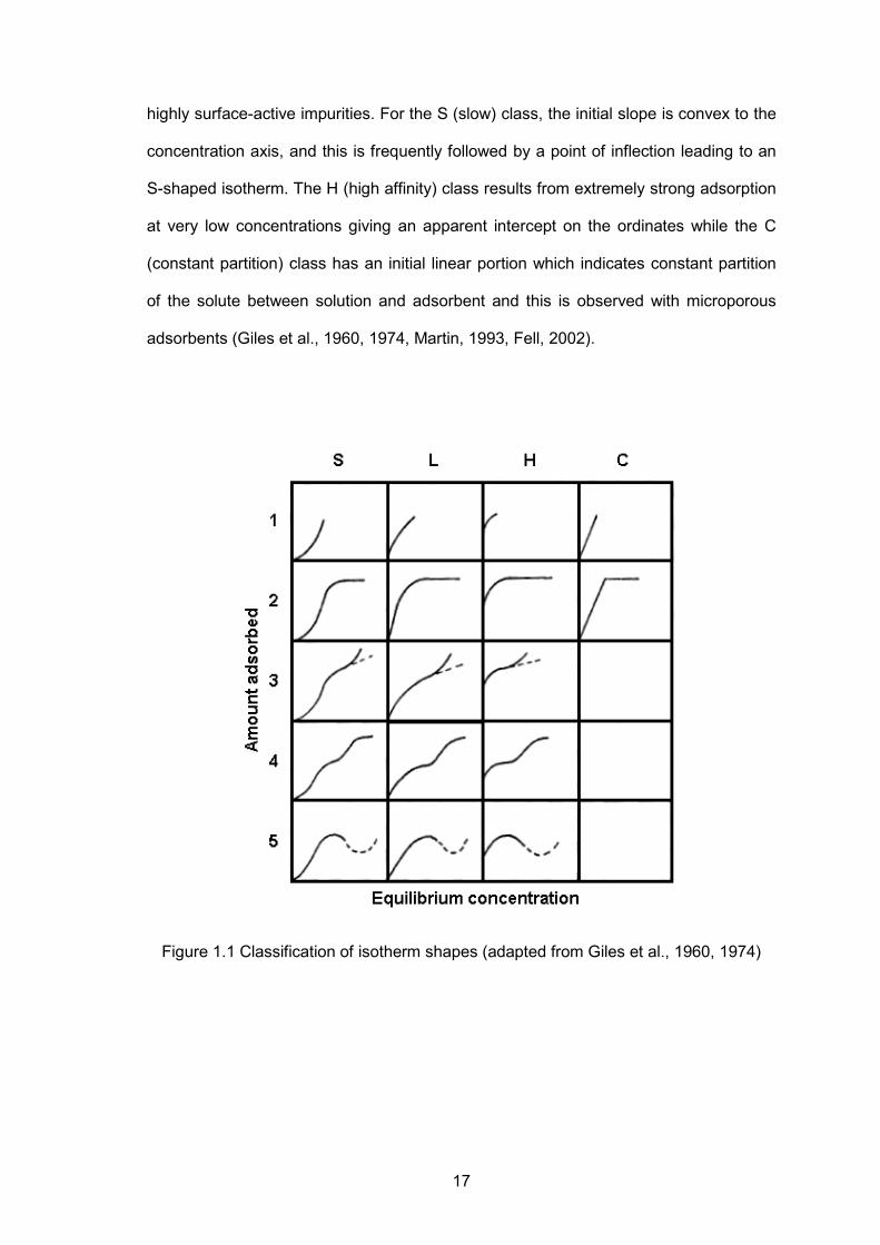
The isotherms obtained can generally be classified into four characteristic classes,
based on the form of the initial part of the isotherm; and each class can be divided into
subgroups in relation to the behaviour at higher concentrations. They are shown in
Figure 1.1. The L (Langmuir) class is the most common and is characterized by an
initial region which is concave to the concentration axis. The L2 isotherm reaches a
plateau and further adsorption above this value gives the L3 isotherm, and if that
reaches a second plateau it is designated as L4. The fifth L type reflects a special set
of circumstances. They are observed with solutes that associate in solution and contain
17
highly surface-active impurities. For the S (slow) class, the initial slope is convex to the
concentration axis, and this is frequently followed by a point of inflection leading to an
S-shaped isotherm. The H (high affinity) class results from extremely strong adsorption
at very low concentrations giving an apparent intercept on the ordinates while the C
(constant partition) class has an initial linear portion which indicates constant partition
of the solute between solution and adsorbent and this is observed with microporous
adsorbents (Giles et al., 1960, 1974, Martin, 1993, Fell, 2002).
Figure 1.1 Classification of isotherm shapes (adapted from Giles et al., 1960, 1974)

18
There have been many attempts to develop equations to fit the experimentally
observed isotherms. Among the most widely used equations are the Langmuir
adsorption isotherm, the Freundlich adsorption isotherm and the Brunauer, Emmett
and Teller (BET) equations (Martin, 1993). However, in the current study, only the first
two adsorption isotherms are used, and they are discussed in the following sections.
1.2.2(a) Langmuir adsorption isotherm
The equation was derived by assuming that only monolayer coverage was possible.
The equation is usually written as
xm =
abC1 + bC
(1.1)
where x is the amount of solute adsorbed by a weight, m, of adsorbent. C is the
concentration of the solution at equilibrium, and b and a are constants. Equation 1.1
can be arranged into the linear form
C
(x/m) =
1ab + Ca (1.2)
Hence plotting C/(x/m) against C should give a straight line with a slope 1/a and
intercept 1/ab (Florence and Attwood, 1988b, Martin, 1993, Fell, 2002).
1.2.2(b) Freundlich adsorption isotherm
The Freundlich equation is given as
xm = kC1/n (1.3)
where n and k are constants for a particular system. Equation 1.3 can be written in a
linear form by taking logarithms of both sides, giving
log ( xm ) = log k + (
1n) log C (1.4)
A plot of log (x/m) against log C should be linear, with an intercept of log k and slope of
n-1. It is generally assumed that, for systems which obey this equation, adsorption
19
results in the formation of multilayer rather than a single monolayer (Florence and
Attwood, 1988b, Fell, 2002).
1.2.3 Factors affecting adsorption
1.2.3(a) Solubility of the adsorbate
Solubility is an important factor affecting adsorption. According to Lundelius’ rule, the
extent of adsorption of a solute is inversely proportional to its solubility in the solvent
from which adsorption occurs (Florence and Attwood, 1988b). In order for adsorption to
occur, solute-solvent bonds must first be broken. Therefore, the greater the solubility,
the stronger are these bonds and hence the smaller the extent of adsorption. Besides,
it was shown that for most cases of adsorption from solution, the relative amount of
solute removed is greater in dilute solutions (Fell, 2002).
1.2.3(b) pH
pH can affect adsorption in various manner, the most important from a pharmaceutical
viewpoint being its effect on the ionisation and solubility of the adsorbate drug
molecules. Generally, adsorption increases as the ionisation of the drug is suppressed
and the extent of adsorption reaching a maximum when the drug is completely
unionised (Florence and Attwood, 1988b).
Normally, pH and solubility effects act in concert, since the unionised form of most
drugs in aqueous solution has a low solubility. The solubility effect is usually the
stronger among the two effects (Florence and Attwood, 1988b). In the adsorption of
hyoscine and atropine on magnesium trisilicate, it was observed that hyoscine,
although in its completely unionised form, was less strongly adsorbed than atropine,
which at the pH of the experiment was 50 per cent ionised. The reason for this result is
clear when the solubilities of the two bases are considered. Hyoscine base is freely
soluble compared with atropine base. Even when it was 50 per cent ionised, atropine is
20
less soluble than hyoscine and consequently more strongly adsorbed (El-Masry and
Khalil, 1974).
1.2.3(c) Nature of the adsorbent
The physicochemical nature of the adsorbent can have profound effects on the rate
and capacity of adsorption. The most important property affecting adsorption is the
surface area of the adsorbent. The extent of adsorption is proportional to the specific
surface area (Florence and Attwood, 1988b). Thus, an increased surface area,
achieved by a reduction in particle size or the use of a porous material, will increase
the extent of adsorption (Fell, 2002).
Besides, adsorbent-adsorbate interactions may affect adsorption. These interactions
are complex. Some particular adsorbents have affinities for particular adsorbates for a
wide variety of reasons. The adsorbent clays such as bentonite, attapulgite and kaolin
have cation-exchange sites on the surface. Such clays have strong affinities for
protonated compounds which they adsorb by an ion-exchange process. Moreover,
different parts of the surface of the same adsorbent may have different affinities for
different types of adsorbents (Florence and Attwood, 1988b).
1.2.3(d) Temperature
Adsorption is generally an exothermic process. Therefore, an increase in temperature
normally leads to a decrease in the amount adsorbed (Fell, 2002). The changes in
enthalpy of adsorption are usually of the order of those for condensation or
crystallisation. Thus, small variations in temperature tend not to alter the adsorption
process to a significant extent (Florence and Attwood, 1988b).
21
1.2.4 Medical and pharmaceutical applications of adsorption
1.2.4(a) Improving drug dissolution/bioavailability
The adsorption of drug molecules onto the surface of excipients can reduce drug
particle size and increase the surface area of drug available to the dissolution medium.
Both of these effects might increase dissolution and, as a result, bioavailability
(Jackson et al., 2000). Microcrystalline cellulose, polyvinylpyrrolidone and kaolin serve
most often as adsorbents (Marchal-Heussler and Barra, 2002).
The absorption of weak acid dicumarol in dogs was shown to be enhanced by
magnesium hydroxide and magnesium oxide as a result of increased dissolution. This
was attributed to the formation of a more readily absorbable dicumarol-magnesium
chelate or to an increase in pH of the microenvironment caused by the addition of the
excipients (Akers et al., 1973). In another study of indomethacin formulated as an
absorbate with kaolin, an increase in dissolution was observed compared with a simple
mixture of the two components. The authors suggested that, indomethacin crystallized
on the surface of the adsorbent particles during the formation of adsorbate, leading to
an increase in the surface area of drug available for dissolution (Alsaidan et al., 1998).
1.2.4(b) Adsorption of noxious substances from the alimentary tract
Adsorption onto finely divided solids has been utilized beneficially for a long time to
remove bacterial toxins and for treatment of intoxication by drugs or chemicals (Nada
et al., 1989). The commonly used oral antidotes for reducing the effects of poisoning
include activated charcoal, magnesium oxide and tannic acid. Several drugs such as
chlorpheniramine, propoxyphene hydrochloride, colchicines, diphenylhydantoin and
acetylsalicylic acid are adsorbed effectively by activated charcoal. The interactions of
drug molecules on the surface of the charcoal particles involved a non-polar,
monolayer adsorption (Florence and Attwood, 1988b).

22
1.2.5 Adsorption problems in drug formulation
Despite the advantages, adsorption may cause problems in formulation. Drugs and
preservatives can be adsorbed by containers and undissolved materials in suspension,
thereby reducing their effective concentrations (Fell, 2002). For example, the parabens
may be adsorbed on to the solid materials present in a suspension, leading to a loss in
antimicrobial activity (Allwood, 1982). The adsorption of insulin on to intravenous
administration sets has been reported, as has the adsorption of phenylmercuric acetate,
used as a preservative in eye drops, on to polyethylene containers (Aspinall et al.,
1980).
1.3 Crospovidone
1.3.1 Introduction
Conventional tablets are formulated to release the active compounds within a relatively
short interval in order to achieve optimal drug bioavailability. This is normally achieved
by incorporating disintegrants to facilitate the break-up of granules and tablets into fine
particles upon exposure to fluids in the gastrointestinal tract (Kottke and Rudnic, 2002).
For several decades, starch has been one of the most widely used disintegrants in
tablet manufacturing. However, starch has several drawbacks such as reduced
effectiveness at high tablet compression forces and large amounts are needed to
achieve the desired performance. Hence, alternative high-performance disintegrants,
termed as super disintegrants, have been introduced into tablet technology. Super
disintegrants permit low use levels and facilitate reliable disintegration (I.S.P., 2000a).
Insoluble polyvinylpyrrolidone or cross-linked polyvinylpyrrolidone (crospovidone) are
classified as super disintegrants.
23
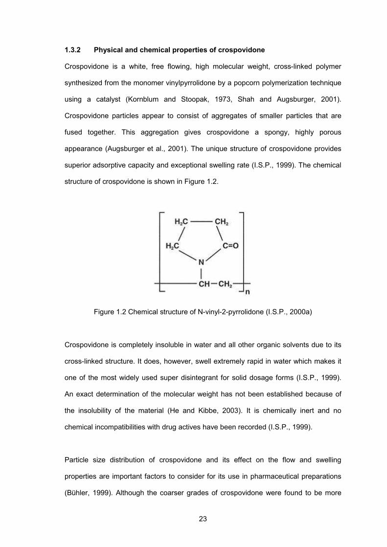
1.3.2 Physical and chemical properties of crospovidone
Crospovidone is a white, free flowing, high molecular weight, cross-linked polymer
synthesized from the monomer vinylpyrrolidone by a popcorn polymerization technique
using a catalyst (Kornblum and Stoopak, 1973, Shah and Augsburger, 2001).
Crospovidone particles appear to consist of aggregates of smaller particles that are
fused together. This aggregation gives crospovidone a spongy, highly porous
appearance (Augsburger et al., 2001). The unique structure of crospovidone provides
superior adsorptive capacity and exceptional swelling rate (I.S.P., 1999). The chemical
structure of crospovidone is shown in Figure 1.2.
Figure 1.2 Chemical structure of N-vinyl-2-pyrrolidone (I.S.P., 2000a)
Crospovidone is completely insoluble in water and all other organic solvents due to its
cross-linked structure. It does, however, swell extremely rapid in water which makes it
one of the most widely used super disintegrant for solid dosage forms (I.S.P., 1999).
An exact determination of the molecular weight has not been established because of
the insolubility of the material (He and Kibbe, 2003). It is chemically inert and no
chemical incompatibilities with drug actives have been recorded (I.S.P., 1999).
Particle size distribution of crospovidone and its effect on the flow and swelling
properties are important factors to consider for its use in pharmaceutical preparations
(Bühler, 1999). Although the coarser grades of crospovidone were found to be more

24
efficient than those with finer particle sizes in tablet disintegration (Shah and
Augsburger, 2001), a grade with relatively fine particle size of around 11 µm was
selected for this present study. This is due to the high surface area of the finer
crospovidone particles which provide superior adsorptive capacity (I.S.P., 1999).
Crospovidone can form complexes with many molecules including a wide range of
drugs as well as toxins. The complexation is reversible, involving the formation of weak
hydrogen bonding or van der Waals forces rather than the covalent chemical bonds
(I.S.P., 1999). However, complex formation of crospovidone is absent in alkaline
medium. Therefore, its ability to reduce the absorption of toxins is controversial and this
formed part of the aim of the present study. The degree of complexation also depends
on the chemical structure of the drugs used. It has been shown that complexation may
increase the rate and extent of drug dissolution (I.S.P., 2000a).
1.3.3 Toxicological and safety profiles
Crospovidone is used in oral pharmaceutical formulations and is generally regarded as
a non-toxic and non-irritant material. Short-term animal toxicity studies showed that no
adverse effect has been associated with crospovidone (He and Kibbe, 2003). The oral
median lethal dose (LD50) in rats was more than 100 g/kg. It is not absorbed dermally
and not a primary irritant or sensitizer. Long-term clinical studies showed that
crospovidone products are not absorbed through the gastrointestinal tract due to its
insolubility. It is essentially 100% excreted from the body. The Joint FAO/WHO Expert
Committee for food additives has assigned crospovidone an Acceptable Daily Intake
status of “Not Specified”, since it is not considered a health hazard (I.S.P., 1999, I.S.P.,
2000b).