Embed Size (px)
Citation preview
R
O
Itd
A
a
b
c
RA
dG
2t
evista de Gastroenterología de México. 2016;81(3):134---140
www.elsevier.es/rgmx
REVISTA DE
DE MEXICOGASTROENTEROLOGIA´
´
RIGINAL ARTICLE
nflammatory fibroid polyp of the gastrointestinalract: 10 years of experience at the Instituto Nacionale Ciencias Médicas y Nutrición Salvador Zubirán�
.F. Romano-Munivea,∗, R. Barreto-Zunigaa, J.A. Rumoroso-Garcíab, P. Ramos-Martínezc
Servicio de Endoscopia Digestiva, Instituto Nacional de Ciencias Médicas y Nutrición Salvador Zubirán, SSA, Mexico City, MéxicoServicio de Radiología, Instituto Nacional de Ciencias Médicas y Nutrición Salvador Zubirán, SSA, Mexico City, MéxicoServicio de Patología Digestiva, Instituto Nacional de Ciencias Médicas y Nutrición Salvador Zubirán, SSA, Mexico City, México
eceived 25 January 2016; accepted 11 March 2016vailable online 17 June 2016
KEYWORDSInflammatory fibroidpolyp;Intussusception;Iron deficiencyanemia;Polypectomy;Stomach
AbstractBackground: Inflammatory fibroid polyp (lFP) is a rare, benign, and solitary neoplasm predom-inantly located in the gastric antrum and small bowel. Its clinical symptoms are heterogeneousand essentially depend on the location and size of the tumor. Definitive diagnosis is madethrough histopathology and this pathology has excellent long-term prognosis.Aim: To identify the cases of IFP seen at the Instituto Nacional de Ciencias Médicas y NutriciónSalvador Zubirán over a 10-year period.Methods: A retrospective, cross-sectional, descriptive, and observational study was conductedthat included patients with histopathologic diagnosis of IFP within the time frame of January2001 and December 2011.Results: Six cases were found and 5/6 (83.3%) of them were women. The median age was 41years (minimum-maximum range of 19-56 years). The most frequent symptoms were weight
loss (n = 3), fever (n = 2), nausea (n = 2), and vomiting (n = 2). Three patients presented withiron deficiency anemia and 2 cases with intussusception. The IFPs were located at the followingsites: esophagus (n = 1), stomach (n = 2), small bowel (n = 2), and colon (n = 1). Treatment wassurgical in 5/6 (83.3%) of the patients.� Please cite this article as: Romano-Munive AF, Barreto-Zuniga R, Rumoroso-García JA, Ramos-Martínez P. Pólipo fibroideo inflamatorio
el tracto gastrointestinal: 10 anos de experiencia del Instituto Nacional de Ciencias Médicas y Nutrición Salvador Zubirán. Revista deastroenterología de México. 2016;82:134---140.∗ Corresponding author. Vasco de Quiroga 15, Sección XVI, Tlalpan, Mexico City, CP 14000. Tel.: 0445520826588; fax: +54870900 ext. 2150.E-mail address: fab [email protected] (A.F. Romano-Munive).
255-534X/© 2016 Asociacion Mexicana de Gastroenterologıa. Published by Masson Doyma Mexico S.A. This is an open access article underhe CC BY-NC-ND license (http://creativecommons.org/licenses/by-nc-nd/4.0/).
Inflammatory fibroid polyp of the gastrointestinal tract 135
Conclusions: IFPs are extremely rare in our population. They usually present with weight lossand iron deficiency anemia and are more frequently located in the stomach and small bowel.This is the largest reported IFP case series in a Mexican population.© 2016 Asociacion Mexicana de Gastroenterologıa. Published by Masson Doyma Mexico S.A. Thisis an open access article under the CC BY-NC-ND license (http://creativecommons.org/licenses/by-nc-nd/4.0/).
PALABRAS CLAVEPólipo fibroideoinflamatorio;Intususcepción;Anemia ferropénica;Polipectomía;Estómago
Pólipo fibroideo inflamatorio del tracto gastrointestinal: 10 anos de experiencia delInstituto Nacional de Ciencias Médicas y Nutrición Salvador Zubirán
ResumenAntecedentes: El pólipo fibroideo inflamatorio (PFI) es una neoplasia rara, benigna y solitaria,predomina en el antro gástrico y el intestino delgado. Los síntomas clínicos son heterogéneos,dependen fundamentalmente de la localización y el tamano del tumor. El diagnóstico definitivose establece mediante histopatología y su pronóstico es excelente a largo plazo.Objetivo: Identificar los casos de PFI en el Instituto Nacional de Ciencias Médicas y NutriciónSalvador Zubirán en un período de 10 anos.Métodos: Estudio observacional, descriptivo, transversal y retrospectivo, se incluyó a lospacientes con diagnóstico histopatológico de PFI desde enero del 2001 hasta diciembre del2011.Resultados: Se encontraron 6 casos, 5/6 (83.3%) fueron mujeres. La mediana de edad fue de 41anos (rango mínimo-máximo de 19 a 56 anos). Los síntomas más frecuentes fueron pérdida depeso (n = 3), fiebre (n = 2), náuseas (n = 2) y vómito (n = 2). Tres pacientes cursaron con anemiaferropénica. Dos casos se presentaron con intususcepción. La localización de los PFI fue lasiguiente: esófago (n = 1), estómago (n = 2), intestino delgado (n = 2) y colon (n = 1). Eltratamiento fue quirúrgico en 5/6 (83.3%) pacientes.Conclusiones: Los PFI son extremadamente raros en nuestra población, suelen presentarse conpérdida de peso y anemia ferropénica, se localizan con mayor frecuencia en el estómago y elintestino delgado. Esta es la serie de casos más grande de PFI que se ha reportado en poblaciónmexicana.© 2016 Asociacion Mexicana de Gastroenterologıa. Publicado por Masson Doyma Mexico S.A.Este es un artıculo Open Access bajo la licencia CC BY-NC-ND (http://creativecommons.org/licenses/by-nc-nd/4.0/).
hs
htt
Iy1
M
Atptime frame of January 2001 and December 2011. The demo-
Introduction
Inflammatory fibroid polyp (IFP) is a rare, benign, and soli-tary neoplasm initially described by Vanek in 1949 as asubmucosal gastric granuloma with eosinophilia.1 Helwigand Ranier proposed the term IFP in 1953.2 Its incidenceis extremely low (0.1%) and usually presents in the sixthand seventh decades of life. It is slightly more frequent inthe male sex.2,3 IFPs predominate in the gastric antrum andsmall bowel.2,4,5 Its etiology is unknown, but some authorssuggest that it is an allergic reaction due to the presence ofeosinophils. However, other factors have been implicated,such as neural hyperplasia, irritants, trauma, genetic alter-ations, and bacterial, physical, or chemical stimulants.2
Activating mutations in the platelet-derived growth factorreceptor alpha (PDGFRA) gene have been associated withthe development of IFPs and of gastrointestinal stromaltumor (GIST), showing a similar physiopathogenesis between
these 2 neoplasms and lending support to the theory of aneoplastic IFP origin.6,7 Mutations have been documentedin 21.7 to 69.6% of the cases.8 The clinical symptoms aregiw
eterogeneous and essentially depend on the location andize of the tumor.9,10
The definitive diagnosis is made throughistopathology.11---13 Currently, the majority of cases can bereated with polypectomy, with the rest requiring surgicalreatment, and the long-term prognosis is excellent.14
The aim of this study was to identify the cases ofFP seen at the Instituto Nacional de Ciencias Médicas
Nutrición Salvador Zubirán (INCMNSZ) over a period of0 years.
ethods
retrospective, cross-sectional, descriptive, and observa-ional study was conducted at the INCMNSZ and includedatients with a histopathologic diagnosis of IFP within the
raphic variables, clinical manifestations, laboratory andmaging studies, and clinical progression of the patientsere obtained from the clinical case records.
136 A.F. Romano-Munive et al.
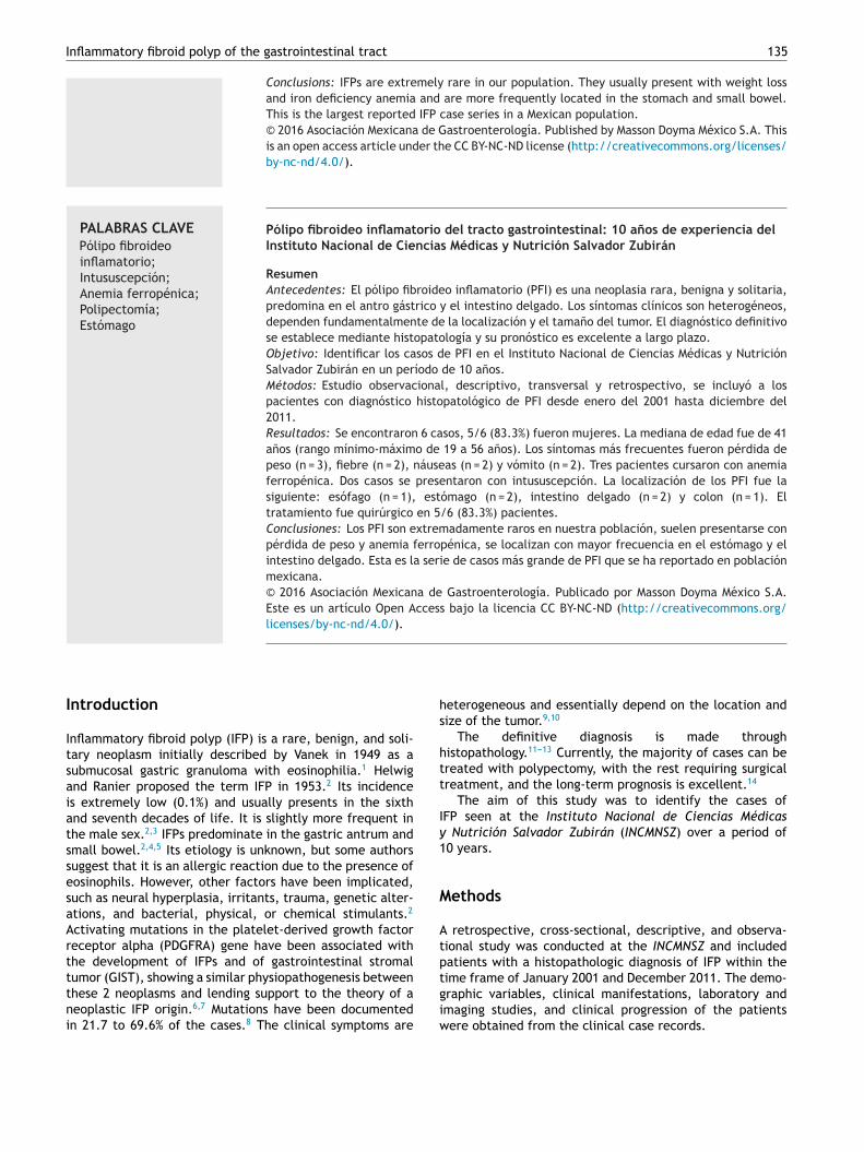
Figure 1 Case 1. The chest CT reported an intraluminal polypoid lesion dependent on the cervical esophagus. Panendoscopyr sounp
crs
R
Swrw(mw
fsc=spta
D
Fs
evealed a polypoid lesion with a long pedicle. Endoscopic ultraropria layer. The surgical specimen reported IFP.
The descriptive statistics included frequencies for theategorical variables and medians and minimum-maximumange for the continuous variables. The SPSS version 20.0tatistical program was utilized (Chicago, Illinois, USA).
esults
ix cases of IFP were found and 5/6 (83.3%) of them were inomen. The median age was 41 years (minimum-maximum
ange of 19 to 56 years). The most frequent symptoms were
eight loss (n = 3), fever (n = 2), nausea (n = 2), and vomitingn = 2). Three patients presented with iron deficiency ane-ia and 2 with intussusception. The mean symptom durationas 7 months (range of 0 to 12 months). The IFPs had the
IIp
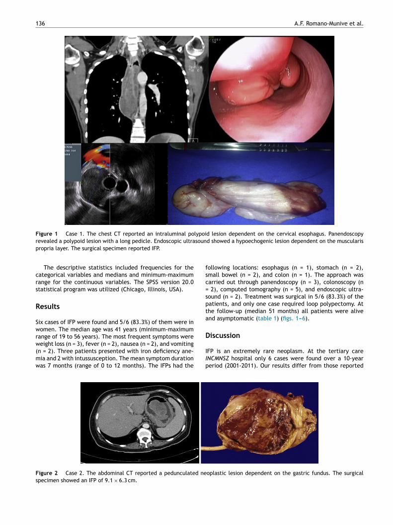
igure 2 Case 2. The abdominal CT reported a pedunculated nepecimen showed an IFP of 9.1 × 6.3 cm.
d showed a hypoechogenic lesion dependent on the muscularis
ollowing locations: esophagus (n = 1), stomach (n = 2),mall bowel (n = 2), and colon (n = 1). The approach wasarried out through panendoscopy (n = 3), colonoscopy (n
2), computed tomography (n = 5), and endoscopic ultra-ound (n = 2). Treatment was surgical in 5/6 (83.3%) of theatients, and only one case required loop polypectomy. Athe follow-up (median 51 months) all patients were alivend asymptomatic (table 1) (figs. 1---6).
iscussion
FP is an extremely rare neoplasm. At the tertiary careNCMNSZ hospital only 6 cases were found over a 10-yeareriod (2001-2011). Our results differ from those reported
oplastic lesion dependent on the gastric fundus. The surgical
Inflam
matory
fibroid polyp
of the
gastrointestinal tract
137
Table 1 Inflammatory fibroid polyps.
Case Sex/age Main symptoms Laboratory tests Location Endoscopy CT EUS Treatment Pathology
1 F/19 Dysphagia,vomiting,protrusion of amass through theoral cavity, weightloss, fever
Hb 9.1 g/dL, MCV75.2 fl, MCH 24.6pg/cel, irondeficiency profile,ESR 74 mm/h and CRP15.32 mg/L
Esophagus Polypoid lesionwith longpedicle,ulcerated inthe distal third
Intraluminalpolypoid lesionin the cervicalesophagus
Hypoechogeniclesiondependent onthe muscularispropria
Left lateralcervicalesophagotomy
IFP 18.4 × 4 cm
2 F/51 Epigastric painand weight loss
Hb 8.8 g/dL, MCV 64fl, MCH 19 pg/cel,iron deficiency profile
Stomach Polypoid tumorin the gastricfundus
Neoplasticpedunculatedlesion in thegastric fundus
Heterogeneoushypoechoiclesion withirregular edgesdependent onthe muscularispropria
Total gastrectomyandesophagojejunalanastomosis inRoux-en-Y
IFP 9.1 × 6.3 cm
3 F/56 Fatigue andmelena
Normal completeblood count
Stomach Ulceratedpedunculatedpolyp in thelessercurvature
Contrast-enhancedlesion
NP Antrectomy andgastroduodenalanastomosis
IFP 3.7 × 2.5 cm
4 F/35 Vomiting,abdominal painand fever, laterpresentation ofacute abdomen
Leukocytes 12.1/mL,neutrophils 82.7%,hemoglobin 14.3g/dL, MCV 74.6 fl andMCH 25.4 pg/cel
Ileum NP 30 cmintussusceptionof the ileum intothe ascendingcolon
NP Righthemicolectomywithileotransverseanastomosis
IFP 7.2 × 3.8 cm,ilealintussusceptionand ischemicenteritis
5 M/36 Weight loss andrectorrhagia
Normal completeblood count
Ileum NeoplasiaExophyticneoplasm inthe right colonwith 90%occlusion ofthe lumenimpeding thepassage of thecolonoscope
Ileocolicintussusception,cecal edema,and free liquid
NP Ileal resection andenteroenteros-tomy
IFP 4.2 × 4.1 cmand Meckel’sdiverticulum
6 F/46 Asymptomatic Normal completeblood count
Colon Polyp 5 mm to12 cm from theanal margin
NP NP Polypectomy withloop
IFP 0.5 × 0.4 cm
CT: Computed tomography; EUS: Endoscopic ultrasound; IFP: Inflammatory fibroid polyp; NP: Not performed
138 A.F. Romano-Munive et al.
Fl
is4
s7awe
afipttpp
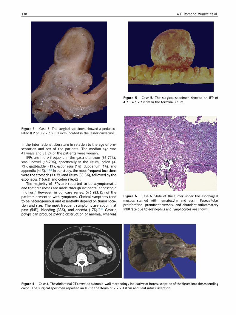
Figure 5 Case 5. The surgical specimen showed an IFP of4.2 × 4.1 × 2.8 cm in the terminal ileum.
Figure 6 Case 6. Slide of the tumor under the esophagealmucosa stained with hematoxylin and eosin. Fusocellular
Fc
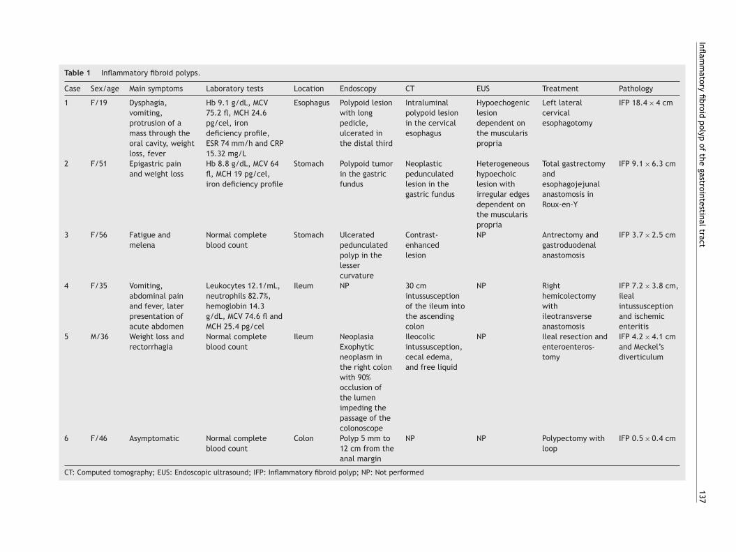
igure 3 Case 3. The surgical specimen showed a peduncu-ated IFP of 3.7 × 2.5 × 0.4 cm located in the lesser curvature.
n the international literature in relation to the age of pre-entation and sex of the patients. The median age was1 years and 83.3% of the patients were women.
IFPs are more frequent in the gastric antrum (66-75%),mall bowel (18-20%), specifically in the ileum, colon (4-%), gallbladder (1%), esophagus (1%), duodenum (1%), andppendix (<1%).1,4,5 In our study, the most frequent locationsere the stomach (33.3%) and ileum (33.3%), followed by thesophagus (16.6%) and colon (16.6%).
The majority of IFPs are reported to be asymptomaticnd their diagnoses are made through incidental endoscopicndings.1 However, in our case series, 5/6 (83.3%) of theatients presented with symptoms. Clinical symptoms tendo be heterogeneous and essentially depend on tumor loca-
ion and size. The most frequent symptoms are abdominalain (54%), bleeding (33%), and anemia (17%).9,10 Gastricolyps can produce pyloric obstruction or anemia, whereasproliferation, prominent vessels, and abundant inflammatoryinfiltrate due to eosinophils and lymphocytes are shown.
igure 4 Case 4. The abdominal CT revealed a double-wall morphology indicative of intussusception of the ileum into the ascendingolon. The surgical specimen reported an IFP in the ileum of 7.2 × 3.8 cm and ileal intussusception.
L1
Taaasp
E
Ptf
Dd
Rd
F
N
C
T
A
Tae
R
Inflammatory fibroid polyp of the gastrointestinal tract
those located in the small bowel present with obstruction orintussusception.1 Dysphagia is the most common symptom ofIFP in the esophagus, followed by gastrointestinal bleedingdue to erosions and ulcers on the surface of the polyp. Thereare reports of cases of regurgitation of the mass and food,as well as asphyxia.15 Fever is attributed to the effect ofcytokines released from the inflammatory cells of the IFP.16
In our study, the most frequent symptoms were weight loss(n = 3), fever (n = 2), nausea (n = 2), and vomiting (n =2). Three patients presented with iron deficiency anemiaand there were 2 cases of intussusception. In 3/6 (50%) ofour cases, endoscopy revealed submucosal, polypoid, intra-luminal, and pedunculated lesions, which frequently have anulcerated surface. These tumors usually measure 2 to 5 cmat the time of diagnosis.4 Our results reported a median of5.7 cm in diameter. Computed tomography is considered themost sensitive radiologic method for diagnosing the polyp orits complications.13 Nevertheless, endoscopic ultrasound isan excellent method for the detailed anatomic descriptionof the tumor. The most common ultrasound pattern is thatof a hypoechogenic, homogeneous, non-encapsulated lesionwith poorly defined margins located in the second and/orthird echo layer of the gastrointestinal wall.12 Endoscopicultrasound also provides information on the vascularity ofthe polyp.13
Definitive diagnosis is made through the histopathologystudy. IFPs are characterized by fusiform, mesenchyma-tous cells with concentric condensation (onion skin), nonecrosis or significant mitosis, as well as by fibroblast andneovessel proliferation and an inflammatory infiltrate rich ineosinophils, lymphocytes, plasma cells, and mastocytes.11,12
Immunohistochemistry studies show important positivity forCD34 and negativity for CD117 (c-kit) in the fusiform cells.IFPs usually display positivity for fascine (a dendritic cellmarker), D1 cyclin (a cell cycle regulation defect marker),calponin, and vimentin and variable reactivity for actin,CD68, desmin, and the S100 protein.13 The differential diag-nosis must be made with gastrointestinal stroma tumor(GIST). Even though both are positive for CD34 and vimentin,GISTs are positive for CD117 (c-kit) and IFPs are not. IFPsshould also be distinguished from schwannoma, fusiformcell leiomyoma, and inflammatory myofibroblastic tumor.Schwannomas are positive for S100 and negative for CD34.Fusiform cell leiomyoma is positive for desmin and actinand only 10 to 15% are positive for CD34. Inflammatorymyofibroblastic tumor displays positivity for actin and CD34.Inflammatory fibrosarcoma and spindle-cell carcinoid tumorare other diagnoses that should be considered.3,4,12,13
Currently, the majority of cases can be treated withpolypectomy and the others require surgery. Their long-term prognosis is excellent.13 Endoscopic polypectomy isadequate for treating small-diameter polyps. Larger polypsrequire surgical resection because they cause complicationssuch as intussusception or obstruction that need emergencysurgery.9 They do not recur after surgery.2 No recurrence hasbeen reported in our patients.
There are two case reports on IFP in a Mexican popula-tion. In 2008, Aguilar-Davidov et al. reported on the case
of a 9.1 cm gastric polyp included in their case series.17 In2011, Morales-Fuentes et al. described a 42-year-old manwith abdominal pain with terminal ileum intussusceptionidentified by abdominal computerized tomography scan.139
aparoscopy was performed, finding a 3 cm IF in the ileum5 cm from the ileocecal valve.18
IFPs are extremely rare in the Mexican population.hey usually present with weight loss and iron deficiencynemia and are more frequently located in the stom-ch and small bowel. They require a multidisciplinarypproach and their prognosis is excellent. The present caseeries on IFP is the largest to be reported in a Mexicanopulation.
thical responsibilities
rotection of persons and animals. The authors declarehat no experiments were performed on humans or animalsor this study.
ata confidentiality. The authors declare that no patientata appear in this article.
ight to privacy and informed consent. The authorseclare that no patient data appear in this article.
inancial disclosure
o financial support was received in relation to this study.
onflict of interest
he authors declare that there is no conflict of interest.
cknowledgments
he authors wish to thank Dr. Miguel Ángel Ramírez-Lunand Dr. Rubén Cortés-González for their participation in thesophageal IFP case.
eferences
1. Joyce KM, Waters PS, Waldron RM, et al. Recurrent adultjejuno-jejunal intussusception due to inflammatory fibroidpolyp - Vanek’s tumour: A case report. Diagn Pathol. 2014;9:127.
2. Kang SH, Kim SW, Moon HS, et al. Inflammatory fibroid polypin the jejunum causing small bowel intussusception. Ann Colo-proctol. 2015;31:106---9.
3. Gara N, Falzarano JS, Limm WM, et al. Ileal inflammatoryfibroid polyp causing chronic ileocolic intussusception and mim-icking cecal carcinoma. World J Gastrointest Oncol. 2009;1:89---92.
4. Akbulut S. Intussusception due to inflammatory fibroid polyp:A case report and comprehensive literature review. World JGastroenterol. 2012;18:5745---52.
5. Kimura N, Hight M, Liang J, et al. Adult intussusception sec-ondary to inflammatory fibroid polyp. West J Emerg Med.2015;16:581---2.
6. Bae JS, Song JS, Hong SM, et al. An unusual presentation of aninflammatory fibroid polyp of the ileum: A case report. Oncol
Lett. 2015;9:327---9.7. Schildhaus HU, Cavlar T, Binot E, et al. Inflammatory fibroidpolyps harbours mutations in the platelet-derived growth factorreceptor alpha (PDGFRA) gene. J Pathol. 2008;216:176---82.
1
1
1
1
1
1
1
1
1
40
8. Abboud B. Vanek’s tumor of the small bowel in adults. World JGastroenterol. 2015;21:4802---8.
9. Sulu B, Günerhan Y, Kösemehmetoglu K. A rare ileal tumor caus-ing anemia and intussusception: Inflammatory fibroid polyp.Turk J Gastroenterol. 2014;25:116---7.
0. Hirasaki S, Matsubara M, Ikeda F, et al. Inflammatory fibroidpolyp occurring in the transverse colon diagnosed by endoscopicbiopsy. World J Gastroenterol. 2007;13:3765---6.
1. Guerra Bautista JA, Ibánez Delgado F, Hernández de la TorreBustillo JM, et al. Pólipo fibroide inflamatorio gástrico. Rev EspEnferm Dig. 2006;98:482---3.
2. Woodward K, Gangarosa LM, Hunt HV. Gastric inflammatory
fibroid polyp. Indian J Pathol Microbiol. 2011;54:622---3.3. Ortiz-Moyano C, Martínez-García RC, Sánchez-Munoz D, et al.Giant fibroid inflammatory polyp. Rev Esp Enferm Dig.2010;102:282---3.
1
A.F. Romano-Munive et al.
4. Modi C, Shah A, Depasquale JR, et al. A large prolapsed inflam-matory fibroid polyp of the esophagus: An unusual presentation.Gastroenterol Hepatol. 2013;9:322---5.
5. Rawashdeh B, Meyer M, Gill J, et al. Unusual presentation of agiant benign inflammatory polyp in the upper esophagus. Int JSurg Case Rep. 2015;6C:206---9.
6. He HY, Shen ZB, Fang Y, et al. Bleeding and hyperpyrexia inan adult with gastric inflammatory fibroid polyp. Chin Med J.2013;126:2594.
7. Aguilar-Davidov B, Chablé-Montero F, Medina-Franco H.Pólipo fibroideo inflamatorio gástrico gigante. Reporte decaso y revisión de la literatura. Rev Gastroenterol Mex.
2008;73:239---41.8. Morales-Fuentes GA, Arino-Suárez M, Zarate-Osorno A, et al.Pólipo de Vanek o pólipo fibroide inflamatorio. Informe de uncaso y revisión de la literatura. Cir Cir. 2011;79:263---7.