Embed Size (px)
Citation preview
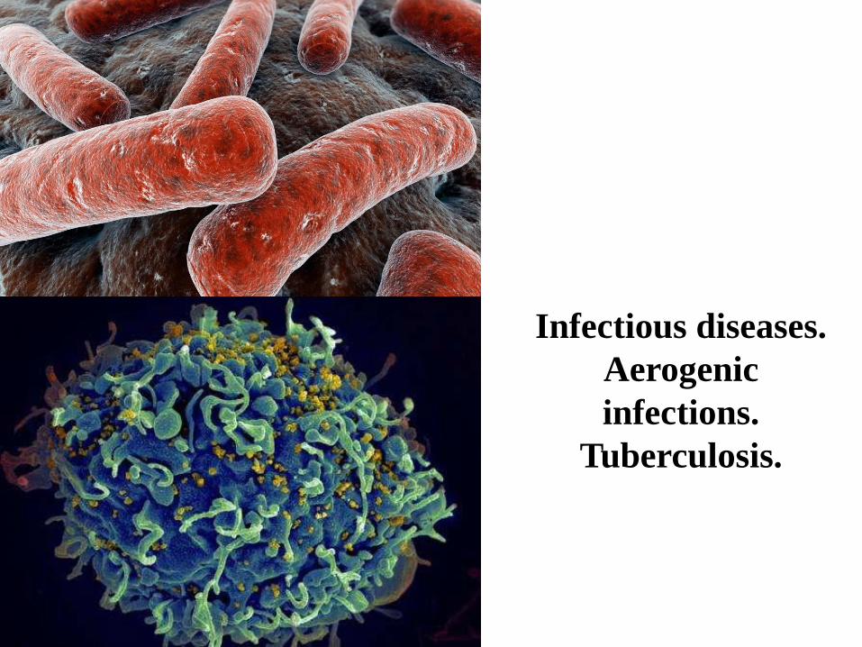
Infectious diseases.
Aerogenic
infections.
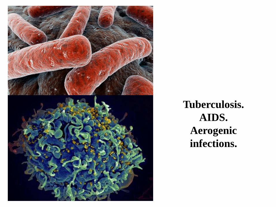
Tuberculosis.
.Infectious diseases.
Aerogenic infections. Tuberculosis.
I. Microspecimens:
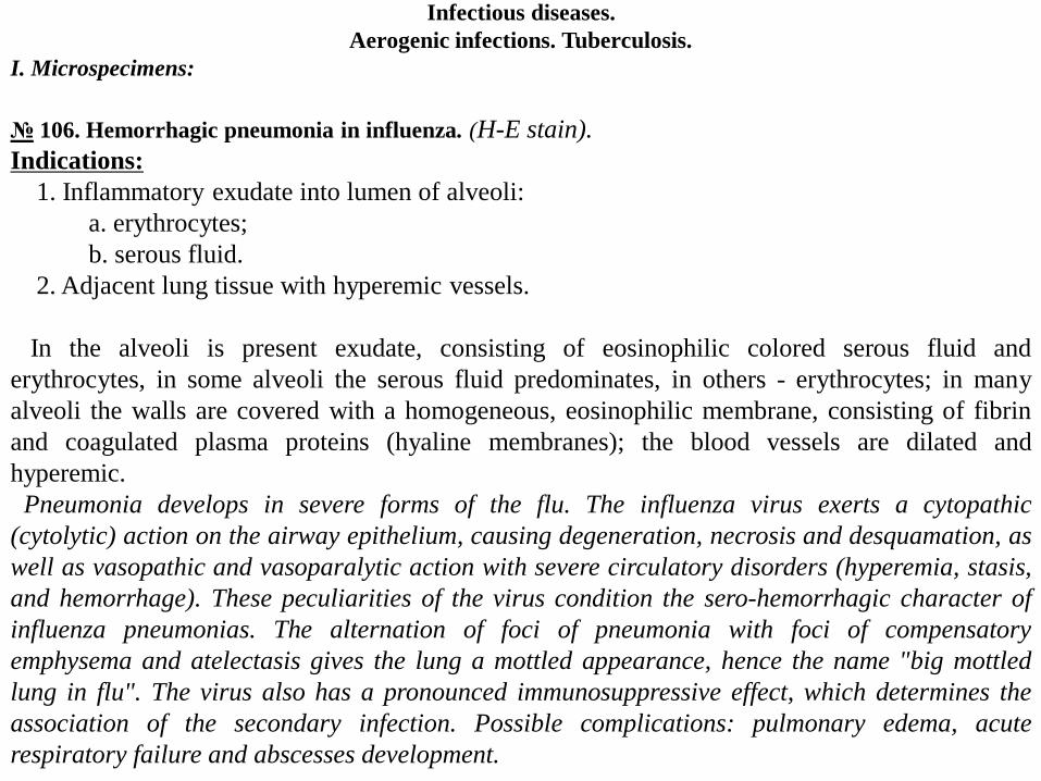
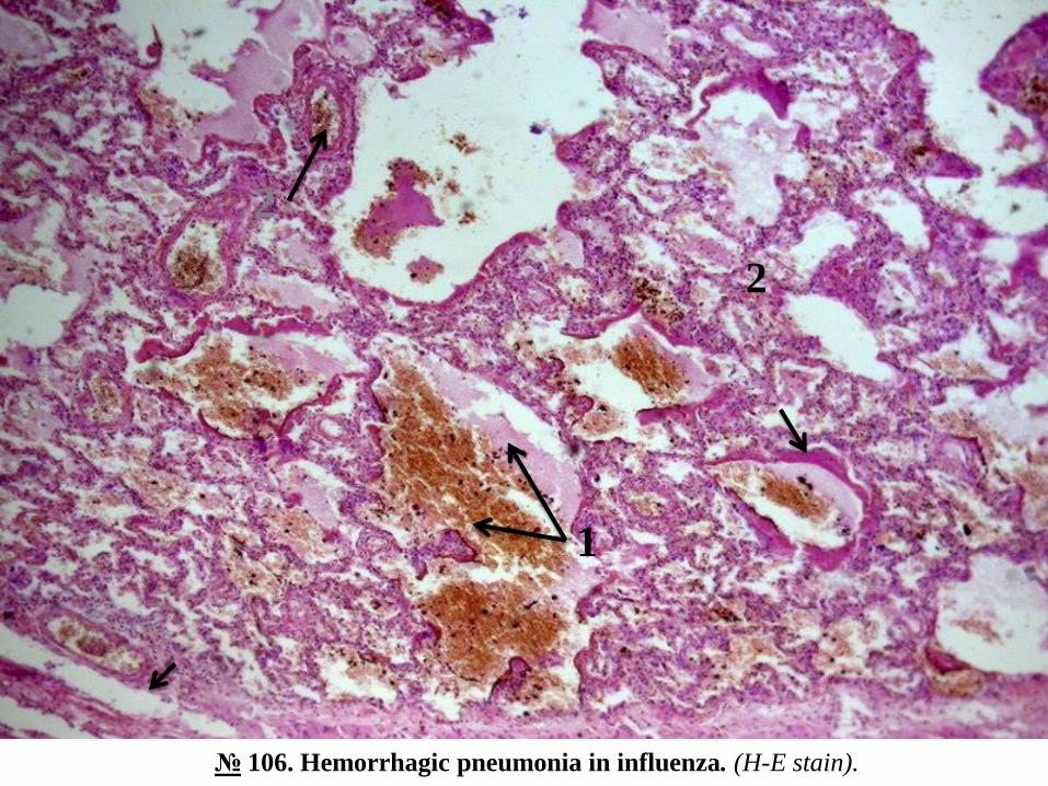
№ 106. Hemorrhagic pneumonia in influenza. (H-E stain).
Indications:
1. Inflammatory exudate into lumen of alveoli:
a. erythrocytes;
b. serous fluid.
2. Adjacent lung tissue with hyperemic vessels.
In the alveoli is present exudate, consisting of eosinophilic colored serous fluid and
erythrocytes, in some alveoli the serous fluid predominates, in others - erythrocytes; in many
alveoli the walls are covered with a homogeneous, eosinophilic membrane, consisting of fibrin
and coagulated plasma proteins (hyaline membranes); the blood vessels are dilated and
hyperemic.
Pneumonia develops in severe forms of the flu. The influenza virus exerts a cytopathic
(cytolytic) action on the airway epithelium, causing degeneration, necrosis and desquamation, as
well as vasopathic and vasoparalytic action with severe circulatory disorders (hyperemia, stasis,
and hemorrhage). These peculiarities of the virus condition the sero-hemorrhagic character of
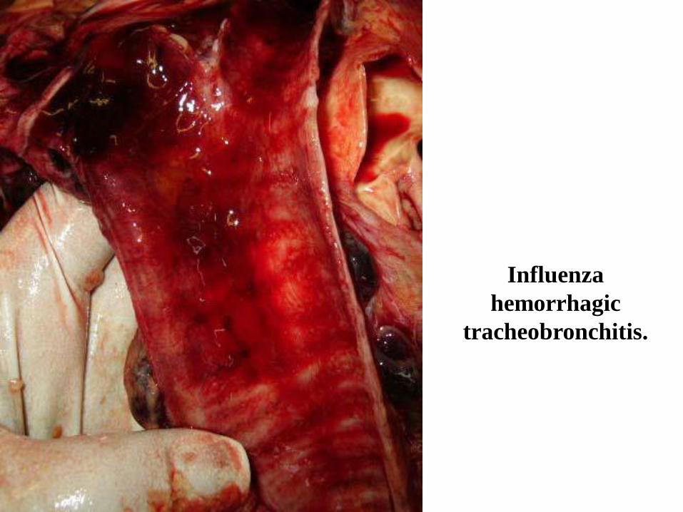
influenza pneumonias. The alternation of foci of pneumonia with foci of compensatory
emphysema and atelectasis gives the lung a mottled appearance, hence the name "big mottled
lung in flu". The virus also has a pronounced immunosuppressive effect, which determines the
association of the secondary infection. Possible complications: pulmonary edema, acute
respiratory failure and abscesses development.
№ 106. Hemorrhagic pneumonia in influenza. (H-E stain).
1
2
2
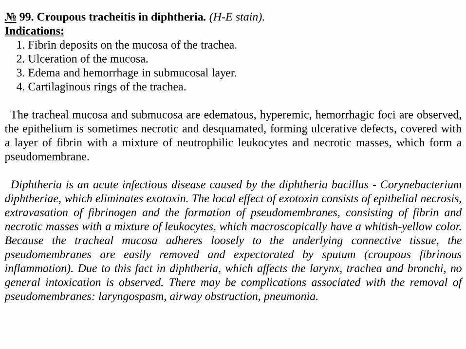
№ 99. Croupous tracheitis in diphtheria. (H-E stain).
Indications:
1. Fibrin deposits on the mucosa of the trachea.
2. Ulceration of the mucosa.
3. Edema and hemorrhage in submucosal layer.
4. Cartilaginous rings of the trachea.
The tracheal mucosa and submucosa are edematous, hyperemic, hemorrhagic foci are observed,
the epithelium is sometimes necrotic and desquamated, forming ulcerative defects, covered with
a layer of fibrin with a mixture of neutrophilic leukocytes and necrotic masses, which form a
pseudomembrane.
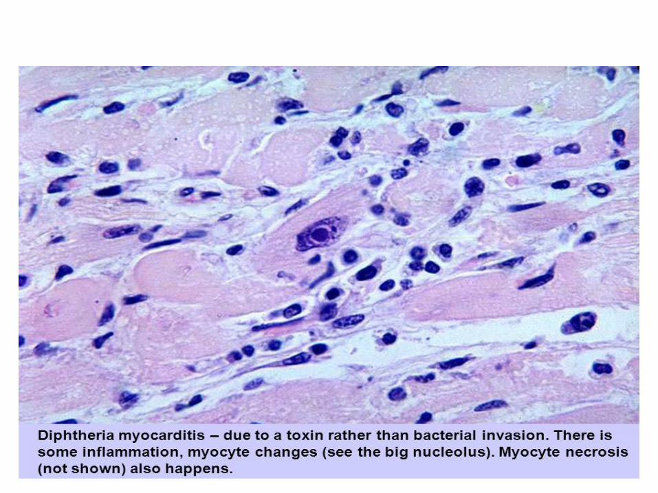
Diphtheria is an acute infectious disease caused by the diphtheria bacillus - Corynebacterium
diphtheriae, which eliminates exotoxin. The local effect of exotoxin consists of epithelial necrosis,
extravasation of fibrinogen and the formation of pseudomembranes, consisting of fibrin and
necrotic masses with a mixture of leukocytes, which macroscopically have a whitish-yellow color.
Because the tracheal mucosa adheres loosely to the underlying connective tissue, the
pseudomembranes are easily removed and expectorated by sputum (croupous fibrinous
inflammation). Due to this fact in diphtheria, which affects the larynx, trachea and bronchi, no
general intoxication is observed. There may be complications associated with the removal of
pseudomembranes: laryngospasm, airway obstruction, pneumonia.
№ 99. Croupous tracheitis in diphtheria. (H-E stain).
2
1
4 3
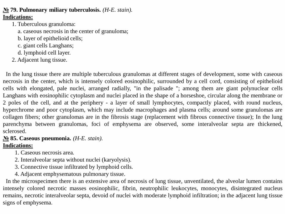
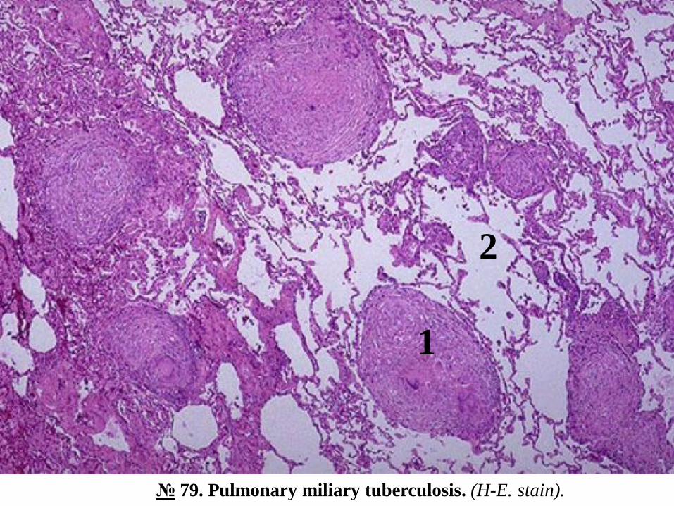
№ 79. Pulmonary miliary tuberculosis. (H-E. stain).
Indications:
1. Tuberculous granuloma:
a. caseous necrosis in the center of granuloma;
b. layer of epithelioid cells;
c. giant cells Langhans;
d. lymphoid cell layer.
2. Adjacent lung tissue.
In the lung tissue there are multiple tuberculous granulomas at different stages of development, some with caseous
necrosis in the center, which is intensely colored eosinophilic, surrounded by a cell cord, consisting of epithelioid
cells with elongated, pale nuclei, arranged radially, "in the palisade "; among them are giant polynuclear cells
Langhans with eosinophilic cytoplasm and nuclei placed in the shape of a horseshoe, circular along the membrane or
2 poles of the cell, and at the periphery - a layer of small lymphocytes, compactly placed, with round nucleus,
hyperchrome and poor cytoplasm, which may include macrophages and plasma cells; around some granulomas are
collagen fibers; other granulomas are in the fibrosis stage (replacement with fibrous connective tissue); In the lung
parenchyma between granulomas, foci of emphysema are observed, some interalveolar septa are thickened,
sclerosed.
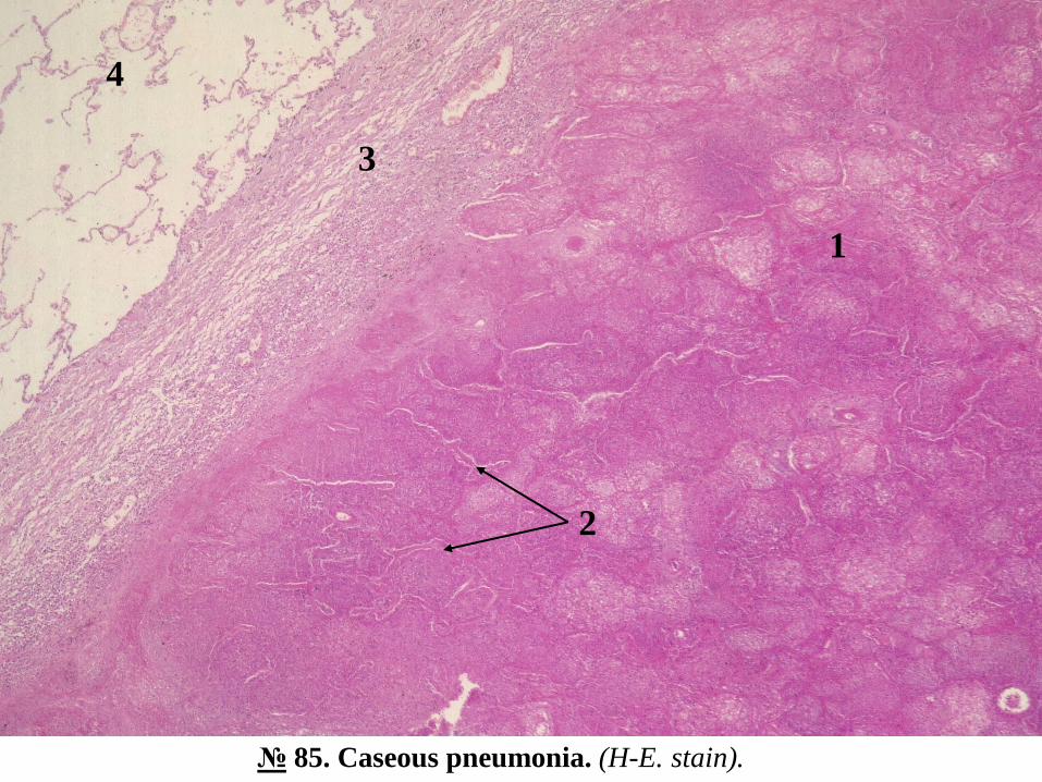
№ 85. Caseous pneumonia. (H-E. stain).
Indications:
1. Caseous necrosis area.
2. Interalveolar septa without nuclei (karyolysis).
3. Connective tissue infiltrated by lymphoid cells.
4. Adjacent emphysematous pulmonary tissue.
In the microspecimen there is an extensive area of necrosis of lung tissue, unventilated, the alveolar lumen contains
intensely colored necrotic masses eosinophilic, fibrin, neutrophilic leukocytes, monocytes, disintegrated nucleus
remains, necrotic interalveolar septa, devoid of nuclei with moderate lymphoid infiltration; in the adjacent lung tissue
signs of emphysema.
1
2
№ 79. Pulmonary miliary tuberculosis. (H-E. stain).
№ 85. Caseous pneumonia. (H-E. stain).
2
1
3
4
II. Macrospecimens:
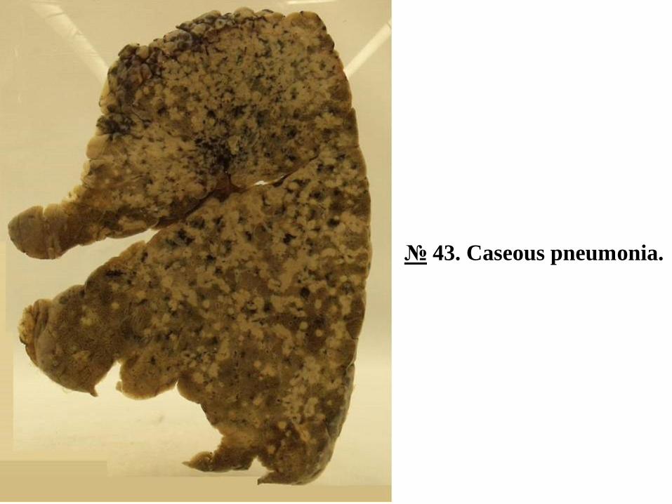
№ 43. Caseous pneumonia.
In the lung there are multiple foci of caseous necrosis, unventilated, of different sizes, white-
yellow color, the necrotic masses have a friable, crumbly appearance, it resembles dry cow's
cheese (lat. Caseum - cheese).
Caseous pneumonia is found in secondary tuberculosis, but can also be in primary tuberculosis.
Initially, acinar, lobular caseous outbreaks appear, which can extend to the level of a segment or
even of an entire lobe - lobar caseous pneumonia. It develops in patients with low immunity,
malnourished. There are deposits of fibrin in the pleura. The curd masses can be subjected to
purulent lysis and liquefaction with the appearance of decomposition cavities - caverns
(cavernous tuberculosis).
№ 43. Caseous pneumonia.
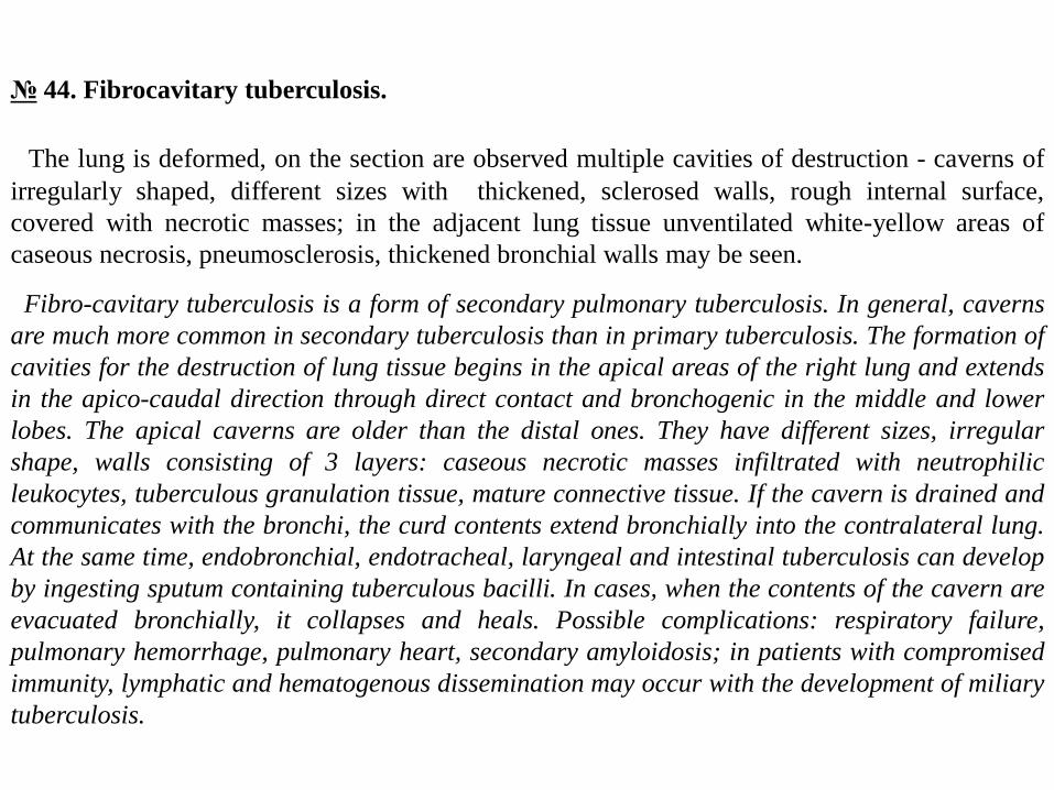
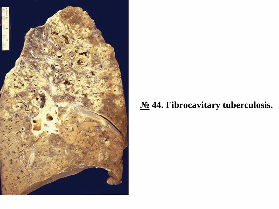
№ 44. Fibrocavitary tuberculosis.
The lung is deformed, on the section are observed multiple cavities of destruction - caverns of
irregularly shaped, different sizes with thickened, sclerosed walls, rough internal surface,
covered with necrotic masses; in the adjacent lung tissue unventilated white-yellow areas of
caseous necrosis, pneumosclerosis, thickened bronchial walls may be seen.
Fibro-cavitary tuberculosis is a form of secondary pulmonary tuberculosis. In general, caverns
are much more common in secondary tuberculosis than in primary tuberculosis. The formation of
cavities for the destruction of lung tissue begins in the apical areas of the right lung and extends
in the apico-caudal direction through direct contact and bronchogenic in the middle and lower
lobes. The apical caverns are older than the distal ones. They have different sizes, irregular
shape, walls consisting of 3 layers: caseous necrotic masses infiltrated with neutrophilic
leukocytes, tuberculous granulation tissue, mature connective tissue. If the cavern is drained and
communicates with the bronchi, the curd contents extend bronchially into the contralateral lung.
At the same time, endobronchial, endotracheal, laryngeal and intestinal tuberculosis can develop
by ingesting sputum containing tuberculous bacilli. In cases, when the contents of the cavern are
evacuated bronchially, it collapses and heals. Possible complications: respiratory failure,
pulmonary hemorrhage, pulmonary heart, secondary amyloidosis; in patients with compromised
immunity, lymphatic and hematogenous dissemination may occur with the development of miliary
tuberculosis.
№ 44. Fibrocavitary tuberculosis.
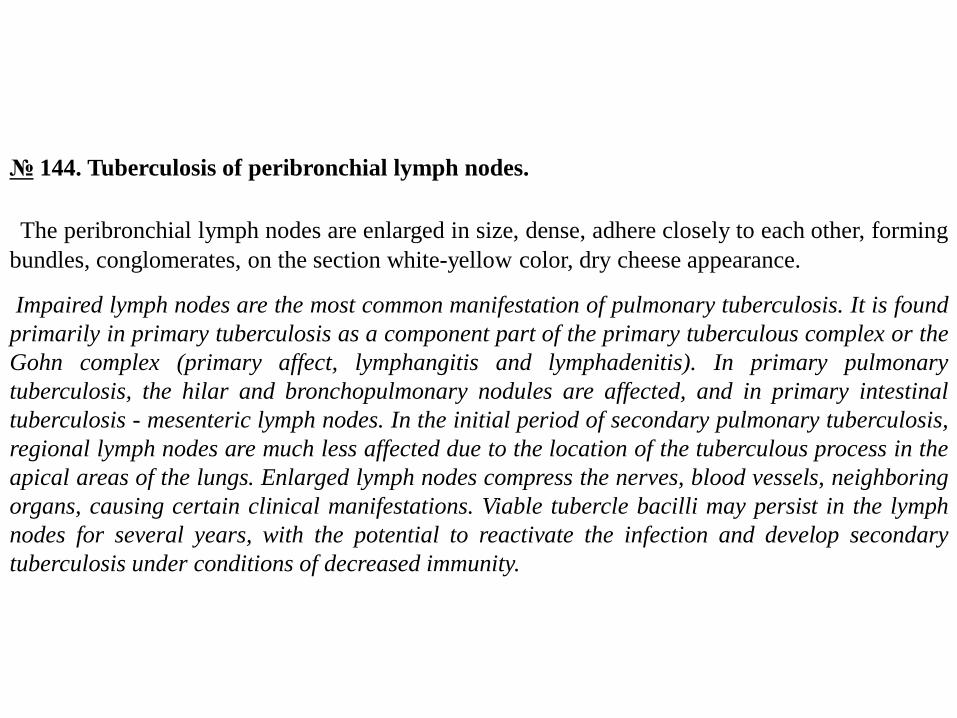
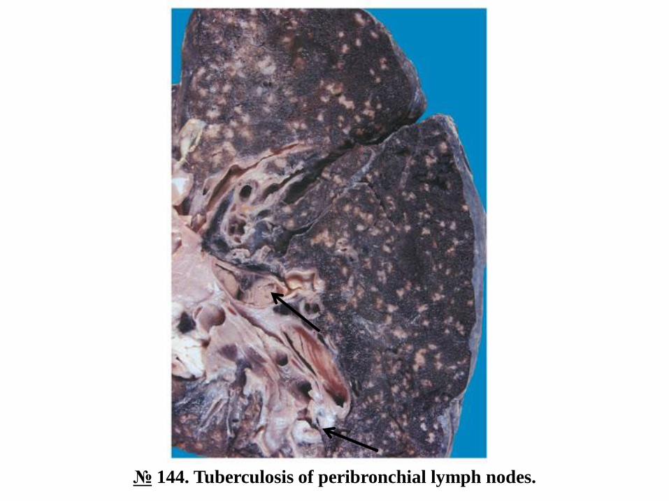
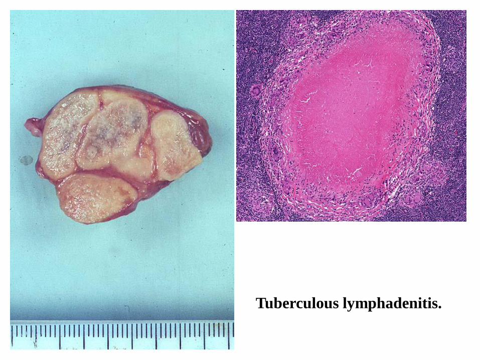
№ 144. Tuberculosis of peribronchial lymph nodes.
The peribronchial lymph nodes are enlarged in size, dense, adhere closely to each other, forming
bundles, conglomerates, on the section white-yellow color, dry cheese appearance.
Impaired lymph nodes are the most common manifestation of pulmonary tuberculosis. It is found
primarily in primary tuberculosis as a component part of the primary tuberculous complex or the
Gohn complex (primary affect, lymphangitis and lymphadenitis). In primary pulmonary
tuberculosis, the hilar and bronchopulmonary nodules are affected, and in primary intestinal
tuberculosis - mesenteric lymph nodes. In the initial period of secondary pulmonary tuberculosis,
regional lymph nodes are much less affected due to the location of the tuberculous process in the
apical areas of the lungs. Enlarged lymph nodes compress the nerves, blood vessels, neighboring
organs, causing certain clinical manifestations. Viable tubercle bacilli may persist in the lymph
nodes for several years, with the potential to reactivate the infection and develop secondary
tuberculosis under conditions of decreased immunity.
№ 144. Tuberculosis of peribronchial lymph nodes.
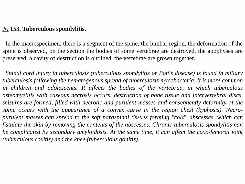
№ 153. Tuberculous spondylitis.
In the macrospecimen, there is a segment of the spine, the lumbar region, the deformation of the
spine is observed, on the section the bodies of some vertebrae are destroyed, the apophyses are
preserved, a cavity of destruction is outlined, the vertebrae are grown together.
Spinal cord injury in tuberculosis (tuberculous spondylitis or Pott's disease) is found in miliary
tuberculosis following the hematogenous spread of tuberculosis mycobacteria. It is more common
in children and adolescents. It affects the bodies of the vertebrae, in which tuberculous
osteomyelitis with caseous necrosis occurs, destruction of bone tissue and intervertebral discs,
seizures are formed, filled with necrotic and purulent masses and consequently deformity of the
spine occurs with the appearance of a convex curve in the region chest (kyphosis). Necro-
purulent masses can spread to the soft paraspinal tissues forming "cold" abscesses, which can
fistulate the skin by removing the contents of the abscesses. Chronic tuberculosis spondylitis can
be complicated by secondary amyloidosis. At the same time, it can affect the coxo-femoral joint
(tuberculous coxitis) and the knee (tuberculous gonitis).
№ 153. Tuberculous spondylitis.
(Pott disease).
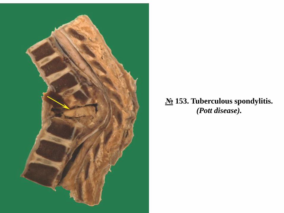
Influenza
hemorrhagic
tracheobronchitis.
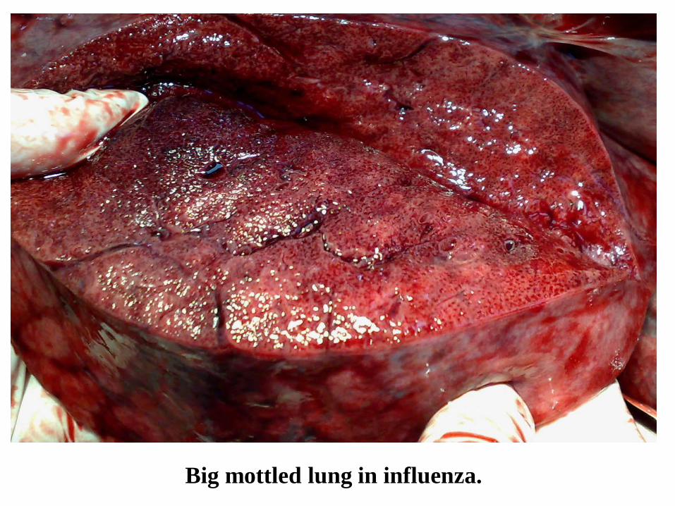
Big mottled lung in influenza.
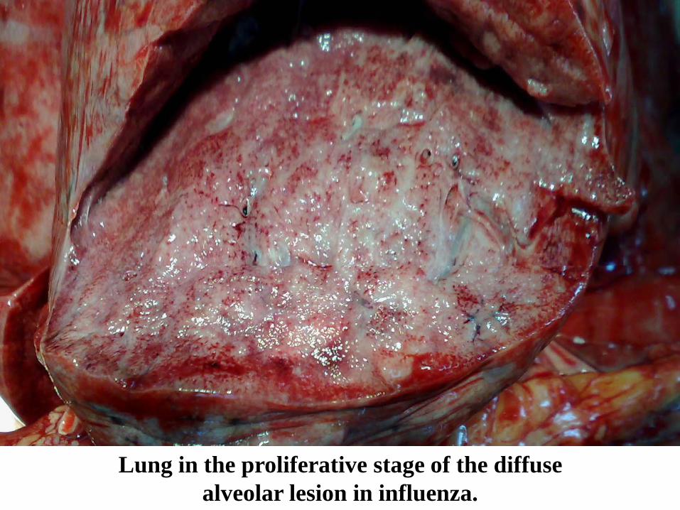
Lung in the proliferative stage of the diffuse
alveolar lesion in influenza.
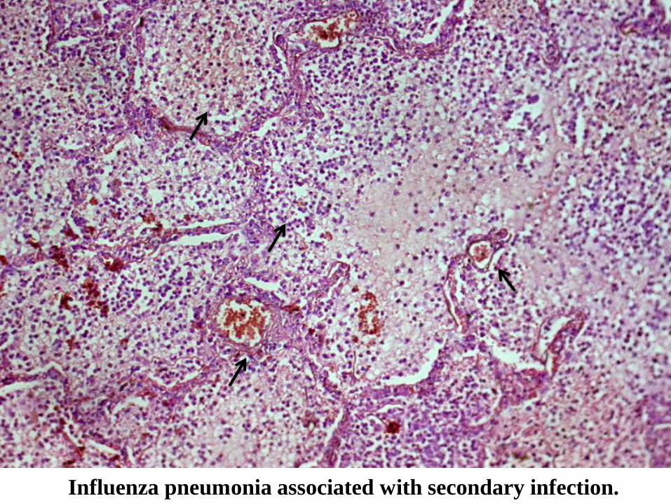
Influenza pneumonia associated with secondary infection.
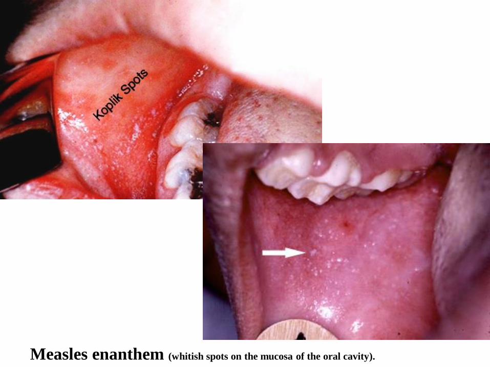
Measles enanthem (whitish spots on the mucosa of the oral cavity).
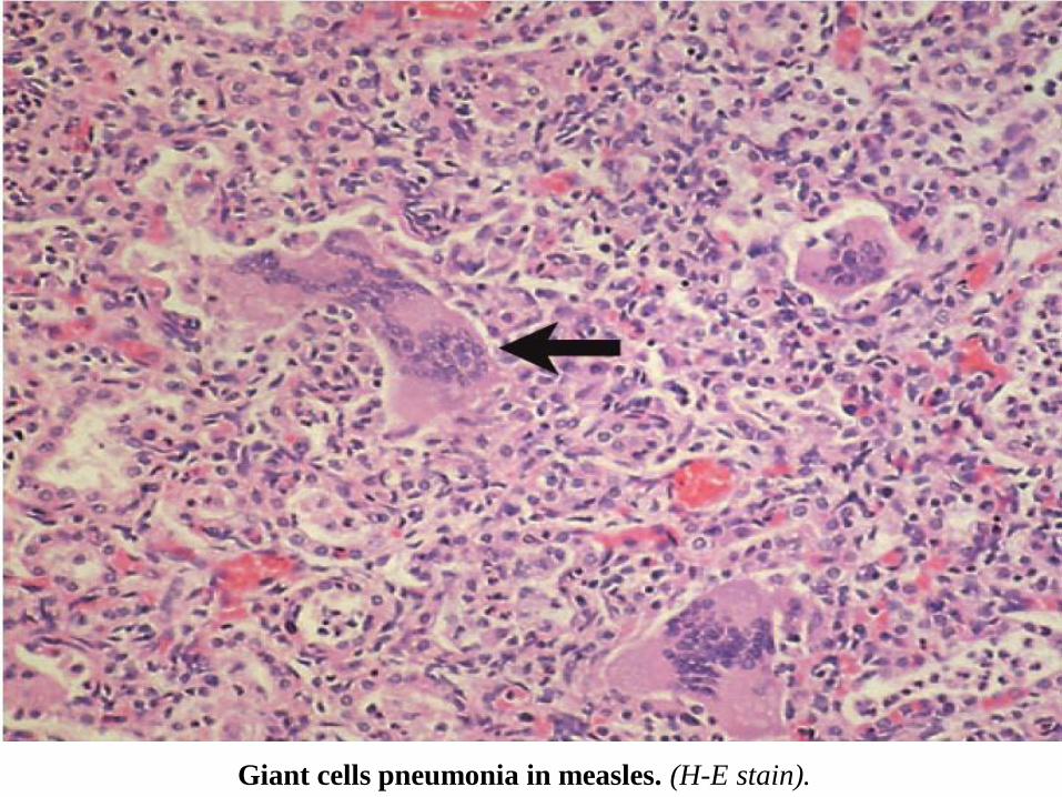
Giant cells pneumonia in measles. (H-E stain).
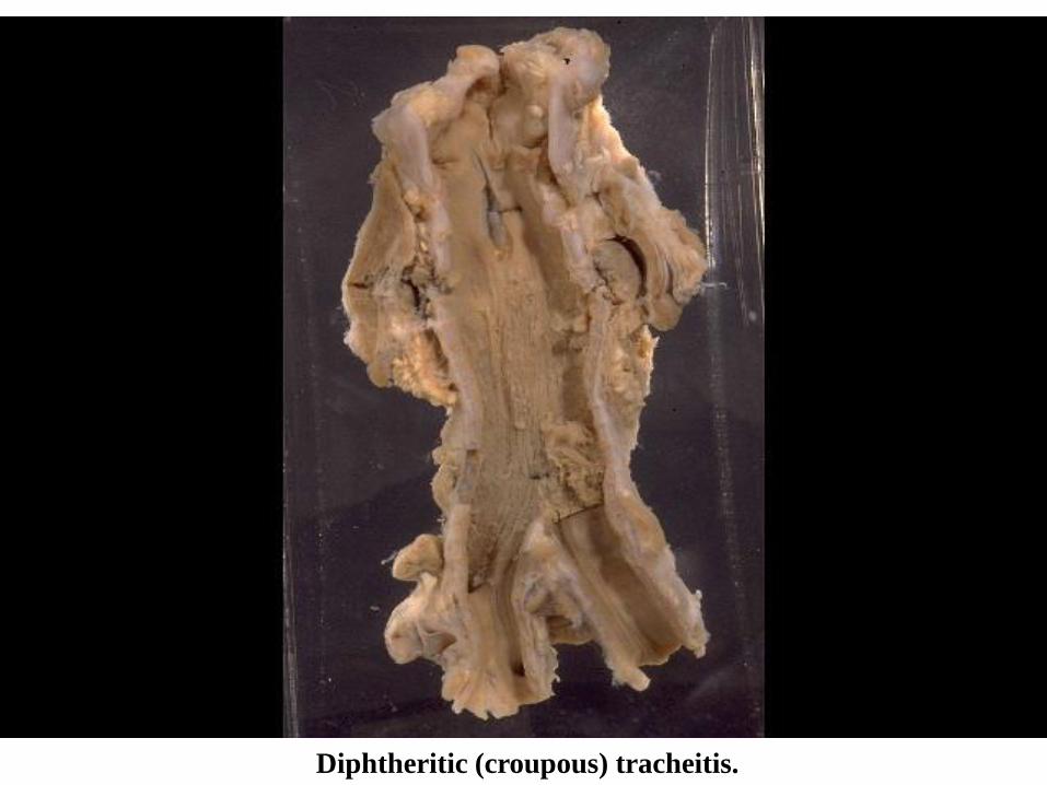
Diphtheritic (croupous) tracheitis.
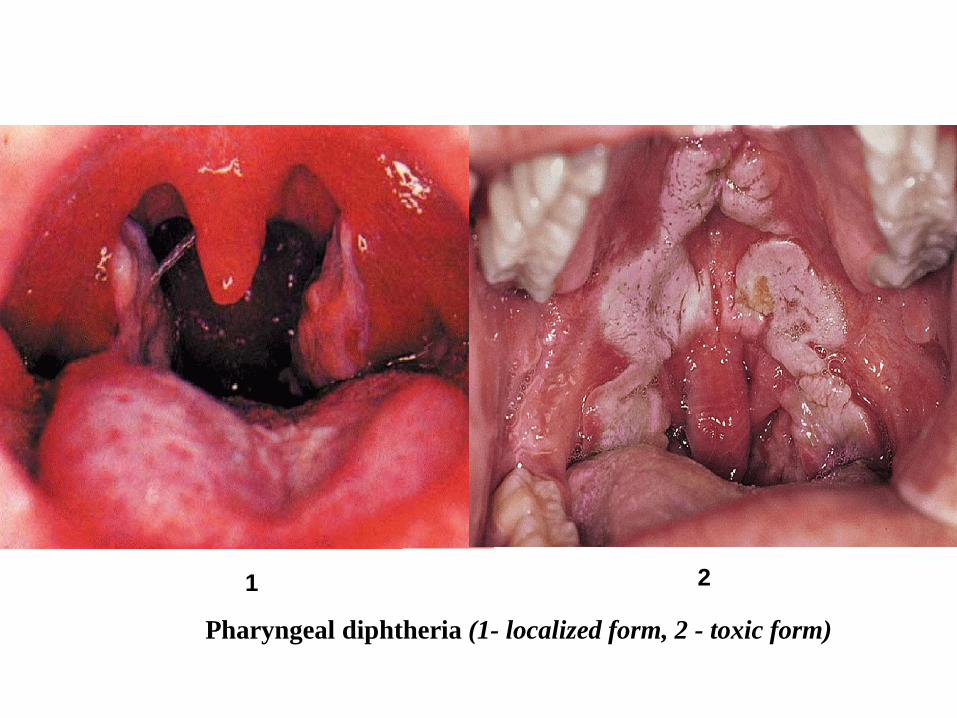
Pharyngeal diphtheria (1- localized form, 2 - toxic form)
1 2
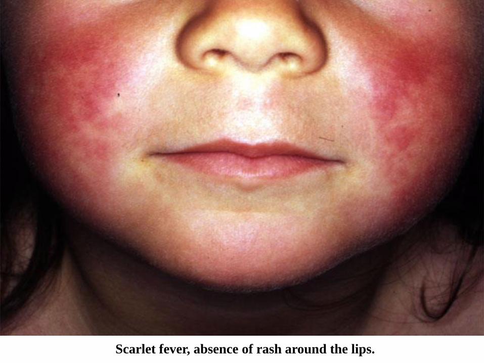
Scarlet fever, absence of rash around the lips.

Strawberry tongue in scarlet fever.
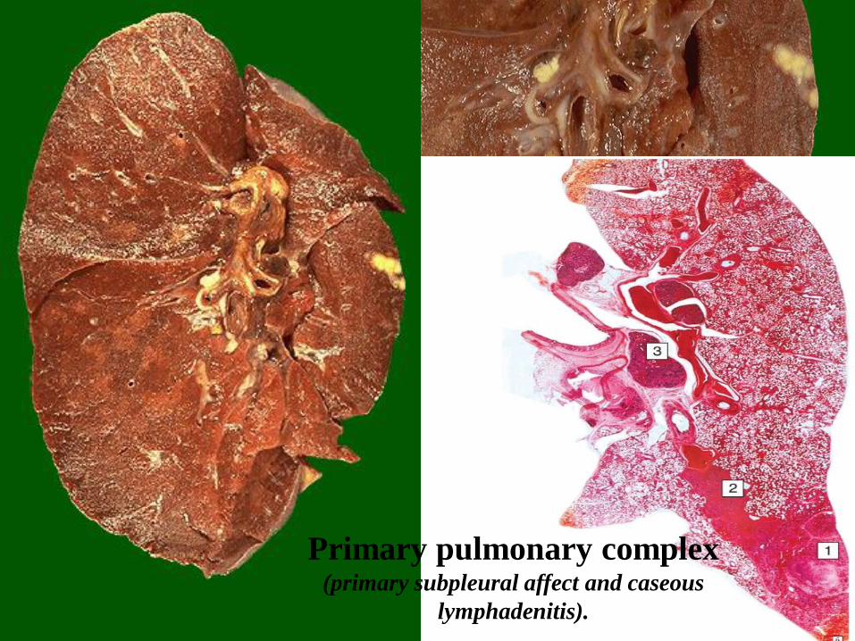
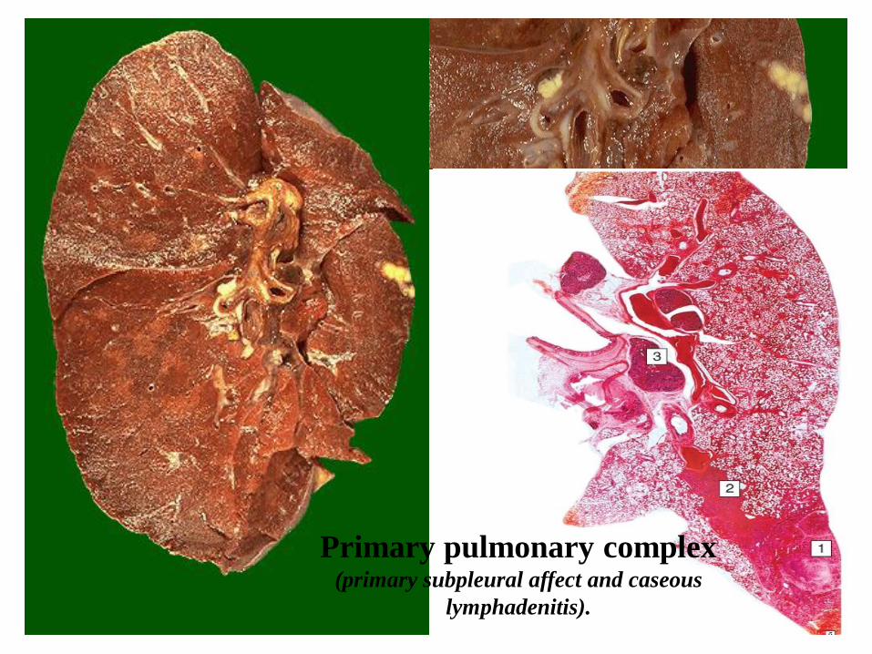
Primary pulmonary complex(primary subpleural affect and caseous
lymphadenitis).
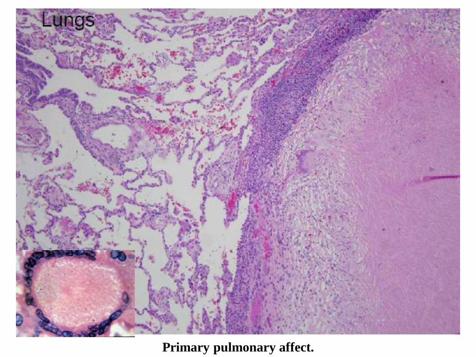
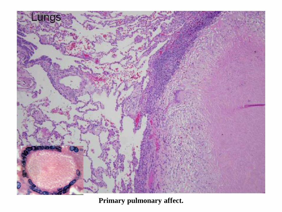
Primary pulmonary affect.
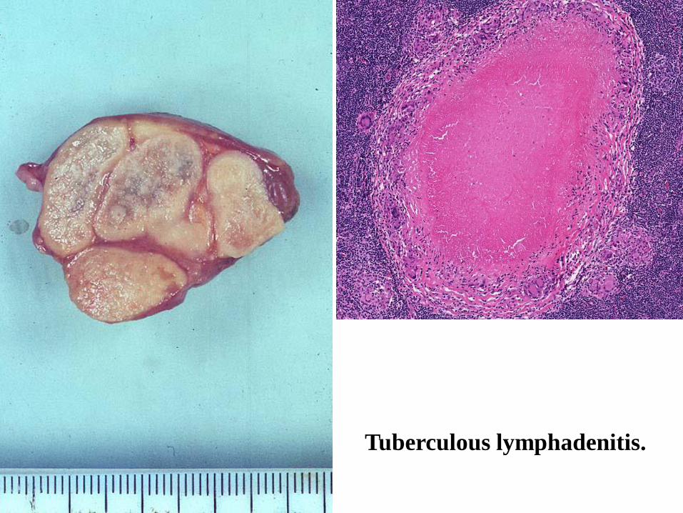
Tuberculous lymphadenitis.
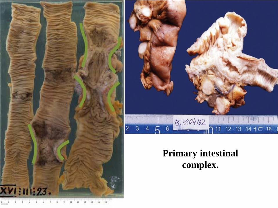
Primary intestinal
complex.
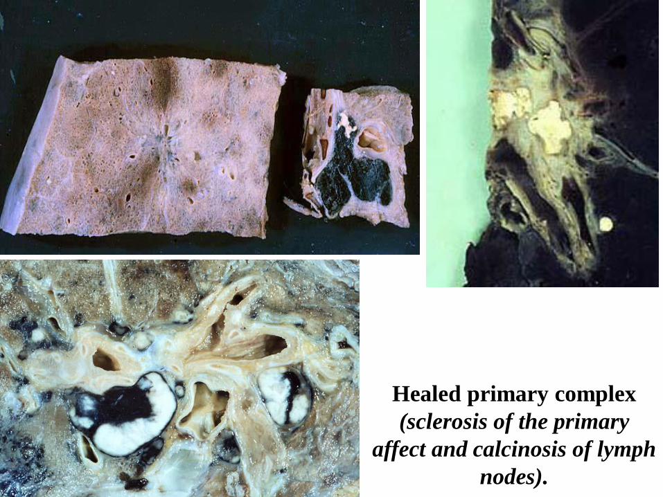
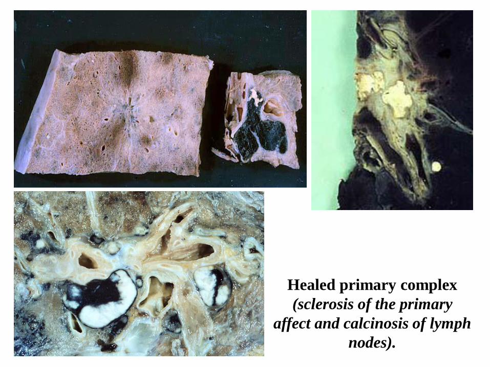
Healed primary complex
(sclerosis of the primary
affect and calcinosis of lymph
nodes).
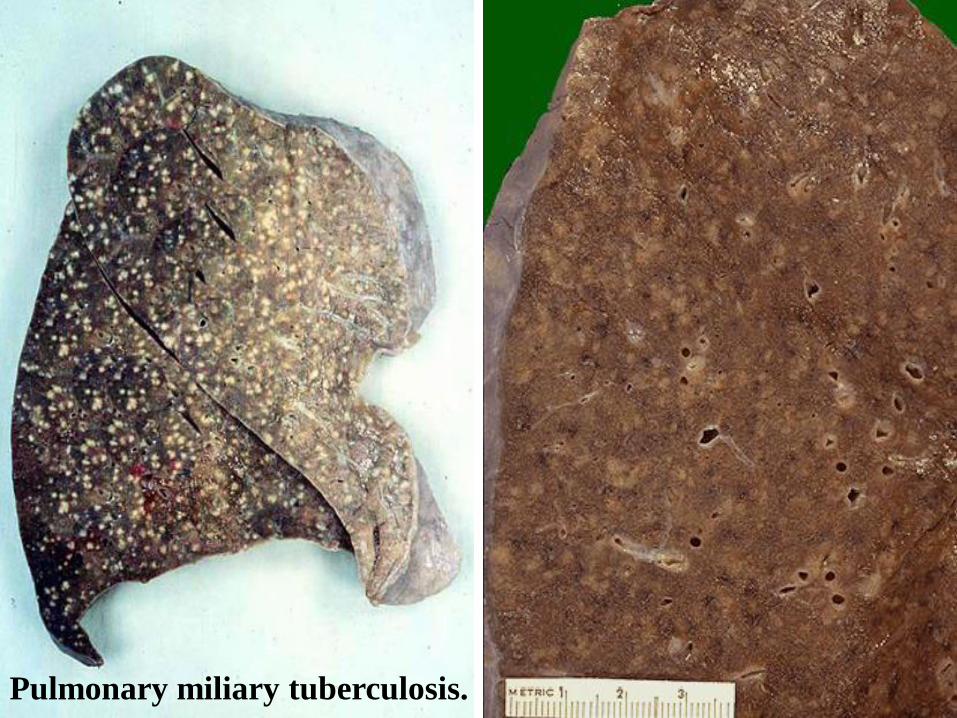
Pulmonary miliary tuberculosis.
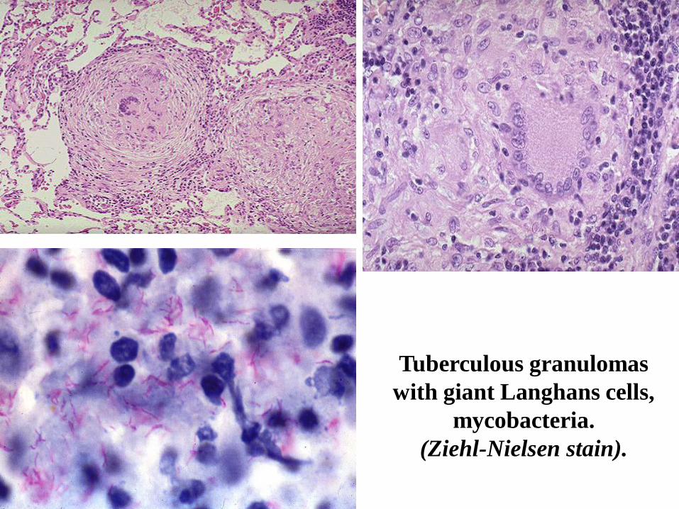
Tuberculous granulomas
with giant Langhans cells,
mycobacteria.
(Ziehl-Nielsen stain).
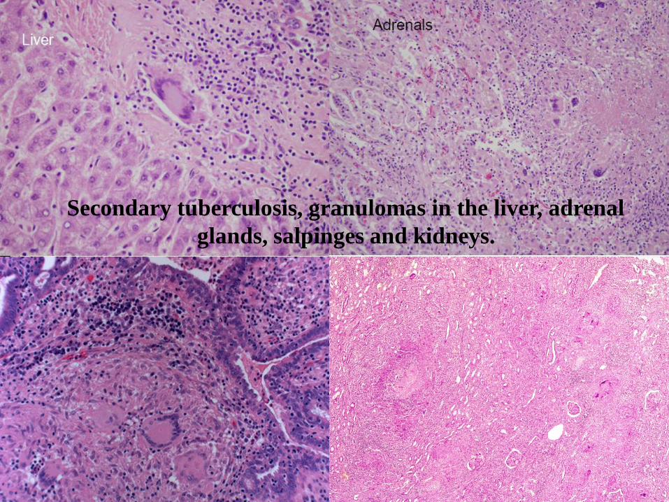
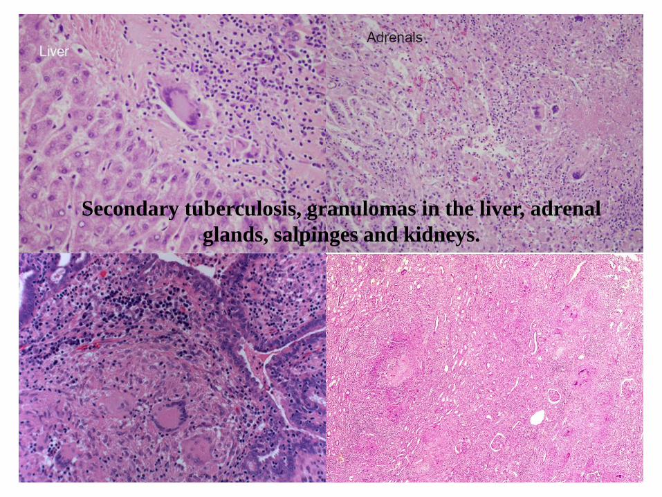
Secondary tuberculosis, granulomas in the liver, adrenal
glands, salpinges and kidneys.
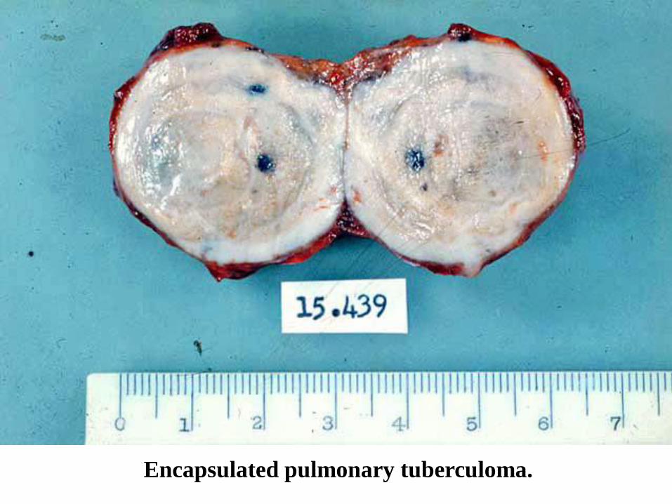
Encapsulated pulmonary tuberculoma.
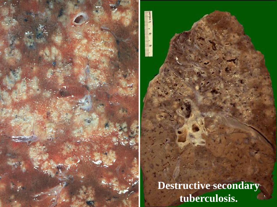
Destructive secondary
tuberculosis.
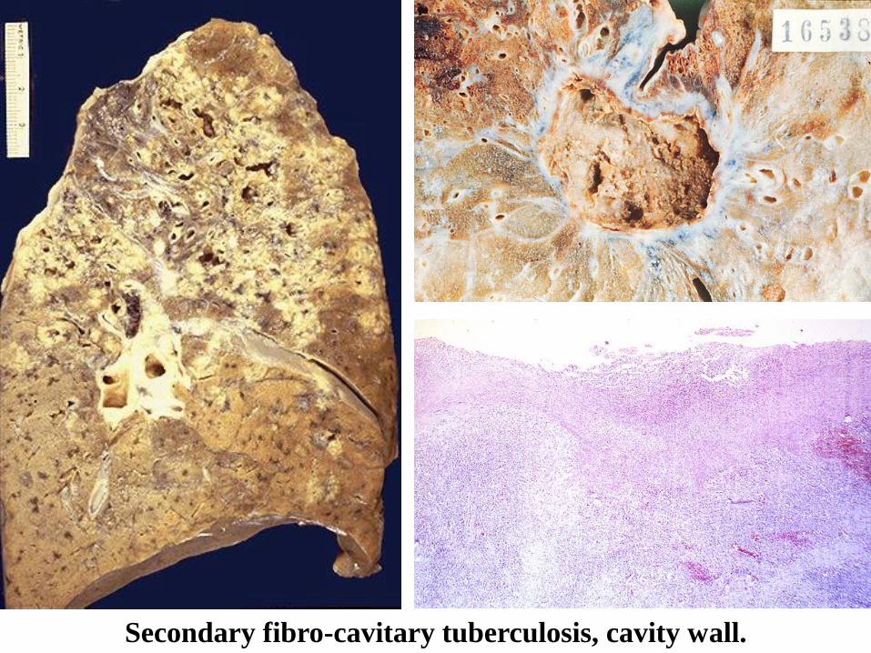
Secondary fibro-cavitary tuberculosis, cavity wall.
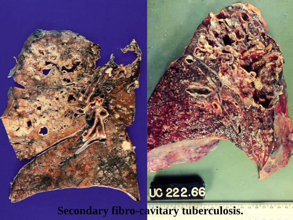
Secondary fibro-cavitary tuberculosis.
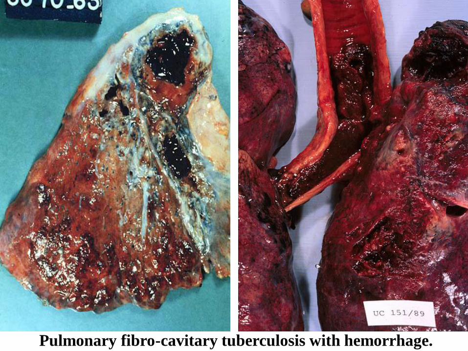
Pulmonary fibro-cavitary tuberculosis with hemorrhage.

Tuberculosis.
AIDS.
Aerogenic
infections.
Introduction
:➢Infects 1/3 to ½ of world population..!
➢3 million deaths due to TB every year
➢Under privileged population -
▪ Crowding, Poverty, malnutrition.
➢Since 1985 incidence is increasing in west
▪ AIDS, Diabetes, Immunosuppressed
patients, Drug resistance.
➢Tuberculosis (TB) remains the leading cause of death
worldwide from a single infectious disease agent.
Indeed up to 1/2 of the world's population is
infected with TB. The registered number of new
cases of TB worldwide roughly correlates with
economic conditions: the highest incidences are seen
in those countries of Africa, Asia, and Latin America
with the lowest gross national products. WHO
estimates that eight million people get TB every year,
of whom 95% live in developing countries. An
estimated 2 million people die from TB every year.
➢ It is estimated that between 2000 and 2021, nearly one billion people will be newly infected, 200 million people will get sick, and 35 million will die from TB - if control is not further strengthened. The mechanisms, pathogenesis, and prophylaxis knowledge is minimal. After a century of decline TB is increasing and there are strains emerging which are resistant to antibiotics. This excess of cases is attributable to the changes in the social structure in cities, the human immunodeficiency virus epidemic, and failure of most cities to improve public health programs, and the economic cost of treating.
➢With the increased incidence of AIDS, TB has
become more a problem in the U.S., and the
world.
➢It is currently estimated that 1/2 of the world's
population (3.1 billion) is infected with
Mycobacterium tuberculosis. Mycobacterium
avium complex is associated with AIDS
related TB.
➢TB is an ancient infectious disease caused by
Mycobacterium tuberculosis. It has been
known since 1000 B.C., so it not a new
disease. Since TB is a disease of respiratory
transmission, optimal conditions for
transmission include:
▪ overcrowding
▪ poor personal hygiene
▪ poor public hygiene
Transmissio
n➢Pulmonary tuberculosis is a disease of
respiratory transmission, Patients with the
active disease (bacilli) expel them into the air
by:
▪ coughing,
▪ sneezing,
▪ shouting,
▪ or any other way that will expel bacilli into the air
➢Once inhaled by a tuberculin free person, the
bacilli multiply 4 -6 weeks and spreads
throughout the body. The bacilli implant in
areas of high partial pressure of oxygen:
➢lung
➢renal cortex
➢reticuloendothelial system
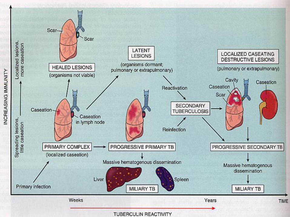
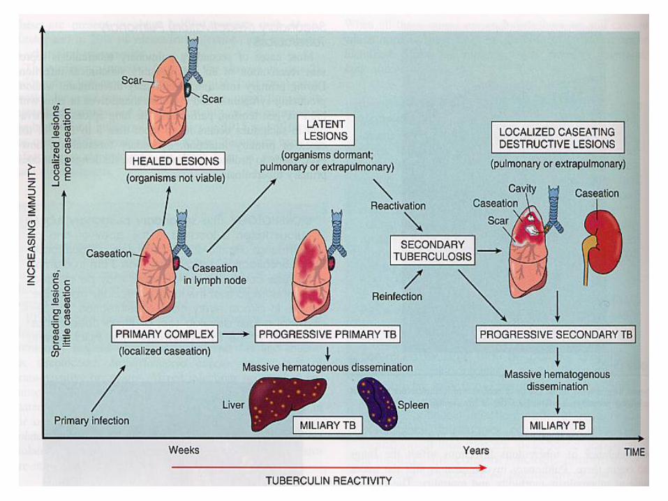
➢This is known as the primary infection. The patient will heal and a scar will appear in the infected loci. There will also be a few viable bacilli/spores may remain in these areas (particularly in the lung). The bacteria at this time goes into a dormant state, as long as the person's immune system remains active and functions normally this person isn't bothered by the dormant bacillus.
➢When a person's immune system is depressed., a secondary reactivation occurs. 85-90% of the cases seen which are of secondary reactivation type occurs in the lungs.
Pathogenesis of
TB:
➢Type IV hypersensitivity – T cells –
Macrophages → Granuloma
➢Activated macrophages – epithelioid cells.
➢Remain viable inside macrophages
➢Self destruction by lysosomal enzymes.
Microbiology of
TB:➢Mycobacteria – ‘fungus like..
➢Bacilli, Aerobic, no toxins, no spore.
➢M. tuberculosis & M. bovis
➢M. avium, M.intracellulare in AIDS -Atypical TB
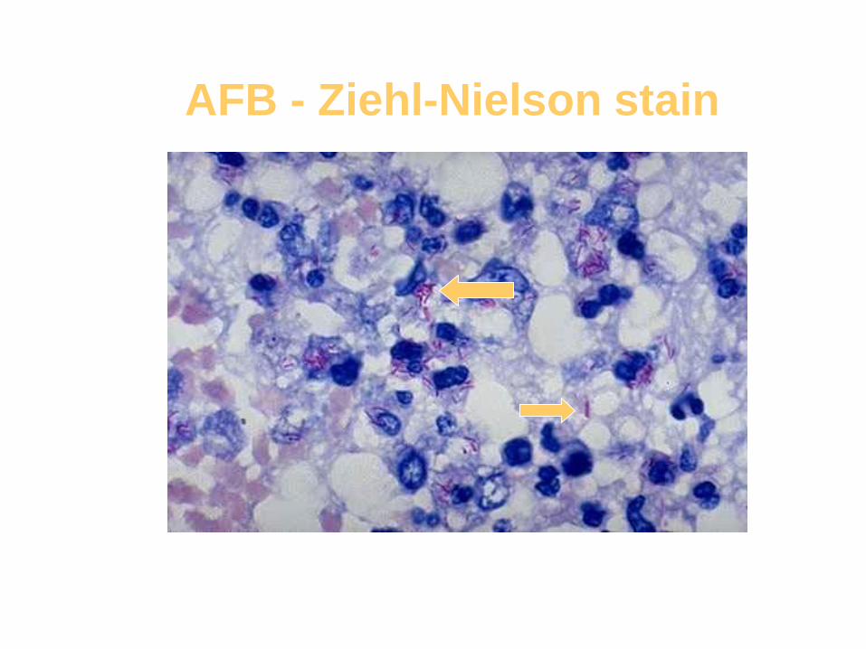
AFB - Ziehl-Nielson stain
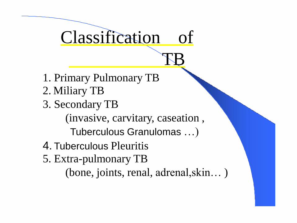
Classification of
TB1. Primary Pulmonary TB
2. Miliary TB
3. Secondary TB
(invasive, carvitary, caseation ,
Tuberculous Granulomas …)
4. Tuberculous Pleuritis
5. Extra-pulmonary TB
(bone, joints, renal, adrenal,skin… )
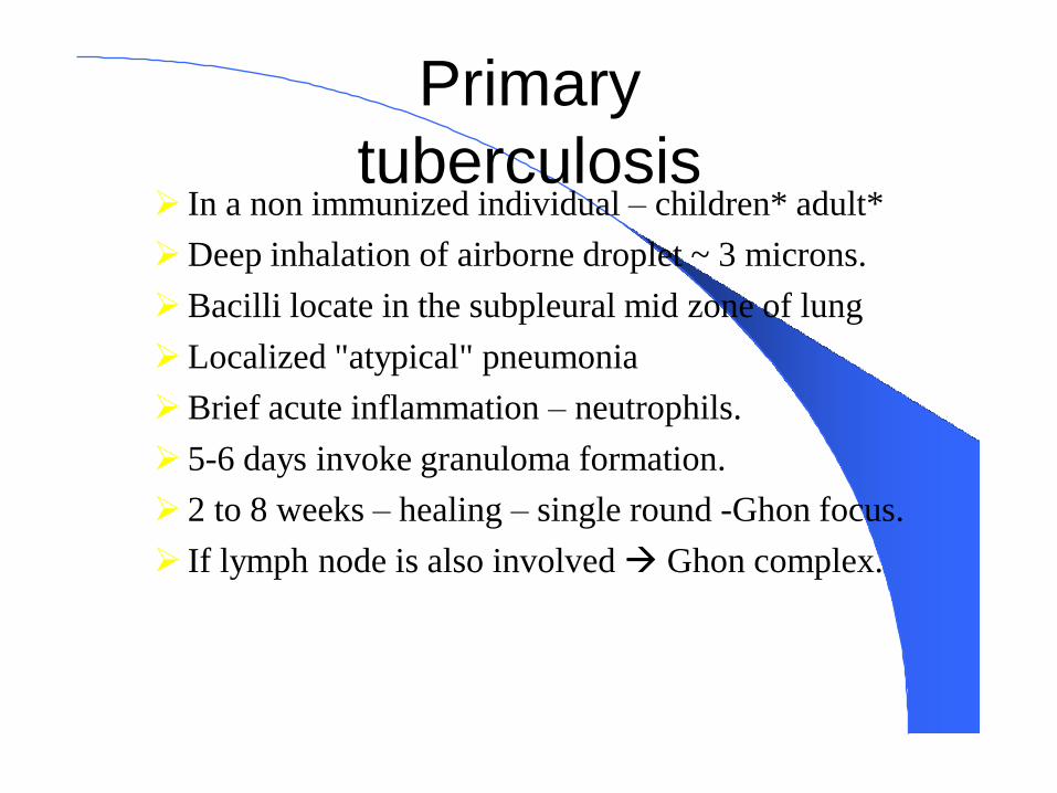
Primary
tuberculosis➢ In a non immunized individual – children* adult*
➢Deep inhalation of airborne droplet ~ 3 microns.
➢Bacilli locate in the subpleural mid zone of lung
➢Localized "atypical" pneumonia
➢Brief acute inflammation – neutrophils.
➢ 5-6 days invoke granuloma formation.
➢ 2 to 8 weeks – healing – single round -Ghon focus.
➢ If lymph node is also involved→ Ghon complex.
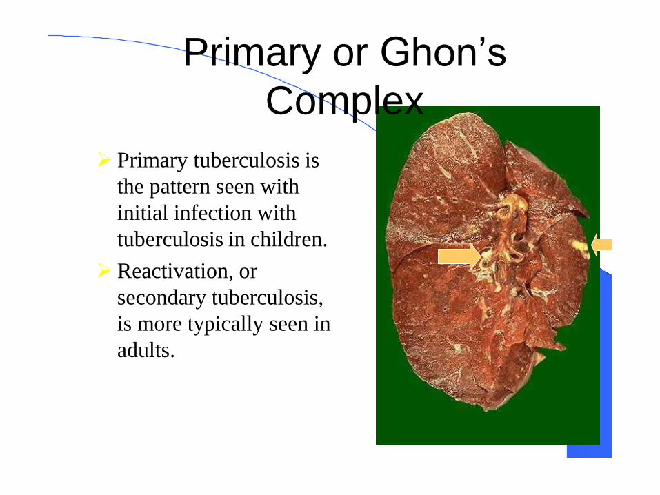
Primary or Ghon’s
Complex
➢ Primary tuberculosis is
the pattern seen with
initial infection with
tuberculosis in children.
➢Reactivation, or
secondary tuberculosis,
is more typically seen in
adults.
Primary
Tuberculosis• In Non Immunized individuals
(Children)
➢Primary Tuberculosis:▪ Self Limited disease
▪ Ghons focus, complex or Primary complex.
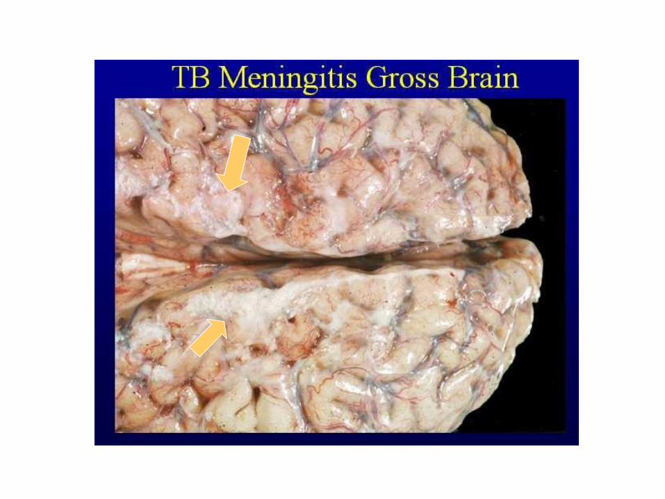
➢Primary Progressive TB ( in US. )▪ Miliary TB and TB Meningitis.
▪ Common in malnourished children
▪ 10% of adults, Immuno-suppressed individuals
Primary pulmonary complex(primary subpleural affect and caseous
lymphadenitis).
Primary pulmonary affect.
Tuberculous lymphadenitis.
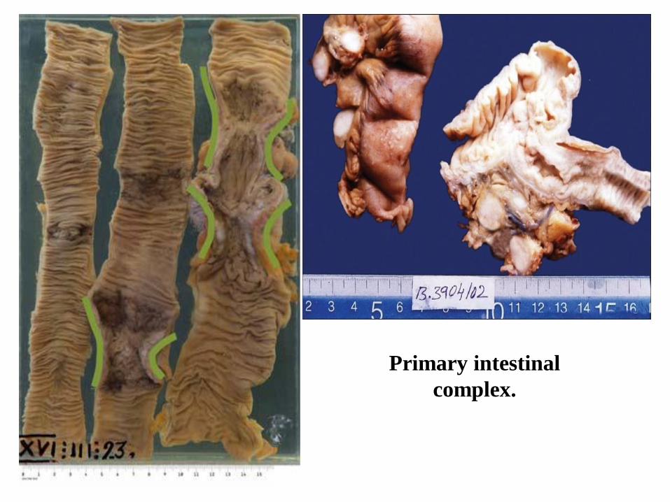
Primary intestinal
complex.
Healed primary complex
(sclerosis of the primary
affect and calcinosis of lymph
nodes).
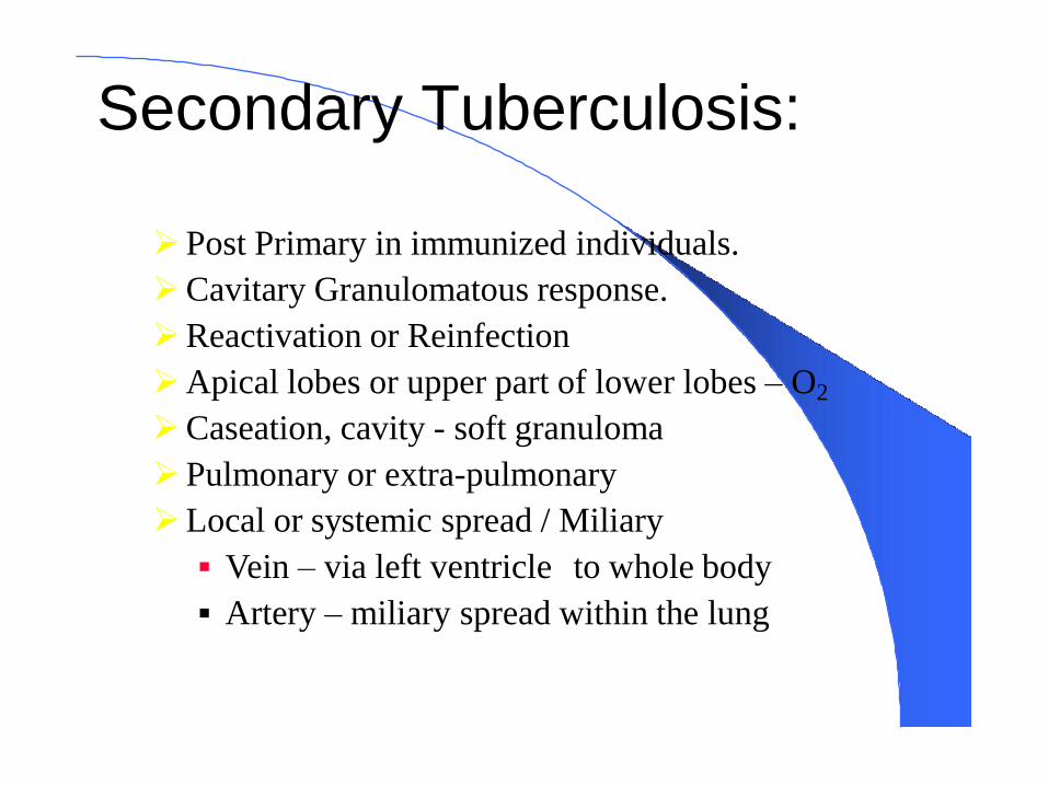
Secondary Tuberculosis:
➢Post Primary in immunized individuals.
➢Cavitary Granulomatous response.
➢Reactivation or Reinfection
➢Apical lobes or upper part of lower lobes – O2
➢Caseation, cavity - soft granuloma
➢Pulmonary or extra-pulmonary
➢Local or systemic spread / Miliary
▪ Vein – via left ventricle to whole body
▪ Artery – miliary spread within the lung
Secondary Tuberculosis:
➢Reactivation occurs in 10-15% of patients.
➢Most commonly males 30-50 y
➢Slowly Progressive (several months)
➢Cough, sputum, Low grade fever, night sweats,
fatigue and weight loss.
➢Hemoptysis or pleuritic pain = severe disease
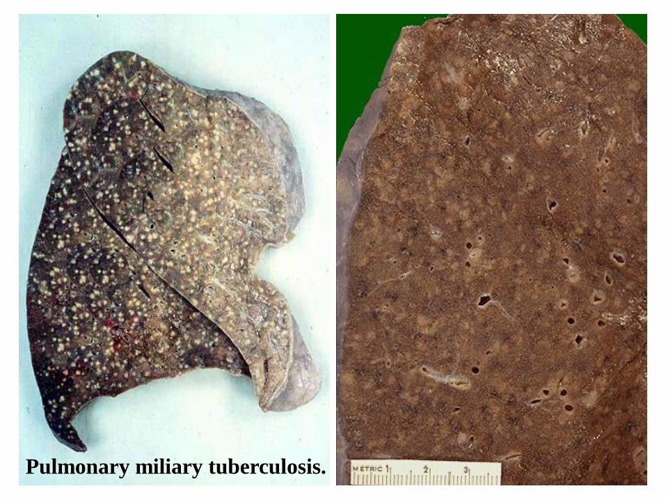
Pulmonary miliary tuberculosis.
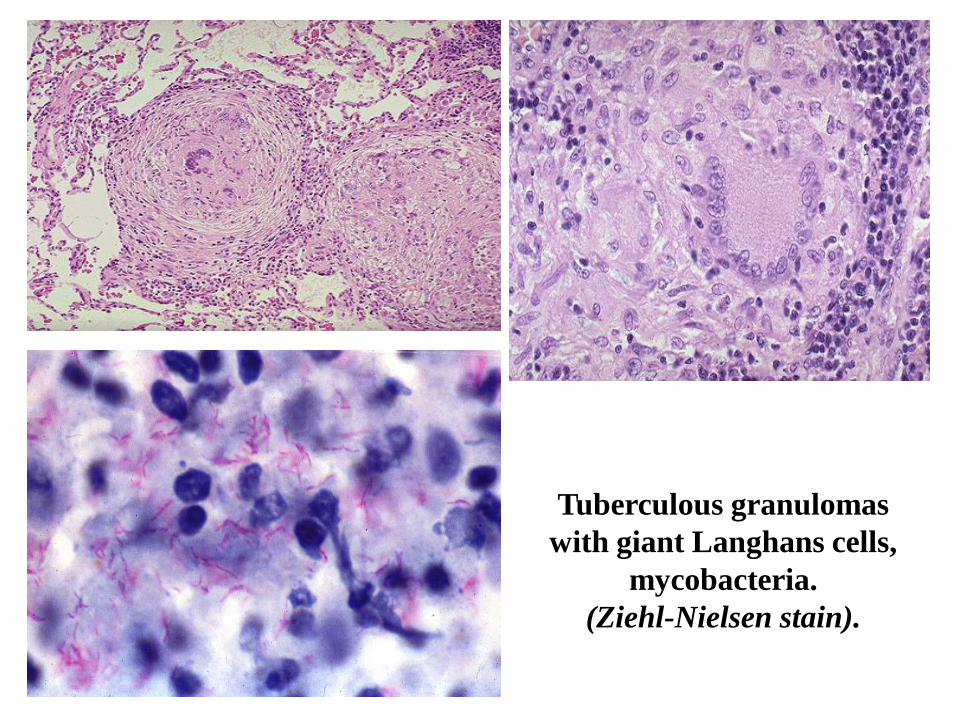
Tuberculous granulomas
with giant Langhans cells,
mycobacteria.
(Ziehl-Nielsen stain).
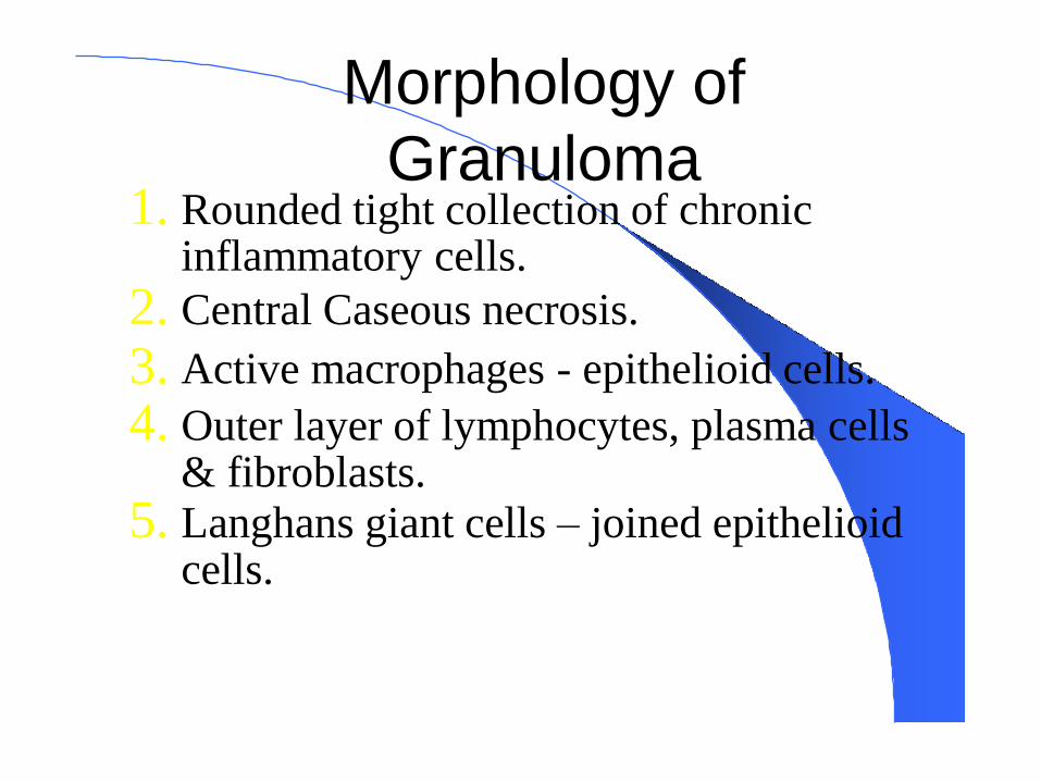
Morphology of
Granuloma1. Rounded tight collection of chronic
inflammatory cells.
2. Central Caseous necrosis.
3. Active macrophages - epithelioid cells.
4. Outer layer of lymphocytes, plasma cells & fibroblasts.
5. Langhans giant cells – joined epithelioid cells.
Secondary tuberculosis, granulomas in the liver, adrenal
glands, salpinges and kidneys.
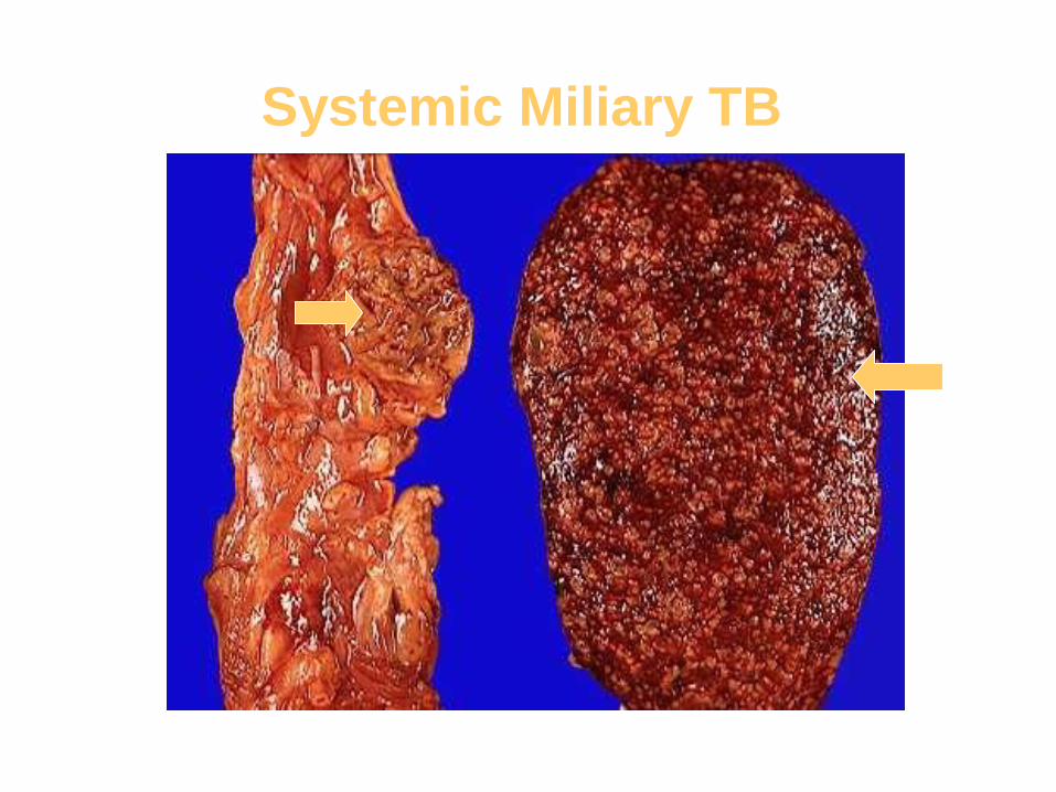
Systemic Miliary TB
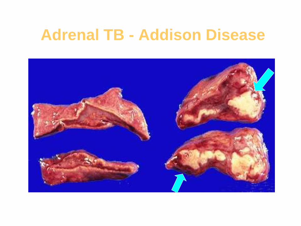
Adrenal TB - Addison Disease
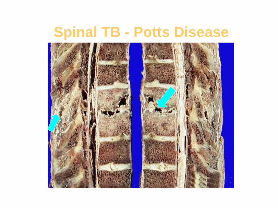
Spinal TB - Potts Disease
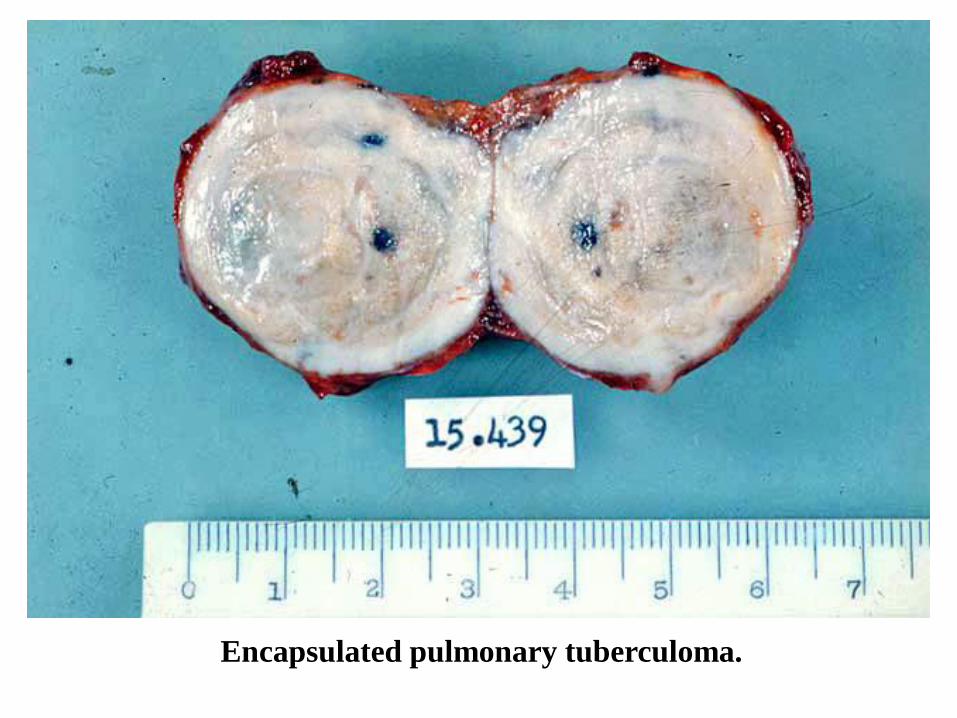
Encapsulated pulmonary tuberculoma.
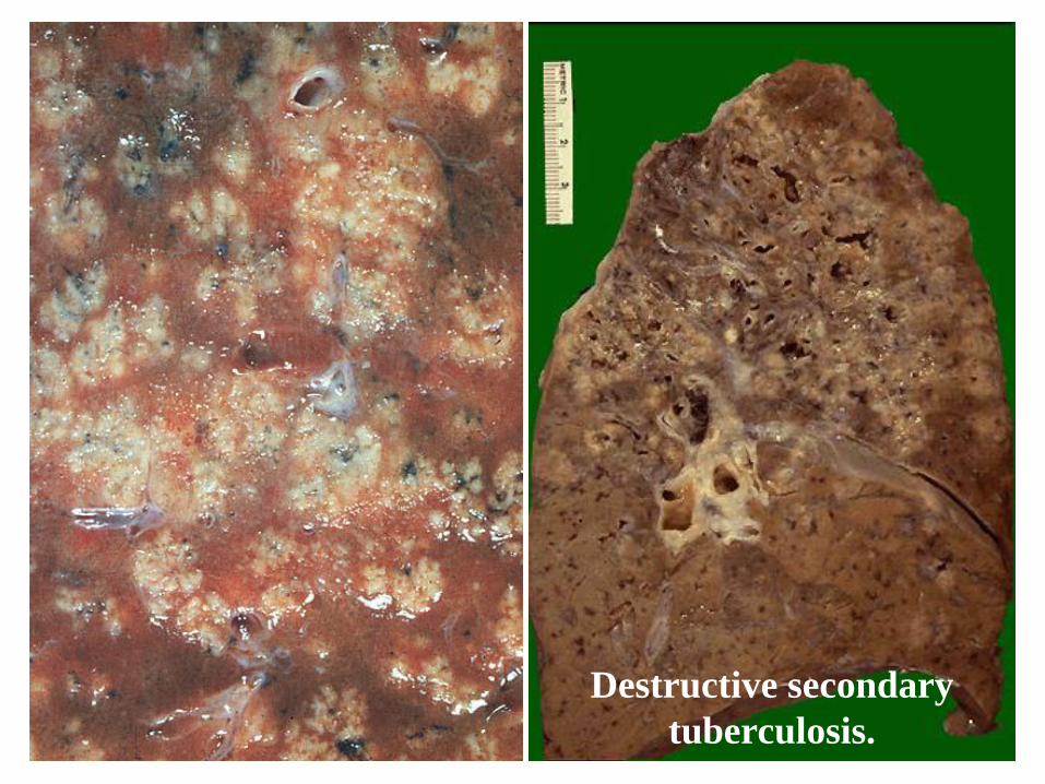
Destructive secondary
tuberculosis.
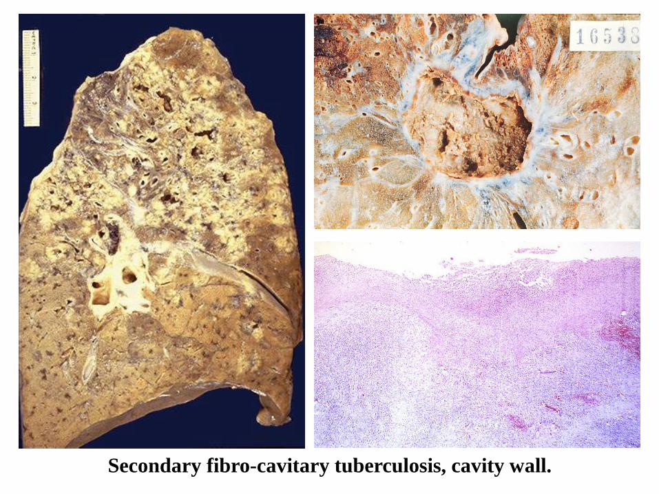
Secondary fibro-cavitary tuberculosis, cavity wall.
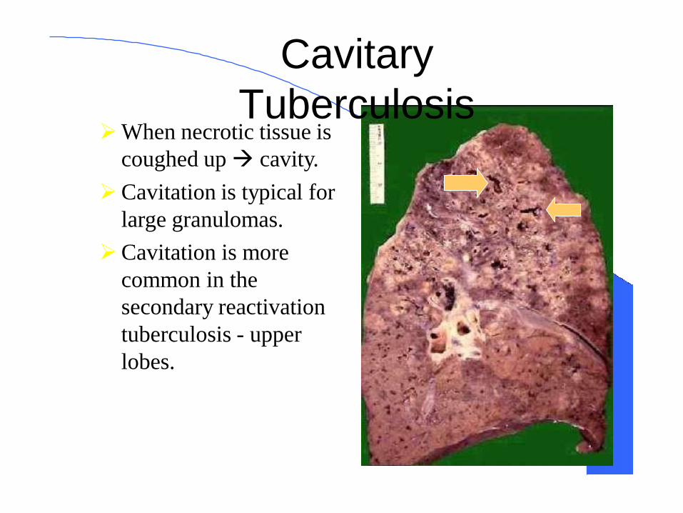
Cavitary
Tuberculosis➢When necrotic tissue is
coughed up→ cavity.
➢Cavitation is typical for
large granulomas.
➢Cavitation is more
common in the
secondary reactivation
tuberculosis - upper
lobes.
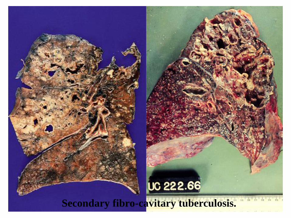
Secondary fibro-cavitary tuberculosis.
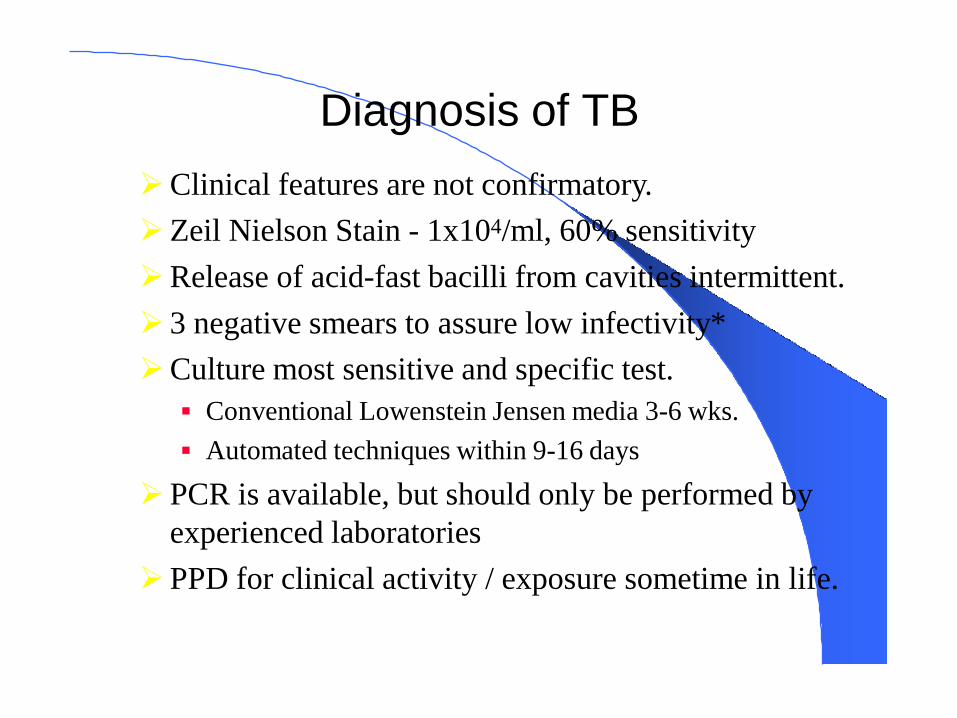
Diagnosis of TB
➢Clinical features are not confirmatory.
➢Zeil Nielson Stain - 1x10⁴/ml, 60% sensitivity
➢Release of acid-fast bacilli from cavities intermittent.
➢ 3 negative smears to assure low infectivity*
➢Culture most sensitive and specific test.
▪ Conventional Lowenstein Jensen media 3-6 wks.
▪ Automated techniques within 9-16 days
➢PCR is available, but should only be performed by
experienced laboratories
➢PPD for clinical activity / exposure sometime in life.
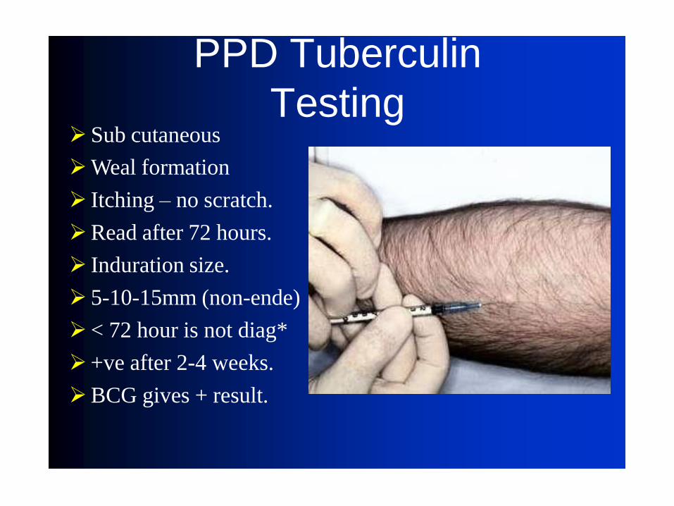
PPD Tuberculin
Testing➢ Sub cutaneous
➢Weal formation
➢ Itching – no scratch.
➢Read after 72 hours.
➢ Induration size.
➢ 5-10-15mm (non-ende)
➢ < 72 hour is not diag*
➢+ve after 2-4 weeks.
➢BCG gives + result.
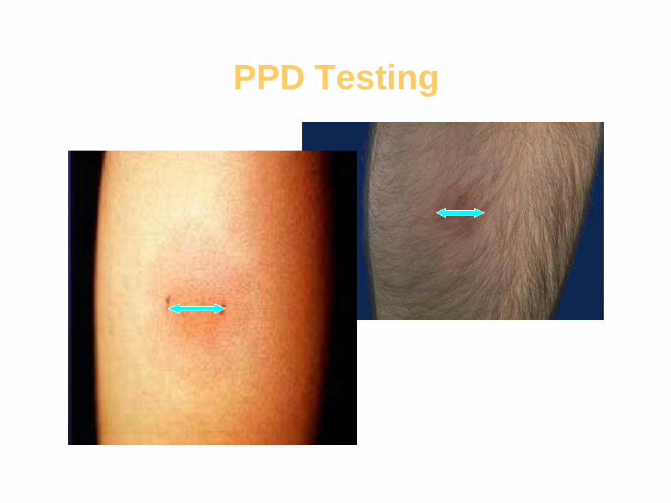
PPD Testing
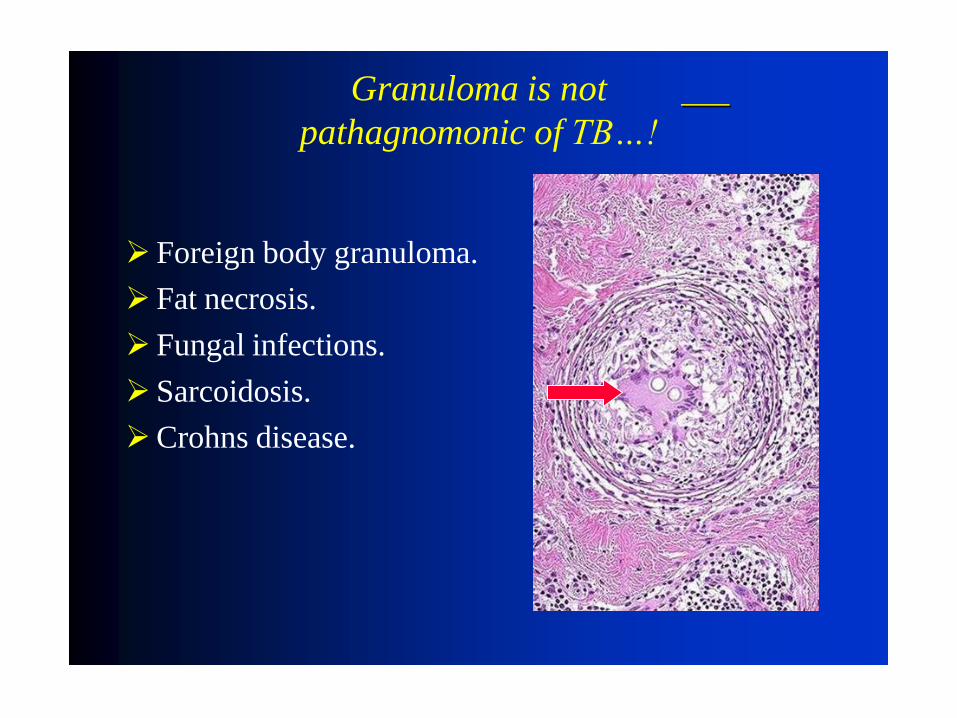
Granuloma is not
pathagnomonic of TB…!
➢ Foreign body granuloma.
➢ Fat necrosis.
➢ Fungal infections.
➢ Sarcoidosis.
➢Crohns disease.
Conclusions:
➢A chronic, common, infectious disease - Weight loss, fever, night sweats, lung damage.
➢Commonest fatal infectious disease in the world.
➢AIDS, Diabetes, malnutrition (poverty), crowding.
➢ Pulmonary,miliary,invasive,pleuritis,extrapulmonary,.
➢ Prevention depends on PPD & INH prophylaxis
What is New…?
➢14-30% of TB patients also HIV infected.
➢New drugs - Rifapentine, Interferons,
Thalidomide.
➢Immune therapy : Killed M. vaccine stimulates
CD8 cells (increased INF and IL-12).
➢The genome of TB has been identified (~4000
genes) potential to develop new vaccines and
tests.
Tuberculosis.
AIDS.
Aerogenic
infections.
Influe nza
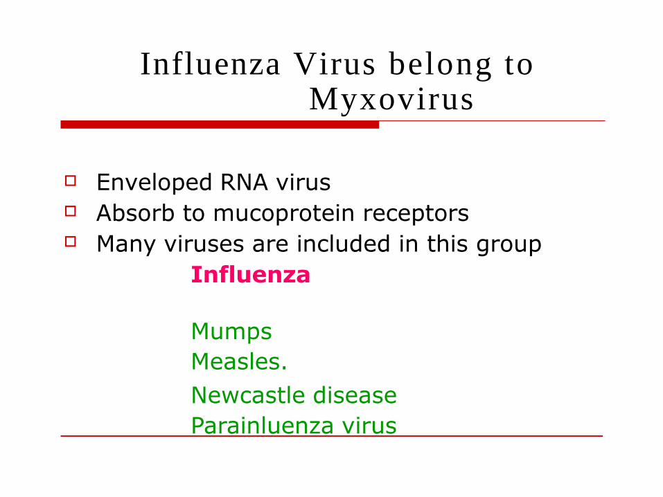
Influenza Virus belong to Myxovirus
Enveloped RNA virus
Absorb to mucoprotein receptors
Many viruses are included in this group
Influenza
Mumps
Measles.
Newcastle disease
Parainluenza virus
INFLUENZA
Cause of the infection of the respiratory tract.
Occurs as
Sporadic
Epidemic
Pandemic
Major pande m ic in 1918 – 1919
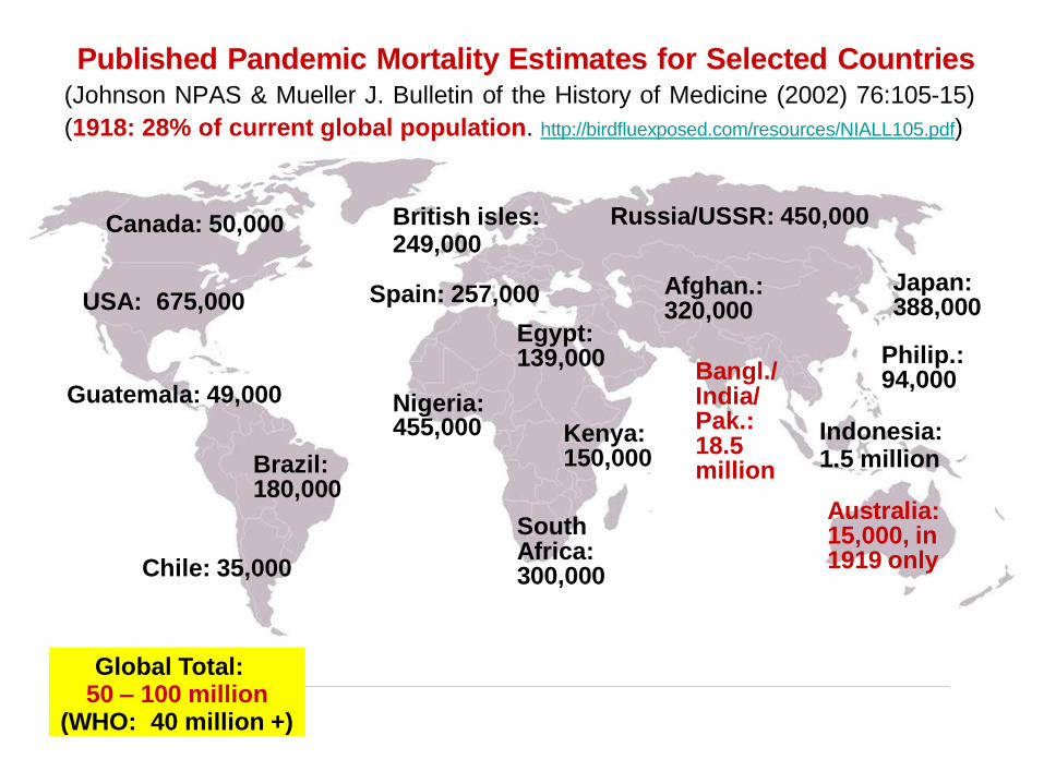
Published Pandemic Mortality Estimates for Selected Countries(Johnson NPAS & Mueller J. Bulletin of the History of Medicine (2002) 76:105-15)
(1918: 28% of current global population. http://birdfluexposed.com/resources/NIALL105.pdf)
USA: 675,000
Bangl./ India/ Pak.: 18.5million
455,000
Guatemala: 49,000
Afghan.: 320,000
Indonesia:1.5 million
Japan: 388,000
Philip.: 94,000
Brazil: 180,000
South Africa: 300,000
Kenya: 150,000
Global Total: 50 – 100 million
(WHO: 40 million +)
Russia/USSR: 450,000Canada: 50,000
Chile: 35,000
Australia: 15,000, in1919 only
British isles: 249,000
Spain: 257,000
Egypt: 139,000
Nigeria:
Definition: Influenza is an acute, febrile, generalized viral
infection that affects the upper and lower respiratory tract.
Etiology: myxovirus influenzae, which has 3 major
antigenic types (A, B, C). Influenza virus (especially A)
is characterized by high antigenic variability. Genes
encoding surface proteins (hemagglutinin and
neuraminidase) are constantly changing, resulting in
new subtypes and antigenic variants, against which the
population is not immunized.
Classification of Influenza virus
What are A B C
Classification on the basis of
Ribonucleoprotein Antigen and Matrix
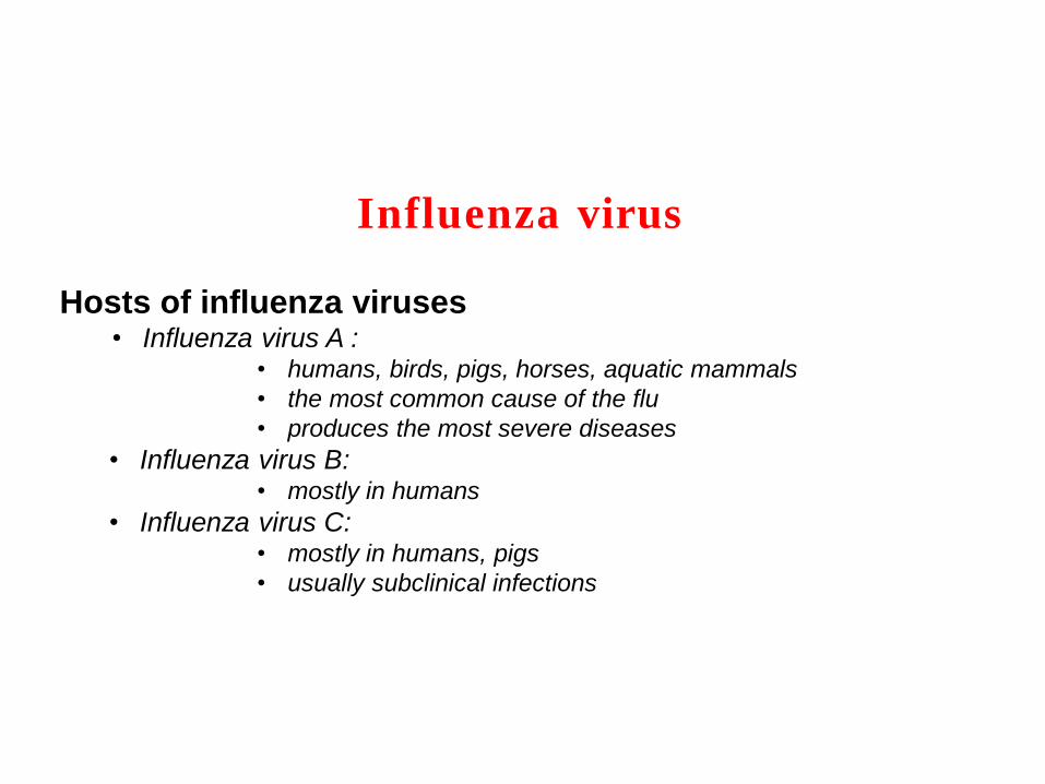
Influenza virus
Hosts of influenza viruses• Influenza virus A :
• humans, birds, pigs, horses, aquatic mammals
• the most common cause of the flu
• produces the most severe diseases
• Influenza virus B: • mostly in humans
• Influenza virus C:• mostly in humans, pigs
• usually subclinical infections
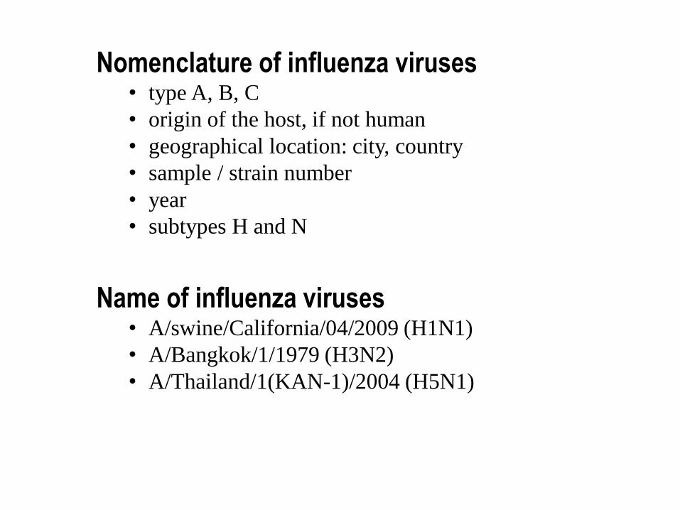
Nomenclature of influenza viruses• type A, B, C
• origin of the host, if not human
• geographical location: city, country
• sample / strain number
• year
• subtypes H and N
Name of influenza viruses• A/swine/California/04/2009 (H1N1)
• A/Bangkok/1/1979 (H3N2)
• A/Thailand/1(KAN-1)/2004 (H5N1)
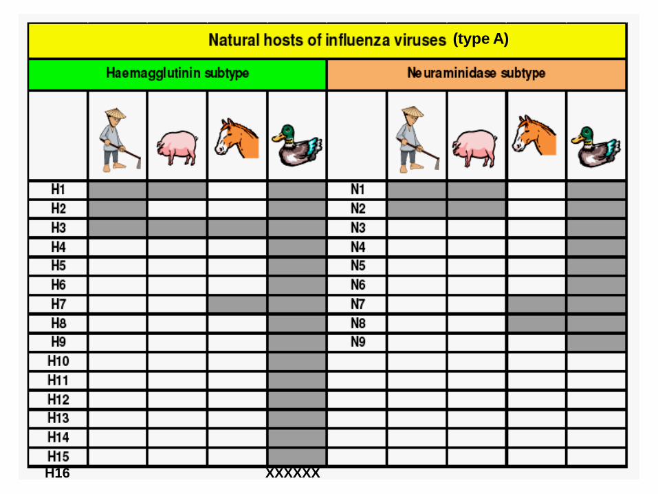
H16 XXXXXX
(type A)
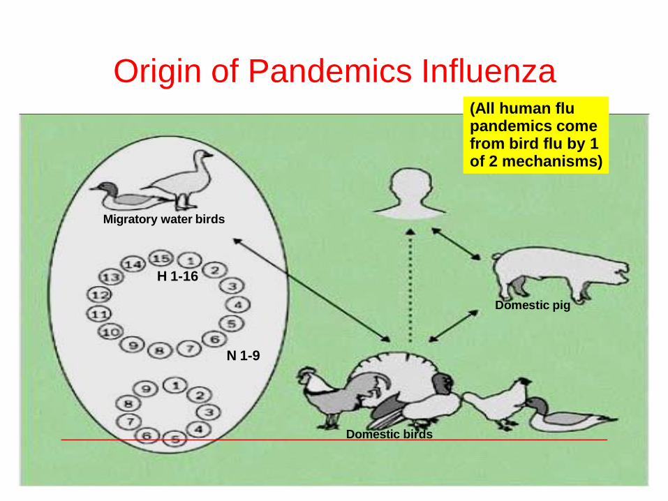
Origin of Pandemics Influenza
Migratory water birds
H 1-16
N 1-9
Domestic pig
Domestic birds
(All human flu pandemics come from bird flu by 1 of 2 mechanisms)
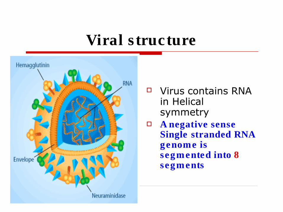
Viral s tructure
Virus contains RNAin Helical symmetry
A negative sense Single stranded RNA genome issegm ented into 8 segm ents
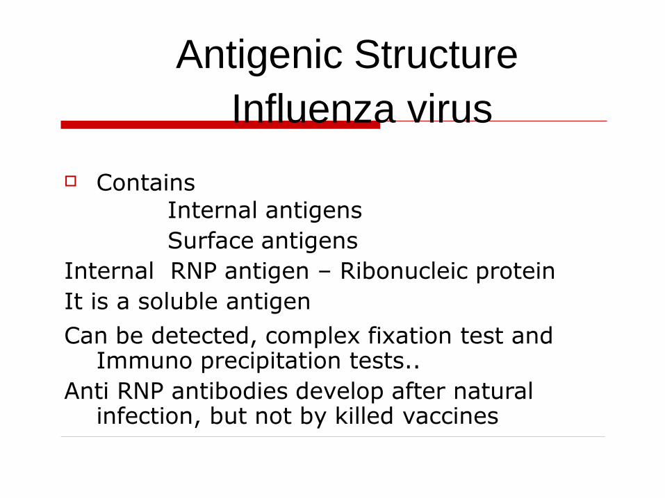
Antigenic Structure
Influenza virus
ContainsInternal antigens
Surface antigens
Internal RNP antigen – Ribonucleic protein
It is a soluble antigen
Can be detected, complex fixation test and Immuno precipitation tests..
Anti RNP antibodies develop after natural infection, but not by killed vaccines
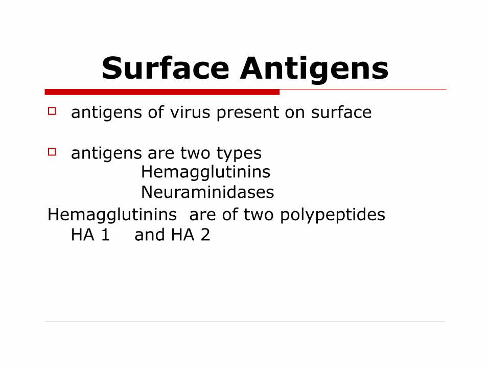
Surface Antigens antigens of virus present on surface
antigens are two typesHemagglutinins
Neuraminidases
Hemagglutinins are of two polypeptidesHA 1 and HA 2
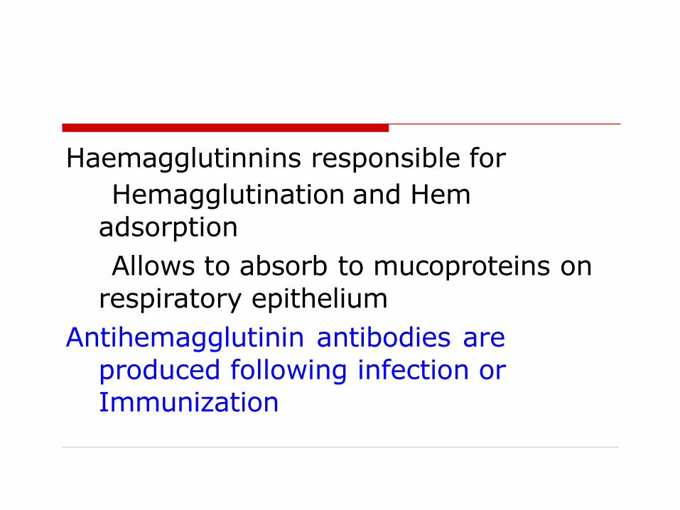
Haemagglutinnins responsible for
Hemagglutination and Hem
adsorption
Allows to absorb to mucoproteins on
respiratory epithelium
Antihemagglutinin antibodies are
produced following infection orImmunization
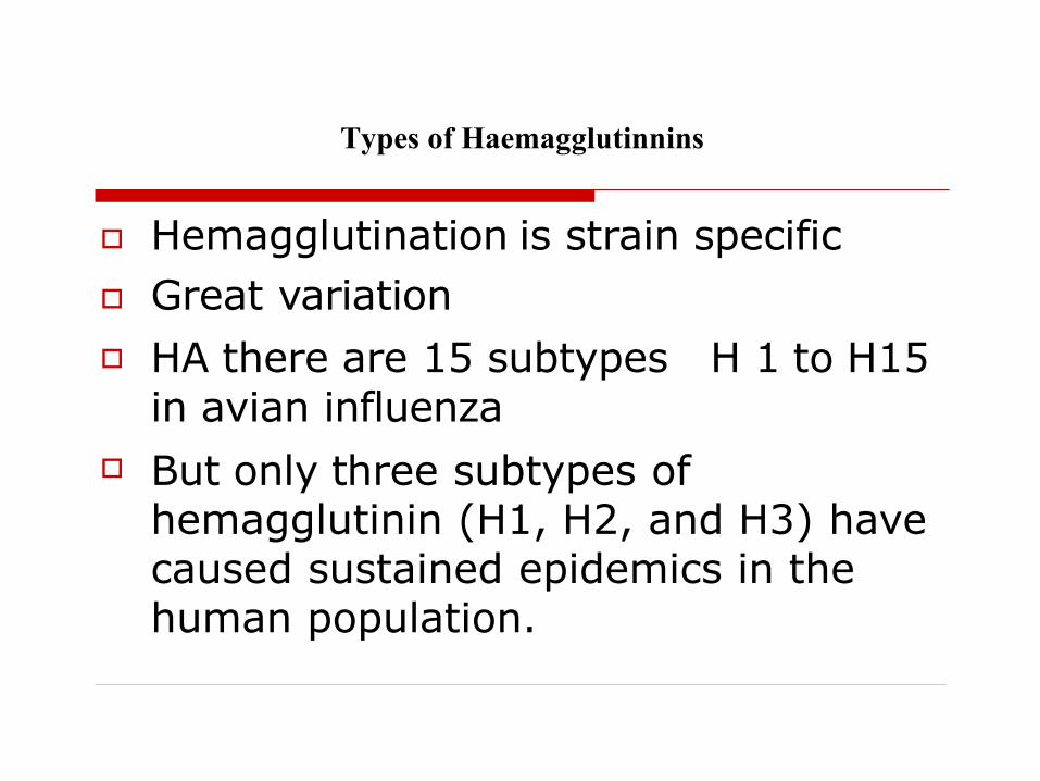
Hemagglutination is strain specific
Great variation
HA there are 15 subtypes H 1 to H15 in avian influenza
But only three subtypes of hemagglutinin (H1, H2, and H3) have caused sustained epidemics in the human population.
Types of Haemagglutinnins

Neuraminidases
Neuraminidase are glycoprotein's
Destroys cell receptors by hydrolysis cleavage
Anti neuraminidase antibodies are produced
following infection and immunization
Not protective as Antihemagglutinin
antibodies
Strain specific exhibit variation, There arenine different subtypesN 1 – N9
But only two subtypes of hemagglutinin (N1 and N2) have caused sustained epidemics in the human population.
Antigenic Variation
Unique feature of this virus lies withantigenic variation.
High in type A virus
Less in type B virus
Not in type C virus
RNP and Matrix proteins are stable Hemagglutination and Neuraminidase
are independ of the variations.
Influenza prominent Antigenic Changes
Antigenic Shift◼
◼
◼
major change, new subtype
caused by exchange of gene segments
may result in pandemic
Example of antigenic shift◼
◼
H2N2 virus circulated in 1957-1967
H3N2 virus appeared in 1968 and
completely replaced H2N2 virus
AntigenicShift
It is abrupt and Drastic
Discontinuous variation in structure in antigens
Results in novel virus and unrelated to previous strains causing infections
Involves – Hemagglutinins, Neuraminidase or both
Subtypes depends only on antigenic shifts, occurs on Hemagglutinins
Influenza Antigenic Changes
Antigenic Driftminor change, same subtype caused by point mutations in gene
◼
◼
◼ may result in epidemic Example of antigenic drift
◼
◼
in 2002-2003, A/Panama/2007/99 (H3N2) virus was dominantA/Fujian/411/2002 (H3N2) appeared inlate 2003 and caused widespread illness in 2003-2004
Pathogenesis
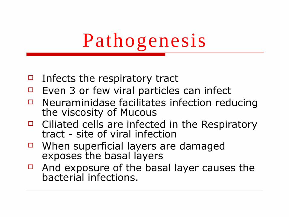
Infects the respiratory tract
Even 3 or few viral particles can infect
Neuraminidase facilitates infection reducing the viscosity of Mucous
Ciliated cells are infected in the Respiratory tract - site of viral infection
When superficial layers are damaged exposes the basal layers
And exposure of the basal layer causes the bacterial infections.
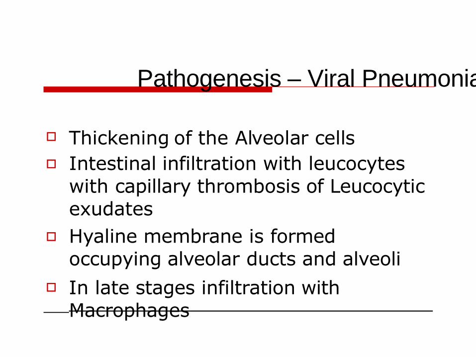
Pathogenesis – Viral Pneumonia
Thickening of the Alveolar cells
Intestinal infiltration with leucocytes
with capillary thrombosis of Leucocytic exudates
Hyaline membrane is formed occupying alveolar ducts and alveoli
In late stages infiltration withMacrophages
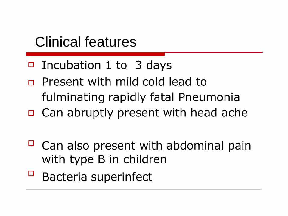
Clinical features
Incubation 1 to 3 days
Present with mild cold lead to
fulminating rapidly fatal Pneumonia
Can abruptly present with head ache
Can also present with abdominal pain
with type B in children
Bacteria superinfect
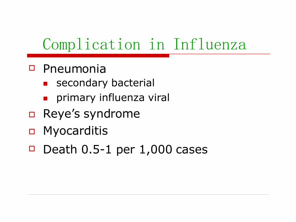
Complication in Influenza
Pneumonia◼
◼
secondary bacterial
primary influenza viral
Reye’s syndrome
Myocarditis
Death 0.5-1 per 1,000 cases
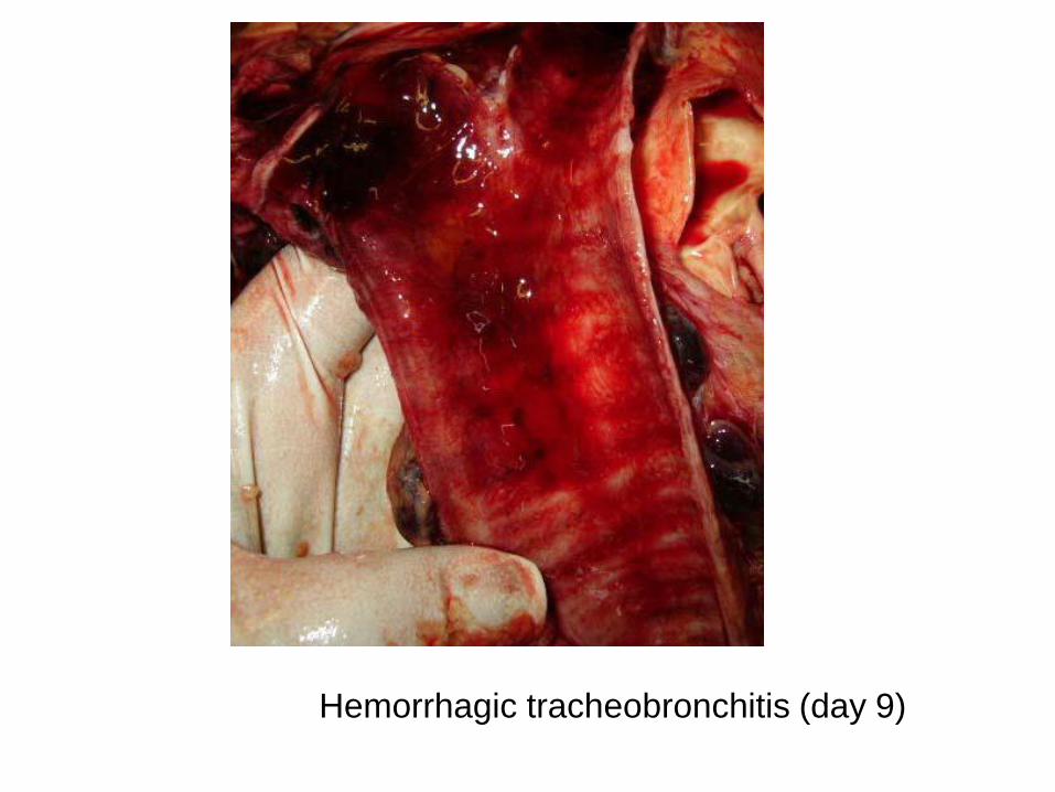
Hemorrhagic tracheobronchitis (day 9)
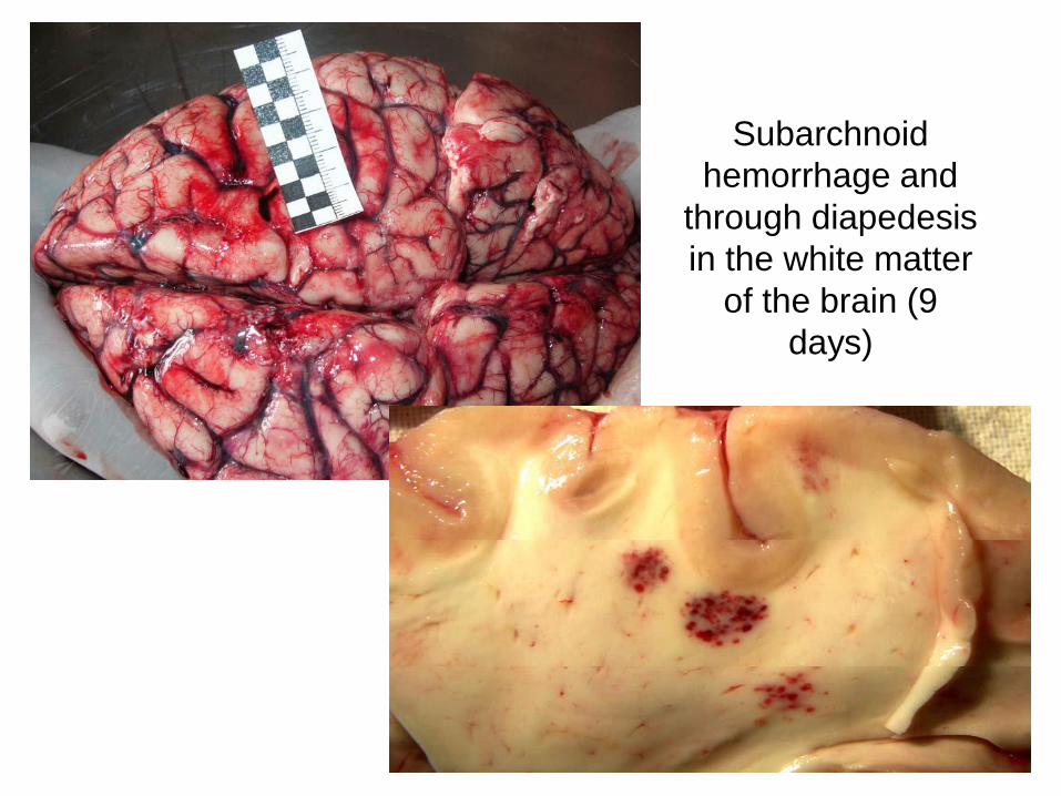
Subarchnoid
hemorrhage and
through diapedesis
in the white matter
of the brain (9
days)
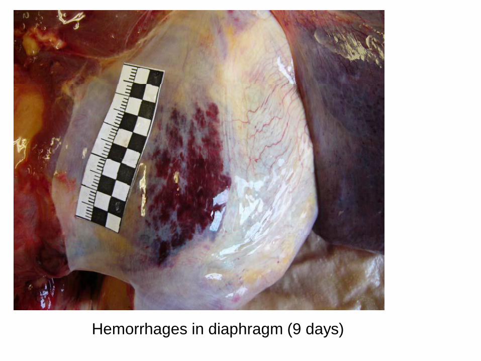
Hemorrhages in diaphragm (9 days)
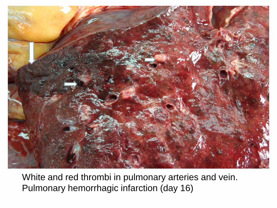
White and red thrombi in pulmonary arteries and vein.
Pulmonary hemorrhagic infarction (day 16)
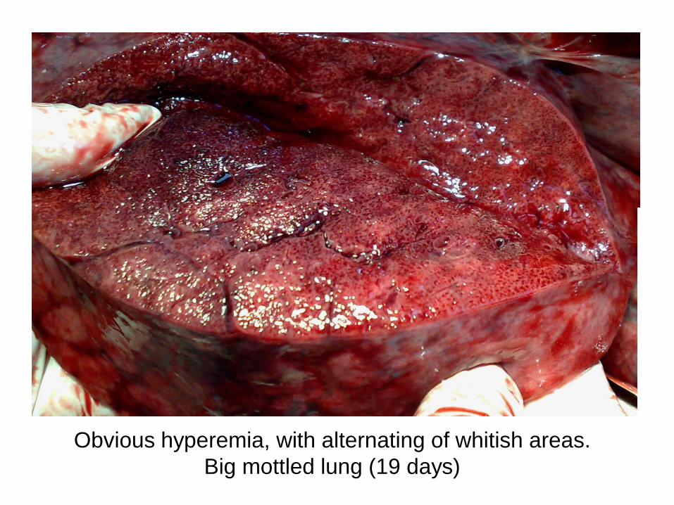
Obvious hyperemia, with alternating of whitish areas.
Big mottled lung (19 days)
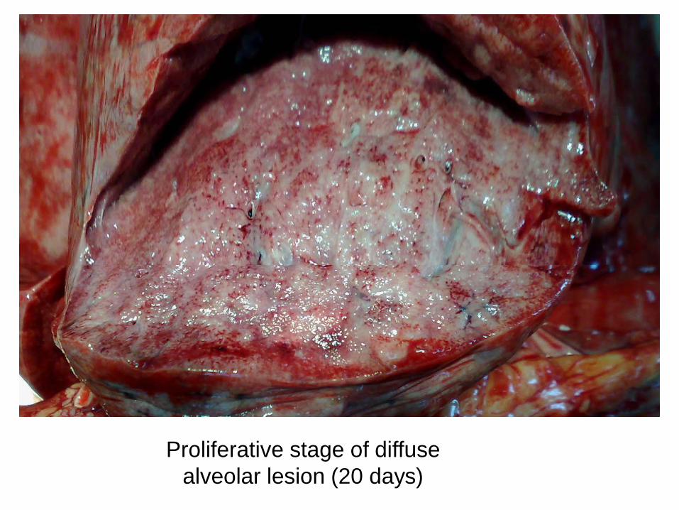
Proliferative stage of diffuse
alveolar lesion (20 days)
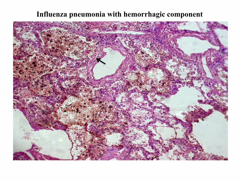
Influenza pneumonia with hemorrhagic component
1) Exudat hemoragic cu prezenţa siderofagilor
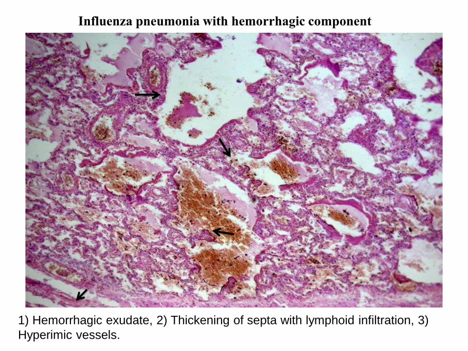
Influenza pneumonia with hemorrhagic component
1) Hemorrhagic exudate, 2) Thickening of septa with lymphoid infiltration, 3)
Hyperimic vessels.
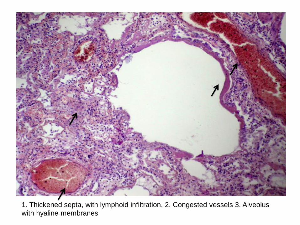
1. Thickened septa, with lymphoid infiltration, 2. Congested vessels 3. Alveolus
with hyaline membranes
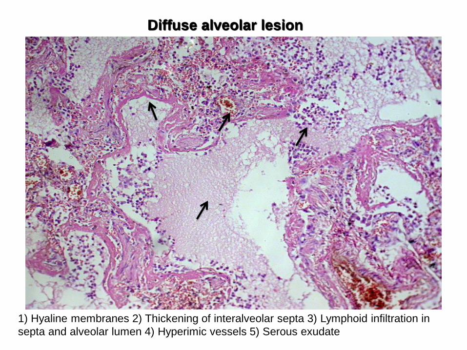
Diffuse alveolar lesion
1) Hyaline membranes 2) Thickening of interalveolar septa 3) Lymphoid infiltration in
septa and alveolar lumen 4) Hyperimic vessels 5) Serous exudate
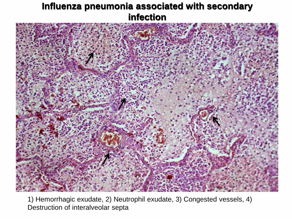
Influenza pneumonia associated with secondary
infection
1) Hemorrhagic exudate, 2) Neutrophil exudate, 3) Congested vessels, 4)
Destruction of interalveolar septa
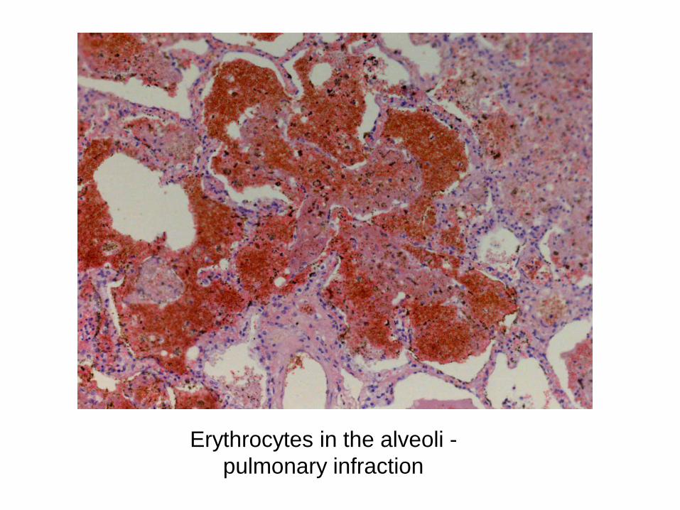
Erythrocytes in the alveoli -
pulmonary infraction
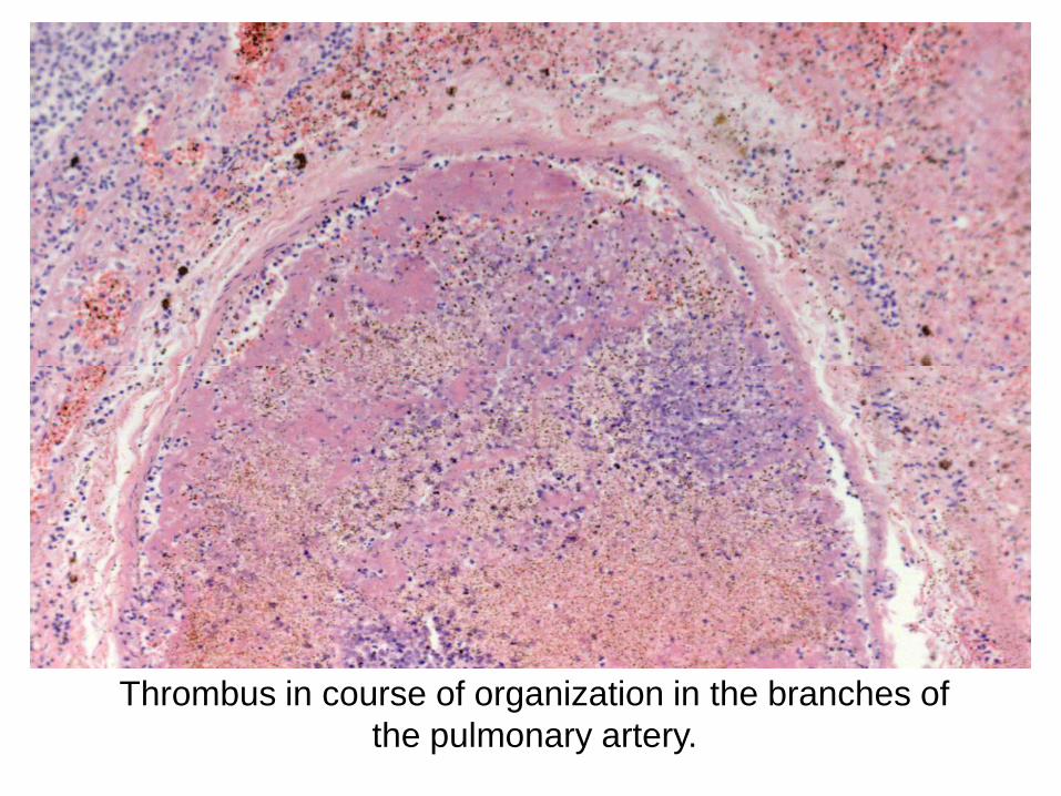
Thrombus in course of organization in the branches of
the pulmonary artery.
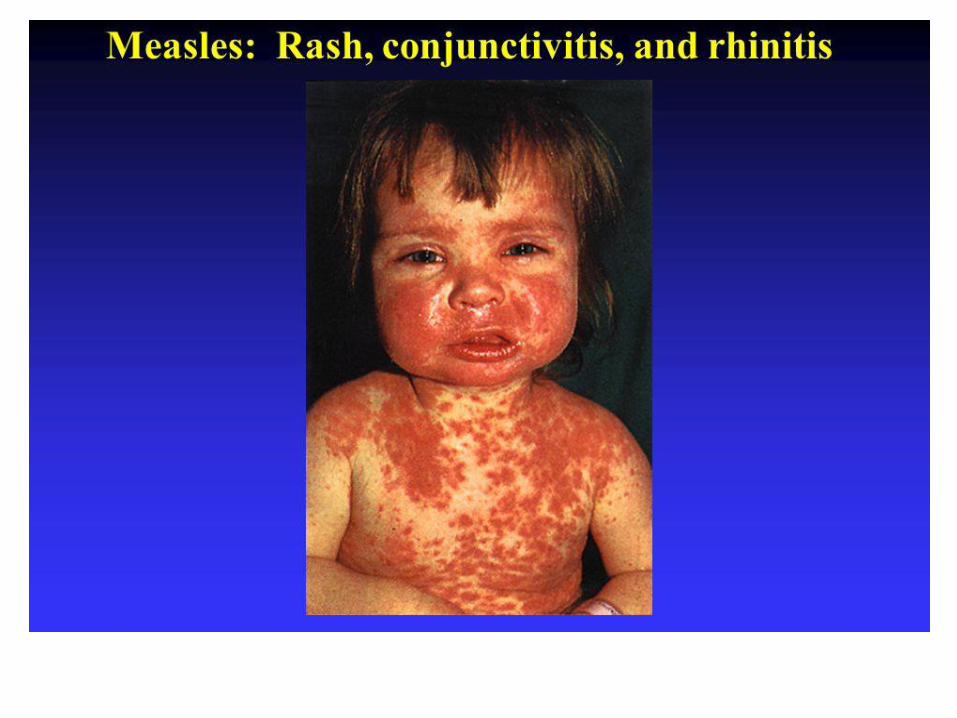
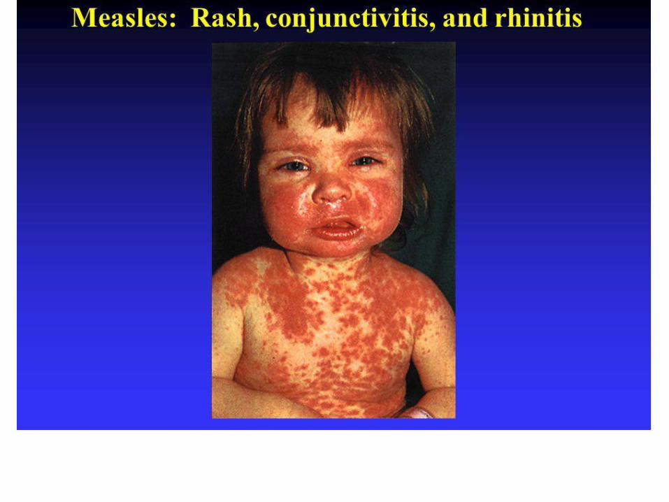
Measles
(Rubeola)
It is an acute viral infection characterized by a final stage with a maculopapular rash erupting successively over the neck and face, trunk, arms, and legs, and accompanied by a high fever.
Etiolog
y
◼ Measles virus, the cause of measles, is an RNA virus of the genus Morbillivirus in the family Paramyxoviridae.
Only one serotype is known◼
Epidemiolog
y◼ Measles is endemic throughout the
world.
In the past, epidemics tended to occur irregularly, appearing in the spring in large cities at 2-4-yr intervals as new groups of susceptible children were exposed.
◼
Epidemiology(Cont.)
◼ It is rarely subclinical.
Prior to the use of measles vaccine, the peak incidence was among children 5-10 yr of age.
◼
Individuals born before 1957 are considered to have had natural infection and to be immune
TRANSMISSI
ON
◼ Measles is highly contagious; approximately 90% of susceptible household contacts acquire the disease.
Maximal dissemination of virus occursby droplet spray during the prodromalperiod (catarrhal stage).
◼
Pathology
◼ The essential lesion of measles is found in the skin, conjunctivae, and the mucous membranes of the nasopharynx, bronchi, and intestinal tract.Serous exudate and proliferation of mononuclear cells and a few polymorphonuclear cells occur around the capillaries.
◼
Pathology (cont.)
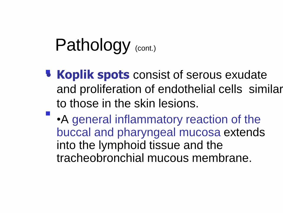
◼• Koplik spots consist of serous exudate
and proliferation of endothelial cells similar
to those in the skin lesions.
•A general inflammatory reaction of the buccal and pharyngeal mucosa extends into the lymphoid tissue and the tracheobronchial mucous membrane.
◼
Pathology(cont.)
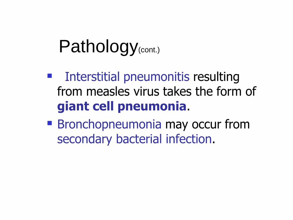
◼ Interstitial pneumonitis resulting from measles virus takes the form of giant cell pneumonia.
Bronchopneumonia may occur from secondary bacterial infection.
◼
Pathology (cont.)
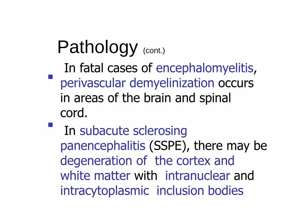
◼
In fatal cases of encephalomyelitis, perivascular demyelinization occursin areas of the brain and spinalcord.
In subacute sclerosingpanencephalitis (SSPE), there may be degeneration of the cortex and white matter with intranuclear andintracytoplasmic inclusion bodies
◼
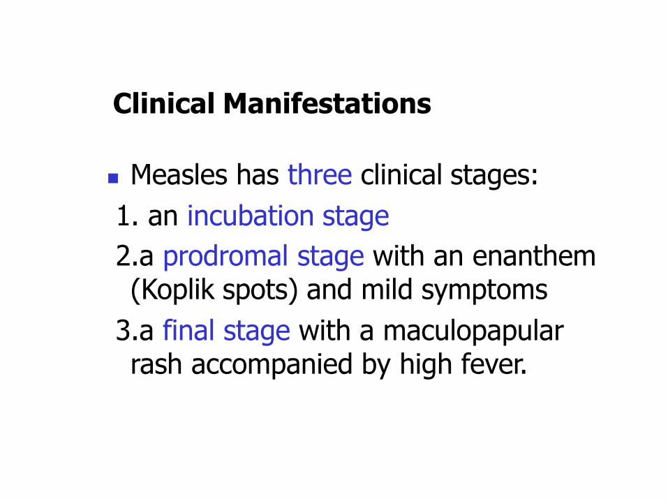
◼ Measles has three clinical stages:
1. an incubation stage
2.a prodromal stage with an enanthem (Koplik spots) and mild symptoms
3.a final stage with a maculopapular rash accompanied by high fever.
Clinical Manifestations
Koplik spots
◼
◼ An enanthem or red mottlingis usually present on the hard and soft palates
the pathognomonic sign of
measles:
Koplik spots (cont.)
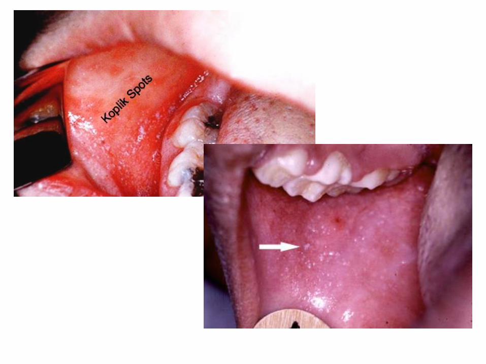
◼ • are grayish white dots, usually as small as grains of sand, that have slight, reddish areolae; occasionally theyare hemorrhagic.
• tend to occur opposite the lower molars but may spread irregularly over the rest ofthe buccal mucosa.
The
rash◼ usually starts as faint macules on the:
* upper lateral parts of the neck
* behind the ears
* along the hairline
* posterior parts of the cheek.
The rash(cont.)
◼ The individual lesions become increasingly maculopapular as the rash spreads rapidly over the:
* entire face
* neck
* upper arms
* upper part of the chest
within approximately the first 24 hr
The rash(cont.)
◼ During the succeeding 24 hr the rash spreads over the back, abdomen, entire arm, and thighs.
As it finally reaches the feet on the 2nd-3rd day, it begins to fade on the face.
◼
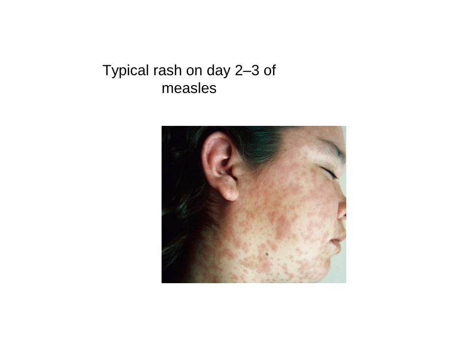
Typical rash on day 2–3 of
measles
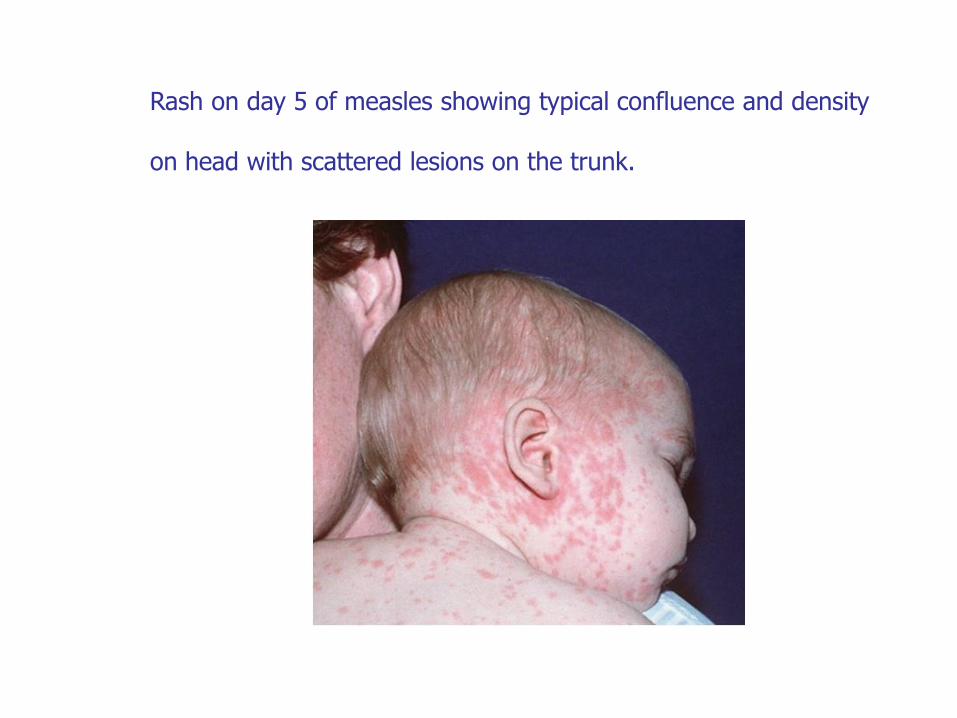
Rash on day 5 of measles showing typical confluence and density
on head with scattered lesions on the trunk.
The prodromal phase
◼ Otitis media
bronchopneumonia
gastrointestinal symptoms such as diarrhea and vomiting
◼
◼
Are more common in infants and small children (especially if they are malnourished) than in older children.
Complicatio
ns◼ The chief complications of measles
are:
◼ otitis media
pneumonia
encephalitis.
◼
◼
Respiratory tract
complications
◼ Interstitial pneumonia may be caused by the measles virus (giant cell pneumonia).
Bacterial superinfection and bronchopneumonia are more frequent, however, usually with pneumococcus, group A Streptococcus, Staphylococcus aureus, and Haemophilus influenzae type b.
Laryngitis, tracheitis, and bronchitis are common and may be due to the virus alone
◼
◼
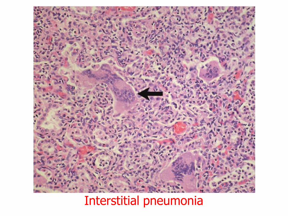
Interstitial pneumonia
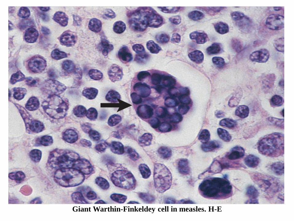
Giant Warthin-Finkeldey cell in measles. H-E
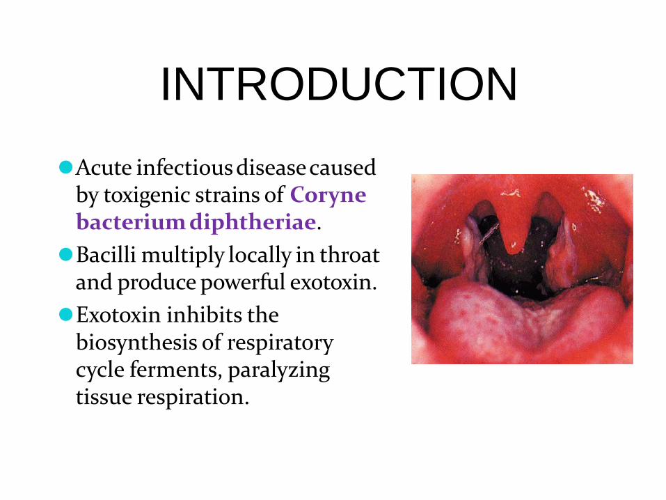
INTRODUCTION
⚫Acute infectiousdiseasecaused by toxigenic strains of Coryne bacterium diphtheriae.
⚫Bacilli multiply locally in throat and produce powerful exotoxin.
⚫Exotoxin inhibits the biosynthesis of respiratory cycle ferments, paralyzing tissue respiration.
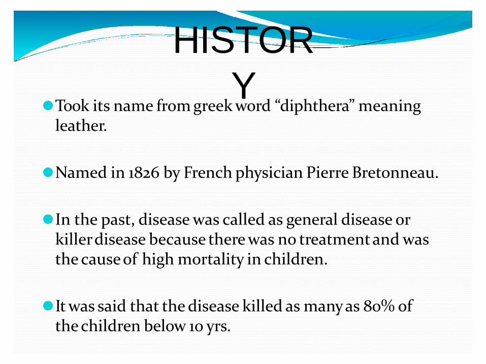
HISTOR
Y⚫Took its name from greek word “diphthera” meaning
leather.
⚫Named in 1826 by French physician Pierre Bretonneau.
⚫In the past, disease was called as general disease or killerdisease because there was no treatmentand was the causeof high mortality in children.
⚫It was said that the disease killed as manyas 80% of the children below 10 yrs.
AGENT
Agent Corynebacterium diphtheria
⚫Gram positive motileorganism
⚫No invasive power but produce powerful exotoxin after multiplication locally in the throat responsible for:
1. Formation of false membrane over tonsils, pharynx or larynx, with well defined edges and membrane cannot be wiped away.
2. Marked congestion, edema, local tissue destruction3. Enlargement of lymph nodes4. Toxaemic signs and symptoms
MODE OF
TRANSMISSION⚫Droplet infections
⚫Can also be transmitted directly to susceptiblepersons from infected cutaneous lesions.
⚫Transmission by objects contaminated by naso-pharyngeal secretionsof patients is also possible.
PORTAL OF
ENTRY⚫Respiratory route- respiratory tract
⚫Non-respiratoryroute-
Portal of entry may be skin wherecuts, ulcersand
wounds not properly attended to or through
umbilicusof new born.
Siteof implantation may be eyes, genitaliaor
middleear.
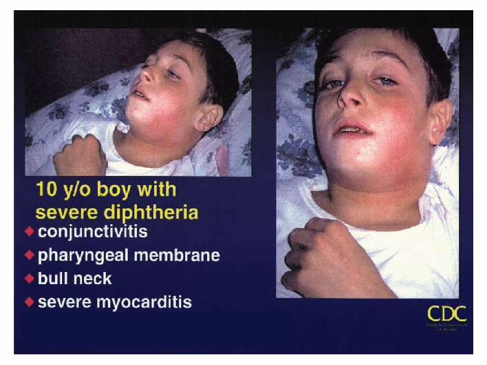
CLINICAL
FEATURES⚫Respiratory tract forms of diphtheria-
pharyngo-tonsillar
laryngo tracheal
nasal
combinations
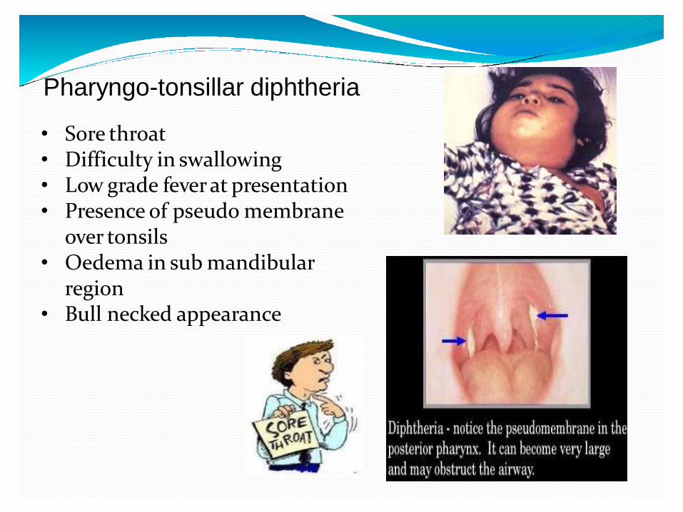
Pharyngo-tonsillar diphtheria
• Sore throat• Difficulty in swallowing• Low grade fever at presentation• Presence of pseudo membrane
over tonsils• Oedema in sub mandibular
region• Bull necked appearance
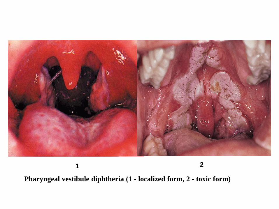
Pharyngeal vestibule diphtheria (1 - localized form, 2 - toxic form)
1 2
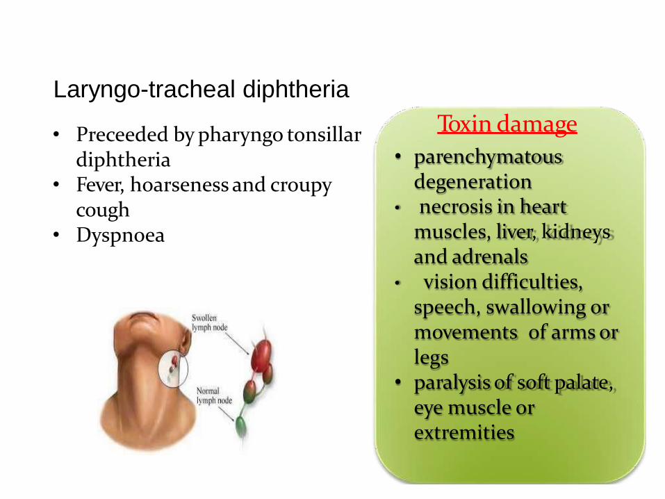
Laryngo-tracheal diphtheria
• Preceeded by pharyngo tonsillardiphtheria
• Fever, hoarseness and croupy cough
• Dyspnoea
• necrosis in heart muscles, liver, kidneys and adrenals
• vision difficulties, speech, swallowing or movements of arms or legs
• paralysis of soft palate, eye muscle or extremities
Toxin damage
• parenchymatous degeneration
Nasal diphtheria
• Mildest form• Localized in septum or turbinates of one
side of nose• Conjunctivaand genitals also sources of
infection• Membrane extends to pharynx.
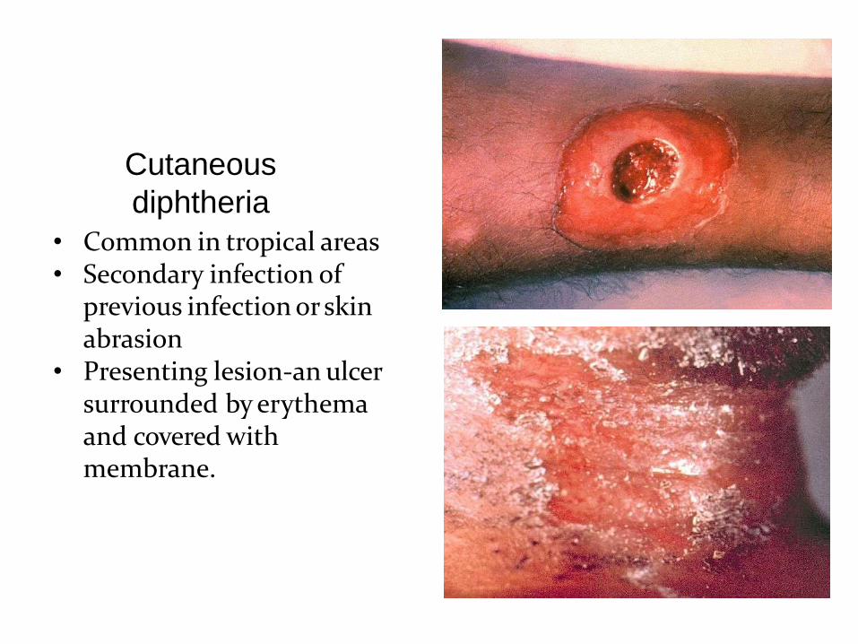
Cutaneous
diphtheria
• Common in tropical areas• Secondary infection of
previous infection or skin abrasion
• Presenting lesion-an ulcer surrounded by erythema and covered with membrane.
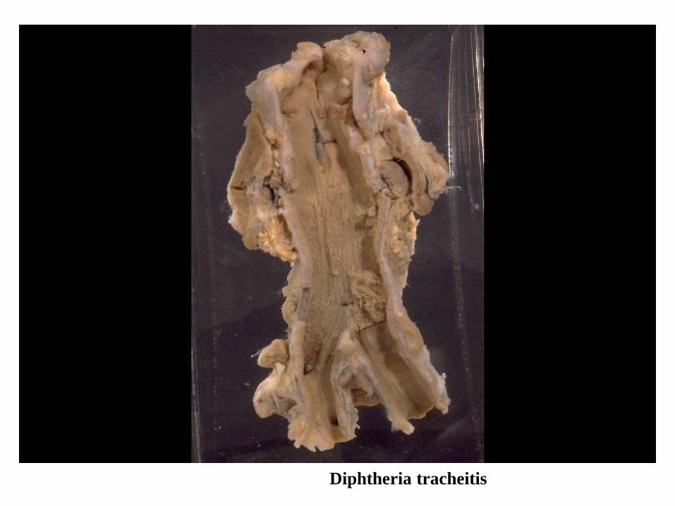
Diphtheria tracheitis
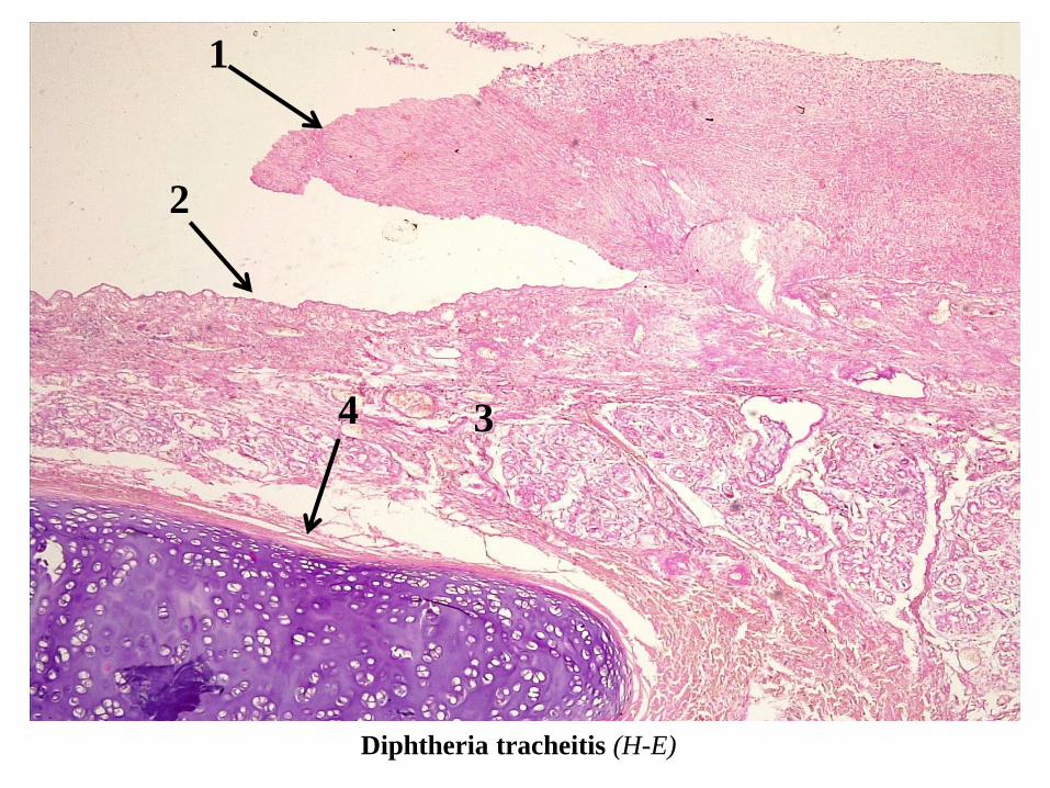
Diphtheria tracheitis (H-E)
2
1
4 3
Scarlet Fever
tractIs an upper respiratory infection associatedcharacteristic
with arash, which is
caused by an infection withpyrogenic exotoxin (erythrogenictoxin)-producing group
Astreptococcus in individuals who
do not have antitoxin antibodies.
Scarlet Fever (cont.)
Page164
• The incidence is cyclic, depending on:1. The prevalence of toxin-
producing strains2.The immune status of the
population• The epidemiologic features
which include:1.Modes of transmission 2.Age distribution
are otherwise similar to those for group A streptococcal pharyngitis.
The rash of Scarlet Fever
•The rash appears within 24-48 hr after onset of
symptoms, although it may appear with the first
signs of illness.
•It often begins around the neck and spreads
over the trunk and extremities.
•It is a diffuse, finely papular, erythematous
eruption producing a bright red discoloration of
the skin, which blanches on pressure.
•It is often more intense on the elbows, axillae,
and groin.
The rash of Scarlet Fever (cont.)
• The skin has a goose-pimple appearance
and feels rough.
• The face is usually spared, although the
cheeks may be erythematous with pallor
around the mouth.
• After 3-4 days, the rash begins to fade and is
followed by desquamation, first on the face
progressing downward, and often resembling
that seen subsequent to a mild sunburn.
• Occasionally, sheetlike desquamation may
occur around the free margins of the
fingernails, the palms, and the soles.
Scarlet Fever (cont.)
Page167
•Examination of the pharynx of a patient with
scarlet fever reveals essentially the same
findings as with group A streptococcal
pharyngitis.
•In addition, the tongue is usually coated and
the papillae are swollen.
•After desquamation, the reddened papillae are
prominent, giving the tongue a strawberry
appearance.