Embed Size (px)
Citation preview
ALL2, a Homologue of ALL1, Has a Distinct Role in Regulating pHHomeostasis in the Pathogen Cryptococcus neoformans
Neena Jain,a Tejas Bouklas,b Anjali Gupta,a Avanish K. Varshney,b Erika P. Orner,c Bettina C. Friesb,c
Department of Medicine (Infectious Diseases), Albert Einstein College of Medicine, Bronx, New York, USAa; Departments of Medicine (Infectious Diseases)b andMicrobiology and Molecular Genetics,c Stony Brook University, Stony Brook, New York, USA
Cryptococcus neoformans is a facultative intracellular fungal pathogen that has a polysaccharide capsule and causes life-threat-ening meningoencephalitis. Its capsule, as well as its ability to survive in the acidic environment of the phagolysosome, contrib-utes to the pathogen’s resilience in the host environment. Previously, we reported that downregulation of allergen 1 (ALL1) re-sults in the secretion of a shorter, more viscous exopolysaccharide with less branching and structural complexity, as well asaltered iron homeostasis. Now, we report on a homologous coregulated gene, allergen 2 (ALL2). ALL2’s function was character-ized by generating null mutants in C. neoformans. In contrast to ALL1, loss of ALL2 attenuated virulence in the pulmonary infec-tion model. The all2� mutant shed a less viscous exopolysaccharide and exhibited higher sensitivity to hydrogen peroxide thanthe wild type, and as a result, the all2� mutant was more resistant to macrophage-mediated killing. Transcriptome analysis fur-ther supported the distinct function of these two genes. Unlike ALL1’s involvement in iron homeostasis, we now present data onALL2’s unique function in maintaining intracellular pH in low-pH conditions. Thus, our data highlight that C. neoformans, ahuman-pathogenic basidiomycete, has evolved a unique set of virulence-associated genes that contributes to its resilience in thehuman niche.
Cryptococcus neoformans is a major fungal pathogen that causesdisease predominantly in patients with AIDS but also in pa-
tients with other immune deficiencies. The most common clinicalpresentation of cryptococcal disease is chronic meningoencepha-litis (CME). Worldwide, approximately 1 million new cases ofcryptococcal meningitis occur every year, resulting in more than600,000 deaths (1). Despite advances made in antifungal treat-ment, intracranial pressure management, and antiretroviral ther-apy, there is a major challenge to rapidly clear C. neoformans fromcerebral spinal fluid (CSF), which is required to achieve a goodoutcome (2). The pathogen has various highly regulated virulencetraits, including capsule, melanin formation, growth at 37°C, andextracellular enzymes (3). Differential gene regulation of thesevirulence traits can promote microevolution in vivo, which mayfacilitate evasion of the host immune response. One process bywhich microevolution can be achieved is phenotypic switching,which generates variants with augmented virulence that also alterhost-pathogen interactions (4, 5).
In our previous studies, we documented that RC2, a serotype DC. neoformans clinical strain that is also a standard laboratorystrain (6), undergoes phenotypic switching from a smooth to mu-coid colony morphology and is associated with downregulation ofa defined set of genes, including allergen 1 (ALL1) (7). Loss ofALL1 function resulted in a hypervirulent phenotype in both se-rotype A- and D-null mutants (all1�). Furthermore, it was shownthat ALL1 is regulated during capsule induction (8) and by thetranscription factor Sp1 and the PKC1 gene under glucose starva-tion (9), as well as by CIR1 in response to changes in iron concen-tration (10). Under starvation, VAD1 downregulates ALL1 tran-scripts via degradation of mRNA (11). Although the precisefunction of ALL1 is not known, its expression indirectly affectspolysaccharide conformation and iron homeostasis (12). Thus,intracisternal infection with the all1� mutant results in aug-mented intracerebral pressure and premature death in infectedrats (7). Extensive homology search for ALL1 revealed a highly
homologous uncharacterized cryptococcal gene, CNM02200(JEC21), which was subsequently named ALL2. Similarly to ALL1,ALL2 is also regulated by Sp1 and PKC1 under glucose starvationand during capsule induction (9).
In the present study, we sought to characterize the function ofthis gene and generated a null mutant of ALL2 (all2�) and a dou-ble mutant of ALL1 and ALL2 (all1� all2�). Results obtained im-ply that these homologous genes differ in their contribution tovirulence and associated function. In contrast to the loss of ALL1,the loss of ALL2 attenuates virulence, and the mutants shed moreof a less viscous exopolysaccharide (exo-PS). Microarray analysissupports distinct functions for these homologous genes and indi-cates that, unlike ALL1, the gene ALL2 is involved in maintainingintracellular pH.
MATERIALS AND METHODSYeast strains and media. The C. neoformans strains used in this study arelisted in Table S1 in the supplemental material. C. neoformans strains werecultured at 37°C in YNB broth (0.67% yeast nitrogen base without aminoacids plus 2% glucose) or grown on YPD broth/agar (20 g/liter glucose, 10g/liter yeast extract, 20 g/liter peptone [for YPD agar, 20 g/liter agar was
Received 10 August 2015 Returned for modification 24 August 2015Accepted 13 November 2015
Accepted manuscript posted online 23 November 2015
Citation Jain N, Bouklas T, Gupta A, Varshney AK, Orner EP, Fries BC. 2016. ALL2, ahomologue of ALL1, has a distinct role in regulating pH homeostasis in thepathogen Cryptococcus neoformans. Infect Immun 84:439 –451.doi:10.1128/IAI.01046-15.
Editor: G. S. Deepe, Jr.
Address correspondence to Bettina C. Fries,[email protected].
Supplemental material for this article may be found at http://dx.doi.org/10.1128/IAI.01046-15.
Copyright © 2016, American Society for Microbiology. All Rights Reserved.
crossmark
February 2016 Volume 84 Number 2 iai.asm.org 439Infection and Immunity
on August 8, 2016 by guest
http://iai.asm.org/
Dow
nloaded from
added]), unless otherwise stated. For glucose starvation studies, YNBbroth supplemented with 0.2% glucose, no glucose, or asparagine saltmedium (1 g/liter asparagine, 10 mM sodium phosphate [pH 6.5], and0.25 g/liter MgSO4) was used. For screening mutants, YPD supplementedwith 100 mg/liter nourseothricin (NAT) or 200 mg/liter neomycin G418(NEO) was used. RNA for microarray analysis was isolated from C. neo-formans cells grown in minimal medium (10 mM magnesium sulfate, 29.3mM potassium phosphate monobasic, 13 mM glycine, and 3 �M thia-mine-HCl; adjusted to pH 5.5 and supplemented with 15 mM glucose asthe carbon source). Other relevant media are specified below.
Strain construction. The C. neoformans serotype A and D referencesequences were accessed originally through TIGR (now available at theNCBI) and the Fungal Genome Initiative database at the Broad Insti-tute (http://www.broadinstitute.org/annotation/genome/cryptococcus_neoformans/MultiHome.html).
Generation of all2�, all2��PACT1-ALL2, and all1� all2� mutants.The ALL2 gene is 1,549 bp long, has four introns, and encodes a putativeprotein with a molecular mass of 25 kDa. In strain JEC21, ALL2(CNM02200) is located on chromosome 13. To generate the all2� mu-tant, the coding region of ALL2 (RC2, 926 bp) was replaced with a neo-mycin resistance marker by homologous recombination as described pre-viously (7). Primers used to generate mutants are listed in Table S2 in thesupplemental material. Homologous recombination was confirmed byPCR, Southern blot analysis, and real-time PCR of ALL2 transcripts. ForALL2 complementation, the ACT1 promoter and the ALL2 open readingframe (ORF) were amplified from genomic DNA of RC2 and fused inframe using the NdeI restriction site. ACT1 and ALL2 fragments werecloned in pJAF13 using XbaI and XhoI cloning sites to generate plasmidpJAF13/PACT1-ALL2. The plasmid pJAF13/PACT1-ALL2 was linearized byXhoI and introduced into the all2� mutant by biolistic transformation.ALL2-positive clones (all2��PACT1-ALL2) were selected on YPD agarwith 100 mg/liter NAT. Gene complementation was confirmed by PCR,and the level of ALL2 expression was determined by reverse transcription-PCR (RT-PCR) of ALL2 transcripts. For generation of the all1� all2�double mutant, the ALL1 gene was replaced with the NAT resistancemarker in RC2-all2� by modifying the construct designed previously todisrupt ALL1 (7). Positive clones were selected on YPD agar containing100 mg/liter NAT and 200 mg/liter NEO. Homologous recombinationwas confirmed by PCR, Southern blot analysis, and determining the tran-scription level of ALL1 and ALL2 by RT-PCR.
Phenotypic characterization. Mutants were phenotypically character-ized for (i) baseline and induced capsule size, (ii) growth rate in variable pHand Dulbecco’s modified Eagle’s medium (DMEM) supplemented with 10%fetal calf serum, 10% NCTC, 1% nonessential amino acids, 1% penicillin-streptomycin, or minimal medium (10 mM magnesium sulfate, 29.3 mMpotassium phosphate monobasic, 13 mM glycine, 3 �M thiamine-HCl; ad-justed to pH 5.5 and supplemented with 15 mM glucose as the carbonsource), (iii) in vitro stress sensitivity (1 M potassium chloride, lithium chlo-ride [100 and 200 mM with glucose and galactose, respectively], 1 M sodiumchloride), (iv) hydrogen peroxide sensitivity, (v) macrophage phagocytosisindex, and (vi) macrophage-mediated killing assays as described previously(13–16). The intrinsic viscosity of exopolysaccharide and glucuronic acid res-idue content was determined as previously described (17, 18). All measure-ments were done in triplicate.
Generation of ALL1::HA-tagged and ALL2:mCherry fusion proteinstrains. The ALL1::HA-tagged strain (where HA is hemagglutinin) wasgenerated using the following strategy (19, 20). The ALL1 gene was am-plified under its own promoter from RC2 genomic DNA using primersALL1-prom-F-XbaI and ALL1-HARev (see Table S2 in the supplementalmaterial). The resulting fragment and pJAF1 were digested with XbaI andNcoI and ligated to generate the 5=ALL1-HA/pJAF1 construct. The 3=untranslated region (3=UTR) of the ALL1 gene was cloned in frame be-hind ALL1-HA using EcoRV and NcoI to generate 5=ALL1-HA/3=UTR/pJAF1. Next, 1,000 bp downstream of ALL1 was cloned into ALL1-HA/3=UTR/pJAF1 using the restriction sites for BamHI and KpnI to generate
5=ALL1-HA/3=UTR/NEO/ALL1-3=pJAF1. The construct was restrictedwith XbaI and KpnI, yielding a linearized fragment of 5=ALL1-HA/3=UTR/NEO/ALL1-3=, which was biolistically delivered into RC2 using astandard procedure as described previously (7). Homologous recombina-tion was confirmed by PCR. ALL1::HA was detected by Western blothybridization using rabbit anti-HA polyclonal antibody and a chemilu-minescence kit (Pierce/Life Technologies) according to the manufactur-er’s instructions.
To generate ALL2-mCherry fusion protein in ALL1::HA strain for lo-calization studies, plasmid YP164 (a generous gift from the Peter William-son lab) was used to express a fusion between ALL2 and a syntheticmCherry protein (21). ALL2 under ACT1 promoter was amplified frompreviously generated plasmid PACT1-ALL2/pJAF13 using primersAll2For-NHEI and All2Rev-PST1 and inserted into the plasmid YP164using standard cloning techniques to generate plasmid ALL2/mCherry/YP142. Further, ALL2-mCherry was amplified from plasmid ALL2/mCherry/YP142 using the primers PAct-XbaFor and mCherry rev-EcoRV,and ALL2-3=UTR was amplified from RC2-SM genomic DNA using theprimers All2 3=UTR-For ECORV and All2 3=UTR-Rev XHOI. Both partswere cloned in frame in plasmid pJAF13 using standard cloning tech-niques. The plasmid ALL2/mCherry/3=UTR/pJAF13 was linearized andtransformed in ALL1::HA cells using the standard biolistic transformationtechnique.
All1p and All2p localization in vivo. For localization studies, immu-nofluorescence was performed as described previously (20) with fewmodifications. Briefly, RC2 wild-type (RC2-wt) cells for All1p::HA andAll2p-mCherry were cultured for 16 h in YNB up to an optical density at600 nm of 0.4 to 0.5. Cells were harvested by centrifugation (5,000 rpm for1 min), washed once with phosphate-buffered saline (PBS), suspended in1 ml of 4% paraformaldehyde in PBS, and incubated for 30 min withrotation. The sample was then washed three times with PBS, suspended in1 ml of lysis buffer (50 mM sodium citrate [pH 6.0], 1 M D-sorbitol, 35mM �-mercaptoethanol) plus 40 mg/ml lysing enzyme (fromTrichoderma harzianum), and incubated for 1 to 2 h at 37°C with occa-sional inversion. To monitor the protein mobility in variable growthphase, the cells were harvested at different time points during the experi-ment. After digestion, the cells were washed twice with heparan sulfate(HS) buffer (100 mM HEPES [pH 7.5], 1 M D-sorbitol) and suspended in200 �l of HS buffer. The washed cell pellet was incubated with HS buffercontaining 1% Triton X-100 and incubated for 10 min; it was then washedthree times with HS buffer and three times with PBS. Next, the cells weretreated with blocking buffer (5% goat serum and 0.02% Triton X-100 inPBS) for 1 h, followed by a high-affinity rabbit anti-HA polyclonal anti-body (1:250/1:500 in blocking buffer [BB] obtained from Roche AppliedScience) at 4°C in a rotator. Samples were then washed six times withblocking buffer and stained with Alexa Fluor 594-goat anti-rabbit IgG (1�g/ml in BB) for 1 h in the dark. The cells were washed three times with BBand three times with PBS. Bright-field and fluorescence images were ac-quired as for the capsule staining assays described above.
Murine virulence studies. BALB/c mice (female, 6 to 8 weeks old)were obtained from the National Cancer Institute (Bethesda, MD). Mice(n � 10 per group) were infected intratracheally (i.t.) with 5 � 104 or intra-venously (i.v.) with 106 of wild type (RC2) or respective mutant cells in PBS.Mouse health was monitored daily, and if moribund, the mice were sacrificedin accordance with Institutional Animal Care and Use Committee (IACUC)regulations at the Albert Einstein College of Medicine (AECOM). To deter-mine CFU studies, infected mice were killed at day 7 or 14, and their lungswere excised. Half of the lung was plated for CFU, and the other half was fixedin 10% formalin for histological analysis. Lung tissues were stained with he-matoxylin and eosin, as well as mucicarmine, by the Histopathology Core atAECOM and Stony Brook University. Survival curves were generated andanalyzed using GraphPad Prism 6.0.
ALL2 expression in glucose starvation. RC2 cells were grown over-night in YNB supplemented with 2% glucose, diluted 1:50, and then againgrown for 8 h in YNB supplemented with 2% glucose, 0.2% glucose, or no
Jain et al.
440 iai.asm.org February 2016 Volume 84 Number 2Infection and Immunity
on August 8, 2016 by guest
http://iai.asm.org/
Dow
nloaded from
glucose. RNA transcript levels were measured by RT-PCR as previouslydescribed (7) in a 10-�l reaction mixture containing SYBR green mastermix (Applied Biosystems/Life Technologies) with the primers listed inTable S2 in the supplemental material. An RT-PCR analysis was per-formed with the appropriate controls, and the relative transcript levelswere determined as described earlier (12).
Microarray and GO enrichment analysis. RC2-wt and mutant cellswere grown in minimal medium for 16 h at 37°C with agitation (150 rpm),diluted 1:100, and regrown for 16 h or up to an optical density of 0.6 to 0.7with agitation (150 rpm) at 37°C. Experiments were performed in dupli-cates (minimal medium arrays). Total RNA was isolated, and the microar-ray was performed by the Genome Technology Access Center at Wash-ington University in St. Louis. Microarray analysis and gene ontology(GO) enrichment analysis were performed by the facility as describedpreviously (12). Microarray results were further confirmed by performingRT-PCR on select genes as described above. A functional network wasgenerated using Cryptonet (http://www.inetbio.org/cryptonet/).
Measurement of vacuolar pH. Intracellular pH (pHi) of cells grown inYNB with variable glucose, as well as total pH, was measured using thepH-sensitive fluorophore BCECF-AM [2=7=-bis-(2-carboxyethyl)-5-(and6)-carboxyfluorescein]. BCECF accumulates in yeast vacuoles, and exci-tation spectra are sensitive to pH (22). pHi measurement was performedby collecting the ratio of fluorescence at two excitation wavelengths (450and 490 nm) and measuring at a single emission wavelength (535 nm)using protocols described elsewhere (23).
ATP measurement. Cellular ATP levels were measured as previouslydescribed (24). Briefly, cells were grown in 50 ml of YNB buffered with 50mM MES (morpholineethanesulfonic acid) (pH 4 and pH 7) for 16 h withconstant agitation of 150 rpm at 37°C. The cultures were centrifuged at3,000 rpm for 10 min to collect the pellet and washed once with PBS. Thecells were counted using a hemocytometer, and a pellet of a similar num-ber of cells was suspended in 1 ml of 50 mM HEPES (pH 7). The cells werefurther disrupted with a bead-beater (Biospec Products) using prewashed0.5-mm zirconia beads. Disrupted samples were centrifuged at 8,000 rpmat 4°C for 10 min. The ATP concentrations of supernatants were deter-mined using an ATP bioluminescence assay kit (Sigma-Aldrich, St. Louis,MO). ATP concentrations were normalized with total protein concentra-tions for each sample. The mean measurements for triplicate sampleswere reported.
Chronological and replicative life spans. The chronological life span(CLS) was determined by adaptation of Saccharomyces cerevisiae methods(25) with some modifications. Briefly, equal numbers of C. neoformanscells were grown in YNB medium with or without 2% dextrose for 3 daysat 37°C until they reached stationary phase. The cells were then trans-ferred to sterile-distilled H2O, and the number of viable cells was mea-sured by plating appropriate dilutions every 2 to 3 days on Sabourauddextrose agar (SDA) plates. CFU on the plates were quantitated at 48 h,and the study was terminated when 99% of the cells were dead. All mea-surements were performed in triplicates.
The replicative life span (RLS) was determined by microdissectionusing a tetrad dissection Axioscope A1 microscope (Zeiss) as describedpreviously in S. cerevisiae (26) with some modifications for C. neoformans.Briefly, the first buds of C. neoformans cells (n � 20 to 40) were arrayed ona YPD agar plate maintained at 37°C and then evaluated every 1 to 2 h byseparating newly arising buds using a 50-�m fiber optic needle (CoraStyles). The RLS of each cell was calculated by determining the sum of thetotal buds until the cessation of divisions.
Statistical analysis. Statistical analysis, including Student’s t test, Wil-coxon rank sum test, and log-rank test, was performed using MicrosoftExcel 2011 and GraphPad Prism 6.0 for Macintosh.
Microarray data accession number. Microarray data for the mutantand the wt were uploaded to the GEO database and assigned GEO acces-sion no. GSE74219.
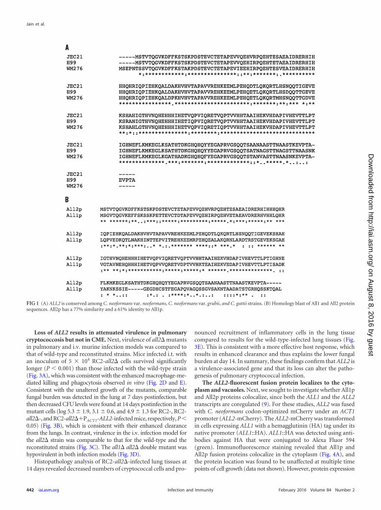
RESULTSIdentification of ALL2 and generation of all2� and all1� all2�mutants. The ALL2 gene is highly conserved among C. neofor-mans var. neoformans and var. grubii, as well as C. gattii, strains(Fig. 1A). Comparison of All1p and All2p amino acid sequencesrevealed that they are highly homologous (77% similarity and61% identity) (Fig. 1B). Homology in the N-terminal part of theprotein (176 amino acids [aa]) was 85%, whereas homology in theC-terminal part was only 35% (58 aa). Extensive searches for pu-tative conserved domains or protein motifs using standard motifsearch programs detected no conserved domains or motifs. BothAll1p and All2p exhibit homology (33 to 38%) only with hypo-thetical proteins in Malassezia sp. and other nonencapsulatedplant-pathogenic fungi, such as Sterenum hirsutum and Rhodospo-ridium toruloides. No homology with proteins of any human-pathogenic fungi was found (see Fig. S1 in the supplemental ma-terial). The all2� null mutant was generated in the C. neoformansRC2 (serotype D) strain by replacing the coding region of ALL2(RC2, 926 bp) with a neomycin resistance marker (NEO) of 2,098bp using biolistic transformation. Successful homologous recom-bination was confirmed by PCR (see Fig. S2 in the supplementalmaterial) and real-time PCR (see Table S3 in the supplementalmaterial). The double mutant strain (all1� all2�) was also gener-ated in RC2 using the previously generated all1� mutant strain (7)as the background strain.
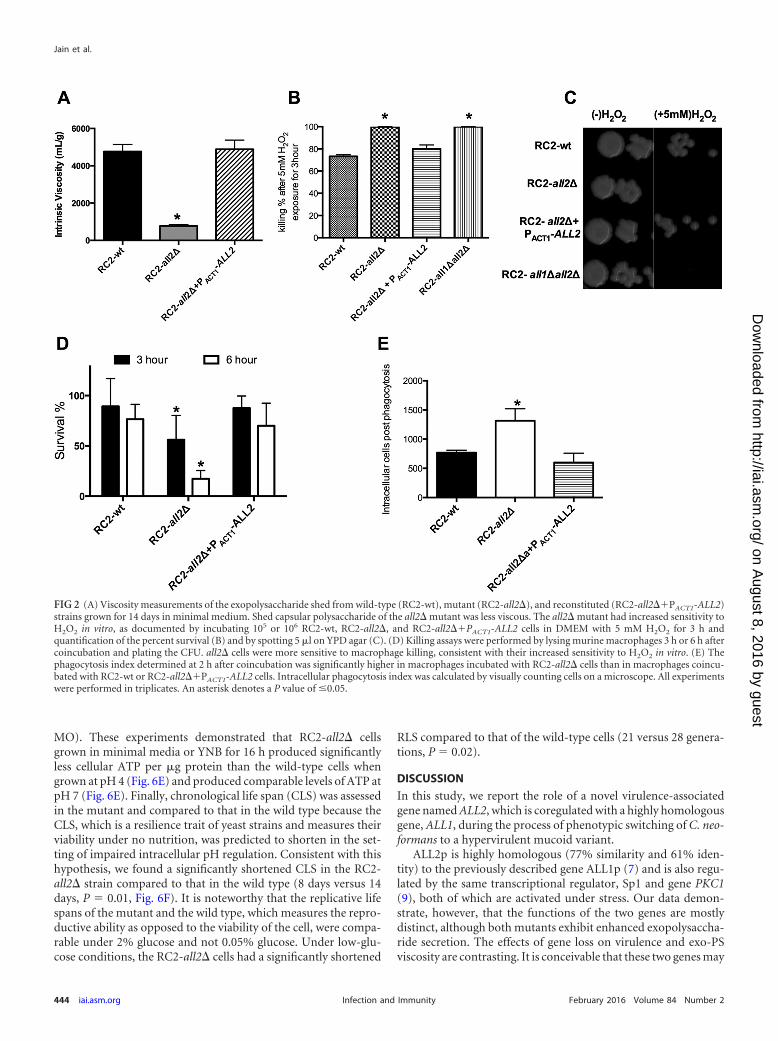
Characterization of disruption mutants. Phenotypic charac-teristics of the null all2� mutant, the all1� all2� double mutant,and the reconstituted all2��PACT1-ALL2 strain are summarizedin Table 1. Baseline capsule size and capsule induction were notaffected by loss of ALL2 either in vitro or in vivo. Measurement ofthe biophysical properties of shed exo-PS demonstrated thatexo-PS from the RC2-all2� mutant was less viscous than exo-PSfrom the RC2-wt and the reconstituted strain (Fig. 2A). To quan-tify shed exo-PS, a phenol-sulfuric colorimetric assay, whichquantifies glucuronic acid residues, was used. This experimentshowed that RC2-all2� cells shed more exo-PS per cell than thewild type (Table 1). Furthermore, RC2-all2� cells have no growthdefect at neutral pH and display normal melanin production. Mu-tant cells exhibit comparable sensitivity to lysing enzyme and var-ious high salt concentrations (data not shown). However, all2�mutant cells exhibit increased sensitivity to oxidative stress. In thepresence of hydrogen peroxide (H2O2), RC2-all2� cells demon-strated a larger zone of inhibition in disc diffusion assays than thewild-type cells (65 versus 57 mm). This defect was resolved withgene reconstitution in RC2-all2��PACT1-ALL2 (58 mm) (Table1). Consistent with this finding, incubation of mutant cells (all2�and all1� all2�) with 5 mM H2O2 for 3 h resulted in significantlyenhanced killing (P � 0.001) compared to that seen with the wildtype and the reconstituted strain (Fig. 2B and C). Macrophage-mediated killing assays showed significantly enhanced (P � 0.001)killing of RC2-all2� cells compared to cells of the wild type andthe reconstituted strains (Fig. 2D). Further, antibody-medi-ated phagocytosis was enhanced (P � 0.042) for the all2� mu-tant (Fig. 2E). Notably, an all2� mutation was also generated inthe standard laboratory strain H99, a serotype A strain and alsoa clinical isolate. The phenotype of the mutant in H99 wassimilar with respect to viscosity and H2O2 sensitivity but notmacrophage-mediated phagocytosis (see Table S4 in the sup-plemental material).
Allergen 2 Regulates Intracellular pH
February 2016 Volume 84 Number 2 iai.asm.org 441Infection and Immunity
on August 8, 2016 by guest
http://iai.asm.org/
Dow
nloaded from
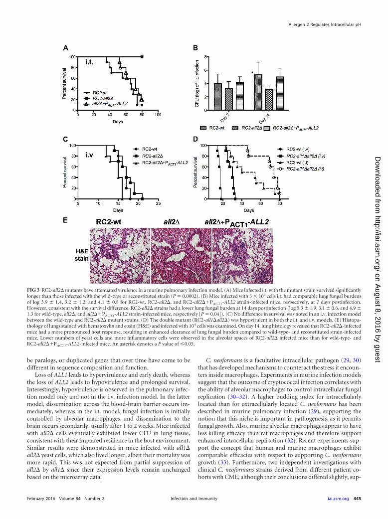
Loss of ALL2 results in attenuated virulence in pulmonarycryptococcosis but not in CME. Next, virulence of all2� mutantsin pulmonary and i.v. murine infection models was compared tothat of wild-type and reconstituted strains. Mice infected i.t. withan inoculum of 5 � 104 RC2-all2� cells survived significantlylonger (P � 0.001) than those infected with the wild-type strain(Fig. 3A), which was consistent with the enhanced macrophage-me-diated killing and phagocytosis observed in vitro (Fig. 2D and E).Consistent with the unaltered growth of the mutants, comparablefungal burden was detected in the lung at 7 days postinfection, butthen decreased CFU levels were found at 14 days postinfection in themutant cells (log 5.3 1.9, 3.1 0.6, and 4.9 1.3 for RC2-, RC2-all2�-, and RC2-all2��PACT1-ALL2-infected mice, respectively, P �0.05) (Fig. 3B), which is consistent with their enhanced clearancefrom the lungs. In contrast, virulence in the i.v. infection model forthe all2� strain was comparable to that for the wild-type and thereconstituted strains (Fig. 3C). The all1� all2� double mutant washypovirulent in both infection models (Fig. 3D).
Histopathology analysis of RC2-all2�-infected lung tissues at14 days revealed decreased numbers of cryptococcal cells and pro-
nounced recruitment of inflammatory cells in the lung tissuecompared to results for the wild-type-infected lung tissues (Fig.3E). This is consistent with a more effective host response, whichresults in enhanced clearance and thus explains the lower fungalburden at day 14. In summary, these findings confirm that ALL2 isa virulence-associated gene and that its loss can alter the patho-genesis of pulmonary cryptococcal infection.
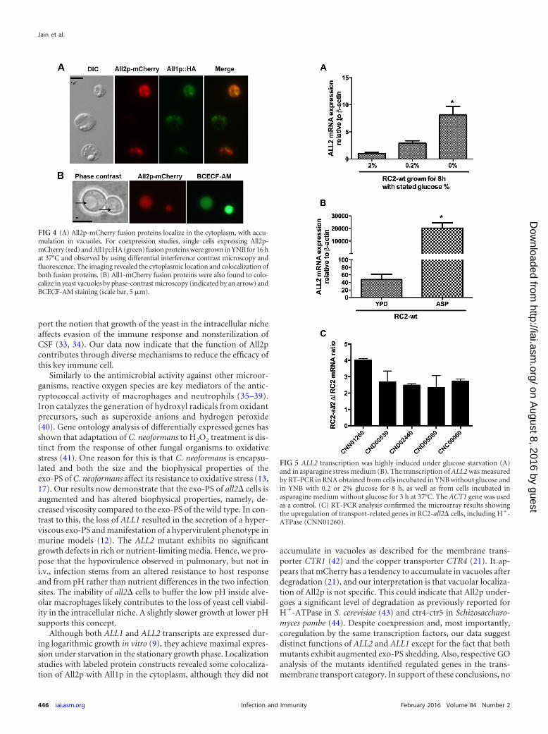
The ALL2-fluorescent fusion protein localizes to the cyto-plasm and vacuoles. Next, we sought to investigate whether All1pand All2p proteins colocalize, since both the ALL1 and the ALL2transcripts are coregulated (9). For these studies, ALL2 was fusedwith C. neoformans codon-optimized mCherry under an ACT1promoter (ALL2-mCherry). The ALL2-mCherry was transformedin cells expressing ALL1 with a hemagglutinin (HA) tag under itsnative promoter (ALL1::HA). ALL1::HA was detected using anti-bodies against HA that were conjugated to Alexa Fluor 594(green). Immunofluorescence staining revealed that All1p andAll2p fusion proteins colocalize in the cytoplasm (Fig. 4A), andthe protein location was found to be unaffected at multiple timepoints of cell growth (data not shown). However, protein expression
FIG 1 (A) ALL2 is conserved among C. neoformans var. neoformans, C. neoformans var. grubii, and C. gattii strains. (B) Homology blast of All1 and All2 proteinsequences. All2p has a 77% similarity and a 61% identity to All1p.
Jain et al.
442 iai.asm.org February 2016 Volume 84 Number 2Infection and Immunity
on August 8, 2016 by guest
http://iai.asm.org/
Dow
nloaded from
varied widely among yeast cells, and a majority of C. neoformans cellsexpressed only either All1p or All2p (82%). Phase-contrast micros-copy indicated that the All2-mCherry fusion protein also localized tothe yeast vacuoles (Fig. 4B). This finding was confirmed by vacuole-specific BCECF-AM staining of yeast cells. Variable pH and glucosedid not alter the amount of All2p accumulation in vacuoles. Further,cells recovered from the lungs and the brains of mice 9 days postin-fection displayed All2p localization in vacuoles (see Fig. S3 in thesupplemental material).
ALL2 is highly expressed under stress conditions, and its lossaffects the expression of genes involved in multiple pathways oftransport. Review of published transcriptional profiles of Sp1�under glucose starvation documented upregulation of ALL2 inaddition to ALL1 (9). Quantification of ALL2 expression by RT-PCR in cells grown for 3 h in asparagine stress media and thentransferred to yeast nitrogen base media under glucose (Fig. 5A)or asparagine (Fig. 5B) starvation confirmed elevated expressionof ALL2 in both stress conditions. Comparison of transcriptomesfrom RC2-all2� and RC2-wt cells grown in minimal media doc-umented downregulation of 20 genes and upregulation of 263genes in RC2-all2� cells (Table 2; see also Fig. S4 in the supple-mental material). Downregulated genes included a hypotheticalprotein involved in carbohydrate transport and metabolism(CNE05330, 7.6-fold), a D-galactonate transporter (CNE05340,4.1-fold), and a cluster of proteins that have in common a domainfor 5-oxoprolinase (CNE05320, 2.1-fold; CNE05310, 2.5-fold).The 5-oxoprolinase (EC 3.5.2.9) enzyme is ATP dependent and isinvolved in glutathione metabolism to synthesize L-glutamatefrom 5-oxoproline.
Interestingly, the most abundant upregulated gene was aplasma membrane H�-ATPase 1 (CNN01260, 9-fold). Othergenes upregulated in the mutant were involved in multipleamino acid biosynthesis pathways: aminoacyl-tRNA biosynthesis(CNF00070, CND06300, CNM01290, CNG01310, and CNB05640),valine, leucine, and isoleucine biosynthesis (CNA02070), and purine(CNK02180, CNN00440), glutamate (CNJ02910), and glutathione(CNN00350 and CNG01150) metabolism (Table 2). Differentialexpression of the plasma membrane H�-ATPase (CNN01260)
and other highly regulated genes was further confirmed by RT-PCR (Fig. 5C). Importantly, the loss of ALL2 or ALL1 did notresult in increased expression of the respective homologous genes.Transcriptomes of all1� all2� and all1� mutants grown in mini-mal media were also compared with that of the all2� mutant. Anoverlap of genes was noted only with all1� all2� cells but not withall1� cells (GEO accession number GSE74219). This further sup-ported our conclusion that ALL1 and ALL2 have distinct func-tions.
Lastly, gene ontology (GO) analysis of differentially expressedgenes in the all2� mutant under starvation conditions was con-sistent with the above findings. This analysis identified specificGO categories, namely, those related to transport, nucleotidebinding, transmembrane transport, and catalytic activityamong others (Table 3).
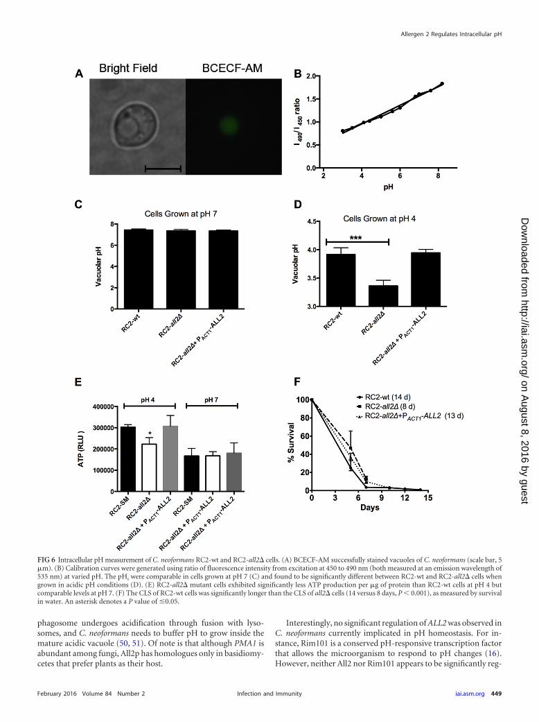
Intracellular pH, ATP levels, and chronological aging are al-tered in the all2� mutant. The main intracellular pH (pHi) reg-ulator in all eukaryotic cells is the plasma membrane H�-ATPasepump (27). To measure vacuolar pH, yeast cells were stained witha pH-sensitive fluorophore, BCECF-AM. Vacuole-specific label-ing was confirmed by microscopy (Fig. 6A), and calibration curveswere generated (Fig. 6B) using the ratio of fluorescence intensityfrom excitation at 450 to 490 nm (both measured at an emissionwavelength of 535 nm) at variable pHs. We measured pHi of yeastcells grown in YNB supplemented with 2 or 0.2% glucose at me-dium pH 4 or 7. The pHi were comparable in cells grown at pH 7under both conditions (Fig. 6C). However, in cells grown at anacidic pH, the mutant was not capable of maintaining intracellularpH compared to the wild type and the reconstituted strains. As aresult, the pHi of RC2-all2� was significantly more acidic thanthat of the wild type (Fig. 6D). Next, we investigated the actualfunction of the plasma membrane H�-ATPase 1 (CNN01260) inthe all2� mutant under different pH conditions. The primaryfunction of ATP synthase in most organisms is ATP synthesis(28). This enzyme is part of complex V ATP synthase in theoxidative phosphorylation pathway in C. neoformans. Accord-ingly, we quantified ATP cellular levels in RC2-all2� cells usingan ATP bioluminescence assay kit (Sigma-Aldrich, St. Louis,
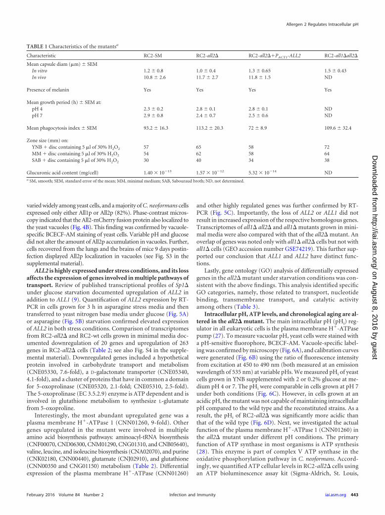
TABLE 1 Characteristics of the mutantsa
Characteristic RC2-SM RC2-all2� RC2-all2��PACT1-ALL2 RC2-all1�all2�
Mean capsule diam (�m) SEMIn vitro 1.2 0.8 1.0 0.4 1.3 0.65 1.5 0.43In vivo 10.8 2.6 11.7 2.7 11.8 1.5 ND
Presence of melanin Yes Yes Yes Yes
Mean growth period (h) SEM at:pH 4 2.3 0.2 2.8 0.1 2.8 0.1 NDpH 7 2.9 0.8 2.4 0.7 2.5 0.6 ND
Mean phagocytosis index SEM 93.2 16.3 113.2 20.3 72 8.9 109.6 32.4
Zone size (mm) on:YNB � disc containing 5 �l of 30% H2O2 57 65 58 72MM � disc containing 5 �l of 30% H2O2 54 62 58 64SAB � disc containing 5 �l of 30% H2O2 30 40 34 38
Glucuronic acid content (mg/cell) 1.40 � 1013 1.57 � 1012 5.32 � 1014 NDa SM, smooth; SEM, standard error of the mean; MM, minimal medium; SAB, Sabouraud broth; ND, not determined.
Allergen 2 Regulates Intracellular pH
February 2016 Volume 84 Number 2 iai.asm.org 443Infection and Immunity
on August 8, 2016 by guest
http://iai.asm.org/
Dow
nloaded from
MO). These experiments demonstrated that RC2-all2� cellsgrown in minimal media or YNB for 16 h produced significantlyless cellular ATP per �g protein than the wild-type cells whengrown at pH 4 (Fig. 6E) and produced comparable levels of ATP atpH 7 (Fig. 6E). Finally, chronological life span (CLS) was assessedin the mutant and compared to that in the wild type because theCLS, which is a resilience trait of yeast strains and measures theirviability under no nutrition, was predicted to shorten in the set-ting of impaired intracellular pH regulation. Consistent with thishypothesis, we found a significantly shortened CLS in the RC2-all2� strain compared to that in the wild type (8 days versus 14days, P � 0.01, Fig. 6F). It is noteworthy that the replicative lifespans of the mutant and the wild type, which measures the repro-ductive ability as opposed to the viability of the cell, were compa-rable under 2% glucose and not 0.05% glucose. Under low-glu-cose conditions, the RC2-all2� cells had a significantly shortened
RLS compared to that of the wild-type cells (21 versus 28 genera-tions, P � 0.02).
DISCUSSION
In this study, we report the role of a novel virulence-associatedgene named ALL2, which is coregulated with a highly homologousgene, ALL1, during the process of phenotypic switching of C. neo-formans to a hypervirulent mucoid variant.
ALL2p is highly homologous (77% similarity and 61% iden-tity) to the previously described gene ALL1p (7) and is also regu-lated by the same transcriptional regulator, Sp1 and gene PKC1(9), both of which are activated under stress. Our data demon-strate, however, that the functions of the two genes are mostlydistinct, although both mutants exhibit enhanced exopolysaccha-ride secretion. The effects of gene loss on virulence and exo-PSviscosity are contrasting. It is conceivable that these two genes may
FIG 2 (A) Viscosity measurements of the exopolysaccharide shed from wild-type (RC2-wt), mutant (RC2-all2�), and reconstituted (RC2-all2��PACT1-ALL2)strains grown for 14 days in minimal medium. Shed capsular polysaccharide of the all2� mutant was less viscous. The all2� mutant had increased sensitivity toH2O2 in vitro, as documented by incubating 105 or 106 RC2-wt, RC2-all2�, and RC2-all2��PACT1-ALL2 cells in DMEM with 5 mM H2O2 for 3 h andquantification of the percent survival (B) and by spotting 5 �l on YPD agar (C). (D) Killing assays were performed by lysing murine macrophages 3 h or 6 h aftercoincubation and plating the CFU. all2� cells were more sensitive to macrophage killing, consistent with their increased sensitivity to H2O2 in vitro. (E) Thephagocytosis index determined at 2 h after coincubation was significantly higher in macrophages incubated with RC2-all2� cells than in macrophages coincu-bated with RC2-wt or RC2-all2��PACT1-ALL2 cells. Intracellular phagocytosis index was calculated by visually counting cells on a microscope. All experimentswere performed in triplicates. An asterisk denotes a P value of �0.05.
Jain et al.
444 iai.asm.org February 2016 Volume 84 Number 2Infection and Immunity
on August 8, 2016 by guest
http://iai.asm.org/
Dow
nloaded from
be paralogs, or duplicated genes that over time have come to bedifferent in sequence composition and function.
Loss of ALL1 leads to hypervirulence and early death, whereasthe loss of ALL2 leads to hypovirulence and prolonged survival.Interestingly, hypovirulence is observed in the pulmonary infec-tion model only and not in the i.v. infection model. In the lattermodel, dissemination across the blood-brain barrier occurs im-mediately, whereas in the i.t. model, fungal infection is initiallycontrolled by alveolar macrophages, and dissemination to thebrain occurs secondarily, usually after 1 to 2 weeks. Mice infectedwith all2� cells eventually exhibited lower CFU in lung tissue,consistent with their impaired resilience in the host environment.Similar results were demonstrated in mice infected with all1�all2� yeast cells, which also lived longer, albeit their mortality wasmore rapid. This was not expected from partial suppression ofall2� by all1� since their expression levels remain unchangedbased on the microarray data.
C. neoformans is a facultative intracellular pathogen (29, 30)that has developed mechanisms to counteract the stress it encoun-ters inside macrophages. Experiments in murine infection modelssuggest that the outcome of cryptococcal infection correlates withthe ability of alveolar macrophages to control intracellular fungalreplication (30–32). A higher budding index for intracellularlylocated than for extracellularly located C. neoformans has beendescribed in murine pulmonary infection (29), supporting thenotion that this niche is important in pathogenesis, as it permitsfungal growth. Also, murine alveolar macrophages appear to haveless killing efficacy than rat macrophages and therefore supportenhanced intracellular replication (32). Recent experiments sup-port the concept that human and murine macrophages exhibitcomparable efficacies with respect to supporting C. neoformansgrowth (33). Furthermore, two independent investigations withclinical C. neoformans strains derived from different patient co-horts with CME, although their conclusions differed slightly, sup-
FIG 3 RC2-all2� mutants have attenuated virulence in a murine pulmonary infection model. (A) Mice infected i.t. with the mutant strain survived significantlylonger than those infected with the wild-type or reconstituted strain (P � 0.0002). (B) Mice infected with 5 � 104 cells i.t. had comparable lung fungal burdensof log 3.9 1.4, 3.2 1.2, and 4.1 0.8 for RC2-wt, RC2-all2�, and RC2-all2��PACT1-ALL2 strain-infected mice, respectively, at 7 days postinfection.However, consistent with the survival difference, RC2-all2� strains had a lower lung fungal burden at 14 days postinfection (log 5.3 1.9, 3.1 0.6, and 4.9 1.3 for wild-type, all2�, and all2��PACT1-ALL2 strain-infected mice, respectively [P � 0.04]). (C) No difference in survival was noted in an i.v. infection modelbetween the wild-type and RC2-all2� mutant strains. (D) The double mutant (RC2-all1�all2�) was hypovirulent in both the i.t. and i.v. models. (E) Histopa-thology of lungs stained with hematoxylin and eosin (H&E) and infected with 104 cells was examined. On day 14, lung histology revealed that RC2-all2�-infectedmice had a more pronounced host response, resulting in enhanced clearance of lung fungal burden compared to wild-type- and reconstituted strain-infectedmice. Lower numbers of yeast cells and more inflammatory cells were observed in the alveolar spaces of RC2-all2� infected mice than for wild-type- andRC2-all2��PACT1-ALL2-infected mice. An asterisk denotes a P value of �0.05.
Allergen 2 Regulates Intracellular pH
February 2016 Volume 84 Number 2 iai.asm.org 445Infection and Immunity
on August 8, 2016 by guest
http://iai.asm.org/
Dow
nloaded from
port the notion that growth of the yeast in the intracellular nicheaffects evasion of the immune response and nonsterilization ofCSF (33, 34). Our data now indicate that the function of All2pcontributes through diverse mechanisms to reduce the efficacy ofthis key immune cell.
Similarly to the antimicrobial activity against other microor-ganisms, reactive oxygen species are key mediators of the antic-ryptococcal activity of macrophages and neutrophils (35–39).Iron catalyzes the generation of hydroxyl radicals from oxidantprecursors, such as superoxide anions and hydrogen peroxide(40). Gene ontology analysis of differentially expressed genes hasshown that adaptation of C. neoformans to H2O2 treatment is dis-tinct from the response of other fungal organisms to oxidativestress (41). One reason for this is that C. neoformans is encapsu-lated and both the size and the biophysical properties of theexo-PS of C. neoformans affect its resistance to oxidative stress (13,17). Our results now demonstrate that the exo-PS of all2� cells isaugmented and has altered biophysical properties, namely, de-creased viscosity compared to the exo-PS of the wild type. In con-trast to this, the loss of ALL1 resulted in the secretion of a hyper-viscous exo-PS and manifestation of a hypervirulent phenotype inmurine models (12). The ALL2 mutant exhibits no significantgrowth defects in rich or nutrient-limiting media. Hence, we pro-pose that the hypovirulence observed in pulmonary, but not ini.v., infection stems from an altered resistance to host responseand from pH rather than nutrient differences in the two infectionsites. The inability of all2� cells to buffer the low pH inside alve-olar macrophages likely contributes to the loss of yeast cell viabil-ity in the intracellular niche. A slightly slower growth at lower pHsupports this concept.
Although both ALL1 and ALL2 transcripts are expressed dur-ing logarithmic growth in vitro (9), they achieve maximal expres-sion under starvation in the stationary growth phase. Localizationstudies with labeled protein constructs revealed some colocaliza-tion of All2p with All1p in the cytoplasm, although they did not
accumulate in vacuoles as described for the membrane trans-porter CTR1 (42) and the copper transporter CTR4 (21). It ap-pears that mCherry has a tendency to accumulate in vacuoles afterdegradation (21), and our interpretation is that vacuolar localiza-tion of All2p is not specific. This could indicate that All2p under-goes a significant level of degradation as previously reported forH�-ATPase in S. cerevisiae (43) and ctr4-ctr5 in Schizosaccharo-myces pombe (44). Despite coexpression and, most importantly,coregulation by the same transcription factors, our data suggestdistinct functions of ALL2 and ALL1 except for the fact that bothmutants exhibit augmented exo-PS shedding. Also, respective GOanalysis of the mutants identified regulated genes in the trans-membrane transport category. In support of these conclusions, no
FIG 4 (A) All2p-mCherry fusion proteins localize in the cytoplasm, with accu-mulation in vacuoles. For coexpression studies, single cells expressing All2p-mCherry (red) and All1p::HA (green) fusion proteins were grown in YNB for 16 hat 37°C and observed by using differential interference contrast microscopy andfluorescence. The imaging revealed the cytoplasmic location and colocalization ofboth fusion proteins. (B) All1-mCherry fusion proteins were also found to colo-calize in yeast vacuoles by phase-contrast microscopy (indicated by an arrow) andBCECF-AM staining (scale bar, 5 �m).
FIG 5 ALL2 transcription was highly induced under glucose starvation (A)and in asparagine stress medium (B). The transcription of ALL2 was measuredby RT-PCR in RNA obtained from cells incubated in YNB without glucose andin YNB with 0.2 or 2% glucose for 8 h, as well as from cells incubated inasparagine medium without glucose for 3 h at 37°C. The ACT1 gene was usedas a control. (C) RT-PCR analysis confirmed the microarray results showingthe upregulation of transport-related genes in RC2-all2� cells, including H�-ATPase (CNN01260).
Jain et al.
446 iai.asm.org February 2016 Volume 84 Number 2Infection and Immunity
on August 8, 2016 by guest
http://iai.asm.org/
Dow
nloaded from
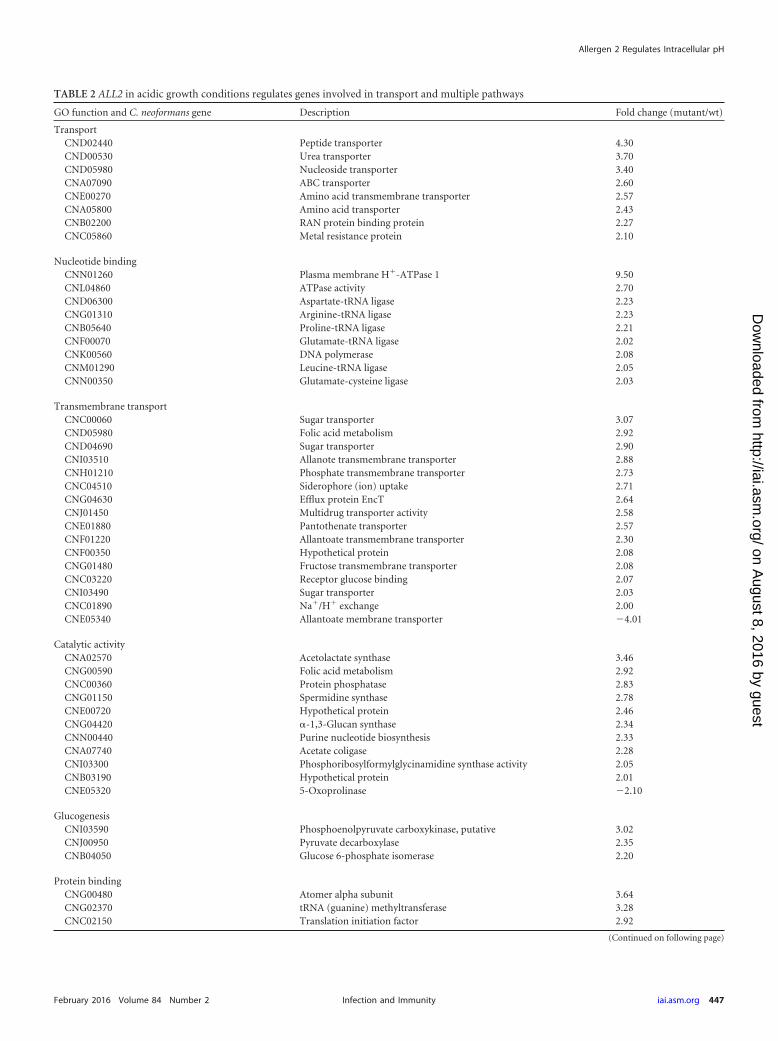
TABLE 2 ALL2 in acidic growth conditions regulates genes involved in transport and multiple pathways
GO function and C. neoformans gene Description Fold change (mutant/wt)
TransportCND02440 Peptide transporter 4.30CND00530 Urea transporter 3.70CND05980 Nucleoside transporter 3.40CNA07090 ABC transporter 2.60CNE00270 Amino acid transmembrane transporter 2.57CNA05800 Amino acid transporter 2.43CNB02200 RAN protein binding protein 2.27CNC05860 Metal resistance protein 2.10
Nucleotide bindingCNN01260 Plasma membrane H�-ATPase 1 9.50CNL04860 ATPase activity 2.70CND06300 Aspartate-tRNA ligase 2.23CNG01310 Arginine-tRNA ligase 2.23CNB05640 Proline-tRNA ligase 2.21CNF00070 Glutamate-tRNA ligase 2.02CNK00560 DNA polymerase 2.08CNM01290 Leucine-tRNA ligase 2.05CNN00350 Glutamate-cysteine ligase 2.03
Transmembrane transportCNC00060 Sugar transporter 3.07CND05980 Folic acid metabolism 2.92CND04690 Sugar transporter 2.90CNI03510 Allanote transmembrane transporter 2.88CNH01210 Phosphate transmembrane transporter 2.73CNC04510 Siderophore (ion) uptake 2.71CNG04630 Efflux protein EncT 2.64CNJ01450 Multidrug transporter activity 2.58CNE01880 Pantothenate transporter 2.57CNF01220 Allantoate transmembrane transporter 2.30CNF00350 Hypothetical protein 2.08CNG01480 Fructose transmembrane transporter 2.08CNC03220 Receptor glucose binding 2.07CNI03490 Sugar transporter 2.03CNC01890 Na�/H� exchange 2.00CNE05340 Allantoate membrane transporter 4.01
Catalytic activityCNA02570 Acetolactate synthase 3.46CNG00590 Folic acid metabolism 2.92CNC00360 Protein phosphatase 2.83CNG01150 Spermidine synthase 2.78CNE00720 Hypothetical protein 2.46CNG04420 �-1,3-Glucan synthase 2.34CNN00440 Purine nucleotide biosynthesis 2.33CNA07740 Acetate coligase 2.28CNI03300 Phosphoribosylformylglycinamidine synthase activity 2.05CNB03190 Hypothetical protein 2.01CNE05320 5-Oxoprolinase 2.10
GlucogenesisCNI03590 Phosphoenolpyruvate carboxykinase, putative 3.02CNJ00950 Pyruvate decarboxylase 2.35CNB04050 Glucose 6-phosphate isomerase 2.20
Protein bindingCNG00480 Atomer alpha subunit 3.64CNG02370 tRNA (guanine) methyltransferase 3.28CNC02150 Translation initiation factor 2.92
(Continued on following page)
Allergen 2 Regulates Intracellular pH
February 2016 Volume 84 Number 2 iai.asm.org 447Infection and Immunity
on August 8, 2016 by guest
http://iai.asm.org/
Dow
nloaded from
change in the expression levels in either mutant to compensate forthe loss of the other gene was observed. Also, efforts to coprecipi-tate the two proteins were not successful (data not shown).
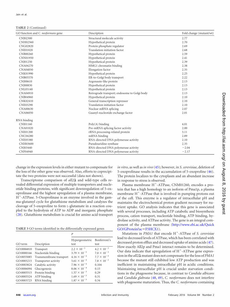
Transcriptome comparison of all2� and wild-type cells re-vealed differential expression of multiple transporters and nucle-otide binding proteins, with significant downregulation of 5-ox-oprolinase and the highest upregulation of a plasma membrane,H�-ATPase. 5-Oxoprolinase is an enzyme involved in the gam-ma-glutamyl cycle for glutathione metabolism and catalyzes thecleavage of 5-oxoproline to form L-glutamate in a reaction cou-pled to the hydrolysis of ATP to ADP and inorganic phosphate(28). Glutathione metabolism is crucial for amino acid transport
in vitro, as well as in vivo (45); however, in S. cerevisiae, deletion of5-oxoprolinase results in the accumulation of 5-oxoproline (46).The protein localizes to the cytoplasm and an abundant increasein response to stress is observed.
Plasma membrane H�-ATPase, CNM01260, encodes a pro-tein that has a high homology to an isoform of Pma1p, a plasmamembrane H�-ATPase that is involved in pumping protons outof the cell. This enzyme is a regulator of intracellular pH andmaintains the electrochemical proton gradient necessary for nu-trient uptake. GO analysis indicates that this gene is associatedwith several processes, including ATP catabolic and biosynthesisprocess, cation transport, nucleotide binding, ATP binding, hy-drolase activity, and ATPase activity. The gene is an integral com-ponent of the plasma membrane (http://www.ebi.ac.uk/QuickGO/GProtein?ac�F5HCX1).
Mutations in PMA1 that encode H�-ATPase of S. cerevisiaelead to decreased levels of ATPase, which has been correlated withdecreased proton efflux and decreased uptake of amino acids (47).How exactly All2p and Pma1 interact remains to be determined.Our data indicate that upregulation of H�-ATPase gene expres-sion in the all2� mutant does not compensate for the loss of PMA1because the mutant still exhibited low ATP production and wasinefficient in maintaining intracellular pH in acidic conditions.Maintaining intracellular pH is crucial under starvation condi-tions in the phagosome because, in contrast to Candida albicansand Candida glabrata (48, 49), C. neoformans does not interferewith phagosome maturation. Thus, the C. neoformans containing
TABLE 2 (Continued)
GO function and C. neoformans gene Description Fold change (mutant/wt)
CNJ02300 Structural molecule activity 2.77CNH02560 Hypothetical protein 2.70CNG02820 Protein phosphate regulator 2.69CNE01020 Translation initiation factor 2.68CNB00260 Hypothetical protein 2.59CNN01950 Hypothetical protein 2.41CNJ01250 Hypothetical protein 2.39CNA04270 HMG1 chromatin binding 2.38CNA04050 Elongation factor 2.35CNK01990 Hypothetical protein 2.25CNB05570 ER-to-Golgi body transport 2.22CNJ00610 Argonaute-like protein 2.13CNJ00830 Hypothetical protein 2.13CNL05140 Hypothetical protein 2.13CNA04910 Retrograde transport; endosome to Golgi body 2.13CNB04960 Hypothetical protein 2.10CNK02410 General transcription repressor 2.10CNE03290 Translation initiation factor 2.10CNA00630 Nuclear mRNA splicing 2.07CNA06050 Guanyl-nucleotide exchange factor 2.01
RNA bindingCNI01160 Poly(A) binding 4.01CNA01020 Pre-mRNA splicing factor activity 2.00CNE01200 rRNA processing-related protein 3.11CNC04280 mRNA binding 2.89CNE05380 RNA-directed DNA polymerase activity 2.10CNH03600 Pseudouridine synthase 2.35CNJ03440 RNA-directed DNA polymerase activity 2.04CNF04940 RNA-directed DNA polymerase activity 2.17
TABLE 3 GO terms identified in the differentially expressed genes
GO term Description
P
Hypergeometrictest
Bonferroni’stest
GO:0006810 Transport 2.2 � 105 4.2 � 103
GO:0000166 Nucleotide binding 3.79 � 105 7.0 � 103
GO:0055085 Transmembrane transport 4.16 � 104 7.7 � 103
GO:0005215 Transporter activity 5.61 � 104 7.8 � 102
GO:0003824 Catalytic activity 7.96 � 104 0.104GO:0006094 Glucogenesis 8.06 � 104 0.15GO:0005515 Protein binding 1.57 � 103 0.29GO:0005524 ATP binding 1.69 � 103 0.31GO:0003723 RNA binding 1.87 � 103 0.34
Jain et al.
448 iai.asm.org February 2016 Volume 84 Number 2Infection and Immunity
on August 8, 2016 by guest
http://iai.asm.org/
Dow
nloaded from
phagosome undergoes acidification through fusion with lyso-somes, and C. neoformans needs to buffer pH to grow inside themature acidic vacuole (50, 51). Of note is that although PMA1 isabundant among fungi, All2p has homologues only in basidiomy-cetes that prefer plants as their host.
Interestingly, no significant regulation of ALL2 was observed inC. neoformans currently implicated in pH homeostasis. For in-stance, Rim101 is a conserved pH-responsive transcription factorthat allows the microorganism to respond to pH changes (16).However, neither All2 nor Rim101 appears to be significantly reg-
FIG 6 Intracellular pH measurement of C. neoformans RC2-wt and RC2-all2� cells. (A) BCECF-AM successfully stained vacuoles of C. neoformans (scale bar, 5�m). (B) Calibration curves were generated using ratio of fluorescence intensity from excitation at 450 to 490 nm (both measured at an emission wavelength of535 nm) at varied pH. The pHi were comparable in cells grown at pH 7 (C) and found to be significantly different between RC2-wt and RC2-all2� cells whengrown in acidic pH conditions (D). (E) RC2-all2� mutant cells exhibited significantly less ATP production per �g of protein than RC2-wt cells at pH 4 butcomparable levels at pH 7. (F) The CLS of RC2-wt cells was significantly longer than the CLS of all2� cells (14 versus 8 days, P � 0.001), as measured by survivalin water. An asterisk denotes a P value of �0.05.
Allergen 2 Regulates Intracellular pH
February 2016 Volume 84 Number 2 iai.asm.org 449Infection and Immunity
on August 8, 2016 by guest
http://iai.asm.org/
Dow
nloaded from
ulated by the other in respective transcriptome studies (16). In amore recent study, Rim101 was shown to share downstream tar-gets with Pka1, and not surprisingly, ALL2 was found to be up-regulated in the pka1� mutant (52). Impaired ability to maintainproper intracellular pH also explains the significantly shortenedchronological life span of the mutants compared to that of the wildtype. In S. cerevisiae, chronological aging has been shown to resultin an acidic intracellular environment (25), and the inability of themutants to neutralize pH results in a shortened chronological lifespan but has no effect on the replicative life span. C. neoformans isacquired by inhaling spores from the environment. Most clinicalmanifestations of infections represent reactivation of latent dis-ease (53). The intracellular environment of macrophages in smallgranulomas is likely the site where the fungus survives nonrepli-cating for decades, similar to the pathogenesis of latent tubercu-losis. Chronological aging is a distinct trait of resilience as it mea-sures the ability of a fungus to survive in a nonreplicating state.Although this trait has not been studied with respect to pathogen-esis, we propose that this form of resilience is critical for the estab-lishment of latency of cryptococcal infection.
It has been reported previously that the overexpression of H�-ATPase is compensated by the downregulation of H�-ATPase ac-tivity in vivo (54) and does not always enhance growth (55). In S.cerevisiae, PTK2 and HRK1 genes encode protein kinases that havebeen identified to activate yeast plasma membrane production inresponse to glucose activation (56). Of note, ALL1 and ALL2 areregulated by the protein kinase Pkc1 signaling pathway (9). Incontrast to the plasma membrane H�-ATPase of S. cerevisiae,which is upregulated by glucose (57), ALL1 and ALL2 are upregu-lated in the setting of low glucose levels.
In summary, we conclude that the attenuated virulence of theall2� mutant in the pulmonary infection model may result fromthe mutant’s inefficiency to maintain intracellular pH in an acidicphagosome environment. The all2� mutant has no dramaticgrowth defect but produces significantly lower levels of ATP thanthe wild type, which affect the energetic state of the all2� mutantin vivo. We reason that this changes its resilience and the outcomeof infection, similarly to Leishmania donovani, where intracellularATP levels correlate with parasite proliferation (58).
We propose that ALL2 should be considered a target for anti-fungal drug discovery and further studied. Our studies highlightthe uniqueness of C. neoformans, one of the few basidiomycetesthat have evolved to be a successful human fungal pathogen. Spe-cifically, these data show that its unique ability to escape hostresponse and survive in the intracellular niche relies on genes,such as ALL2, that are unique to this fungus and contribute to itsexceptional resilience.
FUNDING INFORMATIONThis work was supported by NIH awards R01 AI059681 and R21 A1087564 toB.C.F. The funders had no role in study design, data collection and interpre-tation, or the decision to submit the work for publication.
REFERENCES1. Park BJ, Wannemuehler KA, Marston BJ, Govender N, Pappas PG,
Chiller TM. 2009. Estimation of the current global burden of cryptococcalmeningitis among persons living with HIV/AIDS. AIDS 23:525–530. http://dx.doi.org/10.1097/QAD.0b013e328322ffac.
2. Bicanic T, Meintjes G, Wood R, Hayes M, Rebe K, Bekker LG, HarrisonT. 2007. Fungal burden, early fungicidal activity, and outcome in crypto-coccal meningitis in antiretroviral-naive or antiretroviral-experienced pa-
tients treated with amphotericin B or fluconazole. Clin Infect Dis 45:76 –80. http://dx.doi.org/10.1086/518607.
3. Hamilton AJ, Goodley J. 1996. Virulence factors of Cryptococcus neofor-mans. Curr Top Med Mycol 7:19 – 42.
4. Fries BC, Goldman DL, Cherniak R, Ju R, Casadevall A. 1999. Pheno-typic switching in Cryptococcus neoformans results in changes in cellularmorphology and glucuronoxylomannan structure. Infect Immun 67:6076 – 6083.
5. Fries BC, Taborda CP, Serfass E, Casadevall A. 2001. Phenotypic switch-ing of Cryptococcus neoformans occurs in vivo and influences the outcomeof infection. J Clin Invest 108:1639 –1648. http://dx.doi.org/10.1172/JCI13407.
6. Franzot SP, Mukherjee J, Cherniak R, Chen LC, Hamdan JS, CasadevallA. 1998. Microevolution of a standard strain of Cryptococcus neoformansresulting in differences in virulence and other phenotypes. Infect Immun66:89 –97.
7. Jain N, Li L, Hsueh YP, Guerrero A, Heitman J, Goldman DL, Fries BC.2009. Loss of allergen 1 confers a hypervirulent phenotype that resemblesmucoid switch variants of Cryptococcus neoformans. Infect Immun 77:128 –140. http://dx.doi.org/10.1128/IAI.01079-08.
8. Haynes BC, Skowyra ML, Spencer SJ, Gish SR, Williams M, Held EP,Brent MR, Doering TL. 2011. Toward an integrated model of capsuleregulation in Cryptococcus neoformans. PLoS Pathog 7:e1002411. http://dx.doi.org/10.1371/journal.ppat.1002411.
9. Adler A, Park YD, Larsen P, Nagarajan V, Wollenberg K, Qiu J, MyersTG, Williamson PR. 2011. A novel specificity protein 1 (SP1)-like generegulating protein kinase C-1 (Pkc1)-dependent cell wall integrity andvirulence factors in Cryptococcus neoformans. J Biol Chem 286:20977–20990. http://dx.doi.org/10.1074/jbc.M111.230268.
10. Jung WH, Sham A, White R, Kronstad JW. 2006. Iron regulation of themajor virulence factors in the AIDS-associated pathogen Cryptococcusneoformans. PLoS Biol 4:e410. http://dx.doi.org/10.1371/journal.pbio.0040410.
11. Panepinto J, Liu L, Ramos J, Zhu X, Valyi-Nagy T, Eksi S, Fu J, JaffeHA, Wickes B, Williamson PR. 2005. The DEAD-box RNA helicase Vad1regulates multiple virulence-associated genes in Cryptococcus neoformans.J Clin Invest 115:632– 641. http://dx.doi.org/10.1172/JCI200523048.
12. Jain N, Cordero RJ, Casadevall A, Fries BC. 2013. Allergen 1 regulatespolysaccharide structure in Cryptococcus neoformans. Mol Microbiol 88:713–727. http://dx.doi.org/10.1111/mmi.12216.
13. Zaragoza O, Chrisman CJ, Castelli MV, Frases S, Cuenca-Estrella M,Rodriguez-Tudela JL, Casadevall A. 2008. Capsule enlargement in Cryp-tococcus neoformans confers resistance to oxidative stress suggesting amechanism for intracellular survival. Cell Microbiol 10:2043–2057. http://dx.doi.org/10.1111/j.1462-5822.2008.01186.x.
14. Jain N, Li L, McFadden DC, Banarjee U, Wang X, Cook E, Fries BC.2006. Phenotypic switching in a Cryptococcus neoformans variety gattiistrain is associated with changes in virulence and promotes disseminationto the central nervous system. Infect Immun 74:896 –903. http://dx.doi.org/10.1128/IAI.74.2.896-903.2006.
15. Mandal P, Banerjee U, Casadevall A, Nosanchuk JD. 2005. Dual infec-tions with pigmented and albino strains of Cryptococcus neoformans inpatients with or without human immunodeficiency virus infection in In-dia. J Clin Microbiol 43:4766 – 4772. http://dx.doi.org/10.1128/JCM.43.9.4766-4772.2005.
16. O’Meara TR, Norton D, Price MS, Hay C, Clements MF, Nichols CB,Alspaugh JA. 2010. Interaction of Cryptococcus neoformans Rim101 andprotein kinase A regulates capsule. PLoS Pathog 6:e1000776. http://dx.doi.org/10.1371/journal.ppat.1000776.
17. Cordero RJ, Frases S, Guimaraes AJ, Rivera J, Casadevall A. 2011.Evidence for branching in cryptococcal capsular polysaccharides and con-sequences on its biological activity. Mol Microbiol 79:1101–1117. http://dx.doi.org/10.1111/j.1365-2958.2010.07511.x.
18. DuBois M, Gilles KA, Hamilton JK, Rebers PA, Smith F. 1956. Color-imetric method for determination of sugars and related substances. AnalChem 28:350 –356. http://dx.doi.org/10.1021/ac60111a017.
19. Reilly MC, Aoki K, Wang ZA, Skowyra ML, Williams M, Tiemeyer M,Doering TL. 2011. A xylosylphosphotransferase of Cryptococcus neofor-mans acts in protein O-glycan synthesis. J Biol Chem 286:26888 –26899.http://dx.doi.org/10.1074/jbc.M111.262162.
20. Reilly MC, Levery SB, Castle SA, Klutts JS, Doering TL. 2009. A novelxylosylphosphotransferase activity discovered in Cryptococcus neofor-
Jain et al.
450 iai.asm.org February 2016 Volume 84 Number 2Infection and Immunity
on August 8, 2016 by guest
http://iai.asm.org/
Dow
nloaded from
mans. J Biol Chem 284:36118 –36127. http://dx.doi.org/10.1074/jbc.M109.056226.
21. Waterman SR, Park YD, Raja M, Qiu J, Hammoud DA, O’HalloranTV, Williamson PR. 2012. Role of CTR4 in the virulence of Cryptococ-cus neoformans. mBio 3:e00285-12. http://dx.doi.org/10.1128/mBio.00285-12.
22. Plant PJ, Manolson MF, Grinstein S, Demaurex N. 1999. Alternativemechanisms of vacuolar acidification in H�-ATPase-deficient yeast. J BiolChem 274:37270 –37279. http://dx.doi.org/10.1074/jbc.274.52.37270.
23. Diakov TT, Tarsio M, Kane PM. 2013. Measurement of vacuolar andcytosolic pH in vivo in yeast cell suspensions. J Vis Exp http://dx.doi.org/10.3791/50261.
24. Garcia J, Shea J, Alvarez-Vasquez F, Qureshi A, Luberto C, Voit EO,Del Poeta M. 2008. Mathematical modeling of pathogenicity of Crypto-coccus neoformans. Mol Syst Biol 4:183.
25. Burtner CR, Murakami CJ, Kennedy BK, Kaeberlein M. 2009. A mo-lecular mechanism of chronological aging in yeast. Cell Cycle 8:1256 –1270. http://dx.doi.org/10.4161/cc.8.8.8287.
26. Steffen KK, Kennedy BK, Kaeberlein M. 2009. Measuring replicative lifespan in the budding yeast. J Vis Exp http://dx.doi.org/10.3791/1209.
27. Serrano R. 1988. Structure and function of proton translocating ATPasein plasma membranes of plants and fungi. Biochim Biophys Acta 947:1–28. http://dx.doi.org/10.1016/0304-4157(88)90017-2.
28. Alberts B, Bray D, Lewis J, Raff M, Roberts K, Watson J. 1994.Molecular biology of the cell, 3rd ed. Taylor and Francis, New York, NY.
29. Feldmesser M, Kress Y, Novikoff P, Casadevall A. 2000. Cryptococcusneoformans is a facultative intracellular pathogen in murine pulmonaryinfection. Infect Immun 68:4225– 4237. http://dx.doi.org/10.1128/IAI.68.7.4225-4237.2000.
30. Feldmesser M, Tucker S, Casadevall A. 2001. Intracellular parasitism ofmacrophages by Cryptococcus neoformans. Trends Microbiol 9:273–278.http://dx.doi.org/10.1016/S0966-842X(01)02035-2.
31. Zaragoza O, Alvarez M, Telzak A, Rivera J, Casadevall A. 2007. Therelative susceptibility of mouse strains to pulmonary Cryptococcus neofor-mans infection is associated with pleiotropic differences in the immuneresponse. Infect Immun 75:2729 –2739. http://dx.doi.org/10.1128/IAI.00094-07.
32. Shao X, Mednick A, Alvarez M, van Rooijen N, Casadevall A, GoldmanDL. 2005. An innate immune system cell is a major determinant of spe-cies-related susceptibility differences to fungal pneumonia. J Immunol175:3244 –3251. http://dx.doi.org/10.4049/jimmunol.175.5.3244.
33. Sabiiti W, Robertson E, Beale MA, Johnston SA, Brouwer AE, Loyse A,Jarvis JN, Gilbert AS, Fisher MC, Harrison TS, May RC, Bicanic T.2014. Efficient phagocytosis and laccase activity affect the outcome ofHIV-associated cryptococcosis. J Clin Invest 124:2000 –2008. http://dx.doi.org/10.1172/JCI72950.
34. Alanio A, Desnos-Ollivier M, Dromer F. 2011. Dynamics of Cryptococcusneoformans-macrophage interactions reveal that fungal background influ-ences outcome during cryptococcal meningoencephalitis in humans.mBio 2:e00158-11. http://dx.doi.org/10.1128/mBio.00158-11.
35. Alspaugh JA, Granger DL. 1991. Inhibition of Cryptococcus neoformansreplication by nitrogen oxides supports the role of these molecules aseffectors of macrophage-mediated cytostasis. Infect Immun 59:2291–2296.
36. Granger DL, Hibbs JB, Jr, Perfect JR, Durack DT. 1988. Specific aminoacid (L-arginine) requirement for the microbiostatic activity of murinemacrophages. J Clin Invest 81:1129 –1136. http://dx.doi.org/10.1172/JCI113427.
37. Murray HW, Cartelli DM. 1983. Killing of intracellular Leishmania don-ovani by human mononuclear phagocytes: evidence for oxygen-dependent and -independent leishmanicidal activity. J Clin Invest 72:32– 44.
38. Salas SD, Bennett JE, Kwon-Chung KJ, Perfect JR, Williamson PR. 1996.Effect of the laccase gene CNLAC1, on virulence of Cryptococcus neoformans.J Exp Med 184:377–386. http://dx.doi.org/10.1084/jem.184.2.377.
39. Walker L, Lowrie DB. 1981. Killing of Mycobacterium microti by immu-nologically activated macrophages. Nature 293:69 –71. http://dx.doi.org/10.1038/293069a0.
40. Rosen GM, Pou S, Ramos CL, Cohen MS, Britigan BE. 1995. Freeradicals and phagocytic cells. FASEB J 9:200 –209.
41. Upadhya R, Campbell LT, Donlin MJ, Aurora R, Lodge JK. 2013. Globaltranscriptome profile of Cryptococcus neoformans during exposure to hy-
drogen peroxide induced oxidative stress. PLoS One 8:e55110. http://dx.doi.org/10.1371/journal.pone.0055110.
42. Ding C, Yin J, Tovar EM, Fitzpatrick DA, Higgins DG, Thiele DJ. 2011.The copper regulon of the human fungal pathogen Cryptococcus neofor-mans H99. Mol Microbiol 81:1560 –1576. http://dx.doi.org/10.1111/j.1365-2958.2011.07794.x.
43. Ferreira T, Mason AB, Slayman CW. 2001. The yeast Pma1 protonpump: a model for understanding the biogenesis of plasma membraneproteins. J Biol Chem 276:29613–29616. http://dx.doi.org/10.1074/jbc.R100022200.
44. Ioannoni R, Beaudoin J, Mercier A, Labbe S. 2010. Copper-dependenttrafficking of the Ctr4-Ctr5 copper transporting complex. PLoS One5:e11964. http://dx.doi.org/10.1371/journal.pone.0011964.
45. Griffith OW, Bridges RJ, Meister A. 1978. Evidence that the gamma-glutamyl cycle functions in vivo using intracellular glutathione: effects ofamino acids and selective inhibition of enzymes. Proc Natl Acad Sci U S A75:5405–5408. http://dx.doi.org/10.1073/pnas.75.11.5405.
46. Kumar A, Bachhawat AK. 2010. OXP1/YKL215c encodes an ATP-dependent 5-oxoprolinase in Saccharomyces cerevisiae: functional charac-terization, domain structure and identification of actin-like ATP-bindingmotifs in eukaryotic 5-oxoprolinases. FEMS Yeast Res 10:394 – 401. http://dx.doi.org/10.1111/j.1567-1364.2010.00619.x.
47. Vallejo CG, Serrano R. 1989. Physiology of mutants with reduced expres-sion of plasma membrane H�-ATPase. Yeast 5:307–319. http://dx.doi.org/10.1002/yea.320050411.
48. Lee SC, Kress Y, Zhao ML, Dickson DW, Casadevall A. 1995. Crypto-coccus neoformans survive and replicate in human microglia. Lab Invest73:871– 879.
49. Seider K, Brunke S, Schild L, Jablonowski N, Wilson D, Majer O, BarzD, Haas A, Kuchler K, Schaller M, Hube B. 2011. The facultativeintracellular pathogen Candida glabrata subverts macrophage cytokineproduction and phagolysosome maturation. J Immunol 187:3072–3086.http://dx.doi.org/10.4049/jimmunol.1003730.
50. Levitz SM, Nong SH, Seetoo KF, Harrison TS, Speizer RA, Simons ER.1999. Cryptococcus neoformans resides in an acidic phagolysosome of hu-man macrophages. Infect Immun 67:885– 890.
51. Vylkova S, Carman AJ, Danhof HA, Collette JR, Zhou H, Lorenz MC.2011. The fungal pathogen Candida albicans autoinduces hyphal morpho-genesis by raising extracellular pH. mBio 2:e00055-11. http://dx.doi.org/10.1128/mBio.00055-11.
52. O’Meara TR, Xu W, Selvig KM, O’Meara MJ, Mitchell AP, AlspaughJA. 2014. The Cryptococcus neoformans Rim101 transcription factor di-rectly regulates genes required for adaptation to the host. Mol Cell Biol34:673– 684. http://dx.doi.org/10.1128/MCB.01359-13.
53. Garcia-Hermoso D, Janbon G, Dromer F. 1999. Epidemiological evi-dence for dormant Cryptococcus neoformans infection. J Clin Microbiol37:3204 –3209.
54. Gevaudant F, Duby G, von Stedingk E, Zhao R, Morsomme P, BoutryM. 2007. Expression of a constitutively activated plasma membrane H�-ATPase alters plant development and increases salt tolerance. PlantPhysiol 144:1763–1776. http://dx.doi.org/10.1104/pp.107.103762.
55. Haruta M, Burch HL, Nelson RB, Barrett-Wilt G, Kline KG, MohsinSB, Young JC, Otegui MS, Sussman MR. 2010. Molecular characteriza-tion of mutant Arabidopsis plants with reduced plasma membrane protonpump activity. J Biol Chem 285:17918 –17929. http://dx.doi.org/10.1074/jbc.M110.101733.
56. Goossens A, de La Fuente N, Forment J, Serrano R, Portillo F. 2000.Regulation of yeast H�-ATPase by protein kinases belonging to a family ded-icated to activation of plasma membrane transporters. Mol Cell Biol 20:7654–7661. http://dx.doi.org/10.1128/MCB.20.20.7654-7661.2000.
57. Campetelli AN, Monesterolo NE, Previtali G, Santander VS, AmaidenMR, Arce CA, Valdez-Taubas J, Casale CH. 2013. Activation of H�-ATPase by glucose in Saccharomyces cerevisiae involves a membrane serineprotease. Biochim Biophys Acta 1830:3593–3603. http://dx.doi.org/10.1016/j.bbagen.2013.03.012.
58. Luque-Ortega JR, Rivero-Lezcano OM, Croft SL, Rivas L. 2001. In vivomonitoring of intracellular ATP levels in Leishmania donovani promasti-gotes as a rapid method to screen drugs targeting bioenergetic metabo-lism. Antimicrob Agents Chemother 45:1121–1125. http://dx.doi.org/10.1128/AAC.45.4.1121-1125.2001.
59. Perfect JR, Lang SD, Durack DT. 1980. Chronic cryptococcal meningitis:a new experimental model in rabbits. Am J Pathol 101:177–194.
Allergen 2 Regulates Intracellular pH
February 2016 Volume 84 Number 2 iai.asm.org 451Infection and Immunity
on August 8, 2016 by guest
http://iai.asm.org/
Dow
nloaded from