Embed Size (px)
Citation preview
cases, instruments
and notes
Inexpensive quality television camera suitable for use with operating microscope
H. Wade Faulkner, M.D. Cheryl Blanton Raymond Norman
Mobile, Alabama
ABSTRACT
Video monitoring and video recording are useful in ophthalmic microsurgery for self-instruction and improvement, increased interest by operating room personnel, transfer of useful information to other physicians and improvement of technique.
Specialized video cameras for such use are expensive. There is, however, a newly-marketed, inexpensive and light-weight video camera, which was designed for electronic newsgathering purposes, and which can be modified for microsurgery application, providing excellent results.
One of the new portable ENG-EFP (electronic news gathering/electronic field production) video cameras, with some minor modifications, is providing us with excellent video of anterior segment and vitreo-retinal surgery.
We evaluated available video cameras suitable for mounting on the Zeiss microscope, including cameras specially designed for such application that range in price from $8000 to more than $10,000. The video market now has a wide array of portable color cameras, offering a variety of capabilities. Many are inexpensive in comparison with the special design cameras. We decided to purchase a newly marketed Panasonic WV3150 portable camera, which features a Newvicon pick-up tube. List price of this camera is $1395.
The Newvicon pick-up tube is 1 V2 to 2 times as sensitive as the silicon Vidicon tube featured in the specialized cameras we evaluated. In addition, the N ewvicon tube displays less "blooming" caused by high intensity light, and it is not subject to "burn-in" problems.
As purchased, the WV3150 model camera is equipped with viewfinder, servo-zoom lens, microphone and pistol-grip; the weight of the camera is 6.05 pounds. It is designed to be powered by a videorecorder, or optionally by an AC power adaptor. The color balance is automatic.
AM INTRA-OCULAR IMPLANT SOC J-VOL. 8, SPRING 1982 161
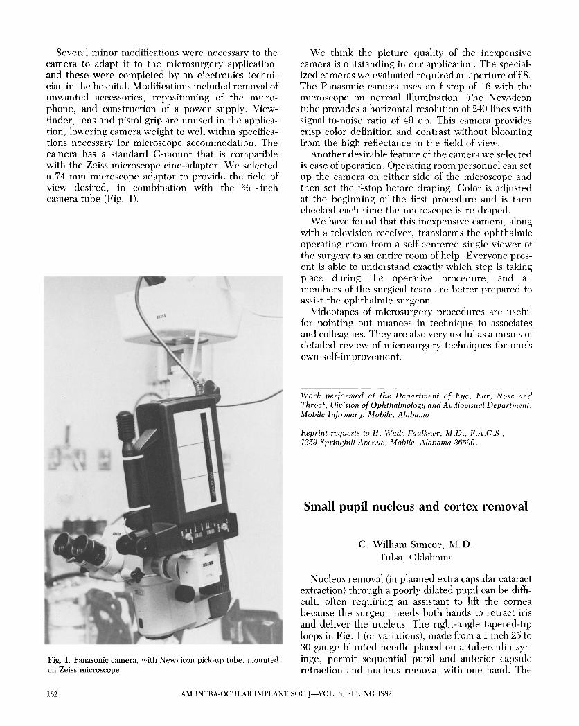
Several minor modifications were necessary to the camera to adapt it to the microsurgery application, and these were completed by an electronics technician in the hospital. Modifications included removal of unwanted accessories, repositioning of the microphone, and construction of a power supply. Viewfinder, lens and pistol grip are unused in the application, lowering camera weight to well within specifications necessary for microscope accommodation. The camera has a standard C-mount that is compatible with the Zeiss microscope cine-adaptor. We selected a 74 mm microscope adaptor to provide the field of view desired, in combination with the % - inch camera tube (Fig. 1).
Fig. 1. Panasonic camera, with Newvicon pick-up tube, mounted on Zeiss microscope.
We think the picture quality of the inexpensive camera is outstanding in our application. The specialized cameras we evaluated required an aperture of f 8. The Panasonic camera uses an f stop of 16 with the microscope on normal illumination. The Newvicon tube provides a horizontal resolution of 240 lines with signal-to-noise ratio of 49 db. This camera provides crisp color definition and contrast without blooming from the high reflectance in the field of view.
Another desirable feature of the camera we selected is ease of operation. Operating room personnel can set up the camera on either side of the microscope and then set the f-stop before draping. Color is adjusted at the beginning of the first procedure and is then checked each time the microscope is re-draped.
We have found that this inexpensive camera, along with a television receiver, transforms the ophthalmic operating room from a self-centered single viewer of the surgery to an entire room of help. Everyone present is able to understand exactly which step is taking place during the operative procedure, and all members of the surgical team are better prepared to assist the ophthalmic surgeon.
Videotapes of microsurgery procedures are useful for pointing out nuances in technique to associates and colleagues. They are also very useful as a means of detailed review of microsurgery techniques for one's own self-improvement.
Work performed at the Department of Eye , Ear, Nose and Throat, Division of Ophthalmology and Audiovisual Department, Mobile Infirmary, Mobile, Alabama.
Reprint requests to H. Wade Faulkner, MD., F.A.C.S., 1359 Springhill Avenue, Mobile, Alabama 36690.
Small pupil nucleus and cortex removal
C. William Simcoe, M.D. Tulsa, Oklahoma
Nucleus removal (in planned extra capsular cataract extraction) through a poorly dilated pupil can be difficult, often requiring an assistant to lift the cornea because the surgeon needs both hands to retract iris and deliver the nucleus. The right-angle tapered-tip loops in Fig. 1 (or variations), made from a 1 inch 25 to 30 gauge blunted needle placed on a tuberculin syringe, permit sequential pupil and anterior capsule retraction and nucleus removal with one hand. The
162 AM INTRA-OCULAR IMPLANT SOC J-VOL. 8, SPRING 1982