Embed Size (px)
Citation preview

INDICATIONS AND OUTCOME OF SCLERAL BUCKLE SURGERY IN
RHEGMATOGENEOUS RETINA DETACHMENT AT THE PCEA KIKUYU
HOSPITAL-EYE UNIT
DR. SANDIA SUMBANE
H58/89159/2016
A DISSERTATION SUBMITTED IN PARTIAL FULFILMENT FOR THE AWARD OF
DEGREE OF MASTER IN MEDICINE (OPHTHALMOLOGY) AT THE UNIVERSITY
OF NAIROBI
2019

ii
DECLARATION
I declare that this dissertation is the result of my own work and has not been submitted either
wholly or in part to any other university for the award of any degree or diploma. Research material
and contributions by others have been properly acknowledged.
Dr. Sandia Sumbane
MBChB( Eduardo Mondlane University, Mozambique)
Registrar, Department of Ophthalmology,
University of Nairobi
Signed………………………………………… Date……………………………………
iii
SUPERVISOR’S APPROVAL
This dissertation has been submitted with our approval as university and hospital supervisors.
Dr. Muchai Gachago
MBChB, M.MED (Ophthalmology), ICO, FEACO, FVRS
Lecturer, Department of Ophthalmology
University of Nairobi
Signed………………………………………… Date……………………………………
Dr. Millicent Muthoni Kariuki
MBChB, M.MED (Ophthalmology), FEACO
Senior Lecturer, Department of Ophthalmology
University of Nairobi
Signed………………………………………… Date……………………………………
Dr. Shafiq Jafferji Shabbir
MBChB, M.MED (Ophthalmology), FEACO, FMRF
Consultant Ophthalmology/Vitreoretinal Specialist, PCEA Kikuyu Hospital
Kikuyu Eye Unit
Signed………………………………………… Date……………………………………

iv
ACKNOWLEDGEMENT
I would like to thank my supervisors; Dr. Muchai Gachago, Dr. Millicent Muthoni Kariuki and
Dr. Shafiq Jafferji Shabbir for their guidance and support on preparation of this dissertation. I also
appreciate the support of my family and friends.
My gratitude also goes to Sight Savers, Mozambique for sponsoring my training.

v
TABLE OF CONTENTS
DECLARATION .......................................................................................................................................... ii
SUPERVISOR’S APPROVAL.................................................................................................................... iii
TABLE OF CONTENTS .............................................................................................................................. v
LIST OF FIGURES .................................................................................................................................... vii
LIST OF ABBREVIATIONS ...................................................................................................................... ix
ABSTRACT .................................................................................................................................................. x
CHAPTER ONE ......................................................................................................................................... 1
1.0 Introduction ............................................................................................................................................. 1
CHAPTER TWO ........................................................................................................................................ 2
2.0 Literature Review .................................................................................................................................... 2
2.1 Rhegmatogenous retinal detachment .................................................................................................. 2
2.2 Risk factors for rhegmatogenous retinal detachment .......................................................................... 3
2.3 Surgical interventions ......................................................................................................................... 4
2.4 Scleral buckle surgery ......................................................................................................................... 4
2.5 Indications for SB surgery .................................................................................................................. 7
2.6 Surgery success rates .......................................................................................................................... 7
2.7 Visual outcomes .................................................................................................................................. 8
2.8 Complications ..................................................................................................................................... 9
2.9 Factors associated with re-detachments ............................................................................................ 10
2.10 Rationale ......................................................................................................................................... 10
2.11 Research Questions ......................................................................................................................... 11
2.12. General objective ........................................................................................................................... 11
2.12.1 Specific objectives ................................................................................................................... 11
CHAPTER THREE .................................................................................................................................. 12
3.0 MATERIALS AND METHODS .......................................................................................................... 12
3.1 Study design ...................................................................................................................................... 12
3.2 Study site ........................................................................................................................................... 12
3.3 Study population ............................................................................................................................... 13
3.4 Eligibility criteria .............................................................................................................................. 13
3.4.1 Inclusion ..................................................................................................................................... 13
3.4.2 Exclusion .................................................................................................................................... 13
3.5 Sample size ....................................................................................................................................... 13
3.6 Sampling procedure .......................................................................................................................... 14

vi
3.7 Data collection methods .................................................................................................................... 14
3.7.1 Study tools ................................................................................................................................. 14
3.7.2 Data abstraction.......................................................................................................................... 14
3.8 Data management and analysis ......................................................................................................... 14
3.9 Ethical considerations ....................................................................................................................... 15
CHAPTER FOUR ..................................................................................................................................... 16
4.0 RESULTS ............................................................................................................................................. 16
5.0 DISCUSSION ....................................................................................................................................... 30
6.0 LIMITATIONS ..................................................................................................................................... 34
7.0 CONCLUSIONS ................................................................................................................................... 35
8.0 RECOMMENDATIONS ...................................................................................................................... 36
9.0 REFERENCES ..................................................................................................................................... 37
10. TIME FRAME ...................................................................................................................................... 41
11. Appendix 1: BUDGET ......................................................................................................................... 42
11.1 Appendix 2: Data Abstraction Tool ................................................................................................ 43
11.2 Appendix 3: Collaborating letter..................................................................................................... 47
11.3 Appendix 4: Ethics Approval .......................................................................................................... 48

vii
LIST OF FIGURES
Figure 1: Kikuyu in Kiambu County in Kenya (Source: map data Google 2018) ....................... 12
Figure 2 : Flow-chart of operated cases ........................................................................................ 16
Figure 3: Number of eyes operated by year (n=75) ...................................................................... 17
Figure 4: Age distribution (n=75) ................................................................................................. 19
Figure 5: Risk factors for RRD ..................................................................................................... 20
Figure 6: Characteristics of RRD (n=75) ...................................................................................... 21
Figure 7: Loss to follow-up........................................................................................................... 23
Figure 8: Visual acuity status at last follow-up ............................................................................ 27
Figure 9: Visual acuity status at 6 months .................................................................................... 28

viii
LIST OF TABLES
Table 1: Baseline characteristics (n=75) ....................................................................................... 18
Table 2: Characteristics of RD ...................................................................................................... 22
Table 3: Types of scleral buckling ................................................................................................ 23
Table 4: Trend of anatomical outcome ......................................................................................... 24
Table 5: Anatomical outcome at last follow up ............................................................................ 24
Table 6: Visual acuity trend .......................................................................................................... 25
Table 7: Comparison of preoperative and post-operative BCVA at last follow up ...................... 26
Table 8: Comparison of preoperative and post-operative BCVA at 6 months ............................. 27
Table 9: Complications of scleral buckling .................................................................................. 28
Table 10: Factors affecting re-detachment ................................................................................... 29

ix
LIST OF ABBREVIATIONS
BCVA Best Corrected Visual Acuity
UCVA Uncorrected Visual Acuity
KEU Kikuyu Eye Unit
PDR Proliferative Diabetic Retinopathy
PPV Pars PlanaVitrectomy
PVD Posterior Vitreous Detachment
PVR Proliferative Vitreoretinopathy
SB Scleral Buckle
SP Scleral Pocket
ST Sclera Thinning
IOL Intraocular Lens
SRF Subretinal Fluid
VA Visual acuity
RD Retinal Detachment
RRD Rhegmatogenous Retinal Detachment

x
ABSTRACT
Background: Scleral buckling is one of the preferred surgical techniques for RRD repair at KEU.
Beside the fact that this technique has been used for many years in this center, the outcomes are
unknown and worldwide studies have shown a high success rate and improvement on visual acuity
after surgery.
Objective: To identify the indications and outcome of scleral buckle surgery in RRD at the KEU.
Study design: Retrospective case series
Methods: In this study 75 RRD cases that underwent scleral buckling at KEU from 1st January
2012 to 31stDecember 2017 were identified and their social demographic data, the indication for
surgery as well as the outcome measures in terms of BCVA and retinal reattachment at 6 months
were entered into a data abstraction tool, then analysed using SPSS version 23.0. Factors associated
with re-detachments were determined using chi square.
Results: Seventy-five (75) eyes of 73 patients were analysed but, only 30(40%) patients had 6
months follow up. The median age was 31 (IQR 22-46) years. Majority of patients (61.3%) were
males, and right eye was involved in 62.7% of the cases. The mean duration of symptoms
experienced prior to presentation was 35.6 (SD ± 30.2) days. In this study scleral buckling was
done in 94.7% phakic eyes with inferior RRD in 72% without or with mild PVR associated with
macula on in 98.7% cases. Less than 3 breaks were identified in 70.4% cases, associated with
atrophic holes in 70.7% of cases. The final anatomical success rate was 96.7% at 6 months. The
visual outcome improved in 19 (63.3%) eyes by more than 2 Snellen lines. In this study only
6.7% of eyes had re-detachment but no factors were identified to be associated with re-
detachments.
Conclusion: Scleral buckling is safe and effective for uncomplicated RRD. Appropriate training
of SB surgery for RRD is necessary and justified in view of the favourable results especially in
phakic patients

1
CHAPTER ONE
1.0 Introduction
Scleral buckle surgery is an ophthalmic technique that has been used to repair rhegmatogenous
retinal detachments for over 60 years(1). Retinal detachment is described as the separation of the
neurosensory retina from the underlying retinal pigment epithelium (RPE). It can be classified
into three main types: rhegmatogenous, traction and exudative retinal detachment. Combined
forms such as tractional rhegmatogenous detachment may also occur but, the most common is the
rhegmatogenous, where liquefied vitreous humor penetrates into the sub-retinal space following
a retinal tear(2).
RRD occurs mainly due to physiological degeneration of the vitreous scaffold in the presence of a
retinal break. The degeneration starts early in life and over time results in hardening of the collagen
fibrils hence posterior vitreous detachment due to the progressive loss of elasticity(2). Early
symptoms of acute detachment may include tiny dark floaters that are linked to photopsia (flashes).
Loss of visual field is a late complication as a result of accumulation of subretinal fluid causing a
corresponding loss of peripheral vision. In addition, patients can be asymptomatic until
involvement of the macula(2).
Surgery is the only effective treatment and scleral buckle (SB) surgery is one of the most widely
surgical techniques used to repair RRD (3). This surgical procedure is classified into radial,
segmental circumferential and encircling circumferential buckling. The preferred type of buckle
to use depends on its indication(4). Functional closure of all retinal breaks and relief of vitro-retinal
traction is the main objective of sclera buckling(5). The sclera buckle(SB) procedure results in
anatomic reattachment lasting up to 20 years with significant longevity in many cases(6).
The aim of the study is to assess retrospectively the indications and the anatomical success rates
of buckling performed for RRD at KEU and also to establish the visual outcome following the
surgical procedure.

2
CHAPTER TWO
2.0 Literature Review
2.1Rhegmatogenous retinal detachment
Rhegmatogenous retinal detachment (RRD) is the most common indication for surgery in
vitreoretinal specialty(7), however, the presentation varies from uncomplicated detachment with a
single localized break to total detachments associated with multiple breaks, giant tears or
proliferative vitreoretinopathy (PVR) [4].
Ware in 1805, Wardrop in 1818 and Panizza in 1826 first described the retinal detachment by
relying mainly on pathological observations(8). Various other people in history have also played
a role by contributing to the current success rate of managing retinal detachments (RDs) that stands
at more than 90%. The evolution of RD is divided into pre-Gonin and post-Gonin eras that were
named after Jules Gonin(9). In the pre-Gonin era, though retinal breaks were associated with RD,
the focus was on the RD without much attention to the causative break. Different theories and
respective treatments were put forward with the first one showing that RD was spontaneous due
to abnormal leakage from the choroid. Breaks in the retina were due to increased pressure from
fluid generated behind the retina. Therefore, treatment was to puncture the sclera and retina to
relieve the pressure.
Draining the subretinal fluid (SRF) resulted in failure as a result of use of various modalities such
as chemical retinopexy and galvanocautery. In the second theory, hypotony and associated
circulatory alterations were thought to be the cause of RD. Many treatments like injection of
materials such as rabbit vitreous and gelatin to increase intraocular pressure were used. Osmotic
agents such as saline, cane sugar, glycerin and mercury salts were also injected into the
subconjunctival space with the hope of reducing SRF. Leber and Nordenson postulated alterations
in the vitreous generated secondary traction on the equatorial retina forming retinal tears with RD.
Non-surgical methods such as dietary modifications with salt restriction and bilateral compression
bandages with bed rest that increases intraocular pressure were also used(9).

3
Jules Gonin (1870-1935) demonstrated the role of the retinal break in the pathogenesis of RD in
which retinal breaks were localized pre- and intra-operatively. Under local anesthesia, the
subretinal space was entered after making a radial sclera incision near the break while SRF was
drained.Thermocautery was then introduced to create retinopexy in which Gonin reported a 63%
success rate(9).
In subsquent decades, different modifications of retinopexy were developed and used to treat the
retinal break. Chemical cauterization for retinopexy using potassium hydroxide introduced by
Guist and Lindner in 1931 and diathermy to bare the sclera (surface diathermy) or after trephining
the sclera (penetrating diathermy) was used. In this case, drainage of the SRF was performed along
with diathermy. Complications of diathermy led to the search for other modalities for retinopexy.
The current use of cryotherapy is credited to Harvey Lincroff and Amoils who created a specially
designed cryo-probe by use of liquid nitrogen. Charles Schepens described the use of scleral
depression and devised the first clinical head mounted indirect ophthalmoscope in 1947. He later
modified it by incorporating the light source and viewing system on the headband as is currently
known(9).
Shortening of the globe by scleral resection was the first step towards scleral buckling. Creating
an inward ridge for supporting a break was initially achieved either by full or partial thickness
sclera resection with SRF drainage and putting mattress sutures across the defect. Ernst Custodies
performed the first scleral buckling procedure using an episcleral exoplant in 1949 while Charles
Schepens did the first scleral buckling surgery in the USA in 1951. Harvey Lincoff in 1965
modified the original procedure by Custodis. The changes included use of silicone sponge, use of
improved scleral needles and use of cryotherapy instead of diathermy for retinopexy. Silicone
sponges were used in a radial or circumferential fashion depending on the clinical scenario(9).
2.2 Risk factors for rhegmatogenous retinal detachment
Typical risk factors for RRD includes, family history, increasing age, trauma to the eye, prior
cataract surgery (pseudophakia or aphakia), fellow eye retinal detachment and shortsightedness
(myopia) which is the main risk factor(2,5). An Indian study reported that prior cataract surgery
was the most common identifiable risk factor at 40% (10).
4
2.3 Surgical interventions
RRD is successfully repaired by scleral buckle surgery, pars plana vitrectomy with retinopexy and
internal tamponade and pneumatic retinopexy. None of the studies done previously has shown
superiority of one technique over another in terms of anatomic or visual outcomes. The choice of
technique used depends on the experience and preference of the surgeon combined with the
type(s), number and distribution of retinal break(s) as well as the nature and extent of RRD(5).
2.4 Scleral buckle surgery
Scleral buckle surgery is a procedure in which a band of silicone, plastic, rubber or sponge is
placed and sewn outside the sclera at the site of a retinal break to create an indentation of the sclera
toward the retinal break. It holds the retina against the sclera until formations of scar tissue at the
site of chorioretinal irritation by laser or cryotherapy is complete(10). The shortening of the sclera
was introduced by Mueller in 1903 to decreases the globe volume, however, Jess became the first
person to use a foreign substance to create an indentation in 1937 where a temporary tampon of
gauze was placed beneath Tenon’s capsule over the retinal break. Ernst Custodis was the first
person to do scleral buckling using retained explants(9).
Scleral buckle (SB) procedure came to revolutionize the treatment of patients with retinal
detachment and has been in place since 1957 with anatomic reattachment success of up to 94%.The
procedure also gives longevity lasting 20 years or more in many cases(6).
Vitrectomy technique has become popular for the treatment of RRD due to the advent of outpatient
ambulatory surgery, improvement in technology, instrumentation, and viewing systems(11).
However, SB is still the primary procedure for many surgeons in young phakic patients with
RRD(12).
Scleral buckling involves various techniques such as encircling buckles and segmental buckles
that can be placed radically, circumferentially or even obliquely(4). The choice of the scleral
buckle used depends on the location, number and size of the retinal breaks. It also depends on the
phakic status, distribution of SRF, presence of proliferative vitreoretinopathy (PVR), the amount
of vitreous traction, the state of the sclera and available eye volume.

5
There are three main SB techniques; they include the radial SB indicated for large U-shaped tears
and posterior breaks, the segmental circumferential SB indicated for small to medium size breaks
in the same location, the encircling circumferential SB used for more complex RD. This includes
large and multiple breaks in three or more different locations, extensive RD without visible breaks,
lattice degeneration in three or more locations, mild PVR, aphakic RD, excessive drainage of SRF
and after segmental SB failed. It confers a permanent 360-degree scleral indentation(7).
General or local anesthesia can be used to perform scleral buckling and the procedure involves
conjunctiva opening performed at either the limbus or several millimeters posterior in patients with
filtering blebs or recent limbal wounds. In many cases, radial conjunctiva relaxation incisions are
suggested to prevent tearing. A 3600 peritomy is necessary if more than two quadrants are to be
buckled. Conjunctiva and Tenon’s capsule can be reflected in the required quadrants using curved
Steven’s scissor and only the appropriate muscles are isolated. After the peritomy, the space
between Tenon’s capsule and sclera is then entered, muscle tendon is later engaged with a muscle
hook, and the connections to Tendon’s capsule are identified and separated from the muscle.
A traction suture using 4.0 braided silk sutures is placed around the muscle, after all rectus have
been isolated, the surface of the sclera is inspected for evidence of thinning, staphyloma, and
anomalous vortex veins(13). The locations of any abnormalities are noted before scleral depression
is started or retinal breaks are marked. Using indirect ophthalmoscopy and scleral indentation all
breaks are localized and the anterior edge is marked, that consist of an area of most vitreoretinal
traction that has to be opposed. The breaks are then sealed with cryopexy trans-sclerally and marks
placed on the sclera to localize the breaks which act as guides to secure the buckle. The buckle is
positioned beneath the rectus or over intact sclera and secured with 5-0 polyester sutures on
spatulated needle placed parallel to the buckle going through partial sclera thickness or within
intrascleral tunnels that will add the surgery duration but the risk of extrusion of the buckle
decreases. If a non-drainage technique is chosen, it is necessary to use medical or surgery
techniques (anterior chamber paracentesis) to counter intraocular pressure elevation. This usually
occurs in cases of encircling bands due to their higher potential to reduce the eye ball volume. If a
drainage approach is chosen, the SRF drainage is carried out by use of scleral cut down and a 30G
needle perforates the choroid. Once adequate fluid removal occurs, the sclerotomy can be closed

6
with 5-0 nylon suture. Restoration of vitreous volume is achieved with air, gas (SF6 or C3 F8) or
fluid if the eye becomes too soft from the amount of fluid drained. The buckle position and
pulsation of central retinal artery is then checked and the conjunctiva closed with 8/0
vicryl.Subconjunctival injection of antibiotic and steroid are then given and temporary eye patch
is placed (14,15).
During SB surgery for RRD, drainage of SRF can be done using an incision parallel to the limbus
in a scleral pocket (SP) or through a simple radial scleral thinning (ST). A study reported that
during surgery, choroidal detachments were observed at a higher percentage in the SP group and
at the end of the surgery a certain amount of SRF behind the buckle was significant in the ST
group. The study thus concluded that SP drainage technique appeared to be an effective and safe
method to drain SRF (16,17).
Drainage of SRF is a critical stage in scleral surgery for RRD that is carried out to facilitate the
localizations of breaks, to visualize the retinal attachment to the buckling band during surgery. It
also allows reduction of the extent of retinal-choroidal-cryo-applications hence a smaller risk of
pigment dispersion in to the vitreous and vitreoretinal retraction processes(16). However, drainage
of subretinal fluid during scleral buckling is controversial with most cases being managed without
draining while others believe that it is a crucial part of the technique. Two reasons for draining are
to decrease the intraocular volume allowing elevation of the buckle without elevating intraocular
pressure (IOP) and to remove fluid from the subretinal space allowing the retina to settle on the
elevated buckle (13,16).
The most significant complications occurring during SRF drainage are subretinal or choroidal
hemorrhage, retinal perforation, vitreoretinal incarceration, ocular hypotony and choroidal
detachment. The occurrence of any of these complications can cause surgery to be unsuccessful
(16). A study reported complications related to drainage of subretinal fluid in 5.6% of the 556 eyes
in which drainage was attempted. Small sub retinal hemorrhage was the most common
complication in 3%, retinal incarceration occurred in 2% and was associated with an iatrogenic
retinal hole in three cases and loss of formed vitreous in two cases. Hyphema was observed during

7
drainage in a single eye with an anterior chamber IOL and an atypical endophthalmitis seen in one
case in which uncomplicated drainage had been performed(17).
2.5 Indications for SB surgery
Scleral buckle surgery procedure is recommended in; RRDs in young and phakic patients with no
posterior vitreous detachment (PVD), retinal dialysis, atrophic or round holes, breaks anterior to
the equator, inferior breaks and certain complex retinal detachments with PVR. However, SB is
contraindicated when detachment is due to significantly posterior breaks, in cases of opaque media
(vitreous hemorrhage) and when it is associated with significant traction such as diabetic tractional
detachments and PVR(18). It is also contra-indicated in patients with vaso-occlusive disease
(sickle cell anemia) to avoid possible anterior ischemia caused by buckling(15). A study done in
Nigerian included all phakic RD patients for buckling (19).
2.6 Surgery success rates
Rhegmatogenous retinal detachment has a high reattachment success rate in 90 percent of cases
following surgery with success rates nearing 100% after secondary surgery(5). Anatomical success
is a reattachment of the retina 6 months after primary surgery with reoperations included(3). In a
retrospective study carried out in USA, 1226 cases with follow up over 1-year reported a success
rate of 86%for scleral buckling, 90% vitrectomy and 94% for combined vitrectomy and scleral
buckling and 63% for pneumatic retinopexy surgery(20).While in Norway, scleral buckling
achieved a success rate of up to 86% reattachment without reoperation and 99% with reoperation
in 6 months(3). In Japan,93.7% primary anatomical success and 100% final anatomical success in
271 eyes treated using SB surgery was reported(21). In Pakistan, a final re-attachment rate of 82%
was also reported(22). In a Nigerian study, anatomical attachment was seen in 95.6% of patients
on the operation table, 90.9% patients at first day postoperatively and 86.5% of patients at six
weeks after surgery(19).
Several factors have been identified in previous studies linked to anatomical success or failure of
SB surgery for RRD. Preoperative factors identified in previous studies include proliferative
vitreoretinopathy, opacity of the media, pseudophakia, and in complicated RRD with multiple
breaks. Primary anatomical failure was found more frequently in older patients with lower VA
8
with p <0.001(12). In a study carried out in India, it was reported that the commonest cause of
failure was a missed break in 32.4%(23) while the prognostic factor for anatomic and visual acuity
success was associated with macular detachment in most cases(15).
2.7 Visual outcomes
Scleral buckling procedure has a good visual outcome when performed in fresh retinal
detachment(19).Visual acuity improves significantly after surgical intervention and there is no
difference between various techniques used to repair rhegmatogenous retinal detachments(20).
Various pre-operative characteristics determine the post-surgical visual outcomes, for instance, in
old age (>70 years) severe PVR, detachment more than 7 days prior to surgery, and macular
detachment are the most important predictive factors in restoring visual acuity. The most reliable
predictive factors for poor postoperative outcome are the extent of initial macular involvement,
poor preoperative visual acuity and intraoperative hemorrhage(15).Only 40% to 60% of patients
with macula-off detachments have visual acuity restored to 20/50 or better. In detachments
however, sparing the macula visual restoration is much higher. In a surgery carried out in one large
series indicated that 90% of patients with macula-on detachments had vision of 20/50 or
better(5).Similarly, in Japan, study findings showed a significant improvement on BCVA in
patients with phakic and macula-off detachments and no change for those with pseudophakia or
macula-on detachments(21).In Pakistan, a mean change in refractive spherical equivalent of -1.478
+ or - 0.698 D in which the final BCVA achieved in 62% of the treated eyes was> 6/60(22).A
retrospective non-comparative case series of 65 patients carried out in Scotland showed a
significant improvement in BCVA from 29% of patients with preoperative VA of 6/6 to 56% with
VA 6/6post-operatively(24).
Several other pre-operative characteristics have been identified to have a significant influence on
post-operative visual outcome. Younger patients achieved significantly better visual outcome and
had shorter duration of symptom compared to the older patients. This was reported in a Korean
study with older age (35 years and older) being a prognostic factor for anatomical failure in SB for
uncomplicated RRD(12).Positive prognostic factors for visual acuity that have also been shown to
be important were better preoperative visual acuity with fewer quadrants involved by
the detachment due to lack of high myopia(15).
9
In Nigeria, a study reported good visual outcome at 38.8% and the primary anatomic reattachment
was at 80.5% while final anatomic success was at 90.2%. VA of 6/60 and better preoperative, mild
PVR, and achievement of primary anatomic success favorably influenced a good visual outcome
after surgery for RRD(25). In another study carried out in Nigeria, over 51.1% of patients reported
an improvement of visual acuity, 20% remained the same and 28.9% got worse six weeks after
scleral buckle surgery and at the end of the follow up 66.7% had BCVA > 3/60. Late presentation
and PVR may have been associated with poor outcome in some patients in which vitrectomy in
these patients helped to improve their visual outcome. It was noted advising the population on the
importance of early presentation and adequate awareness of scleral buckle surgery for RRD could
result in better outcome in sub-Saharan Africa(19).
In summary, the main factors that predict poorer visual function after surgery include poor
preoperative visual acuity, more than three quadrants involvement, multiple breaks, breaks
localized inferiorly, macula-off RRD, proliferative vitreoretinopathy of any grade (PVR) and
intraoperative hemorrhage(5).
2.8 Complications
There is relatively low potential for intra and postoperative complications of SB surgery.
Immediate and early postoperative discomfort, entrapment of rectus muscle leading to strabismus,
explants extrusion, anterior segment ischemia especially after using encircling bands and increased
axial length, decreased equatorial diameter leading to refractive error which is the most common
complication. Other early complications include high IOP resulting from reduction of total globe
volume leading to glaucoma if long standing, macular edema or macular pucker, diplopia,
choroidal bleeding and infection of the buckle(26). Diplopia and strabismus were reported early
after operation in around 15% of cases. Buckle infection was found in 0.3% of cases and buckle
migrates into the eyeball was found in <0.01% of cases(2). In Pakistan, a study reported that at
least one intra-operative complication was found in 32% of the patients. This included 2%
associated with iatrogenic scleral break, 8% inadvertent drainage of sub retinal fluid, 2% with
choroidal hemorrhage, 14% sub retinal hemorrhage, 2% retinal incarceration, vitreous hemorrhage
in 6%, 4% with high intraoperative IOP, 2%, with very low IOP and hyphema in 2%, 22% high

10
IOP and 16% vitreous hemorrhage. Twenty-four percent of patients who developed these
complications had with systemic complications. (22). Complications reported in a Nigerian study
included raised intraocular pressure and reopening of breaks in five patients(19).
2.9 Factors associated with re-detachments
There are various risks that are mainly associated with anatomical failure and re-detachment of
scleral buckling like pre-retinal membrane, undetected retinal tear, inadequate sclera buckle, new
retinal tear, inadequate chorio-retinal reaction, iatrogenic retinal tear and loss of buckle height(27).
A drain-air injection-cryo-exoplant (DACE) procedure can also cause too many air bubbles that
interfere with proper localization of the break as well as appropriateness of the buckle location.
There is a general tendency to choose buckles of narrow width which makes it easier to pass
mattress sutures. However, this should not be at the expense of inadequate coverage of the break.
Another issue that can compromise the outcome is a sub-retinal bleed at the conclusion of SRF
drainage due to cauterization of the knuckle of the choroid that does not always prevent the bleed.
One way to deal with this is controlled drainage.
There are other factors that can affect outcome like inadequate cryo-induced chorio-retinal
adhesion around the retinal break and fish mouthing. The latter is common after placing
circumferential buckles for large horseshoe tears(28).In a USA study,1088 consecutive operations
for retinal detachment were done, it was found that that pre-retinal membrane, accounted for 33%
of failure (27). Re-detachment was found to occur after a mean period of 13.6 months with a
median of 3 months. 55 (12.6%) eyes had re-detachment within the first 3 months after the primary
surgery. In addition, 68 out of 436 eyes (15.6%) had developed re-detachmentat 6 months and 80
out of 436 eyes (18.3%) after 1 year(29).
2.10 Rationale
Scleral buckling is a treatment of choice in patients with uncomplicated RD associated with round
holes and in retinal dialysis. In detachments affecting the macula, 40 to 60% of patients improve
their visual acuity to 20/50 or better. Worldwide studies have shown high sucess rate nearing
100%(5). In Kenya, despite the fact that the SB surgery has been done for many years at KEU no
studies have been done to find out the indications and outcomes of SB surgery in RRD hence it is

11
necessary to determine the proportions of reattachment, the level of visual acuity after surgery and
factors associated with the outcome then to establish the effectiveness of the SB surgery in our
setting.
2.11 Research Questions
1. What are the indications of scleral buckle in rhegmatogenous retinal detachment among
patients attending the Kikuyu Eye Unit (KEU)?
2. What are the retinal reattachment success rates following scleral buckling among patients
attending the KEU?
3. What is the visual outcome 6 months after scleral buckling among patients attending the
KEU?
4. Which factors are associated with re-detachment among patients attending the KEU?
2.12. General objective
To determine the indications and outcome of scleral buckle surgery in rhegmatogenous retinal
detachment among patients attending the Kikuyu Eye Unit.
2.12.1 Specific objectives
1. To identify the indications of scleral buckle surgery in rhegmatogenous retinal detachment
among patients attending the KEU.
2. To determine the retinal reattachment success rates 6 months following scleral buckling
among patients attending the KEU.
3. To determine visual outcome 6 months after scleral buckling among patients attending the
KEU.
4. To identify factors associated with re-detachment among patients attending the KEU.

12
CHAPTER THREE
3.0 MATERIALS AND METHODS
3.1 Study design
This was a retrospective case series.
3.2 Study site
The study was carried out at KEU at the Kikuyu Hospital situated in Kiambu County located in
the Central region of Kenya. Kiambu County is one of the 47 counties in Kenya. It borders Nairobi
and Kajiado Counties to the South, Machakos to the East, Murang‘a to the North and North East,
Nyandarua to the North West, and Nakuru to the West. Kikuyu sub-county covers 175.8 km2 with
an estimated population of 292,022 (Kiambu County, 2018).
The Kikuyu eye unit is a referral center and handles 70-80,000 patients a year (Kikuyu Eye
Hospital, 2018). A total of 176 RRD is seen by year with an average of 18 scleral buckle surgeries
for RRD done per year all by one vitreoretinal surgeon.
Figure 1: Kikuyu in Kiambu County in Kenya (Source: map data Google 2018)
13
3.3 Study population
This study included all case notes of patients with rhegmatogenous retinal detachment (RRD) who
had scleral buckling done at the KEU from 1st January 2012 to 31st December 2017.
3.4 Eligibility criteria
3.4.1 Inclusion
1. Case notes of patients with rhegmatogenous retinal detachment patients who underwent
scleral buckle surgery between 1st January 2012 to 31st December 2017
3.4.2 Exclusion
1. Cases notes of patients with rhegmatogenous retinal detachment who were treated using
combined procedures other than SB surgery only.
2. Missing files after surgery.
3.5 Sample size
Averages of 2 SB surgical cases were carried out every month at the Kikuyu Eye Unit translating
to approximately 20 cases per year. In this study we review records of 6-year period hence the
total number of cases is estimated was 120. A representative sample was drawn from the finite
population and sample size was determined using the formula for finite populations (less than
10,000). The calculation as follows:
Where
n' = sample size with finite population correction,
N = size of the target population = 120
Z1-α/2 - Two-sided significance level (1-alpha)-95% = 1.96
P – Estimated proportion of patients with successful retinal reattachment after SB surgery = 86%
(Schaal et al., 2011)

14
d – Precision error = ±5%
n = 73
A minimum of 73 records will be sampled to estimate within a 5% level of precision.
3.6 Sampling procedure
Hospital registers were used to identify all the case notes of patients who had undergone SB
surgery. All files that fit the criteria within the study period were retrieved and reviewed from the
records using the inpatient numbers and included in the study. All missing file or those not meeting
inclusion criteria were excluded. Data collection was carried out over a period of one month.
3.7 Data collection methods
3.7.1 Study tools
A structured data abstraction tool (Appendix 2) was used to collect the demographic and medical
information about the patient’s characteristics, SB procedure, intra- and post-operative
complications, surgical outcome and post-operative visual acuity.
3.7.2 Data abstraction
The data was collect by the investigator and information from the files was entered in the data
abstraction tool. We ensured completeness of the information collected as much as possible. Those
files with inadequate information for the study’s objectives were excluded. Patients’ records were
allocated study numbers and recorded in the data abstraction tool. To ensure non-duplication of
patients’ records in the study database, colored stickers with study numbers written were placed
on top of every file at the end of abstraction for identification.
3.8 Data management and analysis
The data from the questionnaires was entered and managed in a pre-designed Microsoft Access
database. Data entry was done continuously in the course of data collection. At the end of data
entry, this data was cleaned and analyzed using SPSS version 23.0. The study population was
described by summarizing categorical data into proportions and continuous data into means or
120 x 1.962 x 0.86 x 0.14
0.052 (120-1) + 1.962 x 0.86 x 0.14
=

15
medians. The visual outcome, indications of scleral buckling, complications arising during and
after surgery, retinal reattachment rates was presented as proportions. Factors associated with
reattachments were tested using Chi-square test and odds ratios were presented to show the
estimated risk ratio associated with independent variables. All statistical tests were performed at
5% level of significance (95% confidence interval). The findings of this study were presented
using tables and graphs.
3.9 Ethical considerations
Ethical approval for this study was sought from KNH/UON ethics research committee. In addition,
permission to conduct the study was sought from Kikuyu hospital through the health information
department. Confidentiality was assured at all times by not using patients’ identifiers in the data
collection tools or in any data set or publication. Patients’ files were not be photocopied or names
of clinicians/surgeons recorded. The research documents were only accessible to investigators and
the statistician.

16
CHAPTER FOUR
4.0 RESULTS
A total of 91 scleral buckle surgeries were done between 1st January 2012 and 31st December
2017 and retrieved from the theatre record book, however, only 75 cases were analysed. Those
that were not analysed consisted of 10 files that were missing from the records filing storage and
6 files had missing pages with no theatre notes as shown in the flow chart below.
Figure 2 : Flow-chart of operated cases
Untraced files - 10
Operated cases - 91
Cases Not Analyzed- 16 Analyzed cases - 75
Incomplete data- 6

17
Figure 3: Number of eyes operated by year (n=75)
The average number of surgeries done per year was 13 and all surgeries were performed by the
same surgeon.
13
910
1817
8
0
2
4
6
8
10
12
14
16
18
20
2012 2013 2014 2015 2016 2017
CA
SE
S
YEAR
18
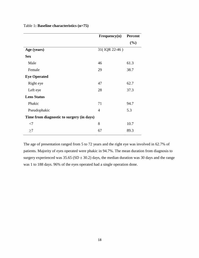
Table 1: Baseline characteristics (n=75)
Frequency(n) Percent
(%)
Age (years) 31( IQR 22-46 )
Sex
Male 46 61.3
Female 29 38.7
Eye Operated
Right eye 47 62.7
Left eye 28 37.3
Lens Status
Phakic 71 94.7
Pseudophakic 4 5.3
Time from diagnostic to surgery (in days)
<7 8 10.7
≥7 67 89.3
The age of presentation ranged from 5 to 72 years and the right eye was involved in 62.7% of
patients. Majority of eyes operated were phakic in 94.7%. The mean duration from diagnosis to
surgery experienced was 35.65 (SD ± 30.2) days, the median duration was 30 days and the range
was 1 to 188 days. 96% of the eyes operated had a single operation done.

19
Figure 4: Age distribution (n=75)
Majority of patients in 25.3% where at age group of 21to 30

20
Figure 5: Risk factors for RRD
Myopia was the most common risk factor for rhegmatogenous retinal detachment in 24 eyes
(32.0%) followed by peripheral degeneration. RRD associated with atrophic hole were identified
in 50 eyes (70.7%). Unknown causes in 14 eyes (18.7%) contributed to a significant number of
the retinal detachments.

21
Figure 6: Characteristics of RRD (n=75)
Macula was attached in 98.7% of cases and retina breaks were identified in 94.7% of eyes. There
was proliferative vitreoretinopathy grade A (tobacco dust) in 4 eyes (5.3%).
0
20
40
60
80
100
120
PVR Macula on Break identified
Nu
mb
ers
of
eyes
(%)
Yes
No
4(5.3%)
71(94.7%)74(98.7%)
1(1.3%)
71(94.7%)
4(5.3%)

22
Table 2: Characteristics of RD
Frequency
(n=75)
Percent
(%)
Number of breaks
1-3 breaks 49 65.3
4-6 breaks 18 24.0
>6 breaks 7 9.3
Missing 1 1.3
Types of breaks
Atrophic hole 53 70.7
Retinal tears 15 20.0
Dialysis
Missing
Location of Breaks
Inferior
Temporal
Nasally
Superior
6
1
54
14
4
3
8.0
1.3
72.0
18.7
5.3
4.0
Majority of eyes 72.0% had inferior RD associated with atrophic hole. Note that one eye was not
documented the type and number of retina breaks.

23
Table 3: Types of scleral buckling
Frequency(n=75) Percent (%)
Segmental circumferential 68 90.7
Encircling 5 6.7
Missing 2 2.7
Sub-retinal fluid drainage
Yes 72 96.0
No 3 4.0
Every eye in the study underwent cryotherapy. Internal tamponade with gas or other agent was
not applied to any of the cases.
Figure 7: Loss to follow-up
Postsurgical follow-up is important in that it ensures that the retina is in place and at the same
time, it also helps to identify unsuccessful cases for repeat surgery. The shortest postoperative
follow-up period was 2 weeks, while the longest postoperative follow-up period was 6 months
for 30(40.0%) eyes.
0
20
40
60
80
100
120
Pre-op 1st POD Week-2 Week-6 Month-3 Month-6
Nu
mb
er o
f ey
es(%
)
Follow up
100.0% 98.7% 97.3% 94.7%
44.0% 40.0%

24
Table 4: Trend of anatomical outcome
Retina
attached
On table
Day 1 6 weeks 3months 6 months
Yes 75(100%) 72(96.0%) 68(90.7%) 32(44.7%) 29(38.7%)
No 0(0.0%) 2(2.7%) 3(4%) 1(1.3%) 0(0.0%)
Missing data
0(0.0%) 1(1.3%) 4(5.3%) 42(56%)
46(61.3%)
Final anatomic success rate was 96.7% at 6 month follow up.
Table 5: Anatomical outcome at last follow up
Frequency(n=75) Percent (%)
Retina attached 70 (93.3%)
Re-detachment 5 (6.7%)
Primary anatomic success rate was 93.3%.
Re-detachment was found in 5(6.7%) eyes after surgery, male/female ratio 3:2.
In all the eyes in which the break was identified the macula was on and there was no PVR and
Cryotherapy and drainage of sub retinal fluid were done and duration of symptoms was more
than 7 days
Three eyes were associated with myopia and two were post-traumatic. One eye was
pseudophakic and had re-detachment at day one after surgery, two eyes had re-detachment
within one month of surgery two eyes had re-detachment secondary to new retinal breaks at three
months period. Three of the re-detached eyes were re-operated and two were observed because
they were inoperable.
For the eyes that underwent re-operation, one eye underwent additional buckling, the other
underwent buckle replacement and the final one underwent pars plana vitrectomy.
Final anatomic success rate was 97.3% after re-operations based on last follow up.

25
Table 6: Visual acuity trend
BCVA Preoperative
Postoperative
2weeks 6 weeks 3 months 6 months
Mild or no visual
impairment
(6/6 – 6/18)
10(13.3%)
8(10.7%) 9(12%) 7(9.3%) 8(10.7%)
Moderate visual
impairment
(<6/18 – 6/60)
23(30.7%)
30(40%) 19(25.3%) 14(18.7%) 13(17.3%)
Severe visual impairment
(<6/60 – 3/60)
3(4%)
4(5.3%) 3(4%) 3(4%) 3(4%)
Blindness
(<3/60)
39(52%)
27 (36%) 13(17.3%) 8(10.7%) 6(8%)
VA not recorded 0
6(8%) 31(41.3%) 44(57.3%) 46(60%)
Total 75 75 75 75 75
Only 30 cases (40%) had visual acuity data at 6 months follow up. The rate of loss to follow-up
was high after 2 weeks.

26
Table 7: Comparison of preoperative and post-operative BCVA at last follow up
Number of eyes
Preop BCVA Postop BCVA
Mild or no visual impairment
(6/6 – 6/18) 10 (13.3%)
13 (17.3%)
Moderate visual impairment
(<6/18 – 6/60) 23 (30.7%)
36 (48%)
Severe visual impairment
(<6/60 – 3/60) 3 (4.0%)
5 (6.7%)
Blindness
(<3/60) 39 (52.0%)
21 (28%)
Total 75 (100.0%) 75 (100.0%)
Postoperative BCVA has been measured at the last follow-up for each patient.
At presentation, 42 eyes (56%) had VA worse than 6/60, 33 eyes (44%) had VA between 6/6 and
6/60. The proportion of eyes with VA better than or equal to 6/60 increased from 44% pre-
operatively to 65.3% post-operatively and the proportion of eyes with VA worse than 6/60
decreased to 34.7% by the last follow up.

27
Figure 8: Visual acuity status at last follow-up
Majority of eyes operated 31 (41.3%) showed improvement in 4.6 (average) in Snellen line from
their pre-operative visual acuity.
Table 8: Comparison of preoperative and post-operative BCVA at 6 months
Number of eyes
Pre-op BCVA Pos-top BCVA
Mild or no visual impairment
(6/6 – 6/18) 5 (16.7%)
8 (26.7%)
Moderate visual impairment
(<6/18 – 6/60) 5 (16.7%)
13 (43.3%)
Severe visual impairment
(<6/60 – 3/60) 2 (6.7%)
3 (10.0%)
Blindness
(<3/60) 18(60.0%)
6 (20.0%)
Total 30(100%)
The proportion of eyes with 6 months post-operative BCVA ≥6/60 increased from 33.3% pre-
operatively to 70% post-operatively and the proportion of eyes with VA<6 /60 decreased to
30.0% from 66.7%
0
5
10
15
20
25
30
35
40
45
Improvement No change Deteriorated
Nu
mb
er
of
eye
s(%
)
Visual acuity status
31(41.3%)
14(18.7%)
30(40.0%)

28
Figure 9: Visual acuity status at 6 months
Majority of eyes operated 19 (63.3%) showed improvement in 4.8 (average) in Snellen line from
their pre-operative visual acuity.
Table 9: Complications of scleral buckling
Frequency (%)
Intraoperative complications 9 (12%)
Retinal incarceration 5(6.7%)
Retina Folds 3(4%)
Vitreous hemorrhage 1(1.3%)
Postoperative early complications 2 (2.7%)
Infection of explants 1(1.35%)
Retina folds 1(1.35%)
Complications were identified in 14.7% of eyes. Of these 12% were noted in the intra-operative
period and retinal incarceration was found in 5 eyes (6.7%) and was the commonest intra-
operative complication. Postoperative early complications were found in 2 eyes (2.7%) but there
were no late complications seen in patients with 6 months data.
0
10
20
30
40
50
60
70
Improvement No change Deteriorated
Nu
mb
er
of
eye
s(%
)
Visual acuity status
19(63.3%)
5(16.7%)6(20.0%)

29
Table 10: Factors affecting re-detachment
Attached Re-detachment OR (95% CI) P value
Duration of symptoms
≤ 7 days 8(11.4%) 0(0.0%)
> 7 days 62(88.6%) 5(100.0%) NA
Age
< 35 years 32(45.7%) 2(40%)
≥ 35 years 38(54.3%) 3(60%) 1.26(1.99-8.03) 0.589
PVR
Yes 3(4.3%) 0(0.0%)
No 67(95.7%) 5(100%) NA
Lens status
Phakic 67(95.7%) 4(80%)
Pseudophakic 3(4.3%) 1(20%) 5.58(0.46-66.5) 0.246
No. of retinal breaks
1-3 breaks 45(62.2%) 4(80%)
>3 breaks 24(37.8%) 1(20%) 0.46(0.50-4.43) 0.657
There was no significant factor which was associated with retinal re-detachment as shown in the
table above.

30
5.0 DISCUSSION
Scleral Buckle is one of the surgical techniques used for repairing RRD at KEU. A total of 91
files were identified from the theatre records, but only 75 cases were analyzed. Those not analysed
consisted of 10 files that were missing from the records filing store and 6 files with missing pages
as show in flow chart 1. All patients were operated on by a single surgeon and 96% of the eyes
had a single operation done. The standard procedure was a SB technique where all patients
received an encircling tire.
The decision of an encircling or segmental buckle to use was made based on the size and location
of the retinal tear. Drainage of subretinal fluid was performed in 72(96%) eyes when judged
necessary by the surgeon. Cryo-coagulation was performed in all cases and no intravitreal
tamponade was used.
Follow up
All eyes were examined on the first day post-operation but the number of patients subsequently
decreases at two weeks follow up to 98.7% and 40% at 6 months as shown in figure 7. That was
similar to a study done by Shakal et al that showed the shortest postoperative follow-up period was
2 weeks, while the longest postoperative follow-up period was 6 months(24). Postsurgical follow-
up is important in that it ensures that the retina is in place and at the same time, it also helps to
identify unsuccessful cases for repeat surgery

31
Demographics
Most patients who had surgery were males at 61.3%. The mean age of presentation was 34.7 (SD±
17.1) years with the range being 5 to 75 years. These findings were almost similar to those in a
study done at Khyber Institute of Ophthalmic Medical Sciences in Pakistan by Khan et al, who
found more male patients with rhegmatogenous retinal detachment at 66.04% mean age at
presentation of 38.87 ± 8.7 years with a range 5 to 90 years(30).
The mean duration from diagnosis to surgery experienced was 35.65 (SD ± 30.2) days, the median
duration was 30 days and the range was 1 to 188 days. This was found to be different with majority
of studies done that showed early presentation. A study done by Oluleye et al in Nigeria found that
the median duration before surgery was 3 months, range: 5 days – 156 months(19).
The most identified risk factor for RRD was myopia in 24(32%) eyes and was the leading
indication for sclera buckling. This may be due to the fact that myopia causes stretching of the
eyeball with consequent peripheral tears and it is easier to tamponade with sclera buckling. This
was comparable to a study done by Koc et al in Beyoglu Eye Training and Research Hospital,
Istanbul that showed the most common risk factors were idiopathic causes and myopia, each found
in 34.7% of eyes, and trauma in 18.8% (31).
Indications for Scleral buckling
In this study the majority of eyes were phakic in 94.7%, associated with inferior RRD in 54(72%)
of eyes with mild or without PVR. Majority had macula on 74(98.7%) associated with atrophic
holes in 70.7% eyes and less than 3 breaks in 50(70.4%) eyes were identified as shown in Table
2. This was similar to a study done by Haritoglou et al where 90.2% of eyes were phakic with no
PVR in 49.2% and less than 3 breaks in 89.5%. This could be because the visual prognosis is
better(32).

32
Outcomes of scleral buckling
Anatomical
Primary anatomic success rate was 90.0% that was defined as retina reattachment after one surgery
and final anatomic success rate was 96.7% at 6 month follow up. This was found to be better in
comparison with the MUSTARD study, one of the largest studies done on scleral buckling in
Europe that showed macula on detachment had a success rate of up to 88.24% and the overall
success rate of all 4325 MUSTARD patients was 83.98%(33). This was comparable to many
studies, in particular, a study done in Nigeria by Oderinlo et al that found primary anatomic
reattachment was achieved in 83 eyes (80.5%), while final anatomic success was achieved in 93
eyes (90.2%). Quinjiro found a success rate of 96% in primary surgery and 100% in secondary
although his study was in phakic uncomplicated detachments(34) while Noori had 100% in young
patients(35).
Functional
The visual outcome at last follow-up period revealed improvement in 41.3% of eyes, 40%
remained the same and 18.7% had vision deterioration as shown in Figure n⁰ 8. This results are
almost similar to a study done by Oluleye et al in Nigeria that showed at 6 weeks, there was an
improvement in visual acuity in 23 eyes (51.1%), while visual acuity remained the same in nine
eyes (20%) and was worse in 13 eyes (28.9%), (19).
The proportion of eyes with 6 months post-operative BCVA ≥6/60 increased from 33.3% pre-
operatively to 70% post-operatively and this was better compared to a study done in Pakistan by
Abdullah AS, et al, that showed final BCVA of > 6/60 was achieved in 62% of the subjects in the
treated eyes (22). At final follow-up, improvement in visual acuity was achieved in 63.3% of eyes
with an average of 4.8 with a mean of two or more Snellen lines and this was also comparable
to a study done in Ireland that showed an improvement of two or more Snellen lines from
presenting acuity in 10 (35.7%) eyes, and 16 (57.1%) of the operated eyes exhibited no change in
vision, whereas a deterioration of two or more Snellen lines was seen in two (7.1%) eyes(36).

33
Complications
In this study complications were identified in 11(14.7%) eyes as shown in Table 9. This was
comparable to a study done by Haritoglou et al. in Munich that showed 9.6% of eyes suffered
complications(32). Retina incarceration seen in 6.7% was the most frequent intra-operative
complication and 1(1.35%) had buckle infection in the post-operative period. This was found to
be different to a study done in Nepal where the intraoperative complications included vitreous
hemorrhage (1.96%) and post-operatively where rise in IOP above 21 mmHg was seen in 17 eyes
(33.3%) and buckle infection (1.96%) (37).
No late post-operative complication (6-weeks to 6-months), was noted. This may be related to the
number of patients who dropped out and may also be related to the facts that the surgery is safe
and effective with few late complications.
Factors affecting re-detachment
In this study only 5(6.7%) eyes had re-detachment. Age, lens status, PVR, number of breaks and
duration of symptoms were assessed and were found not to have any statistically significance on
the rate of retinal re-detachment as shown in the Table 12. In a study done by Park et al in Korea
it was shown that older age (≥35) was an independent prognostic factor for primary anatomical
failure in SB for uncomplicated RRD (12).
34
6.0 LIMITATIONS
1. A high attrition rate was noted in this study and is a major limitation on the study
especially in establishing the long term functional and anatomical outcomes of SB and
the complications.
2. This being a retrospective study only 30 files had complete post-operative visual acuity
recorded at sixth month review visit. This reduced the number of files with complete
post-operative visual acuity data for analysis of visual acuity outcomes.

35
7.0 CONCLUSIONS
1. Scleral buckling was mostly done for younger phakic patients with macula-on RD,
inferior RD and no PVR.
2. Majority (96.7%) of the eyes achieved high success rate at 6 months follow up.
3. Improvement of BCVA at 6 months follow up was archived in 41.3% of the eyes.
4. There were no factors found to be associated with retinal re-detachment

36
8.0 RECOMMENDATIONS
1. Scleral buckling should continue to be indicated for phakic patients with uncomplicated
inferior RRDs.
2. A study to attempt to establish the reasons why there is a low turnout for post-operative
review visits should be done.
3. Scleral buckling technique should continue to be emphasised in vitreoretinal training
because it has been shown to be safe and effective in treatment of uncomplicated RRDs.

37
9.0 REFERENCES
1. Nemet A, Moshiri A, Yiu G. A Review of Innovations in Rhegmatogenous Retinal
Detachment Surgical Techniques. Journal of Ophthalmology. 2017;1-5
2. Feltgen N, Walter P. Rhegmatogenous Retinal Detachment. Deutsches Aerzteblatt Online.
2014 Jan 6;111(1–2):12–22.
3. Haugstad M, Moosmayer S, Bragadόttir R. Primary rhegmatogenous retinal detachment -
surgical methods and anatomical outcome. Acta Ophthalmologica. 2017 May; 95(3):247–
51.
4. Banaee T, Hosseini M, Ghooshkhanei H. Anatomical and Visual Outcomes of Three
Different Scleral Buckling Techniques. Journal Ophthalmic and Vision Research.
2009;4(2):90–6.
5. American Academy of Ophthalmology,. Retina and vitreous. San Francisco,: European
Board of Ophthalmology:; 2016. 218–290 p.
6. Paulus YM, Leung L-S, Pilyugina S. Comparison of Pneumatic Retinopexy and Scleral
Buckle for Primary Rhegmatogenous Retinal Detachment Repair. Ophthalmic Surgery,
Lasers and Imaging Retina. 2017 Nov 1;48(11):887–93.
7. Schepens CL, Okamura ID, Brockhurst RJ. The Scleral Buckling Procedures: 1. Surgical
Techniques and Management. AMA Arch Ophthalmol. 1957 Dec 1;58(6):797–811.
8. Rezaei KA, Abrams GW. The History of Retinal Detachment Surgery. In: Kreissig I, editor.
Primary Retinal Detachment [Internet]. Berlin/Heidelberg: Springer-Verlag; 2005 [cited
2018 Sep 6]. p. 1–24.
9. Pal B, Saurabh K. Evolution of retinal detachment surgery down the ages. Scientific Journal
of Medical & Vision Research Foundations. July 2017;35(2):3–6.
10. Takkar B, Azad S, Bhatia I. Clinical Patterns and risk factors for rhegmatogenous retinal
detachment at a tertiary eye care centre of northern India. Nepalese Journal of
Ophthalmology. 2017 Jun 20;9(1):60-5.
38
11. Dell’Omo R, Scupola A, Viggiano D. Incidence and Factors Influencing Retinal
Displacement in Eyes Treated for Rhegmatogenous Retinal Detachment With Vitrectomy
and Gas or Silicone Oil. Investigative Opthalmology & Visual Science. 2017 Jul
17;58(6):BIO191.
12. Park SW, Kwon HJ, Byon IS. Impact of Age on Scleral Buckling Surgery for
Rhegmatogenous Retinal Detachment. Korean Journal of Ophthalmology. 2017;31(4):328-
35.
13. Abdel K, Habib A-S. Scleral buckling for the management of inferior stationary long-
standing rhegmatogenous retinal detachment in the juvenile age group. Journal of the
Egyptian Ophthalmological Society. 2013;106(3):180.
14. Espinosa AA, Mendoza GB, Millan MAT. Scleral Buckling for Rhegmatogenous Retinal
Detachment.2011;22(5):34–8.
15. Osmanovic S, Dahl A. Scleral Buckle: Overview, Periprocedural Care, Technique. 2018
Jun 20;1–49.
16. Malagola R, Pannanale L, Tortorela P. Drainage of subretinal fluidduring scleral buckling
surgery for rhegmatogenous retinal detachment. G Chir. 2015 Jul 19;36(3):106–11.
17. Wilkinson CP, Bradford RH. The drainage of subretinal fluid. Trans Am Ophthalmol Soc.
1983;81:162–71.
18. Azad R, Sharma Y, Chandra P. Basic Principle in Manegement of Retinal Detachment.
New Dalhi: All India Ophthalmological Society; 2001. 44 p. (CME).
19. Oluleye TS, Ibrahim F, Olusanya B. Scleral buckling for retinal detachment in Ibadan, Sub-
Saharan Africa: anatomical and visual outcome. Clinical Ophthalmology. 2013 May;1049-
52.
20. Schaal S, Sherman MP, Barr CC. Primary retinal detachment repair: comparison of 1-year
outcomes of four surgical techniques. Retina (Philadelphia, Pa). 2011 Sep;31(8):1500–4.

39
21. Kobashi H, Takano M, Yanagita T. Scleral buckling and pars plana vitrectomy for
rhegmatogenous retinal detachment: an analysis of 542 eyes. Curr Eye Res. 2014
Feb;39(2):204–11.
22. Abdullah AS, Jan S, Qureshi MS. Complications of Conventional Scleral Buckling
Occuring During and After Treatment of Rhegmatogenous Retinal Detachment. 2010;20:6.
23. Rishi P, Rishi E, Gupta A. Non-drainage scleral buckling with solid silicone elements.
Oman Journal of Ophthalmology. 2014;7(2):55-60.
24. Shankar V, Lim L, Ah-Kee E. Outcome of rhegmatogenous retinal detachment repair by
scleral buckling: The experience of a tertiary referral center in Scotland. Oman Journal of
Ophthalmology. 2014;7(3):130-4.
25. Oderinlo O, Oluyadi F, Ogunro A. Factors Influencing Visual Outcome after Surgery for
Retinal Detachment. Nigerian Journal of Ophthalmology,2012;20(1):24–9.
26. Papakostas TD, Vavvas D. Postoperative Complications of Scleral Buckling. Seminars in
Ophthalmology. 2018 Jan 2;33(1):70–4.
27. Rachal WF, Burton TC. Changing Concepts of Failures After Retinal Detachment Surgery.
Archives of Ophthalmology. 1979 Mar 1;97(3):480–3.
28. Gopal L. Managing retinal detachment: tips for the beginners. Scientific Journal of Medical
& Vision Research Foundations. 2017;35(2):7-9.
29. Goezinne F, La Heij E, Berendschot T. Incidence of redetachment 6 months after scleral
buckling surgery. Acta Ophthalmologica. 2010 Mar;88(2):199–206.
30. Khan MT, Jan S, Karim S. Outcome of scleral buckling procedures for primary
rhegmatogenous retinal detachment. J Pak Med Assoc. 2010;60(9):4.
31. Koç AA. Results of Scleral Buckling Surgery in Rhegmatogenous Retinal Detachment.
Beyoglu Eye Journal . 2017; 2(3): 77–80

40
32. Haritoglou C, Brandlhuber U, Kampik A. Anatomic Success of Scleral Buckling for
Rhegmatogenous Retinal Detachment – A Retrospective Study of 524 Cases.
Ophthalmologica. 2010;224(5):312–8.
33. Thelen U, Amler S, Osada N, et all. Outcome of surgery after macula-off retinal
detachment - results from MUSTARD, one of the largest databases on buckling surgery in
Europe. Acta Ophthalmol. 2012 Aug;90(5):481–6.
34. Quijano C, Alkabes M, Gómez-Resa M. Scleral Buckling in Phakic Uncomplicated Primary
Rhegmatogenous Retinal Detachment: Long-Term Outcomes. European Journal of
Ophthalmology. 2017 Mar;27(2):220–5.
35. Noori J, Bilonick RA, Eller AW. Scleral Buckle Surgery for Primary Retinal Detachment
Without Posterior Vitreous Detachment. Retina (Philadelphia, Pa). 2016 Nov;36(11):2066–
71.
36. James M, O’Doherty M, Beatty S. Buckle-related complications following surgical repair
of retinal dialysis. Eye. 2008 Apr;22(4):485–90.
37. Shah R, Byanju R, Pradhan S, et all. Factors Affecting the Outcome of Scleral Buckling
Surgery for Primary Rhegmatogenous Retinal Detachment. Journal of Ophthalmology.
2018 May 16/9016302/ref/

41
10. TIME FRAME
March-Aug/18 Sept-Dec/18 Jan.2019 Feb-March April May
Proposal writing
Ethics approval
Data Collection
Data Analysis
Report writing
Thesis Defense

42
11. Appendix 1: BUDGET
Items Quantity Unit cost (Kshs) Total (Kshs)
Proposal development
Printing 6 1,000.00 6,000.00
Binding 6 500 3,000.00
Internet 5,000.00 5,000.00
Ethics committee fee 1 2,000.00 2,000.00
Subtotal 16,000.00
Data collection
Printing data tools 1 5,000.00 5,000.00
Contracted services
Statistician 1 40,000.00 40,000.00
Transport to Kikuyu 2,000,00 2,000.00
Accommodation= 28 days 28x2000 56,000.00
Food 28x2000 56,000.00
Other expenses 1 15,000.00 15,000.00
Subtotal 174,000.00
Data analysis 1 30,000.00 30,000.00
Report writing
Internet 5,000.00 5,000.00
Printing 10 1,000.00 10,000.00
Binding 10 500.00 5,000.00
Subtotal 50,000.00
Grand Total 240,000.00

43
11.1 Appendix 2: Data Abstraction Tool
1) Patient number:
2) Eye operated Right Left
3) Sex Male Female
4) Age Year of birth
5) Duration of symptoms at time of surgery: days_________ weeks__________ months
________________ years_____________
6) Visual acuity at admission (UCVA)
(BCVA)
7) Lens status
i. Phakic
ii. Pseudophakic
iii. Aphakic
8) Etiology of detachment:
i) Post traumatic
ii) Myopia
iii) Previous cataract surgery
iv) Peripheral degenerations
v) Previousintravitreal injections
vi) Unknown
vii) Others ____________________________________
9) Rhegmatogenous retinal detachment
With PVR Yes No
Macular ON Yes No
Retinal breaks identified Yes No

44
i. Number of breaks
ii. Size of breaks
iii. Types of breaks
iv. Location of breaks
10) Type of Scleral buckling done
i. Encircling
ii. Segmental radial
iii. Segmental circumferential
11) Cryotherapy Yes No
12) Sub-retinal fluid drainage Yes No
13) Internal tamponade
i. Yes
a) Air b) SF6 c) C3 F8
ii. No
14) After surgery:
Retina attached
Time Yes No
i. On the table
ii. Day 1
iii. 1 months
iv. 3 months
v. 6 months
Vi 12 months
15) New breaks Re-detachment No

45
Time Number of tears Location of tears
i. Day 1
ii. 1 months
iii. 3 months
iv. 6 months
v. 12 months
16) Re-operations
i) Additional buckle or buckle replacement,
ii) Pneumopexy SF6 C3F8
iii) Vitrectomy
17) Visual acuity
a) After surgery: (UCVA) (BCVA)
i. 2 weeks
ii. 6 weeks
iii. 3 months
iv. 6 months
18) Complications
a) Intraoperative:
i. Choroidal detachment
ii. Vitreous hemorrhage
iii. Choroidal hemorrhage,
iv. Subretinal hemorrhage,
v. Retinal incarceration,
vi. Raised IOP,
vii. Hyphema
viii. Vitreous incarceration

46
ix. Lens damage
x. Corneal damage
xi. Retinal tears
xii. Others
b) Postoperative early complication (24h-6 weeks):
i. Keratitis
ii. Endophthalmitis
iii. Exposure of explants
iv. Infection of explants
v. Elevation IOP
vi. Muscle entrapment
vii. Re-detachment
viii. Others
c) Late complications:
i. Cataract formation
ii. Glaucoma
iii. New break
iv. Proliferative vitreoretinopathy
v. Re-detachment
vi. Cystoid maculopathy
ix) Others

47
11.2 Appendix 3: Collaborating letter
48
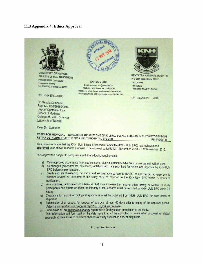
11.3 Appendix 4: Ethics Approval

49