Embed Size (px)
Citation preview
Independent learning program for GPs
Independent learning program for GPs
www.racgp.org.au/check
Unit 528 June 2016
Respiratory disease

DisclaimerThe information set out in this publication is current at the date of first publication and is intended for use as a guide of a general nature only and may or may not be relevant to particular patients or circumstances. Nor is this publication exhaustive of the subject matter. Persons implementing any recommendations contained in this publication must exercise their own independent skill or judgement or seek appropriate professional advice relevant to their own particular circumstances when so doing. Compliance with any recommendations cannot of itself guarantee discharge of the duty of care owed to patients and others coming into contact with the health professional and the premises from which the health professional operates.
While the text is directed to health professionals possessing appropriate qualifications and skills in ascertaining and discharging their professional (including legal) duties, it is not to be regarded as clinical advice and, in particular, is no substitute for a full examination and consideration of medical history in reaching a diagnosis and treatment based on accepted clinical practices.
Accordingly, The Royal Australian College of General Practitioners and its employees and agents shall have no liability (including without limitation liability by reason of negligence) to any users of the information contained in this publication for any loss or damage (consequential or otherwise), cost or expense incurred or arising by reason of any person using or relying on the information contained in this publication and whether caused by reason of any error, negligent act, omission or misrepresentation in the information.
SubscriptionsFor subscriptions and enquiries please call 1800 331 626 or email [email protected]
Published byThe Royal Australian College of General Practitioners 100 Wellington Parade East Melbourne, Victoria 3002, Australia Telephone 03 8699 0414 Facsimile 03 8699 0400 www.racgp.org.au
ABN 34 000 223 807 ISSN 0812-9630
© The Royal Australian College of General Practitioners 2016.
REFERENCES: 1. fl utiform® inhaler Product Information, October 2015. 2. van Noord JA et al. Eur Respir J 1996;9(8):1684−1688. 3. Politiek MJ et al. Eur Respir J 1999;13:988–992. 4. Mager DE et al. J Pharm Sci 2003;92:1521−1525. 5. Johnson M. J Allergy Clin Immunol 1998;101:S434−S439. ® FLUTIFORM is the registered trade mark of Jagotec AG used under licence by Mundipharma Pty Limited. Mundipharma Pty Limited ABN 87 081 322 509, 88 Phillip Street, Sydney, NSW 2000. Tel: 1800 188 009. S&SW. MFF0131_CUFP_V_RHP. ORBIS AU-3310 Mar 16.
*Potent anti-infl ammatory activity in the lungs.1†fl utiform ® inhaler is not indicated for use as a reliever.1
Combining the power* offluticasone propionate withthe speed† of eformoterol.1–5 fluticasone propionate/eformoterol
Potent anti-infl ammatory activity in the lungs.
fluticasone propionate with1–5
Please review Product Information and State and Federal regulations before prescribing. The Product Information
for FLUTIFORM® inhaler can be accessed at https://www.mundipharma.com.au/products/prescription-medicines/
FLUTIFORM® pressurised inhalation 50 micrograms/5 micrograms, 125 micrograms/5 micrograms and 250 micrograms/10 micrograms MINIMUM PRODUCT INFORMATION NAME OF THE MEDICINE fl uticasone propionate and eformoterol fumarate dehydrate. INDICATIONS For the regular maintenance treatment of asthma where the use of a combination product (an inhaled corticosteroid and a long-acting ß2-agonist) is appropriate. This includes patients not adequately controlled with inhaled corticosteroids and inhaled short-acting ß2-agonist on an ‘as required’ basis. CONTRAINDICATIONS Hypersensitivity to any of the active substances or to the excipients. PRECAUTIONS Not to be used as the fi rst treatment for asthma, treat acute asthma symptoms, transfer from oral corticosteroids, abrupt withdrawal, combination with other long-acting ß2-agonists or to children under 12 years of age. Do not initiate during exacerbation/signifi cantly worsening/acutely deteriorating asthma. Use with caution in patients with active/quiescent pulmonary tuberculosis, untreated systemic infections, ocular herpes simplex, fungal/viral/other infections of the airway, pre-existing cardiovascular disease including prolongation of the QTc interval, history of thyrotoxicosis, phaeochromocytoma, diabetes mellitus, uncorrected hypokalaemia, low levels of serum potassium, hypertrophic obstructive cardiomyopathy, idiopathic sub-valvular aortic stenosis, severe hypertension, aneurysm, severe cardiovascular disorders (e.g. ischaemic heart disease, cardiac arrhythmias, severe heart failure), impaired adrenal function, Churg Strauss syndrome, stress, elective surgery, hepatic or renal impairment (not studied), pregnancy or lactation. Consider spacer device, particularly in patients with poor inhaler technique. The prophylactic use of fl utiform® inhaler in exercise-induced asthma has not been studied. INTERACTIONS WITH OTHER MEDICINES CYP3A4 inhibitors (e.g. ritonavir, atazanavir, clarithromycin, indinavir, itraconazole, nelfi navir, saquinavir, ketoconazole, telithromycin), non-potassium sparing diuretics (e.g. loop, thiazide), xanthine derivatives, glucocorticosteroids, digitalis glycosides, halogenated hydrocarbon anaesthetics, MAOIs, furazolidone, procarbazine, tricyclic antidepressants, drugs known to prolong the QTc interval (e.g. quinidine, disopyramide, procainamide, phenothiazines, antihistamines), adrenergic drugs, ß-blockers including eye drops, L-dopa, L-thyroxine, oxytocin, alcohol. ADVERSE EFFECTS Adverse effects of fl utiform® inhaler are uncommon (≥1/1,000 and <1/100) or rare (≥1/10,000 and <1/1,000), and include oral candidiasis, oral fungal infection, sinusitis, hyperglycaemia, sleep disorders including insomnia, abnormal dreams, agitation, headache, tremor, dizziness, dysgeusia, vertigo, palpitations, ventricular extrasystoles, angina pectoris, tachycardia, hypertension, exacerbation of asthma, dysphonia, throat irritation, dyspnoea, cough, dry mouth, diarrhoea, dyspepsia, rash, pruritus, muscle spasms, peripheral oedema, asthenia. DOSAGE AND ADMINISTRATION Adults and children above 12 years: 2 inhalations of fl utiform® inhaler 50/5 mcg or 125/5 mcgtwice daily. If asthma remains poorly controlled on fl utiform® inhaler 50/5 mcg,the total daily dose of the inhaled corticosteroid can be increased by administering the next highest strength combination product (i.e. fl utiform® inhaler 125/5 mcg,2 puffs twice daily). Adults only: 2 inhalations of fl utiform® inhaler 250/10 mcg twice daily. If asthma remains poorly controlled on fl utiform® inhaler 125/5 mcg, the total daily dose can be increased by administering fl utiform® inhaler 250/10 mcg, 2 puffs twice daily. Patients who are currently receiving medium to high doses of inhaled corticosteroid therapy, and whose disease severity clearly warrants treatment with 2 maintenance therapies, the recommended starting dose is 2 inhalations twice daily of fl utiform® inhaler 125/5 mcg. fl utiform® inhaler 50/5 mcg strength is not appropriate in adults and adolescents with severe asthma. If dosages outside the recommended regimen are required, appropriate doses of ß-agonist and/or corticosteroid in single inhalers should be prescribed. No dose adjustments required in the elderly. Monitor patients with hepatic or renal impairment. Not recommended in patients under 12 years of age. Must be used regularly (2 actuations twice daily), even when asymptomatic. The use of 1 actuation twice daily has not been investigated in clinical trials. Patients will need to be trained on the use of the inhaler (with or without spacer) and should be regularly reassessed by a doctor so they receive the optimal dose. The dose should be titrated to the lowest dose at which effective control of symptoms is maintained and the strength of dose should only be increased or decreased on medical advice. DATE OF FIRST INCLUSION IN THE AUSTRALIAN REGISTER OF THERAPEUTIC GOODS (THE ARTG) 14 June 2013. DATE OF MOST RECENT AMENDMENT 21 October 2015.
PBS Information. Restricted benefi t: Asthma. Patient must have previously
had frequent episodes of asthma while receiving treatment with oral corticosteroids or optimal doses of inhaled corticosteroids. Patient
must be aged 12 years or over.
MFF0131_CU_FP_V_RHP.indd 1 6/05/2016 11:29 am
Independent learning program for GPs
Independent learning program for GPs
The five domains of general practice
Communication skills and the patient–doctor relationship
Applied professional knowledge and skills
Population health and the context of general practice
Professional and ethical role
Organisational and legal dimensions
Respiratory diseaseUnit 528 June 2016
About this activity 2
Acronyms 3
Case 1 Danny is too puffed to play rugby 3
Case 2 Paula has run out of puffers 11
Case 3 Kathryn has a productive cough 20
Case 4 Mary feels tired and depressed 24
Case 5 Mark is feeling short of breath 28
Multiple choice questions 33
2
check Respiratory disease
ABOUT THIS ACTIVITY
Respiratory disorders are among the most commonly managed problems in general practice1 and were managed at a rate of about one in five consultations in the period of 2005–06 to 2014–15.2 The results of the 2014–15 National Health Survey show that about 7.1 million Australians suffer from a chronic respiratory condition.1 In 2013, there were 12,465 deaths that were attributable to an underlying acute or chronic respiratory condition.1 In particular, chronic obstructive pulmonary disease and asthma are major causes of morbidity and mortality in Australia and other countries.3 Asthma is listed as one of three National Health Priority Areas in Australia.4 Other common respiratory conditions include bronchiectasis, obstructive sleep apnoea and occupational lung disease.1 Bronchiectasis is being increasingly recognised as a disorder that can cause long-term disability and premature death. Management of bronchiectasis can be complex and often involves multiple healthcare providers; however, general practitioners play a key role in coordinating patient care.5,6 Occupational lung disease is recognised as a major cause of work-related disability.
This edition of check considers the management and treatment of respiratory conditions in general practice.
LEARNING OUTCOMES
At the end of this activity, participants will be able to:
• describe the assessment and management of a patient presenting with shortness of breath
• discuss the approach to managing chronic obstructive pulmonary disease
• identify the signs and symptoms of bronchiectasis
• summarise the assessment and management of obstructive sleep apnoea
• outline the treatment for asthma.
AUTHORS
Ryan Hoy (Case 5) MBBS, FRACP, MOccEnvHlth is a respiratory and sleep disorders physician at Cabrini Medical Centre. Dr Hoy has clinical and research interests in occupational causes of respiratory disease.
Simon Joosten (Case 4) MBBS, BMedSc, FRACP, PhD is a respiratory and sleep medicine physician at Monash Health in Melbourne Australia, and a Research Fellow at the School of Clinical Sciences at Monash University. Dr Joosten is a Board Member of the Sleep Health Foundation.
Paul King (Case 3) MBBS, FRACP, PhD is a respiratory physician at Monash Medical Centre/Monash Health. Dr King completed a PhD in bronchiectasis and has an ongoing clinical interest and research program in lung inflammation and immunity and bronchiectasis.
Lawrence Tan (Case 2) MBBS, DCH, DRCOG, MPH, FRACGP is a part-time general practitioner in southwest Sydney, and part-time senior lecturer in the Department of General Practice, Western Sydney University. Dr Tan enjoys teaching medical students and registrars. His research interests are in cultural and linguistic diversity and how this affects the delivery of primary healthcare.
Judi Wicking (Case 1) RN, BN, GradCert Asthma Ed is a registered nurse who, after 22 years’ experience as a practice nurse specialised
in asthma and respiratory health and management. Ms Wicking developed the Asthma and Respiratory Nurse-Led Clinics model of care in 2001. Currently, Ms Wicking is the project manager for the National Asthma Council Australia health professional education program and continues to run respiratory clinics in general practices in the eastern suburbs of Melbourne. Her interest in how specialist nurse educators working collaboratively with GPs improves health outcomes for people with asthma and associated respiratory conditions has involved her in several published research studies and articles on this topic. Ms Wicking was a Board Member of the Australian Asthma Respiratory Educators Association and has been an invited contributor on many advisory committees including the Australian Asthma Handbook and other National Asthma Council Australia Publications.
PEER REVIEWERS
Kerry Hancock BMBS (Flin), DipObsRACOG is a general practitioner in a group practice in outer metropolitan Adelaide and has more than 30 years’ experience in clinical practice. Dr Hancock is also an executive member of the COPD National Program with the Lung Foundation Australia. She has a special interest in general practice-based respiratory medicine and has strong affiliations with Asthma Australia, the Asthma Foundation of South Australia, the National Asthma Council of Australia, Lung Foundation Australia, and the International Primary Care Respiratory Group. Dr Hancock’s active participation with these organisations has enabled her to be involved in the development of national primary care-focused respiratory management guidelines, educational activities and the development of tools to assist GPs in the management of their patients with asthma and COPD.
Robert Menz MBBS, FRACGP, MClinEdu has been a general practitioner in the inner eastern Adelaide suburbs since 1980. He also has wide experience in non-clinical aspects of medicine through organisations such as the Royal Australian College of General Practitioners (RACGP), Australian Medical Association (AMA), Australian General Practice Accreditation Limited (AGPAL), Divisions of General Practice and National Primary Care Collaborative (NPCC). From 2001–14 Dr Menz was a senior medical adviser for the Commonwealth Department of Human Services (DHS). This role provides advice, education and stakeholder engagement as part of the Health Professionals Branch and a professional link between the DHS and the medical profession. Dr Menz is the RACGP Corlis Fellow for South Auatralia and the Northern Territory. He has been an RACGP examiner since 1984 and was censor for SA/NT from 1997–2003. He was an RACGP nominee to the AGPAL board from 2000–2006, and is still a surveyor. Dr Menz was on SA/NT AMA branch council and chaired the Council of General Practice in 1992–1993. He remains on the editorial committee. Dr Menz teaches undergraduate medical students at Flinders University, general practice registrars through NTGPE, and is a medical educator for the RACGP.
REFERENCES 1. Australian Institute of Health and Welfare. Chronic respiratory conditions
including asthma and COPD. Canberra: AIHW, 2016. Available at www.aihw.gov.au/chronic-respiratory-conditions [Accessed 28 April 2016].
2. Britt H, Miller G, Henderson J, et al. A decade of Australian general practice activity 2005–06 to 2014–15. General practice series no. 39. Sydney: Sydney University Press, 2015.
ABOUT THIS ACTIVITY
ABOUT THIS ACTIVITY
CASE 1
3
check Respiratory disease
3. Poulos LM, Cooper SJ, Ampon R, Reddel HK, Marks GB. Mortality from asthma and COPD in Australia. Cat. no. ACM 30. Canberra: Australian Institute of Health and Welfare, 2014.
4. Australian Institute of Health and Welfare. A picture of Australia’s children 2012. Cat. no. PHE 167. Canberra: AIHW, 2012. Available at www.aihw.gov.au/WorkArea/DownloadAsset.aspx?id=10737423340 [Accessed 28 April 2016].
5. Australian Institute of Health and Welfare. Bronchiectasis. Canberra: AIHW, 2016. Available at www.aihw.gov.au/bronchiectasis [Accessed 28 April 2016].
6. McGuire G. Bronchiectasis. A guide for primary care. Aust Fam Physician 2012;41(11):842–50.
ACRONYMSBMI body mass indexCOPD chronic obstructive pulmonary diseaseCPAP continuous positive airways pressureCXR chest X-rayDXA dual-energy X-ray absorptiometryFEV1 forced expiratory volume in one secondFVC forced vital capacityGPMP general practitioner management plan
HRCT high-resolution computed tomography
ICS inhaled corticosteroids
LAMA long-acting muscarinic antagonist
OA occupational asthma
OSA obstructive sleep apnoea
PBS Pharmaceutical Benefits Scheme
PEF peak expiratory flow
PEP positive expiratory pressure
SABA short-acting beta-2 agonist
QUESTION 1
What information from Danny’s history would help you consider the most common causes of breathlessness in an adult male?
FURTHER INFORMATION
Danny’s general health is good and he keeps fit by playing rugby, including training, and doing ‘cardio’ and weight training at the gym about twice per week.
As well as worsening shortness of breath during and after exertion, he has woken during the night struggling for breath twice in the past fortnight. On one occasion, his breathing ‘sounded wheezy’. Danny has no history of cough, except when he had a respiratory infection about eight weeks ago, which required two days sick leave from work, but from which he completely recovered within a few days. He is not aware of post-nasal drip.
Danny has otherwise been generally well and takes no medicines.
He looks fit and healthy, and is in the healthy weight range – his body mass index (BMI) is 22.6 kg/m2 (height = 182 cm and weight = 75 kg). His blood pressure is 128/76 mmHg and pulse is 68 regular beats per minute. His chest is clear on auscultation, heart sounds are normal, the shape of his chest appears normal, and there is no obvious nasal obstruction.
Danny was a social smoker in his late teens and early twenties, with a four pack–year history, but has since quit. Both Danny’s parents smoked until he was about six or seven years of age.
Danny had eczema as an infant and asthma during early primary school. He recalls using a puffer occasionally, but ‘grew out of it’ before high school. Danny developed hay fever as a teenager, and now has occasional, mild (unmedicated) symptoms during spring. He has no other known allergies. Danny has a sister aged 27 years. His sister and mother have asthma. His mother also has hay fever.
Neither of his parents have cardiovascular disease, but his paternal grandfather ‘died from a heart attack’ in his early 60s.
Note on smoking history: Pack–years are calculated as the number of years smoked, multiplied by the number of cigarettes smoked per day, divided by 20.
CASE 1
DANNY IS TOO PUFFED TO PLAY RUGBY
Danny, 31 years of age, comes to see you because he has increasingly been struggling for breath during his regular social rugby games and training sessions this season. For the last few games, he has become ‘completely out of breath’ about 10–20 minutes into the game. He says this is unusual for him because he started the season in good form and feels otherwise well.
CASE 1
4
check Respiratory disease
Table 1. Danny’s spirometry results
Parameter Predicted value Pre-bronchodilator
(% of predicted)
LLN Post-bronchodilator
(% of predicted)
% Change
(volume)
FEV1 (L) 4.70 3.18 (68%) 3.75 3.79 (81%) 19% (0.61 L)
FVC (L) 5.72 4.88 (85%) 4.62 5.15 (90%) 5% (0.27 L)
FEV1/FVC (%) 82 65 71 74 –
PEF (L/sec) 11.24 7.93 (71%) 7.93 9.38 (83%) 18%
FEV1, forced expiratory volume in one second; FVC, forced vital capacity; LLN, lower limit of normal; PEF, peak expiratory flow
QUESTION 2
What are the differential diagnoses at this stage?
QUESTION 3
Do you have enough information to make a provisional or definitive diagnosis of asthma? What other information do you need?
FURTHER INFORMATION
Danny has a spirometry test before and after taking an inhaled short-acting beta2-agonist (SABA) bronchodilator (salbutamol 400 µg).
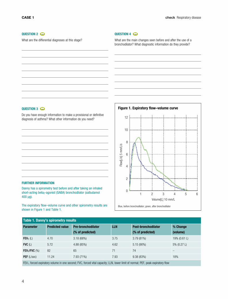
The expiratory flow–volume curve and other spirometry results are shown in Figure 1 and Table 1.
QUESTION 4
What are the main changes seen before and after the use of a bronchodilator? What diagnostic information do they provide?
Figure 1. Expiratory flow–volume curve
Blue, before bronchodilator; green, after bronchodilator
1 2 3 4 5 6
2
0
4
6
8
10
12
Volume[L] 10 mm/L
Flow
[L/s
] 5 m
m/L
/s

CASE 1
5
check Respiratory disease
QUESTION 5
How will you manage Danny’s asthma?
QUESTION 6
How would you follow up Danny?
QUESTION 7
What are the most common problems in asthma management?
QUESTION 8
Given that asthma management requires ongoing monitoring, planned, structured review and significant self-management education for patients, what aspects of practice management might help you achieve all this?
CASE 1
6
check Respiratory disease
CASE 1 ANSWERS
ANSWER 1
The history should aim to find out:
• when symptoms occurred (eg day or night, trigger, frequency, timing)
• whether there is a regular cough (if so, is it dry or productive, as well as the characteristics of sputum)
• smoking history, past and current (including passive exposure, particularly when younger)
• previous relevant history including atopic dermatitis (eczema), allergic rhinitis (hay fever) or cardiovascular disease
• family history of asthma, allergies or cardiovascular disease
• previous and current medications
• previous hospital admissions with respiratory symptoms
• allergies
• exercise regimen (eg frequency, intensity).
ANSWER 2
Conditions that should be considered in an adult with recurrent shortness of breath over this period of time include:1
• poor cardiopulmonary fitness – this is a common cause of breathlessness on exertion, especially among people who are overweight or obese. Some shortness of breath and increased respiratory rate is normal during strenuous exercise. In an active, non-smoking, fit, young adult within a healthy weight range it is usually only worth investigating if symptoms increase over a few months, as they have in Danny’s case
• asthma – Danny’s history includes several factors that would suggest a high probability of asthma (factors include the presence of breathlessness and wheeze, recurrent nocturnal symptoms, history of allergies, triggered by exercise, history of asthma, family history of allergies and asthma)1,2
• other respiratory conditions including bronchiectasis, chronic obstructive pulmonary disease (COPD), hyperventilation/dysfunctional breathing, pulmonary thromboembolism, inhaled foreign body, large airway stenosis, pleural effusion, pulmonary fibrosis, rhinitis/rhinosinusitis, upper airway dysfunction (vocal cord dysfunction), COPD and lung cancer – COPD and lung cancer are relatively unlikely in view of Danny’s age and smoking history. Other conditions would be considered if a diagnosis of asthma is ruled out
• cardiovascular disease (eg hypertrophic cardiomyopathy) – unlikely in view of Danny’s vital signs and history, but would be considered after ruling out the more likely conditions.
ANSWER 3
The diagnosis of asthma is based in the probability that symptoms and clinical findings are due to asthma (Table 2). A thorough history, physical examination and lung function testing are essential.
Table 2. Findings that increase or decrease the probability of asthma in adults1
Asthma is more likely to explain the symptoms if any of these apply
Asthma is less likely to explain the symptoms if any of these apply
More than one of these symptoms:
• wheeze*
• breathlessness
• chest tightness
• cough
Symptoms recurrent or seasonal
Symptoms worse at night or in the early morning
History of allergies (eg allergic rhinitis, atopic dermatitis)
Symptoms obviously triggered by exercise, cold air, irritants, medicines (eg aspirin or beta blockers), allergies, viral infections, laughter
Family history of asthma or allergies
Symptoms began in childhood
Widespread wheeze audible on chest auscultation
FEV1 or PEF lower than predicted, without other explanation
Eosinophilia or raised blood IgE level, without other explanation
Symptoms rapidly relieved by a SABA bronchodilator
Dizziness, light-headedness, peripheral tingling
Isolated cough with no other respiratory symptoms
Chronic sputum production
No abnormalities on physical examination of chest when symptomatic (over several visits)
Change in voice
Symptoms only present during upper respiratory tract infections
Heavy smoker (now or in the past)
Cardiovascular disease
Normal spirometry or PEF when symptomatic (despite repeated tests)
*Wheeze suggests asthma but is not pathognomonic. It can also occur due to other conditions such as respiratory infections, chronic obstructive pulmonary disease or obesity. High-pitched stridor may be mistaken for wheezing in people with upper airway dysfunction – careful auscultation will reveal that the sound is localised to the upper airway (not peripheral airway expiratory wheezing).
FEV1, forced expiratory volume in 1 second; IgE, immunoglobulin E; PEF, peak expiratory flow; SABA, short-acting beta2-agonist
Reproduced with permission from the National Asthma Council Australia. Australian Asthma Handbook. Version 1.1. South Melbourne, Vic: National Asthma Council Australia, 2015. Available at www.asthmahandbook.org.au [Accessed 31 March 2016].
In clinical practice, asthma is defined by the presence of both the following:1
• respiratory symptoms (eg wheeze, shortness of breath, cough, chest tightness) that vary over time and may be present or absent at any point in time – Danny’s history includes these
• excessive variation in lung function (‘variable airflow limitation’, ie variation in expiratory airflow that is greater than that seen in healthy people) – this has not yet been demonstrated in Danny’s case.
To confirm the diagnosis of asthma in an adult, it is necessary to demonstrate variable expiratory airflow limitation by testing lung function.1 This involves:1
CASE 1
7
check Respiratory disease
• identifying expiratory airflow limitation by confirming that the ratio of FEV1 to forced vital capacity (FEV1/FVC) is reduced (less than the lower limit of normal for age*) at a time when FEV1 is lower than predicted
• showing that expiratory airflow limitation is variable (eg by performing spirometry before and after inhalation of a bronchodilator to show a positive response).
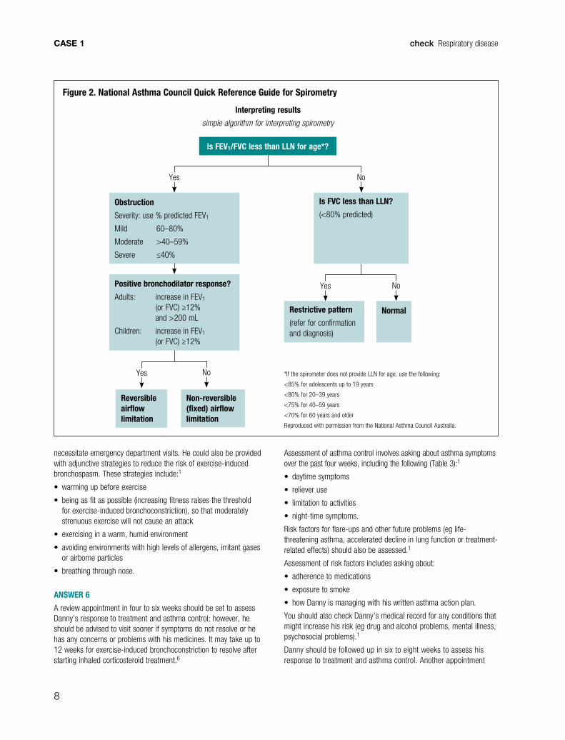
The principle is that the ratio of FEV1 to forced vital capacity (FEV1/FVC) is reduced (less than the lower limit of normal for age*). Severity of obstruction is determined by FEV1 being lower than the predicted value for the person’s age and height. The age, height, sex and ethnicity of the patient should be entered to ensure that the correct reference values are used; however, the reference range used on the spirometer is also important.3,4 Figure 2 shows an algorithm for interpretation of spirometry results.5
Spirometry can be performed in primary care (if reliable equipment and appropriately trained staff are available), or the patient can be referred to an appropriate provider (eg an accredited respiratory function laboratory).1
Ideally, airflow limitation should be confirmed when the patient does not have a respiratory tract infection.1
*If the spirometer does not provide lower limit of normal for age, the following age-based cut-off points can be used to indicate expiratory airflow limitation in adults and older adolescents:
• <0.85 (up to 19 years)
• <0.80 (20–39 years)
• <0.75 (40–59 years)
• <0.70 (60 years and older).
ANSWER 4
Findings
The blue line on Figure 1 (expiratory flow–volume curve before the use of a bronchodilator) is shorter and concave, compared with the green line (after the use of a bronchodilator).
Concavity in the expiratory flow–volume curve and reduced FEV1/FVC ratio (<0.8) indicates an obstructive ventilatory defect.5
Pre-bronchodilator FEV1 is 68% of the predicted value indicating a mild, obstructive ventilatory defect.5
Post-bronchodilator FEV1 values show a substantial increase (19% and 0.61 L). This finding meets the criteria for a clinically significant bronchodilator response (increase in FEV1 of at least 12% and at least 200 mL),1 and indicates that Danny has reversible airflow limitation.
FEV1/FVC ratio increased from 65% to 74%.
Interpretation
Danny’s spirometry results are highly suggestive of asthma.
ANSWER 5
Current Australian asthma guidelines1 recommend the following:
• Regular inhaled corticosteroid treatment should be given for all adults and adolescents who report asthma symptoms twice or
more during the past month, or waking due to asthma symptoms once or more during the past month. The starting dose should be low and the response should be reviewed in six to eight weeks.
• Salbutamol should be used 15 minutes before exercise for initial management of exercise-related symptoms until the full effect of the inhaled corticosteroid has been achieved (usually two to four weeks, but can be up to 12 weeks).
• Patients with asthma should be advised to carry a reliever containing a rapid-onset inhaled beta2-agonist, and to use it when they experience difficulty breathing.
• All patients should be educated about asthma and trained to use inhalers correctly. A written asthma action plan should be provided.
Management: Pharmacological treatment
• Low-dose inhaled corticosteroid – suitable options would include any of the following:2
– beclometasone dipropionate 100 µg: one actuation twice daily
– budesonide 200 µg: one actuation once or twice daily
– ciclesonide 160 µg: one actuation once daily
– fluticasone propionate 125 µg: one actuation twice daily (lowest adult dose available)
• Rapid-onset beta2 agonist bronchodilator – suitable options would include either of the following:2
– salbutamol 100 µg by pressurised metered-dose inhaler: Two to four puffs 15 minutes before exercise and one to two puffs at other times as needed
– terbutaline 500 µg by dry-powder inhaler: One to two inhalations 15 minutes before exercise and one inhalation at other times as needed.
Cost and Danny’s preference should be considered when choosing the regimen. He will also need training on how to use his inhalers correctly. This includes correct inhalation technique and advice to rinse his mouth with water and spit after each dose of inhaled corticosteroid, to avoid candidiasis.2
Management: Education and self-management
To manage his asthma effectively, Danny will need:
• training in correct inhaler technique
• education about:
– asthma
– the purpose and importance of regular preventer medicines
– how to avoid local side effects
– how to avoid or manage triggers
– how to monitor his asthma control based on symptoms (and optional peak expiratory flow monitoring)
– how to manage symptoms
• a written asthma action plan and explanation of how to use it
• advice to keep his immunisations (especially influenza vaccination) up to date.
Danny needs to understand that poor asthma control will increase his risk of flare-ups, which may cause lost work days or even
CASE 1
8
check Respiratory disease
necessitate emergency department visits. He could also be provided with adjunctive strategies to reduce the risk of exercise-induced bronchospasm. These strategies include:1
• warming up before exercise
• being as fit as possible (increasing fitness raises the threshold for exercise-induced bronchoconstriction), so that moderately strenuous exercise will not cause an attack
• exercising in a warm, humid environment
• avoiding environments with high levels of allergens, irritant gases or airborne particles
• breathing through nose.
ANSWER 6
A review appointment in four to six weeks should be set to assess Danny’s response to treatment and asthma control; however, he should be advised to visit sooner if symptoms do not resolve or he has any concerns or problems with his medicines. It may take up to 12 weeks for exercise-induced bronchoconstriction to resolve after starting inhaled corticosteroid treatment.6
Assessment of asthma control involves asking about asthma symptoms over the past four weeks, including the following (Table 3):1
• daytime symptoms
• reliever use
• limitation to activities
• night-time symptoms.
Risk factors for flare-ups and other future problems (eg life-threatening asthma, accelerated decline in lung function or treatment-related effects) should also be assessed.1
Assessment of risk factors includes asking about:
• adherence to medications
• exposure to smoke
• how Danny is managing with his written asthma action plan.
You should also check Danny’s medical record for any conditions that might increase his risk (eg drug and alcohol problems, mental illness, psychosocial problems).1
Danny should be followed up in six to eight weeks to assess his response to treatment and asthma control. Another appointment
Figure 2. National Asthma Council Quick Reference Guide for Spirometry
Is FEV1/FVC less than LLN for age*?
Obstruction
Severity: use % predicted FEV1
Mild 60–80%
Moderate >40–59%
Severe ≤40%
Positive bronchodilator response?
Adults: increase in FEV1 (or FVC) ≥12% and >200 mL
Children: increase in FEV1 (or FVC) ≥12%
Is FVC less than LLN?
(<80% predicted)
*If the spirometer does not provide LLN for age, use the following:
<85% for adolescents up to 19 years
<80% for 20–39 years
<75% for 40–59 years
<70% for 60 years and older
Reproduced with permission from the National Asthma Council Australia.
Yes No
Restrictive pattern
(refer for confirmation and diagnosis)
Normal
Yes No
Reversible airflow limitation
Non-reversible (fixed) airflow limitation
Yes No
Interpreting results
simple algorithm for interpreting spirometry
CASE 1
9
check Respiratory disease
in three to four months for a comprehensive asthma review is recommended.1
ANSWER 7
Common problems include:1
• suboptimal adherence – most patients do not take their preventer medication as often as prescribed, particularly when symptoms improve, or are mild or infrequent. Reasons often include concerns about side effects, interference with lifestyle, forgetting doses, and misunderstanding the purpose or effects of the medicine
• incorrect inhaler technique – most patients do not use their inhaler correctly. To achieve consistently good technique, it is essential to repeatedly check the person’s technique and demonstrate the correct technique for the specific brand of inhaler.
Key message: Whenever asthma control is poor despite apparently adequate treatment, adherence and inhaler technique should be checked before increasing the dose or changing the regimen.
Other problems include:
• continued exposure to known triggers or undetected triggers for asthma symptoms (eg respiratory infections, cold air, smoke, airborne allergens or irritants in the environment, home or workplace, certain medicines)
• comorbid conditions (eg allergic rhinitis, gastro-oesophageal reflux disease, upper airway dysfunction, obesity)
• patient skipping planned asthma reviews and only attending during flare-ups – this makes it impossible to adjust the treatment regimen to maintain control at the lowest dose needed while minimising the risk of adverse treatment-related effects.
ANSWER 8
Asthma management capacity in general practice can be optimised by:
• the general practice nurse, who can provide education, and check and review correct use of his inhaler, explain how to use the written asthma action plan, and (with specific training) perform
spirometry. Some practices employ asthma educators and/or run dedicated asthma clinics
• Asthma Cycle of Care,8 which is an Australian Government initiative to support primary care health professionals (GPs, other medical practitioners and trainees) to provide asthma care. It is implemented through the Practice Incentives Program (PIP) – Asthma Incentive and applies to the clinical care of people with ‘moderate-to-severe asthma’, generally defined to include people who require preventive medication or use bronchodilator at least three times a week9
• chronic disease management Medicare items10 – patients with asthma are eligible for Medicare items associated with GP Management Plans and Team Care Arrangements.
CONCLUSION
Four weeks later, Danny returns for a review. He has had no more night-time symptoms and has taken his reliever ‘a few times in the first couple of weeks’ but not since. His exercise-related symptoms have improved and are almost resolved.
Spirometry results now indicate normal lung function and no significant bronchodilator reversibility.
Danny’s inhaler technique with each of his devices is fairly good when he demonstrates, but you note a few errors and correct these.
Danny’s written asthma action plan use is reviewed.
RESOURCES FOR PATIENTS • National Asthma Council Australia, www.nationalasthma.org.au
• Asthma Australia, www.asthmaaustralia.org.au
• Asthma Helpline, 1800 ASTHMA (1800 278 462)
RESOURCES FOR DOCTORS• National Asthma Council Australia. Australian Asthma Handbook. Version
1.1. South Melbourne, Vic: National Asthma Council Australia, 2015. Available at www.asthmahandbook.org.au [Accessed 31 March 2016].
• National Asthma Council Australia tools and resources for health professionals, www.nationalasthma.org.au
− how-to videos demonstrating correct inhaler technique
− peer-led training in best practice asthma management
− written asthma action plan library of templates
− spirometry resources including Spirometer Users’ and Buyers’ Guide at www.nationalasthma.org.au/health-professionals/spirometry-resources
ACKNOWLEDGEMENTEditorial writing assistance was provided by Ms Jenni Harman, Meducation.
REFERENCES 1. National Asthma Council Australia. Australian Asthma Handbook. Version
1.1. South Melbourne, Vic: National Asthma Council Australia, 2015. Available at www.asthmahandbook.org.au [Accessed 24 March 2016].
2. Expert Group for Respiratory. Asthma in adults and adolescents. In: eTG Complete [Internet]. Melbourne: Therapeutic Guidelines Limited, 2015.
3. Quanjer PH, Stanojevic S, Cole T, et al. Multi-ethnic reference values for spirometry for the 3–95-yr age range: The global lung function
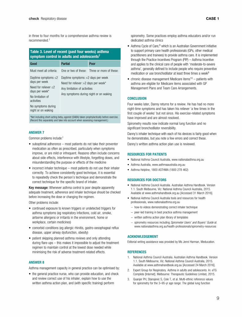
Table 3. Level of recent (past four weeks) asthma symptom control in adults and adolescents7
Good Partial Poor
Must meet all criteria: One or two of these: Three or more of these:
Daytime symptoms ≤2 days per week
Need for reliever ≤2 days per week*
No limitation of activities
No symptoms during night or on waking
Daytime symptoms >2 days per week
Need for reliever >2 days per week*
Any limitation of activities
Any symptoms during night or on waking
*Not including short-acting beta2 agonist (SABA) taken prophylactically before exercise. (Record this separately and take into account when assessing management.)

CASE 1
10
check Respiratory disease
2012 equations. Eur Respir J 2012;40:1324–43. Available at http://erj.ersjournals.com/content/40/6/1324.long [Accessed 24 May 2016].
4. Centers for Disease Control and Prevention. Spirometry training program: NHANES III reference values. Atlanta, GA: CDC, 2015. Available at www.cdc.gov/niosh/topics/spirometry/nhanes.html [Accessed 28 April 2016].
5. National Asthma Council Australia. Quick reference guide for spirometry. South Melbourne, Vic: National Asthma Council Australia, 2016. Available at www.nationalasthma.org.au/health-professionals/spirometry-resources/spirometry-quick-reference-guide [Accessed 24 May 2016].
6. Weiler JM, Anderson SD, Randolph C et al. Pathogenesis, prevalence, diagnosis, and management of exercise-induced bronchoconstriction: A practice parameter. Ann Allergy Asthma Immunol 2010;105(6 Suppl):S1–47.
7. National Asthma Council Australia. Australian Asthma Handbook: Quick reference guide version 1.1. South Melbourne, Vic: National Asthma Council Australia, 2015. Available at www.asthmahandbook.org.au/uploads/555143d72c3e3.pdf [Accessed 24 March 2016].
8. Department of Health. Medical Benefits Schedule – Completion of the Asthma Cycle of Care (Items 2546–2559 and 2664–2677). Woden, ACT: DoH, 2016. Available at www.mbsonline.gov.au [Accessed 31 March 2016].
9. Department of Health. MBS Online: Completion of the asthma cycle of care. Canberra: Commonwealth of Australia, 2016. Avaiable at www9.health.gov.au/mbs/fullDisplay.cfm?type=note&qt=NoteID&q=A44 [Accessed 28 April 2016].
10. Department of Health. Chronic Disease Management (formerly Enhanced Primary Care or EPC) – GP services. Woden, ACT: DoH, 2014. Available at www.health.gov.au/internet/main/publishing.nsf/Content/mbsprimarycare-chronicdiseasemanagement [Accessed 31 January 2016].

11
check Respiratory disease CASE 2
QUESTION 1
What would you look for on further history and examination?
FURTHER INFORMATION
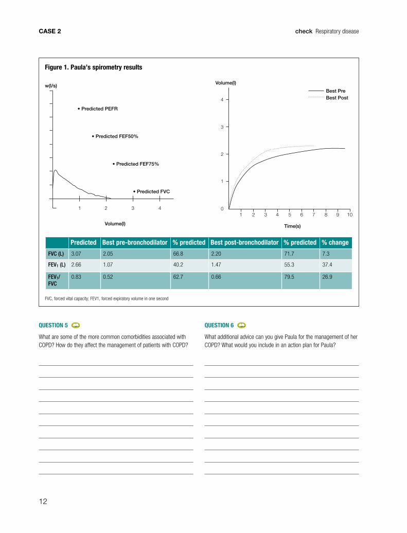
Paula describes shortness of breath on walking up two flights of stairs and is out of breath from walking 100 m from the car park to the shops. Paula tells you that on at least two occasions in the past two winters, she has had to take antibiotics and steroids. She admits that she is not always good at remembering to take her puffers. She has no signs of respiratory distress and her physical examination is unremarkable. You ask the practice nurse to perform spirometry (pre-bronchodilator and post-bronchodilator; Figure 1).
QUESTION 2
How would you interpret Paula’s spirometry results?
QUESTION 3
What are the risk factors associated with chronic obstructive pulmonary disease (COPD)?
QUESTION 4
Would you make any modifications to Paula’s medications, given her history and spirometry findings? If so, what modifications would you make?
CASE 2
PAULA HAS RUN OUT OF PUFFERS
Paula, aged 67 years, presents for a repeat prescription for her puffers. Her usual general practitioner (GP) is away today. She shows you her old salbutamol and fluticasone/salmeterol 250/25 inhalers. ‘It’s for me asthma, Doc’, she offers. Paula’s records show that she has had asthma since she was a teenager. Her last prescriptions were issued three months ago with five repeats. She is also on telmisartan 40 mg, atorvastatin 20 mg, esomeprazole 20 mg and oxazepam 30 mg. Paula is allergic to penicillin. She has smoked 10–15 cigarettes a day since the age of 18 years.
12
check Respiratory diseaseCASE 2
QUESTION 5
What are some of the more common comorbidities associated with COPD? How do they affect the management of patients with COPD?
QUESTION 6
What additional advice can you give Paula for the management of her COPD? What would you include in an action plan for Paula?
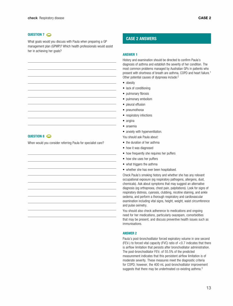
Figure 1. Paula’s spirometry results
Predicted Best pre-bronchodilator % predicted Best post-bronchodilator % predicted % change
FVC (L) 3.07 2.05 66.8 2.20 71.7 7.3
FEV1 (L) 2.66 1.07 40.2 1.47 55.3 37.4
FEV1/FVC
0.83 0.52 62.7 0.66 79.5 26.9
FVC, forced vital capacity; FEV1, forced expiratory volume in one second
1 2 3 4 5 6 7 8 9 10
1
2
3
4
0
Time(s)
Volume(l)
Best PreBest Post
1 2 3 4
Volume(l)
w(l/s)
• Predicted PEFR
• Predicted FEF50%
• Predicted FEF75%
• Predicted FVC
13
check Respiratory disease CASE 2
QUESTION 7
What goals would you discuss with Paula when preparing a GP management plan (GPMP)? Which health professionals would assist her in achieving her goals?
QUESTION 8
When would you consider referring Paula for specialist care?
CASE 2 ANSWERS
ANSWER 1
History and examination should be directed to confirm Paula’s diagnosis of asthma and establish the severity of her condition. The most common problems managed by Australian GPs in patients who present with shortness of breath are asthma, COPD and heart failure.1 Other potential causes of dyspnoea include:2
• obesity
• lack of conditioning
• pulmonary fibrosis
• pulmonary embolism
• pleural effusion
• pneumothorax
• respiratory infections
• angina
• anaemia
• anxiety with hyperventilation.
You should ask Paula about:
• the duration of her asthma
• how it was diagnosed
• how frequently she requires her puffers
• how she uses her puffers
• what triggers the asthma
• whether she has ever been hospitalised.
Check Paula’s smoking history and whether she has any relevant occupational exposure (eg respiratory pathogens, allergens, dust, chemicals). Ask about symptoms that may suggest an alternative diagnosis (eg orthopnoea, chest pain, palpitations). Look for signs of respiratory distress, cyanosis, clubbing, nicotine staining, and ankle oedema, and perform a thorough respiratory and cardiovascular examination including vital signs, height, weight, waist circumference and pulse oximetry.
You should also check adherence to medications and ongoing need for her medications, particularly oxazepam, comorbidities that may be present, and discuss preventive health issues such as immunisations.
ANSWER 2
Paula’s post-bronchodilator forced expiratory volume in one second (FEV1) to forced vital capacity (FVC) ratio of <0.7 indicates that there is airflow limitation that persists after bronchodilator administration. The post-bronchodilator FEV1 of 55.5% of the predicted measurement indicates that this persistent airflow limitation is of moderate severity. These measures meet the diagnostic criteria for COPD; however, the 400 mL post-bronchodilator improvement suggests that there may be undertreated co-existing asthma.3
14
check Respiratory diseaseCASE 2
Diagnosis of co-existing asthma and COPD is often difficult and should include clinical features, including a good history, as well as spirometry.4 Recognising the asthma component of COPD affects the management and prognosis.5
Patients who have asthma require inhaled corticosteroids (ICS) and should not be prescribed long-acting bronchodilator therapy without ICS; however, initial or early therapy in patients with COPD is often long-acting bronchodilator therapy without ICS. Some studies suggest that asthma co-exists with COPD in about 15–20% of patients.6 These patients are often younger and more likely to have frequent exacerbations and higher mortality.7,8
Spirometry is a frequently neglected investigation. The current inaccuracy of diagnosis in community settings and the importance of using spirometry was demonstrated in an Australian study where only 58% of general practice patients being treated for COPD were confirmed to have the diagnosis on post-bronchodilator spirometry; 18% had normal spirometry.9
Every patient with chronic lung disease should have spirometry to clarify their diagnosis and to monitor disease progression. Forced
expiratory volume in six seconds (FEV6) has been used as a surrogate for FVC and portable devices are available to assist with case finding for COPD using a FEV1/FEV6 cut-off of 0.8,10 but spirometry should still be performed for confirmation.
ANSWER 3
Smoking is the major risk factor for developing COPD.11 Passive smoking is thought to account for 1.9 million deaths from COPD in China.12
Smoking is not the only risk factor. The prevalence of COPD in never-smokers worldwide is thought to be 25–40%.13 Chronic asthma is thought to produce irreversible airway changes due to airway remodelling contributing to the development of COPD even in those who have never smoked.14 Other risk factors for developing COPD include:11
• outdoor air pollution
• biomass fuels (coal, animal dung, wood)
• occupational contaminants (diesel, organic dust, chemicals)
• tuberculosis
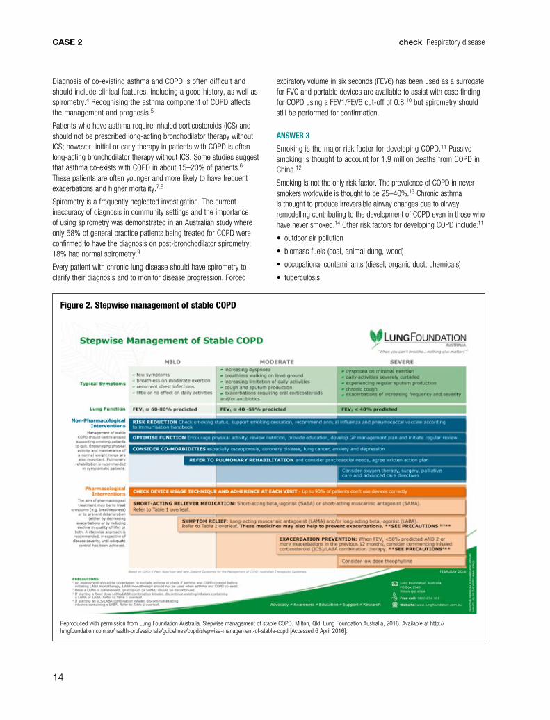
Figure 2. Stepwise management of stable COPD
Reproduced with permission from Lung Foundation Australia. Stepwise management of stable COPD. Milton, Qld: Lung Foundation Australia, 2016. Available at http://lungfoundation.com.au/health-professionals/guidelines/copd/stepwise-management-of-stable-copd [Accessed 6 April 2016].
15
check Respiratory disease CASE 2
• α-1 antitrypsin deficiency
• ageing
• low socioeconomic status.
ANSWER 4
The Lung Foundation Australia has produced a helpful chart (Figure 2).15 According to Australian guidelines, Paula has moderate COPD. Her current diagnosis of co-existing COPD and asthma suggests she should continue to use a short-acting beta2 agonist (SABA) as needed, together with a long-acting beta2 agonist/inhaled corticosteroids (LABA/ICS) combination.4 Patients with COPD and asthma should not be given a LABA alone, and ICS are the mainstay in this subgroup. As Paula is already on this combination, but is still symptomatic, her asthma therapy should be maximised by first checking her medication adherence and technique; if she is still symptomatic, a long-acting muscarinic antagonist (LAMA) can be added.
Patients with COPD who do not have co-existing asthma and are symptomatic on single long-acting bronchodilator therapy
(either LAMA or LABA) may benefit from combined LAMA/LABA. Pharmaceutical Benefits Scheme (PBS) requirements are that the ‘patient must have been stabilised on a combination of a LAMA/LABA’.16
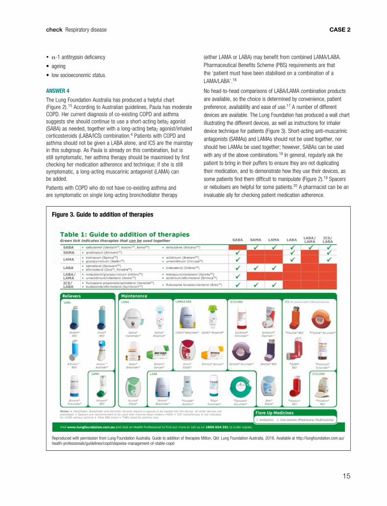
No head-to-head comparisons of LABA/LAMA combination products are available, so the choice is determined by convenience, patient preference, availability and ease of use.17 A number of different devices are available. The Lung Foundation has produced a wall chart illustrating the different devices, as well as instructions for inhaler device technique for patients (Figure 3). Short-acting anti-muscarinic antagonists (SAMAs) and LAMAs should not be used together, nor should two LAMAs be used together; however, SABAs can be used with any of the above combinations.18 In general, regularly ask the patient to bring in their puffers to ensure they are not duplicating their medication, and to demonstrate how they use their devices, as some patients find them difficult to manipulate (Figure 2).19 Spacers or nebulisers are helpful for some patients.20 A pharmacist can be an invaluable ally for checking patient medication adherence.
Figure 3. Guide to addition of therapies
Reproduced with permission from Lung Foundation Australia. Guide to addition of therapies Milton, Qld: Lung Foundation Australia, 2016. Available at http://lungfoundation.com.au/health-professionals/guidelines/copd/stepwise-management-of-stable-copd
16
check Respiratory diseaseCASE 2
ANSWER 5
Multimorbidity affects up to 60% of people with chronic respiratory disease in primary care.21 Common comorbidities include:
• ischaemic heart disease
• peripheral vascular disease
• lung cancer
• anxiety
• depression
• pulmonary hypertension
• cor pulmonale
• sleep apnoea
• atrial fibrillation
• gastrooesophageal reflux disease
• osteoporosis.
Multimorbidity in patients with COPD may be explained by common risk factors (such as smoking and lack of physical activity), activation of the neurohumoral response, systemic inflammation,22 and side-effects of drug treatment. Patients with multimorbidity who are diagnosed with COPD tend to underestimate its importance, which affects their ability to self-manage and use health resources.23
Disease–disease, disease–drug and drug–drug interactions are important when dealing with multimorbidity. ICS are associated with an increased risk of pneumonia,24 and LABAs and LAMAs may have cardiovascular effects.25 Yet, beta-blockers are often omitted in patients with COPD and heart failure,26 despite cardioselective beta-blockers being safe and indicated for patients with COPD and ischaemic heart disease or heart failure.27
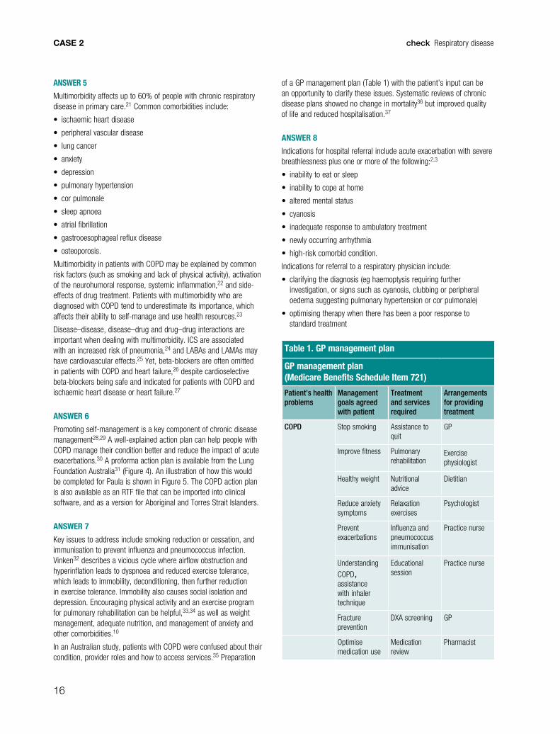
ANSWER 6
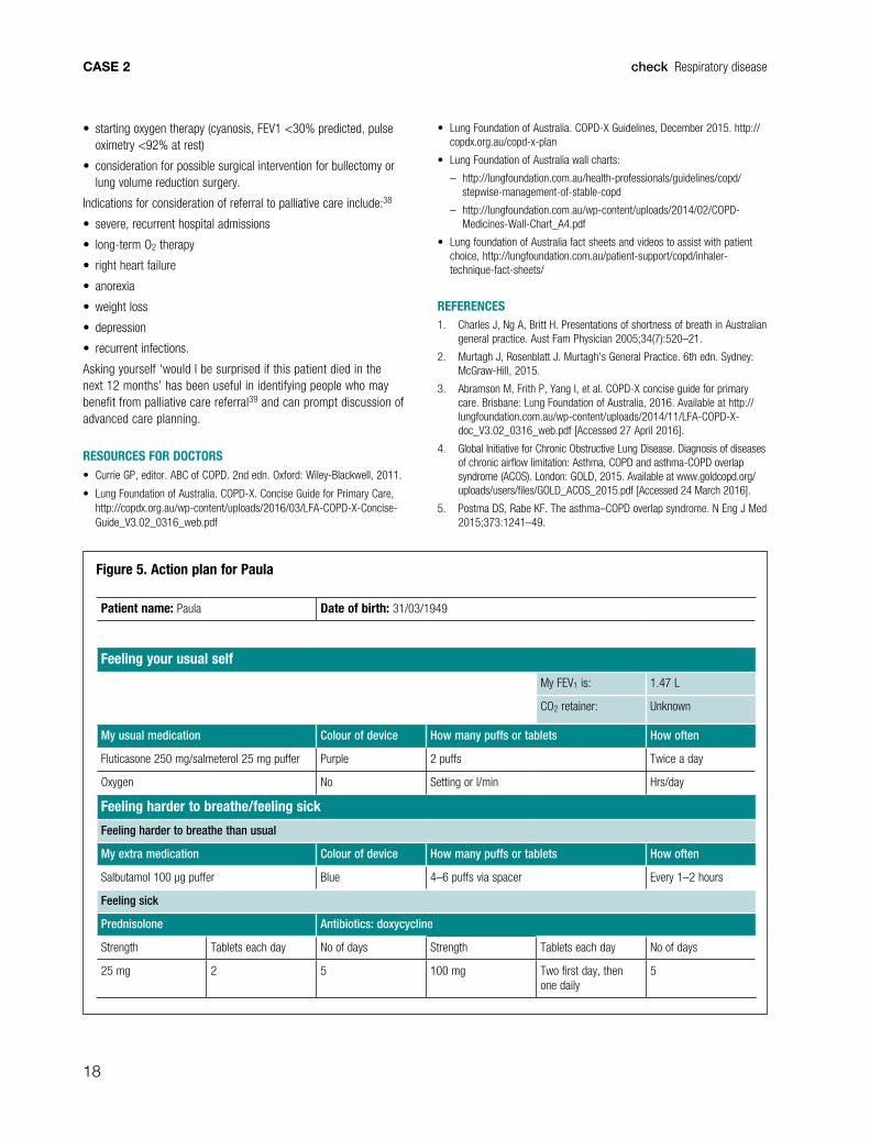
Promoting self-management is a key component of chronic disease management28,29 A well-explained action plan can help people with COPD manage their condition better and reduce the impact of acute exacerbations.30 A proforma action plan is available from the Lung Foundation Australia31 (Figure 4). An illustration of how this would be completed for Paula is shown in Figure 5. The COPD action plan is also available as an RTF file that can be imported into clinical software, and as a version for Aboriginal and Torres Strait Islanders.
ANSWER 7
Key issues to address include smoking reduction or cessation, and immunisation to prevent influenza and pneumococcus infection. Vinken32 describes a vicious cycle where airflow obstruction and hyperinflation leads to dyspnoea and reduced exercise tolerance, which leads to immobility, deconditioning, then further reduction in exercise tolerance. Immobility also causes social isolation and depression. Encouraging physical activity and an exercise program for pulmonary rehabilitation can be helpful,33,34 as well as weight management, adequate nutrition, and management of anxiety and other comorbidities.10
In an Australian study, patients with COPD were confused about their condition, provider roles and how to access services.35 Preparation
of a GP management plan (Table 1) with the patient’s input can be an opportunity to clarify these issues. Systematic reviews of chronic disease plans showed no change in mortality36 but improved quality of life and reduced hospitalisation.37
ANSWER 8
Indications for hospital referral include acute exacerbation with severe breathlessness plus one or more of the following:2,3
• inability to eat or sleep
• inability to cope at home
• altered mental status
• cyanosis
• inadequate response to ambulatory treatment
• newly occurring arrhythmia
• high-risk comorbid condition.
Indications for referral to a respiratory physician include:
• clarifying the diagnosis (eg haemoptysis requiring further investigation, or signs such as cyanosis, clubbing or peripheral oedema suggesting pulmonary hypertension or cor pulmonale)
• optimising therapy when there has been a poor response to standard treatment
Table 1. GP management plan
GP management plan (Medicare Benefits Schedule Item 721)
Patient’s health problems
Management goals agreed with patient
Treatment and services required
Arrangements for providing treatment
COPD Stop smoking Assistance to quit
GP
Improve fitness Pulmonary rehabilitation
Exercise physiologist
Healthy weight Nutritional advice
Dietitian
Reduce anxiety symptoms
Relaxation exercises
Psychologist
Prevent exacerbations
Influenza and pneumococcus immunisation
Practice nurse
Understanding COPD, assistance with inhaler technique
Educational session
Practice nurse
Fracture prevention
DXA screening GP
Optimise medication use
Medication review
Pharmacist
17
check Respiratory disease CASE 2
Figure 4. Action plan proforma
Reproduced with permission from Lung Foundation Australia. COPD action plan. Milton, Qld: Lung Foundation Australia, 2015. Available at http://lungfoundation.com.au/health-professionals/clinical-resources/copd/copd-action-plan/?doing_wp_cron=1459921697.4677140712738037109375 [Accessed 6 April 2016].
18
check Respiratory diseaseCASE 2
• starting oxygen therapy (cyanosis, FEV1 <30% predicted, pulse oximetry <92% at rest)
• consideration for possible surgical intervention for bullectomy or lung volume reduction surgery.
Indications for consideration of referral to palliative care include:38
• severe, recurrent hospital admissions
• long-term O2 therapy
• right heart failure
• anorexia
• weight loss
• depression
• recurrent infections.
Asking yourself ‘would I be surprised if this patient died in the next 12 months’ has been useful in identifying people who may benefit from palliative care referral39 and can prompt discussion of advanced care planning.
RESOURCES FOR DOCTORS• Currie GP, editor. ABC of COPD. 2nd edn. Oxford: Wiley-Blackwell, 2011.
• Lung Foundation of Australia. COPD-X. Concise Guide for Primary Care, http://copdx.org.au/wp-content/uploads/2016/03/LFA-COPD-X-Concise-Guide_V3.02_0316_web.pdf
• Lung Foundation of Australia. COPD-X Guidelines, December 2015. http://copdx.org.au/copd-x-plan
• Lung Foundation of Australia wall charts:
– http://lungfoundation.com.au/health-professionals/guidelines/copd/stepwise-management-of-stable-copd
– http://lungfoundation.com.au/wp-content/uploads/2014/02/COPD-Medicines-Wall-Chart_A4.pdf
• Lung foundation of Australia fact sheets and videos to assist with patient choice, http://lungfoundation.com.au/patient-support/copd/inhaler-technique-fact-sheets/
REFERENCES1. Charles J, Ng A, Britt H. Presentations of shortness of breath in Australian
general practice. Aust Fam Physician 2005;34(7):520–21.
2. Murtagh J, Rosenblatt J. Murtagh’s General Practice. 6th edn. Sydney: McGraw-Hill, 2015.
3. Abramson M, Frith P, Yang I, et al. COPD-X concise guide for primary care. Brisbane: Lung Foundation of Australia, 2016. Available at http://lungfoundation.com.au/wp-content/uploads/2014/11/LFA-COPD-X-doc_V3.02_0316_web.pdf [Accessed 27 April 2016].
4. Global Initiative for Chronic Obstructive Lung Disease. Diagnosis of diseases of chronic airflow limitation: Asthma, COPD and asthma-COPD overlap syndrome (ACOS). London: GOLD, 2015. Available at www.goldcopd.org/uploads/users/files/GOLD_ACOS_2015.pdf [Accessed 24 March 2016].
5. Postma DS, Rabe KF. The asthma–COPD overlap syndrome. N Eng J Med 2015;373:1241–49.
Figure 5. Action plan for Paula
Patient name: Paula Date of birth: 31/03/1949
Feeling your usual self
My FEV1 is: 1.47 L
CO2 retainer: Unknown
My usual medication Colour of device How many puffs or tablets How often
Fluticasone 250 mg/salmeterol 25 mg puffer Purple 2 puffs Twice a day
Oxygen No Setting or l/min Hrs/day
Feeling harder to breathe/feeling sick
Feeling harder to breathe than usual
My extra medication Colour of device How many puffs or tablets How often
Salbutamol 100 µg puffer Blue 4–6 puffs via spacer Every 1–2 hours
Feeling sick
Prednisolone Antibiotics: doxycycline
Strength Tablets each day No of days Strength Tablets each day No of days
25 mg 2 5 100 mg Two first day, then one daily
5

19
check Respiratory disease CASE 2
6. Global Initiative for Chronic Obstructive Lung Disease. COPD diagnosis and management at-a-glance desk reference. London, GOLD, 2015. Available at www.goldcopd.org/guidelines-copd-diagnosis-and-management.html [Accessed 22 March 2016].
7. De Marco R, Pesce G, Marcon A, et al. The co-existence of asthma and chronic obstructive pulmonary disease (COPD): Prevalence and risk factors in young, middle-aged and elderly people from the general population. PLOS ONE 2013;8(5):e62985. Available at http://journals.plos.org/plosone/article?id=10.1371/journal.pone.0062985 [Accessed 15 March 2016].
8. Alshabanat A, Zafari Z, Albanyan O, et al. Asthma and COPD overlap syndrome (ACOS): A systematic review and meta analysis. PLoS ONE 2015;10(9):e0136065. Available at http://journals.plos.org/plosone/article?id=10.1371/journal.pone.0136065 [Accessed 24 March 2016].
9. Zwar NA, Marks GB, Hermiz O, et al. Predictors of accuracy of diagnosis of chronic obstructive pulmonary disease in general practice Med J Aust 2011;195:168–71.
10. Represas-Represas C, Fernández-Villar A, Ruano-Raviña A,et al. Screening for chronic obstructive pulmonary disease: Validity and reliability of a portable device in non-specialized healthcare settings. PLoS ONE 2016;11(1):e0145571. Available at http://journals.plos.org/plosone/article?id=10.1371/journal.pone.0145571 [Accessed 24 March 2016].
11. Lung Foundation of Australia. COPD-X guidelines. Milton, Qld: Lung Foundation Australia, 2015. Available at http://copdx.org.au/copd-x-plan [Accessed 24 March 2016].
12. Yin P, Jiang CQ, Cheng KK, et al. Passive smoking exposure and risk of COPD among adults in China: The Guangzhou biobank cohort study. The Lancet 2007;370(9589):751–57.
13. Currie GP, editor. ABC of COPD. 2nd edn. Oxford: Wiley-Blackwell, 2011.
14. Lung Foundation of Australia. The COPD-X plan: C4.1 confirm or exclude asthma. Milton, QLD: Lung Foundation, 2014. Available at http://copdx.org.au/copd-x-plan/confirm-diagnosis/c4-assessing-acute-response-to-bronchodilators/c41-confirm-or-exclude-asthma/#c4 [Accessed 25 April 2016].
15. Lung Foundation of Australia. Stepwise management of stable COPD. Milton, QLD: Lung Foundation, 2016. Available at http://lungfoundation.com.au/wp-content/uploads/2014/02/LFA-Stepwise-Management-of-COPD_0216_WEB.pdf [Accessed 24 March 2016].
16. NPS MedicineWise. New fixed-dose combination bronchodilators for COPD. Surry Hills, NSW: NPS MedicineWise, 2016. Available at www.nps.org.au/publications/health-professional/nps-radar/latest-issue/brief-item-fdc-copd-bronchodilators [Accessed 18 March 2016].
17. Therapeutic Guidelines Limited. In: eTG Complete [CD-ROM]. Melbourne: Therapeutic Guidelines Ltd, 2015.
18. NPS MedicineWise. Pharmacological therapies for COPD in Australia. Surry Hills, NSW: NPS Radar, 2014. Available at www.nps.org.au/__data/assets/pdf_file/0011/266771/NPS-RADAR-December-2014-complete.pdf [Accessed 18 March 2016].
19. Albertson TE, Schivo M, Zeki AA. The pharmacological approach to the elderly COPD Patient. Drugs Aging 2013;30:479–502.
20. Potter A, Wilkinson A. Management of COPD. InnovAiT 2011;4(7):390–97.
21. O’Kelly A, Smith SM, Lane S. Chronic respiratory disease and multimorbidity: Prevalence and impact in a general practice setting. Respiratory Medicine 2011;105:236e242.
22. Clini EM, Boschetto P, Lainscak M, Janssens W. Comorbidities in chronic obstructive pulmonary disease from assessment to treatment. BioMed Res Int 2014:414928. Available at www.hindawi.com/journals/bmri/2014/414928 [Accessed 19 March 2016].
23. Ansari S, Hosseinzadeh H, Dennis S, Zwar N. Patients’ perspectives on the impact of a new COPD diagnosis in the face of multimorbidity: A qualitative study. Primary Care Respiratory Medicine 2014;24:14036. Available at www.nature.com/articles/npjpcrm201436 [Accessed 11 April 2016].
24. Kew KM, Seniukovich A. Inhaled steroids and risk of pneumonia for chronic obstructive pulmonary disease. Cochrane Database Syst Rev 2014;CD010115. Available at http://onlinelibrary.wiley.com/doi/10.1002/14651858.CD010115.pub2/full [Accessed 11 April 2016].
25. Hillas G, Perlikos F, Tsiligianni I, Tzanakis N. Managing comorbidities in COPD. Int J Chron Obstruct Pulmon Dis 2015;10:95–109.
26. Doos L, Roberts EO, Corp N, Kadam UT. Multi-drug therapy in chronic condition multimorbidity: A systematic review. Fam Pract 2014;31(6):654–63.
27. Salpeter SR, Ormiston TM, Salpeter EE. Cardioselective beta-blockers for chronic obstructive pulmonary disease. Cochrane Database Syst Rev 2005;CD003566.
28. Morgan MDL. Action plans for COPD self management. Integrated care is more than the sum of its parts. Thorax 2011;66:935e936.
29. Effing T, van der Palen J, Frith P. Education in COPD self-management: Only part of the game. Respirology 2014;19:151–52.
30. Effing T, Kerstjens H, ven der Valk P, et al. (Cost)-effectiveness of self-treatment of exacerbations on the severity of exacerbations in patients with COPD: The COPE II study. Thorax 2009;64:956–62.
31. Lung Foundation Australia. COPD action plan. Milton, Qld: Lung Foundation Australia, 2015. Available at http://lungfoundation.com.au/health-professionals/clinical-resources/copd/copd-action-plan [Accessed 31 March 2016].
32. Vincken W. An Update on bronchodilator treatment of chronic obstructive pulmonary disease (COPD). Ann Resp Med 2010;1(2):1–15.
33. Yawn B, Thomashow B. Management of patients during and after exacerbations of chronic obstructive pulmonary disease: The role of primary care physicians. Int J Gen Med 2011;4:665–76.
34. Caferella P, Effing T, Hancock K, Frith P. Partners in COPD management in the primary care setting. Medicine Today 2016;17(1–2):26–34. Available at http://medicinetoday.com.au/2016/january/feature-article/partners-copd-management-primary-care-setting [Accessed 25 April 2016].
35. Kirby SE, Mutimbe M, Vagholkar S, et al. How integrated are services for patients with chronic obstructive pulmonary disease? Perceptions of patients and health care providers. Aust J Prim Health 2014;20(2):158–61.
36. Peytremann-Bridevaux I, Taffe P, Burnand B, et al. Mortality of patients with COPD participating in chronic disease management programmes: A happy end? Thorax 2014;69:865–66.
37. Kruis AL, Smidt N, Assendelft WJJ, et al. Integrated disease management interventions for patients with chronic obstructive pulmonary disease. Cochrane Database Syst Rev 2013;10:CD009437.
38. Sadler J, Corcoran JP. Chronic management of stable COPD. InnovAiT 2013;7(3):141–50.
39. Moroni M, Zocchi D, Bolognesi, et al; on behalf of the SUQ-P group. The ‘surprise’ question in advanced cancer patients: A prospective study among general practitioners. Palliat Med 2014;28(7):959–64.

20
check Respiratory diseaseCASE 3
QUESTION 1
What information do you need from Kathryn at this point in the consultation?
FURTHER INFORMATION
Kathryn tells you that when her cough first started five years ago, the sputum production was intermittent and was mainly mucoid (white in colour). For the past two years, she has had daily production of sputum that is discoloured (ranging from light yellow to dark green). Kathryn also has mild sinus symptoms with intermittent nasal discharge and congestion. She is a lifelong non-smoker and cannot identify any environmental factors that might contribute to her cough.
She states that two to three times a year, over the Past few years, she has an increased volume of sputum and the sputum changes colour from yellow to green. She thinks that these episodes are often precipitated by a cold that she caught from her grandchildren when she was babysitting them.
On examination, she has bibasal crackles.
QUESTION 2
What tests would you order at this stage?
FURTHER INFORMATION
The chest X-ray (CXR) was reported as being normal. Two sputum samples were analysed and both were reported as purulent; one was reported as having no growth, whereas Haemophilus influenzae, which was sensitive to all antibiotics tested, was isolated in the second. A chest high-resolution computed tomography (HRCT) scan showed the presence of mild bilateral lower zone bronchiectasis.
QUESTION 3
What further tests or referrals would you consider at this stage?
CASE 3
KATHRYN HAS A PRODUCTIVE COUGH
Kathryn is a retired schoolteacher, 68 years of age, who presents with a cough. She describes a history of cough with the production of sputum for the past five years, but this has been worse in the past month. Kathryn has been generally well and has no other significant medical problems. However, as a child, she had frequent episodes of bronchitis and was diagnosed as having asthma.
21
check Respiratory disease CASE 3
QUESTION 4
What are the key features of management?
QUESTION 5
How common is bronchiectasis? How does it occur?
QUESTION 6
What is the long-term prognosis of bronchiectasis?
CASE 3 ANSWERS
ANSWER 1
You should ask Kathryn about her smoking history and whether she is a current smoker. Information should also be obtained about:
• any environmental factors that precipitate or aggravate her cough
• the presence of upper respiratory tract symptoms (eg sinusitis, postnasal drip)
• symptoms of reflux
• the presence and frequency of worsening symptoms.
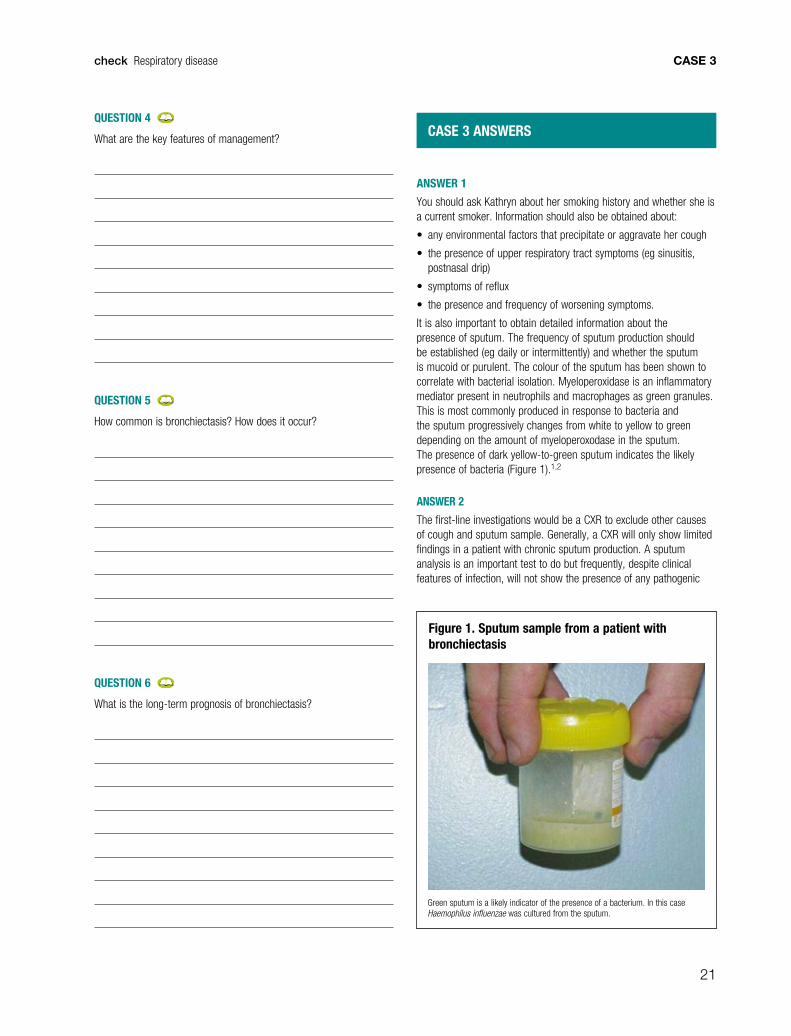
It is also important to obtain detailed information about the presence of sputum. The frequency of sputum production should be established (eg daily or intermittently) and whether the sputum is mucoid or purulent. The colour of the sputum has been shown to correlate with bacterial isolation. Myeloperoxidase is an inflammatory mediator present in neutrophils and macrophages as green granules. This is most commonly produced in response to bacteria and the sputum progressively changes from white to yellow to green depending on the amount of myeloperoxodase in the sputum. The presence of dark yellow-to-green sputum indicates the likely presence of bacteria (Figure 1).1,2
ANSWER 2
The first-line investigations would be a CXR to exclude other causes of cough and sputum sample. Generally, a CXR will only show limited findings in a patient with chronic sputum production. A sputum analysis is an important test to do but frequently, despite clinical features of infection, will not show the presence of any pathogenic
Figure 1. Sputum sample from a patient with bronchiectasis
Green sputum is a likely indicator of the presence of a bacterium. In this case Haemophilus influenzae was cultured from the sputum.
22
check Respiratory diseaseCASE 3
bacteria. Spirometry should also be performed to assess underlying lung function and investigate the possibility of asthma as a cause of cough.
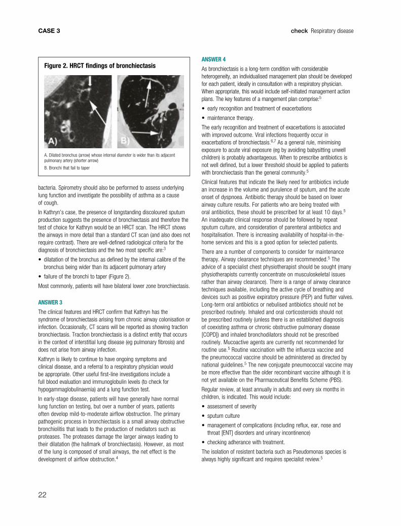
In Kathryn’s case, the presence of longstanding discoloured sputum production suggests the presence of bronchiectasis and therefore the test of choice for Kathryn would be an HRCT scan. The HRCT shows the airways in more detail than a standard CT scan (and also does not require contrast). There are well-defined radiological criteria for the diagnosis of bronchiectasis and the two most specific are:3
• dilatation of the bronchus as defined by the internal calibre of the bronchus being wider than its adjacent pulmonary artery
• failure of the bronchi to taper (Figure 2).
Most commonly, patients will have bilateral lower zone bronchiectasis.
ANSWER 3
The clinical features and HRCT confirm that Kathryn has the syndrome of bronchiectasis arising from chronic airway colonisation or infection. Occasionally, CT scans will be reported as showing traction bronchiectasis. Traction bronchiectasis is a distinct entity that occurs in the context of interstitial lung disease (eg pulmonary fibrosis) and does not arise from airway infection.
Kathryn is likely to continue to have ongoing symptoms and clinical disease, and a referral to a respiratory physician would be appropriate. Other useful first-line investigations include a full blood evaluation and immunoglobulin levels (to check for hypogammaglobulinaemia) and a lung function test.
In early-stage disease, patients will have generally have normal lung function on testing, but over a number of years, patients often develop mild-to-moderate airflow obstruction. The primary pathogenic process in bronchiectasis is a small airway obstructive bronchiolitis that leads to the production of mediators such as proteases. The proteases damage the larger airways leading to their dilatation (the hallmark of bronchiectasis). However, as most of the lung is composed of small airways, the net effect is the development of airflow obstruction.4
ANSWER 4
As bronchiectasis is a long-term condition with considerable heterogeneity, an individualised management plan should be developed for each patient, ideally in consultation with a respiratory physician. When appropriate, this would include self-initiated management action plans. The key features of a mangement plan comprise:5
• early recognition and treatment of exacerbations
• maintenance therapy.
The early recognition and treatment of exacerbations is associated with improved outcome. Viral infections frequently occur in exacerbations of bronchiectasis.6,7 As a general rule, minimising exposure to acute viral exposure (eg by avoiding babysitting unwell children) is probably advantageous. When to prescribe antibiotics is not well defined, but a lower threshold should be applied to patients with bronchiectasis than the general community.5
Clinical features that indicate the likely need for antibiotics include an increase in the volume and purulence of sputum, and the acute onset of dyspnoea. Antibiotic therapy should be based on lower airway culture results. For patients who are being treated with oral antibiotics, these should be prescribed for at least 10 days.5 An inadequate clinical response should be followed by repeat sputum culture, and consideration of parenteral antibiotics and hospitalisation. There is increasing availability of hospital-in-the-home services and this is a good option for selected patients.
There are a number of components to consider for maintenance therapy. Airway clearance techniques are recommended.5 The advice of a specialist chest physiotherapist should be sought (many physiotherapists currently concentrate on musculoskeletal issues rather than airway clearance). There is a range of airway clearance techniques available, including the active cycle of breathing and devices such as positive expiratory pressure (PEP) and flutter valves. Long-term oral antibiotics or nebulised antibiotics should not be prescribed routinely. Inhaled and oral corticosteroids should not be prescribed routinely (unless there is an established diagnosis of coexisting asthma or chronic obstructive pulmonary disease [COPD]) and inhaled bronchodilators should not be prescribed routinely. Mucoactive agents are currently not recommended for routine use.5 Routine vaccination with the influenza vaccine and the pneumococcal vaccine should be administered as directed by national guidelines.5 The new conjugate pneumococcal vaccine may be more effective than the older recombinant vaccine although it is not yet available on the Pharmaceutical Benefits Scheme (PBS).
Regular review, at least annually in adults and every six months in children, is indicated. This would include:
• assessment of severity
• sputum culture
• management of complications (including reflux, ear, nose and throat [ENT] disorders and urinary incontinence)
• checking adherance with treatment.
The isolation of resistent bacteria such as Pseudomonas species is always highly significant and requires specialist review.5
Figure 2. HRCT findings of bronchiectasis
A. Dilated bronchus (arrow) whose internal diameter is wider than its adjacent pulmonary artery (shorter arrow)
B. Bronchi that fail to taper
23
check Respiratory disease CASE 3
ANSWER 5
The prevalence of bronchiectasis is not well defined as definitive CT population screening studies have not been carried out. However, with the widespread use of CT scanning it has now been found to be a very common condition in specialist respiratory practice. A recent study has estimated that more than two million people worldwide have bronchiectasis.8 The largest group of patients with bronchiectasis are those with COPD. Studies have described that in 29–57% of patients with COPD, the presence of bronchiectasis is shown on CT scanning. The combination of COPD and bronchiectasis is associated with increased mortality, worse symptoms and the isolation of bacteria from sputum.9–11 There are no guidelines currently available for the management of the combined entity of COPD with bronchiectasis.
Bronchiectasis occurs when there is a defect in the host defence that allows the persistence of microbial pathogens in the lower respiratory tract. Many potential defects in host defence that cause bronchiectasis have been described, but it is often not possible to define a cause. Two common patterns of bronchiectasis are childhood-onset disease and adult-onset disease.12 Typically, childhood-onset disease starts in the first 10 years of life and is characterised by recurrent sputum production and chest infections, and is often associated with upper respiratory tract disease (eg sinusitis). Usually, this will improve during adolescence and then recur when patients are older (eg over 60 years of age). In adult-onset disease, patients will typically develop a chronic productive cough in their 60s and 70s; after several years of symptoms an HRCT will show mild bronchiectasis.
ANSWER 6
Once established, bronchietasis generally persists no matter how aggressive therapy is.4 It is a heterogeneous condition with various outcomes. Most commonly, the condition becomes gradually worse over a number of years but in the great majority of patients this does not affect mortality. There is a small subset of patients who have rapidly progressive disease, who need careful specialist management; this subgroup is more likely to have airway colonisation with bacterium such as Pseudomonas spp.5
CONCLUSION
Patients with bronchiectasis typically have a long-term cough productive of purulent sputum with bibasal crackles on examination. The diagnosis is made with HRCT scanning. Core principles of management include an individualised management plan, and early and aggressive treatment of exacerbations. It is a common condition and most patients will have persistent symptoms.
RESOURCES FOR DOCTORS AND PATIENTS• The Thoracic Society of Australia and New Zealand guidelines was the main
source for management recommedations in this case study, www.mja.com.au/system/files/issues/cha00287.pdf
• Australian Lung Foundation has useful informaton for patients, www.lungfoundation.com.au
• Gibson PG, Chang AB, Glasgow NJ, et al. CICADA: Cough in children and adults: Diagnosis and Assessment. Australian cough guidelines summary statement. Med J Aust 2010;192(5):265–71; www.mja.com.au/journal/2010/192/5/cicada-cough-children-and-adults-diagnosis-and-assessment-australian-cough
REFERENCES1. Stockley RA, Bayley D, Hill SL, Hill AT, Crooks S, Campbell EJ.
Assessment of airway neutrophils by sputum colour: Correlation with airways inflammation. Thorax 2001;56(5):366–72.
2. Murray MP, Pentland JL, Turnbull K, MacQuarrie S, Hill AT. Sputum colour: A useful clinical tool in non-cystic fibrosis bronchiectasis. Eur Respir J 2009;34(2):361–64.
3. McGuinness G, Naidich DP. CT of airways disease and bronchiectasis. Radiol Clin North Am 2002;40(1):1–19.
4. King PT. The pathophysiology of bronchiectasis. Int J Chron Obstruct Pulm Dis 2009;4:411–19.
5. Chang AB, Bell SC, Torzillo PJ, et al. Chronic suppurative lung disease and bronchiectasis in children and adults in Australia and New Zealand Thoracic Society of Australia and New Zealand guidelines. Med J Aust 2015;202(1):21–23.
6. Kapur N, Mackay IM, Sloots TP, Masters IB, Chang AB. Respiratory viruses in exacerbations of non-cystic fibrosis bronchiectasis in children. Arch Dis Childhood 2014;99(8):749–53.
7. Gao YH, Guan WJ, Xu G, et al. The role of viral infection in pulmonary exacerbations of bronchiectasis in adults: A prospective study. Chest 2015;147(6):1635–43.
8. Polverino E, Cacheris W, Spencer C, Operschall E, O’Donnell AE. Global burden of non-cystic fibrosis bronchiectasis: A simple empidemiologic analysis. Eur Respir J;2012(Supp 56):P3983.
9. O’Brien C, Guest PJ, Hill SL, Stockley RA. Physiological and radiological characterisation of patients diagnosed with chronic obstructive pulmonary disease in primary care. Thorax 2000;55(8):635–42.
10. Patel IS, Vlahos I, Wilkinson TM, et al. Bronchiectasis, exacerbation indices and inflammation in chronic obstructive pulmonary disease. Am J Respir Crit Care Med 2004;170(4):400–07.
11. Martinez-Garcia MA, de la Rosa Carrillo D, Soler-Cataluna JJ, et al. Prognostic value of bronchiectasis in patients with moderate-to-severe chronic obstructive pulmonary disease. Am J Respir Crit Care Med 2013;187(8):823–31.
12. King PT, Holdsworth SR, Farmer M, Freezer N, Villanueva E, Holmes PW. Phenotypes of adult bronchiectasis: onset of productive cough in childhood and adulthood. COPD 2009;6(2):130–36.

24
check Respiratory diseaseCASE 4
QUESTION 1
What are the important historical points to clarify in this situation?
FURTHER INFORMATION
Mary says that she is usually in bed by 9.30–10.00 pm and has difficulty getting to sleep in less than an hour. She describes a number of menopausal symptoms that affect her ability to get to sleep (including hot flushes) and restlessness due to paraesthesia in her legs. Mary feels as though she is off to sleep by around midnight and sleeps through until 7.30 am when her alarm wakes her. She wakes up twice a night on average, at least once to pass urine and sometimes because of hot flushes. On two to three nights a week, Mary finds it difficult to get back to sleep and it can take her over an hour to get back to sleep on those occasions. She awakes with a dry mouth and occasionally a headache, feeling unrefreshed from her sleep. Mary is a snorer, as reported by Phil, but Phil is not sure if she moves her legs at night and is not aware of her breathing stopping. She has never had a motor vehicle accident secondary to falling asleep, but has woken at the traffic lights with cars behind her beeping their horns. Mary occasionally has hay fever but denies any difficulties breathing through her nose. She has never had upper airway surgery. She describes low mood currently but attributes this to being sleepy all the time.
QUESTION 2
What important examination findings will help aid your diagnosis?
FURTHER INFORMATION
Mary has a BMI of 33 kg/m2 and her blood pressure is 150/100 mmHg. She has a crowded oropharynx and a Mallampati score of 4. Her neck circumference is 39 cm and her waist circumference is 86 cm. Mary’s tonsils are not enlarged and her Epworth Sleepiness Score is 14/24.
QUESTION 3
What is your working diagnosis? What are the differential diagnoses?
QUESTION 4
How would you go about differentiating the causes of Mary’s sleepiness?
CASE 4
MARY FEELS TIRED AND DEPRESSED
Mary is 54 years of age and has been attending your practice for the past 20 years. She has a past history of depression (currently not on medication), hypertension (treated with an angiotensin converting enzyme inhibitor [ACEi]) and obesity. Mary presents today complaining of difficulty concentrating at work, low mood and feelings of fatigue. She feels tired in the morning and her husband, Phil, has reported increasing snoring noises over the past 12 months.
25
check Respiratory disease CASE 4
FURTHER INFORMATION
The blood tests are returned with no abnormalities detected. You refer Mary to the sleep medicine physician who requests an in-laboratory polysomnography.
The sleep study shows evidence of moderately severe obstructive sleep apnoea with an apnoea and hypopnoea index (AHI) of 25 events/hour (normal <5 events/hr). There is also evidence of an elevated periodic leg movement index of 20 events/hr (normal <10 events/hr). Mary’s sleep is fragmented with an elevated arousal index and a reduced proportion of rapid eye movement sleep.
QUESTION 5
How should Mary be managed in the context of her sleep study findings?
FURTHER INFORMATION
The sleep physician recommends continuous positive airways pressure (CPAP) to Mary and initiates therapy with a hire/trial period of automatically titrating CPAP.
QUESTION 6
What is automatically titrating CPAP? What are the advantages and disadvantages of recommending a trial of CPAP therapy as opposed to an in-laboratory CPAP titration?
CASE 4 ANSWERS
ANSWER 1
Generally speaking, if a patient has excessive daytime sleepiness it is because of one or more of the following:
• not enough sleep (quantity issue)
• poor quality sleep (quality issue)
• a medical condition causing sleepiness despite adequate quantity or quality sleep (eg depression, anaemia, thyroid dysfunction, sleep-specific disorders, such as obstructive sleep apnoea, periodic limb movement disorder, narcolepsy, idiopathic hypersomnia).
As such, it is important to clarify the patient’s normal sleeping habit:
• What time does the patient go to bed?
• On average, how long does it take for the patient to fall asleep?
• On average, how many times does the patient wake up per night?
• Each time the patient wakes up, what does the patient do and how long does it then take to get back to sleep?
• What time does the patient have to get up?
• Does the patient feel exhausted in the morning?
• Does the patient have a headache first thing in the morning that improves as the day progresses?
Once you have quantified the patient’s likely sleep duration (patients tend to over-estimate sleeping times by approximately 30 minutes), it is important to ask focused questions to help you decide if a sleep quality issue is present (eg obstructive sleep apnoea, periodic limb movements).
• Do you snore?1
• Does your bed partner report apnoeic episodes (eg breathing pauses, gasping, choking)?
• Do you wake with a dry mouth and/or a sore throat?
• Do you find it difficult to breathe through your nose? Do you have hay fever?
• Do you wake up gasping for air?
• Do you have difficulty keeping your legs still in the evening (eg when sitting down in front of the television)?
• Does your bed partner report leg twitching?
• Do you find that you have to move your legs around to get comfortable at the start of the night?
It is important (but often quite difficult) to differentiate between symptoms of fatigue (feeling washed out, low mood, lethargic physically)2 and genuine sleepiness where the patient can fall asleep very quickly in all sorts of environments (eg at work, in a waiting room, at traffic lights).3 Genuine sleepiness typically points to underlying sleep loss (sleep quality or quantity issue).
Depression scales, such as the Hospital Anxiety and Depression Scale (HADS), Depression Anxiety Stress Scales (DASS) or Kessler Psychological Distress Scale (K10), can be useful screening tools for the symptoms of depression, as these can be comorbid with or the primary cause of feelings of sleepiness and fatigue.
26
check Respiratory diseaseCASE 4
ANSWER 2
The suspicion here is obstructive sleep apnoea. A focused physical examination should take note of the following:
• body mass index (BMI)
• neck and waist circumferences4,5
• blood pressure
• upper airway anatomy – graded according to the Mallampati score and taking note of tonsils, tongue size, dentition, ability to breathe through nose, nasal septum position.6
The Mallampati score is a short, visual anaesthetic tool to help estimate the space in the oropharynx as an indicator of ease of intubation (https://en.wikipedia.org/wiki/Mallampati_score). It is performed simply by examining the mouth and does not require instrumentation. The Mallampati score is nominal and has a range of 1–4, with 1 being an open and capacious oropharynx predicting easy intubation, and 4 being a crowded oropharynx predicting a difficult intubation.
ANSWER 3
Mary has subjective sleepiness with an elevated Epworth Sleepiness Score, which is a subjective scale of sleepiness across eight common sedentary scenarios. A score of 10 or more is generally accepted as describing subjective excessive daytime sleepiness.7 Given the physical examination findings, there is a clinical suspicion of obstructive sleep apnoea. However, the pre-test probability is not high as there are other potential contributors to daytime sleepiness, including poor sleep quantity on some nights, symptoms of menopause, low mood and leg restlessness.
Differential diagnoses for Mary’s sleepiness include:
• obstructive sleep apnoea
– moderate pre-test probability with an OSA50* score of 2 and a STOP BANG* score of 48
• fragmented sleep secondary to menopausal symptoms
• periodic leg movements in association with leg parasthesiae
• low mood as a possible symptom of depression.
*The OSA50 and STOP BANG questionnaires are short screening tools that involve simple history and physical examination findings to predict the likelihood of OSA and therefore a pre-test probability.
ANSWER 4
It would be appropriate to undertake a full physical examination and request investigations to exclude causes of the tiredness other than OSA, including full blood evaluation, urea electrolytes and creatinine, glucose, calcium, magnesium, phosphate, thyroid function, iron studies (low ferritin is associated with restless legs syndrome), B12 and folate.
Given the multiple potential contributors to sleepiness and the moderate pre-test probability of obstructive sleep apnoea, a portable diagnostic sleep test is not an appropriate test in this instance. Referral to a sleep medicine physician (FRACP) is warranted. The patient would need to have a high likelihood of OSA on either the OSA50 or STOP BANG to warrant a portable diagnostic sleep test.
ANSWER 5
In the context of sleepiness and hypertension, a trial of CPAP is indicated. Given the sleep fragmentation observed and the elevated periodic leg movement index, close clinical observation for improvements in symptoms of sleepiness are warranted as ongoing symptoms may indicate that the leg movements or other causes of fragmented sleep also require addressing. It is essential to give lifestyle and dietary advice, as any advances in terms of weight loss are critically important in helping manage common OSA comorbidities such as hypertension, hypercholesterolaemia and diabetes. This may involve referral to a dietitian for advice.
ANSWER 6
Automatically titrating CPAP describes a particular mode of CPAP therapy where the device automatically adjusts the pressure delivered to resolve the breathing abnormality. The advantage of initiating CPAP in this way is that the patient:
• can start treatment immediately (ie they do not have to wait for another sleep study to determine what pressure to use)
• will have many nights to acclimatise to treatment
• have the opportunity to swap masks to find the best fit
• uses a device with an output of 95th percentile pressure that can be used to subsequently program a standard/fixed pressure CPAP device.
There are certain populations of patients for whom an automatic CPAP titration strategy might not be advisable, including patients:
• you anticipate will have difficulty operating/caring for the equipment
• with multiple comorbidities that might affect their ability to acclimatise to treatment
• with comorbidities that might have an impact on the effectiveness of CPAP treatment – especially those with congestive cardiac failure or evidence of central sleep apnoea on the sleep study.
In order to initiate automatic CPAP therapy, the patient is usually referred to a CPAP supplier (refer to Resources for patients and doctors).
CONCLUSION
Mary returns to the sleep physician for review. She has been using CPAP via a nasal CPAP mask for one month. The download of data from the CPAP machine reveals usage of 6.5 hours/night. The machine reports a residual AHI of 2.1 events/hour and the mask leak is within normal limits. Mary feels less sleepy during the day and her morning headaches have resolved. She still has fragmented sleep secondary to symptoms of menopause. The downloaded data revealed a 95th centile pressure of 11 cmH2O. Mary was advised that a fixed-pressure CPAP machine set to 11 cmH2O would treat her sleep apnoea successfully and she resolved to trial the fixed-pressure machine before purchasing. Mary was also referred to a dietitian and commenced a graded daily exercise program. She was able to lose 5 kg in weight.
RESOURCES FOR PATIENTS AND DOCTORS• The Sleep Foundation provides a list of accredited CPAP suppliers, www.
sleephealthfoundation.org.au/membership/certified-cpap-suppliers.html

27
check Respiratory disease CASE 4
• The STOP BANG questionnaire, http://www.stopbang.ca
• The OSA50, www.thoracic.org/members/assemblies/assemblies/srn/questionaires/osa50.php
REFERENCES1. Myers KA, Mrkobrada M, Simel DL. Does this patient have obstructive
sleep apnea?: The rational clinical examination systematic review. JAMA 2013;310:731–41.
2. NPS MedicineWise. Fatigue: A diagnostic approach. Sydney: NPS MedicineWise, 2016. Available at www.nps.org.au/publications/health-professional/medicinewise-news/2014/fatigue-diagnostic-approach [Accessed 4 May 2016].
3. Hossain JL, Ahmad P, Reinish LW, Kayumov L, Hossain NK, Shapiro CM. Subjective fatigue and subjective sleepiness: Two independent consequences of sleep disorders? J Sleep Res 2005;14:245–53.
4. Stradling JR, Crosby JH. Predictors and prevalence of obstructive sleep apnoea and snoring in 1001 middle aged men. Thorax 1991;46:85–90.
5. Carmelli D, Swan GE, Bliwise DL. Relationship of 30-year changes in obesity to sleep-disordered breathing in the Western Collaborative Group Study. Obes Res 2000;8:632–37.
6. Epstein LJ, Kristo D, Strollo PJ Jr, et al. Clinical guideline for the evaluation, management and long-term care of obstructive sleep apnea in adults. J Clin Sleep Med 2009;5:263–76.
7. Johns MW. Daytime sleepiness, snoring, and obstructive sleep apnea. The Epworth Sleepiness Scale. Chest 1993;103:30–36.
8. Chung F, Yegneswaran B, Liao P, et al. STOP questionnaire: A tool to screen patients for obstructive sleep apnea. Anesthesiology 2008;108:812–21.
28
check Respiratory diseaseCASE 5
QUESTION 1
What further information should you obtain from Mark? What physical examination would you perform?
FURTHER INFORMATION
Mark is an ex-smoker who quit two years ago. He has never been diagnosed with asthma; however, he has had hay fever from September to December since the age of 14 years. Mark has two brothers who also have hay fever, one of whom has had ‘bad asthma’ since childhood.
When Mark feels short of breath, he has also been aware of associated tightness over his chest and occasionally hears noise when breathing in and out. He experiences frequent coughing bouts and gets very short of breath and dizzy during these episodes. He produces only a small amount of sputum with coughing. Mark feels that the chest tightness is worse towards mid-afternoon, but improves by the next morning when he wakes up. His nasal congestion has been much worse over the past six months, and he tends to sneeze frequently at work. This is his first medical presentation with these symptoms.
Mark has had a dog at home for the last four years. He does not have any nasal or chest symptoms when in contact with the dog, nor with exposure to household dust.
On examination, Mark appears to be a well young man. His respiratory rate is 20 breaths per minute, peak flow is 460 L/minute and blood pressure is 124/68 mmHg. He has a body mass index of 23 kg/m2. There is a soft expiratory wheeze throughout the lung fields. Cardiovascular examination is unremarkable. Nasal examination notes moderate bilateral narrowing in association with turbinate oedema and clear secretions.
QUESTION 2
What further questions would you ask Mark about his work?
FURTHER INFORMATION
Mark commenced working at the bakery four years ago after completing a 24-month apprenticeship. The bakery is a small business in the local shopping centre. The bakery employs eight people in total, including two other bakers. They produce a large range of breads, rolls and pastries on site.
Mark works full time from Tuesday to Saturday, commencing at 5.00 am each day. Following his arrival at work, he makes dough for the bread and buns. This involves weighing flour and other ingredients, which are then tipped into a large mixer. Wheat and rye are the only flours used in the bakery. Once the dough is prepared, he then forms it into loaves and rolls. This job requires him to work the dough on a bench, which is covered with flour to prevent sticking. Mark notes that a considerable amount of dust is generated when tipping flour into the mixer and also when working with the dough on the bench.
Mark also notes that he starts sneezing within one minute of exposure to the flour dust, and coughing after one hour. By the end of his shift, his nose feels blocked and he then uses a nasal decongestant spray. The chest tightness and shortness of breath
CASE 5
MARK IS FEELING SHORT OF BREATH
Mark is a baker, 24 years of age, who presents to you with two months of intermittent shortness of breath and cough. His symptoms wake him from sleep at least three nights a week. He also complains of frequent sneezing and nasal congestion. Mark has a history of seasonal hay fever, but it is not the pollen season and he is worried that his allergies are getting worse. He uses an over-the-counter decongestant nasal spray and takes an antihistamine tablet almost every day with minimal benefit.
29
check Respiratory disease CASE 5
are still present by the time he goes home; and he wakes from sleep coughing about three nights a week. Mark usually feels almost back to normal at the end of two consecutive days of being away from work. During a two-week holiday to Bali, his symptoms resolved completely.
QUESTION 3
What is your working diagnosis and what further investigations would you order?
QUESTION 4
Would you consider referring Mark to a specialist to assist with diagnosis and further management? If so, what type of specialist would you refer Mark to?
QUESTION 5
What is the optimal approach to the management of occupational asthma?
QUESTION 6
Would you contact Mark’s employer to discuss his condition? If so, why and how would you approach this?
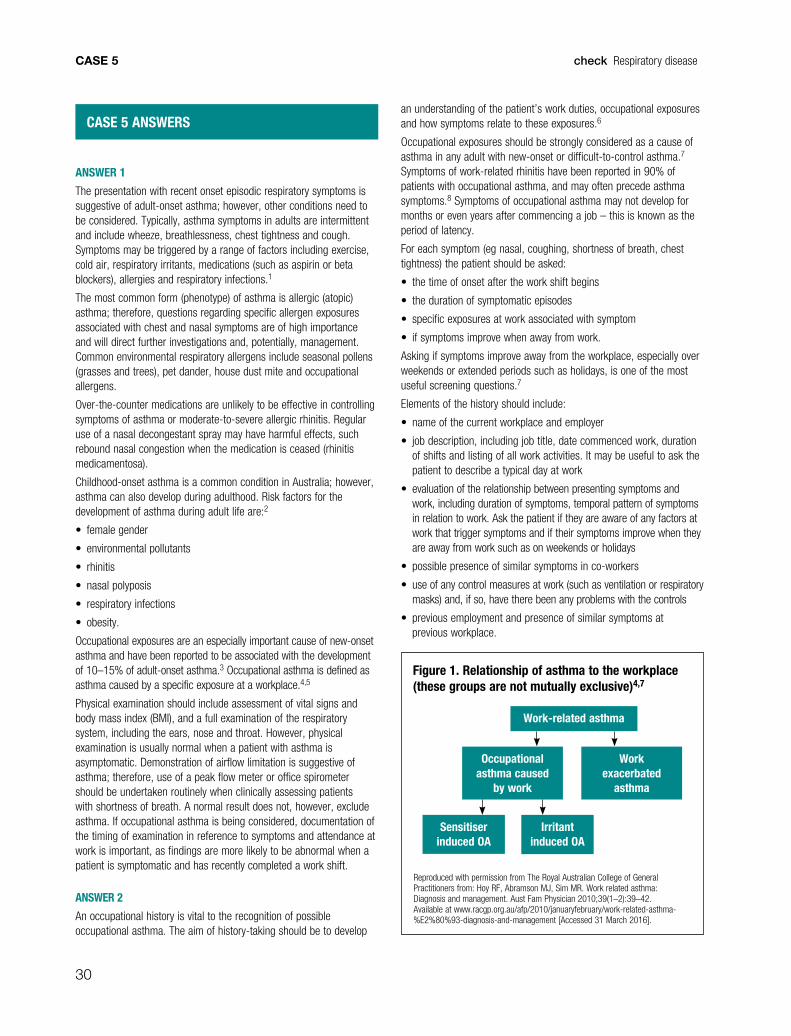
30
check Respiratory diseaseCASE 5
CASE 5 ANSWERS
ANSWER 1
The presentation with recent onset episodic respiratory symptoms is suggestive of adult-onset asthma; however, other conditions need to be considered. Typically, asthma symptoms in adults are intermittent and include wheeze, breathlessness, chest tightness and cough. Symptoms may be triggered by a range of factors including exercise, cold air, respiratory irritants, medications (such as aspirin or beta blockers), allergies and respiratory infections.1
The most common form (phenotype) of asthma is allergic (atopic) asthma; therefore, questions regarding specific allergen exposures associated with chest and nasal symptoms are of high importance and will direct further investigations and, potentially, management. Common environmental respiratory allergens include seasonal pollens (grasses and trees), pet dander, house dust mite and occupational allergens.
Over-the-counter medications are unlikely to be effective in controlling symptoms of asthma or moderate-to-severe allergic rhinitis. Regular use of a nasal decongestant spray may have harmful effects, such rebound nasal congestion when the medication is ceased (rhinitis medicamentosa).
Childhood-onset asthma is a common condition in Australia; however, asthma can also develop during adulthood. Risk factors for the development of asthma during adult life are:2
• female gender
• environmental pollutants
• rhinitis
• nasal polyposis
• respiratory infections
• obesity.
Occupational exposures are an especially important cause of new-onset asthma and have been reported to be associated with the development of 10–15% of adult-onset asthma.3 Occupational asthma is defined as asthma caused by a specific exposure at a workplace.4,5
Physical examination should include assessment of vital signs and body mass index (BMI), and a full examination of the respiratory system, including the ears, nose and throat. However, physical examination is usually normal when a patient with asthma is asymptomatic. Demonstration of airflow limitation is suggestive of asthma; therefore, use of a peak flow meter or office spirometer should be undertaken routinely when clinically assessing patients with shortness of breath. A normal result does not, however, exclude asthma. If occupational asthma is being considered, documentation of the timing of examination in reference to symptoms and attendance at work is important, as findings are more likely to be abnormal when a patient is symptomatic and has recently completed a work shift.
ANSWER 2
An occupational history is vital to the recognition of possible occupational asthma. The aim of history-taking should be to develop
an understanding of the patient’s work duties, occupational exposures and how symptoms relate to these exposures.6
Occupational exposures should be strongly considered as a cause of asthma in any adult with new-onset or difficult-to-control asthma.7 Symptoms of work-related rhinitis have been reported in 90% of patients with occupational asthma, and may often precede asthma symptoms.8 Symptoms of occupational asthma may not develop for months or even years after commencing a job – this is known as the period of latency.
For each symptom (eg nasal, coughing, shortness of breath, chest tightness) the patient should be asked:
• the time of onset after the work shift begins
• the duration of symptomatic episodes
• specific exposures at work associated with symptom
• if symptoms improve when away from work.
Asking if symptoms improve away from the workplace, especially over weekends or extended periods such as holidays, is one of the most useful screening questions.7
Elements of the history should include:
• name of the current workplace and employer
• job description, including job title, date commenced work, duration of shifts and listing of all work activities. It may be useful to ask the patient to describe a typical day at work
• evaluation of the relationship between presenting symptoms and work, including duration of symptoms, temporal pattern of symptoms in relation to work. Ask the patient if they are aware of any factors at work that trigger symptoms and if their symptoms improve when they are away from work such as on weekends or holidays
• possible presence of similar symptoms in co-workers
• use of any control measures at work (such as ventilation or respiratory masks) and, if so, have there been any problems with the controls
• previous employment and presence of similar symptoms at previous workplace.
Figure 1. Relationship of asthma to the workplace (these groups are not mutually exclusive)4,7
Work-related asthma
Occupational asthma caused
by work
Work exacerbated
asthma
Sensitiser induced OA
Irritant induced OA
Reproduced with permission from The Royal Australian College of General Practitioners from: Hoy RF, Abramson MJ, Sim MR. Work related asthma: Diagnosis and management. Aust Fam Physician 2010;39(1–2):39–42. Available at www.racgp.org.au/afp/2010/januaryfebruary/work-related-asthma-%E2%80%93-diagnosis-and-management [Accessed 31 March 2016].
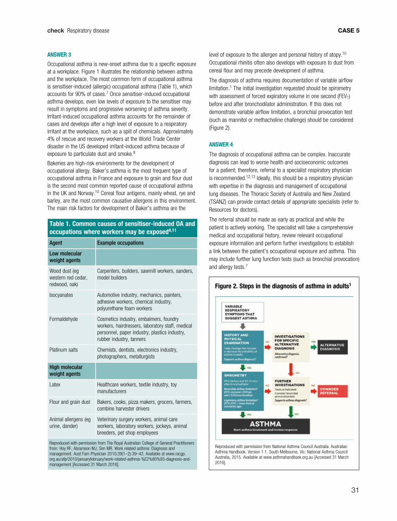
31
check Respiratory disease CASE 5
ANSWER 3
Occupational asthma is new-onset asthma due to a specific exposure at a workplace. Figure 1 illustrates the relationship between asthma and the workplace. The most common form of occupational asthma is sensitiser-induced (allergic) occupational asthma (Table 1), which accounts for 90% of cases.7 Once sensitiser-induced occupational asthma develops, even low levels of exposure to the sensitiser may result in symptoms and progressive worsening of asthma severity. Irritant-induced occupational asthma accounts for the remainder of cases and develops after a high level of exposure to a respiratory irritant at the workplace, such as a spill of chemicals. Approximately 4% of rescue and recovery workers at the World Trade Center disaster in the US developed irritant-induced asthma because of exposure to particulate dust and smoke.8
Bakeries are high-risk environments for the development of occupational allergy. Baker’s asthma is the most frequent type of occupational asthma in France and exposure to grain and flour dust is the second most common reported cause of occupational asthma in the UK and Norway.10 Cereal flour antigens, mainly wheat, rye and barley, are the most common causative allergens in this environment. The main risk factors for development of Baker’s asthma are the
level of exposure to the allergen and personal history of atopy.10
Occupational rhinitis often also develops with exposure to dust from cereal flour and may precede development of asthma.
The diagnosis of asthma requires documentation of variable airflow limitation.1 The initial investigation requested should be spirometry with assessment of forced expiratory volume in one second (FEV1) before and after bronchodilator administration. If this does not demonstrate variable airflow limitation, a bronchial provocation test (such as mannitol or methacholine challenge) should be considered (Figure 2).
ANSWER 4
The diagnosis of occupational asthma can be complex. Inaccurate diagnosis can lead to worse health and socioeconomic outcomes for a patient; therefore, referral to a specialist respiratory physician is recommended.12,13 Ideally, this should be a respiratory physician with expertise in the diagnosis and management of occupational lung diseases. The Thoracic Society of Australia and New Zealand (TSANZ) can provide contact details of appropriate specialists (refer to Resources for doctors).
The referral should be made as early as practical and while the patient is actively working. The specialist will take a comprehensive medical and occupational history, review relevant occupational exposure information and perform further investigations to establish a link between the patient’s occupational exposure and asthma. This may include further lung function tests (such as bronchial provocation) and allergy tests.7
Figure 2. Steps in the diagnosis of asthma in adults1
Reproduced with permission from National Asthma Council Australia. Australian Asthma Handbook. Version 1.1. South Melbourne, Vic: National Asthma Council Australia, 2015. Available at www.asthmahandbook.org.au [Accessed 31 March 2016].
Table 1. Common causes of sensitiser-induced OA and occupations where workers may be exposed4,11
Agent Example occupations
Low molecular weight agents
Wood dust (eg western red cedar, redwood, oak)
Carpenters, builders, sawmill workers, sanders, model builders
Isocyanates Automotive industry, mechanics, painters, adhesive workers, chemical industry, polyurethane foam workers
Formaldehyde Cosmetics industry, embalmers, foundry workers, hairdressers, laboratory staff, medical personnel, paper industry, plastics industry, rubber industry, tanners
Platinum salts Chemists, dentists, electronics industry, photographers, metallurgists
High molecular weight agents
Latex Healthcare workers, textile industry, toy manufacturers
Flour and grain dust Bakers, cooks, pizza makers, grocers, farmers, combine harvester drivers
Animal allergens (eg urine, dander)
Veterinary surgery workers, animal care workers, laboratory workers, jockeys, animal breeders, pet shop employees
Reproduced with permission from The Royal Australian College of General Practitioners from: Hoy RF, Abramson MJ, Sim MR. Work related asthma: Diagnosis and management. Aust Fam Physician 2010;39(1–2):39–42. Available at www.racgp.org.au/afp/2010/januaryfebruary/work-related-asthma-%E2%80%93-diagnosis-and-management [Accessed 31 March 2016].
32
check Respiratory diseaseCASE 5
ANSWER 5
Pharmacological management of occupational asthma uses a stepped approach to medication and should follow guidelines such as those provided by the National Asthma Council Australia.1 However, there is insufficient evidence to determine whether pharmacological treatment can alter the course of asthma in patients who remain exposed to allergens. Complete avoidance of exposure to the causative allergen is the approach that will most likely lead to a recovery from occupational asthma.14 If this requires removal of the worker from their workplace it is likely to be associated with significant negative socioeconomic effects for the worker. The decision to recommend leaving a workplace requires careful understanding of several factors, including the severity of asthma, prognosis if exposure continues, and possible benefits of control measures to minimise exposure as far as is practicable. Unless asthma is severe, the decision to recommend a worker leave their workplace should only be made following specialist assessment of the patient.
ANSWER 6
Mark’s employer can only be contacted if Mark expressly provides permission to allow you to discuss the situation. Discussion with an employer or the Occupational Health and Safety (OH&S) representative can be very useful in the management of occupational asthma. The aims of this discussion include:
• improved understanding of the exposures present at the workplace
• exploration of alternative work duties to avoid or minimise exposure to the causative allergen for the worker
• discussing a plan to closely monitor the progress of the worker
• discussing the condition of current control measures such as the adequacy of ventilation and personal protective equipment as a means of primary prevention and protection of all workers
• consideration of screening other workers as early as possible for possible occupational asthma. One identified case of occupational asthma at a workplace is a sentinel event, which should lead to attempts to identify other workers who may be affected.
Occupational asthma is a preventable form of asthma and occupational disease. Ideally, workplaces should have measures to prevent workers from inhaling agents that can cause asthma. In the setting of Baker’s asthma, flour cannot be removed for the work process, so minimisation of dust levels as far as practicable is key. Elements of a control program include:
• local ventilation concentrated at flour release points such as weighing stations and dough-making machines
• optimal work practices to avoid flour becoming airborne, such as careful bag emptying and empty bag handling
• worker education through training programs15
• appropriately selected and well maintained personal protective equipment (PPE) such as respirators; however, this is less effective than controlling exposure at the source
• use of lids for mixing tubs and divider oils rather than sprinkling workbenches with flour to prevent dough sticking – this has been shown to significantly reduce levels of airborne allergens.16
RESOURCES FOR PATIENTS• Better Health Channel’s Asthma and your workplace, www.betterhealth.vic.
gov.au/health/conditionsandtreatments/asthma-and-your-workplace
RESOUCES FOR DOCTORS• Occupationalasthma.com
• The Thoracic Society of Australia and New Zealand (TSANZ), www.thoracic.org.au
• National Asthma Council Australia, www.nationalasthma.org.au
REFERENCES1. National Asthma Council Australia. Australian Asthma Handbook. Version
1.1. South Melbourne, Vic: National Asthma Council Australia, 2015. Available at www.asthmahandbook.org.au [Accessed 31 March 2016].
2. de Nijs SB, Venekamp LN, Bel EH. Adult-onset asthma: Is it really different? Eur Respir Rev 2013;22(127):44–52.
3. Toren K, Blanc PD. Asthma caused by occupational exposures is common – A systematic analysis of estimates of the population-attributable fraction. BMC Pulm Med 2009;9:7.
4. Hoy RF, Abramson MJ, Sim MR. Work related asthma – Diagnosis and management. Aust Fam Physician 2010;39(1–2):39–42.
5. Tarlo SM, Lemiere C. Occupational asthma. N Engl J Med 2014;370(7):640–49.
6. Tan J, Bernstein JA. Occupational asthma: An overview. Curr Allergy Asthma Rep 2014;14(5):431.
7. Tarlo SM, Balmes J, Balkissoon R, et al. Diagnosis and management of work-related asthma: American College Of Chest Physicians Consensus Statement. Chest 2008;134(3 Suppl):1S–41S.
8. Malo JL, Lemiere C, Desjardins A, et al. Prevalence and intensity of rhinoconjunctivitis in subjects with occupational asthma. Eur Respir J 1997;10(7):1513–15.
9. Wheeler K, McKelvey W, Thorpe L, et al. Asthma diagnosed after 11 September 2001 among rescue and recovery workers: Findings from the World Trade Center Health Registry. Environ Health Perspect 2007;115(11):1584–90.
10. Quirce S, Diaz-Perales A. Diagnosis and management of grain-induced asthma. Allergy Asthma Immunol Res 2013;5(6):348–56.
11. Elder D, Abramson M, Fish D, et al. Surveillance of Australian workplace Based Respiratory Events (SABRE): Notifications for the first 3.5 years and validation of occupational asthma cases. Occup Med (Lond) 2004;54(6):395–99.
12. Anees W, Moore VC, Burge PS. FEV1 decline in occupational asthma. Thorax 2006;61(9):751–55.
13. Vandenplas O. Socioeconomic impact of work-related asthma. Expert Rev Pharmacoecon Outcomes Res 2008;8(4):395–400.
14. Vandenplas O, Dressel H, Nowak D, et al. What is the optimal management option for occupational asthma? Eur Respir Rev 2012;21(124):97–104.
15. Brisman J. Baker’s asthma. Occup Environ Med 2002;59(7):498–502; quiz, 426.
16. Baatjies R, Meijster T, Heederik D, et al. Effectiveness of interventions to reduce flour dust exposures in supermarket bakeries in South Africa. Occup Environ Med 2014;71(12):811–18.
MULTIPLE CHOICE QUESTIONS
MULTIPLE CHOICE QUESTIONS
33
check Respiratory disease
CASE 1 – CAMERON
Cameron is 38 years of age and presents with a two-year history of cough and production of a dark yellow sputum. He sometimes has nasal congestion, shortness of breath and wheezing. Cameron has been a smoker since the age of 20 years, but quit recently and has not had a cigarette for the past six months. In assessing Cameron’s symptoms, you take a thorough history and consider possible diagnoses of asthma, including occupational asthma, bronchiectasis and chronic obstructive pulmonary disease (COPD).
QUESTION 1
Which of the following is the most specific diagnostic feature of bronchiectasis?
A. Internal calibre of the bronchus being wider that the adjacent pulmonary artery
B. Bronchial wall being wider than the bronchial lumen
C. Variable expiratory airflow limitation
D. The presence of Haemophilus influenzae in the sputum
QUESTION 2
What initial treatment would be recommended for Cameron if a diagnosis of bronchiectasis is confirmed?
A. Inhaled corticosteroids
B. Inhaled bronchodilators
C. Oral antibiotics
D. Intramuscular antibiotics
FURTHER INFORMATION
Cameron has a spirometry test, which shows that pre-bronchodilator forced expiratory volume in one second (FEV1) is 3 L. The predicted value is 4.7 L
QUESTION 3
Which of the following values for forced vital capacity (FVC) is consistent with a diagnosis of asthma for Cameron?
A. 3.85 L
B. 3.74 L
C. 3.66 L
D. 3.75 L
QUESTION 4
To meet the criteria for a clinically significant bronchodilator response, Cameron’s post-bronchodilator FEV1 would need to be at least:
A. 3.15 L
B. 3.28 L
C. 3.36 L
D. 3.40 L
QUESTION 5
Which of the following results are consistent with a diagnosis of moderate COPD?
A. FEV1/FVC <0.8; FEV1 65% of predicted
B. FEV1/FVC <0.7; FEV1 55% of predicted
C. FEV1/FVC <0.7; FEV1 65% of predicted
D. FEV1/FVC <0.8; FEV1 55% of predicted
QUESTION 6
What initial treatment would you prescribe for Cameron if he is diagnosed with COPD?
A. Inhaled corticosteroids (ICS)
B. Short-acting bronchodilator
C. Long-acting beta2-agonist (LABA)
D. Combined ICS and LABA
ACTIVITY ID: 50087
RESPIRATORY DISEASE
This unit of check is approved for six Category 2 points in the RACGP QI&CPD program. The expected time to complete this activity is three hours and consists of:
• reading and completing the questions for each case study
– you can do this on hard copy or by logging onto gplearning, http://gplearning.racgp.org.au
• answering the following multiple choice questions (MCQs) by logging onto gplearning, http://gplearning.racgp.org.au
– you must score ≥80% before you can mark the activity as ‘Complete’
• completing the online evaluation form.
You can only qualify for QI&CPD points by completing the MCQs online; we cannot process hard copy answers.
If you have any technical issues accessing this activity online, please contact the gplearning helpdesk on 1800 284 789.
If you are not an RACGP member and would like to access the check program, please contact the gplearning helpdesk on 1800 284 789 to purchase access to the program.
34
check Respiratory diseaseMULTIPLE CHOICE QUESTIONS
FURTHER INFORMATION
During history taking, Cameron had told you that he works in his family’s furniture business. He began there as an accountant, but in the past five years has been involved in manufacturing furniture, which he prefers. He admits that he is exposed to wood dust and paints but did not have any respiratory problems until two years ago.
QUESTION 7
Which of the following questions is most useful in considering occupational asthma as a cause of Cameron’s symptoms?
A. Do his co-workers have similar symptoms?
B. What control measures are used in his workplace minimise exposure to possible allergens and irritants?
C. Does he use protective equipment?
D. Are his symptoms better when away from work, such as on weekends or during holidays?
QUESTION 8
Which of the following statements regarding occupational asthma is true?
A. Sensitiser-induced occupational asthma accounts for 10% of cases.
B. Exposure to very low levels of a sensitiser are unlikely to cause symptoms.
C. Irritant-induced occupational asthma develops after a high level of exposure to a respiratory irritant in the workplace.
D. Irritant-induced occupational asthma is the most common form of occupational asthma.
CASE 2 – JESSICA
Jessica, 40 years of age, presents with fatigue and difficulty concentrating at work. She says she has about seven or eight hours of sleep each night, but wakes feeling unrefreshed and is always tired. On physical examination you find that Jessica has a Mallampati score of 2, neck circumference of 30 cm, waist circumference of 86 cm, body mass index of 25 kg/m2. She scores 15 on an Epworth Sleepiness Score.
QUESTION 9
Which of the following warrants consideration of obstructive sleep apnoea as a possible diagnosis?
A. Epworth sleepiness score of 15
B. A Mallampati score of 2
C. Neck circumference of 30 cm
D. Waist circumference of 85 cm
FURTHER INFORMATION
The sleep physician initiates a trial of automatically titrating continuous positive airway pressure (CPAP) for Jessica.
QUESTION 10
What is the advantage of automatically titrating CPAP?
A. There is a standard mask so the patient does not have to try different sizes.
B. Treatment can commence immediately.
C. It is suitable for patients with multiple comorbidities.
D. Acclimatisation is more rapid than with standard CPAP.

Independent learning program for GPs
Independent learning program for GPs