Embed Size (px)
Citation preview
This article is protected by copyright. All rights reserved.
1
Increased L-dopa-derived dopamine following selective MAO-A or -B inhibition in rat
striatum depleted of dopaminergic and serotonergic innervation.1
O Sader-Mazbar1, Y Loboda1, M J Rabey2 and J P M Finberg1*
1Dept of Molecular Pharmacology, Rappaport Faculty of Medicine,
Technion, Haifa, Israel; 2Department of Neurology, Assaf Harofe Medical Center, Zerifin,
affiliated to Sackler School of Medicine, Tel Aviv University, Israel
Running title: Striatal dopamine levels following L-dopa
*Author for correspondence:
Prof John P.M. Finberg,
Dept of Molecular Pharmacology,
Rappaport Family Faculty of Medicine,
Technion,
Haifa, 31096
Israel
Tel: +972-4-8295272
Fax: +972-4-8295271
Email: [email protected]
This article has been accepted for publication and undergone full peer review but has not been through the copyediting, typesetting, pagination and proofreading process, which may lead to differences between this version and the Version of Record. Please cite this article as doi: 10.1111/bph.12349
Acc
epte
d A
rticl
e
This article is protected by copyright. All rights reserved.
2
Summary
Background and purpose:
Selective MAO-B inhibitors are effective in potentiation of the clinical effect of L-dopa in
Parkinson’s disease, but dopamine is deaminated mainly by MAO-A in rat brain. We sought
to clarify the roles of MAO-A and MAO-B in deamination of dopamine formed from
exogenous L-dopa in rat striatum depleted of dopaminergic, or both dopaminergic and
serotonergic innervations. We also studied the effect of organic cation transporter-3 (OCT-3)
inhibition by decinium-22 on extracellular dopamine levels following L-dopa.
Experimental approach:
Striatal dopaminergic and/or serotonergic neuronal innervations were lesioned by 6-
hydroxydopamine or 5,7-dihydroxytryptamine respectively. Microdialysate dopamine levels
after systemic L-dopa were measured after inhibition of MAO-A or MAO-B by clorgyline or
rasagiline respectively. MAO subtype localization in the striatum was determined by
immunofluorescence.
Key results:
Rasagiline increased dopamine extracellular levels following L-dopa to a greater extent in
double- than in single-lesioned rats (2.8 and 1.8 fold increase respectively relative to saline
treatment), however clorgyline elevated DA levels in both models over 10 fold. MAO-A was
strongly expressed in medium spiny neurons (MSNs) in intact and lesioned striata, while
MAO-B was localized in glia and to a small extent in MSNs. Inhibition of OCT-3 increased
dopamine levels in the double- more than the single-lesion animals.
Conclusions and Implications:
In striatum devoid of dopaminergic and serotonergic inputs, most deamination of L-dopa-
derived dopamine is mediated by MAO-A in MSN and a smaller amount by MAO-B in both
MSN and glia. OCT-3 plays a significant role in uptake of DA from extracellular space. Acc
epte
d A
rticl
e

This article is protected by copyright. All rights reserved.
3
Inhibitors of OCT-3 are potential future targets for anti-Parkinsonian treatments.
Acc
epte
d A
rticl
e
This article is protected by copyright. All rights reserved.
4
Keywords
Parkinson’s disease; L-dopa; rasagiline; clorgyline; 6-hydroxydopamine; glial cells;
microdialysis; OCT-3; medium spiny neurons
Abbreviations used: 6-OHDA, 6-hydroxydopamine; 5,7-DHT, 5,7-dihydroxytryptamine; L-
dopa, L-3,4-dihydroxyphenylalanine; Pd, Parkinson's disease ; DA, dopamine; AAAD, L-
aromatic amino acid decarboxylase; DAT, dopamine transporter; 5-HT, 5-
hydroxytryptamine; SNpc, substantia nigra, pars compacta; DOPAC, 3,4-
dihydroxyphenylacetic acid; HVA, homovanillic acid; 5-HIAA, 5-hydroxyindoleacetic acid;
COMT, catecholamine-O-methyltransferase; PEA, phenylethylamine; OCT-3, organic cation
transporter-3; MSN, medium spiny neuron; MAO, monoamine oxidase; SERT, serotonin
transporter; NET, norepinephrine transporter
Acc
epte
d A
rticl
e
This article is protected by copyright. All rights reserved.
5
1. Introduction:
L-dopa treatment of Parkinson's disease (Pd) is commonly referred to as dopamine (DA)
replacement therapy, however the precise way in which L-dopa replaces DA lost from the
Parkinsonian brain is far from clear. Since L-dopa is decarboxylated to DA by aromatic L-
amino acid decarboxylase (AAAD; EC 4.1.1.28), DA can be produced from L-dopa not only
in dopaminergic neurons but in all cells which possess this enzyme, such as certain
populations of glial cells, blood vessels, medium spiny neurons (MSN), small aspiny
interneurons and serotonergic neurons (Lopez-Real et al., 2003; Mura et al., 1995; Tashiro et
al., 1989; Vialou et al., 2007). Following dopaminergic deafferentation with 6-
hydroxydopamine (6-OHDA) serotonergic axon density increases in striatum (Maeda et al.,
2003), and DA has been detected in serotonergic axonal varicosities following L-dopa
administration in 6-OHDA-treated rats (Yamada et al., 2007). DA production from L-dopa in
6-OHDA-treated rats is markedly reduced following serotonergic deafferentation with 5,7-
dihydroxytryptamine (5,7-DHT; Navailles et al., 2010; Tanaka et al., 1999). These
observations are strongly indicative of a role of serotonergic neurons in production of DA
from L-dopa in parkinsonian striatum.
The level of DA in extracellular fluid, which is proportional to the synaptic level, is
controlled by neuronal and extraneuronal uptake and metabolism by the intracellular enzymes
monoamine oxidase (MAO) and catechol O-methyltransferase (COMT). Following
dopaminergic deafferentation of the striatum in rats, levels of DA in striatal extra-cellular
fluid after systemic L-dopa administration (as detected by microdialysis) increase to values
higher than those seen in intact brain (Miller et al., 1999), reflecting the efficient removal of
DA from the synapse by the dopamine transporter (DAT) in normal brain. In parkinsonian
brain, in which DAT is absent, DA formed from L-dopa can be taken up by serotonergic Acc
epte
d A
rticl
e
This article is protected by copyright. All rights reserved.
6
neurons, since these neurons express the serotonergic transporter (SERT), which is also
capable of transporting DA across the axolemma (Carta et al., 2007). Although serotonergic
perikarya of the raphe nucleus express MAO-B, serotonergic axon terminals are thought to
express MAO-A, since serotonin behaves as a MAO-A substrate in vivo (Green et al., 1975).
Dopamine can also be taken up and deaminated in astrocytes, which express monoamine
oxidase type B (MAO-B; Ekblom et al., 1993), the plasma membrane noradrenaline
transporter (NET, Inazu et al., 2003), and the organic cation transporter -3 (OCT-3; Cui et al.,
2009). The role of the low-affinity transporter OCT-3 in clearance of DA from the
extracellular space has been little studied, however this transporter could become more
important in the clearance of L-dopa-derived DA in Parkinsonian striatum.
In the intact rat brain, both subtypes A and B of MAO are expressed in similar proportions
(Youdim et al., 1983) but MAO-A is the dominant form responsible for both DA and 5-HT
metabolism, as seen by a much larger effect of MAO-A than MAO-B inhibition in
suppressing formation of striatal 3,4-dihydroxyphenylacetic acid (DOPAC), homovanillic
acid (HVA) and 5-hydroxyindoleacetic acid (5-HIAA), and increasing DA microdialysate
levels (Finberg et al., 1995; Green et al., 1975; Wachtel et al., 1994; Waldmeier et al., 1976).
In early stage Pd patients, administration of the selective MAO-B inhibitors selegiline and
rasagiline in monotherapy produces a beneficial dopaminergic effect, and in advanced stage
patients, MAO-B inhibition reduces fluctuations in response to L-dopa (Parkinson Study
Group, 2005). Inhibitors of MAO-A cannot be administered to PD patients treated with L-
dopa, because of the possibility of serious cardiovascular reactions.
The object of this study was to clarify the effect of MAO-A and MAO-B inhibition on L-
dopa-derived extracellular DA levels in striatum devoid of dopaminergic innervation, and
both dopaminergic and serotonergic innervations.. Because the extent of DA metabolism by Acc
epte
d A
rticl
e
This article is protected by copyright. All rights reserved.
7
MAO-A or –B depends on the localization of the MAO subtypes to neuronal and glial
elements, we have further clarified the location of MAO subtypes in striatum by
immunofluorescence, as well as their enzyme activities in striatal tissue following lesioning.
In addition, we have probed the role of low-affinity uptake by OCT-3 in clearance of DA
from the extracellular space in both lesion models.
2. Methods
2.1 Experimental Animals
All procedures with animals were authorized by the Technion Animal Care and Use
Committee, whose ethical standards are based on those detailed in the National Institutes of
Health (Bethesda, MD, USA) Guide for the Care and Use of Laboratory Animals, and whose
general procedures for animal welfare comply with Israeli law on animal experimentation.
The study procedures and results are reported in compliance with the ARRIVE guidelines
(Kilkenny et al., 2010; McGrath et al., 2010). All experiments were carried out on male
Sprague-Dawley rats, which were obtained from Harlan Laboratories, Jerusalem, at 7 weeks
of age, and were maintained in our animal facility for an additional 2 weeks before use.
Animals were fed rat pellets and water ad libitum, and were caged in plastic Individually
Ventilated Cages (IVC) in a non-sterile room of our animal department, under 12 hours
light/dark regimen at an environmemtal temperature of 21±1 oC.
2.2 Materials
5-[2-14C]- hydroxytryptamine binoxalate (14C-5HT) and β-[ethyl-1-14C]- phenylethylamine
hydrochloride (14C-PEA) were purchased from Perkin Elmer (MA, USA), alkaline
phosphatase conjugated anti-rabbit antibody and p-nitrophenyl phosphate from Chemicon
(Millipore, USA). Rasagiline and carbidopa were obtained from Teva pharmaceuticals Acc
epte
d A
rticl
e
This article is protected by copyright. All rights reserved.
8
(Israel). Isoflurane was purchased from Nicholas Piramal (UK). Mouse anti-DARPP-32 (sc-
271111), rabbit anti-MAO-A (H-70) and goat anti-MAO-B (D-16) antibodies were obtained
from Santa Cruz (CA, USA). Rabbit anti-DARPP-32 (19A3) antibody was purchased from
Cell Signaling Technology (MA, USA). Rabbit anti-OCT3 (OCT31-A) antibody was
obtained from Alpha Diagnostics (TX, USA). Monoclonal anti-tyrosine hydroxylase and
monoclonal anti-tryptophan hydrxylase antibodies were obtained from Sigma (Israel). Alexa
fluor conjugated antibodies were purchased from Invitrogen (NY, USA). All other
compounds and antibodies were obtained from Sigma (Israel). Microdialysis guide cannulas
and probes were purchased from CMA Microdialysis (Sweden).
2.3 Lesioning procedure:
Rats were administered desipramine 10 mg.kg-1 s.c., anesthetized with ketamine/xylazine
(70/7 mg.kg-1 i.p.) and placed in a stereotaxic frame (Kopf, CA, USA). A thermostatically-
controlled heating pad maintained body temperature constant at 37º C and blood gases were
monitored. For single (dopaminergic only) lesion, 6-OHDA (8 μg in saline with 0.1%
ascorbic acid) was stereotactically injected into the left medial forebrain bundle (coordinates
4.4 mm posterior, 1.5 mm left, 8 mm ventral with respect to bregma). For additional
serotonergic lesioning (double lesion), rats were injected with 5,7-DHT (150 μg free base in
saline with 0.1% ascorbic acid) into the left lateral ventricle (coordinates: 0.8 mm posterior,
1.4 mm left, 3.4 mm ventral with respect to bregma) during the same operation. Sham-
lesioned rats were subjected to the same procedures using 0.9 % saline (with 0.1% ascorbic
acid) for injections. The extent of dopaminergic lesion was indicated by injection of
apomorphine (0.1 mg.kg-1 s.c.), 3 weeks after lesioning. Only rats with intensive contralateral
rotation (more than 100 complete contralateral turns in the 60 min following injection, and
more than 20 contralateral turns between 15 and 20 min following the injection) induced by Acc
epte
d A
rticl
e
This article is protected by copyright. All rights reserved.
9
apomorphine were used for the present experiments. These criteria have been found in our
laboratory to select rats with more than 99% dopaminergic lesion, which was additionally
verified by: a) determination of striatal tissue DA levels at end of microdialysis experiments,
and b) immunohistological examination of tyrosine-hydroxylase-positive neurons in
substantia nigra in an additional group of 11 double-lesioned and 7 sham-operated rats. The
extent of serotonergic lesion of striatal tissue was measured: a) by determination of striatal
tissue levels of 5-HIAA at the end of microdialysis experiments, and b) immunohistological
examination of tryptophan hydroxylase-positive neurons in dorsal raphe nucleus in the same
additional group of double-lesioned rats.
2.4. Determination of MAO activity
The effect of 6-OHDA and 5,7-DHT lesions on striatal tissue MAO activity was determined
in a separate group of rats not administered MAO inhibitors, but which fulfilled the criteria
for extensive 6-OHDA lesioning as described below for apomorphine-induced turning, and
were killed by decapitation under isoflurane anesthesia one week after apomorphine
administration. MAO activity in rat striatum was determined in vitro using a radiometric
method (Otsuka et al., 1964), with modifications (O'Carroll et al., 1983). Briefly, four weeks
after the induction of the lesions, and one week after apomorphine screening test, rats were
decapitated under ketalar/xylazine anesthesia, striata were dissected and homogenized in
sucrose (0.32 M). For MAO-B activity determination, homogenates were pre-incubated with
clorgyline (0.15 µM) for 60 min then with 14C-PEA for 20 min at 37º C. For MAO-A activity
determination, homogenates were pre-incubated with selegiline (0.15 µM) for 60 min, then
were incubated with 14C-5-HT for 30 min at 37º C. The deaminated products were extracted
into toluene/ethyl acetate (1:1, vol/vol), to which was added a solution of 2,5-diphenyl
oxazole to a final concentration of 0.4% w.v-1 before determination by liquid scintillation Acc
epte
d A
rticl
e
This article is protected by copyright. All rights reserved.
10
counting (LKB-Wallac Rakbeta 1211). MAO activity is expressed as the amount of
radioactive deaminated product per µg protein in a 20 or 30 min incubation period as
appropriate.
2.5. Gliosis quantitation
Because an increase in MAO-B activity was found in some of the lesioned rats, and because
MAO-B is the isoform associated with glial cells, we quantitated the degree of gliosis
induced by single- and double-lesion procedures by determination of the glial marker protein
GFAP in striatal tissue homogenates, and by GFAP-positive cell counting in striatal tissue
sections.
2.5.1 GFAP quantitation
This determination was carried out on the striatal sucrose homogenate obtained in section 2.4.
Aliquots of the sucrose homogenate were diluted in lysis buffer (15 mM Tris, 250 mM
sucrose, 1 mM EDTA, 0.05% NP-40), sonicated and then centrifuged for 10 min at 4º C,
1,000 g. The supernatant was assayed with sandwich ELISA. Briefly, a 96 well plate was
coated with mouse anti-GFAP (dilution 1:500) overnight at 4ºC, homogenates (300 ng
protein each well) were added and incubated for 1 h at room temperature. Non-specific
binding was blocked with 1% bovine serum albumin followed by incubation for 1 h with
rabbit anti-GFAP (dilution 1:500). Finally, alkaline phosphatase conjugated anti-rabbit
antibody and its substrate p-nitrophenyl phosphate were used to detect bound primary
antibody. A linear standard curve was obtained correlating the optical density of the product
with the amount of protein in the homogenate.
2.5.2 GFAP positive cell counts
Additional groups of lesioned rats were prepared for this determination: sham lesion (n=4),
single lesion (n=6) and double lesion (n=8). Four weeks after the induction of the lesions and Acc
epte
d A
rticl
e
This article is protected by copyright. All rights reserved.
11
1 week after the apomorphine screening test, rats were anesthetized with ketamine/xylazine
and perfused via the left cardiac ventricle with phosphate-buffered saline (PBS, pH=7.4, 10
mL) followed by 4% paraformaldehyde in PBS. Brains were removed and post-fixed for 4
days. After dehydration in alcohols (70%, 80%, 96% ethanol, then 100% isopropanol) and
clearing in organic solvent (chloroform) brains were embedded in paraffin. Coronal sections
(5µm) of striatum from levels 1.6, 1.2, 0.8, 0.5 and 0.2 mm anterior to bregma were
deparaffinized in xylene, rehydrated in decreasing concentrations of ethanol and submitted
to antigen retrieving procedure by microwaving in phosphate-citrate buffer pH=6.8 (0.017 M
citric acid and 0.066 M dibasic sodium phosphate in double-distilled water). After
permeabilization in 0.5%Triton X100 in PBS and quenching of endogenous peroxidase in
hydrogen peroxide (0.3% in absolute ethanol), sections were blocked in PBS containing 5%
bovine serum albumen and 5% fetal bovine serum with 0.1% Triton X100 and incubated
overnight in mouse anti-GFAP antibody 1:500 (Sigma). For antibody visualization sections
were treated with biotinylated secondary antibody to mouse, followed by horse radish
peroxidase-streptavidin complex, then exposed to 3-amino 9-ethylcarbazole (AEC)
chromogen reagent and counterstained with hematoxylin (Histostain-SP Kit, Zymed
Laboratories Inc., Invitrogen). For GFAP-positive cell counting, 25 pictures were taken from
left striatum at each level from bregma as above. Only GFAP-positive glial cells sectioned
through the central part of the nucleus were counted (125 pictures for every rat) and data
were statistically analysed by one-way ANOVA followed by Bonferoni multiple comparison
test.
2.6. Protein determination
Protein content was obtained using the Lowry assay (Lowry et al., 1951).
Acc
epte
d A
rticl
e
This article is protected by copyright. All rights reserved.
12
2.7. Microdialysis experiments
One day before the microdialysis study, rats were anesthetized with ketamine/xylazine (70:7
mg/kg i.p.) and a silicon guide cannula (CMA/12) was implanted in the skull with its end
directed to the upper limit of the left striatum (0.2 mm anterior, 2.5 mm left, 3.5 mm ventral
to bregma, according to a rat brain stereotaxic atlas (Paxinos et al., 1982). Rats were left to
recover for 24 h. On the next day, rats were anesthetized using isoflurane inhalation and
maintained on isoflurane anesthesia administered via a nose cone while the rat's body
temperature was maintained at 37ºC with a thermostatically controlled heating pad. A dialysis
probe (CMA12, 4 mm membrane length) was inserted into the guide cannula and perfused
continuously with artificial CSF (composition in mM: NaCl 14.7, KCl 0.27, CaCl2 0.12,
MgCl2 0.085) at a flow rate of 2 μL.min-1. After a 1 h equilibration period, carbidopa (6
mg.kg-1) was given i.p., and then baseline dialysate collection was commenced each 20 min
for 1 h in tubes containing 10 μL of a preservative solution containing 0.1 M perchloric acid
and 0.05mM disodium ethylenediamine tetra-acetic acid. L-dopa (25 mg.kg-1) was then given
i.p., and sample collection was continued for 5 h. At the end of the experiment, rats were
sacrificed and striata were dissected for MAO activity measurement as well as DA and 5-
HIAA content determination, and the position of the probe was determined by direct
observation of the probe track.
2.7.1 Microdialysis following MAO inhibitor treatment
Rats bearing either single or double lesions were randomly assigned for daily MAO inhibitor
treatment 2 weeks after the apomorphine test. Saline, rasagiline 0.05 mg.kg-1 or clorgyline 0.2
mg.kg-1 were administered s.c. daily for 14 days. On day 14 the microdialysis experiment was
carried out as described above, and on this day MAO inhibitors were given 2 h before L-dopa
injection. Acc
epte
d A
rticl
e
This article is protected by copyright. All rights reserved.
13
In a separate experiment designed to test the effect of partial MAO-A inhibition, rats bearing
the double lesion were treated daily with either clorgyline 0.0033 mg.kg-1 s.c. or saline for 14
days. On day 14 the microdialysis experiment was carried out as described above, and on this
day clorgyline was given 2 h before L-dopa injection. The doses of carbidopa and L-dopa
were as above.
2.7.2 Microdialysis following inhibition of the organic cation transporter (OCT-3)
This microdialysis experiment in single- and double-lesion rats was carried out as described
above 7 days after screening with apomorphine, but microdialysis tubings were filled with
either artificial CSF or 50 µM decynium-22 (OCT-3 inhibitor). Carbidopa and L-dopa were
administered as above.
2.7.3. Analysis of microdialysate
Dialysate samples were analyzed for DA, DOPAC (dihydroxyphenylacetic acid) and HVA
(homovanilic acid) by HPLC with electrochemical detection. Separation of DA and its
metabolites was achieved using an Inertsil ODS-2 column (GL Sciences, Japan) with a
mobile phase composed of 100 mM sodium dihydrogen phosphate, 1 mM octanesulfonic acid
and 269 μM disodium EDTA, with 2.5% v/v methanol and 4.5% v/v acetonitrile, in HPLC
grade deionized water, pH 2.75. Detection of compounds was enabled by an ESA Coulochem
II (model 5200, ESA, U.S.A.) electrochemical unit operated in redox mode and coupled to an
ESA guard cell at a potential of +300 mV placed before the analytical cell (ESA model
5010). Column eluates were reduced to +100 mV at detector 1 of the analytical cell, and
measured at -400 mV at detector 2. The limit of detection for DA, DOPAC and HVA was
0.01 pmol.
Acc
epte
d A
rticl
e
This article is protected by copyright. All rights reserved.
14
2.8. Immunofluorescence determination of MAO-A and MAO-B localization in brain sections
Brain sections (5 µm) were prepared as described in section 2.5.2 and permeabilized with
0.5% Triton in PBS for 10 min at room temperature. Afterwards, a blocking solution (for
MAO-A staining, 5% goat serum and 1% bovine serum albumen, for MAO-B only 5%
bovine serum albumen in PBS) was added, and sections were incubated for 2 h at room
temperature. Incubation with primary antibodies [rabbit anti-MAO A 1:100, goat anti-MAO
B 1:150, anti-GFAP 1:1000, rabbit anti-OCT-3 1:100, mouse anti-tyrosine hydroxylase
1:1000, mouse anti-DARPP-32 (dopamine- and cAMP-regulated phosphoprotein, Mr 32
kDa)1:500, rabbit anti-DARPP-32 1:500] was carried out overnight at 4oC and incubation
with secondary fluorescent antibodies (1:1000) for 1 h at room temperature. Sections were
observed with a laser confocal microscope (LSM 510 Meta).
2.9. Immunohistochemical determination of tyrosine hydroxylase- and tryptophan
hydroxylase-positive neurons
This experiment was carried out to verify the degrees of dopaminergic and serotonergic
lesioning produced by the above procedures. An additional group of 11 double-lesioned and 7
control rats were prepared for in vivo fixation as described in section 2.5.2. Coronal 25 µm
sections were cut at topographically-identified levels (for substantia nigra from -4.8 to -6.0
and for raphe nucleus from -7.0 to -9.2 mm from bregma according to stereotactic atlas of
Paxinos and Watson, 1982). For calculation of raphe nucleus cells, sections were taken every
200 µm between the above bregma levels and only cells with visible nucleus were counted.
Immunohistochemical detection of tyrosine hydroxylase and tryptophan hydroxylase was
carried out as described in section 2.5.2. Secondary antibodies were visualized using AEC or
3-3'-diaminobenzidine tetrahydrochloride (DAB).
Acc
epte
d A
rticl
e
This article is protected by copyright. All rights reserved.
15
2.10. Statistical analysis
All data are presented as mean ± SEM or mean + SEM for graphical data. One way ANOVA
was applied on the results of MAO activity and GFAP expression followed by Bonferroni
multiple comparison test. Statistical analysis of microdialysis data was done using two-way
ANOVA followed by Bonferroni multiple comparisons test post hoc for difference between
treatments at each time point. For comparison of maximum peak levels unpaired t-test was
used. All statistical analyses were performed on original data, not on percentages.
3. Results
3.1. Striatal MAO activity, amine depletion and GFAP expression, following single and
double lesions
Initially we determined the alteration in MAO activity in striatal tissue in rats given single
and double lesions. The extent of lesioning of dopaminergic and serotonergic striatal inputs
was determined as described in Methods. In rats given a 6-OHDA lesion, and which showed
the stated rotation criteria, striatal DA tissue levels were reduced by 99.6 ± 0.087 % control
(n = 7 single- and 7 double-lesioned striata, mean ± SEM). The 5,7-dihydroxytryptamine
lesion produced 92.4±1.05 % (mean ± SEM, n=54) reduction in striatal 5-HIAA content
compared to intact striata indicating denervation of most 5-HT terminals innervating the
striatum (Hall et al., 1999; Tanaka et al., 1999). The nearly complete absence of tyrosine
hydroxylase-positive neurons in SNpc in an identically-prepared group of double-lesioned
rats was confirmed by immunohistochemistry (Fig.1), and cresyl violet staining confirmed
the absence of cells with the typical morphology of dopaminergic neurons in lesioned SNpc.
Numbers of tryptophan hydroxylase-positive neurons in dorsal part of dorsal raphe nucleus
were reduced by 86.4±2.9 % in this additional group of lesioned rats (representative section
shown in Fig 1). In view of the very high dopaminergic lesion extent used in this study, in Acc
epte
d A
rticl
e
This article is protected by copyright. All rights reserved.
16
which the extensive lesion develops rapidly (within a few days) and recovery of innervation
does not occur (Schwarting et al., 1996), the exact timing of lesion extent determination in
relation to microdialysis and immunohistochemical determinations is not critical, and was
carried out either 4 or 7 weeks after administration of the neurotoxins. Gliosis extent was also
not expected to alter substantially over this time period, since 4 to 7 weeks is after the
reactive period when gliosis commences.
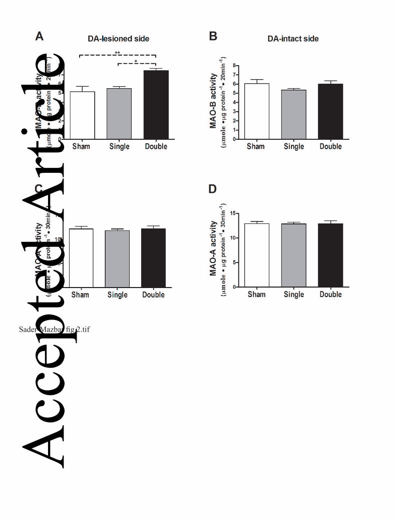
Striatal MAO-A activity was not affected by either single or double lesion (Fig. 2). On the
other hand, there was a significant increase in MAO-B activity in the double lesioned striata
(137.5 ± 0.06 %, p<0.01) compared to that of sham-operated rats four weeks after lesioning.
A significant increase was seen also when compared to single-lesioned striata (P<0.05, Fig
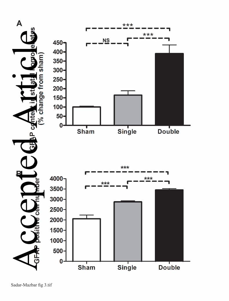
2A). Expression of the glial marker GFAP was measured in the same homogenates of the
striata (the lesioned side) used for the MAO activity assay and significantly increased
(P<0.001) in the double-lesioned striata compared to sham and to single-lesioned striata,
while there was no significant change in its expression in the single-lesioned striata four
weeks after lesioning (Fig. 3A). Glial cell counts were significantly increased (p<0.001) in
both single- and double-lesioned striata immunostained for GFAP expression compared to
sham- lesioned striata, but a larger increase was seen in the double-lesioned compared to the
single-lesioned striata (Fig. 3B). The greater fold increase in the glial marker GFAP as shown
by ELISA compared to GFAP-positive cell counts is probably the result of increased
astrocytic processes in each astrocyte of the newly-formed glial cells, since GFAP is
expressed throughout the astrocytic cell. The fact that double-lesioned rats showed a greater
degree of gliosis than single-lesioned rats is possibly due to the higher exposure of the target
striatal tissue to 5,7-DHT than to 6-OHDA, the latter having been injected to the tissue
surrounding the medial forebrain bundle while the former was injected intra-
cerebroventricularly. Acc
epte
d A
rticl
e
This article is protected by copyright. All rights reserved.
17
3.2. Striatal MAO activity following clorgyline and rasagiline treatment
The dose of clorgyline used for selective inhibition of MAO-A (0.2 mg.kg-1 s.c. daily for 14
days) caused 94.9 ± 2.4 % inhibition of MAO-A and 22.5 ± 12.4 % inhibition of MAO-B.
The dose of rasagiline used for selective inhibition of MAO-B (0.05 mg.kg-1 s.c. daily for 14
days) caused 92.9±1.5 % inhibition of this enzyme form, and 14.9±9.2 % (P>0.05) inhibition
of MAO-A. A similar extent of inhibition of MAO-A as that associated with this dose of
rasagiline was caused by 0.0033 mg.kg-1 s.c. clorgyline daily for 14 days (18.7 ± 5.8 %,
P<0.05) with insignificant inhibition of MAO-B.
3.3. MAO inhibition effects on microdialysate levels of DA and metabolites following L-dopa
administration
Systemic administration of L-dopa causes a marked increase in striatal extracellular DA
levels following lesion of the nigro-striatal pathway, which is reduced by additional lesion of
the serotonergic input (Tanaka et al., 1999). Following on this finding, we probed the role of
MAO-A and MAO-B in metabolism of DA produced from L-dopa in single- and double-
lesioned rats, by use of isoform-selective MAO inhibitors. Microdialysate DA levels before
L-dopa administration in saline-treated single and double lesion rats were very low and close
to detection limit (Fig. 4A and 4B). We confirmed the findings of Tanaka et al., (1999), that
following L-dopa administration, DA levels in saline-treated rats were higher in those given a
single lesion than in double lesion rats (P<0.001 by two-way ANOVA over the time period
20 – 200 min post L-dopa). In both lesion models, the levels of the L-dopa metabolite, 3-O-
methyl dopa, were similar in all treatment groups (not shown), indicating a consistency of L-
dopa dosing and absorption following the i.p. administration. Acc
epte
d A
rticl
e
This article is protected by copyright. All rights reserved.
18
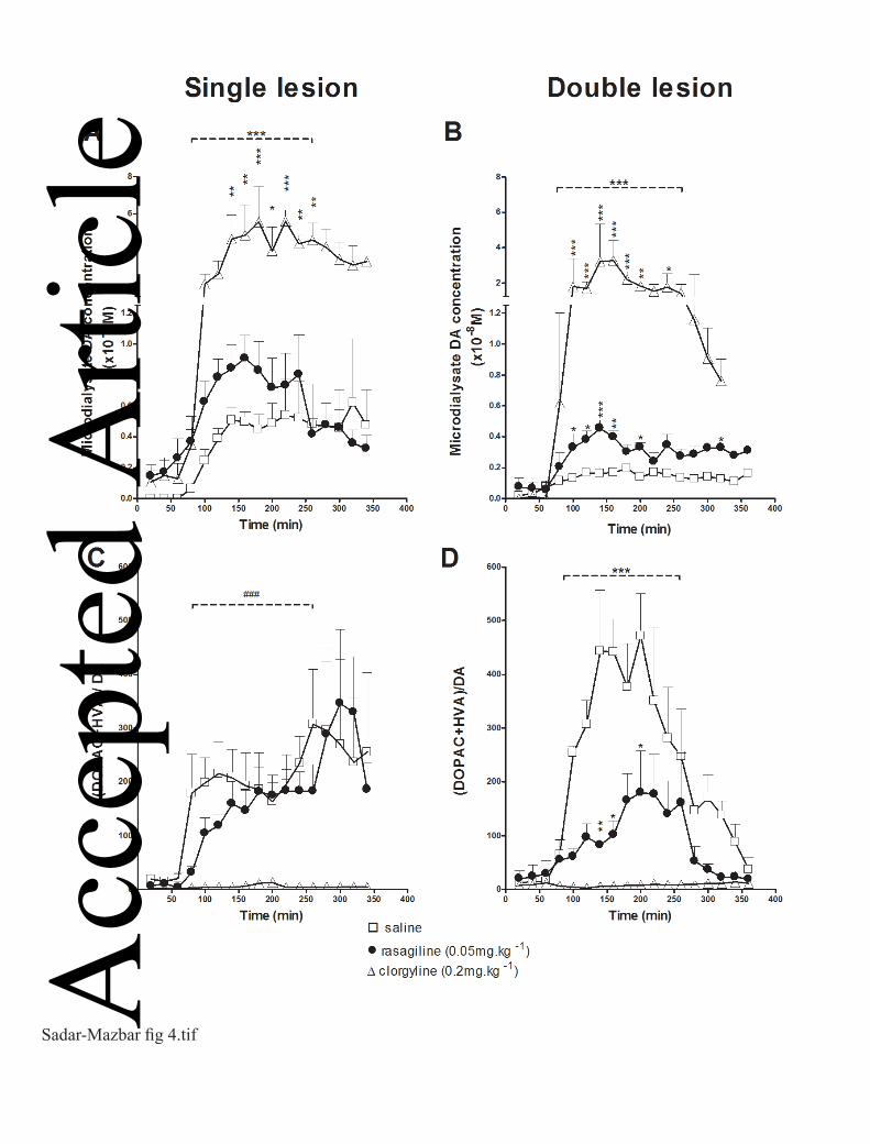
Prior treatment with clorgyline increased significantly DA levels in both lesion models
(P<0.001 by two-way ANOVA over the time period 20–200 min post L-dopa; Fig. 4A and
4B) leading to an increase in peak DA levels following L-dopa of more than 10 fold in both
single (55.4 ± 19.1 versus 4.5 ± 0.9 nM, ratio 12.3, unpaired t-test at 180min P<0.05) and
double (32.1 ± 12.2 versus 1.6 ± 0.3 nM, ratio 19.7, unpaired t-test at 140 min P<0.05) lesion
groups. Rasagiline treatment also significantly increased DA levels following L-dopa in both
lesion models (P<0.001 by two-way ANOVA over the time period 20–200 min post L-dopa),
but to a much smaller extent than clorgyline. It should be noted, however, that the magnitude
of effect of rasagiline compared to saline pre-treatment on peak DA levels post L-dopa in
double-lesion rats (4.5 ± 0.55 versus 1.6 ± 0.2 nM, ratio 2.8, unpaired t-test at 140min
p<0.01) was greater than that in single-lesion rats (9.1 ± 1.5 versus 5.0 ± 0.7 nM, ratio 1.8,
unpaired t-test at 160 min p<0.05). Moreover, higher DA levels were maintained in
rasagiline-treated rats for a longer period of time following L-dopa administration (Fig. 4B)
in the double lesion model. The ratio of (DOPAC + HVA)/DA microdialysate concentrations,
an index of oxidative metabolism of DA by MAO, was significantly reduced by rasagiline in
double-lesioned but not in single-lesioned rats by comparison with saline pretreatment
(P<0.001 by two-way ANOVA over the time period 20–200 min post L-dopa; Fig. 4C and
4D). The ratio (DOPAC + HVA)/DA was reduced to a much greater extent by clorgyline
than by rasagiline in both lesion models and the increase in this ratio following L-dopa was
completely suppressed by clorgyline (Fig. 4C and 4D).
In view of the marked effect of clorgyline to increase DA microdialysate levels, an additional
experiment was carried out in a group of double-lesioned rats to examine the effect of
inhibition of MAO-A by low-dose clorgyline (0.0033 mg.kg-1) to the same extent as was seen
in the rasagiline-treated animals (ie. about 20% inhibition of enzyme activity). In this group Acc
epte
d A
rticl
e
This article is protected by copyright. All rights reserved.
19
of double-lesioned rats, pre-treatment with this low dose of clorgyline did not increase the
post L-dopa DA microdialysate levels (data not shown).
3.4. Effect of inhibition of low-affinity uptake transporters on microdialysate levels of DA
and metabolites following L-dopa administration.
The organic transporter OCT-3 and the plasma-membrane transporter (PMAT) mediate low-
affinity transport of catecholamines and both are expressed in neurons of rat striatum (Cui et
al., 2009; Kilkenny et al., 2012; Vialou et al., 2007). In Parkinsonian striatum, where the
dopamine transporter (DAT) is absent, and the serotonin transporter (SERT) may be reduced,
the low-affinity transporters will play a more important role in uptake of extracellular DA to
sites of metabolism within striatal neurons and glia. We therefore investigated the role of the
low-affinity uptake pathway in clearance of L-DOPA- derived DA microdialysate levels in
striatum of single- and double-lesion rats using decynium-22 (D-22), an inhibitor of OCT-3
and PMAT (Hayer-Zillgen et al., 2002; Engel et al., 2005). In this experiment, the treatment
group received 50 µM D-22 by infusion through the dialysis probe into the striatum while the
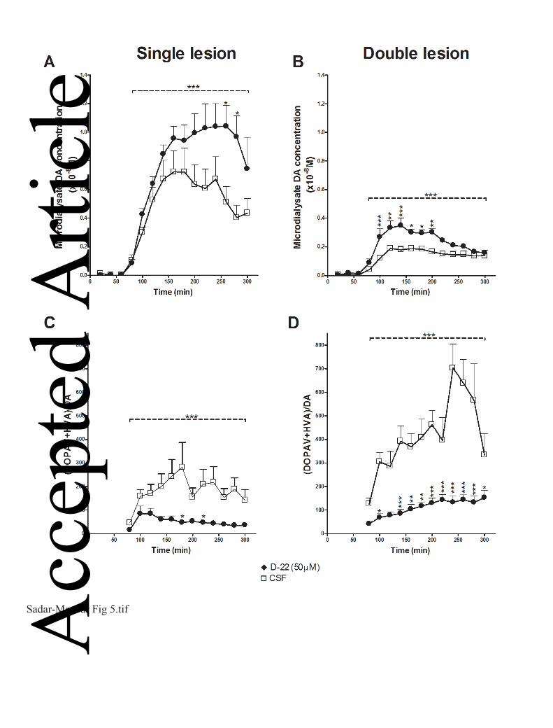
control group received only artificial CSF. Following L-dopa administration, as previously,
DA levels in CSF-treated rats were higher in those given a single-lesion than in double-lesion
rats (P<0.001 by two-way ANOVA over the period 20-240 min post L-dopa; Fig 5, A and B).
DA levels post L-dopa injection in both lesion models were significantly higher in the D-22
treated group compared to the control group throughout the whole time of sampling
(p<0.001) As can be seen in Fig 5B, the peak increase in DA levels in the double lesion
group occurred very soon after the L-dopa injection, whereas in the single lesion group it was
delayed, and it should be noted that in the double-lesioned striata the levels of DA in the D-
22 treated rats were significantly higher when compared at each time point to the CSF-treated
rats (time 20-140 min post L-dopa), while in the single-lesioned striata the difference was Acc
epte
d A
rticl
e
This article is protected by copyright. All rights reserved.
20
significant only for later time points (200-220 min post L-dopa). The ratio (DOPAC +
HVA)/DA was significantly decreased following D-22 treatment in both models (Fig 5, C
and D), mainly as a result of reduction in HVA levels. Levels of 3-OMD were similar in both
treatment groups in each lesion model, indicating consistency of L-dopa administration (not
shown).
3.5 MAO subtypes localization in striatal sections
In order to correlate the results of MAO activity determination in lesioned striata with the
effects of MAO inhibitors on microdialysate DA levels, we conducted immunofluorescence
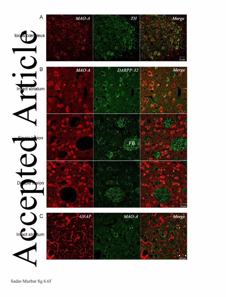
experiments to localize MAO-A and MAO-B in striatal tissue. MAO-A antibody was
validated by positive staining of noradrenergic cells in locus coeruleus (Fig.6A) and MAO-B
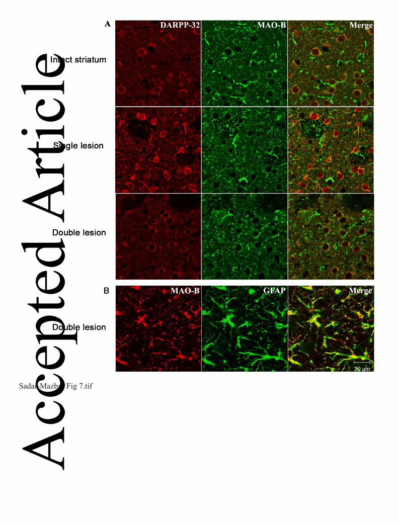
antibody positively stained GFAP-positive cells in the striatum (Fig.7B). Interestingly,
MAO-A was clearly localized in MSNs expressing DARPP-32 in intact striatum (Fig.6B).
Furthermore, MAO-A was still expressed in MSNs even following the application of the
different lesions (Figure 6B). However, no MAO-A staining was seen in glial cells (Figure
6C). MAO-B was not solely localized in glial cells, weaker staining of MAO-B was also
detected in MSN's in both lesion models (Figure 7A), however confirmation of the presence
or absence of MAO-B in MSN requires application of additional techniques, and will be
carried out in a future study. Structures identified morphologically as fiber bundles stained
positively for MAO-A (Figures 6 and 7). The staining of DARPP-32 in striatal MSN occurs
to a variable extent, because of the different degrees of DA activity at striatal dopaminergic
receptors on these cells, which varies the expression level of DARPP-32.
4. Discussion Acc
epte
d A
rticl
e
This article is protected by copyright. All rights reserved.
21
The activity of MAO is critically important in determining effective concentrations of DA at
receptor sites in the striatum following administration of L-dopa. Many techniques have been
applied to the question of the localization of the enzyme sub-types in neurons and glia of the
brain. Although several studies have shown an enrichment of MAO-A in tyrosine
hydroxylase-positive neurons of the locus coeruleus and other catecholaminergic neuronal
groups including SNpc neurons (Westlund et al., 1993), not all agree with the finding of
MAO-A localization in dopaminergic neurons of the SNpc (Arai et al., 1998; Hida et al.,
1999; Konradi et al., 1988; Willoughby et al., 1988). Experiments using human brain
synaptosomes showed that DA nerve endings in caudate nucleus contain exclusively MAO-A
(O'Carroll et al., 1987), but the subtype of the enzyme in synaptic vesicles may differ from
that found in cell bodies of SNpc. Although conclusive direct evidence for the expression of
MAO-A by nigro-striatal dopaminergic neurons is still lacking, indirect evidence points to a
role of MAO-A in deamination of dopamine released from axon terminals of these neurons.
Thus, inhibition of MAO-A by clorgyline but not selective inhibition of MAO-B by selegiline
or rasagiline increases striatal tissue steady-state DA levels and reduces DOPAC in intact rat
brain (Finberg et al., 2002; Waldmeier et al., 1976; Waldmeier et al., 1981).
Our findings of no detectable change in MAO-A content of whole striatal tissue after
destruction of SNpc are at variance with those of other researchers (Agid et al., 1973;
Demarest et al., 1980) who carried out a similar study but found significant reduction in
MAO-A levels following partial destruction of SNpc. It should be pointed out, however, that
in these previous studies the extent of DA depletion would have been much less than in the
present study, since we selected rats for a high degree of striatal DA depletion ( >99%) by
using the functional index of contralateral turning. Under these conditions, serotonergic
neurons hyperinnervate the striatum, which could increase MAO-A activity of the tissue,
assuming the presence of this enzyme form in serotonergic axonal varicosities, and such an Acc
epte
d A
rticl
e
This article is protected by copyright. All rights reserved.
22
increase may offset any reduction caused by the loss of dopaminergic neurons. Our finding of
lack of change of striatal MAO-A activity following DA as well as DA plus 5-HT lesion can
be compared with the study of Carlsson et al., (1981) who saw no change or a modest
increase in striatal MAO-A following unilateral brain hemitransection at the level of the
caudal hypothalamus. Similarly, (Van der Krogt et al., 1983) found no change in striatal
MAO-A content following 6-OHDA-induced lesion of SNpc. Our finding of no change in
striatal MAO-A activity following the double lesion indicates that most of the MAO-A
enzyme in striatum is not located in dopaminergic or serotonergic neurons. In this respect, it
should also be remembered that axonal varicosities comprise only a very small percent by
volume of the tissue even though within axonal varicosities the enzyme is highly
concentrated. When whole tissue enzyme activity is determined, the contribution of this small
compartment to the enzyme activity of whole striatal tissue may be minor, unless the specific
activity of the enzyme in the dopaminergic axonal varicosities is many fold that of the other
neuronal elements in the tissue. Our findings of increased MAO-B content of striatum
following both dopaminergic and serotonergic deafferentation are consistent with the gliosis
caused by this procedure, since MAO-B is the enzyme form which has been detected in
astrocytes in the brain of both rat and man (Ekblom et al., 1993; Levitt et al., 1982; Westlund
et al., 1985) as also in our current findings. The increase in number of GFAP-positive cells
and GFAP tissue content (Fig 3) was greater than the increase in MAO-B activity (Fig 2)
caused by the lesions, which may be related to the much less profound immunohistochemical
staining of MAO-B in glia than that of GFAP (Fig 7). Increased striatal expression of GFAP
has been described following striatal and nigral injection of 6-OHDA (Nomura et al., 2000;
Rodrigues et al., 2001).
Rasagiline is a selective inhibitor of MAO-B, which is currently in clinical use in Pd. In
contrast to selegiline, it is not metabolized to amphetamines, and does not possess Acc
epte
d A
rticl
e

This article is protected by copyright. All rights reserved.
23
amphetamine-like effects (Glezer et al., 2003). Previous studies on DA oxidative metabolism
have revealed that both endogenously released- and L-dopa derived- DA is largely
metabolized by MAO-A in intact and in hemiparkinsonian rats (Butcher et al., 1990; Finberg
et al., 1995; Paterson et al., 1991; Scarr et al., 1994; Wachtel et al., 1994). In the study of
Wachtel and Abercrombie (1994) only a minor effect of clorgyline on DA microdialysate
was seen in unilateral 6-OHDA-lesioned rats, but the degree of lesioning in their study was
probably much lower than in the present and our previous one (Finberg et al., 1995). The
present study is the first to investigate the effect of sub-acute MAO-B inhibition in 6-OHDA
lesioned rats on the metabolism of DA produced from L-dopa. The doses of rasagiline and
clorgyline were chosen to be selective according to previous studies (Finberg et al., 1995;
Lamensdorf et al., 1996; Waldmeier et al., 1981) and achieved 95% inhibition of the target
enzyme with less than 20% inhibition of the other form. The activity of MAO in
catecholaminergic neurons is considered to be in great excess, since it is necessary to inhibit
the enzyme by more than 80% in order to see functional changes in behavior (Youdim and
Finberg, 1983). However, because of the marked effect of MAO-A inhibition on DA levels,
we carried out an additional experiment which showed that 20% reduction in MAO-A
activity by clorgyline did not cause an increase in DA levels in double lesion rats, and so the
effect of rasagiline on DA levels is due to MAO-B inhibition, and the 15% inhibition of
MAO-A produced by rasagiline does not contribute to this effect.
Since 5-HT neurons are a major site for DA production from L-dopa, a larger dose of L-dopa
is needed to initiate turning behavior and induce detectable DA levels in the microdialysate in
the double lesion model compared to the single lesion. We used 25 mg.kg-1 of L-dopa as in a
previous study (Tanaka et al., 1999), in which it was shown that this dose could induce
turning behavior in double lesion rats. The need for increasing the dose of L-dopa with time
is a well-known phenomenon in treating human Pd. This might be explained by the Acc
epte
d A
rticl
e
This article is protected by copyright. All rights reserved.
24
increasing loss of 5-HT as well as DA innervation in the striatum along with the progress of
the disease (see introduction), thus supporting the validity of the double lesion model for
investigating the metabolism of DA from L-dopa in advanced stage PD.
In agreement with our previous studies (Finberg et al., 1995), MAO-A inhibition produced a
larger increase in DA extracellular levels and decrease in oxidative metabolites than did
inhibition of MAO-B in the single lesion model, and in the present study we found that
MAO-A inhibition also caused a greater effect on DA metabolism than did MAO-B
inhibition in the double-lesioned rats. This finding leads to the interesting question as to
where is MAO-A located in the double lesioned striatum, and how does it still contribute to
DA metabolism following L-dopa administration? Our present immunohistochemical study
showed that MAO-A is expressed in GABA-ergic medium spiny neurons which occupy more
than 90% of the striatal neuropil, but not in astrocytes. The role of these neurons in DA
metabolism might become more significant in the absence of the dopaminergic and
serotonergic axon terminals in the striatum.
The effect of rasagiline on DA levels following L-dopa was of a greater relative magnitude in
the double than in the single lesioned striatum. This might be explained by the absence of the
serotonergic terminals, which presumably express MAO-A and which play a major role in the
uptake and metabolism of DA in the case of the single dopaminergic lesion. Consequently,
the contribution of other cells, such as astrocytes expressing MAO-B, in the production and
metabolism of DA becomes more significant in the double-lesion rats. Our results support
this assumption, since the double denervation of dopaminergic and serotonergic terminals
was accompanied by a marked increase in GFAP expression. The recent demonstration of
occurrence of the amine transporter OCT-3 in striatal astrocytes (Cui et al., 2009) is in accord
with a role of astrocytes in metabolism of DA produced from L-dopa in the double-lesioned
striatum. The expression of OCT-3 and PMAT in non-aminergic striatal neurons indicates the Acc
epte
d A
rticl
e
This article is protected by copyright. All rights reserved.
25
possibility that these neurons, which express MAO-A, play a major role in uptake and
metabolism of DA in the absence of DA and 5-HT axonal varicosities, which express the
high-affinity transporters DAT and SERT respectively (Vialou et al., 2007).
In conclusion, when both DA and 5-HT terminals are eliminated, the MAO-B enzyme plays a
larger part in DA metabolism but MAO-A remains the major enzyme. The precise part
played by MSN and glial cells in DA metabolism depends on the degree of expression of the
low affinity transporters in each cell type together with the expression degree and affinities of
the two MAO isoforms for DA, and cannot be stated in a quantitative way in the current
research. In striatum devoid of DA and 5-HT neuronal varicosities, however, our present data
indicate an important role of low affinity transporters, and MAO-A expressed in MSN, in
metabolism of DA derived from L-dopa. These results have clinical relevance, since they
explain the efficacy of MAO-B inhibition in advanced stages of PD. In such patients,
administration of rasagiline together with L-dopa increases "on" period duration and reduces
adverse symptoms in "off" period (Parkinson Study Group, 2005), which correlates with the
increased duration of elevated striatal DA levels following L-dopa seen in double lesion rats.
In addition, the results of our study indicate that inhibitors of OCT-3 could be useful in
treatment of Pd, by enhancing and possibly prolonging the elevation of extracellular levels of
DA in striatum following L-dopa administration.
Conflict of interest
John Finberg is a co-inventor of rasagiline and receives royalties from sale of the drug,
however this fact had no impact on the ideas and concepts expressed in this manuscript.
Acc
epte
d A
rticl
e
This article is protected by copyright. All rights reserved.
26
References
Agid Y, Javoy F, Youdim MB (1973). Monoamine oxidase and aldehyde dehydrogenase activity in the striatum of rats after 6-hydroxydopamine lesion of the nigrostriatal pathway. Br J Pharmacol 48(1): 175-178. Arai R, Horiike K, Hasegawa Y (1998). Dopamine-degrading activity of monoamine oxidase is not detected by histochemistry in neurons of the substantia nigra pars compacta of the rat. Brain Res 812(1-2): 275-278. Butcher SP, Fairbrother IS, Kelly JS, Arbuthnott GW (1990). Effects of selective monoamine oxidase inhibitors on the in vivo release and metabolism of dopamine in the rat striatum. J Neurochem 55(3): 981-988. Carlsson A, Fowler CJ, Magnusson T, Oreland L, Wiberg A (1981). The activities of monoamine oxidase-A and -B, succinate dehydrogenase and acid phosphatase in the rat brain after hemitransection. Naunyn Schmiedebergs Arch Pharmacol 316(1): 51-55. Carta M, Carlsson T, Kirik D, Bjorklund A (2007). Dopamine released from 5-HT terminals is the cause of L-DOPA-induced dyskinesia in parkinsonian rats. Brain 130(Pt 7): 1819-1833. Cui M, Aras R, Christian WV, Rappold PM, Hatwar M, Panza J, et al. (2009). The organic cation transporter-3 is a pivotal modulator of neurodegeneration in the nigrostriatal dopaminergic pathway. Proc Natl Acad Sci U S A 106(19): 8043-8048. Demarest KT, Smith DJ, Azzaro AJ (1980). The presence of the type A form of monoamine oxidase within nigrostriatal dopamine-containing neurons. J Pharmacol Exp Ther 215(2): 461-468. Ekblom J, Jossan SS, Bergstrom M, Oreland L, Walum E, Aquilonius SM (1993). Monoamine oxidase-B in astrocytes. Glia 8(2): 122-132. Engel K, Wang J (2005). Interaction of organic cations with a newly identified plasma membrane monoamine transporter. Mol Pharmacol 68(5): 1397-1407. Finberg JP, Wang J, Goldstein DS, Kopin IJ, Bankiewicz KS (1995). Influence of selective inhibition of monoamine oxidase A or B on striatal metabolism of L-DOPA in hemiparkinsonian rats. J Neurochem 65(3): 1213-1220. Finberg JP, Youdim MB (2002). Pharmacological properties of the anti-Parkinson drug rasagiline; modification of endogenous brain amines, reserpine reversal, serotonergic and dopaminergic behaviours. Neuropharmacology 43(7): 1110-1118. Glezer S, Finberg JP (2003). Pharmacological comparison between the actions of methamphetamine and 1-aminoindan stereoisomers on sympathetic nervous function in rat vas deferens. Eur J Pharmacol 472(3): 173-177. A
ccep
ted
Arti
cle
This article is protected by copyright. All rights reserved.
27
Green AR, Youdim MB (1975). Effects of monoamine oxidase inhibition by clorgyline, deprenil or tranylcypromine on 5-hydroxytryptamine concentrations in rat brain and hyperactivity following subsequent tryptophan administration. Br J Pharmacol 55(3): 415-422. Hall FS, Devries AC, Fong GW, Huang S, Pert A (1999). Effects of 5,7-dihydroxytryptamine depletion of tissue serotonin levels on extracellular serotonin in the striatum assessed with in vivo microdialysis: relationship to behavior. Synapse 33(1): 16-25. Hayer-Zillgen M, Bruss M, Bonisch H (2002). Expression and pharmacological profile of the human organic cation transporters hOCT1, hOCT2 and hOCT3. Br J Pharmacol 136(6): 829-836. Hida T, Hasegawa Y, Arai R (1999). Histochemical study of dopamine-degrading monoamine oxidase activity in dopaminergic neurons of rat brain. Brain Res 842(2): 491-495. Inazu M, Takeda H, Matsumiya T (2003). Functional expression of the norepinephrine transporter in cultured rat astrocytes. J Neurochem 84(1): 136-144. Kilkenny C, Browne W, Cuthill IC, Emerson M, Altman DG (2010). Animal research: reporting in vivo experiments: the ARRIVE guidelines. J Gene Med 12(7): 561-563. Kilkenny C, Browne WJ, Cuthill IC, Emerson M, Altman DG (2012). Improving bioscience research reporting: the ARRIVE guidelines for reporting animal research. Osteoarthritis Cartilage 20(4): 256-260. Konradi C, Svoma E, Jellinger K, Riederer P, Denney R, Thibault J (1988). Topographic immunocytochemical mapping of monoamine oxidase-A, monoamine oxidase-B and tyrosine hydroxylase in human post mortem brain stem. Neuroscience 26(3): 791-802. Lamensdorf I, Youdim MB, Finberg JP (1996). Effect of long-term treatment with selective monoamine oxidase A and B inhibitors on dopamine release from rat striatum in vivo. J Neurochem 67(4): 1532-1539. Levitt P, Pintar JE, Breakefield XO (1982). Immunocytochemical demonstration of monoamine oxidase B in brain astrocytes and serotonergic neurons. Proc Natl Acad Sci U S A 79(20): 6385-6389. Lopez-Real A, Rodriguez-Pallares J, Guerra MJ, Labandeira-Garcia JL (2003). Localization and functional significance of striatal neurons immunoreactive to aromatic L-amino acid decarboxylase or tyrosine hydroxylase in rat Parkinsonian models. Brain Res 969(1-2): 135-146. Lowry OH, Rosebrough NJ, Farr AL, Randall RJ (1951). Protein measurement with the Folin phenol reagent. J Biol Chem 193(1): 265-275. Maeda T, Kannari K, Shen H, Arai A, Tomiyama M, Matsunaga M, et al. (2003). Rapid induction of serotonergic hyperinnervation in the adult rat striatum with extensive dopaminergic denervation. Neurosci Lett 343(1): 17-20. A
ccep
ted
Arti
cle
This article is protected by copyright. All rights reserved.
28
McGrath JC, Drummond GB, McLachlan EM, Kilkenny C, Wainwright CL (2010). Guidelines for reporting experiments involving animals: the ARRIVE guidelines. Br J Pharmacol 160(7): 1573-1576. Miller DW, Abercrombie ED (1999). Role of high-affinity dopamine uptake and impulse activity in the appearance of extracellular dopamine in striatum after administration of exogenous L-DOPA: studies in intact and 6-hydroxydopamine-treated rats. J Neurochem 72(4): 1516-1522. Mura A, Jackson D, Manley MS, Young SJ, Groves PM (1995). Aromatic L-amino acid decarboxylase immunoreactive cells in the rat striatum: a possible site for the conversion of exogenous L-DOPA to dopamine. Brain Res 704(1): 51-60. Navailles S, Bioulac B, Gross C, De Deurwaerdere P (2010). Serotonergic neurons mediate ectopic release of dopamine induced by L-DOPA in a rat model of Parkinson's disease. Neurobiol Dis 38(1): 136-143. Nomura T, Yabe T, Rosenthal ES, Krzan M, Schwartz JP (2000). PSA-NCAM distinguishes reactive astrocytes in 6-OHDA-lesioned substantia nigra from those in the striatal terminal fields. J Neurosci Res 61(6): 588-596. O'Carroll AM, Fowler CJ, Phillips JP, Tobbia I, Tipton KF (1983). The deamination of dopamine by human brain monoamine oxidase. Specificity for the two enzyme forms in seven brain regions. Naunyn Schmiedebergs Arch Pharmacol 322(3): 198-202. O'Carroll AM, Tipton KF, Sullivan JP, Fowler CJ, Ross SB (1987). Intra- and extra-neuronal deamination of dopamine and noradrenaline by the two forms of human brain monoamine oxidase. Implications for the neurotoxicity of N-methyl-4-phenyl-1,2,3,6-tetrahydropyridine. Biogenic Amines 4: 165-178. Otsuka S, Kobayashi Y (1964). Radioisotopic Assay for Monoamine Oxidase Determinations in Human Plasma. Biochem Pharmacol 13: 995-1006. Parkinson Study Group (2005). A randomized placebo-controlled trial of rasagiline in levodopa-treated patients with Parkinson disease and motor fluctuations: the PRESTO study. Arch Neurol 62(2): 241-248. Paterson IA, Juorio AV, Berry MD, Zhu MY (1991). Inhibition of monoamine oxidase-B by (-)-deprenyl potentiates neuronal responses to dopamine agonists but does not inhibit dopamine catabolism in the rat striatum. J Pharmacol Exp Ther 258(3): 1019-1026. Paxinos G, Watson C (1982). The rat brain in stereotaxic coordinates. edn. Academic Press: Sydney. Rodrigues RW, Gomide VC, Chadi G (2001). Astroglial and microglial reaction after a partial nigrostriatal degeneration induced by the striatal injection of different doses of 6-hydroxydopamine. Int J Neurosci 109(1-2): 91-126. A
ccep
ted
Arti
cle
This article is protected by copyright. All rights reserved.
29
Scarr E, Wingerchuk DM, Juorio AV, Paterson IA (1994). The effects of monoamine oxidase B inhibition on dopamine metabolism in rats with nigro-striatal lesions. Neurochem Res 19(2): 153-159. Schwarting RK, Huston JP (1996). The unilateral 6-hydroxydopamine lesion model in behavioral brain research. Analysis of functional deficits, recovery and treatments. Prog Neurobiol 50(2-3): 275-331. Tanaka H, Kannari K, Maeda T, Tomiyama M, Suda T, Matsunaga M (1999). Role of serotonergic neurons in L-DOPA-derived extracellular dopamine in the striatum of 6-OHDA-lesioned rats. Neuroreport 10(3): 631-634. Tashiro Y, Kaneko T, Sugimoto T, Nagatsu I, Kikuchi H, Mizuno N (1989). Striatal neurons with aromatic L-amino acid decarboxylase-like immunoreactivity in the rat. Neurosci Lett 100(1-3): 29-34. Van der Krogt JA, Koot-Gronsveld E, Van den Berg CJ (1983). Localization of rat striatal monoamine oxidase activities towards dopamine, serotonin and kynuramine by gradient centrifugation and nigro-striatal lesions. Life Sci 33(7): 615-623. Vialou V, Balasse L, Dumas S, Giros B, Gautron S (2007). Neurochemical characterization of pathways expressing plasma membrane monoamine transporter in the rat brain. Neuroscience 144(2): 616-622. Wachtel SR, Abercrombie ED (1994). L-3,4-dihydroxyphenylalanine-induced dopamine release in the striatum of intact and 6-hydroxydopamine-treated rats: differential effects of monoamine oxidase A and B inhibitors. J Neurochem 63(1): 108-117. Waldmeier PC, Delini-Stula A, Maitre L (1976). Preferential deamination of dopamine by an A type monoamine oxidase in rat brain. Naunyn Schmiedebergs Arch Pharmacol 292(1): 9-14. Waldmeier PC, Felner AE, Maitre L (1981). Long term effects of selective MAO inhibitors on MAO activity and amine metabolism. In: Youdim MB, Paykel ES (ed)^(eds). Monoamine Oxidase Inhibitors, edn. New York: Wiley. p^pp 87-102. Westlund KN, Denney RM, Kochersperger LM, Rose RM, Abell CW (1985). Distinct monoamine oxidase A and B populations in primate brain. Science 230(4722): 181-183. Westlund KN, Krakower TJ, Kwan SW, Abell CW (1993). Intracellular distribution of monoamine oxidase A in selected regions of rat and monkey brain and spinal cord. Brain Res 612(1-2): 221-230. Willoughby J, Glover V, Sandler M (1988). Histochemical localisation of monoamine oxidase A and B in rat brain. J Neural Transm 74(1): 29-42. Yamada H, Aimi Y, Nagatsu I, Taki K, Kudo M, Arai R (2007). Immunohistochemical detection of L-DOPA-derived dopamine within serotonergic fibers in the striatum and the substantia nigra pars reticulata in Parkinsonian model rats. Neurosci Res 59(1): 1-7. A
ccep
ted
Arti
cle

This article is protected by copyright. All rights reserved.
30
Youdim MBH, Finberg JPM (1983). Monoamine oxidase inhibitor antidepressants. In: Grahame-Smith DG, P.J. C (ed)^(eds). Psychopharmacology, edn, Vol. 1. Amsterdam: Excerpta Medica. p^pp 38-70.
Acc
epte
d A
rticl
e
This article is protected by copyright. All rights reserved.
31
Figure legends
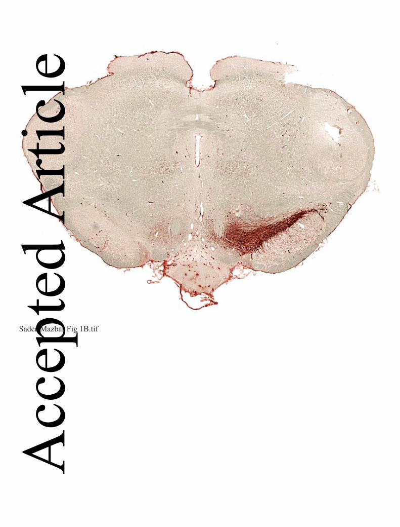
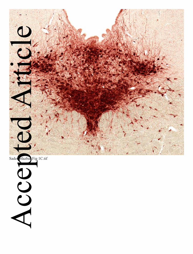
Figure 1 Immunohistochemical demonstration of lesion extent by 6-OHDA and 5,7-DHT. A, B:
tyrosine hydroxylase positive neurons in coronal midbrain sections of normal rat brain (A)
and rat with double lesion (B), 4 weeks after lesioning. C, D: tryptophan hydroxylase-positive
neurons in dorsal raphe nucleus area of normal (C) and double-lesioned (D) brain 4 weeks
after lesioning. Ruler, 1 mm.
Figure 2 Striatal MAO activity in lesioned rats. MAO-B (A and B) and MAO-A (C and D) activities were
determined in DA-intact and DA-lesioned side of striatum from rats bearing either unilateral sham
(n=4), unilateral single (dopaminergic; n=7) or double (unilateral dopaminergic and bilateral
serotonergic; n=6) lesions 4 weeks after lesioning. Data are expressed as mean + SEM of µmole
metabolite.µg protein-1.30min-1 for MAO-A, and µmole metabolite.µg protein-1.20min-1 for MAO-
B. *p<0.05, **p<0.01 by one way ANOVA followed by Bonferroni multiple comparison test.
Acc
epte
d A
rticl
e
This article is protected by copyright. All rights reserved.
32
Figure 3 (A) GFAP quantitation in homogenates of the lesioned striata of rats bearing either unilateral
sham, unilateral single (dopaminergic) or double (unilateral dopaminergic and bilateral
serotonergic) lesions. GFAP content in the striatal homogenates was measured by sandwich
ELISA 4 weeks after the induction of lesion (n=4 for sham, n=7 for single- and n=6 for
double-lesion rats), and is expressed as mean % change from sham-lesioned striata + SEM.
*** p<0.001 by one way ANOVA followed by Bonferroni multiple comparison test. NS - not
significant.
(B) GFAP positive cell counts in 5µm sections of DA-lesioned striata of rats bearing either
unilateral sham (n=4), single (dopaminergic, n=6) or double (dopaminergic and serotonergic,
n=8) lesion. For each rat, 25 coronal photomicrographs were taken from left striatum at each
of the levels1.6, 1.2, 0.8, 0.5 and 0.2 mm anterior to bregma. Data shown represent the
average counts + SEM for each group. *** p<0.001 by one way ANOVA followed by
Bonferroni multiple comparison test.
Acc
epte
d A
rticl
e
This article is protected by copyright. All rights reserved.
33
Figure 4. Microdialysis study of L-dopa-derived DA and DA metabolite levels in DA-lesioned striatum
of single (unilateral dopaminergic only) or double (unilateral dopaminergic plus bilateral
serotonergic) lesion rats. Rats bearing either single or double lesion were treated daily for two
weeks with either saline, rasagiline (0.05mg.kg-1) or clorgyline (0.2mg.kg-1), by s.c. injection.
Rasagiline increased L-dopa- derived DA levels in both lesion models when compared to
saline treatment, however the magnitude of fold increase over saline control in peak DA
levels was larger in the double lesion (B) compared to the single lesion rats (A) (2.8 vs 1.8
fold respectively) and duration of increased DA level was also increased in the double lesion
group. This effect was accompanied by a decrease in DA oxidative metabolism (expressed as
[DOPAC+HVA]/DA ratio) in the double lesion model only (D). Clorgyline treatment caused
a significant difference from saline in all post-L-dopa DA and metabolite levels.
Rats were anesthetized with isoflurane on day 14, microdialysis probes were inserted into
lesioned striatum via a guide cannula implanted 24 h previously and MAO inhibitor was
administered. One h later carbidopa (6 mg.kg-1 i.p.) was administered and microdialysis
collections were started (time 0). L-dopa methyl ester (25mg.kg-1 i.p.) was administered
60min later.
Data are expressed as mean DA or metabolite dialysate concentration + SEM for n=4-6 rats
per group. For horizontal bars in A, B and D, *** p<0.001 over the time period between 20-
200min post L-dopa for difference between both rasagiline and clorgyline curves from saline
curve by two way ANOVA. Bonferroni post hoc test was performed for comparison between
treatments at each time point (*p<0.05, **p<0.01,***p<0.001). In C, ### p<0.001 for
statistical significance over the time period between 20-200 min post L-dopa for difference
between clorgyline curve only from saline curve by two way ANOVA. Acc
epte
d A
rticl
e
This article is protected by copyright. All rights reserved.
34
Figure 5. The effect of decynium-22 (D-22) on L-dopa- derived DA levels in DA-lesioned striatum of
both single (dopaminergic) and double (dopaminergic and serotonergic) lesion models. D-22
increased L-dopa-derived DA levels in both lesion models when compared to artificial
cerebrospinal fluid (CSF) infusion. This increase in DA levels was accompanied by a
decrease in DA oxidative metabolism (expressed as [DOPAC+HVA]/DA ratio) in both lesion
models (C and D).
Rats bearing either type of lesion were infused via the dialysis probe with either 50µM
decynium-22 at 2 µL.min-1 or artificial CSF. Infusion started 1hr before dialysate collection
to achieve stable tissue levels of the drug. Carbidopa (6 mg.kg-1 i.p.) was administered at the
start of microdialysis collections (time 0), and L-dopa (25 mg.kg-1) was administered 60min
later.
Data are expressed as mean + SEM for n=7-9 rats per group. ***p<0.001 for time periods
indicated by horizontal bars (20-240 min post L-dopa) by two way ANOVA for comparison
between treatments (saline and D-22). Bonferroni post hoc test was performed for
comparison between treatments at each time point (*p<0.05, **p<0.01,***p<0.001)
Acc
epte
d A
rticl
e
This article is protected by copyright. All rights reserved.
35
Figure 6 MAO-A localization in brain sections (5µm) using polyclonal anti-MAO-A antibody. A:
Section from intact locus coeruleus, additionally stained for tyrosine hydroxylase using
monoclonal anti-tyrosine hydroxylase antibody, confirming known localization of MAO-A to
noradrenergic neurons of this brain area. B: Striatal sections from intact, single- (ipsilateral
dopaminergic) and double- (ipsilateral dopaminergic plus bilateral serotonergic) lesioned rats,
additionally stained with monoclonal anti-DARPP-32 as indicator of medium spiny neurons
(MSN), showing localization of MAO-A in MSN and fiber bundles (FB), and similar
distribution of MAO-A in intact, single- and double-lesioned rats (representative sections
shown from n = 3, 6 and 5 respectively rats with similar results in each). C: Striatal section
from normal control rat stained for glial cells using monoclonal anti-GFAP antibody,
showing absence of MAO-A from glial cells. Images were obtained using confocal
microscope.
Acc
epte
d A
rticl
e
This article is protected by copyright. All rights reserved.
36
Figure 7. MAO-B localization in brain sections (5µm) using polyclonal anti-MAO-B antibody. A:
Striatal sections from intact, single (ipsilateral dopaminergic) and double (ipsilateral
dopaminergic plus bilateral serotonergic) lesioned rats, additionally stained for medium spiny
neurons (MSN) using polyclonal anti DARPP-32. Most MAO-B containing structures are not
co-localised with DARPP-32. Representative images are shown from n=3, 4 and 4 intact,
single- and double-lesioned rats respectively. B: Striatal section from double-lesion rat
additionally stained for glial cells using monoclonal anti-GFAP antibody. MAO-B
colocalizes with GFAP in most glial cell processes. Images were obtained using confocal
microscope.
Acc
epte
d A
rticl
e

Sader-Mazbar Fig 1A.tif
Acc
epte
d A
rticl
e
Sader-Mazbar Fig 1B.tif
Acc
epte
d A
rticl
e
Sader-Mazbar Fig 1C.tif
Acc
epte
d A
rticl
e

Sader-Mazbar Fig 1D.tif
Acc
epte
d A
rticl
e
Sader-Mazbar fig 2.tif
Acc
epte
d A
rticl
e
Sadar-Mazbar fig 3.tif
Acc
epte
d A
rticl
e
Sadar-Mazbar fig 4.tifAcc
epte
d A
rticl
e
Sadar-Mazbar Fig 5.tifAcc
epte
d A
rticl
e
Sadar-Mazbar fig 6.tif
Acc
epte
d A
rticl
e
Sadar-Mazbar Fig 7.tif
Acc
epte
d A
rticl
e


![Dopa decarboxylaseactivity of the living human · nine (L-dopa). We measured regional dopa decarboxylase activity in brains ofsix healthy volunteers with 6-[18F]fluoro-L-dopaandpositron](https://img.pdfslide.us/doc/110x75/5fd3ff72add4681c6146e1fc/dopa-decarboxylaseactivity-of-the-living-human-nine-l-dopa-we-measured-regional.jpg)