Embed Size (px)
Citation preview
IntroductionHomeobox genes regulate organ development andmorphology by controlling the fate of specific cellpopulations (1–4). Pancreatic duodenal homeobox-1(PDX1), known as IPF-1 in humans, is a homeoboxtranscription factor that is absolutely required for thedevelopment of the pancreas and other foregut struc-tures (5). Humans and mice that do not express thePdx1 gene exhibit pancreatic agenesis (6, 7). Het-erozygosity for inactivating mutations of the IPF1gene in humans leads to MODY4, a rare form ofmaturity-onset diabetes of the young (MODY), inwhich early onset diabetes is characterized byimpaired insulin secretion in response to glucose. (8).Similarly, mice with genetically engineered reductionsin PDX1 levels have impaired glucose tolerance, andmice in which PDX1 has been genetically inactivated
in more than 80% of the β cells develop diabetes(9–13). Based mainly on in vivo studies of these trans-genic mice and experiments conducted on various celllines, this homeobox gene has been ascribed severalimportant “day-to-day” physiological roles in adult βcells, including glucose sensing, insulin biosynthesis,and insulin exocytosis (11). Located preferentially butnot exclusively in β cells, PDX1 has been reported toinfluence the expression of a number of β cell genes,including those coding for insulin (14, 15), islet amy-loid polypeptide (16, 17), glucokinase (13, 14, 18), andGLUT2 (11, 13, 14, 19), suggesting that PDX1 mayplay a broad role. Surprisingly, corresponding studieson isolated islets or β cells that might reveal a cell-autonomous, inherent defect in the ability of individ-ual β cells to sense glucose and secrete insulin havenot been reported.
Our results demonstrate that, while individual iso-lated Pdx1+/– islets and dispersed β cells have normalglucose signaling and stimulus-secretion coupling,Pdx1 haploinsufficient islets exhibit significant apop-tosis at basal glucose concentrations. The develop-mental role of PDX1 persists into adulthood throughthe lifelong maintenance of islet mass, architecture,and plasticity, processes that involve the interaction ofβ cell neogenesis, differentiation, and apoptosis (20).Although the effects are dramatically distinct com-pared with total PDX1 ablation, the phenotype of Pdx1heterozygous mice makes them a highly relevant modelfor study of the pathophysiology of MODY.
The Journal of Clinical Investigation | April 2003 | Volume 111 | Number 8 1147
Increased islet apoptosis in Pdx1+/– mice
James D. Johnson,1 Noreen T. Ahmed,2 Dan S. Luciani,3 Zhiqiang Han,3 Hung Tran,3
Jun Fujita,3 Stanley Misler,1 Helena Edlund,4 and Kenneth S. Polonsky3
1Renal Division, Department of Internal Medicine, Washington University School of Medicine/Barnes-Jewish Hospital, St. Louis, Missouri, USA
2University of Chicago Pritzker School of Medicine, Chicago, Illinois, USA3Metabolism Division, Department of Internal Medicine, Washington University School of Medicine/Barnes-Jewish Hospital,St. Louis, Missouri, USA
4Umea Center for Molecular Medicine, Umea University, Umea, Sweden
Mice with 50% Pdx1, a homeobox gene critical for pancreatic development, had worsening glucose tol-erance with age and reduced insulin release in response to glucose, KCl, and arginine from the per-fused pancreas. Surprisingly, insulin secretion in perifusion or static incubation experiments inresponse to glucose and other secretagogues was similar in islets isolated from Pdx1+/– mice comparedwith Pdx1+/+ littermate controls. Glucose sensing and islet Ca2+ responses were also normal. Depolar-ization-evoked exocytosis and Ca2+ currents in single Pdx1+/– cells were not different from controls,arguing against a ubiquitous β cell stimulus-secretion coupling defect. However, isolated Pdx1+/– isletsand dispersed β cells were significantly more susceptible to apoptosis at basal glucose concentrationsthan Pdx1+/+ islets. BclXL and Bcl-2 expression were reduced in Pdx1+/– islets. In vivo, increased apopto-sis was associated with abnormal islet architecture, positive TUNEL, active caspase-3, and lymphocyteinfiltration. Although similar in young mice, both β cell mass and islet number failed to increase withage and were approximately 50% less than controls by one year. These results suggest that an increasein apoptosis, with abnormal regulation of islet number and β cell mass, represents a key mechanismwhereby partial PDX1 deficiency leads to an organ-level defect in insulin secretion and diabetes.
J. Clin. Invest. 111:1147–1160 (2003). doi:10.1172/JCI200316537.
Received for publication July 30, 2002, and accepted in revised formFebruary 11, 2003.
Address correspondence to: K.S. Polonsky, Department ofMedicine, Washington University School of Medicine, CampusBox 8066, 660 S. Euclid Avenue, St. Louis, Missouri 63110, USA.Phone: (314) 362-8061; Fax: (314) 362-8015; E-mail: [email protected] of interest: The authors have declared that no conflict ofinterest exists.Nonstandard abbreviations used: pancreatic duodenalhomeobox 1 (PDX1); maturity-onset diabetes of the young(MODY); intraperitoneal glucose tolerance test (IPGTT); Krebs-Ringer buffer (KRB); intracellular-like solution (IS);tetraethylammonium (TEA); pico Farad (pF).
MethodsIn vivo characterization of mice. Pdx1 haploinsufficientmice used in the present study, in which exon 2 wasreplaced with the neomycin-resistance gene, have beendescribed previously (6). Mice were genotyped by PCR.Unless otherwise indicated, mice were used for mostexperiments between 8 and 12 weeks of age to avoidpotentially confounding effects of lowered total pancre-atic insulin on our results (see Results below). Intraperi-toneal glucose tolerance tests (IPGTTs) were performedafter a 4-hour fast (2 g dextrose/kg body weight).
Pancreatic perfusion. Pancreata were perfused in ahumidified, temperature-controlled chamber, via theaorta at the level of the celiac artery, as described else-where (21). Oxygenated Krebs-Ringer buffer (KRB) con-taining 119 mM NaCl, 4.7 mM KCl, 25 mM NaHCO3,2.5 mM CaCl2, 1.2 mM MgSO4, 1.2 mM KH2PO4, and0.25% radioimmunoassay-grade BSA with the indi-cated concentrations of glucose was delivered at 1ml/min using a Minipuls 3 peristaltic pump (GilsonInc., Middleton, Wisconsin, USA). Prior to sample col-lection, pancreata were perfused with 5 mM glu-cose/KRB for 30–40 minutes. Samples were collectedat 5-minute intervals. Glucose ramps were generatedas previously described (21).
Islet isolation and characterization. Insulin content wasmeasured by radioimmunoassay (see below) afteracid/ethanol extraction from homogenized whole pan-creata that had been dissected, removed from the ani-mal, and weighed. To obtain pancreatic islets, pancrea-ta were removed and islets were isolated by collagenasedigestion using a protocol adapted (21) from onedescribed by Lacy and Kostianovsky (22). Isolationsfrom Pdx1 heterozygotes older than 16 weeks usuallydid not yield islets. After isolation, islets were culturedfor 4 hours in 10 mM glucose RPMI media (containing100 IU/ml penicillin, 100 µg/ml streptomycin, and 10%FCS; pH was adjusted to 7.4 with NaOH) before beinghand-picked and cultured overnight at 37°C with 5%CO2 and saturated humidity.
Hormone release. For static incubations, groups of5–10 islets were cultured (as above) in small tubes.Experiments were conducted on islets that had beenwashed under the experimental conditions (KRB equil-ibrated with 5% CO2) for 30 minutes. In perifusionexperiments, KRB was pumped at a rate of 1 ml/minaround islets that were loaded into temperature- andCO2-controlled 300-µl plastic chambers and padded onboth sides with beads. Islets were washed under basalconditions for 1 hour prior to the experiment. Insulinand C-peptide were measured from in vivo and in vitrosamples by radioimmunoassay (Linco Research Inc., St.Charles, Missouri, USA).
Islet imaging. Hand-picked islets were allowed toadhere to glass coverslips overnight prior to imagingfor Ca2+ levels or NADH autofluorescence, as describedpreviously (21). Briefly, Fura-2 AM–loaded islets wereperifused at a flow rate of 1 ml/min with KRB (no BSA)at 37°C on the stage of a Nikon inverted microscope
(Nikon Inc., Melville, New York, USA). Changes inintracellular Ca2+ within individual islets were reflect-ed in the ratio of fluorescence emission acquired above510 nM in response to excitation at 340 nm and 380nm. This provides a relative indication of islet Ca2+,which is independent of dye concentration in the tis-sue. NADH autofluorescence was imaged in unloadedislets excited at a wavelength of 365 nM and recordedat a wavelength of 495 nM (23).
Cell dispersion. Islets were gently dispersed after threeconsecutive 1-minute washes with Ca2+/Mg2+-freeMEM (Mediatech Inc., Herndon, Virginia, USA) fol-lowed by a 30-second exposure to trypsin/EDTA solu-tion (Invitrogen Corp., Carlsbad, California, USA)diluted 1:5 in MEM. This was followed by gentle repet-itive pipetting and a final wash with Ca2+/Mg2+-freeMEM. The incorporation of MEM and diluted trypsinsolutions in the present protocol are modificationsdesigned to reduce cellular stress during the dispersionprocess. Unless otherwise indicated, cells were culturedon glass coverslips in RPMI media (as above) and main-tained at 37°C, 5% CO2, and saturated humidity for1–3 days after dispersion.
Single-cell imaging. Islet cell cultures were incubatedwith 1 µM Fura-4F AM (Molecular Probes Inc.,Eugene, Oregon, USA) in RPMI for 30 minutes andrinsed in Ringer’s solution (see below) for 30 minutes.Coverslips in a narrow 35°C chamber (∼300 µl) werecontinuously perifused with preheated solutions. Cells(3–10 per image field) were excited at 340 nM and 380nM using a monochromator (T.I.L.L. PhotonicsGmbH, Grafelfing, Germany) through a 20× objective(IX70; Olympus Optical Co., Tokyo, Japan, USA) andimaged with a CCD camera (T.I.L.L. PhotonicsGmbH). The formula of Grynkiewicz et al. (24) wasused to convert 340/380 ratios to Ca2+ determinations;these were calibrated “in vivo” by exposing cells to 10µM ionomycin for more than 30 minutes to obtainmaximum ratio, and then to 20 µM EGTA and 10 µMionomycin to obtain minimum ratio.
Ca2+ signal quantification was performed essentiallyas described (25) using waveform statistics determinedautomatically with a macro written for IgorPro soft-ware (WaveMetrics Inc., Lake Oswego, Oregon, USA).Ca2+ levels during the 5 minutes prior to treatmentwere averaged to give a pretreatment mean for eachcell. The maximum Ca2+ determination during thetreatment period was subtracted from the pretreat-ment average to give the maximal amplitude of theCa2+ signal. The time from initiation of treatment tomaximal amplitude was measured, and the maximalamplitude was divided by this value to give the effec-tive rate of rise of the Ca2+ signal.
Single-cell assays of hormone release and electrophysiology.Our methods for the perforated-patch variant ofwhole-cell recording using an EPC 9 amplifier (HekaElektronik, Lambrecht, Germany) have been describedpreviously (26). The fire-polished tips of the recordingpipettes contained either (a) high K+ intracellular-like
1148 The Journal of Clinical Investigation | April 2003 | Volume 111 | Number 8
solution (IS) containing 65 mM KCl, 28.4 mM K2SO4,11.3 mM NaCl, 1 mM MgCl2, 0.5 mM EGTA, 47.2 mMsucrose, and 20 mM KHEPES titrated to pH 7.3 or (b)a Cs+-substituted IS containing 65 mM CsCl, 28.4mM Cs2SO4, 11.3 mM NaCl, 1 mM MgCl2, 0.5 mMEGTA, 47.2 mM sucrose, and 20 mM CsHEPES(titrated to pH 7.3). Pipettes were backfilled with theselected IS containing nystatin (250 µg/ml). Record-ings were made at more than 30–32°C in standardextracellular solution containing 5.5 mM KCl, 2 mMCaCl2, 1 mM MgCl2, 20 mM HEPES, 144 mM NaCl,and various glucose concentrations. Giga-seals wereobtained and the membrane potential was held at –70mV except during 200-ms depolarizing steps. Mem-brane capacitance was estimated using the IgorPromodule according to the method of Barnett and Mis-ler (26), where a dual-frequency sine wave voltage isimposed on the cell. For recording Ca2+ currents, theextracellular solution was modified by mole-for-molesubstitution of tetraethylammonium (TEA) (30 mM)for NaCl and the patch pipette was filled with Cs+-containing IS. The largest single cells were selected forrecording because immunohistochemical, cell-sort-ing, and electrophysiological data from rodent isletssuggest these are likely to be β cells.
Quantification of apoptosis susceptibility. We used a PCR-based method for detecting DNA ladders according tothe manufacturer’s instructions (ApoAlert LM-PCRLadder Assay Kit; Clontech Laboratories Inc., Palo Alto,California, USA). Briefly, groups of five islets fromPdx1+/– and Pdx1+/+ mice (three mice each, total of sixmice per experiment) were cultured separately in RPMIcontaining various concentrations of glucose or thap-sigargin, a known inducer of Ca2+ stress–mediatedapoptosis in β cells (23, 27). After 72 hours, genomicDNA was isolated with the DNeasy kit from QIAGENInc. (Valencia, California, USA) according to the man-ufacturer’s instructions for cultured cells, except thatislets were maintained in lysis buffer for 10 minutes at50°C. Genomic DNA was then concentrated by eth-anol precipitation and vacuum-dried before beingresuspended and quantified using UV spectrophotom-etry. Next, 100 µg of DNA was ligated to adapters asdescribed in the ApoAlert Kit. Short cycles of PCR thatselectively amplify adapter-ligated DNA fragments overlonger DNA were used. The resulting PCR productswere run on 1.2% agarose/ethidium bromide gels in0.5× tris-borate-EDTA. Gels were imaged and back-ground-subtracted DNA ladders were quantified usingdensitometry (Eagle Eye II; Stratagene, La Jolla, Cali-fornia, USA). In order to compare data from separategels, band intensity was normalized to the average lad-dering of the three groups of control islets in 5 mM glu-cose. Apoptosis was also measured in single dispersedcells cultured overnight as for imaging experimentsusing the ApoPercentage Kit (Biocolor Ltd., Newtown-abbey, Northern Ireland, United Kingdom). This kituses a dye that stains cells as they undergo the mem-brane “flip-flop” event when phosphatidylserine is
translocated to the outer leaflet (28). This event is con-sidered diagnostic of apoptosis but not necrosis. Apop-totic cells, which appeared bright pink against thewhite background of phenol red–free RPMI (TissueCulture Support Center, Washington University, St.Louis, Missouri, USA), were counted manually in ablinded fashion.
Analysis of gene expression by RT-PCR. The expressionof islet-specific and apoptosis-related genes was ana-lyzed by RT-PCR according to the method describedby Wong et al. (29). Fifteen islets were cultured underconditions identical to those described above for theDNA ladder assay (see Quantification of apoptosis sus-ceptibility). Total RNA was isolated using the RNeasyMini Kit (QIAGEN Inc.) with DNase I treatment anddried. RNA was reverse transcribed using SuperScriptII RNase-H Reverse Transcriptase (Invitrogen Corp.,Carlsbad, California, USA) with oligo d(T)14–18 primer(Invitrogen Corp.) in 20 µl reaction volume, fromwhich 2 µl was used for detection of insulin-1,insulin-2, glucagon, PDX1, and apoptosis-relatedgene transcripts using PCR. GAPDH was used as theinternal standard to which mRNA levels were nor-malized. GAPDH expression, which did not varyunder any of the conditions used, was further vali-dated by comparing its expression to that of β-actinmRNA using QuantumRNA Universal 18S InternalStandards (Ambion Inc., Austin, Texas, USA). As acontrol, we ascertained that PCR of the isolated RNAsamples without reverse transcriptase did not gener-ate detectable products under these conditions (notshown). Where possible, we designed primers thatoverlapped intronic regions that would reveal thepresence of any contaminating genomic DNA.Primer pairs and conditions used were: insulin-1 (261bp), 5′-TCAGAGACCATCAGCAAGCAG-3′ and 5′-GTCT-GAAGGTCCCCGGGGCT-3′, 19 cycles; insulin-2 (267bp), 5′-TCAGAGACCATCAGCAAGCAG-3′ and 5′-GTCT-GAAGGTCACCTGCTCC-3′, 18 cycles; glucagon (215bp), 5′-CATTCACAGGGCACATTGACC-3′ and 5′-ACCAGCCAAGCAATGAATTCCTT-3′, 24 cycles; Pdx1(290 bp), 5′-CCCTTTCCCGTGGATGAAATC-3′ and 5′-GGGTCCCGCTACTACGTTTCTTATC-3′, 32 cycles;GAPDH (532 bp), 5′-GGAGCCAAACGGGTCAT-CATCTC-3′ and 5′-AGTGGGAGTTGCTGTTGAAGTCGC-3′, 23 cycles. PCR reactions for these genes are per-formed at an annealing temperature of 58°C. TheCytoXpress quantitative PCR Detection Kit forMouse Apoptosis Set 2 (BioSource International,Camarillo, California, USA) was used for multiplexPCR of apoptosis-related genes (caspase-3, Bax, Bcl-2,and BclXL; 32 PCR cycles). Twenty microliters of each50-µl PCR reaction was electrophoretically separatedon 2% agarose gels containing 0.5 µg/ml of ethidiumbromide in tris-borate-EDTA buffer. Band intensitywas analyzed by densitometry.
Morphometry and immunohistochemistry. Primary anti-bodies to rat insulin (Linco Research Inc., St. Charles,Missouri, USA), glucagon, active (cleaved) caspase-3
The Journal of Clinical Investigation | April 2003 | Volume 111 | Number 8 1149
(Trevigen Inc., Gaithersburg, Maryland, USA), and poly-clonal Ki67 (Novocastra Laboratories Ltd., Newcastleupon Tyne, United Kingdom) were used. The DeadEndcolorimetric kit for TUNEL (Promega Corp., Madison,Wisconsin, USA) was used per the manufacturer’sinstructions. MIN6 cells exposed to 1 µM thapsigarginwere used as positive controls for active caspase-3 andTUNEL staining. For measurements of islet cell area, isletarchitecture, and in vivo apoptosis, paraffin-embeddedpancreatic tissue sections were stained red with primaryantibody/3-amino,9-ethyl-carbazole and counterstainedwith hematoxylin (Zymed Laboratories Inc., South SanFrancisco, California, USA). Slides were viewed throughthe 1.25× or 20× objective of an Olympus BX41 micro-scope (Olympus Optical Co., Tokyo, Japan). Images wererecorded on a Nikon Coolpix 995 digital camera. Wherenecessary, multiple images were spliced using Adobe Pho-toshop (Adobe Systems Inc., Mountain View, California,USA). Shading defects and non-red background irregu-larities from spliced, monochromatized images were can-celed by subtracting the red channel from the green chan-nel using MetaMorph software (Universal Imaging Corp.,Downingtown, Pennsylvania, USA). Pancreatic area andβ cell area were each estimated using the intensity thresh-olding function of the integrated morphometry packagein MetaMorph. Investigators were blinded to the sourceof the tissue throughout the morphometric analysis.Unless otherwise specified, images are representative ofone to four sections from a total of 11 Pdx1+/+ and tenPdx1+/– mice shown to have a specific phenotype byIPGTT or fasting glucose levels.
Statistical analysis. Results are expressed as mean ± SEM.Differences between means were evaluated usingANOVA or the Student t test as appropriate. Differ-ences were considered significant at P < 0.05.
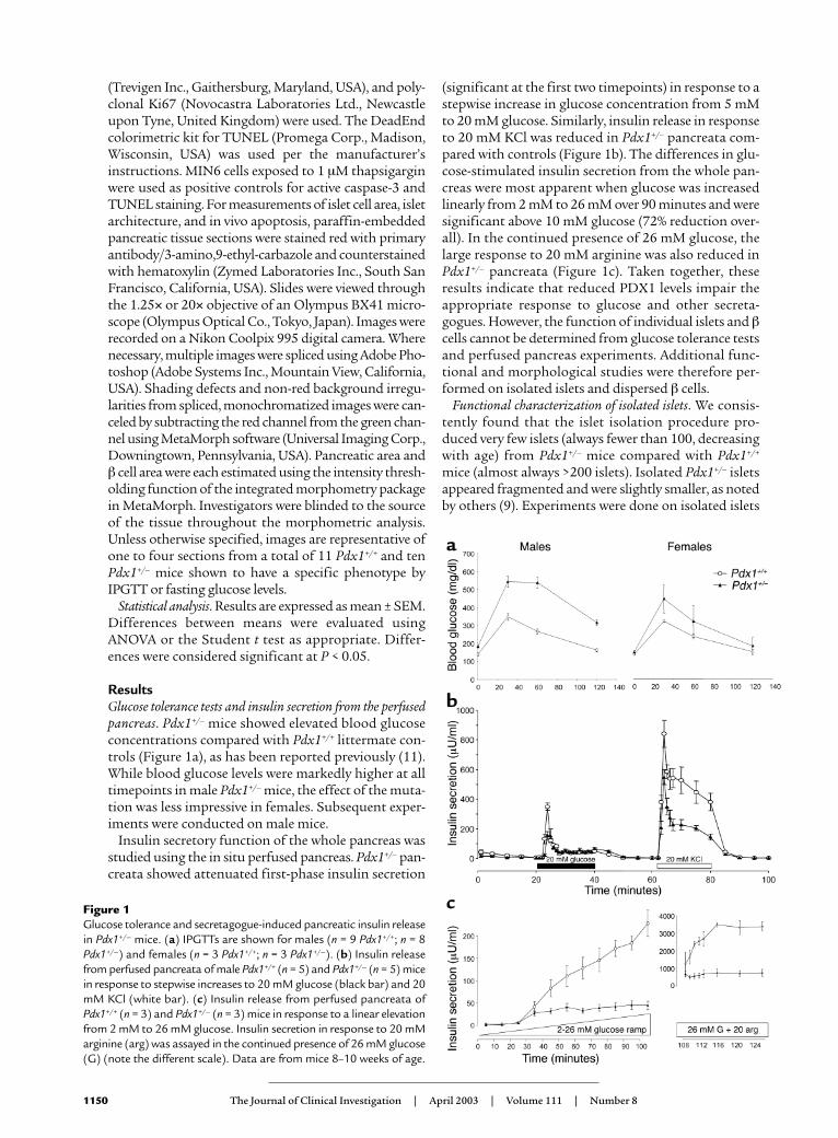
ResultsGlucose tolerance tests and insulin secretion from the perfusedpancreas. Pdx1+/– mice showed elevated blood glucoseconcentrations compared with Pdx1+/+ littermate con-trols (Figure 1a), as has been reported previously (11).While blood glucose levels were markedly higher at alltimepoints in male Pdx1+/– mice, the effect of the muta-tion was less impressive in females. Subsequent exper-iments were conducted on male mice.
Insulin secretory function of the whole pancreas wasstudied using the in situ perfused pancreas. Pdx1+/– pan-creata showed attenuated first-phase insulin secretion
(significant at the first two timepoints) in response to astepwise increase in glucose concentration from 5 mMto 20 mM glucose. Similarly, insulin release in responseto 20 mM KCl was reduced in Pdx1+/– pancreata com-pared with controls (Figure 1b). The differences in glu-cose-stimulated insulin secretion from the whole pan-creas were most apparent when glucose was increasedlinearly from 2 mM to 26 mM over 90 minutes and weresignificant above 10 mM glucose (72% reduction over-all). In the continued presence of 26 mM glucose, thelarge response to 20 mM arginine was also reduced inPdx1+/– pancreata (Figure 1c). Taken together, theseresults indicate that reduced PDX1 levels impair theappropriate response to glucose and other secreta-gogues. However, the function of individual islets and βcells cannot be determined from glucose tolerance testsand perfused pancreas experiments. Additional func-tional and morphological studies were therefore per-formed on isolated islets and dispersed β cells.
Functional characterization of isolated islets. We consis-tently found that the islet isolation procedure pro-duced very few islets (always fewer than 100, decreasingwith age) from Pdx1+/– mice compared with Pdx1+/+
mice (almost always >200 islets). Isolated Pdx1+/– isletsappeared fragmented and were slightly smaller, as notedby others (9). Experiments were done on isolated islets
1150 The Journal of Clinical Investigation | April 2003 | Volume 111 | Number 8
Figure 1Glucose tolerance and secretagogue-induced pancreatic insulin releasein Pdx1+/– mice. (a) IPGTTs are shown for males (n = 9 Pdx1+/+; n = 8Pdx1+/–) and females (n = 3 Pdx1+/+; n = 3 Pdx1+/–). (b) Insulin releasefrom perfused pancreata of male Pdx1+/+ (n = 5) and Pdx1+/– (n = 5) micein response to stepwise increases to 20 mM glucose (black bar) and 20mM KCl (white bar). (c) Insulin release from perfused pancreata ofPdx1+/+ (n = 3) and Pdx1+/– (n = 3) mice in response to a linear elevationfrom 2 mM to 26 mM glucose. Insulin secretion in response to 20 mMarginine (arg) was assayed in the continued presence of 26 mM glucose(G) (note the different scale). Data are from mice 8–10 weeks of age.

from Pdx1+/– mice that were shown to have abnormalglucose tolerance compared with control Pdx1+/+ mice.Islets of similar size and apparent structural integritywere hand-picked to ensure fair comparison. Althougha reduction of approximately 50% in PDX1 in this pop-ulation of islets is confirmed by RT-PCR (see Figure6b), Figure 2a shows that there were no differences inacute insulin release in response to a stepwise increaseto 20 mM glucose between Pdx1+/– islets and Pdx1+/+
islets. Similarly, insulin secretion induced by 20 mMKCl was not reduced in Pdx1+/– islets. Insulin secretionwas also not decreased in the Pdx1+/– islets during a glu-cose ramp (Figure 2b). The response to 20 mM argi-nine, applied after the glucose ramp, was not differentbetween the two groups of islets. We used 1-hour stat-ic incubations to compare the insulin secretoryresponses of Pdx1+/– and Pdx1+/+ islets of similar size toglucose, KCl, and other modulators of β cell secretion.Insulin secretion was similar from the two groups ofislets exposed to 2 mM, 5 mM, or 20 mM glucose (Fig-ure 2c). At basal glucose, insulin release from islets wasalso similar when voltage-gated Ca2+ channels weredirectly activated with 30 mM KCl and when adenylylcyclase was activated with 5 µM forskolin. Insulinrelease was similar in response to 250 µM carbachol,which mobilizes inositol trisphosphate–sensitive intra-cellular Ca2+ stores. Mastoparan (30 µM), which isthought to directly stimulate β cell exocytosis distal toglucose signaling, evoked similar insulin release in
Pdx1+/– and Pdx1+/+ islets. Collectively, the results fromperifusion and static incubation of size-matched intactislets suggest that insulin secretory physiology is nor-mal in isolated Pdx1+/– islets.
To further examine the possible effects of PDX1 defi-ciency on various signaling pathways, we recordedchanges in intracellular Ca2+ and NADH autofluores-cence from physically similar islets. The total Ca2+
response, reflected in the average ratio values abovebasal autofluorescence (area under curve divided bytime ), was not significantly different with activation ofmetabolic pathways using 20 mM glucose, 10 mM glyc-eraldehyde, or 10 mM α-ketoisocaproic acid (Figure3a). PDX1 expression did not alter the responses to 30mM KCl or 250 µM carbachol, suggesting that Ca2+ sig-naling through voltage-gated Ca2+ channels and intra-cellular Ca2+ stores was normal. In agreement with theabove insulin secretion and Ca2+ imaging results, bothgroups of islets generated similar changes in NADHautofluorescence during exposure to 14 mM glucosecompared with controls, further suggesting that glu-cose signaling remains unaffected in large, intactPdx1+/– islets (Figure 3b).
Ca2+ signaling and exocytosis in single cells. To obtain adirect, quantitative assessment of β cell Ca2+ home-ostasis and responsiveness, we imaged cytosolic Ca2+ insingle cells dispersed from Pdx1+/– and control islets.However, it should be noted that because the islet dis-persion requires the maximum amount of startingmaterial, all islets isolated from a given mouse, regard-less of their physical condition, were used. Many dis-persed Pdx1+/– cells failed to adhere to the coverslipsand displayed signs of cell death (e.g., blebbing), result-ing in fewer cells per coverslip.
Resting cytosolic Ca2+ was not significantly differentbetween adherent Pdx1+/– (145 ± 16) and controlPdx1+/+ (161 ± 16 nM) islet cells. The response to 15mM glucose was comparable between cells from Pdx1+/–
islets (63%) and control islets (68%; Figure 3, c and e).Normal mouse islets contain 60–80% β cells. Themajority of glucose-stimulated Ca2+ signals in both cell
The Journal of Clinical Investigation | April 2003 | Volume 111 | Number 8 1151
Figure 2Evoked insulin release from populations of size-matched isolatedislets is not altered in Pdx1+/– mice. (a) Perifused islets were exposedto 20 mM glucose (black bar) and 20 mM KCl (white bar) in thesame experiments. Islets from three mice of each genotype were com-pared. Thirty to fifty islets/column were used. Insulin secretion is nor-malized to pretreatment values to allow pooling of columns with dif-ferent basal insulin release. Basal insulin release was approximately1.5 µU/ml/h. (b) The response of Pdx1+/+ and Pdx1+/– islets to a rampincrease to 26 mM glucose is shown (n = 4). In the presence of 26mM glucose, islets were challenged with 20 mM arginine (right).Note the different scale. (c) Groups of five physically similar isletsfrom Pdx1+/– mice or littermate controls were incubated for 1 hourin 2 mM glucose (G) (n = 33), 5 mM glucose (n = 12), 20 mM glu-cose (n = 33), or 2mM glucose plus either 20 mM KCl (n = 23), 5 µMforskolin (n = 12), 250 µM carbachol (n = 6), or 30 µM mastoparan(n = 15). Unless otherwise indicated, 2 mM glucose was used. forsk,forskolin; Cch, carbachol; masto, mastoparan.
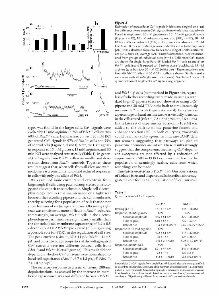
types was found in the larger cells. Ca2+ signals wereevoked by 15 mM arginine in 75% of Pdx1+/– cells versus68% of Pdx1+/+ cells. Depolarization with 30 mM KClgenerated Ca2+ signals in 97% of Pdx1+/– cells and 99%of control cells (Figure 3, d and f). Next, the Ca2+ signalsin response to 15 mM glucose, 15 mM arginine, and 30mM KCl were analyzed statistically (Table 1). In gener-al, Ca2+ signals from Pdx1+/– cells were smaller and slow-er than those from Pdx1+/+ controls. Together, theseresults suggest that, when cells from all islets are exam-ined, there is a general trend toward reduced responsesin cells with only one allele of Pdx1.
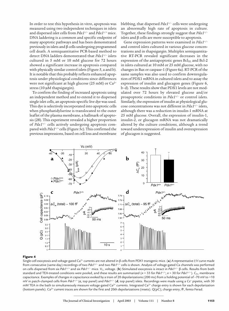
We examined ionic currents and exocytosis fromlarge single β cells using patch-clamp electrophysiolo-gy and the capacitance technique. Single-cell electro-physiology requires the maintenance of a tight sealbetween the recording pipette and the cell membrane,thereby selecting for a population of cells that do notshow features of end-stage apoptosis. Obtaining tightseals was consistently more difficult in Pdx1+/– cultures.Interestingly, on average, Pdx1+/– cells in the electro-physiology experiments were significantly smaller thanthe controls (basal membrane capacitance of 3.9 ± 0.5Pdx1+/– vs. 5.2 ± 0.2 Pdx1+/+ pico Farad (pF), suggestinga possible role for PDX1 in the regulation of cell size.The peak current (Pdx1+/–, 37 ± 11 pA; Pdx1+/+, 41 ± 5pA) and current-voltage properties of the voltage-gatedCa2+ currents were not different between cells fromPdx1+/– and Pdx1+/+ islets (Figure 4a). This result did notdepend on whether Ca2+ currents were normalized tobasal cell capacitance (Pdx1+/–, 6.7 ± 1.2 pA/pF; Pdx1+/+,7.4 ± 0.6 pA/pF).
The secretory response to a train of twenty 200-msdepolarizations, as assayed by the increase in mem-brane capacitance, was not different between Pdx1+/–
and Pdx1+/+ β cells (summarized in Figure 4b), regard-less of whether recordings were made in using a stan-dard high-K+ pipette (data not shown) or using a Cs+
pipette and 30 mM TEA in the bath to simultaneouslymeasure Ca2+ currents (Figure 4, c and d). Exocytosis asa percentage of basal surface area was virtually identicalin the cells tested (Pdx1+/–, 7.2 ± 2.4%; Pdx1+/+, 7.6 ± 1.6%).In the later set of experiments, forskolin (10 µM) wasadded to the bath to mimic paracrine factors andenhance secretion (30). In both cell types, exocytosiscould be enhanced by agents that increase cAMP (datanot shown), suggesting that pathways coupled toparacrine hormones are intact. These results stronglysuggest that the components mediating Ca2+-depend-ent exocytosis are not affected by a reduction ofapproximately 50% in PDX1 expression, at least in thepopulation of seemingly healthy cells from whichrecordings can be made.
Susceptibility to apoptosis in Pdx1+/– islets. Our observationsof isolated islets and dispersed cells described above sug-gested a role for PDX1 in regulation of β cell survival.
1152 The Journal of Clinical Investigation | April 2003 | Volume 111 | Number 8
Figure 3Estimation of intracellular Ca2+ signals in islets and single β cells. (a)No differences were seen in Ca2+ signals from whole islets loaded withFura-2 in response to 20 mM glucose (n = 20), 10 mM glyceraldehyde(Glycer; n = 12), 10 mM α-ketoisocaproic acid (KIC; n = 12), 20 mMKCl (n = 50), or carbachol (Cch; in the presence or absence of 2 mMEGTA; n = 8 for each). Average area under the curve (arbitrary units[AU]) was calculated from raw traces consisting of unitless ratio val-ues (340/380). (b) Average NADH autofluorescence (AU) was meas-ured from groups of individual islets (n = 8). Calibrated Ca2+ tracesare shown for single, large Fura-4F–loaded Pdx1+/+ cells (c and d) orPdx1+/– cells (e and f) exposed to 15 mM glucose (black bars), 15 mMarginine (gray bars), or 30 mM KCl (white bars). Representative tracesfrom 66 Pdx1+/+ cells and 35 Pdx1+/– cells are shown. Similar resultswere seen with 20 mM glucose (not shown). See Table 1 for a fullquantification of single-cell Ca2+ signals. arg, arginine.
Table 1Quantification of Ca2+ signals
Pdx1+/+ Pdx1+/–
Resting [Ca2+]i 161 ± 16 nM 145 ± 16 nMResponse, 15 mM glucose 68% 63%
Maximal amplitude 485 ± 51 nM 329 ± 35 nMA
Time to peak 510 ± 53 s 737 ± 52 sA
Rate of rise 1.3 ± 0.16 nM/s 0.52 ± 0.09 nM/sA
Response to 15 mM arginine 68% 75%Maximal amplitude 422 ± 51 nM 218 ± 32 nMA
Time to peak 78 ± 13 s 124 ± 28 sA
Rate of rise 9.6 ± 2.1 nM/s 3.25 ± 1.2 nM/sA
Response, 30 mM KCI 99% 97%Maximal amplitude 591 ± 47 nM 341 ± 16 nMA
Time to peak 92 ± 17 s 127 ± 8 sA
Rate of rise 6.2 ± 1.1 nM/s 5.6 ± 0.6 nM/s
Intracellular (i) Ca2+ signals from single Fura-4F–loaded islet cells were quantifiedas described in Methods. Cells were obtained from all isolated islets (i.e., not hand-picked or size-matched). Maximal amplitude is calculated as maximum increasefrom baseline. Rate of rise is calculated as maximal amplitude/time to maximalamplitude. ASignificantly different from control. KCI, potassium chloride.
In order to test this hypothesis in vitro, apoptosis wasmeasured using two independent techniques in isletsand dispersed islet cells from Pdx1+/– and Pdx1+/+ mice.DNA laddering is a common and specific endpoint ofmany apoptotic pathways and has been demonstratedpreviously in islets and β cells undergoing programmedcell death. A semiquantitative PCR-based method todetect DNA ladders demonstrated that Pdx1+/– isletscultured in 5 mM or 10 mM glucose for 72 hoursshowed a significant increase in apoptosis comparedwith physically similar control islets (Figure 5, a and b).It is notable that this probably reflects enhanced apop-tosis under physiological conditions since differenceswere not significant at high glucose (25 mM) or Ca2+
stress (10 µM thapsigargin).To confirm the finding of increased apoptosis using
an independent method and to extend it to dispersedsingle islet cells, an apoptosis-specific live dye was used.This dye is selectively incorporated into apoptotic cellswhen phosphatidylserine is translocated to the outerleaflet of the plasma membrane, a hallmark of apopto-sis (28). This experiment revealed a higher proportionof Pdx1+/– cells actively undergoing apoptosis com-pared with Pdx1+/+ cells (Figure 5c). This confirmed theprevious impressions, based on cell loss and membrane
blebbing, that dispersed Pdx1+/– cells were undergoingan abnormally high rate of apoptosis in culture.Together, these findings strongly suggest that Pdx1+/–
islets and β cells are more susceptible to apoptosis.Gene expression patterns were examined in Pdx1+/–
and control islets cultured in various glucose concen-trations and in thapsigargin. Multiplex semiquantita-tive RT-PCR revealed significant decreases in theexpression of the antiapoptotic genes BclXL and Bcl-2in islets cultured at 10 mM or 25 mM glucose, with nochanges in Bax or caspase-1 (Figure 6a). RT-PCR of thesame samples was also used to confirm downregula-tion of PDX1 mRNA in cultured islets and to assay theexpression of insulin and glucagon genes (Figure 6,b–d). These results show that PDX1 levels are not mod-ulated over 72 hours by elevated glucose and/orproapoptotic conditions in Pdx1+/– or control islets.Similarly, the expression of insulin at physiological glu-cose concentrations was not different in Pdx1+/– islets,although there was a reduction in insulin-1 mRNA at25 mM glucose. Overall, the expression of insulin-1,insulin-2, or glucagon mRNA was not dramaticallyaltered by the culture conditions, although a trendtoward underexpression of insulin and overexpressionof glucagon is suggested.
The Journal of Clinical Investigation | April 2003 | Volume 111 | Number 8 1153
Figure 4Single-cell exocytosis and voltage-gated Ca2+ currents are not altered in β cells from PDX1 transgenic mice. (a) A representative I/V curve madefrom consecutive (same-day) recordings of two Pdx1+/– and two Pdx1+/+ cells is shown. Analysis of voltage-gated Ca channels was performedon cells dispersed from six Pdx1+/+ and six Pdx1+/– mice. VC, voltage. (b) Stimulated exocytosis is intact in Pdx1+/– β cells. Results from bothstandard and TEA-treated conditions were pooled, and these results are summarized (n = 33 for Pdx1+/+; n = 30 for Pdx1+/–). Cm, membranecapacitance. Examples of changes in capacitance evoked by a train of 20 depolarizations (200 ms) from a holding potential of –70 mV to +10mV in patch-clamped cells from Pdx1+/+ (c, top panel) and Pdx1+/– (d, top panel) islets. Recordings were made using a Cs+ pipette, with 30mM TEA in the bath to simultaneously measure voltage-gated Ca2+ currents. Integrated Ca2+ charge entry is shown for each depolarization(bottom panels). Ca2+ current traces are shown for the first and 20th depolarizations (insets). Q(pC); charge entry; fF, femto Ferad.

In order to put these apoptosis experiments into con-text with the secretion experiments, the media super-natants from the DNA-ladder experiments was savedand analyzed for hormone secretion. Long-term C-pep-tide secretion was not significantly different in Pdx1+/–
islets under any of the conditions used (data not shown).These results suggest that the apoptosis-related defect infunctional islet cell mass precedes a defect in glucose-stimulated insulin secretion and may also suggest that asubpopulation of islets or islet cells is sufficient to main-tain normal response to glucose over 72 hours.
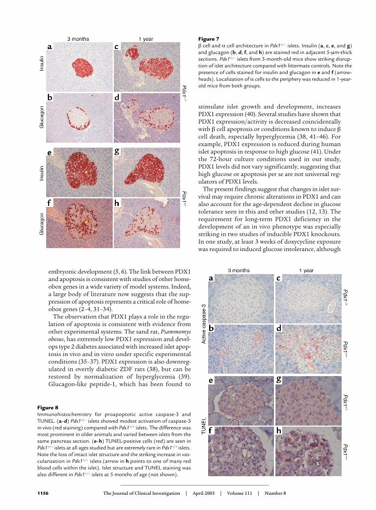
Analysis of pancreatic morphology and β cell mass. Todetermine whether enhanced apoptosis and its eventu-al consequence, reduced β cell mass, could be detectedin vivo, we examined pancreas sections from Pdx1+/–
and Pdx1+/+ mice at 3, 5, and 12 months of age. At allages studied, Pdx1+/– mice showed glucagon-positive αcells within the core of a large percentage of islets (Fig-ure 7). Glucagon staining was observed within the cores
of 1-year-old islets from both genotypes. In agreementwith a previous report (9), we observed an increasedratio of α cells to β cells in younger Pdx1+/– islets. Athigher magnification, adjacent sections showed someislets with cells staining positive for both insulin andglucagon, as previously described for the β cell–specif-ic PDX1 knockout (11). Immunohistochemistry for theproapoptotic (cleaved) form of caspase-3 revealed spo-radic staining that was more common in Pdx1+/– isletsand increased substantially with age (Figure 8, a–d).Similarly, TUNEL-positive cells, though rare, werefound almost exclusively in islets (Figure 8, e–h) andacinar cells (not shown) of Pdx1+/– mice. FrequentlyPdx1+/– islets showed marked expansion of the isletmicrovasculature compared with controls. However,even at 1 year of age, some Pdx1+/– islets showed nosigns of damage or apoptosis. Together, these findingsprovide direct evidence for the regulation of apoptosisby PDX1 in vivo.
The low frequency of cells staining positive forTUNEL or active caspase-3 was not unexpected sinceapoptotic cells are rapidly cleared by the immune sys-tem in vivo. Although more common in 12-month-oldanimals, we observed lymphocyte infiltration at theperiphery of islets in Pdx1+/– mice of all ages, suggest-ing an immune response. Some of the infiltrating lym-phocytes exhibited TUNEL staining (Figure 9a) orcleaved caspase-3 immunoreactivity (not shown). Stain-ing for Ki67, a nuclear antigen specific to proliferatingcells, revealed clusters of positive cells in the invadinglymphocytes in Pdx1+/– islets (Figure 9b), confoundingattempts to quantify β cell proliferation.
Finally, we asked whether the apoptotic effects ofPdx1 haploinsufficiency correspond to altered isletsurvival or neogenesis in vivo by assessing pancreaticβ cell mass. Unlike wild-type mice, which compensatefor age with increased β cell mass, 1-year-old Pdx1+/–
mice had a greater than 50% reduction in normalizedβ cell surface area compared with littermate controls.This difference was due to approximately 50% fewerislets, rather than a significant decrease in the averagearea of individual isles. (Figure 10). The change in βcell mass was mirrored by a decrease in normalizedpancreatic insulin content in older Pdx1+/– mice(expressed as ng/ml/mg pancreatic weight: Pdx1+/–,226 ± 38; Pdx1+/+, 521 ± 70), but not at 8–10 weeks(Pdx1+/–, 560 ± 27 ng/ml/mg; Pdx1+/+, 491 ± 42 ng/ml/mg; n = 6 for each genotype and age). The reduction inpancreatic insulin content was significant whetherexpressed as an absolute or normalized to pancreaticweight. Animal weights and pancreatic weights werenot different at any age studied. Therefore, lower pan-creatic insulin content in older mice can be explainedby a decreased number of functional islets, rather thana reduction in insulin stored in single β cells. The sub-stantial decrease in β cell mass over time, along withdefects in architecture and differentiation of someislets, support the idea that Pdx1+/– islets undergoapoptosis and dedifferentiation in vivo.
1154 The Journal of Clinical Investigation | April 2003 | Volume 111 | Number 8
Figure 5Pdx1+/– islets and cells are prone to apoptosis when cultured in lowglucose concentrations. (a) Apoptosis, measured by PCR-enhancedDNA laddering, was compared in groups of five islets cultured inRPMI with 5 mM, 10 mM, and 25 mM glucose for 72 hours (n = 10for each genotype). Islets cultured in 10 µM thapsigargin (Tg), aknown inducer of islet apoptosis, were used as a positive control.Apoptotic calf-thymus DNA served as an additional reference con-trol, independent of our cultures. DNA-ladder bands were quanti-fied by densitometry and pooled as described in Methods. (b) A rep-resentative gel is shown. (c) The average percentage of apoptotic cellsmeasured in dispersed islet cells cultured overnight in 5 mM glucose,measured by cell uptake of a specific dye (Methods). Cells werecounted manually in phenol red–free RPMI. Shown are pooled resultsfrom three coverslips of islets dispersed from three mice of eachgenotype. Asterisks denote significant differences.

DiscussionThe present study was undertaken to explore themechanisms responsible for the defective insulin secre-tion resulting from decreased expression of PDX1 inmice in vivo. The two major findings are as follows.First, islets and single β cells isolated from Pdx1+/– mice(expressing 50% of the normal levels of PDX1) candemonstrate normal glucose sensing, stimulus-secre-tion coupling, and insulin secretion. Second, a 50%reduction in PDX1 predisposes islets to apoptosis in
vitro and in vivo, leading to disrupted islet architectureand eventually to a marked decrease in β cell area andislet number. Since the maintenance of adequateinsulin secretion involves the relative rates of β cell pro-liferation/neogenesis and death, these findings identi-fy an important factor contributing to the complexphenotype of Pdx1+/– mice. They suggest that PDX1plays an important role in the development and main-tenance of an adequate pool of healthy islets in theadult animal in addition to its well established role in
The Journal of Clinical Investigation | April 2003 | Volume 111 | Number 8 1155
Figure 6RT-PCR analysis of gene expression. (a) The densitometric quantification of four apoptosis-related genes — BclXL, caspase-3 (Casp), BAX,and Bcl-2 — relative to GAPDH in cultured islets (cultured as in Figure 5) is quantified by densitometry (above). Data are pooled from fourgels (representative example shown below). White bars over gel images denote Pdx1+/+ islets, while black bars denote Pdx1+/– islets. EachRT-PCR sample was pooled from the cultured islets of three mice. (b–d) Relative abundance of PDX1, insulin-1 (Ins-1), insulin-2 (Ins-2),and glucagon (Gluca) mRNA are quantified from the same samples. Single asterisks denote significant differences between Pdx1+/– andPdx1+/+ islets. Double asterisks denote significant differences between different treatments to the same type of islet. Insulin content per isletprotein was also not reduced in Pdx1+/– islets (not shown).
embryonic development (5, 6). The link between PDX1and apoptosis is consistent with studies of other home-obox genes in a wide variety of model systems. Indeed,a large body of literature now suggests that the sup-pression of apoptosis represents a critical role of home-obox genes (2–4, 31–34).
The observation that PDX1 plays a role in the regu-lation of apoptosis is consistent with evidence fromother experimental systems. The sand rat, Psammomysobesus, has extremely low PDX1 expression and devel-ops type 2 diabetes associated with increased islet apop-tosis in vivo and in vitro under specific experimentalconditions (35–37). PDX1 expression is also downreg-ulated in overtly diabetic ZDF rats (38), but can berestored by normalization of hyperglycemia (39).Glucagon-like peptide-1, which has been found to
stimulate islet growth and development, increasesPDX1 expression (40). Several studies have shown thatPDX1 expression/activity is decreased coincidentallywith β cell apoptosis or conditions known to induce βcell death, especially hyperglycemia (38, 41–46). Forexample, PDX1 expression is reduced during humanislet apoptosis in response to high glucose (41). Underthe 72-hour culture conditions used in our study,PDX1 levels did not vary significantly, suggesting thathigh glucose or apoptosis per se are not universal reg-ulators of PDX1 levels.
The present findings suggest that changes in islet sur-vival may require chronic alterations in PDX1 and canalso account for the age-dependent decline in glucosetolerance seen in this and other studies (12, 13). Therequirement for long-term PDX1 deficiency in thedevelopment of an in vivo phenotype was especiallystriking in two studies of inducible PDX1 knockouts.In one study, at least 3 weeks of doxycycline exposurewas required to induced glucose intolerance, although
1156 The Journal of Clinical Investigation | April 2003 | Volume 111 | Number 8
Figure 7β cell and α cell architecture in Pdx1+/– islets. Insulin (a, c, e, and g)and glucagon (b, d, f, and h) are stained red in adjacent 5-µm-thicksections. Pdx1+/– islets from 3-month-old mice show striking disrup-tion of islet architecture compared with littermate controls. Note thepresence of cells stained for insulin and glucagon in e and f (arrow-heads). Localization of α cells to the periphery was reduced in 1-year-old mice from both groups.
Figure 8Immunohistochemistry for proapoptotic active caspase-3 andTUNEL. (a–d) Pdx1+/– islets showed modest activation of caspase-3in vivo (red staining) compared with Pdx1+/+ islets. The difference wasmost prominent in older animals and varied between islets from thesame pancreas section. (e–h) TUNEL-positive cells (red) are seen inPdx1+/– islets at all ages studied but are extremely rare in Pdx1+/+ islets.Note the loss of intact islet structure and the striking increase in vas-cularization in Pdx1+/– islets (arrow in h points to one of many redblood cells within the islet). Islet structure and TUNEL staining wasalso different in Pdx1+/– islets at 5 months of age (not shown).

the PDX1 antisense RNA was induced within 24 hours(10). Similarly, after chronic doxycycline treatment,fasting hyperglycemia was seen in 18-month-old, butnot 8-month-old, mice transgenic for an induciblePDX1 antisense/ribozyme construct capable of reduc-ing PDX1 expression in transfected β cells within 72hours (12). It has been noted that in humans, the sever-ity of the PDX1 mutation correlates with age of dia-betes onset (12, 47–49). Interestingly, results frominducible and classical transgenic models of PDX1deficiency concur with our findings that males are pref-erentially affected (13).
The mechanism by which PDX1 affects apoptosis isunclear. However, the coinciding reduction of BclXL
and Bcl-2 gene expression (and their expression relativeto Bax) at 10 mM glucose points to established media-tors of β cell apoptosis. It is also possible that the sus-ceptibility to apoptosis may be related to the subtlechanges in Ca2+ signaling seen in Pdx1+/– cells. Calciumfluxes regulate many cellular functions in neuroen-docrine cells, including metabolism, gene transcrip-tion, protein synthesis, protein sorting/packaging, andexocytosis (50). Ca2+ dysregulation has been implicat-ed in apoptosis in many cell types (51). In excitable cellssuch as neurons and β cells, moderately elevated Ca2+
and electrical activity may have protective effects.Evidence is emerging that PDX1 may act downstream
of prosurvival autocrine insulin signaling in β cells.PDX1 can be activated by insulin and its downstreamsignaling elements, such as Foxo1 (52, 53). Recently,Kushner et al. have reported that transgenic PDX1expression restored β cell mass and pancreatic functionin IRS2–/– mice, a model that displays increased β cellapoptosis (54). These findings provide important sup-port for the hypothesis that PDX1 acts primarilythrough cell survival pathways in vivo and can modu-late prosurvival insulin/IGF signaling.
The results of the present study suggest that PDX1-regulated apoptosis is physiologically important in thelifelong maintenance of β cell mass. Given the disrup-tion in β cell/α cell organization and the altered isletmorphology of Pdx1+/– islets, it is likely that a significantloss of functionalβ cell mass precedes measurable changesin morphological β cell mass. This is not unexpected, since
subtle changes in pancreasphysiology can be readilymeasured with accuracyusing the perfused pancreas,whereas histological parame-ters such as β cell mass aremore difficult to determine.According to a previous study(9), morphological β cell massin Pdx1+/– pancreata is re-duced at 15 weeks by 37%(Pdx1+/–, 1.76 ± 0.13 mg vs.Pdx1+/+, 2.8 ± 0.44 mg), where-as Shih and colleagues foundno decrease in β cell area at a
similar age (55). In experimental models such as Pdx1+/–
mice, functional β cell mass may be substantially lowerthan β cell mass estimated using classical morphomet-ric criteria because apoptotic or poorly functioning isletsare still counted if they contain insulin.
The specialized architecture and relative distribu-tion of islet cell types may also play important roles inislet function and survival. For example, disruption ofcontacts between β cells may reduce the secretory effi-ciency of islets, thereby requiring higher (potentiallytoxic) levels of Ca2+ to maintain secretion. We detect-ed striking changes in islet architecture in Pdx1+/–
The Journal of Clinical Investigation | April 2003 | Volume 111 | Number 8 1157
Figure 9Evidence of enhanced pancreatic immune activity in Pdx1+/– mice. Infiltration of lymphocytes, somepositive for TUNEL (a), were seen within or around islets (arrow) in old mice. Immunofluorescentlocalization of Ki67 nuclear antigen (b) reveals proliferation of lymphocytes within Pdx1+/– islets(arrows in b and c point to the same islet, also delineated by higher-density nuclear DAPI staining(c). Infiltrating lymphocytes are distinguished from islet cells by their small size and low-intensitynuclear staining with DAPI.
Figure 10Reduced β cell mass and islet number in Pdx1+/– mice. (a) Islet cellarea at 3, 5, and 12 months was estimated using insulin immunore-activity as described in Methods and normalized to the total pan-creatic cross-sectional area. Four sections were analyzed from eachanimal. (b) The number of islets was counted in complete pancreat-ic sections at low magnification (1.25× objective). Asterisks denotesignificant difference from wild type. Number of mice studied at 3months, 5 months, and 12 months, respectively: Pdx1+/+, n = 4, 2,and 5; Pdx1+/–, n = 4, 2, and 4.

mice as an early defect. Similarly, islets with condi-tional deletion of PDX1 displayed a marked loss ofthe typical islet architecture (11). Whether this is acause or effect of enhanced apoptosis remains to bedetermined. It has been suggested that specific bind-ing of PDX1 to the glucagon promoter (14) may beimportant for the suppression of the default α celldevelopmental pathway of β cell precursors. Thus, itis possible that apoptosis caused by PDX1 deficiencyleads to, or is associated with, relative dedifferentia-tion of some islets/β cells and the relative enhance-ment of a pool of less differentiated islet cells. In theβ cell–specific PDX1 knockout model, approximately22% of insulin-positive cells also expressed glucagon(11). Our study clearly shows that PDX1 is importantnot only in the normal development of the islet butalso plays a critical role in the maintenance of normalislet function later in life by determining the balanceof cellular proliferation and death (20).
A major finding of the present study is that a 50%reduction in PDX1 gene expression is still compatiblewith normal glucose sensing, insulin gene expression,or stimulus-secretion coupling in isolated islets and βcells. Although several lines of evidence have suggesteda paramount role for PDX1 in pancreatic genesis andislet development, whether PDX1 plays specific physio-logical roles in fully differentiated β cells has remainedless clear. For example, glucose has been shown to phos-phorylate PDX1 and to activate PDX1 DNA bindingactivity (53, 56). Glucose-stimulated changes in the sub-cellular location of PDX1 have also been reported bycertain groups (57). However, the exact relationshipbetween these events and changes in the function ofmaximally differentiated β cells remain unclear.
The major physiological role ascribed to PDX1 inthe adult β cell is the control of insulin gene expres-sion (8). In support of this hypothesis, PDX1 trans-fection permitted the elevation of insulin mRNA inresponse to high glucose in the PDX1–deficientNES2Y cell line derived from a patient with persistenthyperinsulinemic hypoglycemia of infancy (58). Otherevidence for the involvement of PDX1 in insulinexpression comes from the ability of PDX1 to pro-mote ectopic insulin expression in the presence ofother transcription factors (16, 59). However, resultsfrom the present study and others (11, 13, 55) suggesta less important role for PDX1 in the regulation ofinsulin mRNA and protein levels in vivo. Clearly, pan-creatic insulin content and gene expression are nor-mal at a time when PDX1 levels are significantlyreduced, but become abnormal only in older, moreovertly diabetic mice. Even then, part of the reductionin pancreatic insulin content may be accounted for bya reduced number of islets. In other studies, whilePDX1 cotransfection potentiated the ability of otherfactors to activate insulin expression in non–β cells,this transfection alone was not sufficient (60). Simi-larly, there are now many examples in the literaturewhere no correlation between PDX1 and insulin
expression is found, including PDX1-positive cellsexpanded from human islets (61), antisense-treatedMIN6 cells (62), and an insulin-secreting tumor (63).Perhaps most importantly, PDX1 is expressed at somelevel in several other cell types of the pancreas andforegut, whereas insulin is not (64). Therefore, evi-dence that PDX1 is either necessary or sufficient todrive a significant proportion of β cell insulin expres-sion in vivo has not yet been presented. Interestingly,Psammomys obesus islets and NES2Y cells express highbasal levels of insulin despite PDX1 deficiency, butshow marked defects in glucose-stimulated insulingene expression (35, 65). Perhaps PDX1 is involved,directly or indirectly, in glucose-stimulated insulingene transcription, but is redundant for the controlof basal insulin levels under normal conditions invivo. Studies of insulin levels in single PDX1-deficientβ cells are required.
Our measurements of normal pancreatic insulincontent, islet insulin staining, and islet insulin mRNAin young Pdx1+/– mice suggest that apoptosis and theinsulin secretory defects seen in vivo precede anyeffects on insulin levels. It is therefore likely that thereduction of pancreatic insulin content that accom-panies reduced β cell mass, islet number, and alter-ations in islet architecture is secondary to increased βcell apoptosis. Whereas insulin gene expression wasreduced in apoptotic human islets cultured in highglucose (41), we did not find significant differences ininsulin mRNA under acute apoptotic conditions innormal wild-type mouse islets, suggesting a require-ment for chronic exposure.
Our in vivo data confirm the findings of several othergroups using global Pdx1+/–, β cell–specific Pdx1–/–, andinducible transgenic PDX1 knockdown mice (9–13).Our measurements of insulin secretion in response toglucose in the perfused whole pancreas are similar tothose recently reported for another strain of Pdx1+/– mice(13). However, building on these previous observationsto include the characterization of glucose signaling andinsulin secretion from isolated islets and single cells hasled to a greater appreciation of the complexity of thePdx1+/– phenotype. In addition, our study provides anexample wherein the assumption that the response ofthe whole pancreas can be recapitulated in individualisolated islets may not always be correct. Likewise, theheterogeneous responses of single cells are more com-plex than would be predicted from the activity of wholeislets. Unfortunately, many studies of transgenic mice,from which conclusions regarding the cell biology orbiophysics of a certain cell type are made, fail to test suchassumptions by examining single cells. Our study alsomakes clear how difficult it can be to dissociate physiol-ogy from development in such experiments.
In addition to the role of PDX1 in MODY4, recentstudies of European families have suggested that muta-tions in the PDX1 gene may predispose to type 2 diabetes(8). Interestingly, deficiency in HNF-1α (MODY2) selec-tively promotes BclXL-sensitive apoptosis at physiological
1158 The Journal of Clinical Investigation | April 2003 | Volume 111 | Number 8
glucose levels in INS-1 cells (66), implying the existenceof a common pathway regulating apoptosis that maycontribute to both MODY and type 2 diabetes. As inthe Psammomys obesus model, continuous nutrientstress may be necessary to induce pathological conse-quences. Our data show that partial PDX1 deficiencyhas the greatest effect later in life. The age-dependentprogression to type 2 diabetes makes Pdx1+/– mice oneof the most realistic animal models of the disease wherethe pancreatic defect is the primary cause. Examinationof middle-aged mice revealed that the function ofPDX1 persists through adulthood, resulting in β cellmass and islet numbers roughly halfway between thatof wild-type mice and Pdx1–/– mice (which have noislets). The process by which PDX1 heterozygosity iscompensated for until after 3 months of age isunknown, but may represent an important target path-way for therapeutic intervention in type 2 diabetes. Forexample, it has recently been shown that reducingPDX1 in βTC3 cells activates the PDX1 promoter, sug-gesting that PDX1 levels are partially controlled in anautoregulatory loop (12). In summary, our data indi-cate that partial PDX1 deficiency leads to organ-leveldefects in insulin secretion by complex mechanismsthat include islet apoptosis and disrupted islet archi-tecture, but not a ubiquitous or cell-autonomousdefect in β cell stimulus-secretion coupling. Hence, amore complete understanding of the roles of PDX1and apoptosis in the maintenance of functional β cellmass may point to novel approaches to slow the pro-gression of diabetes mellitus.
AcknowledgmentsThis work was supported by NIH grants to K.S. Polon-sky (DK-31842 and DK-44860), S. Misler (DK-37380),and the Diabetes Research and Training Center atWashington University School of Medicine (P60 DK-20579). The support of the Blum Kovler Foundation isalso gratefully acknowledged. We thank ErnestoBernal-Mizrachi and Matteo Levisetti for helpful com-ments, William Pugh for expert technical assistancewith the perfused pancreas, and Eric Ford for technicalassistance and islet isolation. The assistance of SeamusSreenan, Yun-Ping Zhou, and Diane Ostrega is alsoacknowledged. J.D. Johnson was supported by fellow-ship awards from the Juvenile Diabetes Research Foun-dation and the Natural Science and EngineeringResearch Council of Canada.
1. Sommer, R.J., et al. 1998. The Pristionchus HOX gene Ppa-lin-39 inhibitsprogrammed cell death to specify the vulva equivalence group and is notrequired during vulval induction. Development. 125:3865–3873.
2. Rhinn, M., Dierich, A., Le Meur, M., and Ang, S. 1999. Cell autonomousand non-cell autonomous functions of Otx2 in patterning the rostralbrain. Development. 126:4295–4304.
3. Izon, D.J., et al. 1998. Loss of function of the homeobox gene Hoxa-9 per-turbs early T-cell development and induces apoptosis in primitive thy-mocytes. Blood. 92:383–393.
4. Dear, T.N., et al. 1995. The Hox11 gene is essential for cell survival dur-ing spleen development. Development. 121:2909–2915.
5. Offield, M.F., et al. 1996. PDX-1 is required for pancreatic outgrowthand differentiation of the rostral duodenum. Development. 122:983–995.
6. Jonsson, J., Carlsson, L., Edlund, T., and Edlund, H. 1994. Insulin-
promoter-factor 1 is required for pancreas development in mice.Nature. 371:606–609.
7. Stoffers, D.A., Zinkin, N.T., Stanojevic, V., Clarke, W.L., and Habener, J.F.1997. Pancreatic agenesis attributable to a single nucleotide deletion inthe human IPF1 gene coding sequence. Nat. Genet. 15:106–110.
8. McKinnon, C.M., and Docherty, K. 2001. Pancreatic duodenal home-obox-1, PDX-1, a major regulator of beta cell identity and function. Diabetologia. 44:1203–1214.
9. Dutta, S., Bonner-Weir, S., Montminy, M., and Wright, C. 1998. Regula-tory factor linked to late-onset diabetes? Nature. 392:560.
10. Lottmann, H., Vanselow, J., Hessabi, B., and Walther, R. 2001. The Tet-On system in transgenic mice: inhibition of the mouse pdx-1 gene activ-ity by antisense RNA expression in pancreatic beta-cells. J. Mol. Med.79:321–328.
11. Ahlgren, U., Jonsson, J., Jonsson, L., Simu, K., and Edlund, H. 1998. beta-cell-specific inactivation of the mouse Ipf1/Pdx1 gene results in loss ofthe beta-cell phenotype and maturity onset diabetes. Genes Dev.12:1763–1768.
12. Thomas, M.K., et al. 2001. Development of diabetes mellitus in aging trans-genic mice following suppression of pancreatic homeoprotein IDX-1. J. Clin. Invest. 108:319–329. doi:10.1172/JCI200112029.
13. Brissova, M., et al. 2002. Reduction in pancreatic transcription factorPDX-1 impairs glucose-stimulated insulin secretion. J. Biol. Chem.227:11225–11232.
14. Chakrabarti, S.K., James, J.C., and Mirmira, R.G. 2002. Quantitativeassessment of gene targeting in vitro and in vivo by the pancreatic tran-scription factor, Pdx1. Importance of chromatin structure in directingpromoter binding. J. Biol. Chem. 277:13286–13293.
15. Ohlsson, H., Karlsson, K., and Edlund, T. 1993. IPF1, a homeodomain-containing transactivator of the insulin gene. EMBO J. 12:4251–4259.
16. Serup, P., et al. 1996. Induction of insulin and islet amyloid polypeptideproduction in pancreatic islet glucagonoma cells by insulin promoterfactor 1. Proc. Natl. Acad. Sci. U. S. A. 93:9015–9020.
17. Macfarlane, W.M., et al. 2000. Glucose regulates islet amyloid polypep-tide gene transcription in a PDX1- and calcium-dependent manner. J. Biol. Chem. 275:15330–15335.
18. Watada, H., et al. 1996. The human glucokinase gene beta-cell-type pro-moter: an essential role of insulin promoter factor 1/PDX-1 in its acti-vation in HIT-T15 cells. Diabetes. 45:1478–1488.
19. Waeber, G., Thompson, N., Nicod, P., and Bonny, C. 1996. Transcrip-tional activation of the GLUT2 gene by the IPF-1/STF-1/IDX-1 home-obox factor. Mol. Endocrinol. 10:1327–1334.
20. Bonner-Weir, S. 2000. Perspective: postnatal pancreatic beta cell growth.Endocrinology. 141:1926–1929.
21. Pontoglio, M., et al. 1998. Defective insulin secretion in hepatocytenuclear factor 1α-deficient mice. J. Clin. Invest. 101:2215–2222.
22. Lacy, P.E., and Kostianovsky, M. 1967. Method for the isolation of intactislets of Langerhans from the rat pancreas. Diabetes. 16:35–39.
23. Zhou, Y.P., et al. 2000. Overexpression of Bcl-x(L) in beta-cells preventscell death but impairs mitochondrial signal for insulin secretion. Am. J.Physiol. Endocrinol. Metab. 278:E340–E351.
24. Grynkiewicz, G., Poenie, M., and Tsien, R.Y. 1985. A new generation ofCa2+ indicators with greatly improved fluorescence properties. J. Biol.Chem. 260:3440–3450.
25. Johnson, J.D., Van Goor, F., Wong, C.J., Goldberg, J.I., and Chang, J.P.1999. Two endogenous gonadotropin-releasing hormones generate dis-similar Ca(2+) signals in identified goldfish gonadotropes. Gen. Comp.Endocrinol. 116:178–191.
26. Barnett, D.W., and Misler, S. 1997. An optimized approach to membranecapacitance estimation using dual-frequency excitation. Biophys. J.72:1641–1658.
27. Zhou, Y.P., et al. 1998. Apoptosis in insulin-secreting cells. Evidence forthe role of intracellular Ca2+ stores and arachidonic acid metabolism. J. Clin. Invest. 101:1623–1632.
28. Fadok, V.A., et al. 1992. Exposure of phosphatidylserine on the surfaceof apoptotic lymphocytes triggers specific recognition and removal bymacrophages. J. Immunol. 148:2207–2216.
29. Wong, H., Anderson, W.D., Cheng, T., and Riabowol, K.T. 1994. Moni-toring mRNA expression by polymerase chain reaction: the “primer-dropping” method. Anal. Biochem. 223:251–258.
30. Gillis, K.D., and Misler, S. 1993. Enhancers of cytosolic cAMP augmentdepolarization-induced exocytosis from pancreatic B-cells: evidence foreffects distal to Ca2+ entry. Pflugers Arch. 424:195–197.
31. Raman, V., et al. 2000. Compromised HOXA5 function can limit p53expression in human breast tumours. Nature. 405:974–978.
32. Quaggin, S.E., Yeger, H., and Igarashi, P. 1997. Antisense oligonu-cleotides to Cux-1, a Cut-related homeobox gene, cause increased apop-tosis in mouse embryonic kidney cultures. J. Clin. Invest. 99:718–724.
33. Shimamoto, T., Nakamura, S., Bollekens, J., Ruddle, F.H., and Takeshi-ta, K. 1997. Inhibition of DLX-7 homeobox gene causes decreasedexpression of GATA-1 and c-myc genes and apoptosis. Proc. Natl. Acad.Sci. U. S. A. 94:3245–3249.
The Journal of Clinical Investigation | April 2003 | Volume 111 | Number 8 1159
34. Shimamoto, T., Ohyashiki, K., and Takeshita, K. 2000. Overexpressionof the homeobox gene DLX-7 inhibits apoptosis by induced expressionof intercellular adhesion molecule-1. Exp. Hematol. 28:433–441.
35. Leibowitz, G., et al. 2001. IPF1/PDX1 deficiency and beta-cell dysfunc-tion in Psammomys obesus, an animal with type 2 diabetes. Diabetes.50:1799–1806.
36. Leibowitz, G., et al. 2001. Beta-cell glucotoxicity in the Psammomys obe-sus model of type 2 diabetes. Diabetes. 50:S113–S117.
37. Donath, M.Y., Gross, D.J., Cerasi, E., and Kaiser, N. 1999. Hyperglycemia-induced beta-cell apoptosis in pancreatic islets of Psammomys obesusduring development of diabetes. Diabetes. 48:738–744.
38. Seufert, J., Weir, G.C., and Habener, J.F. 1998. Differential expression ofthe insulin gene transcriptional repressor CCAAT/enhancer-bindingprotein β and transactivator islet duodenum homeobox-1 in rat pan-creatic β cells during the development of diabetes mellitus. J. Clin. Invest.101:2528–2539.
39. Harmon, J.S., et al. 1999. In vivo prevention of hyperglycemia also pre-vents glucotoxic effects on PDX-1 and insulin gene expression. Diabetes.48:1995–2000.
40. Stoffers, D.A., et al. 2000. Insulinotropic glucagon-like peptide 1 ago-nists stimulate expression of homeodomain protein IDX-1 and increaseislet size in mouse pancreas. Diabetes. 49:741–748.
41. Marshak, S., et al. 1999. Impaired beta-cell functions induced by chron-ic exposure of cultured human pancreatic islets to high glucose. Diabetes.48:1230–1236.
42. Gremlich, S., Bonny, C., Waeber, G., and Thorens, B. 1997. Fatty acidsdecrease IDX-1 expression in rat pancreatic islets and reduce GLUT2, glu-cokinase, insulin, and somatostatin levels. J. Biol. Chem. 272:30261–30269.
43. Cnop, M., Hannaert, J.C., Hoorens, A., Eizirik, D.L., and Pipeleers, D.G.2001. Inverse relationship between cytotoxicity of free fatty acids in pan-creatic islet cells and cellular triglyceride accumulation. Diabetes.50:1771–1777.
44. Olson, L.K., et al. 1995. Reduction of insulin gene transcription in Hit-T15 beta cells chronically exposed to a supraphysiological glucose con-centration is associated with loss of STF-1 transcription factor expres-sion. Proc. Natl. Acad. Sci. U. S. A. 92:9127–9131.
45. Sharma, A., Olson, L.K., Robertson, R.P., and Stein, R. 1995. The reduc-tion of insulin gene transcription in Hit-T15 beta cells chronicallyexposed to high glucose concentration is associated with the loss ofRIPE3b1 and STF-1 transcription factor expression. Mol. Endocrinol.9:1127–1134.
46. Zangen, D.H., et al. 1997. Reduced insulin, GLUT2, and IDX-1 in beta-cells after partial pancreatectomy. Diabetes. 46:258–264.
47. Stoffers, D.A., Ferrer, J., Clarke, W.L., and Habener, J.F. 1997. Early-onsettype-II diabetes mellitus (MODY4) linked to IPF1. Nat. Genet. 17:138–139.
48. Hani, E.H., et al. 1999. Defective mutations in the insulin promoter fac-tor-1 (IPF-1) gene in late-onset type 2 diabetes mellitus. J. Clin. Invest.104:R41–R48.
49. Macfarlane, W.M., et al. 1999. Missense mutations in the insulin promot-er factor-1 gene predispose to type 2 diabetes. J. Clin. Invest. 104:R33–R39.
50. Johnson, J.D., and Chang, J.P. 2000. Function- and agonist-specific Ca2+signalling: the requirement for and mechanism of spatial and temporalcomplexity in Ca2+ signals. Biochem. Cell Biol. 78:217–240.
51. Berridge, M.J., Bootman, M.D., and Lipp, P. 1998. Calcium—a life anddeath signal. Nature. 395:645–648.
52. Nakai, J., et al. 2002. Regulation of insulin action and pancreatic beta-cell function by mutated alleles of the gene encoding forkhead tran-scription factor Foxo1. Nat. Genet. 32:245–253.
53. Wu, H., et al. 1999. Insulin stimulates pancreatic-duodenal homoeoboxfactor-1 (PDX1) DNA-binding activity and insulin promoter activity inpancreatic beta cells. Biochem. J. 344:813–818.
54. Kushner, J.A., et al. 2002. Pdx1 restores β cell function in Irs2 knockoutmice. J. Clin. Invest. 109:1193–1201. doi:10.1172/JCI200214439.
55. Shih, D.Q., et al. 2002. Profound defects in pancreatic beta-cell functionin mice with combined heterozygous mutations in Pdx-1, Hnf-1alpha,and Hnf-3beta. Proc. Natl. Acad. Sci. U. S. A. 99:3818–3823.
56. Macfarlane, W.M., et al. 1999. Glucose stimulates translocation of thehomeodomain transcription factor PDX1 from the cytoplasm to thenucleus in pancreatic beta-cells. J. Biol. Chem. 274:1011–1016.
57. Elrick, L.J., and Docherty, K. 2001. Phosphorylation-dependent nucleo-cytoplasmic shuttling of pancreatic duodenal homeobox-1. Diabetes.50:2244–2252.
58. Macfarlane, W.M., et al. 2000. Glucose modulation of insulin mRNA lev-els is dependent on transcription factor PDX-1 and occurs independ-ently of changes in intracellular Ca2+. Diabetes. 49:418–423.
59. Ferber, S., et al. 2000. Pancreatic and duodenal homeobox gene 1 inducesexpression of insulin genes in liver and ameliorates streptozotocin-induced hyperglycemia. Nat. Med. 6:568–572.
60. Glick, E., Leshkowitz, D., and Walker, M.D. 2000. Transcription factorBETA2 acts cooperatively with E2A and PDX1 to activate the insulingene promoter. J. Biol. Chem. 275:2199–2204.
61. Beattie, G.M., et al. 1999. Sustained proliferation of PDX-1(+) cellsderived from human islets. Diabetes. 48:1013–1019.
62. Kajimoto, Y., et al. 1997. Suppression of transcription factor PDX-1/IPF1/STF-1/IDX-1 causes no decrease in insulin mRNA in MIN6 cells.J. Clin. Invest. 100:1840–1846.
63. Nakamura, T., et al. 2001. Insulin production in a neuroectodermaltumor that expresses islet factor-1, but not pancreatic-duodenal home-obox 1. J. Clin. Endocrinol. Metab. 86:1795–1800.
64. Kawaguchi, Y., et al. 2002. The role of the transcriptional regulator Ptf1ain converting intestinal to pancreatic progenitors. Nat. Genet.32:128–134.
65. MacFarlane, W.M., et al. 1999. Engineering a glucose-responsive humaninsulin-secreting cell line from islets of Langerhans isolated from apatient with persistent hyperinsulinemic hypoglycemia of infancy. J. Biol.Chem. 274:34059–34066.
66. Wobser, H., et al. 2002. Dominant-negative suppression of HNF-1 alpharesults in mitochondrial dysfunction, INS-1 cell apoptosis, andincreased sensitivity to ceramide-, but not to high glucose-induced celldeath. J. Biol. Chem. 277:6413–6421.
1160 The Journal of Clinical Investigation | April 2003 | Volume 111 | Number 8


![Open Access Genetic Control of -Cell Mass Homeostasis€¦ · Monogenic [MODY6] [146] PDX1 Transcription factor Insulin transcription [147], pancreas development [148] Monogenic [MODY4]](https://img.pdfslide.us/doc/110x75/6110925ed4eda8578404ac9a/open-access-genetic-control-of-cell-mass-homeostasis-monogenic-mody6-146-pdx1.jpg)