Embed Size (px)
Citation preview

Increased chemotaxis and activity of circulatory myeloid progenitor
cells may contribute to enhanced osteoclastogenesis and bone loss in
the C57BL/6 mouse model of collagen-induced arthritis
M. Ikic Matija�sevic,*†1 D. Flegar,*†1
N. Kovacic,†‡ V. Katavic,†‡
T. Kelava,*† A. �Sucur,*† S. Ivcevic,*†
H. Cvija,*† E. Lazic Mosler,†‡
I. Kalajzic,§ A. Maru�sic¶ and
D. Grcevic*†
*Department of Physiology and Immunology,†Laboratory for Molecular Immunology,
Croatian Institute for Brain Research,‡Department of Anatomy, University of Zagreb
School of Medicine, Zagreb, Croatia,§Department of Reconstructive Sciences,
University of Connecticut Health Center,
Farmington, CT, USA, and ¶Department of
Research in Biomedicine and Health, University
of Split School of Medicine, Split, Croatia
Accepted for publication 3 September 2016
Correspondence: D. Grcevic, Department of
Physiology and Immunology, University of
Zagreb School of Medicine, �Salata 3b, 10000
Zagreb, Croatia.
E-mail: [email protected]
1These authors contributed equally to this
work.
Summary
Our study aimed to determine the functional activity of different osteoclast
progenitor (OCP) subpopulations and signals important for their migration
to bone lesions, causing local and systemic bone resorption during the
course of collagen-induced arthritis in C57BL/6 mice. Arthritis was induced
with chicken type II collagen (CII), and assessed by clinical scoring and
detection of anti-CII antibodies. We observed decreased trabecular bone
volume of axial and appendicular skeleton by histomorphometry and micro-
computed tomography as well as decreased bone formation and increased
bone resorption rate in arthritic mice in vivo. In the affected joints, bone loss
was accompanied with severe osteitis and bone marrow hypercellularity,
coinciding with the areas of active osteoclasts and bone erosions. Flow
cytometry analysis showed increased frequency of putative OCP cells
(CD3–B220–NK1.1–CD11b–/loCD1171CD1151 for bone marrow and
CD3–B220–NK1.1–CD11b1CD1151Gr-11 for peripheral haematopoietic
tissues), which exhibited enhanced differentiation potential in vitro.
Moreover, the total CD11b1 population was expanded in arthritic mice as
well as CD11b1F4/801 macrophage, CD11b1NK1.11 natural killer cell and
CD11b1CD11c1 myeloid dendritic cell populations in both bone marrow
and peripheral blood. In addition, arthritic mice had increased expression of
tumour necrosis factor-a, interleukin-6, CC chemokine ligand-2 (Ccl2) and
Ccl5, with increased migration and differentiation of circulatory OCPs in
response to CCL2 and, particularly, CCL5 signals. Our study characterized
the frequency and functional properties of OCPs under inflammatory
conditions associated with arthritis, which may help to clarify crucial
molecular signals provided by immune cells to mediate systemically
enhanced osteoresorption.
Keywords: bone loss, chemokines, collagen-induced arthritis, inflammation,
osteoclast progenitors
Introduction
Collagen-induced arthritis (CIA) is a valuable animal
model of autoimmune arthritis with the symmetrical
inflammation of forepaws and hind paws that includes
stiffness, redness, swelling and pain [1–4], which clinically,
immunologically and pathologically resemble rheumatoid
arthritis (RA) in humans. RA is hallmarked by synovial
inflammation and hyperplasia, followed by cartilage dam-
age, joint destruction and ankylosis [5]. Alongside synovi-
tis, inflammatory infiltrate is found similarly in
subchondral and periarticular bone marrow, detected by
magnetic resonance imaging as bone marrow oedema
(inflammation-induced fluid retention in the bone marrow
compartment) and histologically as osteitis (increased vas-
cularization and substitution of bone marrow fat by
inflammatory cells) [6]. Accumulation of T and B lympho-
cytes, plasma cells and macrophages is associated with dis-
ease activity and development of bone erosions [7–9].
Bone loss in RA occurs locally at affected joints, and sys-
temically in the form of osteopenia and osteoporosis. Local
VC 2016 British Society for Immunology, Clinical and Experimental Immunology, 00: 00–00 1
Clinical and Experimental Immunology ORIGINAL ARTICLE doi:10.1111/cei.12862

changes are presented as marginal bone erosions, subchondral
bone resorption and periarticular bone loss [7,8]. Osteodes-
truction is mediated by increased activity of osteoclasts, speci-
alized bone-resorbing multi-nucleated cells. They originate
from haematopoietic progenitors of monocyte/macrophage
lineage, expressing receptors for the two crucial osteoclasto-
genic factors: macrophage colony-stimulating factor (M-CSF)
receptor (CD115/cFms) and receptor activator of nuclear
factor-jB (CD265/RANK; also known as tumour necrosis fac-
tor receptor superfamily member 11A, TNFRSF11A) [10,11].
Myeloid origin was confirmed by studies demonstrating the
existence of a common monocyte progenitor able to differenti-
ate clonally into mature functional osteoclasts, macrophages
and dendritic cells with high efficiencies in vitro [11–14].
Osteoclastogenesis is induced by the interaction of RANK with
receptor activator of nuclear factor-jB ligand (CD254/
RANKL, tumour necrosis factor ligand superfamily member
11, TNFSF11), expressed on stromal cells, osteoblasts, osteo-
cytes and hypertrophying chondrocytes, as well as on activated
T lymphocytes. Osteoprotegerin (OPG, tumour necrosis factor
receptor superfamily member 11B, TNFRSF11B) counteracts
osteoclastogenesis acting as a soluble decoy receptor for
RANKL, which prevents RANKL binding to RANK [15,16].
Osteoclast progenitors are found physiologically in the bone
marrow as well as in the circulation [17]. The highly osteoclas-
togenic progenitor population accounts for approximately
0�1–0�3% of total nucleated bone marrow cells and resides
within the CD11b–/loCD1151CD1171 immature myeloid sub-
set. The phenotype of peripheral osteoclast progenitors corre-
sponds to circulating monocytes, described as CD11b1Ly-
6ChiCD1151 cells [11–14]. Although murine osteoclast pro-
genitors have been characterized extensively, pathways of
osteoclast differentiation and activation under the inflamma-
tory conditions associated with arthritis are less well
understood.
Immune cells activated by inflammation produce a number
of mediators, acting directly or indirectly to induce excessive
osteoresorption [18,19]. In particular, inflammatory cytokines
[interleukin (IL)-1, IL-6, IL-17, IL-18, tumour necrosis factor
(TNF)-a, etc.] and chemokines (CCL2, CCL3, CCL4, CCL5,
CXCL12, etc.) are produced increasingly in arthritis and, by
autocrine, paracrine and endocrine mechanisms, affect the
number, differentiation, migration and function of osteoclast
progenitor cells [20,21]. However, molecular signals underly-
ing systemic activation and recruitment of osteoclast progeni-
tors to articular and extra-articular bone surfaces are not
defined precisely.
In the present study we aimed to determine the fre-
quency and differentiation potential of osteoclast progeni-
tor subpopulations, paralleled by analysis of the dynamic
changes in the local and systemic bone remodelling over
the course of CIA in C57BL/6 (B6) mice. In addition, we
assessed changes in myeloid cell populations and produc-
tion of inflammatory mediators to identify chemotactic
signals important for osteoclast progenitor migration to
bone lesions and their enhanced functional activity.
Materials and methods
Induction and assessment of CIA
B6 male mice (8–10 weeks old) were used in all experi-
ments. All experimental procedures were approved by the
Ethics Committee of the University of Zagreb School of
Medicine (no. 380-59-10106-14-55/151) and conducted in
accordance with accepted standards of ethical care and use
of laboratory animals.
For CIA induction in B6 mice, we followed a modified pro-
tocol described by Brand et al. [2] and Inglis et al. [3]. Mice,
anaesthetized using tribromoethanol (Avertin), were immu-
nized by injecting 50 ml of 2 mg/ml chicken collagen type II
(CII; Sigma-Aldrich, Saint Louis, MO, USA) emulsified with
complete Freund’s adjuvant (FA; BD Biosciences, San Jose,
CA, USA) intradermally, at the base of the tail. The emulsion
contained 2 mg/ml of heat-killed Mycobacterium tuberculosis
strain H37RA (BD Biosciences). The booster dose containing
the same amount of CII emulsified with incomplete FA was
applied 3 weeks later, near the earlier injection site. Control
mice received phosphate-buffered saline (PBS) by the same
procedure. Mice were sacrificed on day 40 (early arthritis) and
day 70 (late arthritis) after primary immunization, with eight
to 10 mice per group (Fig. 1a).
Visual scoring for clinical signs of arthritis was performed
at 5-day intervals, starting at the time of secondary immuni-
zation by grading arthritis development in each paw: 0 5 no
changes, 1 5 swelling and/or redness limited to one finger/
toe, 2 5 swelling and/or redness of more than one finger/toe,
or slight paw swelling, 3 5 moderate paw swelling and redness
and 4 5 severe paw swelling and redness with ankylosis, with
the maximum clinical score of 16 per mouse [3].
Detection of anti-CII antibodies
Anti-CII antibody isotypes immunoglobulin (Ig)G1 and
IgG2a were assessed in the mouse sera by enzyme-linked
immunosorbent assay (ELISA) [3]. Assay was performed at
room temperature; 96-well ELISA plates were coated overnight
with 100 ml/well chicken CII at concentration 5 mg/ml, washed
four times with PBS/0�05% Tween20, blocked for 1 h with
PBS/2% bovine serum albumin (BSA), washed again and then
incubated for 2 h with sera diluted 1 : 2000 in PBS. Serial dilu-
tions of pooled sera from 10 mice with late arthritis were used
to design the relative standard curve. After washing, 100 ml/
well of rat anti-mouse IgG1 and IgG2a horseradish peroxidase
(HRP)-conjugated antibodies (BD Biosciences) diluted 1 :
1000 in PBS/2% BSA were added and incubated for 1 h, fol-
lowed by washing and adding 3,30,5,50-tetramethylbenzidine
(Sigma-Aldrich) substrate for 15–30 min. To stop the reaction,
10 mM sulphuric acid was used. Optical density was measured
M. I. Matija�sevic et al.
2 VC 2016 British Society for Immunology, Clinical and Experimental Immunology, 00: 00–00
at 450 nm. Antibody concentrations were expressed with rela-
tive quantities in reference to the standard curve.
Assessment of mouse cross-linked C-telopeptide oftype I collagen
Concentrations of mouse cross-linked C-telopeptide of
type I collagen (CTX I) were determined in the mouse sera
using a commercially available ELISA kit (Cusabio Biotech
Co., Wuhan, China), according to the manufacturer’s
directions. Briefly, a 96-well plate was precoated with goat-
anti-rabbit antibody. Samples (40 ml/well) were added with
anti-CTX I antibody and HRP-conjugated CTX I, and then
incubated for 1 h at 378C. After washing, the substrate was
added to the wells for 15 min at 378C and the colour devel-
oped opposite to the amount of CTX I. After stopping the
Circulatory osteoclast progenitors in CIA
VC 2016 British Society for Immunology, Clinical and Experimental Immunology, 00: 00–00 3

reaction, the optical density was measured at 450 nm. CTX
I concentrations were expressed with reference to the
standard curve designed using the standard vials
(range 5 0�075–15 ng/ml), with assay sensitivity of 0�04 ng/
ml.
Osteoclastogenic cultures
Bone marrow cells and cells released from femoral and
tibial bone shafts were cultured overnight with 5 ng/ml
M-CSF (R&D Systems, NE Minneapolis, MN, USA) in a-
minimum essential medium (MEM)/10% FCS to stimulate
the monocyte/macrophage lineage, followed by harvesting
of non-adherent cells as enriched haematopoietic mono-
cyte/macrophage progenitors. Non-adherent cells were
replated into 48-well plates at a density of 0�25 3 106/well
in 0�5 ml/well of a-MEM/10% FCS supplemented with 20
ng/ml M-CSF and 40 ng/ml RANKL (R&D Systems).
Spleen and peripheral blood mononuclear cells [PBMC;
obtained by Histopaque (Sigma-Aldrich) separation] were
cultured in 48-well plates at a density of 0�5 3 106/well in
0�5 ml/well of a-MEM/10% FCS supplemented with 20 ng/
ml M-CSF and 40 ng/mL RANKL. At days 5–7 of culture,
tartrate-resistant acid phosphatase (TRAP)-positive multi-
nucleated osteoclasts (� three nuclei/cell) were identified
using a commercially available kit (Sigma-Aldrich) and
counted by light microscopy [22,23]. In selected experi-
ments, osteoclast cultures were treated with 10 ng/ml CCL5
(PeproTech, Rocky Hill, NJ, USA).
Flow cytometry
Analysis and sorting of osteoclast progenitor cells from
spleen and bone marrow were performed in a BD FACSA-
ria I (BD Biosciences) instrument. Bone marrow cells were
harvested by flushing femora and tibiae with staining
medium (PBS/2% FCS) using a 23-gauge needle. Spleens
were mashed gently in staining medium between a pair of
frosted microscope slides. Erythrocytes were lysed with red
blood cell lysing buffer (Sigma-Aldrich) and single-cell sus-
pensions were obtained by filtering through a 100 lm
Nytex mesh. Cells were counted in a haemocytometer by
trypan blue exclusion and labelled using a mix of commer-
cially available monoclonal antibodies against lymphoid
lineage markers [anti-CD3 fluorescein isothiocyanate
(FITC) (clone 145-2C11) for T lymphocytes, anti-B220
FITC (clone RA3-6B2) for B lymphocytes and NK1.1 FITC
(clone PK136) for natural killer cells], myeloid lineage
marker [anti-CD11b APC-eFluor 780 (clone M1/70)] and
osteoclast progenitor markers [anti-CD115/cFms biotinyl-
ated (clone AFS98), anti-CD117/c-kit allophycocyanin
(APC) (clone 2B8), anti-Gr-1 APC (clone RB6-8C5)], all
from eBiosciences (San Diego, CA, USA). Suspensions
were incubated on ice for 45 min, followed by washing in
staining medium. As a second step (for biotinylated anti-
CD115), cells were stained with streptavidin coupled to
phycoerythrin-cyanin 7 (PE-Cy7) on ice for 45 min.
Finally, cells were resuspended in staining medium contain-
ing 1 lg/ml propidium iodide (PI) for dead cell exclusion.
Sorting gates were defined using unlabelled cells and fluo-
rescence minus one controls. For fluorescence-activated cell
sorting (FACS), labelled cells were acquired at a speed of
approximately 5000 cells/s using the gating strategy
described by Jacquin et al. [13] and Jacome-Galarza et al.
[14]. Briefly, singlets were delineated from total cell popu-
lation depicted on forward- versus side-scatter plot and
then dead cells were excluded from the analysis based on PI
incorporation, followed by plotting lymphoid markers ver-
sus CD11b, and subsequent dissection of CD115 versus
CD117 expression for bone marrow or CD115 versus Gr-1
expression for spleen cells. Defined populations of osteo-
clast progenitor cells were sorted in 2 ml collection tubes
containing a-MEM/20% FCS and used for osteoclastogenic
cultures. Cultures were plated in a density of 5 3 103 cells/
well for bone marrow and 104 cells/well for spleen in 96-
well plates, and supplemented with 30 ng/ml M-CSF and
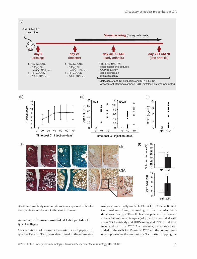
Fig. 1. Development and assessment of collagen-induced arthritis (CIA) in C57BL/6 mice. (a) Model of CIA; arthritis was induced by chicken
collagen type II (CII) emulsified with complete Freund’s adjuvant (CFA), with a booster dose of CII emulsified with incomplete FA (IFA). Mice
were assessed at two time-points defined as early arthritis (day 40/CIA40) and late arthritis (day 70/CIA70) for characterization of osteoclast
progenitor subpopulations and analysis of bone metabolism. PBL 5 peripheral blood; SPL 5 spleen; BM 5 bone marrow; TMT 5 tarsometatarsal
region of hind paws; OCP 5osteoclast progenitor; CTX I 5 cross-linked C-telopeptide of type I collagen; lCT 5micro-computed tomography
(micro-CT). (b) Clinical scoring of CIA by a scale of 0–16 points (0–4 points for each paw) up to 70 days following immunization. The results
shown are pooled data from six independent experiments, presented as mean 6 standard deviation (n 5 8–10 mice per group). (c) Serum levels
of anti-CII immunoglobulin (Ig)G1 and IgG2a antibodies in CIA analysed by enzyme-linked immunosorbent assay (ELISA). Experiments were
repeated three times, and representative results from one set of experiments are shown. Each symbol corresponds to the serum sample from
individual mouse. AU 5 arbitrary unit. (d) Serum level of CTX I in CIA analysed by ELISA. The results shown are pooled data from three
independent experiments (CIA, day 40). Each symbol corresponds to the serum sample from individual mouse. (e) Histological assessment of
the first tarsometatarsal joint in mice developing arthritis (CIA, day 40) reveals osteitis and loss of subchondral bone, with prominent osteoclasts
adjacent to the subchondral regions [haematoxylin and eosin (left), Goldner–Masson trichrome (middle) and tartrate-resistant acid phosphatase
(TRAP) stain (right)]. Magnification 3200. OST 5osteitis; SB 5 subchondral bone; OC 5 osteoclasts. (f) Histomorphometric analysis of
subchondral bone volume and number of active osteoclasts in control (ctrl) and arthritic mice (CIA, day 40). The experiments were repeated
three times and data from a representative experiment are shown as mean 6 standard deviation (n 5 8 mice per group). *Significant difference
from control at P< 0�05, Student’s t-test. [Colour figure can be viewed at wileyonlinelibrary.com]
M. I. Matija�sevic et al.
4 VC 2016 British Society for Immunology, Clinical and Experimental Immunology, 00: 00–00
60 ng/ml RANKL. Sorting parameters and experimental
set-up were optimized for high purity sorting. Sorting
purity was determined by a reanalysis of fractioned popula-
tions and was greater than 99% for all experiments. At days
5–7 of culture, TRAP1 multi-nucleated osteoclasts (�three nuclei/cell) were counted by light microscopy
[22,23].
For the phenotype characterization of spleen, PBMC
and bone marrow myeloid cells, we used directly conju-
gated monoclonal antibodies for myeloid lineage markers
(CD11b, CD11c, F4/80) (all from eBiosciences). In some
experiments, a CD45 marker was used to designate the
total haematopoietic population. In addition, the fre-
quency of osteoclast progenitor subpopulations (defined as
CD3–B220–NK1.1–CD11b–/loCD1151CD1171 for bone
marrow, CD3–B220–NK1.1–CD11b1CD1151Gr-11 for
spleen, CD451CD3–B220–NK1.1–CD11b1CD1151CD265/
RANK1 for osteoclastogenic cultures and CD3–B220–NK1.
1–CD11b1CD1151CD195/CCR51 for migration assay; all
from eBiosciences) was assessed using Attune (Life Tech-
nologies, ABI, Carlsbad, CA, USA) instrument and ana-
lysed by FlowJo software (TreeStar, Ashland, OR, USA).
Migration assay
Migration assays of PBMC were performed in 24-well
Transwell plates (8�0 lm pore size) (Costar, Corning Inc.,
Corning, NY, USA). After stimulation with RANKL and
M-CSF for 48 h, cells from control and CIA groups were
seeded into the upper chamber of the Transwell system at a
concentration of 104 cells/well in 100 ll medium, and the
lower chamber was filled with 10 ng/ml CCL2 or CCL5
(PeproTech for both) in 500 ll medium. After 5 h of incu-
bation at 378C with 5% CO2, the upper surface of the filters
was washed carefully with PBS, and the remaining cells
were removed with a cotton wool swab. The cells that
migrated to the bottom side of the Transwell membrane
inserts were fixed with 4% paraformaldehyde and stained
with 40,6-diamidine-20-phenylindole dihydrochloride
(DAPI). The migrated cells were counted (two wells per
group, four central fields per Transwell) at 3100 magnifi-
cation using a fluorescent microscope (Axiovert 200; Carl
Zeiss, AG, Oberkochen, Germany).
Micro-computed tomography
The distal femoral metaphyses and second lumbar verte-
brae were scanned using micro-computed tomography
(micro-CT) (1172 SkyScan; Bruker, Kontich, Belgium) at
50 kV and 200 mA with a 0�5 aluminum filter using a
detection pixel size of 4�3 lm. Images were captured every
0.78 through 1808 (second lumbar (L2) vertebrae) and
every 0�78 through 3608 (femora) rotation. The scanned
images were reconstructed using the SkyScan Recon soft-
ware and analysed using SkyScan CTAnalyser. Three-
dimensional analysis and reconstruction of trabecular bone
was performed on the bone region 1–2�3 mm distal to the
growth plate. The trabecular bone compartment was
delineated manually from the cortical bone, and the follow-
ing variables were determined: trabecular bone volume
fraction (BV/TV, %), trabecular number (Tb.N/lm), tra-
becular thickness (Tb.Th; lm) and trabecular separation
(Tb.Sp; lm).
Histology and histomorphometry
Femora and hind paws were fixed in 4% paraformaldehyde,
demineralized in 14% ethylendiamine tetraacetic acid in
3% formaldehyde, dehydrated in increasing ethanol con-
centrations and embedded in paraffin. Six-lm sections
were cut with a Leica SM 2000 R rotational microtome
(Lieca SM 2000 R; Leica Biosystems, Nussloch, Germany)
and stained by Goldner–Masson trichrome, haematoxylin
and eosin or TRAP activity. Histomorphometric analysis
was performed under Axio Imager microscope (Carl Zeiss)
using the OsteoMeasure software (Osteo-Metrics, Decatur,
GA, USA) [23].
For static histomorphometry, methaphyseal regions of
Goldner-Masson trichrome-stained femora, 1 mm distally
from the epiphyseal plate, were analysed under 35 objec-
tive magnification. The analysed variables included trabec-
ular volume (BV/TV), trabecular thickness (Tb.Th, lm),
trabecular number (Tb.N/lm) and trabecular separation
(Tb.Sp, lm). On TRAP-stained sections, osteoclasts were
identified as multi-nucleated red cells placed adjacent to
the bone surface. For dynamic histomorphometry, mice
were injected twice with calcein, 20 mg/kg each, at days 6
and 2 prior to sacrificing. Seven-lm sections of undecalci-
fied femora were cut using Leica cryostat and analysed
under a fluorescent microscope (Axio Imager; Carl Zeiss);
bone formation rate (BFR; lm3/lm2/day) was calculated
automatically.
Gene expression analysis
Total RNA from cultured cells and harvested tissues [iso-
lated haematopoietic cells and enzymatically digested tarso-
metatarsal region of hind paws by 0�1% collagenase (type
IV, from Clostridium histolyticum; Sigma-Aldrich)] was
extracted using TRIzol reagent (Applied Biosystems,
Thermo Fisher Scientific, Waltham, MA, USA), reverse-
transcribed (1 mg) to cDNA and amplified (20 ng/well in
duplicate) by quantitative polymerase chain reaction
(PCR) using an AB7500 (Applied Biosystems) instrument.
Expression of osteoclast differentiation genes [cFms/colony
stimulating factor 1 receptor (Csf1r), Rank, cathepsin K
(Ctsk), calcitonin receptor (Calcr)], chemokines (Ccl2,
Ccl5) and cytokines (Il6, Tnf) was determined using com-
mercially available TaqMan Gene Expression Assays
(Applied Biosystems). The expression of a specific gene was
calculated according to the relative standard curve of gene
expression in the calibrator sample (cDNA from
Circulatory osteoclast progenitors in CIA
VC 2016 British Society for Immunology, Clinical and Experimental Immunology, 00: 00–00 5

mononuclear cells or osteoclast culture), and then normal-
ized to the expression level of the b-actin gene as an endog-
enous control [22,23].
Statistics
Each set of experiments was performed at least three times
and data are presented as mean value 6 standard deviation
(s.d.). Statistical analysis of the group difference was per-
formed using Student’s t-test or analysis of variance
(ANOVA) with Bonferroni’s correction for multiple testing.
Levels of anti-collagen antibodies and measurements of
dynamic histomorphometry were presented as individual
values with median, analysed by Mann–Whitney U-test.
For migration assays, values were presented as median with
interquartile range and compared using the Kruskal–Wallis
test with Mann–Whitney for group-to-group comparisons.
The methodological studies of quantitative PCR analysis
suggest that the minimal difference in gene expression that
could be detected reproducibly is approximately 100%
[22,23]. Therefore, we assumed the change in the gene
expression that is statistically significant (ANOVA, P< 0�05)
and more than twofold different compared with control,
repeating through all experiments, as biologically signifi-
cant. Correlations were assessed by Spearman’s rho (q)
coefficient with 95% confidence interval (CI). For all
experiments, a-level was set at 0�05. Statistical analysis was
performed using the software packages MedCalc (version
12.5; MedCalc Inc., Mariakerke, Belgium).
Results
Clinical and serological assessment of CIA
Dark brown Agouti (DBA)/1 mice, the most susceptible
strain, develop CIA with a high incidence of 80–90% and
very pronounced symptoms during the acute disease
course. However, specific protocols for induction of CIA in
the B6 strain have been developed and optimized [2–4].
Upon induction of arthritis, mice were assessed at two
time-points defined as early arthritis (day 40/CIA40) and
late arthritis (day 70/CIA70) for the frequency and activity
of osteoclast progenitor subpopulations in parallel with the
changes in bone metabolism (Fig. 1a).
By applying the modified protocols of the model of CIA
in B6 mice [3,4,24], we were able to reach the arthritis inci-
dence of approximately 50–60% in total of 14 independent
experiments (approximately 200 mice). There were no
signs of arthritis in any of the control (PBS-injected) ani-
mals. However, the arthritis was mild in most cases (total
clinical score mean was 6�97 6 3�91), with one or more
inflamed toes per paw and without ankylosis (Fig. 1b).
During the course of the disease, mice developed mainly
symmetric arthritis, with more severe affection of the hind
paws. Immunization to chicken collagen was confirmed by
detection of circulating anti-CII antibodies (classes IgG1
and IgG2a) in serum samples taken individually at the time
of termination for each time-point (Fig. 1c). Statistical
analysis revealed a significant positive correlation between
the serum level of anti-CII class IgG2a antibodies and clini-
cal score of arthritis (q 5 0�522, CI 5 0�169–0�756,
P 5 0�009). Bone degradation during development of
arthritis was confirmed by increased serum levels of CTX I
as a metabolic marker of bone resorption (Fig. 1d).
Histological confirmation of joint destruction in CIA
Histological analysis of the hind paws in the affected ani-
mals demonstrated severe destruction of small joints of tar-
sometatarsal region in arthritic mice, paralleled by the
increased number of active osteoclasts adjacent to the sub-
chondral bone surfaces compared to the control group
(Fig. 1e). The majority of the affected animals exhibited
bone lesions accompanied by severe osteitis, with the
replacement of bone marrow adipocytes with haemato-
poietic (inflammatory) cells, matching the bone marrow
oedema described in human RA [6]. Very few mice showed
profound synovial inflammation and pannus invasion of
the articular cartilage, which could probably be assigned to
the specific features of CIA in the B6 mouse strain
[4,24,25].
Hypercellularity of bone marrow compartment was most
intensive at subchondral bone regions, coinciding with the
areas of active osteoclasts and bone erosions (Fig. 1e). In
addition, histomorphometric analysis of the first tarsometa-
tarsal joint revealed loss of subchondral bone and an
increased number of active osteoclasts in animals affected
by early CIA compared to the control group (Fig. 1f).
Systemic bone loss in CIA
In addition to local osteoclast activation and joint destruc-
tion, we aimed to confirm systemic alterations in bone
metabolism paralleled by the increased osteoclast activity
and enhanced bone resorption in CIA (Fig. 1d). Using
micro-CT analysis, we assessed axial (at the level of L2 ver-
tebra) and appendicular (at the level of distal femoral
metaphysis) skeleton (Fig. 2a).
In early arthritis (CIA40), despite thinning of trabeculae in
the L2 vertebral body (Tb.Th 42�1 6 4�4 in control versus
38�4 6 4�7 in CIA, P 5 0�04), the percentage of trabecular
bone volume was unaffected (Fig. 2b). With the development
of arthritis (CIA70), trabecular bone volume was reduced sig-
nificantly (Fig. 2b) in addition to decreased trabecular thick-
ness (Tb.Th 43�1 6 6�2 lm in control versus 35�4 6 2�4 lm in
CIA, P 5 0�001), confirming systemic bone loss.
Appendicular skeleton was assessed by both micro-CT
analysis and histology at the level of distal femoral meta-
physis, showing even more prominent changes in early
(CIA40) and late (CIA70) arthritis, with a significant
decrease in trabecular bone volume (Fig. 2a,b), trabecular
M. I. Matija�sevic et al.
6 VC 2016 British Society for Immunology, Clinical and Experimental Immunology, 00: 00–00

thickness and trabecular number (Tb.Th 39�6 6 4�0 lm in
control versus 34�6 6 5�3 lm in CIA, P 5 0�008 and TbN
1�64 6 0�23/lm in control versus 1�36 6 0�22/lm in CIA,
P 5 0�001 by micro-CT). Moreover, dynamic histomoph-
ometry showed decreased bone formation rate as an indi-
cator of suppressed osteoformation in vivo (Fig. 2c).
Osteoclastogenic potential of haematopoietic cells inCIA
To assess in vitro osteoclastogenic potential, we isolated
haematopoietic cells from bone marrow, adjacent endosteal
and periosteal regions, spleen and peripheral blood of mice
with early arthritis, and cultured them with RANKL and
M-CSF to induce differentiation of osteoclast-like multi-
nucleated cells (Fig. 3a). The number of TRAP1 osteoclasts
was significantly higher in peripheral as well as bone mar-
row compartments in CIA compared with control animals
(Fig. 3b).
As unfractionated haematopoietic tissues are heterogene-
ous, containing variable proportions of non-osteoclastogenic
cells, we further assessed frequency and activity of defined
osteoclast progenitor subpopulations according to the reports
by Jacquin et al. [13] and Jacome-Galarza et al. [14]. Arthritic
mice had a higher frequency of putative osteoclast progenitor
cells in the bone marrow compartment, bearing the
phenotype CD3–B220–NK1.1–CD11b–/loCD1171CD1151
or CD3–B220–NK1.1–CD11b–/loCD117–CD1151 and among
Fig. 2. Effect of collagen-induced arthritis (CIA)
on bone remodelling in axial and appendicular
skeleton. Second lumbar vertebra and distal
femora from control (ctrl) and arthritic mice
(early arthritis/CIA 40 and late arthritis/CIA70)
were analysed for trabecular bone volume and
bone formation rate. (a) Three-dimensional
reconstruction of selected areas of the second
lumbar vertebrae scanned by micro-computed
tomography (lCT) (LV lCT, left) and distal
femur (DF lCT, middle) and representative
micrographs of distal femoral section (DF
histology, right) stained with Goldner–Masson
trichrome in CIA70 and control mice. (b) mCT
analysis of trabecular bone volume [BV/TV (%),
bone volume/total volume] in the second lumbar
vertebra (LV lCT) and distal femoral metaphysis
(DF lCT); histomorphometric analysis of BV/TV
(%) at the level of distal femoral metaphysis (DF
histology). Analysis was repeated in three
independent sets of experiments for each time-
point and cumulative results are shown (n 5 15–
25 mice for each group and time-point). All
results are displayed as mean 6 standard
deviation. *Significant difference from the
respective control at P< 0�05, Student’s t-test. (c)
Representative microphotographs of femoral
sections from control (ctrl) and arthritic mice
(CIA, day 40) injected with calcein, and
histomorphometric evaluation of bone formation
rate relative to bone surface area (BFR/BS, lm3/
lm2/day). The experiments were repeated three
times and data from a representative experiment
are shown (n 5 10–13 mice per group). Symbols
represent values of individual mice and
horizontal lines represent median. Statistically
significant difference was determined at P< 0�05,
Mann–Whitney U-test. [Colour figure can be
viewed at wileyonlinelibrary.com]
Circulatory osteoclast progenitors in CIA
VC 2016 British Society for Immunology, Clinical and Experimental Immunology, 00: 00–00 7

spleen haematopoietic cells, bearing the phenotype
CD3–B220–NK1.1–CD11b1CD1151Gr-11 (Fig. 4a). Bone
marrow and spleen progenitors were FACS-sorted and con-
firmed to possess potent osteoclastogenic activity in vitro,
with higher numbers of differentiated TRAP1 osteoclasts
in cultures from mice with CIA compared to control mice
(Fig. 4b).
Studies taken as a starting-point for osteoclast progenitor
dissection were performed in control (untreated) mice [13,14].
Therefore, we additionally tested osteoclastogenic efficiency of
selected osteoclast progenitor subsets in the context of arthritis.
Osteoclastogenic potential of lymphoid-negative cells express-
ing M-CSF receptor (CD115) were compared with respect to
CD11b, as Jacquin et al. [13] reported that CD11bhi fraction of
lymphoid-negative bone marrow cells may possess certain
osteoclastogenic activity. In arthritic mice, the CD11b–/lo subset
generated significantly more TRAP1 osteoclasts compared to
the CD11b1 subset (300 6 53 osteoclasts for
CD3–B220–NK1.1– CD11b–/loCD1151 cells versus 16 6 6 for
CD3–B220–NK1.1–CD11b1CD1151 cells, P 5 0�011) when
plated at the same density. In contrast to bone marrow, osteo-
clast progenitors in spleen were contained within the CD11b1
subpopulation of lymphoid-negative cells, and expression of
M-CSF receptor (CD115) seems to be indispensable for osteo-
clastogenic activity – the CD1151 subset readily gave rise to
TRAP1 osteoclasts in vitro (178 6 34 TRAP1 osteoclasts for
CD3–B220–NK1.1–CD11b1CD1151 cells), with no osteoclas-
togenic activity of CD115– cells (no TRAP1 osteoclasts for
CD3–B220–NK1.1–CD11b1CD115– cells).
In addition to generating a higher number of TRAP1
osteoclasts, differentiated osteoclast progenitors from
arthritic mice exhibited enhanced expression of osteoclast-
specific genes (Rank, Csfr1, Calcr and Ctsk) compared to
the control, for both bone marrow and spleen (Fig. 4c). We
also compared expression of early osteoclast-specific differ-
entiation genes (Rank and Csfr1) between osteoclastogenic
cultures of sorted and unfractionated bone marrow and
spleen cells (Supporting information, Fig. S1). Relative
Rank expression was higher in cultures of sorted cells, indi-
cating that this marker is enriched specifically in osteoclast
progenitor populations. Relative Csfr1 (CD115) expression
was high in unfractionated bone marrow cells and compa-
rable between groups, due probably to the contribution of
non-osteoclastogenic (myeloid) cells expressing M-CSF
receptor. The more obvious difference in the expression of
osteoclast-specific genes between control and arthritic sam-
ples in sorted compared to unfractionated cultures indi-
cates enrichment of osteoclast progenitor cells.
Changes in myeloid populations and cytokineexpression in CIA
As the results showed that osteoclastogenic potential was
enhanced systemically in arthritis, we further assessed
changes in myeloid cell populations among bone marrow
and peripheral blood cells. We hypothesized that osteoclas-
togenic activity could be enhanced due to systemic activa-
tion of the total myeloid population (not just osteoclast
progenitor cells), as a part of the autoinflammatory/auto-
immune response in CIA.
Flow cytometric analysis showed expansion of the total
myeloid CD11b1 population in bone marrow and
Fig. 3. Osteoclastogenic potential of mice with collagen-induced arthritis (CIA). (a) Representative microphotographs of osteoclasts differentiated
in vitro from bone marrow (BM) and spleen (SPL) of control (ctrl) and arthritic mice (CIA, day 40), cytochemically stained for tartrate-resistant
acid phosphatase (TRAP) activity. (b) Number of TRAP1 osteoclasts in cultures of bone marrow (BM), cells derived from bone shaft (BS),
spleen (SPL) and peripheral blood mononuclear cells (PBL), stimulated by receptor activator of nuclear factor kappa-B ligand (RANKL) and
macrophage colony-stimulating factor (M-CSF) from control (ctrl) and arthritic mice (CIA, day 40). The experiments were repeated four times
and data from a representative experiment are shown. Values are presented as mean 6 standard deviation (n 5 4–6 wells per group). *Significant
difference from the respective control at P< 0�05, Student’s t-test. [Colour figure can be viewed at wileyonlinelibrary.com]
M. I. Matija�sevic et al.
8 VC 2016 British Society for Immunology, Clinical and Experimental Immunology, 00: 00–00

Fig. 4. Frequency and differentiation potential of sorted osteoclast progenitor cells. (a) Representative flow cytometry data of the frequency of
osteoclast progenitor cells, bearing the phenotype CD3–B220–NK1.1–CD11b–/loCD1151CD1171 or CD3–B220–NK1.1–CD11b–/loCD1151CD117–
in bone marrow (BM); and CD3–B220–NK1.1–CD11b1CD1151Gr-11 in spleen (SPL) of control (ctrl) and arthritic mice (CIA, day 40). Values
are presented as mean 6 standard deviation for three independent experiments. (b) Osteoclastogenic potential of sorted osteoclast progenitors in
BM (CD3–B220–NK1.1–CD11b–/loCD1151) and SPL (CD3–B220–NK1.1–CD11b1CD1151), stimulated by receptor activator of nuclear factor jB
ligand (RANKL) and macrophage colony-stimulating factor (M-CSF), and assessed by the number of tartrate-resistant acid phosphatase
(TRAP)1 osteoclasts, presented as mean 6 standard deviation (n 5 4–6 wells per group). Flow cytometry analysis and sorting were performed by
plotting the total cell population (excluding doublets and dead cells) for lymphoid markers versus CD11b, subsequently dissected for CD115
versus CD117 expression for BM or CD115 versus Gr-1 expression for SPL cells. Sorting gates were defined using unlabelled cells and
fluorescence minus one controls. *Significant difference from the respective control at P< 0�05, Student’s t-test. (c) Expression of osteoclast-
specific genes in osteoclastogenic cultures of sorted BM-derived and SPL-derived osteoclast progenitors, isolated from control (ctrl) and arthritic
mice (CIA, day 40). Relative RNA quantities were presented as mean 6 standard deviation (n 5 4–6 wells per group). *Biologically significant
difference in the expression (� 100%) in comparison to the respective control group. Csf1r 5 colony stimulating factor 1 receptor (CD115/cFms);
Rank 5 receptor activator of nuclear factor jB; Calcr 5 calcitonin receptor; Ctsk 5 cathepsin K. [Colour figure can be viewed at
wileyonlinelibrary.com]
Circulatory osteoclast progenitors in CIA
VC 2016 British Society for Immunology, Clinical and Experimental Immunology, 00: 00–00 9

peripheral blood (Fig. 5a). In relation to specific myeloid
subpopulations, higher percentages associated with arthri-
tis were noticed for CD11b1F4/801 macrophage,
CD11b1NK1.11 natural killer cell and CD11b1CD11c1
myeloid dendritic cell populations.
Finally, we assessed the expression of several cytokines
and chemokines known to be associated with the systemic
proinflammatory state which could, at the same time, be
responsible for the enhanced activity of myeloid lineage
cells that comprise osteoclast progenitors [17,18,21,26].
Tnf, Il6, Ccl2 and Ccl5 were expressed increasingly in
PBMCs from mice developing arthritis compared to con-
trols (Fig. 5b). Similar changes of Ccl2 and Ccl5 expression
were observed for spleen and bone marrow cells as well as
for tarsometatarsal region of hind paws (Fig. 5b).
Effects of CCL2 and CCL5 on osteoclast progenitormigration and differentiation
In the last set of experiments we evaluated the effects of CCL2
and CCL5 on circulatory osteoclast progenitor cells. These che-
mokines were able to cause enhanced migration of activated
peripheral blood osteoclast progenitors in the migration assay
Fig. 5. Changes of myeloid cell population frequencies and inflammatory cytokine/chemokine expressions associated with arthritis. (a)
Representative flow cytometry data of the frequency of myeloid subpopulations in early arthritis (CIA, day 40): myeloid lineage (CD11b1),
macrophages (CD11b1F4/801), myeloid dendritic cells (CD11b1CD11c1) and natural killer cells (CD11b1NK1.11) in bone marrow (BM) and
peripheral blood mononuclear cells (PBL). Values are presented as mean 6 standard deviation for three independent experiments. (b) Gene
expression of tumour necrosis factor-a (Tnf) and interleukin-6 (Il6) in PBL, and chemokine ligands Ccl2 and Ccl5 in PBL, spleen (SPL), cells
derived from bone shaft (BS), BM and tarsometatarsal region of hind paws (TMT) from control (ctrl) and arthritic mice (CIA, day 40). Relative
RNA quantities were presented as mean 6 standard deviation (n 5 3 replicates for representative set of experiments). *Biologically significant
difference in the expression (� 100%) in comparison to the respective control group. [Colour figure can be viewed at wileyonlinelibrary.com]
M. I. Matija�sevic et al.
10 VC 2016 British Society for Immunology, Clinical and Experimental Immunology, 00: 00–00
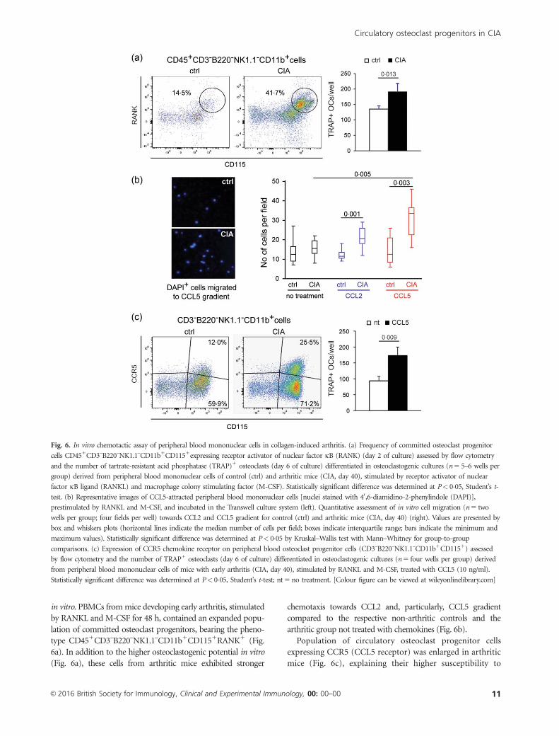
in vitro. PBMCs from mice developing early arthritis, stimulated
by RANKL and M-CSF for 48 h, contained an expanded popu-
lation of committed osteoclast progenitors, bearing the pheno-
type CD451CD3–B220–NK1.1–CD11b1CD1151RANK1 (Fig.
6a). In addition to the higher osteoclastogenic potential in vitro
(Fig. 6a), these cells from arthritic mice exhibited stronger
chemotaxis towards CCL2 and, particularly, CCL5 gradient
compared to the respective non-arthritic controls and the
arthritic group not treated with chemokines (Fig. 6b).
Population of circulatory osteoclast progenitor cells
expressing CCR5 (CCL5 receptor) was enlarged in arthritic
mice (Fig. 6c), explaining their higher susceptibility to
Fig. 6. In vitro chemotactic assay of peripheral blood mononuclear cells in collagen-induced arthritis. (a) Frequency of committed osteoclast progenitor
cells CD451CD3–B220–NK1.1–CD11b1CD1151expressing receptor activator of nuclear factor jB (RANK) (day 2 of culture) assessed by flow cytometry
and the number of tartrate-resistant acid phosphatase (TRAP)1 osteoclasts (day 6 of culture) differentiated in osteoclastogenic cultures (n 5 5–6 wells per
group) derived from peripheral blood mononuclear cells of control (ctrl) and arthritic mice (CIA, day 40), stimulated by receptor activator of nuclear
factor jB ligand (RANKL) and macrophage colony stimulating factor (M-CSF). Statistically significant difference was determined at P< 0�05, Student’s t-
test. (b) Representative images of CCL5-attracted peripheral blood mononuclear cells [nuclei stained with 40,6-diamidino-2-phenylindole (DAPI)],
prestimulated by RANKL and M-CSF, and incubated in the Transwell culture system (left). Quantitative assessment of in vitro cell migration (n 5 two
wells per group; four fields per well) towards CCL2 and CCL5 gradient for control (ctrl) and arthritic mice (CIA, day 40) (right). Values are presented by
box and whiskers plots (horizontal lines indicate the median number of cells per field; boxes indicate interquartile range; bars indicate the minimum and
maximum values). Statistically significant difference was determined at P< 0�05 by Kruskal–Wallis test with Mann–Whitney for group-to-group
comparisons. (c) Expression of CCR5 chemokine receptor on peripheral blood osteoclast progenitor cells (CD3–B220–NK1.1–CD11b1CD1151) assessed
by flow cytometry and the number of TRAP1 osteoclasts (day 6 of culture) differentiated in osteoclastogenic cultures (n 5 four wells per group) derived
from peripheral blood mononuclear cells of mice with early arthritis (CIA, day 40), stimulated by RANKL and M-CSF, treated with CCL5 (10 ng/ml).
Statistically significant difference was determined at P< 0�05, Student’s t-test; nt 5 no treatment. [Colour figure can be viewed at wileyonlinelibrary.com]
Circulatory osteoclast progenitors in CIA
VC 2016 British Society for Immunology, Clinical and Experimental Immunology, 00: 00–00 11

CCL5 gradient. Moreover, CCL5 exhibited direct osteoclas-
togenic effect by increasing the number of differentiated
TRAP1 cells upon addition to M-CSF/RANKL-stimulated
cultures (Fig. 6c), supporting the hypothesis that once they
are attracted to the inflamed joints, osteoclast progenitors
could be enhanced further in their differentiation by proin-
flammatory chemokines.
Discussion
Although it is known that there is a systemic enhancement of
osteoclast activity and generalized bone loss in addition to local
joint destruction, even in patients with early RA [7,8], the pre-
cise mechanisms of osteoclast progenitor migration and activa-
tion in arthritis as well as functional relations between
circulatory and local osteoclast progenitor populations have not
been revealed fully. Several studies have attempted to define the
phenotype of putative osteoclast progenitor cells within the
myeloid lineage population of bone marrow, spleen and PBMC
pools [11–14,27]. It has been demonstrated that the lymphoid-
negative CD11b2/lo fraction recapitulates the early osteoclasto-
genic activity of total bone marrow [13,14], whereas reports are
still inconsistent regarding the osteoclastogenic efficiency of
CD11b1 bone marrow cells [11,13,28]. In addition to these
findings, we observed high osteoclastogenic potential of
the CD3–B220–NK1.1–CD11b–/lo fraction and inefficiency of the
CD3–B220–NK1.1–CD11b1 bone marrow subset to form
TRAP1 osteoclasts in vitro in the context of arthritis. Jacome-
Galarza et al. showed further that most of the osteoclastogenic
activity of CD3–B220–CD11b–/lo fraction was included in the
CD115hiCD117hi population, able to generate bone-resorbing
osteoclasts at a clonal level [14]. CD115/cFms, the receptor for
M-CSF, is expressed on myeloid lineage cells including osteoclast
progenitors, whereas the CD117/c-kit, the receptor for stem
cell factor, is a consistent marker of early haematopoietic frac-
tion [12–14]. This distinct subset of the most potent osteo-
clastogenic cells in bone marrow, bearing the phenotype
CD3–B220–NK1.1–CD11b–/loCD1151CD1171, was expanded
under the inflammatory condition associated with arthritis.
Monocyte progenitors with osteoclastogenic activity were also
confirmed in spleen and peripheral blood [11,14,29–31]. In con-
trast to bone marrow, the peripheral osteoclastogenic activity is
contained within the CD11b1 fraction of lymphoid-negative
(CD3–B220–NK1.1–) cells [14]. Further dissection of this frac-
tion revealed that CD1151Ly6Chi cells exhibited the highest
potential to form osteoclasts in vitro. This distinct phenotype is
consistent with peripheral inflammatory-type circulating mono-
cytes [14]. By using the similar approach to define peripheral
osteoclast progenitors, we observed enlargement of the
CD3–B220–NK1.1–CD11b1CD1151Gr-11 subset in arthritic
mice. These peripheral CD11b1CD1151Gr-11 osteoclast pro-
genitors overlap phenotypically with the myeloid-derived sup-
pressor cells, which have been shown to differentiate into
osteoclasts contributing to bone erosions in CIA [29]. Moreover,
the CD3–B220–NK1.1–CD11b1CD1151RANK1-committed
osteoclast progenitor population was enlarged significantly in
CIA after stimulation of PBMCs in vitro with RANKL and M-
CSF. De Klerck et al., using CIA established in the IFN-gR
knock-out mice, observed a higher number of CD11b1RANK1
splenocytes, possibly serving as a source of osteoclasts responsi-
ble for bone destruction [30]. Another study using the CIA
model revealed that in vivo depletion of CD11b1Gr-11CCR21
monocytes by anti-CCR2 antibody attenuated the severity of
arthritis significantly [31]. In the human TNF-a transgenic
(hTNF-Tg) mouse model, a significant increase in the propor-
tion of bone marrow and peripheral blood CD11b1Gr-1
(Ly6G)–/lo cells was observed [28]. Osteoclast progenitors, iden-
tified as the CD3–B220–Ter119–CD11b–/loLy6Chi population,
were expanded in the bone marrow of the Sakaguchi mouse
model of inflammatory arthritis, and had the capacity to differ-
entiate into multi-nucleated bone-resorbing osteoclasts in vitro
and in vivo [32].
In addition to osteoclast progenitor cells, myeloid line-
age is induced in general as a part of autoinflammatory
and autoimmune responses in CIA [26]. In our study,
changes of myeloid lineage included the total CD11b1
population as well as subsets of CD11b1F4/801 macro-
phages and CD11b1CD11c1 myeloid dendritic cells.
Monocyte/macrophage and dendritic cells contribute to
arthritis pathogenesis by serving as antigen-presenting cells
for xenogeneic collagen and by secreting proinflammatory
cytokines, including IL-1, IL-6 and TNF-a [26,31,33]. A
systemic increase in TNF-a mediated an enlargement of
the peripheral CD11bhiCD1151 osteoclast progenitor pop-
ulation in the hTNF-Tg mouse model [34]. In a similar
mouse model Binder et al. found that anti-TNF therapy
reduced the number of CD1151 osteoclast progenitors and
intensity of subchondral lesions, without a significant
impact on synovial inflammation [35]. Maintenance of the
expanded osteoclast progenitor number and sustained
osteoclast renewal is crucial to enable osteoclasts, whose
individual lifespan is approximately 2–4 weeks, to create
bone erosions during the chronic course of arthritis [36].
Osteoclastogenic assays of sorted osteoclast progenitor
cells further showed greater differentiation potential and
higher expression of osteoclast-specific genes in cultures
obtained from arthritic mice. Under pathological osteore-
sorptive conditions, systemically activated osteoclast pro-
genitors can respond to chemokine signals and home
efficiently to bone surfaces causing bone resorption [8].
Expression of Ccl2 and Ccl5 was enhanced in our model of
CIA, and both chemokines have been shown to be involved
in the pathogenesis of RA [21,37,38]. CCL2 is produced
within the affected joints by different cell types (including
macrophages, endothelial cells, synovial fibroblasts and
chondrocytes) and is elevated in synovial fluid and serum
of RA patients [39]. Talbot et al. showed the importance of
the CCR2/CCL2 interaction for neutrophil infiltration in
the model of antigen-induced arthritis [40]. CCL5 could be
released by inflammatory cells as well as by osteoclasts,
M. I. Matija�sevic et al.
12 VC 2016 British Society for Immunology, Clinical and Experimental Immunology, 00: 00–00

osteoblasts and chondrocytes [41,42], and may attract
osteoclast progenitors to the sites of inflammation. In our
study the migration assay identifies CCL5 and, to a lesser
extent, CCL2 as potent chemotactic factors for committed
osteoclast progenitors isolated from PBMC of arthritic
mice. In addition, CCL5 exhibited direct osteoclastogenic
effects on differentiation of peripheral blood osteoclast
progenitors, which expressed CCR5 (receptor for CCL5) in
significant proportion. CCR5 expression was observed on
synovial tissue macrophages and synovial fluid-derived
monocytes in patients with RA [43]. Moreover, CCL5 was
significantly higher in synovium of RA patients compared
to patients with osteoarthritis [44], and the anti-CCR5
antibody was able to block CCL5-induced chemotaxis of
peripheral blood RA monocytes [45]. The susceptibility of
osteoclast progenitors to chemotactic signals and increased
production of respective chemokines create optimal condi-
tions for enhanced homing of osteoclast progenitors not
only to affected joints, but also at the systemic level,
explaining local as well as systemic bone loss.
In addition to proinflammatory chemokines, we
observed enhanced expression of Tnf and Il6 in haemato-
poietic cells derived from bone marrow as well as from cir-
culation. Even a small increase in systemic inflammation
may trigger a self-amplifying proinflammatory state, lead-
ing to increased osteoclast activation and exaggerated bone
loss [11,17,35,38,46,47]. Within the affected joints, thin-
ning of the articular cartilage and loss of subchondral bone
were most prominent in the tarsometatarsal regions, simi-
lar to previous studies on the CIA model in B10.RIII and
DBA/1 mice [31]. In contrast to profound pannus invasion
of the articular cartilage seen in DBA/1 strain [3–5,24],
joint pathology in B6 mice is characterized by pronounced
osteitis within the subchondral compartment, coinciding
with the areas of TRAP1 osteoclasts adjacent to the sub-
chondral bone surfaces. As well as local changes, systemic
bone loss of axial and appendicular skeleton was observed
in different rodent models of arthritis [48–50]. In our
model, in vivo bone formation was suppressed and bone
resorption was enhanced resulting in decreased trabecular
bone volume. Enhanced production of proinflammatory
cytokines (including IL-1, IL-6 and TNF-a) is known to
suppress osteoblasts function, leaving them unable to com-
pensate for excessive osteoclast activity and bone resorp-
tion [7,19].
In conclusion, the model of CIA in B6 mouse strain
seems to be particularly useful to study changes within the
subchondral compartment (including osteitis and sub-
chondral bone loss) of the affected joints in the absence of
prominent synovial hyperplasia. The altered cytokine and
chemokine milieu created by inflammatory and immune
cells within the subchondral compartment may be crucial
for the recruitment of osteoclast progenitors and activation
of bone-resorbing osteoclasts. Inflammatory mediators
produced by accumulated cells egress into circulation,
thereby producing a systemic impact on osteoresorption,
not only in arthritis but also in other inflammatory proc-
esses. In this context, characterization of osteoclast progen-
itors could be used to monitor disease progression and
intensity of bone resorption.
Acknowledgements
We thank Professor Hector Leonardo Aguila for critical dis-
cussion of the FACS data. We also thank Ms Katerina
Zrinski-Petrovic for her technical assistance. This work was
supported by the Croatian Science Foundation under the
project 5699.
Author contributions
I. K., A. M. and D. G. designed the study; M. I. M., D. F., S.
I., N. K., V. K. and T. K. performed the experiments; M. I.
M., D. F., N. K., V. K., T. K., A. �S., S. I., H. C. and E. L. M.
acquired and analysed the data; A. �S., H. C., E. L. M., I. K.,
A. M. and D. G. interpreted the results; M. I. M., D. F. and
D. G. prepared the manuscript. All authors critically
revised the manuscript and approved the final version.
Disclosure
The authors declare no conflicts of interest.
References
1 Bevaart L, Vervoordeldonk MJ, Tak PP. Evaluation of therapeu-
tic targets in animal models of arthritis: how does it relate to
rheumatoid arthritis? Arthritis Rheum 2010; 62:2192–205.
2 Brand DD, Latham KA, Rosloniec EF. Collagen-induced arthri-
tis. Nat Protoc 2007; 2:1269–75.
3 Inglis JJ, Simelyte E, McCann FE, Criado G, Williams RO. Pro-
tocol for the induction of arthritis in C57BL/6 mice. Nat Protoc
2008; 3:612–8.
4 Campbell IK, Hamilton JA, Wicks IP. Collagen-induced arthritis
in C57BL/6 (H-2b) mice: new insights into an important dis-
ease model of rheumatoid arthritis. Eur J Immunol 2000; 30:
1568–75.
5 Holmdahl R, Malmstrom V, Burkhardt H. Autoimmune pri-
ming, tissue attack and chronic inflammation – the three stages
of rheumatoid arthritis. Eur J Immunol 2014; 44:1593–9.
6 Duer-Jensen A, Horslev-Petersen K, Hetland ML et al. Bone
edema on magnetic resonance imaging is an independent predic-
tor of rheumatoid arthritis development in patients with early
undifferentiated arthritis. Arthritis Rheum 2011; 63:2192–202.
7 Deal C. Bone loss in rheumatoid arthritis: systemic, periarticu-
lar, and focal. Curr Rheumatol Rep 2012; 14:231–7.
8 Goldring SR, Purdue PE, Crotti TN et al. Bone remodelling in
inflammatory arthritis. Ann Rheum Dis 2013; 72 Suppl 2:ii52–5.
9 Bugatti S, Manzo A, Bombardieri M et al. Synovial tissue heter-
ogeneity and peripheral blood biomarkers. Curr Rheumatol Rep
2011; 13:440–8.
10 Asagiri M, Takayanagi H. The molecular understanding of
osteoclast differentiation. Bone 2007; 40:251–64.
Circulatory osteoclast progenitors in CIA
VC 2016 British Society for Immunology, Clinical and Experimental Immunology, 00: 00–00 13

11 Sucur A, Katavic V, Kelava T, Jajic Z, Kovacic N, Grcevic D. Induc-
tion of osteoclast progenitors in inflammatory conditions: key to
bone destruction in arthritis. Int Orthop 2014; 38:1893–903.
12 Miyamoto T, Ohneda O, Arai F et al. Bifurcation of osteoclasts
and dendritic cells from common progenitors. Blood 2001; 98:
2544–54.
13 Jacquin C, Gran DE, Lee SK, Lorenzo JA, Aguila HL. Identifica-
tion of multiple osteoclast precursor populations in murine
bone marrow. J Bone Miner Res 2006; 21:67–77.
14 Jacome-Galarza CE, Lee SK, Lorenzo JA, Aguila HL. Identifica-
tion, characterization, and isolation of a common progenitor for
osteoclasts, macrophages, and dendritic cells from murine bone
marrow and periphery. J Bone Miner Res 2013; 28:1203–13.
15 Walsh MC, Choi Y. Biology of the RANKL–RANK–OPG system
in immunity, bone, and beyond. Front Immunol 2014; 5:511.
16 Guerrini MM, Takayanagi H. The immune system, bone and
RANKL. Arch Biochem Biophys 2014; 561:118–23.
17 Adamopoulos IE, Mellins ED. Alternative pathways of osteoclas-
togenesis in inflammatory arthritis. Nat Rev Rheumatol 2015;
11:189–94.
18 Souza PP, Lerner UH. The role of cytokines in inflammatory
bone loss. Immunol Invest 2013; 42:555–622.
19 Baum R, Gravallese EM. Impact of inflammation on the osteo-
blast in rheumatic diseases. Curr Osteoporos Rep 2014; 12:9–16.
20 Braun T, Zwerina J. Positive regulators of osteoclastogenesis and
bone resorption in rheumatoid arthritis. Arthritis Res Ther
2011; 13:235.
21 Szekanecz Z, Koch AE. Successes and failures of chemokine-
pathway targeting in rheumatoid arthritis. Nat Rev Rheumatol
2016; 12:5–13.
22 Cvija H, Kovacic N, Katavic V et al. Chemotactic and immunor-
egulatory properties of bone cells are modulated by endotoxin-
stimulated lymphocytes. Inflammation 2012; 35:1618–31.
23 Kovacic N, Grcevic D, Katavic V et al. Fas receptor is required
for estrogen deficiency-induced bone loss in mice. Lab Invest
2010; 90:402–13.
24 Kai H, Shibuya K, Wang Y et al. Critical role of M. tuberculosis
for dendritic cell maturation to induce collagen-induced arthri-
tis in H-2b background of C57BL/6 mice. Immunology 2006;
118:233–9.
25 Pan M, Kang I, Craft J, Yin Z. Resistance to development of
collagen-induced arthritis in C57BL/6 mice is due to a defect in
secondary, but not in primary, immune response. J Clin Immu-
nol 2004; 24:481–91.
26 Billiau A, Matthys P. Collagen-induced arthritis and related ani-
mal models: how much of their pathogenesis is auto-immune,
how much is auto-inflammatory? Cytokine Growth Factor Rev
2011; 22:339–44.
27 Muto A, Mizoguchi T, Udagawa N et al. Lineage-committed
osteoclast precursors circulate in blood and settle down into
bone. J Bone Miner Res 2011; 26:2978–90.
28 Yao Z, Li P, Zhang Q et al. Tumor necrosis factor-alpha
increases circulating osteoclast precursor numbers by promoting
their proliferation and differentiation in the bone marrow
through up-regulation of c-Fms expression. J Biol Chem 2006;
281:11846–55.
29 Zhang H, Huang Y, Wang S et al. Myeloid-derived suppressor
cells contribute to bone erosion in collagen-induced arthritis by
differentiating to osteoclasts. J Autoimmun 2015; 65:82–9.
30 De Klerck B, Carpentier I, Lories RJ et al. Enhanced osteoclast
development in collagen-induced arthritis in interferon-gamma
receptor knock-out mice as related to increased splenic
CD11b1 myelopoiesis. Arthritis Res Ther 2004; 6:R220–31.
31 Br€uhl H, Cihak J, Plach�y J et al. Targeting of Gr-11,CCR21
monocytes in collagen-induced arthritis. Arthritis Rheum 2007;
56:2975–85.
32 Charles JF, Hsu L-Y, Niemi EC, Weiss A, Aliprantis AO,
Nakamura MC. Inflammatory arthritis increases mouse osteo-
clast precursors with myeloid suppressor function. The J Clin
Invest 2012; 122:4592–605.
33 Khan S, Greenberg JD, Bhardwaj N. Dendritic cells as targets
for therapy in rheumatoid arthritis. Nat Rev Rheumatol 2009;
5:566–71.
34 Li P, Schwarz EM, O’Keefe RJ et al. Systemic tumor necrosis
factor alpha mediates an increase in peripheral CD11bhigh
osteoclast precursors in tumor necrosis factor alpha-transgenic
mice. Arthritis Rheum 2004; 50:265–76.
35 Binder NB, Puchner A, Niederreiter B et al. Tumor necrosis
factor-inhibiting therapy preferentially targets bone destruction
but not synovial inflammation in a tumor necrosis factor-driven
model of rheumatoid arthritis. Arthritis Rheum 2013; 65:608–17.
36 Le Goff B, Berthelot JM, Maugars Y, Heymann D. Osteoclasts in
RA: diverse origins and functions. Joint Bone Spine 2013; 80:
586–91.
37 Kim MS, Magno CL, Day CJ, Morrison NA. Induction of che-
mokines and chemokine receptors CCR2b and CCR4 in authen-
tic human osteoclasts differentiated with RANKL and osteoclast
like cells differentiated by MCP-1 and RANTES. J Cell Biochem
2006; 97:512–8.
38 Ikic M, Jajic Z, Lazic E et al. Association of systemic and intra-
articular osteoclastogenic potential, pro-inflammatory mediators
and disease activity with the form of inflammatory arthritis. Int
Orthop 2014; 38:183–92.
39 Kim MS, Day CJ, Selinger CI, Magno CL, Stephens SR,
Morrison NA. MCP-1-induced human osteoclast-like cells are
tartrate-resistant acid phosphatase, NFATc1, and calcitonin
receptor-positive but require receptor activator of NFkappaB
ligand for bone resorption. J Biol Chem 2006; 281:1274–85.
40 Talbot J, Bianchini FJ, Nascimento DC et al. CCR2 expression
in neutrophils plays a critical role in their migration into the
joints in rheumatoid arthritis. Arthritis Rheumatol 2015; 67:
1751–9.
41 Huh YH, Lee G, Lee KB, Koh JT, Chun JS, Ryu JH. HIF-2a-
induced chemokines stimulate motility of fibroblast-like syno-
viocytes and chondrocytes into the cartilage-pannus interface in
experimental rheumatoid arthritis mouse models. Arthritis Res
Ther 2015; 17:302.
42 Yano S, Mentaverri R, Kanuparthi D et al. Functional expression
of beta-chemokine receptors in osteoblasts: role of regulated
upon activation, normal T cell expressed and secreted
(RANTES) in osteoblasts and regulation of its secretion by
osteoblasts and osteoclasts. Endocrinology 2005; 146:2324–35.
43 Katschke KJ Jr, Rottman JB, Ruth JH et al. Differential expres-
sion of chemokine receptors on peripheral blood, synovial fluid,
and synovial tissue monocytes/macrophages in rheumatoid
arthritis. Arthritis Rheum 2001; 44:1022–32.
44 Yoshida S, Arakawa F, Higuchi F et al. Gene expression analysis
of rheumatoid arthritis synovial lining regions by cDNA micro-
array combined with laser microdissection: up-regulation of
M. I. Matija�sevic et al.
14 VC 2016 British Society for Immunology, Clinical and Experimental Immunology, 00: 00–00

inflammation-associated STAT1, IRF1, CXCL9, CXCL10, and
CCL5. Scand J Rheumatol 2012; 41:170–9.
45 Lebre MC, Vergunst CE, Choi IY et al. Why CCR2 and CCR5
blockade failed and why CCR1 blockade might still be effective
in the treatment of rheumatoid arthritis. PLOS ONE 2011; 6:
e21772.
46 Totoson P, Maguin-Gat�e K, Nappey M, Wendling D,
Demougeot C. Endothelial dysfunction in rheumatoid arthritis:
mechanistic insights and correlation with circulating markers of
systemic inflammation. PLOS ONE 2016; 11:e0146744.
47 Wong PK, Quinn JM, Sims NA, van Nieuwenhuijze A,
Campbell IK, Wicks IP. Interleukin-6 modulates production of
T lymphocyte-derived cytokines in antigen-induced arthritis
and drives inflammation-induced osteoclastogenesis. Arthritis
Rheum 2006; 54:158–68.
48 Chao CC, Chen SJ, Adamopoulos IE et al. Structural, cellular,
and molecular evaluation of bone erosion in experimental mod-
els of rheumatoid arthritis: assessment by lCT, histology, and
serum biomarkers. Autoimmunity 2010; 43:642–53.
49 Nishida S, Tsurukami H, Sakai A et al. Stage-dependent changes
in trabecular bone turnover and osteogenic capacity of marrow
cells during development of type II collagen-induced arthritis in
mice. Bone 2002; 30:872–9.
50 Richter J, Capkov�a K, H�r�ıbalov�a V et al. Collagen-induced
arthritis: severity and immune response attenuation using mul-
tivalent N-acetyl glucosamine. Clin Exp Immunol 2014; 177:
121–33.
Supporting information
Additional Supporting information may be found in the
online version of this article at the publisher’s web-site:
Fig. S1. Expression of early osteoclast-specific genes in
osteoclastogenic cultures of mice with collagen-induced
arthritis. Expression of early osteoclast-specific differentia-
tion genes (Rank and Csf1r) in bone marrow (BM)-derived
and spleen (SPL)-derived osteoclastogenic cultures from
control mice (ctrl) and mice with early arthritis (CIA, day
40). Rank 5 receptor activator of nuclear factor jB;
Csf1r 5 colony stimulating factor 1 receptor (CD115/
cFms). Relative RNA quantities were presented as mean 6
standard deviation (n 5 4–6 wells per group). *Biologically
significant difference in the expression (� 100%) in com-
parison to the respective control group.
Circulatory osteoclast progenitors in CIA
VC 2016 British Society for Immunology, Clinical and Experimental Immunology, 00: 00–00 15