Embed Size (px)
Citation preview
Ele Ferrannini,1 Stefania Camastra,2 Brenno Astiarraga,2 Monica Nannipieri,2
Jose Castro-Perez,3 Dan Xie,3 Liangsu Wang,3 Manu Chakravarthy,3 andRebecca A. Haeusler 4
Increased Bile Acid Synthesis andDeconjugation After BiliopancreaticDiversionDiabetes 2015;64:3377–3385 | DOI: 10.2337/db15-0214
Biliopancreatic diversion (BPD) improves insulin sensi-tivity and decreases serum cholesterol out of proportionwith weight loss. Mechanisms of these effects areunknown. One set of proposed contributors to meta-bolic improvements after bariatric surgeries is bile acids(BAs). We investigated the early and late effects of BPDon plasma BA levels, composition, and markers of BAsynthesis in 15 patients with type 2 diabetes (T2D). Wecompared these to the early and late effects of Roux-en-Ygastric bypass (RYGB) in 22 patients with T2D and 16 withnormal glucose tolerance. Seven weeks after BPD, insulinsensitivity had doubled and serum cholesterol had halved.At this time, BA synthesis markers and total plasma BAs,particularly unconjugated BAs, had markedly risen; thiseffect could not be entirely explained by low FGF19. Incontrast, after RYGB, insulin sensitivity improved gradu-ally with weight loss and cholesterol levels declinedmarginally; BA synthesis markers were decreased at anearly time point (2 weeks) after surgery and returned tothe normal range 1 year later. These findings indicate thatBA synthesis contributes to the decreased serum cho-lesterol after BPD. Moreover, they suggest a potentialrole for altered enterohepatic circulation of BAs inimproving insulin sensitivity and cholesterol metabo-lism after BPD.
Among bariatric operations, biliopancreatic diversion(BPD) stands out as having the strongest effect on weightloss and the highest frequency of type 2 diabetes (T2D)resolution (1,2). It also significantly reduces cardiovascu-lar risk (3,4). Many of the beneficial metabolic effects ofBPD might be expected to be due to weight loss, as withother bariatric surgeries. However, the improvements in
insulin sensitivity after BPD exceed predictions based onweight loss (5). Moreover, even in the absence of exces-sive weight loss, BPD can resolve or improve T2D andreduce serum cholesterol (6,7). The former effects havebeen attributed to improved insulin sensitivity and en-hanced b-cell function, but the mechanisms of theseimprovements, and the reduced serum cholesterol, re-main unknown (6).
Bile acids (BAs), which are synthesized from choles-terol in the liver, potentially play a role in the metaboliceffects of bariatric surgery. Several studies have shownthat plasma BAs increase after Roux-en-Y gastric bypass(RYGB) and sleeve gastrectomy (8–11). Because BAs canact in an endocrine fashion to regulate a variety of glucoseand lipid metabolic pathways (12–15), it has been specu-lated that increased BAs may contribute to at least someof the metabolic improvements after surgery (16,17). In-deed, it was recently shown that the BA receptor farnesoidX receptor (FXR) is required for metabolic improvements inmice subjected to sleeve gastrectomy (18). However, de-spite the abundant evidence for increases in plasma BAsafter bariatric surgery, the cause is unknown. Moreover,effects of BPD on BAs have not yet been reported, andBPD may have distinct effects on BAs compared withRYGB.
Plasma BA levels mainly reflect two major aspects ofBA transport: 1) absorption of these molecules from theintestine and 2) uptake from the plasma, which occursprimarily in hepatocytes (19,20). Under physiologic con-ditions, the rate of BA synthesis is not a major contribu-tor to plasma BA levels. However, increasing BA synthesisin mice by threefold overexpression of the rate-limitingenzyme of BA synthesis (cytochrome P450, family 7,
1Institute of Clinical Physiology, National Research Council, Pisa, Italy2Department of Clinical and Experimental Medicine, University of Pisa, Pisa, Italy3Cardiometabolic Disease, Merck Research Laboratories, Kenilworth, NJ4Department of Pathology and Cell Biology, Columbia University, New York, NY
Corresponding author: Rebecca A. Haeusler, [email protected].
Received 13 February 2015 and accepted 21 May 2015.
This article contains Supplementary Data online at http://diabetes.diabetesjournals.org/lookup/suppl/doi:10.2337/db15-0214/-/DC1.
© 2015 by the American Diabetes Association. Readers may use this article aslong as the work is properly cited, the use is educational and not for profit, andthe work is not altered.
Diabetes Volume 64, October 2015 3377
METABOLISM
subfamily A, polypeptide 1 [Cyp7a1]) leads to approxi-mately twofold higher levels of plasma BAs (21). BA syn-thesis occurs primarily in hepatocytes and is tightlyregulated through two negative feedback loops. The firstfeedback loop is initiated in ileum, where FXR activationinduces fibroblast growth factor 19 (FGF19), a secretedprotein that binds to FGF receptors on hepatocytes. Thispathway suppresses the expression of CYP7A1, encodingthe first and rate-limiting enzyme of BA synthesis, andcytochrome P450, family 8, subfamily B, polypeptide 1(CYP8B1), encoding the enzyme responsible for BA 12a-hydroxylation. The second feedback loop takes place inhepatocytes, where FXR activation induces the small het-erodimer partner (SHP), which also suppresses the ex-pression of CYP8B1 and, potentially to a lesser extent,CYP7A1 (22,23).
After synthesis of BAs in the liver, these molecules areconjugated to an amino acid and secreted into thecanaliculi. After reaching the distal small intestine, mostBAs are actively transported across enterocytes into theportal circulation. A portion of BAs enters the colon,where these molecules are deconjugated and dehydroxy-lated by gut microbes. Unconjugated BAs can undergopassive diffusion, allowing them to be reabsorbed. Once inthe portal vein, the majority of BAs are taken up intohepatocytes, thus completing the enterohepatic circula-tion, but a fraction enters the systemic circulation. Thus,increases in plasma BAs after bariatric surgery may be dueto alterations in BA transport or a combination of alteredBA transport and increased BA synthesis.
One might expect BPD to alter both BA transport andsynthesis. For example, 1) there may be reduced absorp-tion of BAs in ileum, because the surgery reduces thesurface area of ileum that is exposed to BAs, and 2) thismay result in poor activation of the FXR-FGF19 pathwayin enterocytes, thereby releasing feedback inhibition ofBA synthesis. The latter effect has been speculated asthe cause of decreased plasma cholesterol after BPD(24), but this possibility has not yet been investigated.
The goal of this study was to investigate BA levels,composition, and markers of BA synthesis after BPD,in comparison with RYGB. We examined 15 patientswith T2D undergoing BPD and 38 patients undergoingRYGB, 22 with T2D and 16 that displayed normal glucosetolerance (NGT). We measured fasting plasma BAs,markers of BA synthesis, and FGF19 at baseline and attwo follow-up visits.
RESEARCH DESIGN AND METHODS
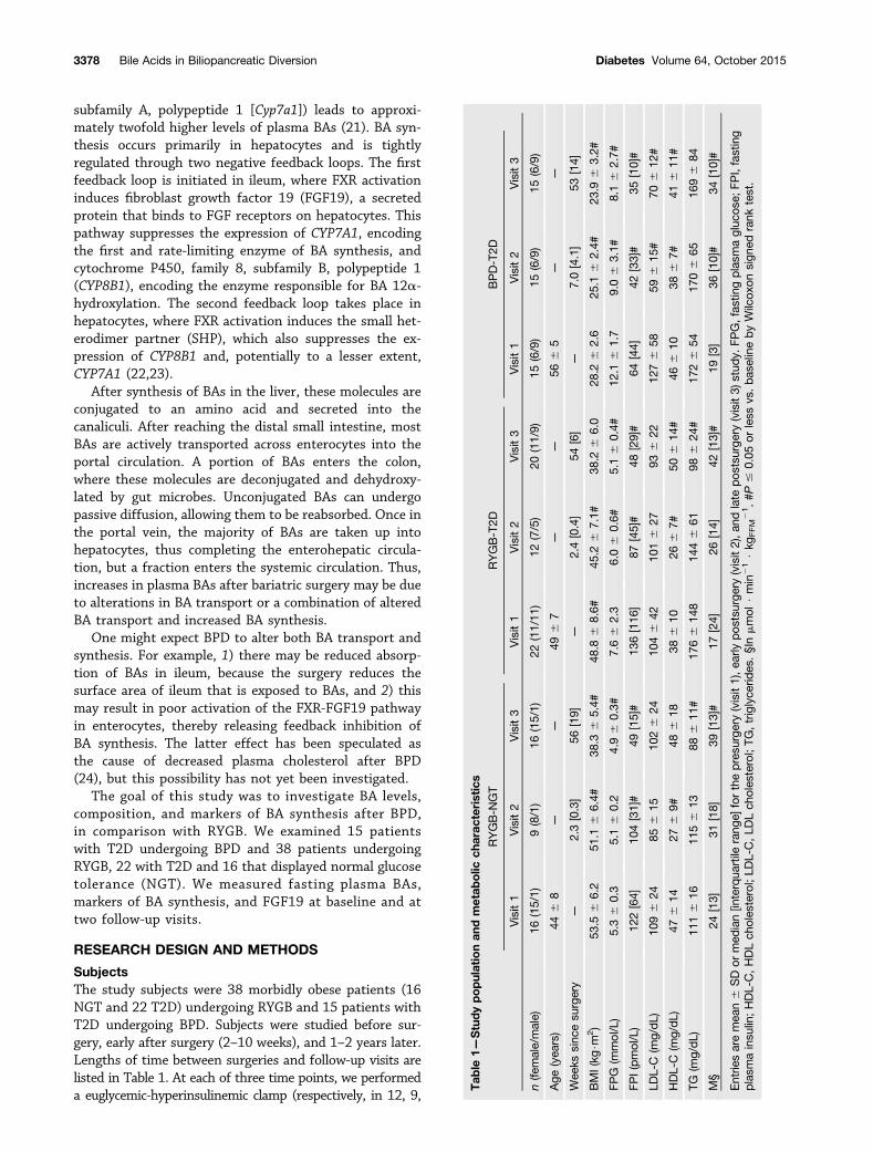
SubjectsThe study subjects were 38 morbidly obese patients (16NGT and 22 T2D) undergoing RYGB and 15 patients withT2D undergoing BPD. Subjects were studied before sur-gery, early after surgery (2–10 weeks), and 1–2 years later.Lengths of time between surgeries and follow-up visits arelisted in Table 1. At each of three time points, we performeda euglycemic-hyperinsulinemic clamp (respectively, in 12, 9,
Tab
le1—
Studypopulationan
dmetab
olic
charac
teristics
RYGB-N
GT
RYGB-T2D
BPD-T2D
Visit1
Visit2
Visit3
Visit1
Visit2
Visit3
Visit1
Visit2
Visit3
n(fe
male/male)
16(15/1)
9(8/1)
16(15/1)
22(11/11
)12
(7/5)
20(11/9)
15(6/9)
15(6/9)
15(6/9)
Age
(yea
rs)
446
8—
—49
67
——
566
5—
—
Wee
kssinc
esu
rgery
—2.3[0.3]
56[19]
—2.4[0.4]
54[6]
—7.0[4.1]
53[14]
BMI(kg
$m2)
53.5
66.2
51.1
66.4#
38.3
65.4#
48.8
68.6#
45.2
67.1#
38.2
66.0
28.2
62.6
25.1
62.4#
23.9
63.2#
FPG
(mmol/L)
5.36
0.3
5.16
0.2
4.96
0.3#
7.66
2.3
6.06
0.6#
5.16
0.4#
12.1
61.7
9.06
3.1#
8.16
2.7#
FPI(pmol/L)
122[64]
104[31]#
49[15]#
136[116
]87
[45]#
48[29]#
64[44]
42[33]#
35[10]#
LDL-C
(mg/dL)
1096
2485
615
1026
2410
46
4210
16
2793
622
1276
5859
615
#70
612
#
HDL-C
(mg/dL)
476
1427
69#
486
1838
610
266
7#50
614
#46
610
386
7#41
611
#
TG(m
g/dL)
1116
1611
56
1388
611
#17
66
148
1446
6198
624
#17
26
5417
06
6516
96
84
M§
24[13]
31[18]
39[13]#
17[24]
26[14]
42[13]#
19[3]
36[10]#
34[10]#
Entrie
saremea
n6
SDor
med
ian[in
terqua
rtile
rang
e]forthepresu
rgery(visit1),ea
rlypos
tsurge
ry(visit2),a
ndlate
pos
tsurge
ry(visit3)
stud
y.FP
G,fas
tingplasm
agluc
ose;
FPI,fasting
plasm
ainsu
lin;HDL-C,HDLch
oles
terol;LD
L-C,LD
Lch
oles
terol;TG
,triglyce
rides
.§Inmmol
$min
21$kg
FFM21.#P
#0.05
orless
vs.ba
selinebyWilcox
onsign
edrank
test.
3378 Bile Acids in Biliopancreatic Diversion Diabetes Volume 64, October 2015
and 12 RYGB-NGT patients; in 14, 12, and 13 RYGB-T2Dpatients; and in 15, 15, and 15 BPD-T2D patients). In theBPD group, the patients were treated by oral hypoglycemicagents and/or insulin (metformin in 6, metformin plussulfonylurea in 3, and metformin plus insulin in 6), andin the RYGB group, 5 patients were treated by diet aloneand 17 by oral hypoglycemic agents and/or insulin (met-formin in 13 and metformin plus sulfonylurea and insulinin 2 each). The patients with diabetes on oral antidiabeticagents were asked to stop them 48–72 h before surgery; inthose on insulin, injections were discontinued 16 h beforethe metabolic study (patients on bedtime glargine had beenswitched to NPH 2 days before the study). Glucose fluxdata from the 15 BPD-T2D patients (7) and from 12RYGB-NGT and 13 RYGB-T2D patients (25) have beenpreviously reported.
For comparison purposes, subjects without diabetesfrom a previous study (26) were selected to match the BMIof subjects at the second follow-up for each surgery. Sub-jects were matched to post-RYGB: n = 23, BMI = 39.76 1.4kg $ m22, and P = 0.56 compared with post–RYGB-NGTand P = 0.49 compared with post–RYGB-T2D. Subjects werematched to post-BPD: n = 10, BMI = 23.56 0.7 kg $m22,and P = 0.98 compared with post BPD-T2D.
Euglycemic-Hyperinsulinemic ClampAfter an overnight (12-h) fast, two catheters wereinserted into an antecubital vein for infusion of all testsubstances and retrogradely into a vein on the dorsumof the hand for blood drawing. The hand was heated at55°C to achieve arterialization of venous blood. At 9:00 A.M.,baseline blood samples were drawn. At time –20, –10,and 0 min, blood samples were obtained from the arte-rialized vein for the measurement of glucose and in-sulin. At time 0, a primed-continuous insulin (Humulin R;Eli Lilly and Company, Indianapolis, IN) infusion (at arate of 240 pmol $ min21 . m22) was started and con-tinued for 180 min; plasma glucose levels were mea-sured every 5 min throughout the clamp. Plasma insulinconcentrations were measured every 20 min between120 and 180 min after the start of insulin infusion.Fat-free mass (FFM) was estimated by electrical bioim-pedance (TBF 300; Tanita, Tokyo, Japan) as previouslydescribed (25).
Analytical ProceduresPlasma glucose was measured by the glucose oxidasetechnique on a Beckman Glucose Analyzer (Beckman,Fullerton, CA). Plasma insulin was assayed by a specificradioimmunoassay (Linco Research, St. Charles, MO,and MYRIA, Technogenetics, Milan, Italy, respectively).BAs were measured by liquid chromatography–tandemmass spectrometry (LC-MS/MS) (Waters Quattro Microwith Waters 2795 Alliance HPLC) as previously described(26). FGF19 was measured by ELISA (R&D Systems) inthe three surgical groups (n at each of three time points,respectively): RYGB-NGT (n = 12, 6, and 11), RYGB-T2D(n = 13, 10, and 14), and BPD-T2D (n = 13, 10, and 6).
BA Synthesis MarkersSample plasma (150 mL) was transferred into a 2-mL,96-deep well plate; this was followed by the addition of585 mL ice-cold acetonitrile containing 0.1% formic acidsolution and 5 mL 60 ng/mL internal standard mixture madeof d6-7a,12a-dihydroxy-4-cholesten-3-one (d6-7,12-diHCO)and d7-7a-hydroxy-4-cholesten-3-one (d7-7-HCO). Theplate was sealed and vortexed for 1 min. Then, the platewas centrifuged for 20 min at 4,000 rpm. After this,600 mL of supernatant was passed under positive pres-sure through a protein precipitation plate, which retainedphospholipids but eluted the BA synthesis markers(Ostro plate; Waters Corp., Milford, MA). The eluent wascollected and evaporated under a constant flow of N2
at 45°C. Samples were then reconstituted in 100 mLof 80% acetonitrile + 0.1% formic acid/20% water.The resultant extract was injected (10 mL) onto an LC-MS/MS system operated in positive ion mode electro-spray (UPLC/Waters TQS mass spectrometer). Isotopicdilution quantitation was conducted to obtain concen-trations of 7a,12a-dihydroxy-4-cholesten-3-one (7,12-diHCO) and 7a-hydroxy-4-cholesten-3-one (7-HCO).
Data AnalysisWhole-body insulin-stimulated glucose disposal (M, inmmol/min) was calculated as the mean exogenous glucoseinfusion rate during the last 40 min of the clamp cor-rected for changes in glucose concentration within adistribution volume of 200 mL per kilogram of bodyweight. M values were expressed per kilogram of FFM (inmmol $ min21 . kgFFM
21) (26).
Statistical AnalysisData are reported as mean6 SD (or median [interquartilerange] for variables with a skewed distribution). Groupdifferences were analyzed by Mann-Whitney U test andpaired observations by Wilcoxon signed rank test. Forselected variables, two-way ANOVA for repeated measureswas used to compare surgical operations across time ofstudy. Linear and nonlinear associations were tested bystandard methods. A P# 0.05 was considered statisticallysignificant.
RESULTS
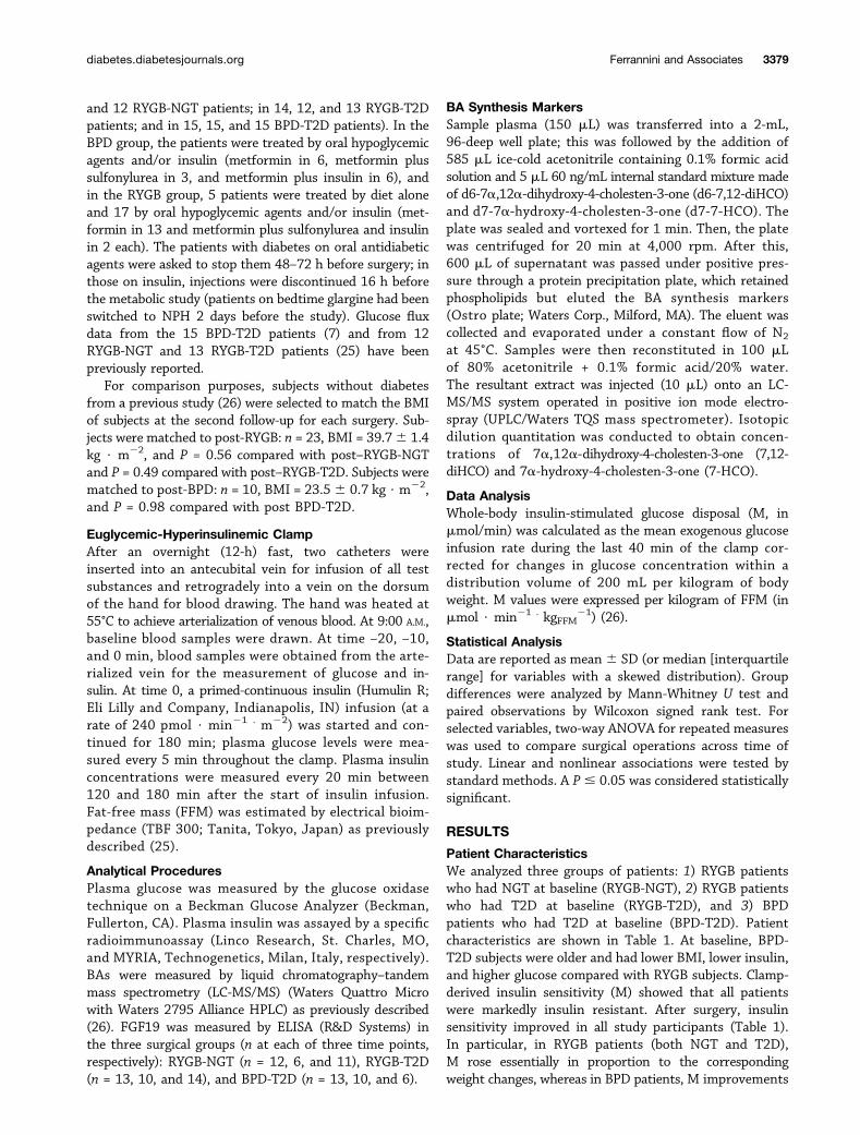
Patient CharacteristicsWe analyzed three groups of patients: 1) RYGB patientswho had NGT at baseline (RYGB-NGT), 2) RYGB patientswho had T2D at baseline (RYGB-T2D), and 3) BPDpatients who had T2D at baseline (BPD-T2D). Patientcharacteristics are shown in Table 1. At baseline, BPD-T2D subjects were older and had lower BMI, lower insulin,and higher glucose compared with RYGB subjects. Clamp-derived insulin sensitivity (M) showed that all patientswere markedly insulin resistant. After surgery, insulinsensitivity improved in all study participants (Table 1).In particular, in RYGB patients (both NGT and T2D),M rose essentially in proportion to the correspondingweight changes, whereas in BPD patients, M improvements
diabetes.diabetesjournals.org Ferrannini and Associates 3379
peaked at the earlier time (when weight changes weremodest) and were of greater magnitude than in RYGBpatients (14 [14] vs. 7 [21] mmol $ min21 . kgFFM
21,P = 0.013) (Fig. 1). In support of this different timecourse, two-way ANOVA was significant (P = 0.0018)for the time 3 group interaction; viewed otherwise, theslope of the relationship between M and BMI was signif-icantly steeper (P , 0.02) in BPD than RYGB patients(Supplementary Fig. 1).
Serum lipids changed differentially between the twooperations. After RYGB, LDL cholesterol levels wereonly marginally (and not significantly) reduced in bothNGT and T2D patients. In contrast, after BPD, LDLcholesterol halved already at 7 weeks, and this dropwas maintained longer term. Accordingly, two-wayANOVA was significant (P , 0.01) for the time 3 groupinteraction.
On the other hand, with RYGB, serum triglyceridesdecreased late after surgery (by 20 and 45% in NGT andT2D, respectively), whereas they were essentially unchangedwith BPD. HDL cholesterol levels were acutely decreased inall groups but recovered (in NGT) or improved (in T2D)after RYGB, whereas they remained slightly, if significantly,lower with BPD (Table 1).
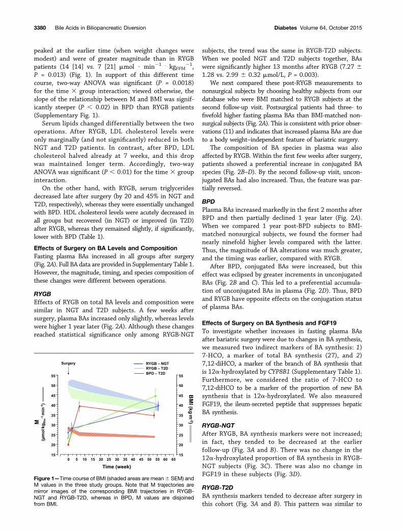
Effects of Surgery on BA Levels and CompositionFasting plasma BAs increased in all groups after surgery(Fig. 2A). Full BA data are provided in Supplementary Table 1.However, the magnitude, timing, and species composition ofthese changes were different between operations.
RYGBEffects of RYGB on total BA levels and composition weresimilar in NGT and T2D subjects. A few weeks aftersurgery, plasma BAs increased only slightly, whereas levelswere higher 1 year later (Fig. 2A). Although these changesreached statistical significance only among RYGB-NGT
subjects, the trend was the same in RYGB-T2D subjects.When we pooled NGT and T2D subjects together, BAswere significantly higher 13 months after RYGB (7.27 61.28 vs. 2.99 6 0.32 mmol/L, P = 0.003).
We next compared these post-RYGB measurements tononsurgical subjects by choosing healthy subjects from ourdatabase who were BMI matched to RYGB subjects at thesecond follow-up visit. Postsurgical patients had three- tofivefold higher fasting plasma BAs than BMI-matched non-surgical subjects (Fig. 2A). This is consistent with prior obser-vations (11) and indicates that increased plasma BAs are dueto a body weight–independent feature of bariatric surgery.
The composition of BA species in plasma was alsoaffected by RYGB. Within the first few weeks after surgery,patients showed a preferential increase in conjugated BAspecies (Fig. 2B–D). By the second follow-up visit, uncon-jugated BAs had also increased. Thus, the feature was par-tially reversed.
BPDPlasma BAs increased markedly in the first 2 months afterBPD and then partially declined 1 year later (Fig. 2A).When we compared 1 year post-BPD subjects to BMI-matched nonsurgical subjects, we found the former hadnearly ninefold higher levels compared with the latter.Thus, the magnitude of BA alterations was much greater,and the timing was earlier, compared with RYGB.
After BPD, conjugated BAs were increased, but thiseffect was eclipsed by greater increments in unconjugatedBAs (Fig. 2B and C). This led to a preferential accumula-tion of unconjugated BAs in plasma (Fig. 2D). Thus, BPDand RYGB have opposite effects on the conjugation statusof plasma BAs.
Effects of Surgery on BA Synthesis and FGF19To investigate whether increases in fasting plasma BAsafter bariatric surgery were due to changes in BA synthesis,we measured two indirect markers of BA synthesis: 1)7-HCO, a marker of total BA synthesis (27), and 2)7,12-diHCO, a marker of the branch of BA synthesis thatis 12a-hydroxylated by CYP8B1 (Supplementary Table 1).Furthermore, we considered the ratio of 7-HCO to7,12-diHCO to be a marker of the proportion of new BAsynthesis that is 12a-hydroxylated. We also measuredFGF19, the ileum-secreted peptide that suppresses hepaticBA synthesis.
RYGB-NGTAfter RYGB, BA synthesis markers were not increased;in fact, they tended to be decreased at the earlierfollow-up (Fig. 3A and B). There was no change in the12a-hydroxylated proportion of BA synthesis in RYGB-NGT subjects (Fig. 3C). There was also no change inFGF19 in these subjects (Fig. 3D).
RYGB-T2DBA synthesis markers tended to decrease after surgery inthis cohort (Fig. 3A and B). This pattern was similar to
Figure 1—Time course of BMI (shaded areas are mean6 SEM) andM values in the three study groups. Note that M trajectories aremirror images of the corresponding BMI trajectories in RYGB-NGT and RYGB-T2D, whereas in BPD, M values are disjoinedfrom BMI.
3380 Bile Acids in Biliopancreatic Diversion Diabetes Volume 64, October 2015
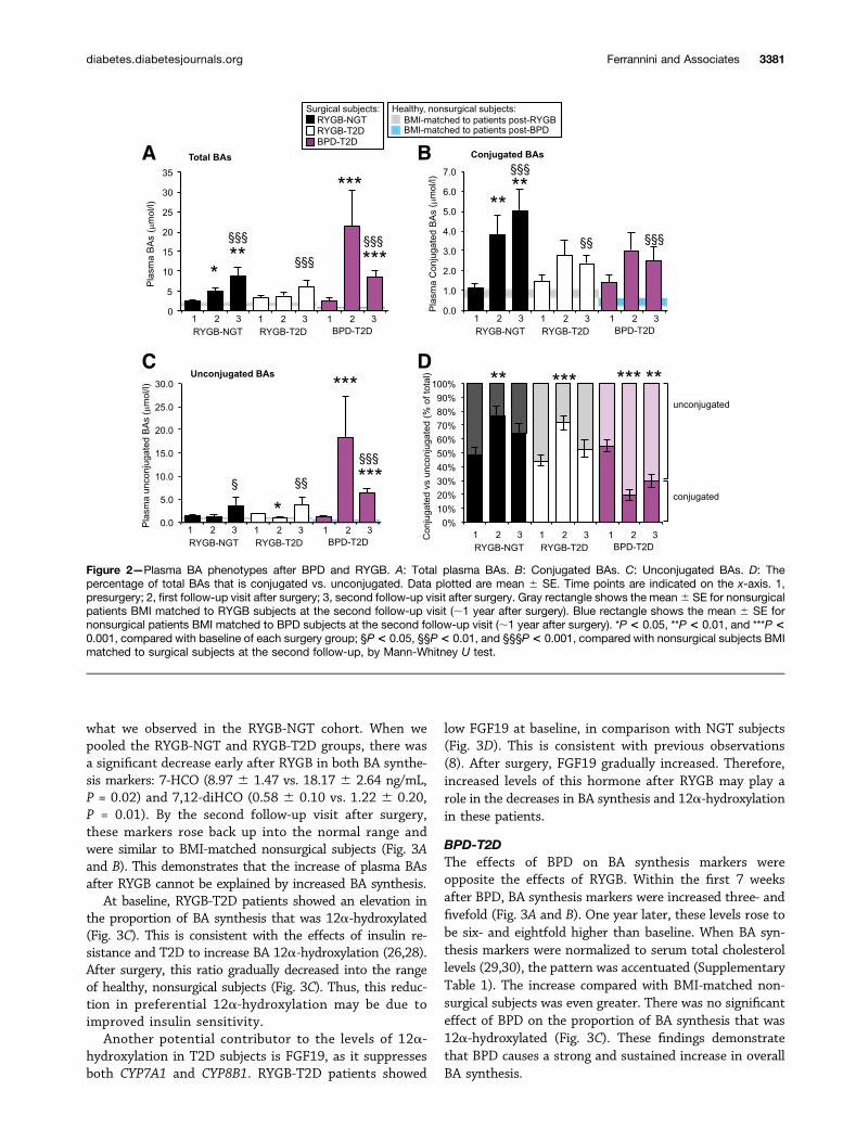
what we observed in the RYGB-NGT cohort. When wepooled the RYGB-NGT and RYGB-T2D groups, there wasa significant decrease early after RYGB in both BA synthe-sis markers: 7-HCO (8.97 6 1.47 vs. 18.17 6 2.64 ng/mL,P = 0.02) and 7,12-diHCO (0.58 6 0.10 vs. 1.22 6 0.20,P = 0.01). By the second follow-up visit after surgery,these markers rose back up into the normal range andwere similar to BMI-matched nonsurgical subjects (Fig. 3Aand B). This demonstrates that the increase of plasma BAsafter RYGB cannot be explained by increased BA synthesis.
At baseline, RYGB-T2D patients showed an elevation inthe proportion of BA synthesis that was 12a-hydroxylated(Fig. 3C). This is consistent with the effects of insulin re-sistance and T2D to increase BA 12a-hydroxylation (26,28).After surgery, this ratio gradually decreased into the rangeof healthy, nonsurgical subjects (Fig. 3C). Thus, this reduc-tion in preferential 12a-hydroxylation may be due toimproved insulin sensitivity.
Another potential contributor to the levels of 12a-hydroxylation in T2D subjects is FGF19, as it suppressesboth CYP7A1 and CYP8B1. RYGB-T2D patients showed
low FGF19 at baseline, in comparison with NGT subjects(Fig. 3D). This is consistent with previous observations(8). After surgery, FGF19 gradually increased. Therefore,increased levels of this hormone after RYGB may play arole in the decreases in BA synthesis and 12a-hydroxylationin these patients.
BPD-T2DThe effects of BPD on BA synthesis markers wereopposite the effects of RYGB. Within the first 7 weeksafter BPD, BA synthesis markers were increased three- andfivefold (Fig. 3A and B). One year later, these levels rose tobe six- and eightfold higher than baseline. When BA syn-thesis markers were normalized to serum total cholesterollevels (29,30), the pattern was accentuated (SupplementaryTable 1). The increase compared with BMI-matched non-surgical subjects was even greater. There was no significanteffect of BPD on the proportion of BA synthesis that was12a-hydroxylated (Fig. 3C). These findings demonstratethat BPD causes a strong and sustained increase in overallBA synthesis.
Figure 2—Plasma BA phenotypes after BPD and RYGB. A: Total plasma BAs. B: Conjugated BAs. C: Unconjugated BAs. D: Thepercentage of total BAs that is conjugated vs. unconjugated. Data plotted are mean 6 SE. Time points are indicated on the x-axis. 1,presurgery; 2, first follow-up visit after surgery; 3, second follow-up visit after surgery. Gray rectangle shows the mean6 SE for nonsurgicalpatients BMI matched to RYGB subjects at the second follow-up visit (;1 year after surgery). Blue rectangle shows the mean 6 SE fornonsurgical patients BMI matched to BPD subjects at the second follow-up visit (;1 year after surgery). *P < 0.05, **P < 0.01, and ***P <0.001, compared with baseline of each surgery group; §P< 0.05, §§P< 0.01, and §§§P< 0.001, compared with nonsurgical subjects BMImatched to surgical subjects at the second follow-up, by Mann-Whitney U test.
diabetes.diabetesjournals.org Ferrannini and Associates 3381
We expected that one reason for the increase in BAsynthesis would be decreased FGF19, because the area ofileum that is exposed to BAs is shortened by the surgery.In fact, FGF19 levels were highly variable and showedonly a trend toward decreased levels after BPD (Fig. 3D).Thus, additional mechanisms may be involved in the in-creased BA synthesis after BPD.
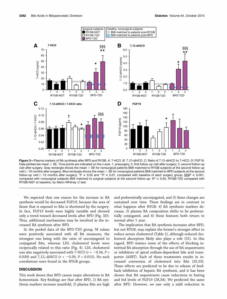
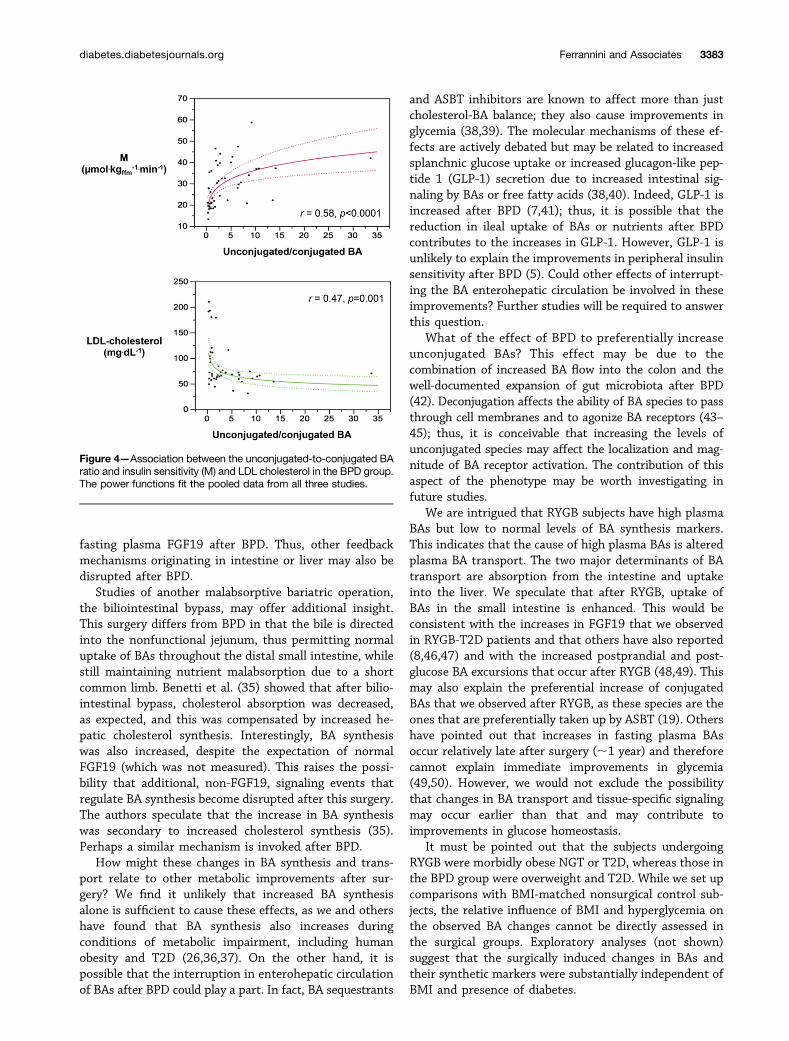
In the pooled data of the BPD-T2D group, M valueswere positively associated with all BA measures, thestrongest one being with the ratio of unconjugated toconjugated BAs, whereas LDL cholesterol levels werereciprocally related to this ratio (Fig. 4). LDL cholesterolwas also negatively associated with 7-HCO (r =20.34, P =0.039) and 7,12,-diHCO (r = 20.39, P = 0.015). No suchcorrelations were found in the RYGB groups.
DISCUSSION
This work shows that BPD causes major alterations in BAhomeostasis. Key findings are that after BPD, 1) BA syn-thesis markers increase manifold, 2) plasma BAs are high
and preferentially unconjugated, and 3) these changes aresustained over time. These findings are in contrast towhat happens after RYGB: 4) BA synthesis markers de-crease, 5) plasma BA composition shifts to be preferen-tially conjugated, and 6) these features both return tonormal after 1 year.
The implication that BA synthesis increases after BPD,but not RYGB, may explain the former’s stronger effect toreduce serum cholesterol (Table 1), although reduced cho-lesterol absorption likely also plays a role (31). In thisregard, BPD mimics some of the effects of blocking in-testinal BA absorption through the use of BA sequestrantsor inhibitors of apical sodium-dependent bile acid trans-porter (ASBT). Each of these treatments results in in-creased conversion of cholesterol into BAs (32,33).These effects are predicted to be due to release of feed-back inhibition of hepatic BA synthesis, and it has beenshown that BA sequestrants cause reductions in fastingand fed levels of FGF19 (28,34). We predicted the sameafter BPD. However, we saw only a mild reduction in
Figure 3—Plasma markers of BA synthesis after BPD and RYGB. A: 7-HCO. B: 7,12-diHCO. C: Ratio of 7,12-diHCO to 7-HCO. D: FGF19.Data plotted are mean6 SE. Time points are indicated on the x-axis. 1, presurgery; 2, first follow-up visit after surgery; 3, second follow-upvisit after surgery. Gray rectangle shows the mean 6 SE for nonsurgical patients BMI matched to RYGB subjects at the second follow-upvisit (;16 months after surgery). Blue rectangle shows the mean6 SE for nonsurgical patients BMI matched to BPD subjects at the secondfollow-up visit (;13 months after surgery). *P < 0.05 and **P < 0.01, compared with baseline of each surgery group; §§§P < 0.001,compared with nonsurgical subjects BMI matched to surgical subjects at the second follow-up; †P < 0.05, RYGB-T2D compared withRYGB-NGT at baseline, by Mann-Whitney U test.
3382 Bile Acids in Biliopancreatic Diversion Diabetes Volume 64, October 2015
fasting plasma FGF19 after BPD. Thus, other feedbackmechanisms originating in intestine or liver may also bedisrupted after BPD.
Studies of another malabsorptive bariatric operation,the biliointestinal bypass, may offer additional insight.This surgery differs from BPD in that the bile is directedinto the nonfunctional jejunum, thus permitting normaluptake of BAs throughout the distal small intestine, whilestill maintaining nutrient malabsorption due to a shortcommon limb. Benetti et al. (35) showed that after bilio-intestinal bypass, cholesterol absorption was decreased,as expected, and this was compensated by increased he-patic cholesterol synthesis. Interestingly, BA synthesiswas also increased, despite the expectation of normalFGF19 (which was not measured). This raises the possi-bility that additional, non-FGF19, signaling events thatregulate BA synthesis become disrupted after this surgery.The authors speculate that the increase in BA synthesiswas secondary to increased cholesterol synthesis (35).Perhaps a similar mechanism is invoked after BPD.
How might these changes in BA synthesis and trans-port relate to other metabolic improvements after sur-gery? We find it unlikely that increased BA synthesisalone is sufficient to cause these effects, as we and othershave found that BA synthesis also increases duringconditions of metabolic impairment, including humanobesity and T2D (26,36,37). On the other hand, it ispossible that the interruption in enterohepatic circulationof BAs after BPD could play a part. In fact, BA sequestrants
and ASBT inhibitors are known to affect more than justcholesterol-BA balance; they also cause improvements inglycemia (38,39). The molecular mechanisms of these ef-fects are actively debated but may be related to increasedsplanchnic glucose uptake or increased glucagon-like pep-tide 1 (GLP-1) secretion due to increased intestinal sig-naling by BAs or free fatty acids (38,40). Indeed, GLP-1 isincreased after BPD (7,41); thus, it is possible that thereduction in ileal uptake of BAs or nutrients after BPDcontributes to the increases in GLP-1. However, GLP-1 isunlikely to explain the improvements in peripheral insulinsensitivity after BPD (5). Could other effects of interrupt-ing the BA enterohepatic circulation be involved in theseimprovements? Further studies will be required to answerthis question.
What of the effect of BPD to preferentially increaseunconjugated BAs? This effect may be due to thecombination of increased BA flow into the colon and thewell-documented expansion of gut microbiota after BPD(42). Deconjugation affects the ability of BA species to passthrough cell membranes and to agonize BA receptors (43–45); thus, it is conceivable that increasing the levels ofunconjugated species may affect the localization and mag-nitude of BA receptor activation. The contribution of thisaspect of the phenotype may be worth investigating infuture studies.
We are intrigued that RYGB subjects have high plasmaBAs but low to normal levels of BA synthesis markers.This indicates that the cause of high plasma BAs is alteredplasma BA transport. The two major determinants of BAtransport are absorption from the intestine and uptakeinto the liver. We speculate that after RYGB, uptake ofBAs in the small intestine is enhanced. This would beconsistent with the increases in FGF19 that we observedin RYGB-T2D patients and that others have also reported(8,46,47) and with the increased postprandial and post-glucose BA excursions that occur after RYGB (48,49). Thismay also explain the preferential increase of conjugatedBAs that we observed after RYGB, as these species are theones that are preferentially taken up by ASBT (19). Othershave pointed out that increases in fasting plasma BAsoccur relatively late after surgery (;1 year) and thereforecannot explain immediate improvements in glycemia(49,50). However, we would not exclude the possibilitythat changes in BA transport and tissue-specific signalingmay occur earlier than that and may contribute toimprovements in glucose homeostasis.
It must be pointed out that the subjects undergoingRYGB were morbidly obese NGT or T2D, whereas those inthe BPD group were overweight and T2D. While we set upcomparisons with BMI-matched nonsurgical control sub-jects, the relative influence of BMI and hyperglycemia onthe observed BA changes cannot be directly assessed inthe surgical groups. Exploratory analyses (not shown)suggest that the surgically induced changes in BAs andtheir synthetic markers were substantially independent ofBMI and presence of diabetes.
Figure 4—Association between the unconjugated-to-conjugated BAratio and insulin sensitivity (M) and LDL cholesterol in the BPD group.The power functions fit the pooled data from all three studies.
diabetes.diabetesjournals.org Ferrannini and Associates 3383
Overall, these findings highlight that elevated plasma BAsafter different bariatric surgeries can arise through distinctmechanisms. The upregulation of BA synthesis after BPDconfirms our prediction and may partially explain thepowerful decrease in plasma cholesterol in these subjects.However, the mechanism responsible for the upregulation ofBA synthesis is likely to involve signaling other than FGF19,and this warrants further investigation. Finally, we wouldsuggest that further examination of changes in BA transportand BA signaling may shed additional light on the impact ofBAs on glucose and lipid metabolism after bariatric surgeries.
Acknowledgments. The authors would like to acknowledge DomenicoAccili from Columbia University and Mark Erion, Stephen Previs, Martin Brenner,and David Kelley from Merck Research Laboratories for their scientific discussion,support, and input into this manuscript.Funding. This study was funded in part by the Italian Ministry of Education,University, and Research (2010329EKE), the National Institutes of Health (NIH)(HL-111206 to R.A.H.), and Columbia Clinical and Translational Science Awardgrant UL1-TR-000040 (National Center for Advancing Translational Sciences/NIH).Duality of Interest. This study was funded in part by an unrestricted grantfrom Merck Research Laboratories. E.F. has been a speaker and consultant forBoehringer Ingelheim, Merck, Sanofi, Eli Lilly and Company, Johnson & Johnson,Astellas, Daiichi Sankyo, Bristol-Myers Squibb/AstraZeneca, and Novartis. J.C.-P.,D.X., L.W., and M.C. are employees of Merck Research Laboratories. No otherpotential conflicts of interest relevant to this article were reported.
Merck Research Laboratories had no input into the design of the study or theanalysis of the data.Author Contributions. E.F. and R.A.H. designed experiments, analyzeddata, and wrote the manuscript. S.C., B.A., and M.N. performed in vivo experi-ments and contributed to discussions. J.C.-P., D.X., L.W., and M.C. measured BAsynthesis markers in a blinded fashion and contributed to discussions. All authorsedited the manuscript. E.F. and R.A.H. are the guarantors of this work and, assuch, had full access to all the data in the study and take responsibility for theintegrity of the data and the accuracy of the data analysis.
References1. Buchwald H, Estok R, Fahrbach K, et al. Weight and type 2 diabetes afterbariatric surgery: systematic review and meta-analysis. Am J Med 2009;122:248.e5–256.e52. Garrido-Sanchez L, Murri M, Rivas-Becerra J, et al. Bypass of the duode-num improves insulin resistance much more rapidly than sleeve gastrectomy.Surg Obes Relat Dis 2012;8:145–1503. Piché ME, Martin J, Cianflone K, et al. Changes in predicted cardiovasculardisease risk after biliopancreatic diversion surgery in severely obese patients.Metabolism 2014;63:79–864. Pontiroli AE, Laneri M, Veronelli A, et al. Biliary pancreatic diversion andlaparoscopic adjustable gastric banding in morbid obesity: their long-term effectson metabolic syndrome and on cardiovascular parameters. Cardiovasc Diabetol2009;8:375. Ferrannini E, Mingrone G. Impact of different bariatric surgical procedureson insulin action and beta-cell function in type 2 diabetes. Diabetes Care 2009;32:514–5206. Scopinaro N, Adami GF, Papadia FS, et al. Effects of biliopanceratic di-version on type 2 diabetes in patients with BMI 25 to 35. Ann Surg 2011;253:699–7037. Astiarraga B, Gastaldelli A, Muscelli E, et al. Biliopancreatic diversion innonobese patients with type 2 diabetes: impact and mechanisms. J Clin Endo-crinol Metab 2013;98:2765–2773
8. Gerhard GS, Styer AM, Wood GC, et al. A role for fibroblast growth factor 19and bile acids in diabetes remission after Roux-en-Y gastric bypass. DiabetesCare 2013;36:1859–18649. Kohli R, Bradley D, Setchell KD, Eagon JC, Abumrad N, Klein S. Weight lossinduced by Roux-en-Y gastric bypass but not laparoscopic adjustable gastric bandingincreases circulating bile acids. J Clin Endocrinol Metab 2013;98:E708–E71210. Nakatani H, Kasama K, Oshiro T, Watanabe M, Hirose H, Itoh H. Serum bileacid along with plasma incretins and serum high-molecular weight adiponectinlevels are increased after bariatric surgery. Metabolism 2009;58:1400–140711. Patti ME, Houten SM, Bianco AC, et al. Serum bile acids are higher inhumans with prior gastric bypass: potential contribution to improved glucose andlipid metabolism. Obesity (Silver Spring) 2009;17:1671–167712. de Aguiar Vallim TQ, Tarling EJ, Edwards PA. Pleiotropic roles of bile acidsin metabolism. Cell Metab 2013;17:657–66913. Lefebvre P, Cariou B, Lien F, Kuipers F, Staels B. Role of bile acids and bileacid receptors in metabolic regulation. Physiol Rev 2009;89:147–19114. Kuipers F, Bloks VW, Groen AK. Beyond intestinal soap—bile acids inmetabolic control. Nat Rev Endocrinol 2014;10:488–49815. Ma H, Patti ME. Bile acids, obesity, and the metabolic syndrome. Best PractRes Clin Gastroenterol 2014;28:573–58316. Kohli R, Seeley RJ. Diabetes: the search for mechanisms underlying bar-iatric surgery. Nat Rev Endocrinol 2013;9:572–57417. Pournaras DJ, le Roux CW. Are bile acids the new gut hormones? Lessonsfrom weight loss surgery models. Endocrinology 2013;154:2255–225618. Ryan KK, Tremaroli V, Clemmensen C, et al. FXR is a molecular target forthe effects of vertical sleeve gastrectomy. Nature 2014;509:183–18819. Trauner M, Boyer JL. Bile salt transporters: molecular characterization,function, and regulation. Physiol Rev 2003;83:633–67120. Dawson PA, Lan T, Rao A. Bile acid transporters. J Lipid Res 2009;50:2340–235721. Li T, Owsley E, Matozel M, Hsu P, Novak CM, Chiang JY. Transgenic ex-pression of cholesterol 7alpha-hydroxylase in the liver prevents high-fat diet-induced obesity and insulin resistance in mice. Hepatology 2010;52:678–69022. Kim I, Ahn SH, Inagaki T, et al. Differential regulation of bile acid homeostasisby the farnesoid X receptor in liver and intestine. J Lipid Res 2007;48:2664–267223. Kong B, Wang L, Chiang JY, Zhang Y, Klaassen CD, Guo GL. Mechanism oftissue-specific farnesoid X receptor in suppressing the expression of genes inbile-acid synthesis in mice. Hepatology 2012;56:1034–104324. Scopinaro N. Thirty-five years of biliopancreatic diversion: notes on gas-trointestinal physiology to complete the published information useful for a betterunderstanding and clinical use of the operation. Obes Surg 2012;22:427–43225. Camastra S, Gastaldelli A, Mari A, et al. Early and longer term effects ofgastric bypass surgery on tissue-specific insulin sensitivity and beta cell functionin morbidly obese patients with and without type 2 diabetes. Diabetologia 2011;54:2093–210226. Haeusler RA, Astiarraga B, Camastra S, Accili D, Ferrannini E. Human insulinresistance is associated with increased plasma levels of 12a-hydroxylated bileacids. Diabetes 2013;62:4184–419127. Gälman C, Arvidsson I, Angelin B, Rudling M. Monitoring hepatic cholesterol7alpha-hydroxylase activity by assay of the stable bile acid intermediate 7alpha-hydroxy-4-cholesten-3-one in peripheral blood. J Lipid Res 2003;44:859–86628. Brufau G, Stellaard F, Prado K, et al. Improved glycemic control with co-lesevelam treatment in patients with type 2 diabetes is not directly associatedwith changes in bile acid metabolism. Hepatology 2010;52:1455–146429. Gälman C, Angelin B, Rudling M. Bile acid synthesis in humans has a rapiddiurnal variation that is asynchronous with cholesterol synthesis. Gastroenterol-ogy 2005;129:1445–145330. Honda A, Yoshida T, Xu G, et al. Significance of plasma 7alpha-hydroxy-4-cholesten-3-one and 27-hydroxycholesterol concentrations as markers for he-patic bile acid synthesis in cholesterol-fed rabbits. Metabolism 2004;53:42–4831. Buchwald H, Avidor Y, Braunwald E, et al. Bariatric surgery: a systematicreview and meta-analysis. JAMA 2004;292:1724–1737
3384 Bile Acids in Biliopancreatic Diversion Diabetes Volume 64, October 2015
32. Einarsson K, Ericsson S, Ewerth S, et al. Bile acid sequestrants: mecha-nisms of action on bile acid and cholesterol metabolism. Eur J Clin Pharmacol1991;40(Suppl. 1):S53–S5833. Root C, Smith CD, Sundseth SS, Pink HM, Wilson JG, Lewis MC. Ileal bileacid transporter inhibition, CYP7A1 induction, and antilipemic action of 264W94.J Lipid Res 2002;43:1320–133034. Beysen C, Murphy EJ, Deines K, et al. Effect of bile acid sequestrants onglucose metabolism, hepatic de novo lipogenesis, and cholesterol and bile acidkinetics in type 2 diabetes: a randomised controlled study. Diabetologia 2012;55:432–44235. Benetti A, Del Puppo M, Crosignani A, et al. Cholesterol metabolism afterbariatric surgery in grade 3 obesity: differences between malabsorptive andrestrictive procedures. Diabetes Care 2013;36:1443–144736. Steiner C, Othman A, Saely CH, et al. Bile acid metabolites in serum: in-traindividual variation and associations with coronary heart disease, metabolicsyndrome and diabetes mellitus. PLoS ONE 2011;6:e2500637. Ståhlberg D, Rudling M, Angelin B, et al. Hepatic cholesterol metabolism inhuman obesity. Hepatology 1997;25:1447–145038. Prawitt J, Caron S, Staels B. Glucose-lowering effects of intestinal bile acidsequestration through enhancement of splanchnic glucose utilization. TrendsEndocrinol Metab 2014;25:235–24439. Chen L, Yao X, Young A, et al. Inhibition of apical sodium-dependent bileacid transporter as a novel treatment for diabetes. Am J Physiol Endocrinol Metab2012;302:E68–E7640. Hofmann AF. Bile acid sequestrants improve glycemic control in type 2diabetes: a proposed mechanism implicating glucagon-like peptide 1 release.Hepatology 2011;53:1784
41. Valverde I, Puente J, Martín-Duce A, et al. Changes in glucagon-likepeptide-1 (GLP-1) secretion after biliopancreatic diversion or vertical bandedgastroplasty in obese subjects. Obes Surg 2005;15:387–39742. Picard M, Frédéric Simon H, Stéfane L, Simon M, Simon B. Complications ofcombined gastric restrictive and malabsorptive procedures: part 2. Curr Surg2003;60:274–279; discussion 279–28143. Makishima M, Okamoto AY, Repa JJ, et al. Identification of a nuclear re-ceptor for bile acids. Science 1999;284:1362–136544. Parks DJ, Blanchard SG, Bledsoe RK, et al. Bile acids: natural ligands for anorphan nuclear receptor. Science 1999;284:1365–136845. Wang H, Chen J, Hollister K, Sowers LC, Forman BM. Endogenous bile acidsare ligands for the nuclear receptor FXR/BAR. Mol Cell 1999;3:543–55346. Jansen PL, van Werven J, Aarts E, et al. Alterations of hormonally activefibroblast growth factors after Roux-en-Y gastric bypass surgery. Dig Dis 2011;29:48–5147. Pournaras DJ, Glicksman C, Vincent RP, et al. The role of bile after Roux-en-Y gastric bypass in promoting weight loss and improving glycaemic control.Endocrinology 2012;153:3613–361948. Ahmad NN, Pfalzer A, Kaplan LM. Roux-en-Y gastric bypass normalizes theblunted postprandial bile acid excursion associated with obesity. Int J Obes 2013;37:1553–155949. Dutia R, Embrey M, O’Brien CS, et al. Temporal changes in bile acid levelsand 12a-hydroxylation after Roux-en-Y gastric bypass surgery in type 2 diabetes.Int J Obes 2015;39:806–81350. Steinert RE, Peterli R, Keller S, et al. Bile acids and gut peptide secretionafter bariatric surgery: a 1-year prospective randomized pilot trial. Obesity (SilverSpring) 2013;21:E660–E668
diabetes.diabetesjournals.org Ferrannini and Associates 3385