Embed Size (px)
Citation preview

Title: Increased axonal bouton stability during learning in the mouse model of
MECP2 duplication syndrome
Abbreviated title: Bouton hyperstability in MECP2 duplication syndrome
Ryan T. Ash1,2,3, Paul G. Fahey2,3, Jiyoung Park3, Huda Y. Zoghbi3,4,5,6,7, Stelios M.
Smirnakis1* 5
1Department of Neurology, Brigham and Women’s Hospital and Jamaica Plain Veterans Administration Hospital, Harvard Medical School, Boston, MA 02115 2Medical Scientist Training Program, Baylor College of Medicine, Houston, TX 77030 3Department of Neuroscience, Baylor College of Medicine, Houston, TX 77030 10 4Department of Pediatrics, Texas Children’s Hospital and Baylor College of Medicine, Houston, TX 77030 5Department of Molecular and Human Genetics, Baylor College of Medicine, Houston, TX 77030 USA 6Jan and Dan Duncan Neurological Research Institute at Texas Children’s Hospital, 15 United States 7Howard Hughes Medical Institute, Baylor College of Medicine, Houston, TX 77030
*Correspondence to:
60 Fenwood Road
Boston MA 02115
Number of pages: 19
Number of figures: 4 25
Word counts. Abstract: 190 Intro: 580 Discussion: 1700
Conflict of interest: None.
Acknowledgments: R.T.A. received support from the Autism Speaks Weatherstone Fellowship and the BCM Medical Scientist Training Program. This work was supported by grants from the Simons Foundation and March of Dimes to S.M.S., the Howard 30 Hughes Medical Institute and NINDS HD053862 to H.Y.Z., and the Baylor Intellectual and Developmental Disabilities Research Center (P30HD024064) Mouse Neurobehavioral Core. We are grateful to S. Torsky, B. Suter, J. Patterson, S. Shen, and D. Yu for technical and theoretical advice on experiments and comments on the manuscript. 35
.CC-BY-NC-ND 4.0 International licensepeer-reviewed) is the author/funder. It is made available under aThe copyright holder for this preprint (which was not. http://dx.doi.org/10.1101/186239doi: bioRxiv preprint first posted online Sep. 8, 2017;
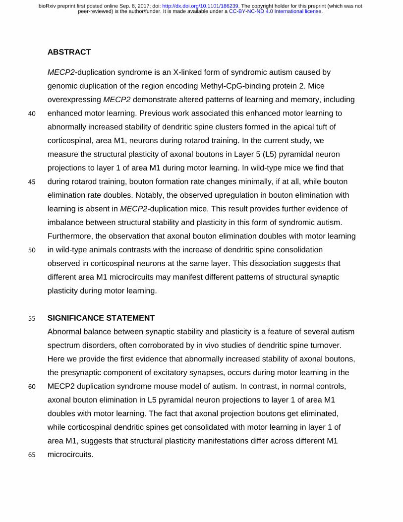
ABSTRACT
MECP2-duplication syndrome is an X-linked form of syndromic autism caused by
genomic duplication of the region encoding Methyl-CpG-binding protein 2. Mice
overexpressing MECP2 demonstrate altered patterns of learning and memory, including
enhanced motor learning. Previous work associated this enhanced motor learning to 40
abnormally increased stability of dendritic spine clusters formed in the apical tuft of
corticospinal, area M1, neurons during rotarod training. In the current study, we
measure the structural plasticity of axonal boutons in Layer 5 (L5) pyramidal neuron
projections to layer 1 of area M1 during motor learning. In wild-type mice we find that
during rotarod training, bouton formation rate changes minimally, if at all, while bouton 45
elimination rate doubles. Notably, the observed upregulation in bouton elimination with
learning is absent in MECP2-duplication mice. This result provides further evidence of
imbalance between structural stability and plasticity in this form of syndromic autism.
Furthermore, the observation that axonal bouton elimination doubles with motor learning
in wild-type animals contrasts with the increase of dendritic spine consolidation 50
observed in corticospinal neurons at the same layer. This dissociation suggests that
different area M1 microcircuits may manifest different patterns of structural synaptic
plasticity during motor learning.
SIGNIFICANCE STATEMENT 55
Abnormal balance between synaptic stability and plasticity is a feature of several autism
spectrum disorders, often corroborated by in vivo studies of dendritic spine turnover.
Here we provide the first evidence that abnormally increased stability of axonal boutons,
the presynaptic component of excitatory synapses, occurs during motor learning in the
MECP2 duplication syndrome mouse model of autism. In contrast, in normal controls, 60
axonal bouton elimination in L5 pyramidal neuron projections to layer 1 of area M1
doubles with motor learning. The fact that axonal projection boutons get eliminated,
while corticospinal dendritic spines get consolidated with motor learning in layer 1 of
area M1, suggests that structural plasticity manifestations differ across different M1
microcircuits. 65
.CC-BY-NC-ND 4.0 International licensepeer-reviewed) is the author/funder. It is made available under aThe copyright holder for this preprint (which was not. http://dx.doi.org/10.1101/186239doi: bioRxiv preprint first posted online Sep. 8, 2017;

INTRODUCTION
The rewiring of synaptic connections in neural microcircuits provides a compelling
mechanism for learning and memory throughout development and adult life(Chklovskii et al., 70
2004). Two-photon imaging of fluorescently-labeled neurons has recently enabled the direct
measurement of synaptic rewiring in vivo, revealing that new synapses form in motor cortex
(M1) during motor training, and that the stability of these synapses correlates with how well
the animal learns to perform the motor task (Xu et al., 2009; Yang et al., 2009). The layer 1
(L1) apical tuft dendritic spines that turn over during learning receive inputs from a range of 75
sources, including L2/3, L5, and L6 cortical pyramidal neurons, thalamocortical neurons, and
others. It is currently not known how synaptic inputs from axonal projections to area M1
behave during learning.
Experimental LTP and LTD paradigms in vitro can induce axonal bouton formation and
elimination (Antonova et al., 2001; Becker et al., 2008; Bourne et al., 2013). In vivo, axonal 80
boutons are spontaneously formed and eliminated in adult sensory cortex (De Paola et al.,
2006; Majewska et al., 2006; Stettler et al., 2006; Grillo et al., 2013), while learning has been
shown to alter bouton turnover in parallel fiber inputs to the cerebellum (Carrillo et al.,
2013)and in orbitofrontal inputs to the medial prefrontal cortex (Johnson et al., 2016).
However, bouton plasticity has yet to be measured in area M1 during motor learning to our 85
knowledge. In this work we examine the turnover of boutons, the pre-synaptic component
of synapses, in L5 pyramidal neuron axons that project to layer 1 of area M1.
Furthermore, we begin to assess whether learning-associated plasticity in inputs to area
M1 is altered in the MECP2 duplication model of autism. MECP2 duplication syndrome is
caused by a genomic duplication that spans the methyl-CpG-binding protein 2 (MECP2) 90
gene and leads to a progressive X-linked disorder of intellectual disability, autism, spasticity,
and epilepsy (Ramocki et al., 2010). Overexpression of the MECP2 gene in mice produces
a similar progressive neurological phenotype including autistic features (abnormal social
behavior, anxiety, and stereotypies), spasticity, and epilepsy (Collins et al., 2004).
Interestingly, before 24 weeks of age MECP2-duplication mice (Tg1) demonstrate a striking 95
enhancement in motor learning and memory on the rotarod task (Collins et al., 2004).
Previous work associated this enhanced learning with an increase in the formation and
.CC-BY-NC-ND 4.0 International licensepeer-reviewed) is the author/funder. It is made available under aThe copyright holder for this preprint (which was not. http://dx.doi.org/10.1101/186239doi: bioRxiv preprint first posted online Sep. 8, 2017;

stabilization of dendritic spine clusters in apical dendritic tufts of corticospinal neurons in
primary motor cortex (M1) (Ash et al., 2017), pointing to a possible mechanism for altered
learning and memory in these animals. 100
MeCP2 and other autism-associated proteins contribute to the development of mature
axons and presynaptic structures (Antar et al., 2006; Belichenko et al., 2009; Degano et al.,
2009; Chen et al., 2014; Garcia-Junco-Clemente and Golshani, 2014). Presynaptic
electrophysiological function has been shown to be altered in MECP2-duplication mice
(increased paired pulse facilitation, Collins et al., 2004) and other autism mouse models 105
(Deng et al., 2013), and mice with mutations in the proteins mediating presynaptic
plasticity often demonstrate autistic features (Blundell et al., 2010). Long term depression
(LTD), a form of synaptic weakening that has a major pre-synaptic component (Collingridge
et al., 2010), has been shown to be defective in several models of autism (D’Antoni et al.,
2014). These findings implicate pre-synaptic dysfunction in autism, but axonal bouton 110
structural plasticity has not been explored directly in a model of autism to our knowledge.
We measured learning-associated axonal bouton structural plasticity in layer 1 of
mouse M1 during rotarod training in the Tg1 mouse model of the MECP2 duplication
syndrome and compared with wild-type (WT) littermates. We found that the rate of bouton
formation does not change significantly with rotarod training in either genotype, 115
remaining approximately the same as the spontaneous bouton formation rate at rest. In
contrast, bouton elimination rate is dramatically accelerated during rotarod learning in
WT mice, whereas this effect is completely abolished in MECP2-duplication mice. This
supports the argument that increased synaptic stability manifests in the MECP2-duplication
syndrome during learning (Ash et al., 2017). 120
MATERIALS & METHODS
Animals. FVB-background MECP2-duplication (Tg1) mice (Collins et al., 2004), were
crossed to C57 thy1-GFP-M (Feng et al., 2000) homozygotes obtained from Jackson
Laboratories, to generate male F1C57;FVB MECP2-duplication;thy1-GFP-M mice and thy1-125
GFP-M littermate controls.
.CC-BY-NC-ND 4.0 International licensepeer-reviewed) is the author/funder. It is made available under aThe copyright holder for this preprint (which was not. http://dx.doi.org/10.1101/186239doi: bioRxiv preprint first posted online Sep. 8, 2017;

In vivo two-photon imaging. All surgeries and imaging were performed blind to genotype.
At least two weeks prior to the first imaging session (~12-14 week-old-mice), a 3 mm-wide
opening was drilled over motor cortex, centered at 1.6 mm lateral to bregma (Tennant et al.,
2011), and a glass coverslip was placed over the exposed brain surface to allow chronic 130
imaging of neuronal morphology (Mostany and Portera-Cailliau, 2008; Holtmaat et al., 2009;
Mostany et al., 2013). Neural structures were imaged using a Zeiss in vivo 2-photon
microscope with Zeiss 20x 1.0 NA water-immersion objective lens. High-quality craniotomies
had a characteristic bright-field appearance with well-defined vasculature and pale grey
matter (Fig. 1A). Under two-photon scanning fluorescent structures were reliably clear and 135
visible with low laser power (<20 mW).
Only high quality preparations (low background noise across all time points, <5 pixel i.e.
<0.5µm slow motion artifact, <2 pixel i.e. <0.2 µm fast motion artifact, and axons well
isolated from other fluorescent structures) were used in the blinded analysis. Pyramidal
neuron axons were imaged at high resolution (310x310 to 420x420 µm FOV, 0.1 µm/pixel, 1 140
µm Z-step size) to adequately capture individual boutons. Laser power was maintained under
20 mW (average ~10 mW) during image stack acquisition.
Motor training. The Ugo Basile mouse rotarod was used for motor training. At least two hours
after imaging sessions, in the late afternoon, mice were placed on the rotarod, and the rotarod
gradually accelerated from 5 to 80 rpm over 3 minutes. Single-trial rotarod performance was 145
quantified as the time right before falling or holding on to the dowel rod for two complete
rotations without regaining footing. A 7-10 minute rest period occurred between each trial.
Four trials were performed per day.
Analysis of bouton plasticity. Analysis was performed blind to genotype. Axons were
chosen from the imaging field based on characteristic appearance, including the 150
absence of dendritic spines, minimal branching, and the presence of synaptic boutons,
as well as decreased width compared to dendrites. In the thy1-GFP M mouse line (Feng
et al., 2000) we employed, the vast majority of GFP-labeled axons in the cerebral cortex
arise from L5 pyramidal neurons, though occasional L2/3, L6 pyramidal neurons and
thalamocortical neurons may also be labeled (De Paola et al., 2006). Pyramidal neuron 155
axons were targeted based on their thin shafts, high density of small (<1 µm diameter)
en-passant boutons, low tortuosity, and rare branching (type A3 axons), allowing them
.CC-BY-NC-ND 4.0 International licensepeer-reviewed) is the author/funder. It is made available under aThe copyright holder for this preprint (which was not. http://dx.doi.org/10.1101/186239doi: bioRxiv preprint first posted online Sep. 8, 2017;

to be clearly distinguished from i) L6 pyramidal neuron axons, which have high
branching and a high density of terminaux boutons, and from ii) thalamocortical
neurons, which have thicker axons and high branching (De Paola et al., 2006). Given 160
the very sparse labeling of L2/3 neurons in the thy1-GFP M mouse line, we are
confident that the great majority of axonal segments we imaged represent L5 pyramidal
neuron projections to area L1 from other regions, i.e. chiefly from the premotor, the
somatosensory and the contralateral motor cortex (Hooks et al., 2013).
Segments of axon that were clearly visualized in all three time points were selected for 165
analysis (length range 30 – 360 µm, mean 138 µm). The presence of en-passant
boutons or terminaux boutons was noted by a blinded investigator, who further
classified synaptic boutons as alpha (> ~2 µm or 20 pixel diameter) or beta (<~2 µm or
20 pixel diameter). The threshold used for bouton classification was based on the
bimodal distribution of boutons, separable at ~2 µm diameter, present in the analyzed 170
data set (Grillo et al., 2013). The presence of a bouton was determined by a clear
increase in axon diameter and increased fluorescence compared to the background
axon, as well as the judgment of an experienced investigator (Fig. 1B). Only
varicosities that were more than twice as bright as the axonal backbone and extended
at least 3 pixels (~0.3 microns) outside the axonal shaft diameter, which corresponds 175
approximately to 2 SDs of the noise blur on either side of the axonal shaft, were counted as
boutons. This is similar to (Grillo et al., 2013), which analyzed boutons that were twice as
bright as the axon.
Boutons located greater than 50 µm away from the nearest other bouton were excluded
from the analysis, so that stretches of bouton-free axon would not bias bouton density 180
calculations. Four to twenty axons were analyzed from 1-3 imaging fields per mouse for
13 mice (6 WT, 7 MECP2-duplication mice). Unless the investigator could clearly trace
the continuity of axon segments, segments were analyzed as individual units. Though
unlikely, the possibility cannot be completely excluded that, on occasion, more than one
segment from a single axon were counted. Bouton formation and elimination (Fig. 1C, 185
2B,C) was calculated as (boutons formed or boutons eliminated) / (total number of
boutons observed across imaging sessions), analogous to the measure used in (Grillo et
.CC-BY-NC-ND 4.0 International licensepeer-reviewed) is the author/funder. It is made available under aThe copyright holder for this preprint (which was not. http://dx.doi.org/10.1101/186239doi: bioRxiv preprint first posted online Sep. 8, 2017;

al., 2013). Bouton survival was calculated as the percent of boutons identified in the first
imaging time point that are present in subsequent imaging time points. Bouton
stabilization was calculated as the percent of newly formed boutons in the second 190
imaging time point, which persisted in the third imaging time point.
Statistics. Except where indicated, the Mann-Whitney U test was used for two-group
statistical comparisons, and 2-way ANOVA with Tukey multiple-comparison correction
was used for multi-group comparisons.
RESULTS [1460 words – how many allowed?] 195
The Tg1 mouse model for MECP2 duplication syndrome (FVB background) was
crossed to the thy1-GFP-M mouse line (C57 background) to generate F1 hybrid males
for experiments. A cranial window was placed over motor cortex (1.6 mm lateral to
bregma) at 12-14 weeks of age, and at least 2 weeks following the surgery the mouse
was placed under the 2-photon microscope to image GFP-labeled axons in layer 1 of 200
area M1 (Fig. 1A; see methods).
L5 pyramidal neuron axons are typically visualized as a thin string of fluorescence
interspersed with fluorescent expansions or varicosities (en passant boutons) and rare
spine-like terminaux boutons. They are readily differentiated morphologically from L6
neuron axons and thalamocortical axons (De Paola et al., 2006), which, in any case, are 205
rarely fluorescent in these animals. The thy1-GFP M line primarily labels L5 pyramidal
neurons in neocortex, and therefore the majority of axonal arbors we imaged are
expected to arise from L5 of the somatosensory cortex, the premotor cortex, or the
contralateral motor cortex, all of which project to L1 of area M1 (Colechio and Alloway,
2009; Mao et al., 2011; Hooks et al., 2013). Area M1 L5 neurons rarely send projections 210
locally to layer 1 (Cho et al., 2004).
First, we report on axonal bouton structure and plasticity analyzed in littermate
controls with normal MECP2 expression. Axonal boutons were identified as periodic
thickenings or extensions along the axon, at least twice as bright as and extending at
least 0.3 µm from the axonal backbone (Grillo et al., 2013) (this corresponds to 215
approximately 2 SDs of the noise blur on either side of the axonal shaft). We observed a
bimodal distribution of bouton sizes, the two modes separated at approximately 2µm
.CC-BY-NC-ND 4.0 International licensepeer-reviewed) is the author/funder. It is made available under aThe copyright holder for this preprint (which was not. http://dx.doi.org/10.1101/186239doi: bioRxiv preprint first posted online Sep. 8, 2017;
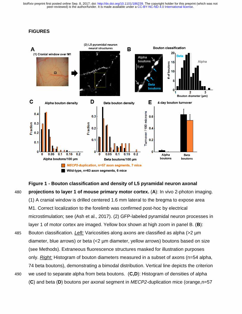
diameter (Fig. 1B, right panel). These large (alpha) and small (beta) boutons were
analyzed separately. The density of alpha boutons was 2.7±0.3 boutons/100µm
(mean±SEM, n=63 axonal segments), and the density of beta boutons was 4.0±0.4 220
boutons/100µm (Fig. 1C), similar to a previous study (see Methods, Grillo et al., 2013).
As expected given their large size (Grillo et al., 2013), alpha boutons were much more
stable than beta boutons (Fig. 1D). Across 4 days of rest the 4-day turnover rate (TOR =
(gain rate+loss rate) /2) of alpha boutons was 0.5±0.25%, while the TOR of beta
boutons was 23±4%. These results are comparable to a previous study in 225
somatosensory cortex, which found 0.1±0.06% 4-day turnover for large boutons and
30±3% 4-day turnover for small boutons (see Fig. 4E,F in Grillo et al., 2013). Since
alpha boutons were stable over time, hardly changing over the time course of the
experiment, we restricted further analysis of structural plasticity to beta boutons.
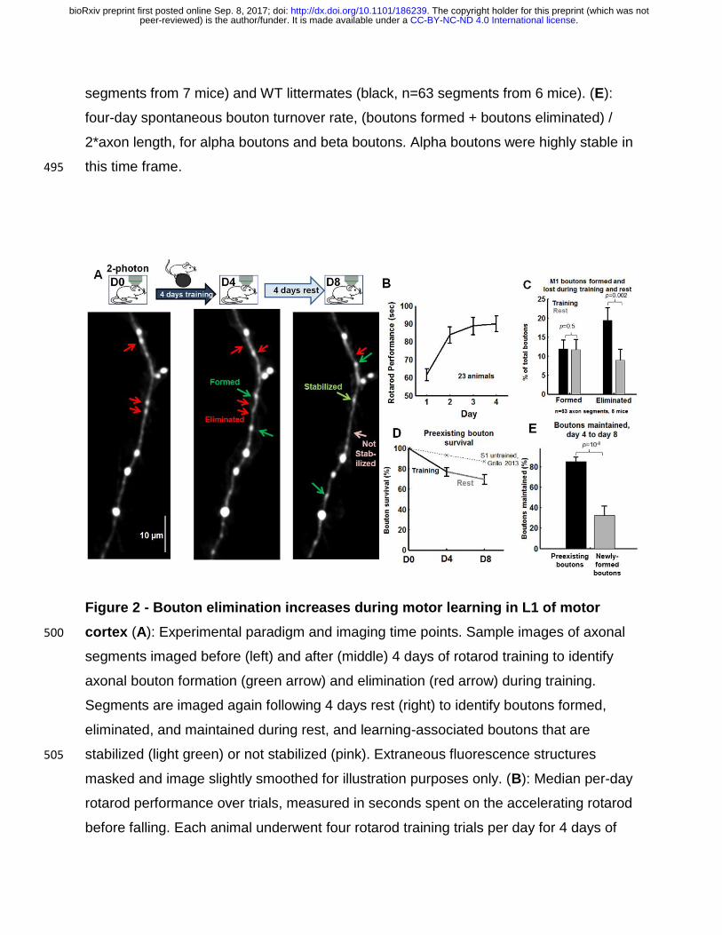
The experimental design is diagrammed in Fig. 2A. L5 pyramidal neuron axonal 230
projections to layer 1 (L1) of area M1 were initially imaged to identify baseline boutons.
Then mice underwent four days of training on the accelerating rotarod task. Axons were
re-imaged to quantify learning-associated bouton turnover. Mice rested in the home
cage for four days, and axons were imaged again to observe bouton turnover during
rest. WT mice performed progressively better on the rotarod across 4 days of training as 235
reported before (Fig. 2B; see also (Buitrago et al., 2004; Collins et al., 2004)). Training
on the rotarod did not significantly alter the formation rate of beta boutons (Fig. 2C;
training: 12±2% of total boutons across time points, rest: 12±3% of total boutons, p=
0.5, Mann-Whitney U test n=63 axon segments from 6 mice). The measured formation
rate was comparable to the spontaneous 4-day bouton formation rate previously 240
observed in L5 pyramidal neuron axons in somatosensory cortex (8±1%, Fig. S4C in
Grillo et al., 2013). Interestingly, rotarod training led to a dramatic increase in bouton
elimination compared to rest: 19±3% of total boutons were lost after 4 days of training
compared to 9±3% of total boutons lost after 4 days of rest, p = 0.002, Mann-Whitney U
test across axonal segments; p = 0.07, paired t test across animals). The M1 245
spontaneous elimination rate we observed was also comparable to prior published
results in area S1 (8.0±0.2%, Grillo et al., 2013, Fig. S4D). Overall, in control animals,
.CC-BY-NC-ND 4.0 International licensepeer-reviewed) is the author/funder. It is made available under aThe copyright holder for this preprint (which was not. http://dx.doi.org/10.1101/186239doi: bioRxiv preprint first posted online Sep. 8, 2017;

motor learning induces a doubling of bouton elimination in M1 without a concomitant
change in the rate of bouton formation.
Plotting the survival fraction of pre-existing (“baseline”) boutons revealed that 250
putative L5 pyramidal axons projecting to L1 of area M1 maintained 77±4% of their
baseline boutons (boutons present pre-training, on day 0) through 4 days of training
(Fig. 2D). This value is significantly lower than prior estimates of spontaneous 4-day
survival fraction of L5 pyramidal neuron axonal boutons (~90% of baseline boutons,
dotted line in Fig. 2D, see Fig. 7B of De Paola et al., 2006, Fig. 3C of Grillo et al., 2013, 255
Fig. 5 of Majewska et al., 2006). Note that elimination rates (Fig. 1C) and survival
curves (Fig. 1D) do not sum exactly to 100% because elimination rate was calculated as
a fraction of the total number of boutons observed across all time points to avoid outlier
turnover rates in axons which had very few baseline boutons, following Grillo et al.,
2013 (see Methods). 260
We also compared the survival rate of newly formed training-related boutons with
that of pre-existing boutons. In the four days of rest following training, 85±4% of
baseline preexisting boutons (boutons present on day zero that were also present on
post-training day 4) were maintained, while newly formed boutons were maintained at a
much lower rate of 32±9% (Fig. 2E, p=10-6), consistent with the reported stabilization 265
rate of spontaneously formed boutons in somatosensory cortex (newly formed: 35±5%
of all boutons over 4 days, Grillo et al., 2013).
We then assessed learning-associated axonal bouton turnover in MECP2-duplication
mice. MECP2-duplication mice performed significantly better on the rotarod than
control littermates (Fig. 3A) as previously described (Collins et al., 2004; Ash et al., 270
2017). The average length of analyzed axonal segments was not significantly different
between mutants and WT littermates (WT: 142±73 µm, MECP2-duplication: 133±73 µm,
mean±SD). The density of alpha boutons (Fig. 1C) and beta boutons (Fig. 1D) was also
similar between the genotypes (alpha boutons, control: 2.7±0.3 boutons/100µm,
MECP2-duplication: 2.4±0.3 boutons/100µm, p=0.4; beta boutons, control: 4±0.4 275
boutons/100µm, MECP2-duplication: 5.8±0.7 boutons/100µm. p=0.2, Mann-Whitney U
test). Similar to WT, alpha boutons were highly stable compared to beta boutons in
MECP2-duplication mice (data not shown). The rate of beta bouton formation was not
.CC-BY-NC-ND 4.0 International licensepeer-reviewed) is the author/funder. It is made available under aThe copyright holder for this preprint (which was not. http://dx.doi.org/10.1101/186239doi: bioRxiv preprint first posted online Sep. 8, 2017;

significantly different between MECP2-duplication mice and WT controls, neither during
the training (Fig. 3B, control: 12±2% of total boutons, n=63 axon segments from 6 mice; 280
MECP2-duplication: 9±3% of total boutons, n=57 axon segments from 7 mice) nor
during the rest phase (control: 12±3 % of total boutons, MECP2-duplication: 7±1 % of
total boutons, effect of genotype: F=2.8, p =0.09; effect of training vs. rest: F=0.78,
p=0.4; genotype x training interaction: F=0.6, p=0.4). Interestingly, the increased bouton
elimination rate during training observed in WT mice (training: 19±3%, rest: 9±3% of 285
total boutons) did not occur in MECP2-duplication mice (Fig. 3C, training: 5±1% of total
boutons; rest: 5±1% of total boutons). Significantly fewer boutons were eliminated
during training in MECP2-duplication mice compared to littermate controls (p < 0.001,
bouton elimination during training in WT vs. all other groups, 2-way ANOVA with Tukey-
test for multiple comparisons. Effect of genotype: F=12.9, p=0.0004; effect of training 290
vs. rest: F=5.3, p = 0.02; genotype x training interaction: F=3.71, p = 0.055). A linear
mixed-effects model ANOVA, with genotype and imaging time point implemented as
fixed effects, and mouse implemented as a random effect generated similarly significant
results (Effect of genotype: t=-2.4, p=0.015; effect of training vs. rest: t=-2.6, p = 0.009;
genotype x training interaction: t=1.9, p = 0.053). 295
Plotting the survival fraction of boutons revealed that baseline boutons were
significantly more stable in MECP2-duplication mice vs. littermate controls (Fig. 3E, p <
0.0001, 2-way ANOVA. Effect of genotype: F=26.7, p<0.0001; effect of training vs. rest:
F=3.25, p = 0.07; genotype x training interaction: F=0.06, p = 0.8). MECP2-duplication
axons maintained 95±1% of their boutons after 4 days of training, while control 300
littermate axons maintained only 77±4%. Subsequently, in the four days of rest following
training, bouton loss was comparable between genotypes. MECP2-duplication axons
lost a further 6±1% of baseline boutons to reach 89±2% bouton survival on day eight,
while littermate controls lost a further 8±2% to end at 69±4%. Learning-associated
bouton stabilization rate, defined as the fraction of boutons formed during the four days 305
of training that were still present after four further days of rest, was not significantly
altered in MECP2-duplication mice (40±8%) comped to controls (32±9%, Fig. 3F, p =
0.3). Again, note that elimination rates (Fig. 3C) and survival curves (Fig. 3D) do not
sum to 100%, as explained above, but note that the measured differences remain
.CC-BY-NC-ND 4.0 International licensepeer-reviewed) is the author/funder. It is made available under aThe copyright holder for this preprint (which was not. http://dx.doi.org/10.1101/186239doi: bioRxiv preprint first posted online Sep. 8, 2017;

significant if the elimination rate is calculated as a fraction of baseline boutons instead 310
of as a fraction of total boutons across time points.
Bouton formation, elimination, and stabilization rates did not correlate well with
rotarod performance in individual animals for either genotype (p > 0.05, t test on linear
regression, all comparisons, data not shown), suggesting that other factors are
potentially more important for the behavioral manifestations of motor learning. 315
DISCUSSION
The stability and plasticity of synaptic connections is a tightly regulated process that
unfolds throughout life. A pathological imbalance between stability and plasticity could
lead to the altered patterns of learning and forgetting observed in autism mouse models
(Collins et al., 2004; Rothwell et al., 2014) and in autistic patients (Treffert, 2014). In 320
prior work (Ash et al., 2017) an abnormal increase in learning-associated dendritic spine
stability was found after motor training in the apical tuft of area M1 corticospinal neurons
in the Tg1 mouse model of MECP2 duplication syndrome. Here we investigated how
axonal boutons in the L5 pyramidal neuron projection to L1 of primary motor cortex turn
over during motor training in these animals. First, we find in WT mice that: 1) bouton 325
formation rate is unaffected by motor learning (Fig.2 C), and 2) bouton elimination rate
doubles from ~10% to ~20% during motor learning (Fig. 2C,D). In contrast, we find that
the increase in learning-associated bouton elimination observed in littermate controls
does not occur in MECP2-duplication mice (Fig. 3C), which exhibit increased bouton
stability during training (Fig. 3D). Bouton formation rate during motor learning was 330
similar between MECP2-duplication animals and littermate controls (Fig. 3B), and was
not significantly different from the rate of bouton formation observed at rest in either
genotype. A similar fraction of learning-associated boutons was stabilized in both
genotypes (Fig. 3E).
335
Bouton formation and elimination with motor learning in controls
Our spontaneous 4-day bouton turnover results are in agreement with a previous
study of axonal bouton formation and elimination in L5 pyramidal neuron axons
.CC-BY-NC-ND 4.0 International licensepeer-reviewed) is the author/funder. It is made available under aThe copyright holder for this preprint (which was not. http://dx.doi.org/10.1101/186239doi: bioRxiv preprint first posted online Sep. 8, 2017;

projecting to layer 1 of somatosensory cortex (Grillo et al., 2013), suggesting that
baseline axonal bouton turnover in L1 is similar in sensory and motor areas. Here, we 340
found that, in normal animals, the rate of axonal bouton elimination increases markedly
during motor training in L5 pyramidal neuron projections to L1 of area M1, without a
concomitant increase in the rate of bouton formation (Fig. 2C).
At face value this suggests that training leads to a weakening of L5 pyramidal inputs
to layer 1 of the primary motor cortex, at least as evidenced by structural analysis. L5 345
Axonal projections to L1 have several potential synaptic partners, including apical
dendritic arbors of L5B corticospinal pyramidal neurons, L5A
corticostriatal/corticocallosal neurons, L2/3 pyramidal neurons, and L1 interneuron
dendrites (Fig. 4). Since L1 interneurons are sparse, most of the postsynaptic partners
of the axonal boutons we studied are likely formed with one or more of the 350
aforementioned classes of pyramidal neurons. The increased elimination of pre-synaptic
axonal boutons would then lead us to expect a corresponding loss in their post-synaptic
partners, i.e. of dendritic spines located in the apical dendritic tufts of the target
neurons. However, we and others have previously observed an increase in the
formation rate of dendritic spines during motor learning in the apical tuft terminal 355
dendrites of L5 neurons in layer 1 of area M1 (Xu et al., 2009; Yang et al., 2009; Liston
et al., 2013; Ash et al., 2017) without an increase in spine elimination during training.
This dissociation between L5 neuron dendritic spine formation and axonal bouton
elimination in layer 1 of area M1 during motor learning suggests that at least a subset of
the axonal projections we imaged are synapsing on dendritic processes not previously 360
analyzed. Indeed, there is evidence that projections to L1 of M1 from different brain
areas preferentially target different cell types (Hooks et al., 2013). Ergo it is also
possible that the pre-synaptic partners of the L5 apical tuft dendritic spines studied
previously during motor learning (Xu et al., 2009; Yang et al., 2009) may arise from
thalamocortical, L2/3, or L6 projections which we did not study here. Overall, these 365
results raise the interesting possibility that different pathways projecting to L1 of mouse
area M1 may have different signatures of structural plasticity during motor learning.
.CC-BY-NC-ND 4.0 International licensepeer-reviewed) is the author/funder. It is made available under aThe copyright holder for this preprint (which was not. http://dx.doi.org/10.1101/186239doi: bioRxiv preprint first posted online Sep. 8, 2017;
We note the alternative possibility that the axonal boutons (or varicosities) whose
elimination rate increases with learning are ones that have not yet formed a synapse, 370
and therefore do not have post-synaptic partners. It has been estimated that ~10-20%
of axonal varicosities (defined as a swelling of the axon exceeding the shaft diameter by
more than 50%) may be non-synaptic (Shepherd and Harris, 1998; White et al., 2004;
Bourne et al., 2013), although this has not been assessed in pyramidal neuron
projections to layer 1 to our knowledge. 375
This still leaves us with the puzzle of why we do not observe an increase in the rate
of bouton formation with learning, to serve as pre-synaptic partners to the increased
number of dendritic spines that form in apical dendritic tufts of L5 pyramidal neurons
(Fig. 4). One possibility mentioned above is that we have not examined the correct pre-380
synaptic axons, particularly as we did not study thalamo-cortical, L2/3 or L6 pyramidal
projections. Another possibility is that, rather than connecting with a new axonal bouton,
newly formed spines rather form a second synapse onto large pre-existing boutons
already harboring a synapse, as has been shown in vivo in the somatosensory cortex
and ex vivo in the hippocampus (~70% of newly formed spines synapse with a multi-385
synapse bouton, compared to 20-30% of preexisting spines (Knott et al., 2006; Nagerl
et al., 2007); see also (Woolley et al., 1996; Toni et al., 1999; Geinisman et al., 2001;
Yankova et al., 2001; Federmeier et al., 2002; Nicholson and Geinisman, 2009; Lee et
al., 2013). Dendritic spines formed during learning may largely synapse on already
existing, large, pre-synaptic boutons (alpha boutons in our study) where they compete 390
with the previously present connections. Over time, some of these connections
withdraw, re-establishing a new equilibrium that favors the new skill learning.
Presumably, in the days-to-weeks following learning, bouton formation modestly
increases and/or bouton elimination decreases to bring bouton densities back to
baseline levels. 395
Increased bouton stability in MECP2-duplication mice
.CC-BY-NC-ND 4.0 International licensepeer-reviewed) is the author/funder. It is made available under aThe copyright holder for this preprint (which was not. http://dx.doi.org/10.1101/186239doi: bioRxiv preprint first posted online Sep. 8, 2017;

We found that the learning-associated increase in bouton elimination rate occurring
in WT mice is abolished in MECP2-duplication mice. This increased synaptic stability in
L5 pyramidal neuron axonal projections could reflect a general increase in synaptic 400
stability during motor learning across multiple circuits, or it may be a manifestation of
increased stability along specific subcircuits projecting to L1 of area M1 (Fig. 4).
Increased bouton stability in MECP2-duplication M1 could also be due in part to more
robust capture and stabilization of pre-existing boutons by newly formed learning-
associated spines, boutons that would have otherwise been eliminated due to loss of 405
their prior post-synaptic targets during the training period (Knott et al., 2006; Nagerl et
al., 2007). Quantification of multi-synapse bouton density with and without training in
mutants could address these possibilities. As in the WT, again it is interesting to note
that the learning-associated increase in spine formation in mutants is not associated
with a concomitant increase in bouton formation (Fig. 4). This provides further basis for 410
the idea that different microcircuit projections to M1 may manifest different patterns of
structural synaptic plasticity during learning.
It is interesting to speculate that the learning-associated bouton elimination that
occurs in littermate controls is a natural end result of strong long-term depression
(Becker et al., 2008; Wiegert and Oertner, 2013). In this case, the lack of bouton 415
elimination in mutants may connote a disruption in processes regulating LTD. Taken
along with the fact that abnormal LTD is observed in many other autism models
(D’Antoni et al., 2014), it will be interesting to experimentally test if LTD is indeed altered
in motor cortex of MECP2-duplication mice, and to see if decreased LTD underlies the
mutant’s increased learning-associated bouton stability. 420
The behavioral implications of increased L1 axonal bouton stabilization remain a
matter of speculation. The rate of pre-existing bouton formation and elimination did not
correlate with behavioral performance across individual animals in either genotype. This
suggests that although bouton elimination is a robust structural phenotype resulting
from motor training, its link to behavioral performance is at best weak. It is certainly 425
weaker than dendritic spine formation and stabilization in apical dendrites of L5
pyramidal neurons, which correlates well with behavior (Yang et al., 2009; Ash et al.,
.CC-BY-NC-ND 4.0 International licensepeer-reviewed) is the author/funder. It is made available under aThe copyright holder for this preprint (which was not. http://dx.doi.org/10.1101/186239doi: bioRxiv preprint first posted online Sep. 8, 2017;

2017). It is possible that our study is not adequately powered to detect a weak
correlation between bouton turnover and rotarod performance.
Potential Limitations 430
It is important to note a number of limitations with the study. First of all, our
quantification of presynaptic terminals depends entirely on morphological measures.
Although we used conservative criteria similar to that which in prior experimenters’
hands has been shown to reliably detect synapse-forming puncta (De Paola et al.,
2006), it is still possible that a fraction (~10%) of the counted varicosities are non-435
synaptic (Shepherd and Harris, 1998; White et al., 2004; Bourne et al., 2013).
Second, the rest phase occurred following training, so it is possible that some of the
corresponding bouton turnover may reflect enduring consolidation processes that
persist beyond training rather than a true rest phase. Having said that, the measured
spontaneous axonal bouton formation and elimination is in very close agreement to 440
previous studies (Grillo et al., 2013), suggesting that the measurements reflect baseline
turnover.
Third, we cannot precisely determine the origin of the axonal afferents imaged in our
study (Fig. 4). Some of the heterogeneity in plasticity observed across imaged axons
could be due to projection-specific differences. For example, it would be interesting to 445
speculate that coarse sensorimotor training induced by the rotarod may drive greater
bouton remodeling in somatosensory cortical inputs to area M1, while fine motor training
requiring higher-order motor planning, such as the seed-grabbing task used by (Xu et
al., 2009), may induce greater remodeling in premotor cortical inputs.
Fourth, the postsynaptic partners of the imaged axons are unknown. The precise 450
connectivity of inputs to M1, with S1 pyramidal neuron axons preferentially synapsing
on L2/3 and L5A neurons and premotor cortex pyramidal neuron axons preferentially
synapsing on L5B neurons (Mao et al., 2011; Hooks et al., 2013), enables a rich
potential repertoire of synaptic reorganization during training. New methods targeting
fluorescent proteins to specific input areas, as well as combinatorial techniques labeling 455
pre-and postsynaptic partners (Kim et al., 2011; Druckmann et al., 2014), will enable
scientists to tackle this question in the future.
.CC-BY-NC-ND 4.0 International licensepeer-reviewed) is the author/funder. It is made available under aThe copyright holder for this preprint (which was not. http://dx.doi.org/10.1101/186239doi: bioRxiv preprint first posted online Sep. 8, 2017;

Conclusions and implications
In conclusion, we report here that L5 pyramidal neuron axonal projections to layer 1
of WT mouse motor cortex exhibit a selective escalation in bouton elimination during 460
motor learning, a plasticity process that is disrupted in the MECP2-duplication syndrome
mouse model of autism. These data constrain models of motor cortex plasticity
underlying learning and underscore the possibility that different synaptic pathways
within the cortical circuit may manifest different patterns of structural synaptic plasticity
during learning. Future work studying plasticity along different synaptic pathways that 465
link various areas along the motor circuit will shed further light on these issues.
Our results provide further evidence for an altered balance between stability and
plasticity of synaptic connections in favor of stability in the MECP2 duplication syndrome
mouse model (Ash et al., 2017). This bias favors enhanced motor learning on the
rotarod and may play a role in other types of learning, such as fear conditioning or 470
social learning. More generally, an abnormal bias toward synaptic stability in relevant
circuits could potentially play a role in explaining the combination of savant-like
phenotypes and behavioral rigidity seen at times in autism.
475
.CC-BY-NC-ND 4.0 International licensepeer-reviewed) is the author/funder. It is made available under aThe copyright holder for this preprint (which was not. http://dx.doi.org/10.1101/186239doi: bioRxiv preprint first posted online Sep. 8, 2017;
FIGURES
Figure 1 - Bouton classification and density of L5 pyramidal neuron axonal
projections to layer 1 of mouse primary motor cortex. (A): In vivo 2-photon imaging. 480
(1) A cranial window is drilled centered 1.6 mm lateral to the bregma to expose area
M1. Correct localization to the forelimb was confirmed post-hoc by electrical
microstimulation; see (Ash et al., 2017). (2) GFP-labeled pyramidal neuron processes in
layer 1 of motor cortex are imaged. Yellow box shown at high zoom in panel B. (B):
Bouton classification. Left: Varicosities along axons are classified as alpha (>2 µm 485
diameter, blue arrows) or beta (<2 µm diameter, yellow arrows) boutons based on size
(see Methods). Extraneous fluorescence structures masked for illustration purposes
only. Right: Histogram of bouton diameters measured in a subset of axons (n=54 alpha,
74 beta boutons), demonstrating a bimodal distribution. Vertical line depicts the criterion
we used to separate alpha from beta boutons. (C,D): Histogram of densities of alpha 490
(C) and beta (D) boutons per axonal segment in MECP2-duplication mice (orange,n=57
.CC-BY-NC-ND 4.0 International licensepeer-reviewed) is the author/funder. It is made available under aThe copyright holder for this preprint (which was not. http://dx.doi.org/10.1101/186239doi: bioRxiv preprint first posted online Sep. 8, 2017;
segments from 7 mice) and WT littermates (black, n=63 segments from 6 mice). (E):
four-day spontaneous bouton turnover rate, (boutons formed + boutons eliminated) /
2*axon length, for alpha boutons and beta boutons. Alpha boutons were highly stable in
this time frame. 495
Figure 2 - Bouton elimination increases during motor learning in L1 of motor
cortex (A): Experimental paradigm and imaging time points. Sample images of axonal 500
segments imaged before (left) and after (middle) 4 days of rotarod training to identify
axonal bouton formation (green arrow) and elimination (red arrow) during training.
Segments are imaged again following 4 days rest (right) to identify boutons formed,
eliminated, and maintained during rest, and learning-associated boutons that are
stabilized (light green) or not stabilized (pink). Extraneous fluorescence structures 505
masked and image slightly smoothed for illustration purposes only. (B): Median per-day
rotarod performance over trials, measured in seconds spent on the accelerating rotarod
before falling. Each animal underwent four rotarod training trials per day for 4 days of
.CC-BY-NC-ND 4.0 International licensepeer-reviewed) is the author/funder. It is made available under aThe copyright holder for this preprint (which was not. http://dx.doi.org/10.1101/186239doi: bioRxiv preprint first posted online Sep. 8, 2017;
training (n=23). (C): Bouton formation and elimination during training (black) and during
rest (grey). Bouton elimination was significantly elevated during training, p=0.001, n=63 510
segments, Mann-Whitney U test. 314 baseline boutons, 40 formed during training, 42
formed during rest, 64 eliminated during training, 23 eliminated during rest. Data
acquired from 6 mice. Statistics performed across axonal segments (p = 0.07 when
calculated across animals). (D): Pre-existing bouton survival curves across imaging
days. Dotted line depicts baseline bouton survival, reproduced from (Grillo et al., 2013). 515
(E): The fraction of boutons maintained during the rest period, measured for pre-existing
boutons (present on day 0) that were still present on day 4 following training (black) and
boutons formed during training (training-associated boutons, grey). p=10-6, Mann-
Whitney U test.
520
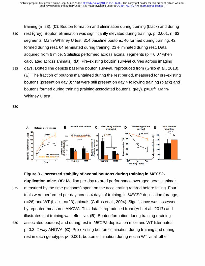
Figure 3 - Increased stability of axonal boutons during training in MECP2-
duplication mice. (A): Median per-day rotarod performance averaged across animals,
measured by the time (seconds) spent on the accelerating rotarod before falling. Four 525
trials were performed per day across 4 days of training, in MECP2-duplication (orange,
n=26) and WT (black, n=23) animals (Collins et al., 2004). Significance was assessed
by repeated-measures ANOVA. This data is reproduced from (Ash et al., 2017) and
illustrates that training was effective. (B): Bouton formation during training (training-
associated boutons) and during rest in MECP2-duplication mice and WT littermates, 530
p=0.3, 2-way ANOVA. (C): Pre-existing bouton elimination during training and during
rest in each genotype, p< 0.001, bouton elimination during rest in WT vs all other
.CC-BY-NC-ND 4.0 International licensepeer-reviewed) is the author/funder. It is made available under aThe copyright holder for this preprint (which was not. http://dx.doi.org/10.1101/186239doi: bioRxiv preprint first posted online Sep. 8, 2017;

conditions, 2-way ANOVA with Tukey test for multiple comparisons. Effect of genotype:
F=12.9, p=0.0004; effect of training vs. rest: F=5.3, p = 0.02; genotype x training
interaction: F=3.71, p = 0.055. Linear mixed-effects model ANOVA results: Effect of 535
genotype: t=-2.4, p=0.015; effect of training vs. rest: t=-2.6, p = 0.009; genotype x
training interaction: t=1.9, p = 0.053. (D): Pre-existing bouton survival curves across
imaging, p<0.0001 effect of genotype, 2-way ANOVA. Effect of genotype: F=26.7,
p<0.0001; effect of training vs. rest: F=3.25, p = 0.07; genotype x training interaction:
F=0.06, p = 0.8. (E): Training-associated bouton stabilization rate – the number of 540
boutons formed during training and still present after 4 days of post-training rest is not
significantly different across genotypes. Data are plotted as percentage of boutons
formed during training. Mann-Whitney U test.
545
.CC-BY-NC-ND 4.0 International licensepeer-reviewed) is the author/funder. It is made available under aThe copyright holder for this preprint (which was not. http://dx.doi.org/10.1101/186239doi: bioRxiv preprint first posted online Sep. 8, 2017;
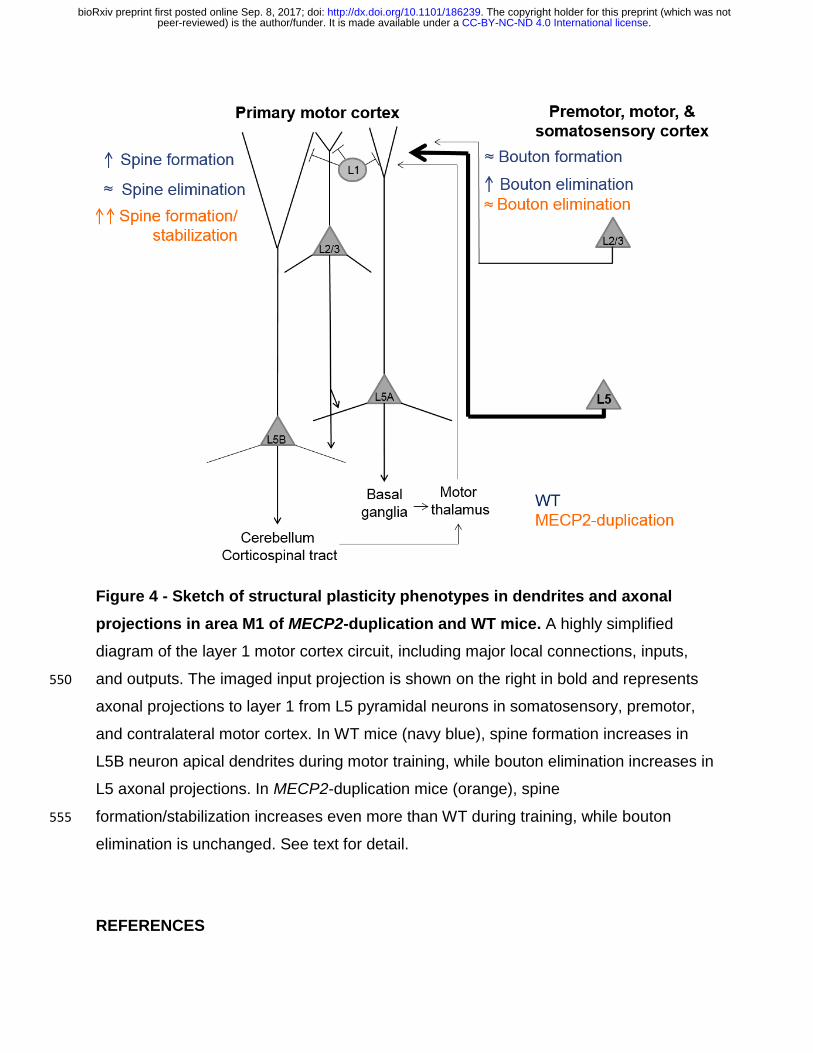
Figure 4 - Sketch of structural plasticity phenotypes in dendrites and axonal
projections in area M1 of MECP2-duplication and WT mice. A highly simplified
diagram of the layer 1 motor cortex circuit, including major local connections, inputs,
and outputs. The imaged input projection is shown on the right in bold and represents 550
axonal projections to layer 1 from L5 pyramidal neurons in somatosensory, premotor,
and contralateral motor cortex. In WT mice (navy blue), spine formation increases in
L5B neuron apical dendrites during motor training, while bouton elimination increases in
L5 axonal projections. In MECP2-duplication mice (orange), spine
formation/stabilization increases even more than WT during training, while bouton 555
elimination is unchanged. See text for detail.
REFERENCES
.CC-BY-NC-ND 4.0 International licensepeer-reviewed) is the author/funder. It is made available under aThe copyright holder for this preprint (which was not. http://dx.doi.org/10.1101/186239doi: bioRxiv preprint first posted online Sep. 8, 2017;

Antar LN, Li C, Zhang H, Carroll RC, Bassell GJ (2006) Local functions for FMRP in
axon growth cone motility and activity-dependent regulation of filopodia and spine 560
synapses. Mol Cell Neurosci 32:37–48.
Antonova I, Arancio O, Trillat a C, Wang HG, Zablow L, Udo H, Kandel ER, Hawkins
RD (2001) Rapid increase in clusters of presynaptic proteins at onset of long-
lasting potentiation. Science 294:1547–1550.
Ash RT, Buffington SA, Park J, Costa-Mattioli M, Zoghbi HY, Smirnakis SM (2017) 565
Excessive ERK-dependent synaptic clustering with enhanced motor learning in the
MECP2 duplication syndrome mouse model of autism. bioRxiv.
Becker N, Wierenga CJ, Fonseca R, Bonhoeffer T, Nägerl UV (2008) LTD Induction
Causes Morphological Changes of Presynaptic Boutons and Reduces Their
Contacts with Spines. Neuron 60:590–597. 570
Belichenko P V., Wright EE, Belichenko NP, Masliah E, Li HH, Mobley WC, Francke U
(2009) Widespread changes in dendritic and axonal morphology in Mecp2-mutant
mouse models of Rett syndrome: Evidence for disruption of neuronal networks. J
Comp Neurol 514:240–258.
Blundell J, Kaeser PS, Südhof TC, Powell CM (2010) RIM1 and Interacting Proteins 575
Involved in Presynaptic Plasticity Mediate Prepulse Inhibition and Additional
Behaviors Linked to Schizophrenia. J Neurosci 30:5326–5333 Available at:
http://www.jneurosci.org/cgi/doi/10.1523/JNEUROSCI.0328-
10.2010%5Cnpapers3://publication/doi/10.1523/JNEUROSCI.0328-10.2010.
Bourne JN, Chirillo MA, Harris KM (2013) Presynaptic ultrastructural plasticity along 580
CA3???CA1 axons during long-term potentiation in mature hippocampus. J Comp
Neurol 521:3898–3912.
Buitrago MM, Schulz JB, Dichgans J, Luft AR (2004) Short and long-term motor skill
learning in an accelerated rotarod training paradigm. Neurobiol Learn Mem 81:211–
216. 585
.CC-BY-NC-ND 4.0 International licensepeer-reviewed) is the author/funder. It is made available under aThe copyright holder for this preprint (which was not. http://dx.doi.org/10.1101/186239doi: bioRxiv preprint first posted online Sep. 8, 2017;

Carrillo J, Cheng S-Y, Ko KW, Jones T a, Nishiyama H (2013) The long-term structural
plasticity of cerebellar parallel fiber axons and its modulation by motor learning. J
Neurosci 33:8301–8307 Available at:
http://www.pubmedcentral.nih.gov/articlerender.fcgi?artid=3680104&tool=pmcentre
z&rendertype=abstract. 590
Chen J, Yu S, Fu Y, Li X (2014) Synaptic proteins and receptors defects in autism
spectrum disorders. Front Cell Neurosci 8:276 Available at:
http://www.pubmedcentral.nih.gov/articlerender.fcgi?artid=4161164&tool=pmcentre
z&rendertype=abstract.
Chklovskii DB, Mel BW, Svoboda K (2004) Cortical rewiring and information storage. 595
Nature 431:782–788.
Cho RH, Segawa S, Okamoto K, Mizuno A, Kaneko T (2004) Intracellularly labeled
pyramidal neurons in the cortical areas projecting to the spinal cord: II. Intra- and
juxta-columnar projection of pyramidal neurons to corticospinal neurons. Neurosci
Res 50:395–410. 600
Colechio EM, Alloway KD (2009) Differential topography of the bilateral cortical
projections to the whisker and forepaw regions in rat motor cortex. Brain Struct
Funct 213:423–439.
Collingridge GL, Peineau S, Howland JG, Wang YT (2010) Long-term depression in the
CNS. Nat Rev Neurosci 11:459–473. 605
Collins AL, Levenson JM, Vilaythong AP, Richman R, Armstrong DL, Noebels JL,
Sweatt D, J, Zoghbi HY (2004) Mild overexpression of MeCP2 causes a
progressive neurological disorder in mice. Hum Mol Genet 13:2679–2689.
D’Antoni S, Spatuzza M, Bonaccorso CM, Musumeci SA, Ciranna L, Nicoletti F, Huber
KM, Catania MV (2014) Dysregulation of group-I metabotropic glutamate (mGlu) 610
receptor mediated signalling in disorders associated with Intellectual Disability and
Autism. Neurosci Biobehav Rev 46:228–241.
.CC-BY-NC-ND 4.0 International licensepeer-reviewed) is the author/funder. It is made available under aThe copyright holder for this preprint (which was not. http://dx.doi.org/10.1101/186239doi: bioRxiv preprint first posted online Sep. 8, 2017;

De Paola V, Holtmaat A, Knott G, Song S, Wilbrecht L, Caroni P, Svoboda K (2006) Cell
type-specific structural plasticity of axonal branches and boutons in the adult
neocortex. Neuron 49:861–875. 615
Degano AL, Pasterkamp RJ, Ronnett G V. (2009) MeCP2 deficiency disrupts axonal
guidance, fasciculation, and targeting by altering Semaphorin 3F function. Mol Cell
Neurosci 42:243–254.
Deng PY, Rotman Z, Blundon JA, Cho Y, Cui J, Cavalli V, Zakharenko SS, Klyachko VA
(2013) FMRP Regulates Neurotransmitter Release and Synaptic Information 620
Transmission by Modulating Action Potential Duration via BK Channels. Neuron
77:696–711.
Druckmann S, Feng L, Lee B, Yook C, Zhao T, Magee JC, Kim J (2014) Structured
Synaptic Connectivity between Hippocampal Regions. Neuron 81:629–640.
Federmeier KD, Kleim JA, Greenough WT (2002) Learning-induced multiple synapse 625
formation in rat cerebellar cortex. Neurosci Lett 332:180–184.
Feng G, Mellor RH, Bernstein M, Keller-Peck C, Nguyen QT, Wallace M, Nerbonne JM,
Lichtman JW, Sanes JR (2000) Imaging neuronal subsets in transgenic mice
expressing multiple spectral variants of GFP. Neuron 28:41–51 Available at:
http://www.ncbi.nlm.nih.gov/pubmed/11086982. 630
Garcia-Junco-Clemente P, Golshani P (2014) PTEN: A master regulator of neuronal
structure, function, and plasticity. Commun Integr Biol 7.
Geinisman Y, Berry RW, Disterhoft JF, Power JM, Van der Zee E a (2001) Associative
learning elicits the formation of multiple-synapse boutons. J Neurosci 21:5568–
5573. 635
Grillo FW, Song S, Teles-Grilo Ruivo LM, Huang L, Gao GG, Knott GW, Maco B,
Ferretti V, Thompson D, Little GE, De Paola V (2013) Increased axonal bouton
dynamics in the aging mouse cortex. Proc Natl Acad Sci 110:1–10 Available at:
http://www.pnas.org/cgi/doi/10.1073/pnas.1218731110%5Cnhttp://www.pubmedce
.CC-BY-NC-ND 4.0 International licensepeer-reviewed) is the author/funder. It is made available under aThe copyright holder for this preprint (which was not. http://dx.doi.org/10.1101/186239doi: bioRxiv preprint first posted online Sep. 8, 2017;

ntral.nih.gov/articlerender.fcgi?artid=3631669&tool=pmcentrez&rendertype=abstrac640
t.
Holtmaat A, Bonhoeffer T, Chow DK, Chuckowree J, De Paola V, Hofer SB, Hübener M,
Keck T, Knott G, Lee W-CA, Mostany R, Mrsic-Flogel TD, Nedivi E, Portera-Cailliau
C, Svoboda K, Trachtenberg JT, Wilbrecht L (2009) Long-term, high-resolution
imaging in the mouse neocortex through a chronic cranial window. Nat Protoc 645
4:1128–1144 Available at: http://www.nature.com/doifinder/10.1038/nprot.2009.89
[Accessed February 16, 2017].
Hooks BM, Mao T, Gutnisky DA, Yamawaki N, Svoboda K, Shepherd GMG (2013)
Organization of Cortical and Thalamic Input to Pyramidal Neurons in Mouse Motor
Cortex. J Neurosci 33:748–760 Available at: 650
http://www.jneurosci.org/content/33/2/748.long%5Cnhttp://www.jneurosci.org/cgi/do
i/10.1523/JNEUROSCI.4338-12.2013.
Johnson CM, Peckler H, Tai L-H, Wilbrecht L (2016) Rule learning enhances structural
plasticity of long-range axons in frontal cortex. Nat Commun 7:10785 Available at:
http://www.nature.com/ncomms/2016/160307/ncomms10785/full/ncomms10785.ht655
ml.
Kim J, Zhao T, Petralia RS, Yu Y, Peng H, Myers E, Magee JC (2011) mGRASP
enables mapping mammalian synaptic connectivity with light microscopy. Nat
Methods 9:96–102 Available at: http://dx.doi.org/10.1038/nmeth.1784.
Knott GW, Holtmaat A, Wilbrecht L, Welker E, Svoboda K (2006) Spine growth 660
precedes synapse formation in the adult neocortex in vivo. Nat Neurosci 9:1117–
1124 Available at: http://www.ncbi.nlm.nih.gov/pubmed/16892056 [Accessed
February 16, 2017].
Lee KJ, Park IS, Kim H, Greenough WT, Pak DTS, Rhyu IJ (2013) Motor Skill Training
Induces Coordinated Strengthening and Weakening between Neighboring 665
Synapses. J Neurosci 33:9794–9799 Available at:
http://www.jneurosci.org/content/33/23/9794%5Cnhttp://libsta28.lib.cam.ac.uk:2356
.CC-BY-NC-ND 4.0 International licensepeer-reviewed) is the author/funder. It is made available under aThe copyright holder for this preprint (which was not. http://dx.doi.org/10.1101/186239doi: bioRxiv preprint first posted online Sep. 8, 2017;

/content/33/23/9794%5Cnhttp://www.jneurosci.org/content/33/23/9794.full.pdf%5C
nhttp://www.ncbi.nlm.nih.gov/pubmed/23739975.
Liston C, Cichon JM, Jeanneteau F, Jia Z, Chao M V, Gan W-B (2013) Circadian 670
glucocorticoid oscillations promote learning-dependent synapse formation and
maintenance. Nat Neurosci 16:698–705 Available at:
http://dx.doi.org/10.1038/nn.3387.
Majewska AK, Newton JR, Sur M (2006) Remodeling of Synaptic Structure in Sensory
Cortical Areas In Vivo. J Neurosci 26. 675
Mao T, Kusefoglu D, Hooks BM, Huber D, Petreanu L, Svoboda K (2011) Long-Range
Neuronal Circuits Underlying the Interaction between Sensory and Motor Cortex.
Neuron 72:111–123.
Mostany R, Anstey JE, Crump KL, Maco B, Knott G, Portera-Cailliau C (2013) Altered
Synaptic Dynamics during Normal Brain Aging. J Neurosci 33:4094–4104 Available 680
at: http://www.jneurosci.org/cgi/doi/10.1523/JNEUROSCI.4825-12.2013.
Mostany R, Portera-Cailliau C (2008) A Craniotomy Surgery Procedure for Chronic
Brain Imaging. J Vis Exp:e680–e680 Available at:
http://www.jove.com/index/Details.stp?ID=680 [Accessed September 1, 2016].
Nagerl U V, Kostinger G, Anderson JC, Martin KA, Bonhoeffer T (2007) Protracted 685
synaptogenesis after activity-dependent spinogenesis in hippocampal neurons. J
Neurosci 27:8149–8156 Available at:
http://www.ncbi.nlm.nih.gov/pubmed/17652605.
Nicholson DA, Geinisman Y (2009) Axospinous synaptic subtype-specific differences in
structure, size, ionotropic receptor expression, and connectivity in apical dendritic 690
regions of rat hippocampal CA1 pyramidal neurons. J Comp Neurol 512:399–418.
Ramocki MB, Tavyev YJ, Peters SU (2010) The MECP2 duplication syndrome. Am J
Med Genet Part A 152:1079–1088.
Rothwell PE, Fuccillo M V., Maxeiner S, Hayton SJ, Gokce O, Lim BK, Fowler SC,
.CC-BY-NC-ND 4.0 International licensepeer-reviewed) is the author/funder. It is made available under aThe copyright holder for this preprint (which was not. http://dx.doi.org/10.1101/186239doi: bioRxiv preprint first posted online Sep. 8, 2017;

Malenka RC, Südhof TC (2014) Autism-associated neuroligin-3 mutations 695
commonly impair striatal circuits to boost repetitive behaviors. Cell 158:198–212.
Shepherd GM, Harris KM (1998) Three-dimensional structure and composition of CA3 -
CA1 axons in rat hippocampal slices: implications for presynaptic connectivity and
compartmentalization. J Neurosci 18:8300–8310.
Stettler DD, Yamahachi H, Li W, Denk W, Gilbert CD (2006) Axons and synaptic 700
boutons are highly dynamic in adult visual cortex. Neuron 49:877–887.
Tennant KA, Adkins DL, Donlan NA, Asay AL, Thomas N, Kleim JA, Jones TA (2011)
The organization of the forelimb representation of the C57BL/6 mouse motor cortex
as defined by intracortical microstimulation and cytoarchitecture. Cereb Cortex
21:865–876. 705
Toni N, Buchs P a, Nikonenko I, Bron CR, Muller D (1999) LTP promotes formation of
multiple spine synapses between a single axon terminal and a dendrite. Nature
402:421–425.
Treffert DA (2014) Savant syndrome: Realities, myths and misconceptions. J Autism
Dev Disord 44:564–571. 710
White EL, Weinfeld E, Lev DL (2004) Quantitative analysis of synaptic distribution along
thalamocortical axons in adult mouse barrels. J Comp Neurol 479:56–69.
Wiegert JS, Oertner TG (2013) Long-term depression triggers the selective elimination
of weakly integrated synapses. Proc Natl Acad Sci U S A 110:E4510–E4519
Available at: http://www.pnas.org/cgi/doi/10.1073/pnas.1315926110. 715
Woolley CS, Wenzel HJ, Schwartzkroin PA (1996) Estradiol increases the frequency of
multiple synapse boutons in the hippocampal CA1 region of the adult female rat. J
Comp Neurol 373:108–117.
Xu T, Yu X, Perlik AJ, Tobin WF, Zweig JA, Tennant K, Jones T, Zuo Y (2009) Rapid
formation and selective stabilization of synapses for enduring motor memories. 720
Nature 462:915–919 Available at: http://www.ncbi.nlm.nih.gov/pubmed/19946267.
.CC-BY-NC-ND 4.0 International licensepeer-reviewed) is the author/funder. It is made available under aThe copyright holder for this preprint (which was not. http://dx.doi.org/10.1101/186239doi: bioRxiv preprint first posted online Sep. 8, 2017;

Yang G, Pan F, Gan WB (2009) Stably maintained dendritic spines are associated with
lifelong memories. Nature 462:920–924 Available at:
http://www.neuroscience.ubc.ca/CourseMat/Yang_et_al_2009.pdf%5Cnhttp://www.
ncbi.nlm.nih.gov/pubmed/19946265. 725
Yankova M, Hart S a, Woolley CS (2001) Estrogen increases synaptic connectivity
between single presynaptic inputs and multiple postsynaptic CA1 pyramidal cells: a
serial electron-microscopic study. Proc Natl Acad Sci U S A 98:3525–3530
Available at:
http://www.pubmedcentral.nih.gov/articlerender.fcgi?artid=30686&tool=pmcentrez&730
rendertype=abstract.
.CC-BY-NC-ND 4.0 International licensepeer-reviewed) is the author/funder. It is made available under aThe copyright holder for this preprint (which was not. http://dx.doi.org/10.1101/186239doi: bioRxiv preprint first posted online Sep. 8, 2017;