Embed Size (px)
Citation preview
haematologica | 2012; 97(11) 1699
Monoclonal Gammopathy of Undetermined Significance Articles and Brief Reports
Introduction
Schnitzler syndrome is a rare plasma cell disorder character-ized by the presence of a monoclonal IgM immunoglobulin inassociation with a chronic urticarial skin rash and at least 2 ofthe following minor criteria: intermittent fever, arthralgia orarthritis, bone pain, enlarged lymph nodes, splenomegalyand/or hepatomegaly, increased neutrophil counts, increasederythrocyte sedimentation rate (ESR), and abnormal bone find-ings with imaging evidence of osteosclerosis.1,2
The pathogenesis of the syndrome is unclear, with only onedescribed case of spontaneous remission.3 The presence of highlevels of interleukins (ILs) in patients with Schnitzler syn-drome4,5 suggests that this plasma-cell disorder may be anacquired auto-inflammatory disease due to an unregulatedsecretion of cytokines via interaction of a clonal product (e.g.the M-protein) with a key component of the IL-1 pathway. Thisis further supported by the successful treatment of the syn-drome with anakinra,6 a synthetic analog of the endogenous IL-1 receptor antagonist, or with immuno-suppressant agents.7,8
However, the increased levels of the above inflammatorycytokines are not sufficient to explain several disease features,such as bone densification.9 Bone remodeling has never beenevaluated in a series of patients with Schnitzler syndrome and
there are very limited data in the literature on the coupling ofbone formation and bone resorption in this disease.Furthermore, although angiogenesis is implicated in the patho-genesis of other plasma cell disorders, including multiple myelo-ma (MM)10 and POEMS syndrome,11 no information is availableon the role of angiogenesis in Schnitzler syndrome. Therefore,the aim of this study was to evaluate angiogenic cytokines andbone remodeling in a large series of patients with Schnitzlersyndrome in order to obtain a better understanding of the biol-ogy of the disease.
Design and Methods
PatientsWe studied 13 patients (12 males and one female, median age 55
years, range 39-79 years) with a well characterized Schnitzler syn-drome. Patients were diagnosed, treated and followed at the HôpitalSaint-Louis, Paris, France, and in Alexandra Hospital, Athens, Greece,between 1989 and 2009. Serum had been collected from all patients at the time of diagnosis
and at the time of best response to treatment; this was stored at -80°Cuntil use. Time of best response was defined as the time of the bestcontrol of symptoms (mainly urticarial rash, bone pain and fever). Allpatients had given their written informed consent to the sampling and
Increased angiogenesis and enhanced bone formation in patients with IgM monoclonal gammopathy and urticarial skin rash: new insight into the biology of Schnitzler syndromeEvangelos Terpos,1 Bouchra Asli,2,3 Dimitrios Christoulas,1 Jean-Claude Brouet,2,3 Efstathios Kastritis,1Michel Rybojad,4 Djaouida Bengoufa,5 Meletios A. Dimopoulos,1 and Jean Paul Fermand2,3
1Department of Clinical Therapeutics, University of Athens School of Medicine, Athens, Greece; 2Service d'Immuno-Hématologie,Hôpital Saint Louis, Paris, France; 3EA3963, Université Paris Diderot, Sorbonne Paris Cité, Institut Universitaire d’Hématologie, Paris,France; 4Service de Dermatologie, Hôpital Saint Louis, Paris, France; and 5Immunology Laboratory, Hôpital Saint Louis, Paris, France
The paper has been presented as a poster presentation at the ASH 2011 Annual Meeting [Blood 2011;118(21):abstract 1802].Manuscript received on April 2, 2012. Revised version arrived on May 1, 2012. Manuscript accepted on May 22, 2012. Correspondence: Evangelos Terpos, Department of Clinical Therapeutics, University of Athens School of Medicine, Alexandra Hospital, 80 Vas. SofiasAvenue, 11528, Athens, Greece. Phone: international +30.213.2162846. Fax: international +30.210.3381511. E-mail: [email protected]/[email protected]
Schnitzler syndrome is a rare plasma cell disorder the patho-genesis of which is still not fully understood. We evaluatedthe circulating levels of four major angiogenic cytokines(VEGF, angiogenin, angiopoietin-1 and angiopoietin-2) andsix bone remodeling markers (sRANKL, osteoprotegerin,dickkopf-1, CTX, osteocalcin and bone-specific alkalinephosphatase-bALP) in 13 patients with Schnitzler syndrome.At diagnosis, patients had elevated angiogenic cytokines.The mean VEGF levels were almost 3.5-fold higher inSchnitzler syndrome compared to controls, while 10 of 13patients had higher VEGF than the upper control value.Successful treatment led to a significant reduction in VEGF.Patients with Schnitzler syndrome had increased bone for-mation (high bALP, osteocalcin and osteoprotegerin) whichwas not balanced by an increase in bone resorption (normalCTX and sRANKL). These data support a role for VEGF as a
new minor criterion in the diagnosis and follow up ofSchnitzler syndrome, while the uncoupling of bone remodel-ing in favor of bone formation justifies the presence of bonedensification.
Key words: Schnitzler syndrome, VEGF, angiogenesis, boneformation.
Citation: Terpos E, Asli B, Christoulas D, Brouet J-C, Kastritis E,Rybojad M, Bengoufa D, Dimopoulos MA and Fermand JP.Increased angiogenesis and enhanced bone formation in patientswith IgM monoclonal gammopathy and urticarial skin rash: newinsight into the biology of Schnitzler syndrome. Haematologica2012;97(11)1699-1703. doi:10.3324/haematol.2012.067306
©2012 Ferrata Storti Foundation. This is an open-access paper.
ABSTRACT
storage of their serum for research purposes. At the time of diagnosis, patients had had a complete skeletal
survey using conventional X-ray to evaluate bone involvement; 11of 13 patients also had a technetium bone scintigraphy.
Measurement of angiogenic cytokinesand bone remodeling markers The following circulating angiogenic cytokines were evaluated
at diagnosis and at the time of best response to therapy: i) vascularendothelial growth factor (VEGF); ii) angiogenin; and iii) angiopoi-etin-1 and angiopoietin-2. The following serum indices weremeasured at diagnosis to evaluate bone remodeling: i) the osteo-clast regulators, soluble receptor activator of nuclear factor kappa-B ligand (sRANKL) and osteoprotegerin (OPG); ii) the osteoblastinhibitor dickkopf-1 (Dkk-1); iii) the bone resorption marker C-telopeptide of collagen type-1 (CTX); and iv) the bone formationmarkers, bone-specific alkaline phosphatase (bALP) and osteocal-cin. Angiogenic cytokines and bone markers were measured usingcommercially available ELISA kits, according to the manufactur-ers’ instructions, as previously described.12-14
The above molecules were also evaluated in 24 gender- and age-matched healthy subjects (22 males and 2 females, median age 55years, range 30-80 years) who served as controls. Each healthycontrol was examined to ensure that there was no evidence ofbone disease: the presence of osteoporosis was excluded by bonedensitometry (DXA) while the presence of osteoarthritis wasexcluded by X-ray. Furthermore, none of the healthy individualshad taken medication that could alter their normal bone turnoverover the previous six months, or had infections or autoimmunedisorder at the time of sampling; all had normal liver and renalfunction, there were no cases of heart disease and none were tak-ing medication for hypertension.The research protocol was approved by the institutional review
board of Alexandra Hospital.
Statistical analysisWilcoxon’s signed rank test was used to test for differences
within groups. Pearson’s (r) coefficient of correlation was used totest correlation between variables.
Results and Discussion
Patients’ clinical characteristicsAll patients presented with urticaria and monoclonal
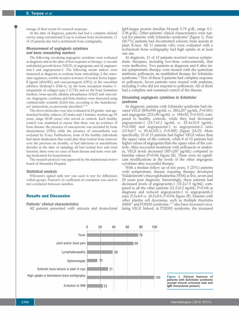
IgM-kappa protein (median M-peak 0.79 g/dL, range 0.1-2.36 g/dL). Other patients' clinical characteristics were typ-ical for patients with Schnitzler syndrome2 (Figure 1). Four(30.7%) patients had documented sclerotic bone lesions inplain X-rays. All 11 patients who were evaluated with atechnetium bone scintigraphy had high uptake in at leastone site.At diagnosis, 11 of 13 patients received various sympto-
matic therapies, including low-dose corticosteroids, thatwere ineffective. Two patients at diagnosis and 6 after ini-tial symptomatic therapy were treated with the quinoloneantibiotic pefloxacin, an established therapy for Schnitzlersyndrome.15 Five of these 8 patients had complete responseto pefloxacin. Seven patients were treated with anakinra,including 3 who did not respond to pefloxacin. All of themhad a complete and sustained control of the disease.
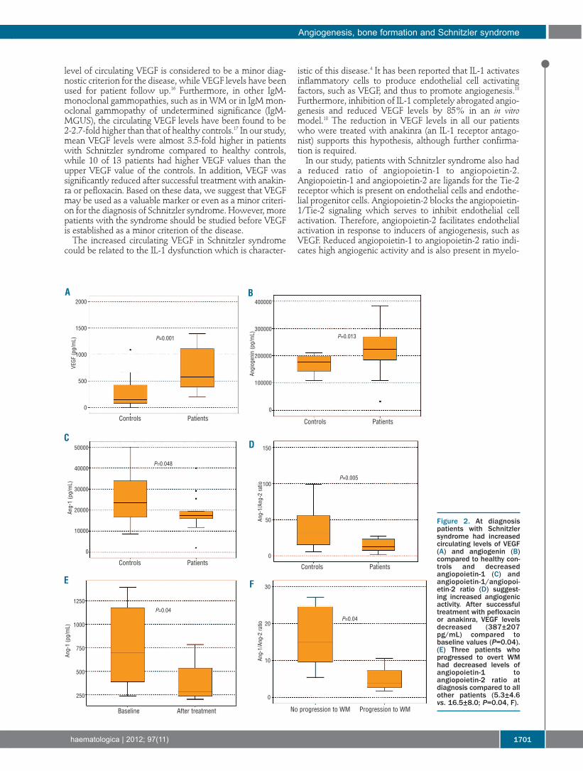
Circulating angiogenic cytokines in Schnitzler syndromeAt diagnosis, patients with Schnitzler syndrome had ele-
vated VEGF (809±598 pg/mL vs. 263±257 pg/mL; P=0.001)and angiogenin (221±96 ng/mL vs. 169±33; P=0.013) com-pared to healthy controls, while they had decreasedangiopoietin-1 (18.7±9.1 ng/mL vs. 25.4±10.9 ng/mL;P=0.048) and angiopoietin-1 to angiopoietin-2 ratio(13.9±8.7 vs. 63.4±102.1; P=0.005) (Figure 2A-D). Morespecifically, 10 of 13 patients had higher VEGF values thanthe upper value of the controls, while 6 of 13 patients hadhigher values of angiogenin than the upper value of the con-trols. After successful treatment with pefloxacin or anakin-ra, VEGF levels decreased (387±207 pg/mL) compared tobaseline values (P=0.04; Figure 2E). There were no signifi-cant modifications in the levels of the other angiogeniccytokines after successful therapy.With a median follow up of ten years, 3 (23%) patients
with symptomatic disease requiring therapy developedWaldenström’s macroglobulinemia (WM) at five, seven and20 years post diagnosis. Interestingly, these patients haddecreased levels of angiopoietin-1 (10.2±7.5 ng/mL) com-pared to all the other patients (21.2±8.2 ng/mL; P=0.04) atdiagnosis and reduced angiopoietin-1 to angiopoietin-2ratio (5.3±4.6 vs. 16.5±8.0; P=0.04; Figure 2F). Patients withother plasma cell dyscrasias, such as multiple myeloma(MM)11 and POEMS syndrome,12,16 also have increased circu-lating VEGF. Indeed, in POEMS syndrome, the increased
e. terpos et al.
1700 haematologica | 2012; 97(11)
Figure 1. Clinical features ofpatients with Schnitzler syndrome(except chronic urticarial rash andIgM monoclonal protein).
0 20 40 60 80 100
76
69
38
15
31
100
23
Fever
Joint and/or bone pain
Lymphadenopathy
Splenomegaly
Sclerotic bone lesions in plain X-rays
High uptake in technetium bone scintigraphy
Evolution to WM
level of circulating VEGF is considered to be a minor diag-nostic criterion for the disease, while VEGF levels have beenused for patient follow up.16 Furthermore, in other IgM-monoclonal gammopathies, such as in WM or in IgM mon-oclonal gammopathy of undetermined significance (IgM-MGUS), the circulating VEGF levels have been found to be2-2.7-fold higher than that of healthy controls.17 In our study,mean VEGF levels were almost 3.5-fold higher in patientswith Schnitzler syndrome compared to healthy controls,while 10 of 13 patients had higher VEGF values than theupper VEGF value of the controls. In addition, VEGF wassignificantly reduced after successful treatment with anakin-ra or pefloxacin. Based on these data, we suggest that VEGFmay be used as a valuable marker or even as a minor criteri-on for the diagnosis of Schnitzler syndrome. However, morepatients with the syndrome should be studied before VEGFis established as a minor criterion of the disease.The increased circulating VEGF in Schnitzler syndrome
could be related to the IL-1 dysfunction which is character-
istic of this disease.4 It has been reported that IL-1 activatesinflammatory cells to produce endothelial cell activatingfactors, such as VEGF, and thus to promote angiogenesis.18Furthermore, inhibition of IL-1 completely abrogated angio-genesis and reduced VEGF levels by 85% in an in vitromodel.18 The reduction in VEGF levels in all our patientswho were treated with anakinra (an IL-1 receptor antago-nist) supports this hypothesis, although further confirma-tion is required. In our study, patients with Schnitzler syndrome also had
a reduced ratio of angiopoietin-1 to angiopoietin-2.Angiopoietin-1 and angiopoietin-2 are ligands for the Tie-2receptor which is present on endothelial cells and endothe-lial progenitor cells. Angiopoietin-2 blocks the angiopoietin-1/Tie-2 signaling which serves to inhibit endothelial cellactivation. Therefore, angiopoietin-2 facilitates endothelialactivation in response to inducers of angiogenesis, such asVEGF. Reduced angiopoietin-1 to angiopoietin-2 ratio indi-cates high angiogenic activity and is also present in myelo-
Angiogenesis, bone formation and Schnitzler syndrome
haematologica | 2012; 97(11) 1701
Figure 2. At diagnosispatients with Schnitzlersyndrome had increasedcirculating levels of VEGF(A) and angiogenin (B)compared to healthy con-trols and decreasedangiopoietin-1 (C) andangiopoietin-1/angiopoi-etin-2 ratio (D) suggest-ing increased angiogenicactivity. After successfultreatment with pefloxacinor anakinra, VEGF levelsdecreased (387±207pg/mL) compared tobaseline values (P=0.04).(E) Three patients whoprogressed to overt WMhad decreased levels ofangiopoietin-1 toangiopoietin-2 ratio atdiagnosis compared to allother patients (5.3±4.6vs. 16.5±8.0; P=0.04, F).
A B
CD
E F
Controls Patients
P=0.001
2000
1500
1000
500
0
50000
40000
30000
20000
10000
0
1250
1000
750
500
250
150
100
50
0
30
20
10
0
400000
300000
200000
100000
0
P=0.048
P=0.04
Ang-1 (pg/mL)
Ang-1 (pg/mL)
Ang-1/An
g-2 ratio
Ang-1/An
g-2 ratio
VEGF
(pg/mL)
Angiog
enin (p
g/mL)
P=0.04
P=0.005
P=0.013
Controls Patients
Controls Patients Controls Patients
Baseline After treatment No progression to WM Progression to WM
ma, where it correlates with advanced disease and poorprognosis.12 The reduced angiopoietin-1 to angiopoietin-2ratio in our patients confirms that angiogenesis is elevatedin Schnitzler syndrome and is implicated in its biology.Another interesting finding of our study is the correlationbetween low angiopoietin-1 to angiopoietin-2 ratio andprogression of Schnitzler to WM. Although the number ofour patients who progressed to WM was low (3 of 13;23%), but similar to that reported in the literature (15%),2and progression took place several years after diagnosis, thisresult may suggest that low angiopoietin-1 to angiopoietin-2 ratio at diagnosis may indicate a predisposition for evolu-tion of the gammopathy towards an overt WM. However,this has to be confirmed prospectively in a larger number ofpatients.
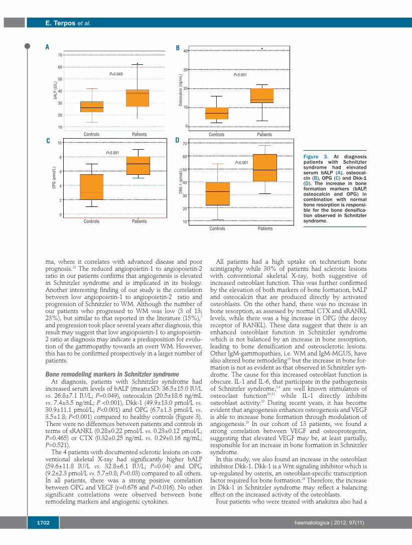
Bone remodeling markers in Schnitzler syndromeAt diagnosis, patients with Schnitzler syndrome had
increased serum levels of bALP (mean±SD: 36.5±15.0 IU/Lvs. 26.8±7.1 IU/L; P=0.049), osteocalcin (20.5±18.6 ng/mLvs. 7.4±3.5 ng/mL; P <0.001), Dkk-1 (49.9±13.0 pmol/L vs.30.9±11.1 pmol/L; P<0.001) and OPG (6.7±1.3 pmol/L vs.3.5±1.8; P<0.001) compared to healthy controls (Figure 3).There were no differences between patients and controls interms of sRANKL (0.28±0.22 pmol/L vs. 0.23±0.12 pmol/L;P=0.465) or CTX (0.32±0.25 ng/mL vs. 0.29±0.16 ng/mL;P=0.521). The 4 patients with documented sclerotic lesions on con-
ventional skeletal X-ray had significantly higher bALP(59.6±11.8 IU/L vs. 32.8±6.1 IU/L; P=0.04) and OPG(9.2±2.3 pmol/L vs. 5.7±0.8; P=0.03) compared to all others.In all patients, there was a strong positive correlationbetween OPG and VEGF (r=0.676 and P=0.016). No othersignificant correlations were observed between boneremodeling markers and angiogenic cytokines.
All patients had a high uptake on technetium bonescintigraphy while 30% of patients had sclerotic lesionswith conventional skeletal X-ray, both suggestive ofincreased osteoblast function. This was further confirmedby the elevation of both markers of bone formation, bALPand osteocalcin that are produced directly by activatedosteoblasts. On the other hand, there was no increase inbone resorption, as assessed by normal CTX and sRANKLlevels, while there was a big increase in OPG (the decoyreceptor of RANKL). These data suggest that there is anenhanced osteoblast function in Schnitzler syndromewhich is not balanced by an increase in bone resorption,leading to bone densification and osteosclerotic lesions.Other IgM-gammopathies, i.e. WM and IgM-MGUS, havealso altered bone remodeling19 but the increase in bone for-mation is not as evident as that observed in Schnitzler syn-drome. The cause for this increased osteoblast function isobscure. IL-1 and IL-6, that participate in the pathogenesisof Schnitzler syndrome,2,4 are well known stimulators ofosteoclast function20,21 while IL-1 directly inhibitsosteoblast activity.22 During recent years, it has becomeevident that angiogenesis enhances osteogenesis and VEGFis able to increase bone formation through modulation ofangiogenesis.23 In our cohort of 13 patients, we found astrong correlation between VEGF and osteoprotegerin,suggesting that elevated VEGF may be, at least partially,responsible for an increase in bone formation in Schnitzlersyndrome. In this study, we also found an increase in the osteoblast
inhibitor Dkk-1. Dkk-1 is a Wnt signaling inhibitor which isup-regulated by osterix, an osteoblast-specific transcriptionfactor required for bone formation.24 Therefore, the increasein Dkk-1 in Schnitzler syndrome may reflect a balancingeffect on the increased activity of the osteoblasts. Four patients who were treated with anakinra also had a
e. terpos et al.
1702 haematologica | 2012; 97(11)
Figure 3. At diagnosispatients with Schnitzlersyndrome had elevatedserum bALP (A), osteocal-cin (B), OPG (C) and Dkk-1(D). The increase in boneformation markers (bALP,osteocalcin and OPG) incombination with normalbone resorption is responsi-ble for the bone densifica-tion observed in Schnitzlersyndrome.
A B
C DControls Patients
Controls PatientsControls Patients
Controls Patients
P=0.049
P<0.001
P<0.001
P<0.001
70
60
50
40
30
20
10
70
60
50
40
30
20
10
10
8
6
4
2
0
40
30
20
10
0
bALP
(U/L)
OPG (pmol/L)
DKK-1 (pmol/L)
Osteoc
alcin (ng/mL)
bone scintigraphy at a median six months post-treatment.All patients showed a dramatic reduction in osteoblasticlesions but in none of them did the lesions completely dis-appear. In conclusion, our analysis shows altered circulating
angiogenic cytokines in Schnitzler syndrome reflectingincreased angiogenic activity. Furthermore, we documentenhanced bone formation with no alterations in boneresorption; this explains the presence of sclerotic bonelesions in this disease entity. Successful therapy with eitheranakinra or pefloxacin is associated with a reduction in themajor angiogenic cytokine VEGF. These data support apotential role for VEGF in the diagnosis and follow up of
patients with Schnitzler syndrome, and suggest that VEGFmay be used as a minor criterion for the diagnosis of thedisease.
Authorship and Disclosures
The information provided by the authors about contributions frompersons listed as authors and in acknowledgments is available withthe full text of this paper at www.haematologica.org.Financial and other disclosures provided by the authors using the
ICMJE (www.icmje.org) Uniform Format for Disclosure ofCompeting Interests are also available at www.haematologica.org.
Angiogenesis, bone formation and Schnitzler syndrome
haematologica | 2012; 97(11) 1703
References
1. Lipsker D, Veran Y, Grunenberger F, CribierB, Heid E, Grosshans E. The Schnitzler syn-drome. Four new cases and review of the lit-erature. Medicine (Baltimore). 2001;80(1):37-44.
2. de Koning HD, Bodar EJ, van der Meer JW,Simon A; Schnitzler Syndrome Study Group.Schnitzler syndrome: beyond the casereports: review and follow-up of 94 patientswith an emphasis on prognosis and treat-ment. Semin Arthritis Rheum. 2007;37(3):137-48.
3. Asli B, Brouet JC, Fermand JP. Spontaneousremission of Schnitzler syndrome. AnnAllergy Asthma Immunol. 2011;107(1):87-8.
4. van Deuren M, Kroot JJ, Swinkels DW.Time-course analysis of serum hepcidin, ironand cytokines in a C282Y homozygouspatient with Schnitzler's syndrome treatedwith IL-1 receptor antagonist.Haematologica. 2009;94(9):1297-300.
5. Migliorini P, Del Corso I, Tommasi C,Boraschi D. Free circulating interleukin-18 isincreased in Schnitzler syndrome: a newautoinflammatory disease? Eur CytokineNetw. 2009;20(3):108-11.
6. Gran JT, Midtvedt Ø, Haug S, Aukrust P.Treatment of Schnitzler's syndrome withanakinra: report of three cases and review ofthe literature. Scand J Rheumatol.2011;40(1):74-9.
7. Ramadan KM, Eswedi HA, El-Agnaf MR.Schnitzler syndrome: a case report of suc-cessful treatment using the anti-CD20 mon-oclonal antibody rituximab. Br J Dermatol.2007;156(5):1072-4.
8. Bozeman S, Lewis S. Schnitzler syndromesuccessfully treated with methotrexate. AnnAllergy Asthma Immunol. 2008;101 (2):219.
9. Dinarello CA. Blocking IL-1 in systemicinflammation. J Exp Med. 2005;201(9): 1355-9.
10. Rajkumar SV, Mesa RA, Fonseca R,
Schroeder G, Plevak MF, Dispenzieri A, et al.Bone marrow angiogenesis in 400 patientswith monoclonal gammopathy of undeter-mined significance, multiple myeloma, andprimary amyloidosis. Clin Cancer Res.2002;8(7):2210-6.
11. Kastritis E, Terpos E, Anagnostopoulos A,Xilouri I, Dimopoulos MA. Angiogenetic fac-tors and biochemical markers of bonemetabolism in POEMS syndrome treatedwith high-dose therapy and autologous stemcell support. Clin Lymphoma Myeloma.2006;7(1):73-6.
12. Terpos E, Anargyrou K, Katodritou E,Kastritis E, Papatheodorou A, Christoulas D,et al. Circulating angiopoietin-1 to angiopoi-etin-2 ratio is an independent prognostic fac-tor for survival in newly diagnosed patientswith multiple myeloma who received thera-py with novel antimyeloma agents. Int JCancer. 2012;130 (3):735-42.
13. Terpos E, Szydlo R, Apperley JF, HatjiharissiE, Politou M, Meletis J, et al. Soluble receptoractivator of nuclear factor kappaB ligand-osteoprotegerin ratio predicts survival inmultiple myeloma: proposal for a novelprognostic index. Blood. 2003;102(3):1064-9.
14. Terpos E, Fragiadaki K, Konsta M,Bratengeier C, Papatheodorou A, Sfikakis PP.Early effects of IL-6 receptor inhibition onbone homeostasis: a pilot study in womenwith rheumatoid arthritis. Clin ExpRheumatol. 2011;29(6):921-5.
15. Asli B, Bienvenu B, Cordoliani F, Brouet JC,Uzunhan Y, Arnulf B, et al. Chronic urticariaand monoclonal IgM gammopathy(Schnitzler syndrome): report of 11 casestreated with pefloxacin. Arch Dermatol.2007;143(8):1046-50.
16. D'Souza A, Hayman SR, Buadi F,Mauermann M, Lacy MQ, Gertz MA, et al.The utility of plasma vascular endothelialgrowth factor levels in the diagnosis and fol-low-up of patients with POEMS syndrome.Blood. 2011;118(17):4663-5.
17. Anagnostopoulos A, Eleftherakis-
Papaiakovou V, Kastritis E, Tsionos K,Bamias A, et al. Serum concentrations ofangiogenic cytokines in Waldenstrommacroglobulinaemia: the ration of angiopoi-etin-1 to angiopoietin-2 and angiogenin cor-relate with disease severity. Br J Haematol.2007;137(6):560-8.
18. Carmi Y, Voronov E, Dotan S, Lahat N,Rahat MA, Fogel M, et al. The role ofmacrophage-derived IL-1 in induction andmaintenance of angiogenesis. J Immunol.2009;183(7):4705-14.
19. Terpos E, Anagnostopoulos A, Kastritis E,Bamias A, Tsionos K, Dimopoulos MA.Abnormal bone remodelling and increasedlevels of macrophage inflammatory protein-1 alpha (MIP-1alpha) in Waldenströmmacroglobulinaemia. Br J Haematol. 2006;133(3):301-4.
20. Lee YM, Fujikado N, Manaka H, Yasuda H,Iwakura Y. IL-1 plays an important role in thebone metabolism under physiological condi-tions. Int Immunol. 2010;22(10):805-16.
21. Liu XH, Kirschenbaum A, Yao S, Levine AC.Cross-talk between the interleukin-6 andprostaglandin E(2) signaling systems resultsin enhancement of osteoclastogenesisthrough effects on the osteoprotegerin/receptor activator of nuclear factor-{kappa}B(RANK) ligand/RANK system.Endocrinology. 2005;146(4):1991-8.
22. Stashenko P, Obernesser MS, Dewhirst FE.Effect of immune cytokines on bone.Immunol Invest. 1989;18(1-4):239-49.
23. Samee M, Kasugai S, Kondo H, Ohya K,Shimokawa H, Kuroda S. Bone morpho-genetic protein-2 (BMP-2) and vascularendothelial growth factor (VEGF) transfec-tion to human periosteal cells enhancesosteoblast differentiation and bone forma-tion. J Pharmacol Sci. 2008;108(1):18-31.
24. Zhang C, Dai H, de Crombrugghe B.Characterization of Dkk1 gene regulation bythe osteoblast-specific transcription factorOsx. Biochem Biophys Res Commun.2012;420(4):782-6.