Embed Size (px)
Citation preview
In vivo regulation of apolipoprotein A-I gene expression by estradiol and testosterone occurs by different mechanisms in inbred strains of mice'
Jingjing Tang, Rai Ajit K. Srivastava, Elaine S. Krul, Dirk Baumann, Barbara A. Pfleger, Robert T. Kitchens, and Gustav Schonfeld
Division of Atherosclerosis and Lipid Research, Department of Internal Medicine, Washington University School of Medicine, St. Louis, M O 63110
Abstract We tested the hypothesis that testosterone and estrogen modulate apoA-I gene expression and metabolism by different mechanisms that may be influenced by genetic factors. Male and female C3H/HeJ (atherosclerosis-resistant) and C57BL/6J (atherosclerosis-susceptible) mice (n = 5/group) were castrated (Placebo). Castrates were given 17P-estradiol (E2) at 0.16 pg/g (E2L) or 5 pglg (E2H) body weight per day, or testosterone (Testo) 1 pglg per day, 14 days after surgery, for 14 days. Plasma total cholesterol concentrations (TC) were higher in male Placebo mice than in females. Testosterone altered TC and high density lipopro- tein (HDL) cholesterol by gender and strain; however (HDL-C)/TC ratios and apoA-I concentrations were unaltered. Testosterone did reduce HDL particle diameters in both genders of C3H mice only. Low density lipoprotein-cholesterol (LDL-C)/TC ratios remained constant and apoB increased in males only. E2L and E2H decreased TC, HDL-C/TC ratios, and apoA-I. Decrements varied by strain. HDL diameters decreased in both genders in C3H mice only; however, HDL size distributions were altered in both strains. LDL-C/TC ratios increased in all groups. E2L mice showed variable responses of apoB, but apoB rose uniformly in all E2H groups. Testosterone increased and E2H decreased hepatic apoA-I synthesis. ApoA-I mRNA concentrations remained stable in both Testo and E2 groups. ApoA-I gene transcription varied by strain and gender, but all changes were <twofold. Testosterone did not affect hepatic apoB or LDL receptor mRNA, however, E2H increased both mRNAs in males but not in females. O n Western blotting of liver membranes, E2H had little effect on mouse LDL receptor protein mass; by contrast, E2H increased LDL receptor -threefold in rats. B In summary, responsive- ness of mouse lipids to testosterone and E2 vary by strain and gender. Testosterone and E2 differ in their regulation of apoA-I production mainly at the level of translation. Hormones operate at several levels of gene regulation, suggesting that complex mechanisms are involved. Mice differ from rats and rabbits in their LDL receptor responsiveness to estradiol treatment.-Tang, J., R. A. K. Srivastava, E. S. Krul, D. Baumann, B. A. Pfleger, R. T. Kitchens, and G. Schonfeld. In vivo regulation of apolipo- protein A-I gene expression by estradiol and testosterone occurs by different mechanisms in inbred strains of mice. J Lipid Res. 1991. 32: 1571-1585.
Supplementary key words gen translation transcription synthesis - post-transcription translation
apoA-I gene expression estrogen andro- post-
Plasma HDL concentrations are potent predictors of coronary disease in humans (1, 2). Apolipoprotein (apo) A-I is the major apolipoprotein of HDL and its concentra- tions also are useful predictors of coronary disease risk. M e n have a greater risk of coronary heart disease than pre-menopausal women perhaps because of the lower con- centrations of HDL a n d apoA-I in their plasma, whereas post-menopausal or castrated women have coronary risk and plasma lipoprotein profiles that more closely resem- ble those of men. Estrogen replacement therapy returns the lipoprotein profiles and perhaps the coronary risk towards pre-menopausal levels. Thus , some of the differ- ences between m e n and women with respect t o lipoprotein and coronary risk may be d u e to sex hormones rather than gender.
ApoA-I is synthesized in liver a s well as in intestine (3), a n d its hepatic synthesis is influenced by estrogens a n d androgens (4-8). These hormones also alter plasma total cholesterol a n d HDL-cholesterol levels in many species (9-13). In some studies, these hormones influence the rate of synthesis of apoA-I (14-16), while in others, they alter its clearance from plasma (17). Differences in hormone re- sponsiveness between individuals a n d species exist a n d
Abbreviations: apo, apolipoprotein; HDL, high density lipoproteins; LDL, low density lipoproteins; VLDL, very low density lipoproteins; HTGL, hepatic triglyceride lipase; LPL, lipoprotein lipase; FPLC, fast protein liquid chromatography; DEPC, diethyl pyrocarbonate; SDS, so- dium dodecyl sulfate; EDTA, ethylenediamine tetraacetic acid; PAGE, polyacrylamide gel electrophoresis; PEG, polyethylene glycol; BSA, bovine serum albumin; TCA, trichloroacetic acid; PMSF, phenylmethyl- sulfonyl fluoride; HEPES, N-[Z-hydroxyethyllpiperazine-N'-[Z-ethane- sulfonic acid]; DTT, dithiothreitol; PIPES, piperazine-N,N'-bis[2- ethanesulfonic acid]; EGTA, ethylene glycol-bis(P-amino ethy1)ether; SSC, 0.15 M sodium citrate, pH 7.0; HSS, high speed supernatant; PC, personal computer; LDL-R, LDL receptor.
'This work was presented in part at the 63rd Scientific Sessions of the American Heart Association, Dallas, TX, 14 November 1990.
Journal of Lipid Research Volume 32, 1991 1571
by guest, on April 24, 2019
ww
w.jlr.org
Dow
nloaded from
probably reflect the genetic diversity of the subjects under study.
Relatively little is known about the hormonal regula- tion of apoA-I production at the molecular level. Even less is known about any interactions of hormonal and genetic factors, which are difficult to study in outbred humans or experimental animals. However, using inbred strains of mice, comparisons can be made between identically treated, but genetically distinct strains to determine what role genetic factors may play in the animals’ responsive- ness to various experimental perturbations. In inbred mice, strain-related differences exist in susceptibility to diet-induced atherosclerosis and in lipoprotein profiles (18-22). For example, the mouse strains C57BL/6J and G57L/J are susceptible to diet-induced atherosclerosis, while the C3H/HeJ and NZB/BINJ strains are resistant (19). Plasma lipoprotein profiles also are affected in char- acteristic ways when these mice are administered athero- genic diets.
We hypothesized that C57BL/6J and C3H mice, two of the strains known to have differing lipoprotein profiles and differing susceptibilities to diet-induced atherosclero- sis, would also differ with respect to the regulation of their apoA-I metabolism by sex steroids. We further hypothe- sized that testosterone and estrogen may differ in their effects on the molecular regulation of apoA-I. Accord- ingly, we measured plasma concentrations and composi- tions of HDL, plasma concentrations of apoA-I, rates of hepatic apoA-I synthesis and the in vivo regulation of apoA-I gene expression in castrated male and female C3H/HeJ and C57BL/6J mice administered either testos- terone propionate or 17P-estradiol. We found that indeed there were differences in responsiveness by mouse strain, gender, and hormone, and that the regulation of apoA-I production occurred at both transcriptional and transla- tional steps.
METHODS
Animals and groups of treatment
Five- to six-week-old, male and female C3H/HeJ and C57BL/6J mice were obtained from Jackson Laboratory, Bar Harbor, ME. All mice were housed at 24°C in a room illuminated in 12-h light-dark cycles and maintained on Purina chow 5015 diet and tap water ad libitum. Mice of each strain were divided into four groups (n=5/group); placebo, castrated mice treated with vehicle injections; Testo, castrated mice administered testosterone propionate (Tp) at 1 pg/g body weight per day; E2L, castrated mice administered 176-estradiol (E2) 0.16 pg/g body weight per day; and E2H, castrated mice administered 17P-estradiol 5 pg/g body weight per day. Castrations were performed under metofane anesthesia at least 7 days after the arrival and housing of mice in our local facility and animals were
allowed to recover for 14 days after surgery. The indicated doses of testosterone propionate and 17P-estradiol dis- solved in propylene glycol were injected subcutaneously along the back at the same time each day for 14 days, while placebo mice received propylene glycol only. Ani- mals were weighed before and after 14 days of hormone administration. At the end of the experiment, animals were fasted overnight and killed by exsanguination under ether anesthesia. Plasma (anticoagulated by EDTA) from individual mice was pooled in each group for FPLC and HDL particle size determination. Serum was collected in- dividually for determination of total cholesterol, sex hor- mones, and apolipoprotein concentrations. Livers were removed immediately and pooled in each group for deter- mining hepatic apoA-I synthesis and for isolating nuclei and total RNA.
Testosterone and 17P-estradiol levels in the serum
Testosterone and estradiol radioimmunoassay kits were purchased from Amersham (DSL 4100 and IMB 100). Standard curves were constructed by plotting the log hor- mone levels (pg/ml) versus cpm of lz5I-1abeled testoster- one or estrogen. The standard curves ranged from 100 to 25,000 pg/ml for testosterone and 20 to 3,000 pg/ml for es- tradiol. One hundred pl of individual mouse sera were used for each assay.
Nondenaturing gradient gel electrophoresis
Nondenaturing gradient gel electrophoresis was per- formed as previously described (22). Briefly, plasma pools from each group (40 pl) were adjusted to d 1.21 g/ml by adding solid KBr and overlayered with d 1.21 g/ml KBr solution containing 1 mmol/l EDTA. Total lipoproteins (d < 1.21 g/ml) were collected (in 20 1.1) after ultracentrifu- gation in a Beckman 42.2 Ti rotor at 42,000 rpm for 24 h at 10°C. The d < 1.21 g/ml lipoprotein fractions (10 p1 per lane) were electrophoresed in PAA 4/30 polyacrylamide gradient gels (Pharmacia, Uppsala, Sweden). Electropho- reses were carried out at 125 V at 10°C for 24 h, and the gels were stained in 0.04% Coomassie brilliant blue G-250. Thyroglobulin and bovine serum albumin (BSA) were added to each sample as internal size standards. Five reference proteins were used for calibrating particle sizes: thyroglobulin (Stoke’s diameter 17.0 nm), apoferritin (12.2 nm), catalase (9.6 nm), lactate dehydrogenase (8.2 nm), and BSA (7.1 nm). Standard curves were plotted as the relative migration of the reference proteins compared to BSA versus the diameter of the reference proteins. Stan- dard curves were used to determine the sizes of the mouse lipoprotein particles.
Gel permeation chromatography and analysis of fractions
Separation of mouse plasma lipoproteins on FPLC Superose-6 columns was performed as previously described
1572 Journal of Lipid Research Volume 32, 1991
by guest, on April 24, 2019
ww
w.jlr.org
Dow
nloaded from
(18). Briefly, 400 pl of pooled plasmas from each group was centrifuged at 100,000 g min and the clear super- natants were loaded onto two Superose-6 columns (FPLC system, Pharmacia) connected in series. Fractions were eluted at 0.5 ml/min with 1 mmol/l EDTA, 154 mmol/l NaCl, and 0.02% NaN3 (pH 8.2). The first 12 ml of effluent was discarded, and 50 fractions of 0.5 ml each were subsequently collected. Cholesterol analyses on the column fractions and individual mouse serum samples were performed using enzymatic kits from Wako Chemi- cals USA (Richmond, VA).
Determination of serum apoA-I and apoB concentrations
Serum apoA-I and apoB concentrations were deter- mined by electroimmunoassay according to the method of Laurel1 (23) with some modifications. For apoA-I, electro- immunoassay plates were prepared as follows: 1.5% agarose (Seakam) with 5% Dextran T10 (Pharmacia) in Tris-Tricine buffer (0.8 M Tris, 0.024 M Tricine, 0.034 mM calcium lactate, 0.05% sodium azide, pH 8.6). For apoB, the plates contained 1% agarose, 3% PEG (mol wt 8000, Sigma), and 0.16% Triton. Agarose solutions were boiled and allowed to cool to 55OC, after which rabbit anti- mouse apoA-I or apoB monospecific polyclonal antibodies were added at 1350 and 1:200 dilutions, respectively. The agaroselantibody solutions were poured onto CelBond film (FMC) (cast gel dimensions = 21.6 x 10 x 0.15 cm). Twenty-three wells of 4 mm diameter were punched out with center-to-center distances of 9 mm. Pooled plasmas from each mouse group or individual mouse serum sam- ples were diluted 1:25 with Tris-Tricine buffer containing 1% BSA (Sigma A-7030) and 8 M urea for apoA-I. No urea was added for apoB. Five pl of standard or diluted samples was applied to the gel wells. Electrophoresis was carried out in Tris-Tricine buffer at 2 V/cm for 16-18 h in an LKB Multiphor chamber connected to a water circula- tor maintained at 15OC. Washing, staining, and drying of the electroimmunoassay plates were performed as de- scribed in the LKB 2117 Multiphor I1 Electrophoresis System Laboratory Manual (24). Results were calculated by area in mm2 (rocket height x $$ width at $$ height) per pl sample applied. Mouse apoA-I and LDL isolated from plasma were used as standards. The standard curves were linear over the range of 50-400 ng for apoA-I and 300-1200 ng for apoB.
Hepatic apoA-I synthesis
ApoA-I synthesis rates in mouse livers were determined according to the method of Williams and Dawson (25). Freshly isolated liver was chopped into 5-mg slices with a razor blade, and rinsed twice with bicarbonate-buffered Krebs-Ringer solution (KRB, 120 mM NaC1, 4.7 mM KCl, 2.5 mM CaCI,, 1.2 mM KH2P04, 1.2 mM MgS04, 25 mM NaHC03, pH 7.4), containing 50 U/ml penicillin
and 50 pglml streptomycin. The washes removed blood and minimized the contamination with plasma lipopro- teins. Forty mg of pooled total liver slices from each group (equal aliquots from each mouse) was incubated for 1 h at 37OC in 200 pl KRB containing antibiotics, 0.1% glucose, and 200 pCi[35S]methionine (1 mCi/ml) which was lyophilized and dissolved in the buffer (sp act: 1106 Ci/mmol, TRAN 3 5 s LABEL, ICN, Irvine, CA) under an atmosphere of 95% 0 2 / 5 % C 0 2 . The tissue incuba- tion was brief (1 h) to ensure that the spectrum of newly synthesized proteins was representative of the proteins being synthesized at the time the tissue was removed from the animal and so that more than 95% of synthesized pro- tein would remain in the tissue. After 1 h of incubation, the liver slices were washed twice with 2 ml ice-cold KRB and homogenized at 0-2OC with 600 p1 homogenization buffer (0.02 M sodium phosphate, pH 7.4, 0.15 M NaCl, 0.005 M EDTA, 200 pg/ml phenylmethylsulfonyl fluoride (PMSF), 1% Triton X-100). The homogenate was ultra- centrifuged at 2OC at 112,000 g for 1 h to prepare a high- speed supernatant (HSS). The HSS of each group were stored at -7OOC until analysis.
ApoA-I hepatic synthesis in each group was measured by immunoprecipitation. Briefly, 10 pl of rabbit anti- mouse apoA-I antiserum (prepared in this laboratory) in 1% Triton X-100 and 200 pg/ml PMSF were incubated with 20 pl HSS overnight at 4OC (performed in triplicate). Immune complexes were precipitated with 30 p1 of a solu- tion of 10% Immuno-Precipitin (Formalin-fixed Staph A cells) (Bethesda Research Laboratories, Life Technolo- gies, Inc., Gaithersburg, MD) in homogenization buffer for 30 min at room temperature. The antibody-antigen complexes were pelleted by spinning at 12,000 rpm for 3 min, and the pellets were washed twice with 1 ml homogenization buffer. Fifty p1 of SDS gel-loading buffer (26 mM Tris, pH 6.8, 10.6% glycerol, 1.1% SDS, 2 % s-mercaptoethanol, 0.024% bromophenol blue) was added to the pellets after the last wash and the solutions were boiled for 5 min. The immunoprecipitates were then run on 10% SDS polyacrylamide gels as previously described (25). After staining with Coomassie blue, the gel was treated with EN3HANCE (NEN Research Products, Boston, MA) for 1 h, followed by a cold water precipita- tion for 30 min and 1% glycerol for 1 h. The gel was then dried and exposed to Kodak XAR-5 film at -7OOC for autoradiography. The peaks corresponding to apoA-I were quantified using the video analysis system ('JAVA Jandel Scientific, Cortex Madera, CA) on a PC. ApoA-I synthesis was expressed as the area under the apoA-I peaks (cm2)/total protein synthesis (cpm)/h. Total protein synthesis was assessed by trichloroacetic acid ( E A ) pre- cipitation. One ml dHzO was added to 20 pl HSS. One ml 20% E A was then added to this mixture, and after incubating at 4°C for 15 min, the precipitates were iso- lated by centrifugation and washed twice with 10% cold
Tang et al. Regulation of hepatic apoA-I production 1573
by guest, on April 24, 2019
ww
w.jlr.org
Dow
nloaded from
E A . The precipitates were dissolved in 200 pl Soluene 350 (Packard, Downers Grove, IL) and counted for 3 5 s
radioactivity in a Beckman LS 2800 scintillation counter. ApoA-I synthesis in placebo group was considered as 100%.
Determination of apoA-I mRNA concentrations in liver and intestine by solution hybridization
Hepatic and intestinal total RNAs were isolated by the method of Chirgwin et al. (26). The rat apoA-I cDNA probe, which was constructed in pGEM-2, was kindly provided by Dr. J. Gordon (Department of Biological Chemistry, Washington University). The probe showed good cross-reaction with mouse apoA-I mRNA on North- ern blots. A 552 bp insert including 469 bp of rat apoA-I cDNA, a 17-mer poly C sequence, and the 66 bp poly- linker of pGEM-2 was cut out by EcoRI and purified by 1% low melting agarose gel (Bethesda Research Labora- tories) and then subcloned into the EcoRI site of the pGEM-3Zf( +) vector. The orientation of the ligated rat apoA-I cDNA in pGEM-SZf(+) was tested both by se- quencing and by hybridizing the 32P-labeled apoA-I cRNA probe (obtained by transcribing the vector with T 7 RNA polymerase, see below) with ssDNA standard made from the recombinant vector.
The pGEM-3Zf( +) containing the apoA-I cDNA insert was linearized by Kpn I and followed by conversion of the 3' overhang produced by Kpn I to a blunt end by adding Klenow DNA polymerase before transcription. A 32P- labeled rat apoA-I cRNA probe was then transcribed by T 7 RNA polymerase from the linearized vector according to Promega Guide protocol (27). The reaction mixture contained (Promega transcription kit): 4 pl of 5 x tran- scription buffer, 2 p1 of 100 mM D T T , 20 U RNAsin (ribonuclease inhibitor), 1 p1 linearized template (1 pgIpl), and Klenow DNA polymerase at 5 U/pg DNA. The mix- ture was incubated at 22°C for 15 min and then the fol- lowing was added: 1 p1 each of 10 mM ATP, GTP, and C T P , 2.4 pl 100 pM U T P , 5 ~ ~ [ c Y - ~ * P ] U T P (50 pCi at 650 Ci/mmol, ICN), 1 pl T7 RNA polymerase (15-20 UIpI). The mixture was incubated at 37OC for 60 min. In order to remove the DNA template, 1 pl RQl RNase free DNase I (1 U/pG DNA) (Boehringer Mannheim Bio- chemicals) and 0.5 p1 RNAsin (20 U) (Promega) was added after the transcription reaction and incubated for a further 15 min at 37OC. To purify the transcribed probes, the reaction mixture was extracted with phenol- chloroform followed by an extraction with chloroform. The cRNA probe was precipitated by ethanol and dis- solved in 20 pl DEPC-treated H20. The unincorporated nucleotide triphosphates were removed from the probe solution by chromatography on an RNA Sephadex G-50 (BMB) quick-spin column. The eluted probe was then ready for use.
An 858 bp cRNA standard was transcribed from the same vector by SP6 RNA polymerase after linearization
with Nael (which is a restriction enzyme site on the pGEM-3Zf( +) vector outside the polylinker region). The cRNA standard was then purified as described above and dissolved in DEPC-treated water. Aliquots were stored at -7OOC until use.
The solution hybridization assay for apoA-I mRNA was carried out at 65OC (optimal temperature determined in earlier experiments) (28). Initial experiments were also carried out to determine the range of total RNA concen- trations that yielded a linear response. Ten pg of total cel- lular RNA was chosen in subsequent experiments to quantify the apoA-I message. The range of the standard curve was from 10 pg to 200 pg of RNA standard. The RNA samples (10 pg) in triplicate were dried in a SpeedVac (Savant, Farmingdale, NY) and dissolved in 25 p1 hybridi- zation buffer (40% formamide, 400 mM NaCl, 1 mM EDTA, 40 mM PIPES, pH 6.5-6.7). Five pl 32P-labeled apoA-I probe (40,000 cpm) was added to each tube and the content of the tube was covered by 5 pl paraffin oil. Tubes were incubated at 65OC overnight. Three hundred pl of ice-cold RNase solution (40 pg/ml RNase A, 2 pg/ml RNase T1, 0.3 M NaCl, 5 mM EDTA, 10 mM Tris.HC1, p H 7.5) was added and incubated at 30°C for 1 h. One ml cold TCA solution (10% TCA, 1.5% sodium pyrophos- phate) was then added and the tubes were placed on ice for 15 min. The samples were then filtered through a G F/L glass fiber filter (Schleicher & Schuell, Keene, NH). The filters were washed with 10% cold TCA, dried at 8OoC for 1 h, and counted for 32P radioactivity. The calculation factor based on this assay was 1100 bp (length of apoA-I cDNA) divided by 858 bp (length of cRNA standard used in the assay) which equals 1.28. The hepatic apoA-I mRNA concentrations were expressed as pg apoA-I mRNA/pg total RNA.
Determination of apoA-I mRNA transcription rates in isolated liver nuclei
Mouse liver nuclei were isolated according to the method of Groudine, Peretz, and Weintrauts (29) with some modification. A section of mouse liver was excised immediately after killing and rinsed with ice-cold buffer A (60 mM KCI, 15 mM NaCI, 0.15 mM spermine, 0.5 mM spermidine, 14 mM 2-mercaptoethanol, 0.5 mM EGTA, 2 mM EDTA, 15 mM HEPES, p H 7.5). After rinsing, the liver (total of 1 g pooled from each group) was homoge- nized in 10 volumes of ice-cold buffer A containing 0.3 A4
sucrose and centrifuged for 10 min at 2,500 rpm in a swinging-bucket rotor. The pellet containing crude nuclei was resuspended in 2 ml of 2 M sucrose in buffer B (same as buffer A but with 0.1 mM each of EGTA and EDTA). This was then layered over a cushion of 2 M sucrose in buffer B and centrifuged for 20 min at 25,000 rpm at 4'C in a Beckman T L 100 ultracentrifuge. The clean nuclear pellet was resuspended in storage buffer (20 mM Tris- HCl, p H 7.9, 7.5 mM NaCl, 0.5 mM EDTA, 0.85 mM DTT, 0.125 mM PMSF, 50% glycerol). The concentra-
1574 Journal of Lipid Research Volume 32, 1991
by guest, on April 24, 2019
ww
w.jlr.org
Dow
nloaded from
tion of the nuclei was about 5 x lO5/pl. Nuclei in storage buffer could be stored for several weeks at - 7OoC without loss of activity.
tion rates in the groups of mice were expressed as a per- centage of the transcription rate in the placebo group.
In vitro transcription rates for apoA-I in isolated nuclei were determined according to the method of Schibler et al. (30). One to 2 x 10⁷ nuclei equivalent to 200-250 pg DNA, in triplicate, were used in the in vitro elongation reactions. The reaction buffer contained: 100 mM Tris- HCl, pH 7.9, 50 mM NaCl, 0.4 mM EDTA, 0.1 mM PMSF, 1.2 mM PMSF, 1.2 mM DTT, 1 mg/ml heparin sulfate, 2 mM MnC12, 4 mM MgC12, 1 mM each of GTP, ATP, CTP, 10 mM creatine phosphate, 130 U/ml ribo- nuclease inhibitor, 30% glycerol, and 100 ~ C ~ [ ~ X - ~ ~ P ] U T P (650 Ci/mM, ICN). The transcription reaction was al- lowed to occur at 3OoC for 40 min and was terminated by the addition of DNase 1 (RNase free) and further in- cubated at 3OoC for 5 min. Twenty pg Proteinase K, 5 mM EDTA, and 1% SDS were then added and the reaction mixtures were incubated at 37OC for a further 30 min. The reaction mixtures were extracted by hot (65OC) phenol-chloroform-isoamylalcohol 25:24:1 (v/v/v), and the RNA was subsequently precipitated by ethanol. The transcription products were checked by running ali- quots on urea SDS-polyacrylamide gels.
The hybridization assay was carried out according to the method of Chazenbalk, Wadsworth, and Rapoport (31). Recombinant pGEM-3Zf( +) vector containing the 550 bp rat apoA-I cDNA was linearized with Kpn I, de- natured in 0.2 M NaOH, and neutralized with l M HEPES, pH 6.5. Two pg of treated plasmid DNA was ap- plied to nitrocellulose paper and dried at 8OOC. The nitrocellulose papers containing the apoA-I probe were prehybridized for 2 h at 42OC in hybridization buffer (20 mM PIPES, pH 6.7, 50% formamide, 2 mM EDTA, 0.8 M NaC1, 0.2% SDS, 0.02% Ficoll, 0.02% polyvinyl- pyrrolidone, 100 pg/ml poly(A), and 0.02% BSA). The hybridization reaction was carried out by adding 100 p1 hybridization buffer with 1-10 x 106 cpm of extracted nuclear [32P]RNA to the prehybridized nitrocellulose paper and incubating for 60 h at 42OC. Fifty pl of paraffin oil was added to each tube to prevent evaporation. After hybridization the nitrocellulose papers were washed twice with 2 x SSC and 0.1% SDS for 30 min at room tempera- ture and treated with RNase A solution (10 pg/ml in 2 x SSC) at 37OC for 30 min followed by washing with 2 x SSC twice at room temperature. Further washings of the filters were done twice at 65OC in 0 . 1 ~ SSC containing 0.1% SDS for 30 min. The filters were dried at room tem- perature and exposed to Kodak XAR-5 film using intensi- fying screens at - 70OC. To elute the hybridized RNA for quantitation, the filters were incubated with 200 pl of 0.3 M NaOH for 15 min at 65OC followed by the addition of 50 pl glacial acetic acid and 4 ml scintillation fluid and counted. P-Actin mRNA transcription rates were used as internal standards. The hepatic apoA-I mRNA transcrip-
Comparison of LDL-R up-regulation by estrogen between rats and mice
Male C3H/HeJ mice (n=J/group) and 250-275 g male Sprague-Dawley rats (Harlan Sprague-Dawley, Inc., In- dianapolis, IN) (n =l/group) were divided into three groups: placebo administered at 1.6 pl/g body weight propylene glycol per day; 17 a-ethinyl estradiol (17a-EE) administered at 5 pglg body weight per day; and 170- estradiol (17P-E2) administered at 5 pg/g body weight per day. The 17a-ethinyl estradiol and the 17P-estradiol were dissolved in propylene glycol and delivered subcutaneous- ly at the same time each day for 6 days. After an overnight fast, the animals were exsanguinated under ether anesthe- sia on the seventh day. Plasma, anticoagulated with EDTA, was collected for total cholesterol concentration determination. Livers were immediately removed, and liver membrane preparations were performed as de- scribed by Kovanen, Brown, and Goldstein (32). Briefly, the livers were homogenized in 10 ml Tris buffer (150 mM NaCl, 1 mM CaC12, and 10 mM Tris-HC1, pH 7.5) with two 10-sec pulses in a Tekmar homogenizer (Cincinnati, OH) at 4OC. The whole homogenates were then cen- trifuged at 500 g for 5 min. The 500 g supernatant was centrifuged at 8,000 g for 15 min, and the 8,000 g super- natant was recentrifuged at 100,000 g for 60 min. The 100,000 g pellets were resuspended in 6 ml Tris buffer by repeatedly passing the pellet through a 21-gauge needle. This suspension was centrifuged at 100,000 g for 60 min, and the pellet was frozen quickly in liquid nitrogen and stored at -8OOC.
LDL-R protein determination
Within a week, the frozen pellets were resuspended in 10 ml buffer (50 mM NaCl, 1 mM CaC12, 20 mM Tris- HCl, pH 7.5) and solubilized using a 21-gauge needle. The solubilized pellets were sonicated for 20 sec in a Branson Sonifier cell disruptor (Model W-l85E, Heat Systems-Ultrasonics, Inc., Plainview, NY) using a micro- probe at a setting of 6, and protein concentrations were determined by a modification of the Lowry procedure (33).
The solubilized liver membranes were mixed with a so- lution of 2.5% SDS, 50% glycerol, and 0.5% bromo- phenyl blue in a ratio of 4:l. Two hundred pg of total liver membrane protein was loaded onto each lane of a 6% polyacrylamide gel containing 0.1% SDS and electro- phoresed without preheating and in the absence of dithio- threitol for 3 h at 4°C in a 25 mM Tris-glycine (pH 8.6) and 0.1% SDS running buffer.
Transfer to nitrocellulose paper was accomplished using an alteration of the method of Towbin, Staehelin, and Gordon (34) except that methanol was omitted from
Tang et al. Regulation of hepatic apoA-I production 1575
by guest, on April 24, 2019
ww
w.jlr.org
Dow
nloaded from
the transfer buffer. Nitrocellulose membranes (Micron Separations, Inc., Westboro, MA) presoaked in transfer buffer (25 mM Tris-glycine) for 30 min, were placed on
were quantified on a video analysis system (JAVA Jandel Scientific).
the anode side of the gel between two sheets bf porous filter paper. Electrotransfer was performed overnight at 4OC at 4 V/cm.
After the transfer, the nitrocellulose paper was dried and then immersed in Bovine Lacto Transfer Technique Optimizer (BLOTTO) blocking buffer consisting of 5% (w/v) nonfat powdered dry milk in phosphate-buffered saline (PBS) with 0.01% antifoam A emulsion (Sigma Chemical Co.) and 0.001% merthiolate (Sigma Chemical Co.) for 2 h. The nitrocellulose paper was then incubated overnight with rabbit anti-bovine adrenal LDL-R anti- serum (#4526, generously provided by Dr. Janet Boyles, San Francisco, CA) diluted 1:lOOO in BLOTTO at room temperature. The nitrocellulose paper was washed with 250 ml of 0.05% NP-40 in PBS, and a '251-labeled goat anti-rabbit IgG antibody diluted in BLOTTO to a con- centration of approximately 7.5 x lo5 cpm/ml was in- cubated with the nitrocellulose paper for 4 h at room tem- perature. The nitrocellulose paper was again washed with 0.05% NP-40 in PBS, dried, and placed in film cassettes for exposure at -80°C for autoradiography. Densities of the bands on autoradiographs corresponding to LDL-R
Determination of LDL-R and apoB mRNA in liver by solution hybridization
Rat LDL-R cDNA clone was obtained from Dr. A. D. Cooper (Palo Alto Medical Foundation, Palo Alto, CA). A 550 bp fragment was cut out by Pst I and subcloned into Pst I site of pGEM-3Zf( +). The 32P-labeled LDL-R cRNA probe was transcribed by T7 RNA polymerase after linearization by Hind111 from pGEM-3Zf( +) con- taining LDL-R cDNA. The cRNA standard was tran- scribed from the same vector by sp6 RNA polymerase, after linearization by Nael (27). Rat apoB cDNA clone was obtained from Dr. A. J. Lusis (University of Califor- nia, Los Angeles, CA). A 240 bp apoB cDNA was sub- cloned into EcoRI site of pGEM-SZf(+) and linearized by BamHI and Nael for standard and probe, respectively, apoB cRNA standard and probe were transcribed by T7
and sp6 RNA polymerase, respectively (27). Fifty pg of total RNA was used for both LDL-R and apoB. The solu- tion hybridization assays were carried out in triplicate as above and reference 28. The hybridization temperature was 60°C for LDL-R and 55°C for apoB. Both LDL-R
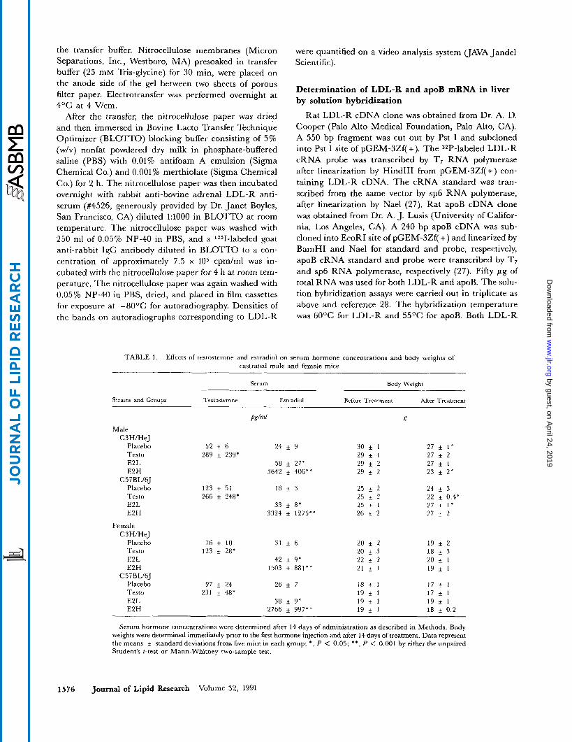
TABLE I . Effects of testosterone and estradiol on serum hormone concentrations and body weights of castrated male and female mice
Serum Body Weight
Strains and Croups Trrtosterone Estradiol Before Treatment After Treatment
Ps/"l E Male
C3H/HeJ Placebo 52 f 6 24 f 9 30 1 27 f 1 ' Testo 289 * 239* 29 f 1 2 7 t 2 E2L 58 f 27* 29 f 2 27 I E2H 3642 t 406** 29 f 2 23 f 2'
Placebo 123 * 51 18 k 3 25 f 2 24 f 3 Testo 266 t 248* 25 f 2 22 & 0.4' E2L 33 t 8* 25 _+ 1 2 7 _+ 1 ' E2H 3324 f 1275" 26 * 2 27 f 2
C57BLhJ
Female C3H/HeJ
Placebo 76 + 10 31 t 6 20 f 2 19 f 2
E2L 42 f 9* 22 f 2 20 i 1 E2H 1503 f 881** 21 f 1 19 k 1
Testo 123 f 28' 20 f 3 18 + 3
C57 €3 L/6J Placebo 97 i 24 26 & 7 18 f 1 1 7 + 1 Testo 231 f 48' 19 f 1 17 f 1 E2L 38 + 9' 19 * 1 19 t 1 E2H 2766 + 997" 19 * 1 18 i 0.2
Serum hormone concentrations were determined after 14 days of administration as described in Methods. Body weights were determined immediately prior to the first hormone injection and after 14 days of treatment. Data represent the means f standard deviations from five mice in each group; *, P < 0.05; **, P < 0.001 by either the unpaired Student's t-test or Mann-Whitney two-sample test.
1576 journal of Lipid Research Volume 32, 1991
by guest, on April 24, 2019
ww
w.jlr.org
Dow
nloaded from
and apoB cRNA probes showed good cross-reaction with mouse mRNA on Northern blots.
RESULTS
Body weights and hormone levels
Body weights of both castrated male and ovariec- tomized female mice were measured before and after es- tradiol or testosterone propionate administration (Table 1). Body weights did not change in female mice, but there were statistically significant differences in four of the eight groups in male mice that could not be attributed simply to hormonal administration since the placebo group re- ceived propylene glycol injections only. Serum estradiol and testosterone concentrations were comparable in both genders and mouse strains after castration. Administra- tion of testosterone propionate increased testosterone levels significantly to mimic physiological ranges in the testosterone groups. Estradiol levels were increased more than 1.4-fold with low dose estradiol treatment (to physio- logic ranges for females), and 50-fold or more with high dose treatment (Table 1).
Effects of hormones on plasma lipid, apoA-I, and apoB concentrations
Total cholesterol. Baseline total plasma cholesterol con- centrations were 19% higher in male C3H/HeJ (205 mg/dl, placebo) than in female C3H/HeJ mice (172 mg/dl, placebo) and 14% higher in male C57BLbJ (142 mg/dl, placebo) than female C57BL/6J mice (125 mg/dl, placebo) (Table 1). Total cholesterol concentrations were variably affected by testosterone administration. In male C3H/HeJ and female C57BL/6J mice, there were no changes, while significant increases were seen in male C57BL/6J and significant decreases in female C3H/HeJ mice. Both low and high doses of estradiol decreased plasma total cho- lesterol by more than 33% in C3H/HeJ mice of either gender, while the effects were very small in either male or female C57BL/6J mice.
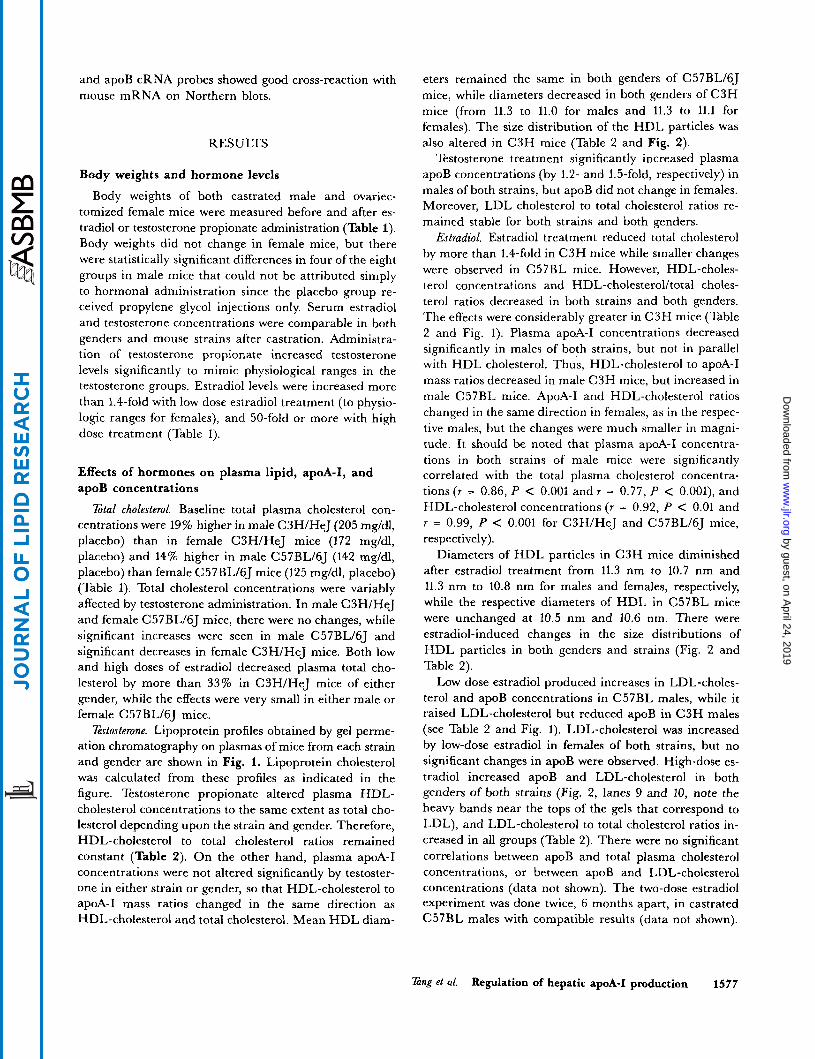
Estosterone. Lipoprotein profiles obtained by gel perme- ation chromatography on plasmas of mice from each strain and gender are shown in Fig. 1. Lipoprotein cholesterol was calculated from these profiles as indicated in the figure. Testosterone propionate altered plasma HDL- cholesterol concentrations to the same extent as total cho- lesterol depending upon the strain and gender. Therefore, HDL-cholesterol to total cholesterol ratios remained constant (Table 2). On the other hand, plasma apoA-I concentrations were not altered significantly by testoster- one in either strain or gender, so that HDL-cholesterol to apoA-I mass ratios changed in the same direction as HDL-cholesterol and total cholesterol. Mean HDL diam-
eters remained the same in both genders of C57BL/6J mice, while diameters decreased in both genders of C3H mice (from 11.3 to 11.0 for males and 11.3 to 11.1 for females). The size distribution of the HDL particles was also altered in C3H mice (Table 2 and Fig. 2).
Testosterone treatment significantly increased plasma apoB concentrations (by 1.2- and 1.5-fold, respectively) in males of both strains, but apoB did not change in females. Moreover, LDL cholesterol to total cholesterol ratios re- mained stable for both strains and both genders.
Estradiol. Estradiol treatment reduced total cholesterol by more than 1.4-fold in C3H mice while smaller changes were observed in C57BL mice. However, HDL-choles- terol concentrations and HDL-cholesterol/total choles- terol ratios decreased in both strains and both genders. The effects were considerably greater in C3H mice (Table 2 and Fig. 1). Plasma apoA-I concentrations decreased significantly in males of both strains, but not in parallel with HDL cholesterol. Thus, HDL-cholesterol to apoA-I mass ratios decreased in male C3H mice, but increased in male C57BL mice. ApoA-I and HDL-cholesterol ratios changed in the same direction in females, as in the respec- tive males, but the changes were much smaller in magni- tude. It should be noted that plasma apoA-I concentra- tions in both strains of male mice were significantly correlated with the total plasma cholesterol concentra- tions(r = 0.86, P < 0.001 andr = 0.77, P < O.OOl), and HDL-cholesterol concentrations (r = 0.92, P < 0.01 and r = 0.99, P < 0.001 for C3H/HeJ and C57BLhJ mice, respectively).
Diameters of HDL particles in C3H mice diminished after estradiol treatment from 11.3 nm to 10.7 nm and 11.3 nm to 10.8 nm for males and females, respectively, while the respective diameters of HDL in C57BL mice were unchanged at 10.5 nm and 10.6 nm. There were estradiol-induced changes in the size distributions of HDL particles in both genders and strains (Fig. 2 and Table 2).
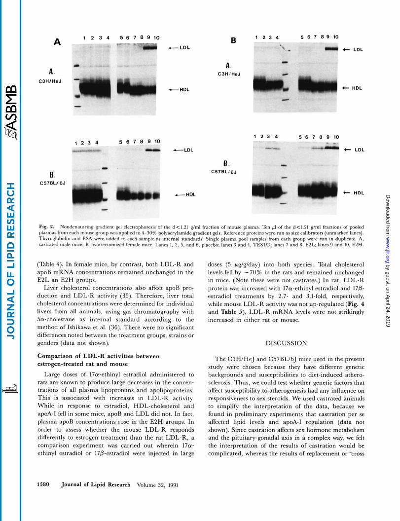
Low dose estradiol produced increases in LDL-choles- terol and apoB concentrations in C57BL males, while it raised LDL-cholesterol but reduced apoB in C3H males (see Table 2 and Fig. 1). LDL-cholesterol was increased by low-dose estradiol in females of both strains, but no significant changes in apoB were observed. High-dose es- tradiol increased apoB and LDL-cholesterol in both genders of both strains (Fig. 2, lanes 9 and 10, note the heavy bands near the tops of the gels that correspond to LDL), and LDL-cholesterol to total cholesterol ratios in- creased in all groups (Table 2). There were no significant correlations between apoB and total plasma cholesterol concentrations, or between apoB and LDL-cholesterol concentrations (data not shown). The two-dose estradiol experiment was done twice, 6 months apart, in castrated C57BL males with compatible results (data not shown).
Zing et al. Regulation of hepatic apoA-I production 1577
by guest, on April 24, 2019
ww
w.jlr.org
Dow
nloaded from
A. CW/M.J
e. CSlBLI6J
A. CW/U.J
II t plubo - I 40 I
I I h
40i d I 0 P .
m a t 801 E2L
i 401
! ““t ’7
t
40 ‘T R I 2:mw 0 5 10 15 20 25 30 35 40 4 5 5 0 5 10 15 20 23 30 35 4 0 I 5 50
../?niiJ:Pu ‘ 0 5 IO 15 20 25 30 35 40 4 5 50 5 10 15 20 25 30 35 40 4 5 50
FRACflON(o.8 mi)
Fig. 1. Elution profiles of lipoproteins by gel permeation chromatography on Superose columns (FPLC system, Pharmacia); males, left and females, right. Pooled plasmas from each experimental group were applied to two Superose 6 columns attached in series as described in Methods. Lipoproteins were eluted at 0.5 ml/min with 1 mmol/l EDTA, 154 mmol/l NaCl, and 0.02% NaN, (pH 8.2). Fractions (0.5 ml) were used analyzed for cholesterol and values represent the p g of cholesterol per fraction. Isolated human lipoproteins were used to calibrate the columns. Peaks of human LDL and HDL eluted between fractions 25-30 and 42-46, respectively. Profiles of plasma cholesterol from untreated non-castrated female mice fed Purina Chow 5105 diet are shown by the dashed line in (A) for comparison (see reference 18). To calculate LDL- and HDL-cholesterol concentrations in mouse plasma, the cholesterol contents in fractions 16 through 32 and 33 through 50 were summed, respectively (see Table 2).
Hepatic apoA-I synthesis and its regulation although it is possible that values for the absolute amount of apoA-I synthesized per mg tissue may be different than the values expressed as a percent of total protein synthe- sis, changes in apoA-I synthesis after hormone treatment relative to the placebo controls wouId still hold.) While hepatic apoA-I mRNA concentrations remained unchanged or decreased slightly, apoA-I transcription rates were significantly increased by 21-75% (except in female C57BL/6J mice). Thus, testosterone influences both tran-
Estostemne. Hepatic apoA-I synthesis was increased 1.4-fold or more by testosterone treatment. This was true in both strains and both genders (Table 3 and Fig. 3). (It should be noted that although synthesis rates for apoA-I were expressed as a percent of the total TCA-precipitable amino acid radioactivity, similar amounts (-40 mg) of liver tissue were used for each determination. Therefore,
1578 Journal of Lipid Research Volume 32, 1991
by guest, on April 24, 2019
ww
w.jlr.org
Dow
nloaded from
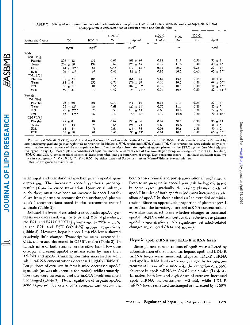
TABLE 2. Effects of testosterone and estradiol administration on plasma HDL- and LDL-cholesterol and apolipoprotein A-I and apolipoprotein B concentrations of castrated male and female mice
HDL-C" LDL-C' __ HDL - HDL-C4 Strains and GrouDs TC HDL-C TC ApoA-I ApoA-I Dia. TC ApoB
mg/dl mg/dl mg/dl nm mg/dl
Male C3H/HeJ
Placebo Testo E2L E2H
C57BL16J Placebo Testo E2L E2H
Female C3H/HeJ
Placebo Testo E2L E2H
C 5 7BL/6J Placebo Testo E2L E2H
205 i 22 208 _+ 10 113 * 12" 109 f 13.'
142 f 14 184 f 6* 157 f 11 149 * 37
0.66 0.67 0.45 0.49
161 f 16 175 f 15 112 i 13'* 82 f 7
0.84 0.79 0.46 0.65
11.3 11.0 10.7 10.7
0.30 33 f 2 0.30 39 f 4* 0.34 22 f 4' 0.40 65 f 7"
135 139 51 53
105 132 84 70
0.74 0.72 0.54 0.47
164 f 13 174 f 18 107 f 9'* 95 f 21'*
0.64 0.76 0.79 0.74
10.5 10.5 10.5 10.5
0.24 30 ~t 2 0.26 44 f 5.' 0.38 42 f 4" 0.50 82 + 14**
172 f 28 125 t 12" 129 f 12" 125 f 17''
125 i 8 116 f 13 111 f 4' 137 f 10
121 84 75 57
0.70 0.68 0.58 0.46
141 f 14 120 f 11' 119 + 13* 79 f 6"
136 34 124 f 25 134 f 34 95 f 13'
0.86 0.70 0.63 0.72
11.3 11.1 10.8 10.8
0.28 22 f 3 0.28 25 f 7 0.34 27 f 6 0.50 72 f 8''
0.30 36 f 13 0.28 31 f 5 0.33 30 i 2 0.47 65 f 5**
84 74 71 61
0.69 0.64 0.64 0.44
0.62 0.60 0.53 0.64
10.6 10.6 10.6 10.6
Plasma total cholesterol (TC), apoA-I, and apoB concentrations were determined as described in Methods. HDL diameters were determined by non-denaturing gradient gel electrophoresis as described in Methods. HDL-cholesterol (HDL-C) and LDL-C concentrations were calculated by sum- ming the cholesterol contents of the appropriate column fractions after chromatography of mouse plasma on the FPLC system (see Methods and the legend to Fig. 1). Pools of plasma consisting of equal volumes derived from each of five animals per group were subjected to FPLC. Therefore, WDL-C and LDL-C concentrations consist of single determinations per experimental group. Data represent means + standard deviations from five mice in each group; *, P C 0.05; * * , P < 0.001 by either unpaired Student's t-test or Mann-Whitney two-sample test.
"Results are given as mass ratios.
scriptional and translational mechanisms in apoA-I gene expression. The increased apoA-I synthesis probably resulted from increased translation. However, simultane- ously there must have been an increase in apoA-I catab- olism from plasma to account for the unchanged plasma apoA-I concentrations noted in the testosterone-treated animals (Table 2).
Estradiol. In livers of estradiol-treated males apoA-I syn- thesis was decreased, e.g., to 36% and 11% of placebo in the E2L and E2H C3HlHeJ groups and to 29% and 7 % in the E2L and E2H C57BL/6J groups, respectively (Table 3). However, hepatic apoA-I mRNA levels showed relatively little change. Transcription rates increased in C3H males and decreased in C57BL males (Table 3). In female mice of both strains, on the other hand, low dose estrogen increased apoA-I synthesis rates by more than 1.9-fold and apoA-I transcription rates increased as well, while mRNA concentrations decreased slightly (Table 3). Large doses of estrogen in female mice decreased apoA-I synthesis (as was also seen in the males), while transcrip- tion rates were increased and the mRNA levels remained unchanged (Table 3). Thus, regulation of hepatic apoA-I gene expression by estradiol is complex and occurs via
both transcriptional and post-transcriptional mechanisms. Despite an increase in apoA-I synthesis by hepatic tissue in some cases, gradually decreasing plasma levels of apoA-I in mice of both genders indicates increased catab- olism of apoA-I in these animals after estradiol adminis- tration. Since an appreciable proportion of plasma apoA-I arises from the intestine, intestinal mRNA concentrations were also measured to see whether changes in intestinal apoA-I mRNA could account for the reductions in plasma apoA-I concentrations. No significant estradiol-related changes were noted (data not shown).
Hepatic apoB mRNA and LDL-R mRNA levels
Since plasma concentrations of apoB were affected by administration of the hormones, hepatic apoB and LDL-R mRNA levels were measured. Hepatic LDL-R mRNA and apoB mRNA levels were not changed by testosterone treatment in any of the mice with the exception of a 36% decrease in apoB mRNA in C57BL male mice (Table 4). In males, both low and high doses of estrogen increased apoB mRNA concentrations - 2-fold, while LDL-R mRNA levels remained unchanged or increased by < 50%
TanE at al. Regulation of hepatic apoA-I production 1579
by guest, on April 24, 2019
ww
w.jlr.org
Dow
nloaded from
1 2 3 4 A
A. C3HIHeJ
I,
H. C57BL/6J -
5 6 7 8 9 1 0
-*y -LDL
1 2 3 4 B
A. C3H/HeJ
-HDL
5 6 7 0 9 1 0
n -LDL
B . c57BL/6J
c
1 2 3 4
b. -HDL
5 6 7 8 9 10
5 6 7 8 9 1 0
- 4 M - -
4- LDL
4- HDL
t LDL
4- HDL
Fig. 2. Nondenaturing gradient gel electrophoresis of the d < 1.21 glml fraction of mouse plasma. Ten pI of the d < 1.21 glml fractions of pooled plasmas from each mouse group was applied to 4-30% polyacrylamide gradient gels. Reference proteins were run as size calibrators (unmarked lanes). Thyroglobulin and BSA were added IO each sample as internal standards. Single plasma pool samples from each group were run in duplicate. A. castrated male mice; R. ovariectomized female mice. Lanes 1, 2, 5 . and 6, placebo; lanes 3 and 4, TESTO; lanes 7 and 8, E2L; lanes 9 and 10, E2H.
(Table 4). In female mice, by contrast, both LDL-R and apoB mRNA concentrations remained unchanged in the E2L an E2H groups.
Liver cholesterol concentrations also affect apoB pro- duction and LDL-R activity (35). Therefore, liver total cholesterol concentrations were determined for individual livers from all animals, using gas chromatography with Sa-cholestane as internal standard according to the method of Ishikawa et al. (36). There were no significant differences noted between the treatment groups, strains or genders (data not shown).
Comparison of LDL-R activities between estrogen-treated rat and mouse
Large doses of l7a-ethinyl estradiol administered to rats are known to produce large decreases in the concen- trations of all plasma lipoproteins and apolipoproteins. This is associated with increases in LDL-R activity. While in response to estradiol, HDL-cholesterol and apoA-I fell in some mice, apoB and LDL did not. In fact, plasma apoB concentrations rose in the E2H groups. In order to assess whether the mouse LDL-R responds differently to estrogen treatment than the rat LDL-R, a comparison experiment was carried out wherein 17a- ethinyl estradiol or 170-estradiol were injected in large
doses (5 pg/g/day) into both species. Total cholesterol levels fell by - 70% in the rats and remained unchanged in mice. (Note these were not castrates.) In rat, LDL-R protein was increased with 17a-ethinyl estradiol and 170- estradiol treatments by 2.7- and 3.1-fold, respectively, while mouse LDL-R activity was not up-regulated (Fig. 4 and Table 5). LDL-R mRNA levels were not strikingly increased in either rat or mouse.
DISCUSSION
The C3H/HeJ and C57BL/6J mice used in the present study were chosen because they have different genetic backgrounds and susceptibilities to diet-induced athero- sclerosis. Thus, we could test whether genetic factors that affect susceptibility to atherogenesis had any influence on responsiveness to sex steroids. We used castrated animals to simplify the interpretation of the data, because we found in preliminary experiments that castration per se affected lipid levels and apoA-I regulation (data not shown). Since castration affects sex hormone metabolism and the pituitary-gonadal axis in a complex way, we felt the interpretation of the results of castration would be complicated, whereas the results of replacement or “cross
1580 Journal of Lipid Research Volume 32, 1991
by guest, on April 24, 2019
ww
w.jlr.org
Dow
nloaded from
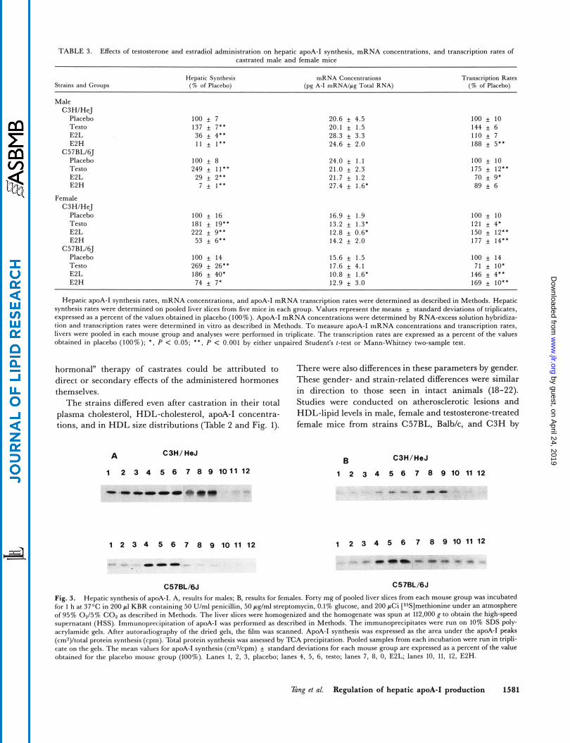
TABLE 3. EKects of testosterone and estradiol administration on hepatic apoA-I synthesis, mRNA concentrations, and transcription rates of castrated male and female mice
Hepatic Synthesis mRNA Concentrations Transcription Rates Strains and Groups (7% of Placebo) (pg A-I mRNA/pg Total RNA) (% of Placebo)
Male C3HIHeJ
Placebo 100 f 7 20.6 f 4.5 100 f 10
E2 L 36 f 4" 28.3 + 3.3 110 f 7 Testo 137 f 7'. 20.1 f 1.5 144 f 6
E2 H C 5 7RLI6J
Placebo Testo E2L E2H
Female C3HIHeJ
Placebo Testo E2L E2 H
C57RW6J Placebo Testo E2L E2H
11 f 1..
100 + 8 249 f 11.. 29 f 2..
7 f 1'.
100 f 16 181 f 19.' 222 f 9..
53 f 6"
100 f 14 269 f 26" 186 f 40. 74 f 7'
24.6 f 2.0
24.0 f 1.1 21.0 f 2.3 21.7 f 1.2 27.4 f 1.6'
16.9 f 1.9 13.2 f 1.3. 12.8 f 0.6. 14.2 f 2.0
15.6 f 1.5 17.6 f 4.1 10.8 f 1.6. 12.9 f 3.0
188 f 5"
100 f 10 175 f 12.. 70 f 9' 89 f 6
100 f 10 121 f 4. 150 f 12.. 177 f 14''
100 f 14 71 f 10'
146 f 4.. 169 f 10'.
~~
Hepatic apoA-I synthesis rates, mRNA concentrations, and apoA-I mRNA transcription rates were determined as described in Methods. Hepatic synthesis rates were determined on pooled liver slices from five mice in each group. Values represent the means f standard deviations of triplicates, expressed as a percent of the values obtained in placebo (100%). ApoA-I mRNA concentrations were determined by RNA-excess solution hybridiza- tion and transcription rates were determined in vitro as described in Methods. To measure apoA-I mRNA concentrations and transcription rates, livers were pooled in each mouse group and analyses were performed in triplicate. The transcription rates are expressed as a percent of the values obtained in placebo (100%); *. P < 0.05; **, P < 0.001 by either unpaired Student's t-test or Mann-Whitney two-sample test.
hormonal" therapy of castrates could be attributed to direct or secondary effects of the administered hormones them selves.
The strains differed even after castration in their total plasma cholesterol, HDL-cholesterol, apoA-I concentra- tions, and in HDL size distributions (Table 2 and Fig. 1).
A C3H/ HeJ
1 2 3 4 5 6 7 8 9 101112
1 2 3 4 5 6 7 8 9 1 0 1 1 1 2
. _ - 0 0 ...
C57BL/6J
There were also differences in these parameters by gender. These gender- and strain-related differences were similar in direction to those seen in intact animals (18-22). Studies were conducted on atherosclerotic lesions and HDL-lipid levels in male, female and testosterone-treated female mice from strains C57BL. Balblc, and C3H by
C3H/HeJ
1 2 3 4 5 6 7 8 9 1 0 1 1 1 2
B
C5?BL/6J
Fig. 3. Hepatic synthesis of apoA-I. A, results for males; B, results for females. Forty mg of pooled liver slices from each mouse group was incubated for 1 h at 37OC in 200 pI KBR containing 50 Ulml penicillin, 50 pglml streptomycin, 0.1% glucose, and 200 pCi ['5S]methionine under an atmosphere of 95% OZ/5% C 0 2 as described in Methods. The liver slices were homogenized and the homogenate was spun at 112,000 g to obtain the high-speed supernatant (HSS). Immunoprecipitation of apoA-I was performed as described in Methods. The immunoprecipitates were run on 10% SDS poly- acrylamide gels. After autoradiography of the dried gels, the film was scanned. ApoA-I synthesis was expressed as the area under the apA-I peaks (cmz)/total protein synthesis (cpm). Total protein synthesis was assessed by E A precipitation. Pooled samples from each incubation were N n in tripli- cate on the gels. The mean values for apoA-1 synthesis (cmzkpm) f standard deviations for each mouse group are expressed as a percent of the value obtained for the placebo mouse group (100%). Lanes 1, 2, 3, placebo; lanes 4, 5, 6, testo; lanes 7, 8. 0, E2L; lanes 10. 11, 12, E2H.
Tang et al. Regulation of hepatic apoA-I production 1581
by guest, on April 24, 2019
ww
w.jlr.org
Dow
nloaded from
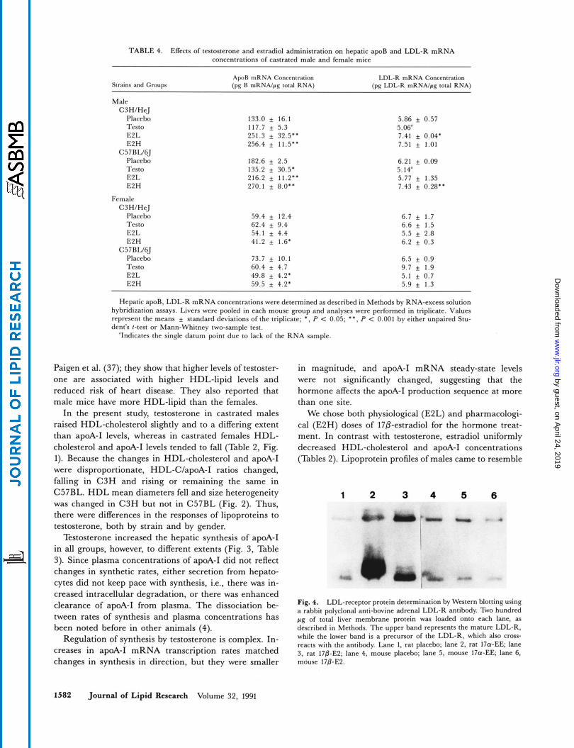
TABLE 4. Effects of testosterone and estradiol administration on hepatic apoB and LDL-R mRNA concentrations of castrated male and female mice
Strains and Groups ApoR mRNA Conccntration (pg H mRNA/pp; total RNA)
LDL-R mRNA Conccntration (pg LDL-R mRNA/pg total RNA)
Male C3HIHeJ
Placebo Testo E2 L E2 H
C57 RLI6J Placebo Testo E2 L E2H
Female C3HIHeJ
Placebo Tesro E2 L E2 H
C5 7 RLI6J Placebo Testo E2 L E2 H
133.0 f 16.1 117.7 f 5.3 251.3 f 32.5.. 256.4 f 11.5'.
182.6 f 2.5 135.2 30.5. 216.2 f 11.2.. 270.1 f 8.0..
59.4 f 12.4 62.4 f 9.4 54.1 f 4.4 41.2 f 1.6.
73.7 f 10.1 60.4 f 4.7 49.8 f 4.2. 59.5 f 4.2.
5.86 + 0.57 5.06" 7.41 f 0.04. 7.51 f 1.01
6.21 f 0.09 5.14' 5.77 f 1.35 7.43 f 0.28..
6.7 f 1.7 6.6 f 1.5 5.5 f 2.8 6.2 + 0.3
6.5 f 0.9 9.7 f 1.9 5.1 f 0.7 5.9 f 1.3
Hepatic apoB, LDL-R mRNA concentrations were determined as described in Methods by RNA-excess solution hybridization assays. Livers were pooled in each mouse group and analyses were performed in triplicate. Values represent the means standard deviations of the triplicate; *, P < 0.05; **. P < 0.001 by either unpaired Stu- dent's t-test or Mann-Whitney two-sample test.
"Indicates the single datum point due to lack of the RNA sample.
Paigen et al. (37); they show that higher levels of testoster- one are associated with higher HDL-lipid levels and reduced risk of heart disease. They also reported that male mice have more HDL-lipid than the females.
In the present study, testosterone in castrated males raised HDL-cholesterol slightly and to a differing extent than apoA-I levels, whereas in castrated females HDL- cholesterol and apoA-I levels tended to fall (Table 2, Fig. 1). Because the changes in HDL-cholesterol and apoA-I were disproportionate, HDL-C/apoA-I ratios changed, falling in C3H and rising or remaining the same in C57BL. HDL mean diameters fell and size heterogeneity was changed in C3H but not in C57BL (Fig. 2). Thus, there were differences in the responses of lipoproteins to testosterone, both by strain and by gender.
Testosterone increased the hepatic synthesis of apoA-I in all groups, however, to different extents (Fig. 3, Table 3). Since plasma concentrations of apoA-I did not reflect changes in synthetic rates, either secretion from hepato- cytes did not keep pace with synthesis, i.e., there was in- creased intracellular degradation, or there was enhanced clearance of apoA-I from plasma. The dissociation be- tween rates of synthesis and plasma concentrations has been noted before in other animals (4).
Regulation of synthesis by testosterone is complex. In- creases in apoA-I mRNA transcription rates matched changes in synthesis in direction, but they were smaller
in magnitude, and apoA-I mRNA steady-state levels were not significantly changed, suggesting that the hormone affects the apoA-I production sequence at more than one site.
We chose both physiological (E2L) and pharmacologi- cal (E2H) doses of 17P-estradiol for the hormone treat- ment. In contrast with testosterone, estradiol uniformly decreased HDL-cholesterol and apoA-I concentrations (Tables 2). Lipoprotein profiles of males came to resemble
1 2 3 4 5 6
Fig. 4. LDL-receptor protein determination by Western blotting using a rabbit polyclonal anti-bovine adrenal LDL-R antibody. Two hundred pg of total liver membrane protein was loaded onto each lane, as described in Methods. The upper band represents the mature LDL-R, while the lower band is a precursor of the LDL-R, which also cross- reacts with the antibody. Lane 1, rat placebo; lane 2, rat 17a-EE; lane 3, rat 17&E2; lane 4, mouse placebo; lane 5, mouse I7a-EE lane 6, mouse 170-E2.
1582 Journal of Lipid Research Volume 32, 1991
by guest, on April 24, 2019
ww
w.jlr.org
Dow
nloaded from
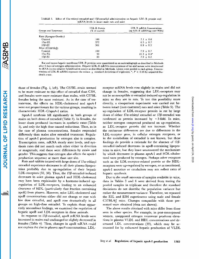
TABLE 5 . Effect of 17a-ethinyl estradiol and 170-estradiol administration on hepatic LDL-R protein and mRNA levels in intact male rats and mice
LDL-R Protein LDL-R mRNA Concentrations (% of control) Groups and Treatments
Rats (Sprague-Dawley)
(pg LDL-R mRNAlpg total RNA)
Control 100 7 . 1 f 0.6 1 7a-EE 272 6 .5 * 0.7 170-E2 30 1 6.9 + 0.5
Control 100 7.9 + 0.7 17a-EE 93 11.3 5 0.4'
Mice (C3H/HeJ)
170-E2 83 7.9 f 0.2
Rat and mouse hepatic membrane LDL-R proteins were quantitated on an autoradiograph as described in Methods after 6 days of estrogen administration. Hepatic LDL-R mRNA concentrations of rat and mouse were determined by RNA-excess solution hybridization assays as described in Methods. Livers were pooled in each group. Concen- trations of LDL-R mRNA represent the means standard deviations of triplicates; *, P < 0.05 by unpaired Stu- dent's t-test
those of females (Fig. 1, left). The C57BL strain seemed to be more resistant to this effect of estradiol than C3H, and females were more resistant than males, with C57BL females being the most resistant. As in the case of tes- tosterone, the effects on HDL-cholesterol and apoA-I were not proportionate for the various groups, resulting in characteristic HDL-C/apoA-I ratios.
ApoA-I synthesis fell significantly in both groups of males on both doses of estradiol (Table 3). In females, the lower doses produced increases in synthetic rates (Table l), and only the high dose caused reductions. Thus, as in the case of plasma concentrations, females responded differently than males after estradiol treatment. Regula- tion of apoA-I production by estrogen also is complex. Transcription rates, mRNA steady state levels, and syn- thesis rates did not match each other either in direction or magnitude, and there were differences by strain and gender. This suggests that estrogen also affects the apoA-I production sequence at mure than one site.
Rats and rabbits treated with large doses of 17a-ethinyl estradiol experience decreases in all their plasma lipopro- teins probably due to up-regulation of their hepatic LDL-receptors (32, 38). Thus, the 17@-estradiol-induced decreases in mice plasma apoA-I and HDL-cholesterol may have been explainable by a hormone-induced up- regulation of LDL-receptors, leading to an enhanced clearance of HDL (particularly that fraction containing apoE) from plasma. However, mice LDL-cholesterol and apoB concentrations either did not change or rose with low dose estradiol, and apoB rose dramatically in all groups on high-dose estradiol. To explain these appar- ently anomalous findings, we examined the regulation of hepatic apoB and LDL-receptors in these animals.
In response to 17P-estradio1, apoB mRNA levels were increased in males and unchanged or slightly decreased in females (Table 4). Thus, changes in apoB mRNA could not explain the rise in plasma apoB concentrations. LDL-
receptor mRNA levels rose slightly in males and did not change in females, suggesting that LDL-receptors may not be as susceptible to estradiol-induced up-regulation in mice as they are in rats. To test this possibility more directly, a comparison experiment was carried out be- tween intact (non-castrated) rats and mice (Table 5). The up-regulation of LDL-receptor protein in rat by large doses of either 17a-ethinyl estradiol or 17P-estradiol was confirmed as protein increased by -3-fold. In mice, neither estrogen compound produced an up-regulation, as LDL-receptor protein did not increase. Whether the ratlmouse differences are due to differences in the LDL-receptor gene, in cellular estrogen receptors, or in the metabolism of estradiol is not known, but these findings do provide a rationale for the absence of 170- estradiol-induced decreases in apoB-containing lipopro- teins in mice, but they leave unanswered the mechanism by which decreases in plasma apoA-I and HDL-choles- terol were produced by estrogen. Perhaps other receptors such as the LDL-receptor-related protein or the HDL- receptors were up-regulated by estrogen, or as mentioned, apoA-I secretion or catabolism may not reflect rates of hepatic synthesis.
Due to the small amounts of samples available in mice, data in Tables 3 and 4 were derived from testing the pooled samples in triplicate and therefore the standard deviations do not describe the population variance but rather the measurement variance. Therefore, we repeated the E2L and E2H experiments using male and female C57BL/6J mice. Changes compatible with those pre- sented were obtained (data not shown).
The above results obtained with mice differ from those seen in other species. For example, in post-menopausal women, unopposed estrogen treatment produces eleva- tions in plasma VLDL and HDL concentrations and de- creased LDL concentrations (39), which may be ac- counted for by enhanced hepatic production of VLDL
Tang et al. Regulation of hepatic apoA-I production 1583
by guest, on April 24, 2019
ww
w.jlr.org
Dow
nloaded from
and HDL. In addition, estradiol produces marked reduc- tions in the activity of HTGL (hepatic triglyceride lipase) with little or no change in LPL (lipoprotein lipase) ac- tivity (39). Since HTGL possesses phospholipase activity that catalyzes the hydrolysis of HDL-phospholipids which in turn destabilizes HDL and promotes its catabolism, estrogen-induced inhibition of HTGL activity in humans may stabilize HDL particles a n d also contribute to the rise in plasma HDL concentrations. Reduced LDL con- centrations a re attributed to estrogen-induced increases in hepatic LDL-receptors (40), and this has been well documented in rabbits a n d rats treated with large doses of ethinyl estradiol as mentioned above. Androgens, in general, produce effects opposite to those produced by es- trogens, but there is less available information o n andro- gen effects. T h e effects of sex hormones in mice on the lipase enzymes remains to be determined.
Rats respond differently to estradiol treatment than d o humans. For example, in contrast with humans, replace- ment doses of estrogens given to castrated female rats result in reductions in total triglyceride, cholesterol, and apoA-I, while HDL concentrations are unaffected (41). In castrated male rats, estrogens in doses meant to mimic female plasma concentrations of estradiol produce de- creases in VLDL a n d increases in HDL concentration (41). Supraphysiologic doses of 17a-ethinyl estradiol drastically lower plasma concentrations of all lipoprotein classes probably due to up-regulation of the LDL- receptor. Clearly, the lipoprotein responses of mice to sex steroid administration appear to differ from both humans and rats as is seen in this report. Whether these species differences are d u e to differences in the metabolism of sex hormones or the regulation of the genes in question re- mains to be elucidated. I
The authors acknowledge the generous gifts of antisera against bovine adrenal LDL-receptor produced by Dr. Janet Boyles, Gladstone Foundation Laboratories; rat apoA-I cDNA probe from Dr. J. Gordon, Washington University, St. Louis, MO; rat LDL-R cDNA probe from Dr. A. D. Cooper, Palo Alto Medical Foundation, Palo Alto, CA; and rat apoB cDNA probe from Dr. A. J. Lusis, University of California, Los Angeles, CA. This work was supported by NIH Grant #PO1 DK33487-01A1. Rai Ajit K. Srivastava was supported by a Fellowship from the Missouri Affiliate of the American Heart Association. Manmcnpt received 14 January 1991 and i n reuisedform # ,Ju ly 1991
REFERENCES
1. Miller, G. J., and N. E. Miller. 1975. Plasma-high-density- lipoprotein concentration and development of ischaemic heart-disease. Lancet. 1: 16-19.
2. Gordon, T., W. P. Castelli, M. C. Hjortland, W. B. Kannel, and T. R. Dawber. 1977. High density lipoprotein as a pro- tective factor against coronary heart disease. Am. J Med.
Haddad, I. A,, J. M . Ordovas, T. Fitzpatrick, and S. K. 62: 707-714.
3.
4.
5.
6.
7.
8.
9.
10.
11.
12.
13.
14.
15.
16.
17.
18.
Karathanasis. 1986. Linkage, evolution and expression of the rat apolipoprotein A-I, C-111 and A-IV genes. J Biol. Chem. 261: 13268-13277. Staels, B., J. Auwerx, L. Chan, A. van Tol, M. Rosseneu, and G. Verhoeven. 1989. Influence of development, estro- gens, and food intake on apolipoprotein A-I, A-11, and E mRNA in rat liver and intestine. J Lipid Res. 30: 1137- 1145. Hazzard, W. R., J. D. Brunzell, D. M. Applebaum, A. E'. Goldberg, C. Gagne, J. J. Albers, P. W. Wahl, and J. J. Hoover. 1977. Steroid contraceptives and human lipopro- tein metabolism: effects and mechanisms. In Pharmacology of Steroid Contraceptive Drugs. S. Garattini and H. W. Berendes, editors. Raven Press, New York. 251-266. Kim, H-J., and R. K. Kalkhoff. 1978. Altered apolipopro- teins in sex steroid-treated rats. Metabolism. 27: 571-587. Kushwaha, R. S., and W. R. Hazzard. 1981. Effect of ex- ogenous estrogens on catabolism of VLDL in cholesterol- fed rabbits. Am. J Physiol. 241: E372-E377. Luskey, K. L., M. S. Brown, and J. L. Goldstein. 1974. Stimulation of the synthesis of very low density lipoproteins in rooster liver by estradiol. J Bid. Chem. 249: 5939-5947. Wilcox, H. G., R. Kenagy, I. Weinstein, and M. Heim- berg. 1981. Alterations of plasma HDL lipids and apo- lipoproteins in female rats treated with ethinyl estradiol. Bi- ochim. Biophys. Acta. 666: 348-355. Weinstein, I., H. G. Wilcox, and M. Heimberg. 1986. Effects of high-dose ethinyl estradiol on serum concentra- tions and hepatic secretion of the very-low-density lipopro- tcin, triacylglycerol, cholesterol, and apolipoprotein A-I in the rat. Biochim. Biophys. Acta. 876: 450-459. Alexander, J. J., M. Hoenig, D. Graham, and A. Imbembo. 1989. Effect of estradiol on low density lipoprotein uptake by bovine aortic endothelial cel1s.J Sur6 Res. 46: 537-542. Henriksson, P., M. Stamberger, M. Eriksson, M. Rudling, U. Diczfdusy, L. Berglund, and B. Angelin. 1989. Oestrogen- induced changes in lipoprotein metabolism: role in preven- tion of atherosclerosis in the cholesterol-fed rabbit. Eur. J Clin. Invest. 19: 395-403. Eriksson, M., L. Berglund, M. Rudling, P. Henriksson, and B. Angelin. 1989. Effects of estrogen on low density lipoprotein metabolism in males. J Clin. Invest. 84: 802- 810. Chan, L., R . L. Jackson, and A. R. Means. 1978. Regula- tion of lipoprotein synthesis. Studies on the molecular mechanisms of lipoprotein synthesis and their regulation by estrogen in the cockerel. Circ. Res. 43: 209-217. Lin, C-T., and L. Chan. 1980. Effects of estrogen on specific protein synthesis in the cockerel liver: an immunocyto- chemical study on major apoproteins in very low density and high density lipoproteins and albumin. Endocrinolou.
Schaefer, E. J., D. M. Foster, L. A. Zech, E T. Lindgren. H. B. Brewer, Jr,, and R. I. Levy. 1983. The effects of estro- gen administration on plasma lipoprotein metabolism in premenopausal females. J Clin. Endocrinol. Metab. 57:
Hazzard, W. R., S. M. Haffner, R. S. Kushwaha, B. D. Applebaum, and D. M. Foster. 1984. Preliminary report: kinetic studies on the modulation of high-density lipo- protein, apolipoprotein, and subfraction metabolism by sex steroids in a postmenopausal woman. Metabolism. 33: 779- 784. Jiao, S., T. G. Cole, R. T. Kitchens, B. Pfleger, and G. Schonfeld. 1990. Genetic heterogeneity of lipoproteins in inbred strains of mice: analysis by gel permeation chro- matography. Metabolism. 39: 155-160.
107: 70-75.
262-267.
1584 Journal of Lipid Research Volume 32, 1991
by guest, on April 24, 2019
ww
w.jlr.org
Dow
nloaded from
19. Paigen, B., B. Y. Ishida, J. Verstuyft, R. B. Winters, and D. Albee. 1990. Atherosclerosis susceptibility differences among progenitors of recombinant inbred strains of mice. Arteriosclerosis. 10: 316-3 23.
20. LeBoeuf, R. C., D. L. Puppione, V. N. Schumaker, and A. J. Lusis. 1983. Genetic control of lipid transport in mice. I. Structural properties and polymorphisms of plasma lipo- proteins. J. Biol. Ghem. 258: 5063-5070. Lusis, A. J., B. A. Taylor, R. W. Wangenstein, and R. C. LeBoeuf. 1983. Genetic control of lipid transport in mice. 11. Genes controlling structure of high density lipoproteins. 1. Biol. Chem. 258: 5071-5078.
22. Jiao, S., T. G. Cole, R. T. Kitchens, B. Pfleger, and G. Schonfeld. 1990. Genetic heterogeneity of plasma lipo- proteins in the mouse: control of low density lipoprotein particle sizes by genetic factors. J. Lipid Res. 31: 467-477.
23. Laurel], C. B. 1966. Quantitative estimation of proteins by electrophoresis in agarose gel containing antibodies. Anal. Biochem. 15: 45-52.
24. LKB 2117 Multiphor I1 Electrophoresis System Laboratory Manual. 1986. LKB Produkter AB, Bromma, Sweden.
25. Williams, D. L., and P. A. Dawson. 1986. Immunochemical measurement of apolipoprotein synthesis in cell and organ culture. Methodr Enzymol. 128: 254-271.
26. Chirgwin, J. M., A. E. Przybla, R. J. McDonald, and W. J. Rutter. 1979. Isolation of biologically active ribonucleic acid from sources enriched in ribonuclease. Biochemisty. 18: 5294-5299.
27. Promega Guide. 1989/90 Protocols and Applications. 28. Azrolan, N., and J. L. Breslow. 1990. A solution hybridiza-
tionlRNase protection assay with riboprobes to determine absolute levels of apoB, A-I, and E mRNA in human hepa- toma cell lines. J. Lipid Res. 31: 1141-1146.
29. Groudine, M., M. Peretz, and H. Weintrauts. 1981. Tran- scriptional regulation of hemoglobin switching in chicken embryos. Mol. Cell. Bid . 1: 281-288.
30. Schibler, U., 0. Hagenbuchle, P. K. Wellauer, and A. C. Pittet. 1983. Two promoters of different strengths control the transcription of the mouse alpha-amylase gene Amy-]” in the parotid gland and the liver. Cell. 33: 501-508. Chazenbalk, G. D., M . L. Wadsworth, and B. Rapoport. 1990. Transcriptional regulation of ferritin H messenger RNA levels in FRTL5 rat thyroid cells by thyrotropin. J Biol. Chem. 265: 666-670.
21.
31.
32.
33.
34.
35.
36.
37.
38.
39.
40.
41.
Kovanen, P. T., M. S. Brown, and J. L. Goldstein. 1979. In- creased binding of low density lipoprotein to liver mem- brane from rats treated with 17a-ethinyl estradiol. J Biol. Chem. 254: 11367-11373. Sata, T., R. J. Have], and A. L. Jones. 1972. Characteriza- tion of subfractions of triglyceride-rich lipoproteins sepa- rated by gel chromatography from blood plasma of normo- lipidemic and hyperlipidemic humans. J Lipid Res. 13:
Towbin, H., T. Staehelin, and J. Gordon. 1979. Electro- phoretic transfer of proteins from polyacrylamide gels to nitrocellulose: procedures and some applications. Proc. Natl. Acad. Sci. USA. 76: 4350-4354. Fuki, I. V., S. N. Preobrazhensky, A. Y. Misharin, N. G. Bushmakina, G. B. Menschikov, V. S. Repin, and R. S. Karpov. 1989. Effect of cell cholesterol content on apoB secretion and LDL receptor activity in human hepatoma cell line, HepG2. Biochim. Biophys. Acta. 1001: 235-238. Ishikawa, T. T., J. MacGee, A. Morrison, and C. J. Glueck. 1974. Quantitative analysis of cholesterol in 5 to 20 pl of plasma. J. Lipid Res. 15: 286-291. Paigen, B., P. A. Holmes, D. Mitchell, and D. Albee. 1987. Comparison of atherosclerotic lesions and HDL-lipid levels in male, female, and testosterone-treated female mice from strains C57BL/6J, Balb/c, and C3H. Atherosclerosis. 64:
Ma, P. T. S., T. Yamamoto, J. L. Goldstein, and M. S. Brown. 1986. Increased mRNA for low density lipoprotein receptor in livers of rabbits treated with 17a-ethinyl es- tradiol. Proc. Natl. Acad. Sci. USA. 83: 792-796. Applebaum-Bowden, D., P. McLean, A. Steinmetz, D. Fontana, C. Matthys, G. R. Warnick, M. Cheung, J. J. Al- bers, and W. R. Hazzard. 1989. Lipoprotein, apolipopro- tein, and lipolytic enzyme changes following estrogen ad- ministration in postmenopausal women. J. Lipid Res. 30:
Nanjee, M. N., D. R. Koritnik, J. Thomas, and N. E. Miller. 1990. Hormonal determinants of apolipoprotein B,E receptor expression in human liver. Positive association of receptor expression with plasma estrone concentration in middle-aged elderly women. Biochim. Biophys. Acta. 1046:
Patsch, W., K. Kim, W. Wiest, and G. Schonfeld. 1980. Effects of sex hormones on rat lipoproteins. Endocrinology.
757-768.
215-221.
1895-1906.
151-158.
107: 1085-1094.
Tang et al. Regulation of hepatic apoA-I production 1585
by guest, on April 24, 2019
ww
w.jlr.org
Dow
nloaded from