Embed Size (px)
Citation preview
American Journal of Environmental Science 9 (6): 470-482, 2013
ISSN: 1553-345X
©2013 Science Publication
doi:10.3844/ajessp.2013.470.482 Published Online 9 (6) 2013 (http://www.thescipub.com/ajes.toc)
Corresponding Author: Kantha Deivi Arunachalam, Center for Environmental Nuclear Research, Directorate of Research, SRM
University, Chennai, Tamil Nadu, 603203, India Tel: 044-27417144; Fax: 044-27417146
470 Science Publications
AJES
In-Vivo Evaluation of Hexavalent
Chromium Induced DNA Damage by Alkaline
Comet Assay and Oxidative Stress in Catla catla
Kantha Deivi Arunachalam,
Sathesh Kumar Annamalai and Jaya Krishna Kuruva
Center for Environmental Nuclear Research,
Directorate of Research, SRM University, Chennai, Tamil Nadu, 603203, India
Received 2013-10-25; Revised 2014-01-14; Accepted 2014-01-15
ABSTRACT
In the present study, the acute toxicity of Chromium in fingerlings of Catla catla, an Indian major carp, was evaluated with renewal bioassay method. In vivo studies were designed to assess the extent of Micronucleus Assay, Comet Assay under the exposure of common heavy-metal compounds, namely, Chromium Nitrate, using Catla catla (2n = 20), as a test model. The laboratory acclimatized fishes were divided into four groups. Group I served as positive control and the other three as exposed groups for three different time durations of 7, 14 and 21 days and were subjected to uninterrupted sub lethal concentrations (50% of 96 h LC50). The experiments were planned in such a way that fish from all the groups were sacrificed on the same day. The frequencies of micronuclei and bi-nuclei were evaluated comparatively in peripheral erythrocytes. As a result, it was observed that, the fishes and different tissues showed differential sensitivity to the heavy-metal treatment. A significant increase in the frequencies of micronucleated and binucleated cells and percentage increase in DNA tail (p<0.001) through Alkaline Comet Assay were observed after 21 days of exposure to chromium. Our results also showed decrease in enzyme Superoxide Dismutase (SOD) activity and increase in catalase enzyme due to increasing chromium concentration. The bio-concentration factor profiles of Chromium in Catla catla during sub lethal toxicity study was also calculated.
Keywords: Chromium, Micronucleus, Catla catla, Cytotoxicity, Comet Assay
1. INTRODUCTION
Aquatic ecosystem is the final sink for many chemicals used in industry and agriculture and has become a global problem (Adeogun and Chukwuka, 2012). When contaminants are released into the Aquatic Ecosystem, they finally get accumulated in the major aquatic organisms, almost invariably (Lavanya et al., 2011). Among all the contaminants, chromium is the one which are directly or indirectly released into aquatic ecosystem (Gheju, 2011). The influx of this global environmental toxicant into aquatic ecosystems from naturally occurring and anthropogenic sources is a serious problem throughout the world (Kumar et al., 2009). Chromium is a common contaminant in surface water and groundwater
because it is used widely in electroplating and other industries and occurs naturally at high concentrations in ultramafic rocks. Under oxidizing conditions, Cr is highly soluble and mobile as the Cr (VI) anions chromate (CrO4
2−) and bichromate (HCrO
4−) (Ellis et al., 2002). The
health effects and toxicity or carcinogenicity of chromium are mainly related to the oxidation state of the metal at exposure. Trivalent (Cr[III]) and Hexavalent (Cr[VI]) compounds are thought to be the most biologically significant, (Jomova and Valko, 2011).
Hexavalent chromium is widely used in many
industrial processes such as electroplating, wood
preservation. The remediation of chromium contaminated
sites poses several unique challenges. Ranipet regions of
Tamil Nadu, a Province in India, have Leather Tanneries
Kantha Deivi Arunachalam et al. / American Journal of Environmental Science 9 (6): 470-482, 2013
471 Science Publications
AJES
located in an industrial development area, from which
treated and untreated effluents are released into Palar
river. The granitic formation in the northern part of Palar
River catchment has the high infiltration rates and results
in fast migration of the contamination to the water table.
The Chromium levels in groundwater of these areas were
found up to 275 mg L−1
as reported by (Rao et al., 2011).
Taking the Paler River catchment into consideration to
access the genotoxic effects of major Indian carp Catla
catla is chosen for our study. Ingestion of large amounts
of chromium can lead to severe respiratory,cardiovascular,
gastrointestinal, hepatic and renal damage and potentially
death (Fatemi et al., 2013).
Many studies have directed their attention to asses the effect of chromium in the tannery and electrochemical works. For example, the genetic alterations in direct and indirect exposures of Hexavalent Chromium [Cr(VI)] in leather tanning industry workers have been reported by (Balachandar et al., 2010), while, the indirect effect has been studied by dietary uptake of chromium by fishes. Fish are at the top of the aquatic food chain and normal metabolism of fish may accumulate large amounts of certain metals from water, food, or sediment. However, like essential metals, nonessential metals are also taken up by fish and gets accumulated in their tissues (Yılmaz et al., 2010). As fish fauna serves as a food source for humans, it is essential to know the impact of water pollution on these organisms. Any change in the natural conditions of aquatic medium causes several physiological adjustments in fish (Garg et al., 2009). Fish has attracted much attention in the biomonitoring of water pollution because of its special biological characters such as relatively big body size, long life cycle, easy to raise. More importantly, fish species are at the top of the aquatic food chain and may directly affect the health of humans, which makes it more significance for the biomonitoring using fish. (Foster et al., 2012; Zhou et al., 2008).
Therefore, to provide data supporting the usefulness
of freshwater fish as indicators of heavy metal pollution,
it has been proposed in the present study, to examine the
bioaccumulation and genotoxic evaluation of chromium
in the selected organs of freshwater fingerlings Catla
catla. In the current investigation, the freshwater
fingerlings Catla catla was used, because it is one of the
most common Indian carp and withstands a wide range
of experimental conditions. It occurs in the principle
rivers of India and is a moderately fast growing
freshwater major carp. In addition, it is of great
commercial importance and is renowned for its taste. The DNA damage was evaluated by Micronucleus
(MN) and comet assays which are the two sensitive, rapid and extensively used tools for detecting the
mutagenic and genotoxic effects of chemicals in the environment, since Micronucleus (MN) assay is an easy and ideal monitoring system that uses aquatic organisms to assess the Genotoxicity of water in the field and in the laboratory (Ali et al., 2008a; 2009; Bucker et al., 2012). Muid et al. (2012) has pointed out that the comet assay is considered a suitable and rapid test for DNA-damaging potential in environmental and biomonitoring studies.Research reports indicate that it can be applicable to freshwater and marine fishes and that gill cells are more sensitive than the hematopoietic cells to micronucleus inducing agents which has been reported by most of the researchers (Al-Sabti and Metcalfe, 1995; Cavas et al., 2005; Fontanetti et al., 2010; Palus et al., 2003).
The concern over aquatic pollution has recently gained importance; thus, monitoring of genotoxic effects is of major importance. The genotoxic effects of environmental pollutants can be monitored using a broad range of both in vitro and in-vivo biomarker assays, while the comet assay is gaining popularity and acceptance over other assays since its advantages include its sensitivity for detecting low levels of DNA damage (0.1 DNA break/10
9
Daltons) (Ali et al., 2008b) To quantify DNA lesions in individual cells for environmental monitoring was also reported (De Andrade et al., 2004). Also many researchers also conducted the pilot study to access the genotoxic efface under the laboratory scale (Emmanouil et al., 2006; Hartmann, 1997; Heuser et al., 2002).
In the present study, we aim to investigate the effects of exposure to sublethal concentrations of chromium in freshwater fish Catla catla, under in-vivo conditions. The mutagenic and genotoxic effects of chromium exposure was assessed using MN assay and comet assay, by monitoring the frequencies of binucleated cells, as an indicator of cytotoxicity, were in addition to the micronuclei. Micronucleus and binucleus frequencies in erythrocytes were analyzed, including the single-stranded DNA break by comet assay analysis. Further, the response of Catalase and Superoxide Dismutase (SOD) activity in the tissues was determined to get a clear outline of the antioxidant stress potential. This data could provide a useful database for future investigations of pollutant effects in freshwater fish in aquatic environments.
2. MATERIALS AND METHODS
2.1. Experimental Fish and Chemicals
Single breed fingerling of Catla catla with a mean length of about 6.00±2.00 cms and an average weight of
about 10.00±2.00 g were procured from commercial fish seed hatchery and safely transported to the laboratory in
Kantha Deivi Arunachalam et al. / American Journal of Environmental Science 9 (6): 470-482, 2013
472 Science Publications
AJES
syntax tanks containing oxygenated water. Fish were stocked in large aquarium tanks disinfected with potassium
permanganate to prevent fungal infection and washed thoroughly prior to introduction of fish (Ali et al., 2009). The specimens were given prophylactic treatment by bathing them twice in 0.05% potassium permanganate (KMnO4) solution for 2 min to avoid any dermal infections. The fishes were then acclimatized for one month under
laboratory condition (Hernández et al., 2006). The faucal matter and other waste materials were siphoned off daily to reduce ammonia content in water (Company et al., 2010). Fish were fed at libitum with rice bran and groundnut oil cake once a day in their experimental tanks. The same food was used for controls and exposed fish and it was always
consumed rapidly so soaking of the food by the exposure water was limited (Eyckmans et al., 2011). Water quality was checked every day before and during the exposure. The physical and chemical parameters of the tank water, when the fish were placed in it, are shown in Table 1, in comparison with the water of the Krishna River, a major
river in South India. Chromium nitrate (analytical grade) used in the present study was purchased from Merck specialties Pvt Ltd, Mumbai, India.
2.2. Determination of Sublethal Concentrations
Preliminary series of short-term (96 h) static toxicity tests were run to determine the median Lethal Concentrations of chromium (LC50) in Catla catla. They were estimated by both the Arithmetic-Karber method (Dede and Kaglo, 2001) and Finney’s Probit analysis method described by (Finney, 1971).
Experiments were performed in 125l glass aquaria with 100l experimental water, by introducing 35 fish in each tank. Water was continuously aerated, with light and darkness regime at 14 and 10h respectively. The physiochemical parameters of the experimental tank were maintained within the limits as described in Table 1. The Physiochemical parametrs are maintained in comparision of the Krishna river, Nagarjuna sagar Dam. Prior to the determination of the sublethal concentration, the fishes were subdivided into groups consisting of 35 Nos (Hernández et al., 2006). The acclimatized animals were each exposed to one of the following nominal Chromium concentrations: 0 (control), 30, 60, 90 and 150 mg L
−1
total Chromium, obtained after range finding test and the experiment was set in triplicate to obtain the 96 h LC50 value of the test chemical for the species. Mortality and abnormal behavioral responses were recorded every 24 h, until 96 h (Company et al., 2010). During the experiment, dead fish were removed immediately because such mortality in static bioassays may deplete the DO, affecting tolerance limits (Alkassasbeh et al., 2009).
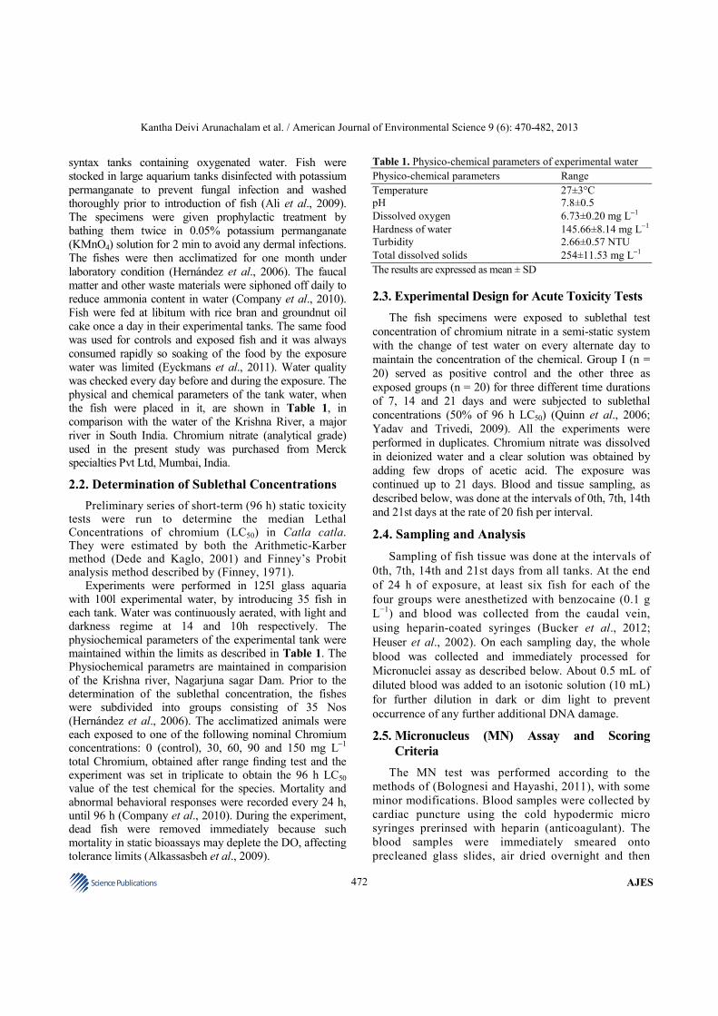
Table 1. Physico-chemical parameters of experimental water
Physico-chemical parameters Range
Temperature 27±3°C
pH 7.8±0.5
Dissolved oxygen 6.73±0.20 mg L−1
Hardness of water 145.66±8.14 mg L−1
Turbidity 2.66±0.57 NTU
Total dissolved solids 254±11.53 mg L−1
The results are expressed as mean ± SD
2.3. Experimental Design for Acute Toxicity Tests
The fish specimens were exposed to sublethal test
concentration of chromium nitrate in a semi-static system
with the change of test water on every alternate day to
maintain the concentration of the chemical. Group I (n =
20) served as positive control and the other three as
exposed groups (n = 20) for three different time durations
of 7, 14 and 21 days and were subjected to sublethal
concentrations (50% of 96 h LC50) (Quinn et al., 2006;
Yadav and Trivedi, 2009). All the experiments were
performed in duplicates. Chromium nitrate was dissolved
in deionized water and a clear solution was obtained by
adding few drops of acetic acid. The exposure was
continued up to 21 days. Blood and tissue sampling, as
described below, was done at the intervals of 0th, 7th, 14th
and 21st days at the rate of 20 fish per interval.
2.4. Sampling and Analysis
Sampling of fish tissue was done at the intervals of
0th, 7th, 14th and 21st days from all tanks. At the end
of 24 h of exposure, at least six fish for each of the
four groups were anesthetized with benzocaine (0.1 g
L−1
) and blood was collected from the caudal vein,
using heparin-coated syringes (Bucker et al., 2012;
Heuser et al., 2002). On each sampling day, the whole
blood was collected and immediately processed for
Micronuclei assay as described below. About 0.5 mL of
diluted blood was added to an isotonic solution (10 mL)
for further dilution in dark or dim light to prevent
occurrence of any further additional DNA damage.
2.5. Micronucleus (MN) Assay and Scoring
Criteria
The MN test was performed according to the
methods of (Bolognesi and Hayashi, 2011), with some
minor modifications. Blood samples were collected by
cardiac puncture using the cold hypodermic micro
syringes prerinsed with heparin (anticoagulant). The
blood samples were immediately smeared onto
precleaned glass slides, air dried overnight and then
Kantha Deivi Arunachalam et al. / American Journal of Environmental Science 9 (6): 470-482, 2013
473 Science Publications
AJES
fixed in absolute methanol for 15 min. Each slide was
stained with 5% Giemsa solution (Medox Biotech India
Pvt. Ltd., Chennai) for 20 min. At least 1,500
erythrocytes for each specimen were identified, counted
and scored microscopically under 1,000 X in Carl Zeiss
microscope (Mortazavi et al., 2005). The main criteria
for scoring the Micronucleus (MN) were based on those
of Al-Sabti and Matcalfe (1995) considering the
absence of connections with the main nucleus, similar
coloration and a size of between 1/10 to 1/30 of the size
of the main nucleus, since for most fish, chromosomes
are much smaller than mammalian chromosomes, as
pointed out by (Schmid, 1975). The nuclear
abnormalities observed were classified into five
categories, adapting the classification proposed by
(Ayllon and Garcia-Vazquez, 2000): (a) Micronuclei;
(b) Binucleated nucleus-two completely separated
nuclei in the same erythrocyte’s cytoplasm; (c) Lobed
nucleus-evaginations of the nuclear envelope of
different sizes; (d) Notched nucleus-having a
noticeable depression into the nucleus that does not
contain nuclear material and (e) Other Nuclear
Abnormalities-all the other types of nuclear
morphological alterations found in the nuclei that
could not be fitted under the previous four categories.
2.6. Comet Assay
The alkaline comet assay was performed as described briefly, as follows; 100 µL of cell suspension was mixed
with 200 µL of 2% low melting temperature agarose at 37°C and then placed on a slide precoated with thin layer of 0.5% normal melting agarose. The cell suspension was immediately covered with a cover glass to obtain a uniform layer and the slides were kept at 4°C for five min, to allow solidification of the agarose
(Rojas et al., 1999). The gel was allowed to solidify by keeping the slide in a steel tray on ice for a minimum period of three minutes. The coverslip was removed and a final layer of 0.5% Low Melting Point (LMP) agarose (100 µL) was placed on the slide and covered with a cover slip (Tice et al., 2000).
After removing the cover glass, the cells were lysed
in a lysing solution (2.5M NaCl, 100mM EDTA, 10mM
Tris, 1% Triton X-100, pH 10) for one hour. After
washing in redistilled water, the slides were placed in a
horizontal gel electrophoresis chamber. The chamber
was filled with cold electrophoretic buffer (1mM EDTA,
300mM NaOH, pH 13) and slides were kept at 4°C for
40 min to allow the DNA to unwind. Electrophoresis was
performed for 25 min (1 V/cm, 300 mA). After
electrophoresis, the slides were washed three times with
neutralization buffer (0.4M Tris, pH 7.5). All preparative
steps were conducted in yellow light to prevent
occurance of additional DNA damage (Avishai et al.,
2003; Tice et al., 2000; Velma and Tchounwou, 2010).
The slides were stained with Ethidium Bromide.
Air-dried slides were immersed for five minutes in
cold water and then stained for five minutes with 80 µL
EtBr (20 µg mL−1
) (Cavallo et al., 2009; Kim et al.,
2002). The slides were rinsed in cold water to remove
excess stain and covered with a coverslip and analyzed
with a fluorescence microscope (NIKON Eclipse 400)
equipped with a CCD-4230A video camera. The nuclei
were analyzed by use of a fluorescence microscope. For
EtBr, a BP 546/10 nm excitation filter and a 590 nm
emission filter were used. For each slide, 25 randomly
chosen nuclei were analyzed. Three slides were evaluated
per treatment and was repeated twice. From the repeated
experiments, the averaged median percentage of tail DNA
as the primary measure of DNA migration was calculated
for each treatment group. Digital images were acquired
and analyzed by the CASP software (Anitha et al., 2000;
Emmanouil et al., 2006).
2.7. Catalase (CAT) Activity
The activity of catalase (CAT) in the liver was
determined spectrophotometrically, at wavelength of 570
nm (Uv-Vis, 3000+, lab India Pvt. Ltd., India), according
to the method of Sinha (1972) and was expressed in ml
mol of decomposed hydrogen peroxide per sec per gram
of tissues wet wt. Sinha (1972) tissue samples were
homogenized in 10 volume of ice-cold 0.1 phosphate
buffers, pH 7.0 and centrifuged for 20 min at 4°C and
15,000 g. The supernatant was used for CAT. The
activity of catalase was determined
spectrophotometrically, by measuring the decrease in the
absorbance of hydrogen peroxide at 240 nm, with a
specific absorption coefficient of 0.0392 cm2 µmoL
H2O2−1
. 2.5 mL of substrate made up of 10 mM
hydrogen peroxide in a 50 mM phosphate buffer of pH
7.0 was added onto 2.5 mg of immobilized CAT
preparation. Reaction was carried out at 25°C for 2 min
and stopped by adding 0.5 mL of 1.0 M HCl. One unit of
activity is defined as the decomposition of 1 µmoL
hydrogen peroxide per minute at 25°C and pH 7.0
(Sayeed et al., 2003; Sun et al., 2006).
2.8. Superoxide Dismutase (SOD) Activity
The activity of Superoxide Dismutase (SOD) was
determined spectrophotometrically at wavelength of
Kantha Deivi Arunachalam et al. / American Journal of Environmental Science 9 (6): 470-482, 2013
474 Science Publications
AJES
480 nm by epinephrine method according to Sun et al.
(2006). SOD was determined spectrophotometrically
in Liver, Muscle Tissue and Gill samples by
measuring the inhibition of the ratio of autocatalytic
adrenochrome formation at 480 nm in a reaction
medium containing 1 mM adrenaline and 50 mM
glycine (pH 10.2). This reaction was conducted at a
constant temperature of 30°C for 3 min. Enzyme
activity is expressed as superoxide dismutase units per
gram of protein. One unit is defined as the amount of
enzymes that inhibits the ratio of adrenochrome
formation by 50% (Sun et al., 2006; Velma and
Tchounwou, 2010).
2.9. Metal Analysis in Tissue Samples
The muscle tissues were washed with distilled
water, dried to a constant weight at 60°C for 48 h until
constant weight is obtained. Dried samples were
homogenized, packed in small pre-cleaned
polyethylene bottles and kept at-20°C until analysis.
All reagents were of analytical grade. Unless
otherwise stated, double deionized water was used for
all dilutions. The dried fish samples were placed in a
high form porcelain crucible and kept in the muffle
furnace. The furnace temperature was slowly
increased to 100°C in two hours. The samples were
ashed at about 450°C for one night, until a white or
grey ash residue was obtained. About 5g of the ash
samples were used for digestion. The samples were
digested with 25 mL of Nitric Acid (70% V/V) and 5
mL of sulphuric acid (98 % V/V) and 5 mL perchloric
acid (%V/V) at 110°C. The Formation of the milky
white precipitate confirms the complete digestion of
sample. The sample is then filtered in Whatman No-
42 filter paper and made upto 50 mL with double
deionized water (Al-Yousuf et al., 2000; Mendil et al.,
2010; Oyoo-Okoth et al., 2010). The samples were
analyzed for heavy metals using Inductively Coupled
Plasma Optical Emission Spectrometry (ICPOES) at
Sophisticated Analytical Instrument Facility (SAIF),
the Indian Institute of Technology, Madras.
2.10. Bio Concentration Factor (BCF)
The Bio concentration factor is the biological
sequestering of a substance at a higher concentration
than at which it occurs in the surrounding
environment or medium. The bioaccumulation factor
is the ratio of the contaminant in an organism to the
concentration in the surrounding environment at a
steady state, where the organism can take the
contaminant through ingestion with its food as well as
through direct content.
Bioconcentration factors between the fish tissues and
the water were calculated, using the mean metal
concentration in each tissue and the corresponding metal
concentration in simulated water. Bioconcentration
Factor (BCF) is defined as the ratio of the concentration
of a specific heavy metal in the organism to the
concentration of the metal in the water in which the fish
lives (Sun et al., 2006). The BCF was calculated using
the formula 1 as described below Equation (1):
f
w
CBCF
C= (1)
where, Cf and Cw are the concentrations of metal in fish
and water respectively, expressed in the same units
(mg/kg and mg/L). BCF will, hence, be a simple
number, without any units.
2.11. Statistical Analysis
One-way Analysis of Variance (ANOVA) was
applied to determine significant differences in the results
of various groups. P-Values <0.001 were considered
significant. The data obtained from analytical methods
were treated statistically using SPSS software (version
17.0 for windows). Descriptive data analysis was
performed, including the calculation of mean, SD.
3. RESULTS AND DISCUSSION
The data obtained from acute toxicity test of water-
born chromium for Catla catla revealed that chromium toxicity increased with increasing concentration and with exposure time. The number of dead fish in relation to the chromium concentrations (20, 60, 100 mg L
−1) were assessed and counted
during the exposure period and were removed. No
mortality was observed during the 96 h at control (0.0 mg Cr L
−1) and 100% mortality rate was observed
only at 100 mg L−1
(Table 2).
The fish exposed to 20 and 60 mg L−1
chromium
showed abnormal behavior like erratic swimming and
loss of equilibrium. The exposed fish swam to surface
more often than the control fish. Neither mortality nor
any visible changes in behavior were viewed in the
control group. Summary of the different behavioral
changes observed due to chromium exposure is
presented in Table 3.
Kantha Deivi Arunachalam et al. / American Journal of Environmental Science 9 (6): 470-482, 2013
475 Science Publications
AJES
Table 2. LC50 determination based on arithmetic method of karber
Concentration Concentration No. of alive No. of Mean death Probit
in ppm difference (X) fish (p) mean fish (p) Q1 Q2
Y2
+= (X×Y)
0 (control) - 35 0 _ 0
20 20 18 17 8.5 170
60 40 0 35 26.0 1040
100 40 0 35 35.0 1400
XY 2610=∑
50 100
Pr obhitLC LC
Noof organisms In each group= −
∑ LC50 = 100 – 2610/35; LC50= 100- 74.58; LC50 = 25.42 mg L−1
Table 3. Determination of 96 h LC50 Value using Finney’s Probit analysis (1964)
Conc.(mg/L) Log (Conc.) No. of fish alive (96 h) No. of fish dead (96 h) Mortality (%) Probit
0 0.00 35 0 0.0 - 74.6 1.87 18 17 48.5 4.95 223.7 2.35 0 35 100.0 7.37 373 2.57 0 35 100.0 7.37
The relationship between the chromium concentration
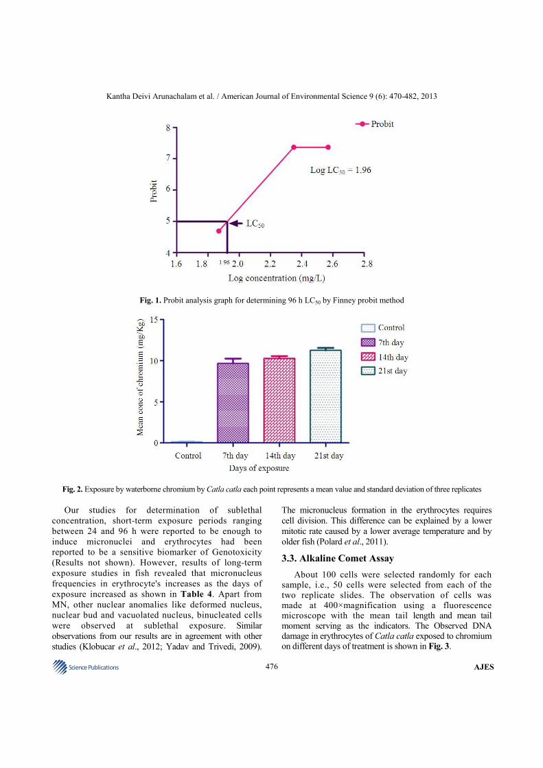
and the mortality rate of Catla catla for determination of LC50 value was determined according to Finney’s Probit Analysis and the results are shown in Fig. 1. A dose-dependent increase and time dependent decrease were observed in mortality rate until 96 h. The LC50 value of chromium was determined as 25.42 mg L
−1 for Catla catla
by Arithmetic Method of Karber (Dede and Kaglo, 2001; Mishra and Mohanty, 2008) and Finney’s Probit analysis (Finney, 1971). The mean 96-h LC50 value with 95% confidence limits for Catla catla by Finney’s probit analysis was found to be 25.42 mg L
−1 as shown in Table
3. This value was estimated to be 25.42 mg L−1
with the Karber’s method (Table 2). The two methods are in good agreement and this suggests that the water-born chromium is definitely a toxic heavy metal to Catla catla.
Based on the 96 h LC50 value, the sublethal concentration (1/2 of 96 h LC50 value = 25.42 mg L
−1) of
chromium were estimated. This value was used for the bioconcentration and genotoxicity studies. The exposed fish swam to surface more often than the control fish. Neither mortality nor any visible changes in behavior were observed in the control group. The hypertrophy and hyperplasia of the gill epithelial cells were absorbed as the gills were swollen compared to the Control fish (Quinn et al., 2006).
3.1. Accumulation of Chromium in Catla Catla
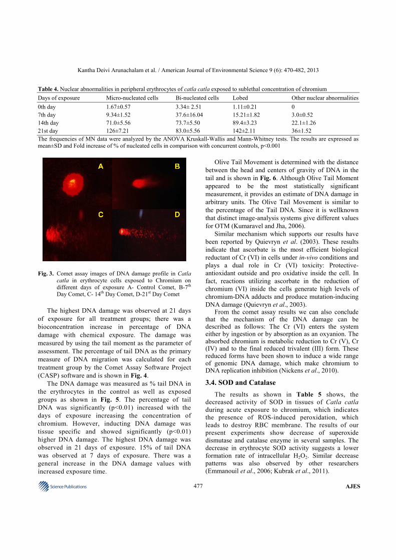
The sublethal concentration was used for this study and therefore, no mortality was recorded during the experimental period for all treatment groups studied. The increased levels of accumulation during the exposure period suggested a rather rapid absorption of this metal.
Similar results have been reported in freshwater isopods exposed to Cu, Pb and Zn (Palaniappan and Karthikeyan, 2009). In the case of bioaccumulation of chromium in the muscle, the accumulation pattern varies with the degree of concentration of the toxicants. Occasionally, fish exposed to Chromium showed vertical and downward swimming patterns, swimming near the water surface, drowsy and erratic swimming and loss of schooling behavior.
During the sublethal toxicity studies the fishes were exposed to ½ of LC50 concentration. As the days of exposure increased, the chromium concentration in the fish tissues increased gradually. As the exposure period continued till 21 days. The accumulation of chromium was maximum within 7 days of exposure (9.68 mg kg
−1) and
after 7 days the chromium is slowly bioaccumulated in the tissues. The maximum accumulation of the chromium was observed on the 21st day of exposure (11.25 mg kg
−1). The
pattern of accumulation of chromium in Catla catla during sublethal toxicity studies were shown in the Fig. 2.
3.2. Genotoxicity of Chromium in Catla Catla by
Micronucleus Assay
Micronucleus was encountered in fish subjected to chromium accumulation. Induction of micronuclei showed a remarkable increase with increased exposure time, when compared with the control. The results suggested that the incidence of micronuclei induction was not dose dependent. In contrast, the group treated with Chromium did not show significant difference in micronuclei frequencies with respect to the control group, although differences between frequencies of the treated and control groups for the other nuclear lesions were highly significant (Anbumani and Mohankumar, 2012).
Kantha Deivi Arunachalam et al. / American Journal of Environmental Science 9 (6): 470-482, 2013
476 Science Publications
AJES
Fig. 1. Probit analysis graph for determining 96 h LC50 by Finney probit method
Fig. 2. Exposure by waterborne chromium by Catla catla each point represents a mean value and standard deviation of three replicates
Our studies for determination of sublethal
concentration, short-term exposure periods ranging
between 24 and 96 h were reported to be enough to
induce micronuclei and erythrocytes had been
reported to be a sensitive biomarker of Genotoxicity
(Results not shown). However, results of long-term
exposure studies in fish revealed that micronucleus
frequencies in erythrocyte's increases as the days of
exposure increased as shown in Table 4. Apart from
MN, other nuclear anomalies like deformed nucleus,
nuclear bud and vacuolated nucleus, binucleated cells
were observed at sublethal exposure. Similar
observations from our results are in agreement with other
studies (Klobucar et al., 2012; Yadav and Trivedi, 2009).
The micronucleus formation in the erythrocytes requires
cell division. This difference can be explained by a lower
mitotic rate caused by a lower average temperature and by
older fish (Polard et al., 2011).
3.3. Alkaline Comet Assay
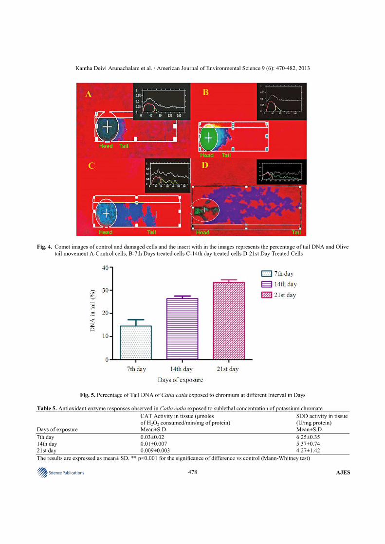
About 100 cells were selected randomly for each sample, i.e., 50 cells were selected from each of the two replicate slides. The observation of cells was made at 400×magnification using a fluorescence microscope with the mean tail length and mean tail moment serving as the indicators. The Observed DNA damage in erythrocytes of Catla catla exposed to chromium on different days of treatment is shown in Fig. 3.
Kantha Deivi Arunachalam et al. / American Journal of Environmental Science 9 (6): 470-482, 2013
477 Science Publications
AJES
Table 4. Nuclear abnormalities in peripheral erythrocytes of catla catla exposed to sublethal concentration of chromium
Days of exposure Micro-nucleated cells Bi-nucleated cells Lobed Other nuclear abnormalities
0th day 1.67±0.57 3.34± 2.51 1.11±0.21 0
7th day 9.34±1.52 37.6±16.04 15.21±1.82 3.0±0.52
14th day 71.0±5.56 73.7±5.50 89.4±3.23 22.1±1.26
21st day 126±7.21 83.0±5.56 142±2.11 36±1.52
The frequencies of MN data were analyzed by the ANOVA Kruskall-Wallis and Mann-Whitney tests. The results are expressed as
mean±SD and Fold increase of % of nucleated cells in comparison with concurrent controls, p<0.001
Fig. 3. Comet assay images of DNA damage profile in Catla
catla in erythrocyte cells exposed to Chromium on
different days of exposure A- Control Comet, B-7th
Day Comet, C- 14th Day Comet, D-21st Day Comet
The highest DNA damage was observed at 21 days
of exposure for all treatment groups; there was a
bioconcentration increase in percentage of DNA
damage with chemical exposure. The damage was
measured by using the tail moment as the parameter of
assessment. The percentage of tail DNA as the primary
measure of DNA migration was calculated for each
treatment group by the Comet Assay Software Project
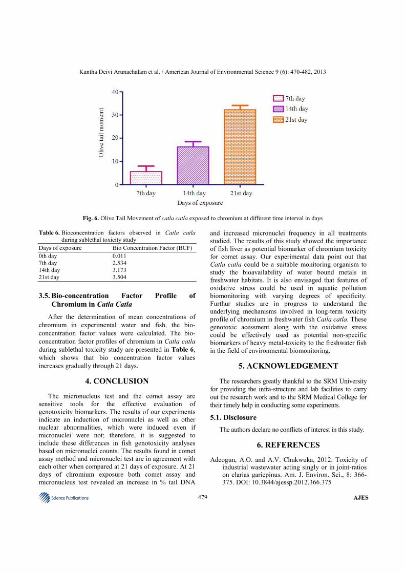
(CASP) software and is shown in Fig. 4.
The DNA damage was measured as % tail DNA in
the erythrocytes in the control as well as exposed
groups as shown in Fig. 5. The percentage of tail
DNA was significantly (p<0.01) increased with the
days of exposure increasing the concentration of
chromium. However, inducting DNA damage was
tissue specific and showed significantly (p<0.01)
higher DNA damage. The highest DNA damage was
observed in 21 days of exposure. 15% of tail DNA
was observed at 7 days of exposure. There was a
general increase in the DNA damage values with
increased exposure time.
Olive Tail Movement is determined with the distance
between the head and centers of gravity of DNA in the
tail and is shown in Fig. 6. Although Olive Tail Moment
appeared to be the most statistically significant
measurement, it provides an estimate of DNA damage in
arbitrary units. The Olive Tail Movement is similar to
the percentage of the Tail DNA. Since it is wellknown
that distinct image-analysis systems give different values
for OTM (Kumaravel and Jha, 2006).
Similar mechanism which supports our results have
been reported by Quievryn et al. (2003). These results
indicate that ascorbate is the most efficient biological
reductant of Cr (VI) in cells under in-vivo conditions and
plays a dual role in Cr (VI) toxicity: Protective-
antioxidant outside and pro oxidative inside the cell. In
fact, reactions utilizing ascorbate in the reduction of
chromium (VI) inside the cells generate high levels of
chromium-DNA adducts and produce mutation-inducing
DNA damage (Quievryn et al., 2003). From the comet assay results we can also conclude
that the mechanism of the DNA damage can be described as follows: The Cr (VI) enters the system either by ingestion or by absorption as an oxyanion. The absorbed chromium is metabolic reduction to Cr (V), Cr (IV) and to the final reduced trivalent (III) form. These reduced forms have been shown to induce a wide range of genomic DNA damage, which make chromium to DNA replication inhibition (Nickens et al., 2010).
3.4. SOD and Catalase
The results as shown in Table 5 shows, the
decreased activity of SOD in tissues of Catla catla
during acute exposure to chromium, which indicates
the presence of ROS-induced peroxidation, which
leads to destroy RBC membrane. The results of our
present experiments show decrease of superoxide
dismutase and catalase enzyme in several samples. The
decrease in erythrocyte SOD activity suggests a lower
formation rate of intracellular H2O2. Similar decrease
patterns was also observed by other researchers
(Emmanouil et al., 2006; Kubrak et al., 2011).
Kantha Deivi Arunachalam et al. / American Journal of Environmental Science 9 (6): 470-482, 2013
478 Science Publications
AJES
Fig. 4. Comet images of control and damaged cells and the insert with in the images represents the percentage of tail DNA and Olive
tail movement A-Control cells, B-7th Days treated cells C-14th day treated cells D-21st Day Treated Cells
Fig. 5. Percentage of Tail DNA of Catla catla exposed to chromium at different Interval in Days
Table 5. Antioxidant enzyme responses observed in Catla catla exposed to sublethal concentration of potassium chromate
CAT Activity in tissue (µmoles SOD activity in tissue
of H2O2 consumed/min/mg of protein) (U/mg protein)
Days of exposure Mean±S.D Mean±S.D
7th day 0.03±0.02 6.25±0.35
14th day 0.01±0.007 5.37±0.74
21st day 0.009±0.003 4.27±1.42
The results are expressed as mean± SD. ** p<0.001 for the significance of difference vs control (Mann-Whitney test)
Kantha Deivi Arunachalam et al. / American Journal of Environmental Science 9 (6): 470-482, 2013
479 Science Publications
AJES
Fig. 6. Olive Tail Movement of catla catla exposed to chromium at different time interval in days
Table 6. Bioconcentration factors observed in Catla catla
during sublethal toxicity study
Days of exposure Bio Concentration Factor (BCF)
0th day 0.011
7th day 2.534
14th day 3.173
21st day 3.504
3.5. Bio-concentration Factor Profile of
Chromium in Catla Catla
After the determination of mean concentrations of
chromium in experimental water and fish, the bio-
concentration factor values were calculated. The bio-
concentration factor profiles of chromium in Catla catla
during sublethal toxicity study are presented in Table 6,
which shows that bio concentration factor values
increases gradually through 21 days.
4. CONCLUSION
The micronucleus test and the comet assay are
sensitive tools for the effective evaluation of
genotoxicity biomarkers. The results of our experiments
indicate an induction of micronuclei as well as other
nuclear abnormalities, which were induced even if
micronuclei were not; therefore, it is suggested to
include these differences in fish genotoxicity analyses
based on micronuclei counts. The results found in comet
assay method and micronuclei test are in agreement with
each other when compared at 21 days of exposure. At 21
days of chromium exposure both comet assay and
micronucleus test revealed an increase in % tail DNA
and increased micronuclei frequency in all treatments
studied. The results of this study showed the importance
of fish liver as potential biomarker of chromium toxicity
for comet assay. Our experimental data point out that
Catla catla could be a suitable monitoring organism to
study the bioavailability of water bound metals in
freshwater habitats. It is also envisaged that features of
oxidative stress could be used in aquatic pollution
biomonitoring with varying degrees of specificity.
Furthur studies are in progress to understand the
underlying mechanisms involved in long-term toxicity
profile of chromium in freshwater fish Catla catla. These
genotoxic acessment along with the oxidative stress
could be effectively used as potential non-specific
biomarkers of heavy metal-toxicity to the freshwater fish
in the field of environmental biomonitoring.
5. ACKNOWLEDGEMENT
The researchers greatly thankful to the SRM University
for providing the infra-structure and lab facilities to carry
out the research work and to the SRM Medical College for
their timely help in conducting some experiments.
5.1. Disclosure
The authors declare no conflicts of interest in this study.
6. REFERENCES
Adeogun, A.O. and A.V. Chukwuka, 2012. Toxicity of industrial wastewater acting singly or in joint-ratios on clarias gariepinus. Am. J. Environ. Sci., 8: 366-375. DOI: 10.3844/ajessp.2012.366.375
Kantha Deivi Arunachalam et al. / American Journal of Environmental Science 9 (6): 470-482, 2013
480 Science Publications
AJES
Ali, D., N.S. Nagpure, S. Kumar, R. Kumar and B.
Kushwaha et al., 2009. Assessment of genotoxic and
mutagenic effects of chlorpyrifos in freshwater fish
Channa punctatus (Bloch) using micronucleus assay
and alkaline single-cell gel electrophoresis. Food
Chem. Toxicol., 47: 650-656. PMID: 19141310 Ali, D., N.S. Nagpure, S. Kumar, R. Kumar and B.
Kushwaha, 2008a. Genotoxicity assessment of acute exposure of chlorpyrifos to freshwater fish Channa punctatus (Bloch) using micronucleus assay and alkaline single-cell gel electrophoresis. Chemosphere, 71: 1823-1831. DOI: 10.1016/j.chemosphere.2008.02.007
Ali, F.K., A.M. El-Shehawi and M.A. Seehy, 2008b.
Micronucleus test in fish genome: A sensitive
monitor for aquatic pollution. Afr. J. Biotechnol., 7:
606-612.
Alkassasbeh, J.Y.M., L.Y. Heng, S. Surif and U.K.M.
Bangi, 2009. Toxicity testing and the effect of
landfill leachate in malaysia on behavior of common
carp (Cyprinus Carpio L., 1758; Pisces,
Cyprinidae). Am. J. Environ. Sci., 5: 209-217. DOI:
10.3844/ajessp.2009.209.217
Al-Sabti, K. and C.D. Metcalfe, 1995. Fish micronuclei
for assessing genotoxicity in water. Mutat. Res.,
343: 121-135. DOI: 10.1016/0165-1218(95)90078-0
Al-Yousuf, M.H., M.S. El-Shahawi and S.M. Al-Ghais,
2000. Trace metals in liver, skin and muscle of
Lethrinus lentjan fish species in relation to body
length and sex. Sci. Total Environ., 256: 87-94.
DOI: 10.1016/S0048-9697(99)00363-0
Anbumani, S. and M.N. Mohankumar, 2012. Gamma
radiation induced micronuclei and erythrocyte
cellular abnormalities in the fish Catla catla. Aquat.
Toxicol., 122-123: 125-132. PMID: 22771702
Anitha, B., N. Chandra, P.M. Gopinath and G. Durairaj,
2000. Genotoxicity evaluation of heat shock in gold
fish (Carassius auratus). Mutat. Res., 469: 1-8.
PMID: 10946237
Avishai, N., C. Rabinowitz and B. Rinkevich, 2003. Use
of the comet assay for studying environmental
genotoxicity: Comparisons between visual and
image analyses. Environ. Mol. Mutagen., 42: 155-
165. PMID: 14556223
Ayllon, F. and E. Garcia-Vazquez, 2000. Induction of
micronuclei and other nuclear abnormalities in
European minnow Phoxinus phoxinus and mollie
Poecilia latipinna: an assessment of the fish
micronucleus test. Mutat. Res., 467: 177-186.
PMID: 10838205
Balachandar, V., M. Arun, M.S. Devi, P. Velmurugan
and P. Manikantan et al., 2010. Evaluation of the
genetic alterations in direct and indirect exposures of
hexavalent chromium [Cr(VI)] in leather tanning
industry workers North Arcot District, South India.
Int. Arch. Occup. Environ. Health 83: 791-801.
PMID: 20617332
Bolognesi, C. and M. Hayashi, 2011. Micronucleus assay
in aquatic animals. Mutagenesis, 26: 205-213. DOI:
10.1093/mutage/geq073, PMID: 21164204
Bucker, A., M. Carvalho, M. Conceicao and J. Alves-
Gomes, 2012. Micronucleus test and comet assay in
erythrocytes of the Amazonian electric fish
Apteronotus bonapartii exposed to benzene. J.
Brazilian Soc. Ecotoxicol., 7: 65-73.
Cavallo, D., C.L. Ursini, B. Rondinone and S. Iavicoli,
2009. Evaluation of a suitable DNA damage
biomarker for human biomonitoring of exposed
workers. Environ. Mol. Mutagen., 50: 781-790.
PMID: 19449396
Cavas, T., N.N. Garanko and V.V. Arkhipchuk, 2005.
Induction of micronuclei and binuclei in blood, gill
and liver cells of fishes subchronically exposed to
cadmium chloride and copper sulphate. Food Chem.
Toxicol., 43: 569-574. PMID: 15721204
Company, R., A. Serafim, R.P. Cosson, A. Fiala-
Medioni and L. Camus et al., 2010. Sub-lethal
effects of cadmium on the antioxidant defence
system of the hydrothermal vent mussel
Bathymodiolus azoricus. Ecotoxicol. Environ. Saf.,
73: 788-795. PIMD: 20137812
De Andrade, V.M., T.R.O. De Freitas and J. Da Silva,
2004. Comet assay using mullet (Mugil sp.) and sea
catfish (Netuma sp.) erythrocytes for the detection
of genotoxic pollutants in aquatic environment.
Mutat. Res., 560: 57-67. PMID: 15099825
Dede, E.B. and E.D. Kaglo, 2001. Aqua-toxicological
Effects of Water Soluble Fractions (WSF) of diesel
fuel on O. Niloticus fingerlings. J. Applied Sci.
Environ. Mgt., 5: 93-96.
Ellis, A.S., T.M. Johnson and T.D. Bullen, 2002.
Chromium isotopes and the fate of hexavalent
chromium in the environment. Science, 295: 2060-
2062. PMID: 11896274
Emmanouil, C., D.J. Smart, N.J. Hodges and J.K.
Chipman, 2006. Oxidative damage produced by
Cr(VI) and repair in mussel (Mytilus edulis L.) gill.
Mar. Environ. Res., 62: S292-S296. PMID:
16698074
Kantha Deivi Arunachalam et al. / American Journal of Environmental Science 9 (6): 470-482, 2013
481 Science Publications
AJES
Eyckmans, M., N. Celis, N. Horemans, R. Blust and
D.G. Boeck, 2011. Exposure to waterborne copper
reveals differences in oxidative stress response in
three freshwater fish species. Aquat. Toxicol., 103:
112-120. PMID: 21419094
Fatemi, S.J.A., M. Iranmanesh and F.D. Balooch, 2013.
Effect of Chromium(VI) on serum iron and removal of
its toxicity by combining deferasirox and deferiprone
chelators in rats. Am. J. Pharmacol. Toxicol., 8: 164-
169. DOI: 10.3844/ajptsp.2013.164.169
Finney, D.J., 1971. Probit Analysis. 1st Edn., Cambridge
University Press, Cambridge, ISBN-10:
0521135907, pp: 272.
Fontanetti, C.S., C.A. Christofoletti, T.G. Pinheiro, T.S.
Souza and J. Pedro-Escher, 2010. Microscopy as a
tool in toxicological evaluations. University
Estadual Paulis.
Foster, W.J., A.D.E. Chatelet and M. Rogerson, 2012.
Testing benthic foraminiferal distributions as a
contemporary quantitative approach to
biomonitoring estuarine heavy metal pollution. Mar.
Pollut. Bull., 64: 1039-1048. DOI:
10.1016/j.marpolbul.2012.01.021
Garg, S., R.K. Gupta and K.L. Jain, 2009. Sublethal
effects of heavy metals on biochemical composition
and their recovery in Indian major carps. J. Hazard.
Mater., 163: 1369-1384. PMID: 18775601
Gheju, M., 2011. Hexavalent Chromium Reduction with
Zero-Valent Iron (ZVI) in Aquatic Systems. Water,
Air, Soil Pollut., 222: 103-148. DOI:
10.1007/s11270-011-0812-y
Hartmann, A., 1997. The contribution of cytotoxicity to
DNA-effects in the single cell gel test (comet assay).
Toxicol. Lett., 90: 183-188. PMID: 9067486
Hernández, P.P., V. Moreno, F.A. Olivari and M.L.
Allende, 2006. Sub-lethal concentrations of
waterborne copper are toxic to lateral line
neuromasts in zebrafish (Danio rerio). Hear. Res.,
213: 1-10. PMID: 16386394
Heuser, V.D., D.J. Silva, H.J. Moriske, J.F. Dias and
M.L. Yoneama et al., 2002. Genotoxicity
biomonitoring in regions exposed to vehicle
emissions using the comet assay and the
micronucleus test in native rodent Ctenomys
minutus. Environ. Mol. Mutagen., 40: 227-235.
PMID: 12489112
Jomova, K. and M. Valko, 2011. Advances in metal-
induced oxidative stress and human disease.
Toxicology, 283: 65-87. PMID: 21414382
Kim, B.S., J.J. Park, L. Edler, V.D. Fournier and W.
Haase et al., 2002. New measure of DNA repair in
the single-cell gel electrophoresis (comet) assay.
Environ. Mol. Mutagen., 40: 50-6. PMID: 12211076
Klobucar, G.I.V., O. Malev, M. Srut, A. Stambuk and S.
Lorenzon et al., 2012. Genotoxicity monitoring of
freshwater environments using caged crayfish
(Astacus leptodactylus). Chemosphere, 87: 62-67.
PMID: 22178377
Kubrak, O.I., V.V. Husak, B.M. Rovenko, J.M. Storey
and K.B. Storey et al., 2011. Cobalt-induced
oxidative stress in brain, liver and kidney of goldfish
Carassius auratus. Chemosphere, 85: 983-999.
PMID: 21777937
Kumar, S., R. Budhwar, A. Nigam and S. Priya, 2009.
Cytoprotection against Cr(6+)-induced DNA
damage by alpha-lipoic acid: Implications in
reducing occupational cancer risk. Mutagenesis, 24:
495-500. PMID: 19710206
Kumaravel, T.S. and A.N. Jha, 2006. Reliable Comet
assay measurements for detecting DNA damage
induced by ionising radiation and chemicals. Mutat.
Res., 605: 7-16. PMID: 16621680
Lavanya, S., M. Ramesh, C. Kavitha and A. Malarvizhi,
2011. Hematological, biochemical and
ionoregulatory responses of Indian major carp Catla
catla during chronic sublethal exposure to inorganic
arsenic. Chemosphere, 82: 977-985. PMID:
21094981
Mendil, D., O.F. Unal, M. Tuzen and M. Soylak, 2010.
Determination of trace metals in different fish
species and sediments from the River Yesilırmak in
Tokat, Turkey. Food Chem. Toxicol., 48: 1383-
1392. DOI: 10.1016/j.fct.2010.03.006
Mishra, A.K. and B. Mohanty, 2008. Acute toxicity
impacts of hexavalent chromium on behavior and
histopathology of gill, kidney and liver of the
freshwater fish, Channa punctatus (Bloch). Environ.
Toxicol. Pharmacol., 26: 136-141. DOI:
10.1016/j.etap.2008.02.010
Mortazavi, S.B., A. Safari, A. Khavanin, A. Kazemnejad
and S.M. Moazzeni et al., 2005. Induction of
micronuclei in mice lymphocytes exposed to
microwave and toluene. Am. J. Applied Sci., 2:
1321-1324. DOI: 10.3844/ajassp.2005.1321.1324 Muid, K.A., R.M. Shahjahan, R. Begum and R.A.
Begum, 2012. Zinc phosphide induced DNA damage in the blood cells of Gallus sp. using comet DNA assay. Am. J. Agric. Biol. Sci., 7: 82-87. DOI: 10.3844/ajabssp.2012.82.87
Kantha Deivi Arunachalam et al. / American Journal of Environmental Science 9 (6): 470-482, 2013
482 Science Publications
AJES
Nickens, K.P., S.R. Patierno and S. Ceryak, 2010.
Chromium genotoxicity: A double-edged sword.
Chem. Biol. Interact., 188: 276-288. PMID:
20430016
Oyoo-Okoth, E., W. Admiraal, O. Osano, V. Ngure and
M.H.S. Kraak et al., 2010. Monitoring exposure to
heavy metals among children in Lake Victoria,
Kenya: Environmental and fish matrix. Ecotoxicol.
Environ. Saf., 73: 1797-1803. PMID: 20705339
Palaniappan, P.R. and S. Karthikeyan, 2009.
Bioaccumulation and depuration of chromium in the
selected organs and whole body tissues of
freshwater fish Cirrhinus mrigala individually and in
binary solutions with nickel. J. Environ. Sci., 21:
229-236. DOI: 10.1016/S1001-0742(08)62256-1
Palus, J., K. Rydzynski, E. Dziubaltowska, K.
Wyszynska and A. Natarajan et al., 2003. Genotoxic
effects of occupational exposure to lead and
cadmium. Mutat. Res. Toxicol. Environ. Mutagen.,
540: 19-28. PMID: 12972055
Polard, T., S. Jean, G. Merlina, C. Laplanche and E.
Pinelli et al., 2011. Giemsa versus acridine orange
staining in the fish micronucleus assay and
validation for use in water quality monitoring.
Ecotoxicol. Environ. Saf., 74: 144-119. PMID:
20828819
Quievryn, G., E. Peterson, J. Messer and A. Zhitkovich,
2003. Genotoxicity and mutagenicity of
chromium(VI)/ascorbate-generated DNA adducts in
human and bacterial cells. Biochemistry, 42: 1062-
1070. PMID: 12549927
Quinn, B., F. Gagne, C. Blaise, M.J. Costello and J.G.
Wilson et al., 2006. Evaluation of the lethal and sub-
lethal toxicity and potential endocrine disrupting
effect of nonylphenol on the zebra mussel
(Dreissena polymorpha). Comp. Biochem. Physiol.
C. Toxicol. Pharmacol., 142: 118-127. PMID:
16377254
Rao, G.T., V.V.S.G. Rao, K. Ranganathan, L. Surinaidu
and J. Mahesh et al., 2011. Assessment of
groundwater contamination from a hazardous dump
site in Ranipet, Tamil Nadu, India. Hydrogeol. J.,
19: 1587-1598. DOI: 10.1007/s10040-011-0771-9
Rojas, E., M.C. Lopez and M. Valverde, 1999. Single cell gel electrophoresis assay: Methodology and applications. J. Chromatogr. B. Biomed. Sci. Applic., 722: 225-254. PMID: 10068143
Sayeed, I., S. Parvez, S. Pandey, B. Bin-Hafeez and R. Haque et al., 2003. Oxidative stress biomarkers of exposure to deltamethrin in freshwater fish, Channa punctatus Bloch. Ecotoxicol. Environ. Saf., 56: 295-301. PMID: 12927561
Schmid, W., 1975. The micronucleus test. Mutat. Res.,
31: 9-15. PMID: 48190
Sinha, A.K., 1972. Colorimetric assay of catalase. Anal.
Biochem., 47: 389-394. DOI: 10.1016/0003-
2697(72)90132-7
Sun, Y., H. Yu, J. Zhang, Y. Yin and H. Shen et al.,
2006. Bioaccumulation and antioxidant responses in
goldfish Carassius auratus under HC Orange No. 1
exposure. Ecotoxicol. Environ. Saf., 63: 430-437.
PMID: 16406596 Tice, R.R., E. Agurell, D. Anderson, B. Burlinson and A.
Hartmann et al., 2000. Single cell gel/comet assay: Guidelines for in vitro and in vivo genetic toxicology testing. Environ. Mol. Mutagen., 221: 206-221. PMID: 10737956
Velma, V. and P.B. Tchounwou, 2010. Chromium-induced biochemical, genotoxic and histopathologic effects in liver and kidney of goldfish, Carassius auratus. Mutat. Res., 698: 43-51. PMID: 20348018
Yadav, K.K. and S.P. Trivedi, 2009. Sublethal exposure of heavy metals induces micronuclei in fish, Channa punctata. Chemosphere, 77: 1495-1500. PMID: 19880156
Yılmaz, A.B., M.K. Sangun, D. Yaglıoglu and C. Turan,
2010. Metals (major, essential to non-essential)
composition of the different tissues of three
demersal fish species from Iskenderun Bay, Turkey.
Food Chem., 123: 410-415. DOI:
10.1016/j.foodchem.2010.04.057
Zhou, Q., J. Zhang, J. Fu, J. Shi and G. Jiang, 2008.
Biomonitoring: An appealing tool for assessment of
metal pollution in the aquatic ecosystem. Anal.
Chim. Acta, 606: 135-150. PMID: 18082645