Embed Size (px)
Citation preview
IN VITRO GENE AND DRUG DELIVERY AND TARGETING TO HUMAN
GLIOMA CELLS BY LIPOPROTEIN MIMICS
by
GUANGLIANG PAN
(Under the direction of D. Robert Lu)
ABSTRACT
Malignant glioma represents a very difficult therapeutic challenge. One new therapeutic strategy is gene therapy, which involves the delivery to and expression of the therapeutic genes in the cancer cells. In this research, a novel artificial lipoprotein delivery system, which consists of nanoemulsion particles and the incorporated lipidized poly-L-lysine, was developed and evaluated. A model plasmid DNA, pSV-β-Gal, was carried by this system and transfected human glioma cell line SF-767 in vitro. The plasmid DNA was effectively delivered by this system and the reporter gene was expressed. Compared to Lipofectamine system, this new delivery system demonstrated similar transfection efficiency but a much lower cytotoxicity. Targeted delivery of therapeutics is another new strategy to treat glioma. The cellular uptake of a cholesterol-based anti-tumor compound, BCH, in liposomal formulation by normal neuron cells and glioma cells was compared. It was found that the cellular uptake of BCH by glioma cells was up to 11 times as high as that by normal neuron cells. In the presence of monoclonal anti-LDL receptor antibody in the culture medium, the cellular uptake of BCH in liposomal formulation by the glioma cells was greatly reduced. The effect of serum on the cellular uptake of BCH in liposome formulation by the glioma cells was also investigated by replacing normal serum with lipoprotein deficient serum. LDL could help the cellular uptake of BCH in liposome formulation by the glioma cells. In addition, the effect of divalent calcium ion and temperature on cellular uptake of BCH in liposome formulation was investigated. These results suggested that LDL receptor played an important role in the uptake of BCH in liposome formulation by the glioma cells. The cytotoxicity of another cholesterol-based anticancer drug, methotrexate-cholesterol, on the glioma cells was also evaluated. In addition, the application of biological protein nanostructure in targeted drug delivery was described. INDEX WORDS: Gene delivery, Glioma, Nanoemulsion, Artificial lipoprotein,
Plasmid DNA, β-Galactosidase, Poly-L-lysine, Lipidized, Transfection, Cytotoxicity, Endocytosis, BCH, Normal neuron cells, LDL receptor, Monoclonal anti-LDL receptor antibody, Lipoprotein deficient serum (LPDS), Calcium ion, Methotrexate-cholesterol
IN VITRO GENE AND DRUG DELIVERY AND TARGETING TO HUMAN
GLIOMA CELLS BY LIPOPROTEIN MIMICS
by
GUANGLIANG PAN
B.S., Shandong University, P. R. China, 1988
M.S., Nanjing Agricultural University, P. R. China, 1991
M.S., University of North Texas, 1998
A Dissertation Submitted to the Graduate Faculty of the University of Georgia in Partial
Fulfillment of the Requirement for the Degree
DOCTOR OF PHILOSOPHY
ATHENS, GEORGIA
2003

2003
Guangliang Pan
All Rights Reserved
IN VITRO GENE AND DRUG DELIVERY AND TARGETING TO HUMAN
GLIOMA CELLS BY LIPOPROTEIN MIMICS
by
GUANGLIANG PAN
Major Professor: D. Robert Lu
Committee: Warren Beach Anthony C. Capomacchia H. Won Jun James C. Price
Electronic Version Approved Maureen Grasso Dean of the Graduate School The University of Georgia August 2003

iv
To My Beloved Family
v
ACKNOWLEDGEMENTS
I am sincerely grateful to my major professor, Dr. D. Robert Lu, for providing the
opportunity to work in his lab, for his continued encouragement, support and guidance.
His willingness to share his knowledge and experience in pharmaceutical and biomedical
research are much appreciated.
I also thank all my committee members, Dr. Warren Beach, Dr. Anthony C.
Capomacchia, Dr. Won H. Jun, and Dr. James C. Price, for their professional wisdom,
guidance, advice, and comments throughout the course of my Ph.D. program
Special thanks goes to Dr. Svein ∅ie, Dean of the College of Pharmacy, for much
scientific advice during the research and writing.
Heart felt gratitude must go to all my family members for their encouragement,
support and understanding. I wish to thank my wife, Lianqi Xie and my lovely daughter,
Chenfei Pan, for their endless love and patience.
vi
TABLE OF CONTENTS
Page ACKNOWLEDGEMENTS……………………………………………………………….v CHAPTER
1 INTRODUCTION AND LITERATURE REVIEW………………………….1
2 IN VITRO GENE TRANSFECTION IN HUMAN GLIOMA CELLS USING
A NOVEL AND LESS CYTOTOXIC ARTIFICIAL LIPOPROTEIN
DELIVERY SYSTEM……………………………………………………….32
3 IN VITRO CELLULAR UPTAKE OF A NEW CHOLESTERYL
CARBORANE ESTER COMPOUND BY HUMAN NORMAL NEURON
CELLS AND GLIOMA CELLS…………………………………………….69
4 UPTAKE OF CARBORANE DERIVATIVE OF CHOLESTERYL ESTER
BY GLIOMA CANCER CELLS IS MEDIATED THROUGH LDL
RECEPTORS………………………………………………………………...88
5 IN VITRO EVALUATION OF THE ANTICANCER EFFECT OF A
METHOTREXATE-CHOLESTEROL CONJUGATE ON GLIOMA CELL
LINE SF-767………………………………………………………………..114
6 CONCLUSIONS……………………………………………………….…..135
APPENDIX
BIOLOGICAL PROTEIN NANOSTRUCTURES AND TARGE DRUG
DELIVERY……………………………………………………………………..137
CHAPTER 1
INTRODUCTION AND LITERATURE REVIEW
1. Lipid-based formulations for drug delivery and targeting
Lipids are a diverse group of biological substances that are ubiquitously
distributed in living organisms. They play very fundamental roles in the architecture and
functionality of all living cells. Because they are primarily made up of non-polar groups,
they are typically more readily to dissolve in non-polar solvents such as chloroform,
acetone, and ether rather than in water. Based on the chemical composition and structure,
there are three major classes of lipids, i.e., neutral lipids, phospholipids, and steroids.
Because of their properties such as the ubiquity in the food-chain, being nature to the
body, and their unique incorporation capabilities for both hydrophilic and hydrophobic
compounds, the research on the utilization of lipids for drug delivery system has been for
more than 30 years.
The most common lipid-based drug delivery systems are oily solution,
suspension, emulsions, microemulsion, and liposomes. Pharmaceutical suspension is a
particular class of dispersion or dispersion system in which the internal or suspended
phase is dispersed uniformly with mechanical agitation throughout the external phase
(suspending vehicle). It includes oral suspension, topical suspension, ophthalmic
suspension, and parenteral suspension (Nash, 1996). Pharmaceutical emulsions are
dispersions in which one phase (internal phase) exists as droplet within the other phase
(external phase). Based on the size distribution and the approach of preparation, there are
microemulsions, submicron emulsions, nanoemulsions, and self-emulsifying emulsions
1
(Block, 1996). Liposomes are microscopic spherical vesicles that form when
phospholipids are hydrated. These vesicles consist of a lipid bilayer membrane that
encloses an aqueous phase. Thus liposomes can be used to encapsulate both hydrophilic
and hydrophobic drugs. They can be formulated as a solution, dry powder, aerosol, cream
or lotion. Practically all of the conventional administration routes can be employed for
liposomal formulations (Gregoriadis, 1998).
Lipid formulations provide many advantages for drug delivery and targeting.
Depending on the nature of drug, lipid formulations exhibit versatility for different drugs.
In addition, they can provide good and reproducible bioavailability for drugs. The
renaissance in lipid-based formulation over the past 10 years has led to the development
of novel lipid-based drug delivery systems and is hot research area. The rapid
development in biotechnology and combinatorial chemistry make lipid-based
formulations even more attractive in the delivery and targeting of biologics and
hydrophobic new chemical entities.
2. Emulsions and their pharmaceutical applications
Emulsions are generally considered to be dispersions, which are composed of two
phases, i.e., an oil phase, and an aqueous phase. One of the two phases (internal phase) is
dispersed as droplets with the other (external phase). Two most common structures of
emulsions are oil-in-water (O/W) in which water is the external phase, and water-in-oil
(W/O) in which oil is the external phase. The structure of emulsion is dependent upon the
surfactant and the phase volume ratio. An O/W emulsion is generally formed in the
presence of small amount of oil, while a W/O emulsion is more likely to be formed in the
presence of small amount of water. The surfactant is the most important factor in the
2

formation of structure of emulsion. O/W emulsions can be formed with surfactants that
have a hydrophilic lipophilic balance (HLB) range of 8-18, and W/O emulsions can be
formed using surfactants with a HLB range of 3-8. If the internal phase itself comprises
emulsified system, the polydisperse systems, such as oil-in-water-in-oil (O/W/O) and
water-in-oil-in-water (W/O/W) can be formed. Based on the size of emulsion droplets,
emulsions can be subcategorized into microemulsion, submicron emulsions or
nanoemulsions (<100 nm).
2.1 Emulsions for oral drug delivery
The interest in emulsions for drug delivery is mainly due to the possibility of
dissolving lipophilic drugs in a safe and tolerable matrix. With the introduction of new
automated synthesis methods, combinatorial chemistry, and high-throughput screening
techniques, more and more active drug compounds have been synthesized; a large portion
of these compounds are lipophilic. Among different routes of drug delivery, the oral route
is the most convenient and accepted method. However, lipophilic drugs have very poor
solubility in the gastrointestinal tract. On the other hand, the rapid advances in
biotechnology leads to the discovery of many peptide and protein drugs, which cannot
overcome the physical and enzymatic barrier imposed by the intestinal tract. Emulsion
vehicles have the potential to improve the oral delivery of these drugs. The application of
lipid emulsions for improving drug dissolution and oral absorption has been reviewed in
many papers (Constantinides, 1995; Charman, 2000; Mizushima, 1996; Tomii, 2002).
One good example of this is to utilize a microemulsion vehicle for oral drug delivery of
cyclosporin A, which has been developed and widely used recently (Arumugam et al.,
1998). The emulsion vehicles can not only improve drug solubilization, but also protect
3
the drug against enzymatic hydrolysis. It has been reported that microemulsions have the
potential to deliver peptide drugs, whose delivery has been thought to be difficult because
of their large molecular weight (Ritschel, 1993; Constantinides et al., 1994, 1995).
2.2 Emulsions for topical drug delivery
Besides the potential for oral drug delivery, emulsion vehicles can also be used to
improve topical bioavailability of drugs (Friedman et al., 1987; Kriwet and Muller-
Goymann, 1995; Ktistis, 1997; Osborne et al., 1991; Fevrier et al., 1991). The topical
application of emulsions has been previously reviewed by Block (Block, 1995). It is
believed that the improved bioavailability by emulsion vehicles is contributed by the
prolonged absorption resulting from the reservoir effect of the dispersed phase. Increased
skin penetration and drug flux across the skin were observed when emulsions were used
as vehicles for the drugs (Fevrier et al. 1991; Thevenin et al. 1996).
2.3 Emulsion for parenteral drug delivery
Because of their high solubilizing capability and thermodynamic stability,
emulsions are attractive vehicles for parenteral administration. Phospholipid-stabilized
lipid emulsions by the parenteral route have been used as energy sources for more than
three decades. These emulsions have also been viewed as vehicles for drug delivery,
controlled drug release and targeting to specific sites in the body for more than ten years
(Davis et al., 1987; Muller, 1991). The lipid emulsion particles undergo similar transport,
distribution and clearance as the natural lipids in the body. Emulsion particles are
primarily cleared by liver, adipose tissue, heart muscles, and lactating mammary glands.
Thus they can serve as good drug delivery systems to these sites, especially for lipophilic
drugs. The clearance of emulsion particles from the body is largely dependent on the
4

interaction with the reticuloendothelial system (RES). The critical factors that affect the
clearance are the size and charge. The smaller the particle size, the slower the clearance
by the RES. Charged particles are cleared more quickly than neutral particles. In addition,
emulsion particles with higher molecular weight emulsifier are cleared more slowly than
those with smaller molecular weight emulsifier.
Both two-phase emulsion systems, such as oil-in-water (O/W) and water-in-oil
(W/O), and polydisperse emulsion systems, such as water-in-oil-in-water (W/O/W) and
oil-in-water-in-oil (O/W/O), can be used for parenteral drug delivery. The choice of
emulsions is dependent on the characteristic of the drug. For example, O/W is usually
given as intravenous route as drug carrier. However, the pharmaceutical applications of
emulsions are limited by the availability of suitable surfactants because most surfactants
are toxic. Among the nontoxic surfactants, lecithin is the most widely used since it is
naturally occurring biological surfactant and is a major component of cell membrane
lipid. So it is the ideal surfactant to prepare pharmaceutically acceptable emulsion,
especially for parenteral administration. Many researches have been conducted in the
development of emulsion vehicles for parenteral use (Shinoda et al., 1991; Trotta et al.,
1998; von Corswant et al., 1997).
3. Liposomes and their pharmaceutical applications
Liposomes are colloidal particles in which a lipid bilayer membrane encapsulates
part of the aqueous phase where they are dispersed (Bangham et al., 1965; Lasic, 1993).
Based on their size and the number of bilayers, they are classified into three classes.
Multilamellar vesicles (MLV) have a size range of 0.1-5.0 µm, large unilamellar vesicles
5
(LUV) have a size range 0.06 µm, and small unilamellar vesicles (SUV) have a size
range of 0.02-0.05 µm.
The liposome was first proposed as a pharmaceutical delivery system for enzymes
used to treat lysosomal storage disease (Roerdink et al. 1987). Since then, a variety of
pharmaceutical applications of liposomes have been proposed. Liposomes have the
unique advantages of being both nontoxic and biodegradable because they are composed
of naturally occurring substances. In addition, the unique ability of liposomes to entrap
both hydrophilic and hydrophobic drugs makes them attractive delivery systems for these
drugs. The advancement in liposome formulation has made it possible to efficiently
entrap drug molecules into liposomes. Utilization of liposomes as carriers for drugs to
treat cancer (Ahmad et al. 1993; Mayhew et al., 1987), fungal diseases (Lopez-Berstein et
al., 1985), leishmaniasis (Alving et al, 1978), and rheumatoid arthritis (RA) (Watson-
Clark et al., 1998) was conducted in many different labs and some of liposome-based
anticancer drugs, such as Doxil, Myocdet, have been commercialized. In addition,
cationic liposomes are widely used for gene delivery research.
3.1 Liposome for anticancer drug delivery
Liposomal technology has been advanced significantly in the last two decades.
These techniques mainly include the production of homogenous liposome particles by
extrusion science, customized entrapment for controlled release, and the production of
long circulating liposomes such as PEG-liposomes. These techniques can greatly enhance
drug therapy, especially cancer therapy. In cancer therapy, cytotoxicity of anticancer drug
is one of the major concerns. Liposomes can selectively deliver anticancer drugs to the
6
tumor site resulting in increased therapeutic index and reduced cytotoxicity to normal
cells (Cabizon et al. 1982; Olson et al., 1982; Conley et al., 1993; Cowens et al., 1993).
The therapeutic effect of anticancer drugs is dependent on both tumor physiology
and tumor cell heterogeneity. An ideal anticancer drug must access the target cancer cells
in sufficient amount to cause cytostatic and cytotoxic effect. Among the commonly used
anticancer drugs, only a limited number of them have been administered via drug
carriers. Because of this, the therapeutic dose must be limited to schedules and amount to
reduce the non-specific toxic effect and to allow the regeneration of blood cells and the
cells of the immune system. Targeted drug delivery to cancer cells can be achieved by
coating monoclonal antibody against tumor-cell specific antigens. For example, when
anticancer drug doxorubicin was entrapped into monoclonal antibody coated PEG-
liposomes, more effectiveness and less cytotoxicity was obtained to treat squamous-cell
carcinoma in mouse model (Ahmad et al., 1993).
3.2 Liposomes for anti-infection drug delivery
Infections caused by bacteria and fungi are frequently met in clinical practice.
Antibiotic treatment remains the major choice for these infections. However, antibiotic
treatment failure often occurs due to the moderate antibiotic susceptibility of the
microorganism, insufficient availability of antibiotic caused by the low half-life in blood,
or the low dose limited by the toxic side effect. In order to increase the antibiotic
concentration in the target site, liposomes can be used to achieve the site-specific delivery
for the antibiotic. For example, long circulating liposomes can remain in the vascular
compartment for prolonged period of time without the requirement of high lipid dose or
rigid nature of the lipid bilayers (Woodle et al., 1994; Marjan and Allen, 1996). In order
7
to overcome the toxic side effects of some antibiotics, e.g., amphotericin B (AMB), the
rationale to use liposomes as carriers for antibiotics is to achieve site-avoidance drug
delivery. It is believed that the reduction of toxicity of the AMB in lipid formulation was
caused by the reduced affinity of AMB to cholesterol in the human cell membrane, as
compared to the lipids in the lipid carrier and the ergosterol in the fungus membrane.
Used this way, high therapeutic index could be achieved (Leenders and De Marie, 1996;
Hiemenz and Walsh, 1996).
Liposomes can also be used in the therapy of HIV infection. Drugs for HIV
infection such as the reverse transcriptase inhibitor 3’-azido-3’-deoxythymidine (AZT)
can be encapsulated into liposomes and targeted to cells and tissues infected with HIV-1
to enhance the efficacy and reduce the toxicity of the drugs. In order to overcome the
low oral availability of water-insoluble drugs such as some protease inhibitors, liposomes
can be used to entrap these drugs in their membrane phase and to deliver them
intravenously or subcutaneously. For large molecular weight drugs such as antisense
oligonucleotides and therapeutic genes, liposomes can not only effectively deliver these
medicines but also protect them from the digestion by nucleases.
3.3 Liposome for vaccine delivery
Liposomes have been widely used to encapsulate and deliver peptide and protein
antigens (Harding et al., 1991; Collins et al., 1992; Walker et al., 1992; Wassef et al.,
1994). The rationale to use liposomes as immunological vectors is that liposomes are able
to sequester antigens and release them slowly (Allison and Gregoriadis, 1974;
Gregoriadis and Allison, 1974). In addition, liposomes resemble the structure of natural
immune systems (Garcon and Six, 1991). They have the potential to stimulate both
8

antibody and T-cell responses simultaneously by gaining entry to both the MHC class I
and class II pathways.
4. Lipid-based formulation for gene delivery Gene therapy can generally be defined as a process by which genetic information
is delivered to cells to correct, repair or modify the gene-derived malfunction. It has most
often been discussed in the context of treating lethal and disabling diseases. Since the aim
of gene therapy is to eradicate the cause rather than the symptoms of disease, it is
believed to be the therapy of 21st century. The rapid advances in human genomics and
molecular biology have revealed the wide relationships between diseases and genetic
component. This makes the promise of gene therapy continue to grow. As a gene
medicine, it consists of three basic components: a gene encoding a specific therapeutic
protein, a plasmid-based gene expression system that controls the functioning of the gene
within a target cell, and a gene delivery system that controls the effective delivery of the
gene expression system to the target cells within a specific organ (Mahato et al., 1999). A
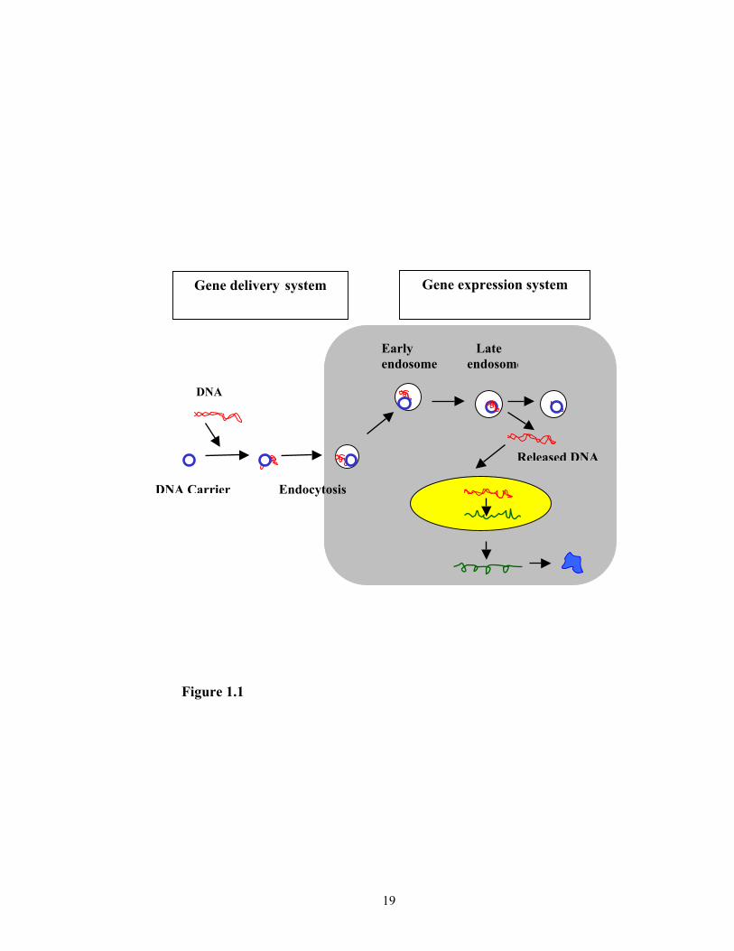
diagram of gene delivery and gene expression system is shown in Figure 1.1. The major
challenge to gene therapy is how to effectively deliver the gene to the target tissue and
then transport the gene to the nucleus of cells for DNA replication and transcription.
There are two major gene delivery systems, i.e. viral and nonviral vectors. The
main viral vectors include adenovirus, retrovirus, lentivirus, adeno-associated virus, and
herpes simplex virus. Most viral vectors show high transfection efficiency, however, their
applications are limited by the immune response and possible mutagenesis caused by the
insertion of viral vectors into the host chromosome. As an alternative gene delivery
system, nonviral vectors provide a safer profile. In addition, nonviral vectors are easier to
manufacture and control. The most commonly investigated nonviral vectors include
9
naked DNA, cationic liposomes, polymers, and protein based cations. The limitation to
the current available nonviral vectors for gene delivery lies in the relative low
transfection efficiency.
Among the nonviral gene delivery system, liposomes are the most widely studied
because their phospholipid compositions can facilitate the delivery of DNA by promoting
membrane fusion. Liposomes are classified according to charge, phospholipid
composition, pH-sensitivity, and structure. Because of their positive surface charge,
cationic liposome can condense the negatively charged DNA by electrostatic interaction
and will enhance the cellular uptake via endocytosis. In addition, cationic liposomes can
protect the DNA from the attack by DNases. Many investigations have shown that
cationic liposome can efficiently deliver DNA into target cells (Gao and Huang, 1996;
Zhou and Huang, 1994; Zabner et al., 1995; Xu and Szoka, 1996; Zelphati and Szoka,
1996; Tan et al., 2001). Currently, the major challenges for cationic liposomes for gene
delivery are the systemic barriers and cellular barriers. In addition, the toxicity of cationic
lipids also greatly limits the application of these delivery systems.
Besides the liposomal formulations, other lipid-based formulations may provide
new opportunities for gene delivery. By mimicking the natural chylomicron remnants, an
emulsion system was developed to deliver the hydrophobic DNA complex to liver of
mice and the delivered DNA was successfully expressed (Hara et al., 1997). This is the
first nonviral vector that resembles a natural lipoprotein carrier. Recently, Kim et al.
developed a new gene delivery system called Terplex system, which is based on a
complex formed by natural low-density lipoprotein (LDL) and stearyl-poly-L-lysine
(Kim et al., 1997; 1998). Through hydrophobic interaction, stearyl-poly-L-lysine can be
10
incorporated into the LDL particles. The assembled complex possessed positive charge
and was able to carry negatively charged DNA and successfully deliver the DNA into
vascular smooth muscle cells. Since this system requires natural LDL, its application was
limited by the availability of natural LDL.
Based on the chemical composition and structure of natural LDL, an artificial
lipoprotein based gene delivery system has been proposed in our lab. Similar to the
structure of natural lipoproteins, this artificial lipoprotein delivery system consists of
nanoemulsion cores made of natural lipids and a polar shell, and lipidized poly-L-lysine,
which replaces the surface protein as in natural lipoproteins. With the proper weight ratio
of poly-L-lysine to the lipids in a nanoemulsion, the artificial lipoprotein delivery system
will be able to carry DNA for gene delivery. The comparison of Terplex system and
artificial lipoprotein system is shown in Figure 1.2. Since the lipids used in the system are
all natural substances, the cytotoxicity of this delivery system was expected to be low.
Another advantage of this system is that it can be readily assembled using commercially
available materials including phospholipids, cholesterol and poly-L-lysine. In addition,
the chemical composition, particle size, and type of surface poly-peptide or surface
protein can be controlled and optimized, allowing widely-diversified gene or drug
delivery and targeting. The development and evaluation of this system for gene delivery
to human glioma cell line will be reported in Chapter 2.
5. Receptor-mediated drug delivery and targeting to cancer cells
The aim of drug targeting is to restrict the access of pharmacological agents to
selected cells within a tissue. The first step involved in this process is the recognition and
interaction of the carrier with specific target cells. The second step is the delivery of the
11
therapeutics into the target cells with little or no uptake by the non-target cells (Poste,
1983). This process is either cell-surface receptor mediated by ligand-receptor interaction
or cell-surface epitope mediated by antigen-antibody interaction. The cellular targeting is
largely dependent on the specificity of the target cell surface proteins.
In the past several decades, research on cancer treatment is still discouraging in
both laboratory and the clinic (Brun et al., 1997; Dunton, 1997). One reason for this is
that the cytotoxic anticancer therapy is largely dependent on the rapid dividing rate of
cancer cells and the anticancer drugs mostly act on the DNA, tubulin, and enzymes such
as the topoisomerase that are important to the DNA replication. Such drugs will also act
on the normal host cells and result in severe side effects. This will not only limit the dose
to kill the cancer cells but also cause the induction of drug resistance and metastasis.
Targeting the cytotoxic drugs to cancer cells becomes an apparent approach to solve
these problems in cancer therapy.
One of the most common methods for anticancer drug targeting is to employ the
monoclonal antibody against the tumor-associated antigens (Hellstrom and Hellstrom,
1997; Wick and Groner, 1997). Cytotoxic drugs could be attached to the lysine residues
that are distributed over the entire protein surface or the thiol groups generated by the
reduction of interchain disulfides at the hinge region (Jinno et al., 1996).
It is well known that mammalian cells have developed various mechanisms to
internalize specific substrates or targets into the cells. These mechanisms are collectively
termed as endocytosis which comprises phagocytosis, pinocytosis, receptor-mediated
endocytosis (clatherin-mediated), and potocytosis (non-clatherin-mediated) (Mukherjee
et al., 1997; Lehr, 1994). Receptor-mediated endocytosis is a highly specific biological
12

process which requires that specific binding and interaction between ligand and receptors.
Such a process is initiated by the binding of an exogenous ligand to a receptor across the
cell membrane with the binding domain oriented to the outside of the cell. When the
ligand binds to receptors, which cluster in domains in the plasma membrane known as
coated pits. These domains eventually invaginate to form coated vesicles. The loss of
coating with the aid of chaperone proteins will result in the formation of endosome,
which may further fuse with lysosome, in which the contents in the vesicles will be
degraded enzymatically.
The cell surface receptors are a group of complex transmembrane proteins. They
play very important roles in regulating cellular functions such as growth differentiation,
metabolism, secretion, contraction, and migration (Hirt et al. 1993). Although various
receptors are expressed in different types of cells, they share common structural features,
i.e. all the receptors have an extracellular ligand binding domain, a single hydrophobic
transmembrane domain, and a cytosolic domain that encodes for the endocytosis and
other functional signals (Schwartz, 1995). Since different cell types have different types
and levels of receptor and specific binding of ligand with receptor is dependent on the
extracellular binding domain, this provides the basis for targeted drug delivery. A specific
ligand can serve as a homing device for specific cell types by recognizing its cell surface
receptors.
It has been found that many cancer cells over-express some cell surface receptors
such as transferrin receptor (Plant et al. 1989; Huwyler et al., 1996), folate receptor
(Garin-Chesa et al. 1993; Ross et al. 1994; Lee and Low, 1994; Reddy et al., 1998), and
LDL receptor (Vitols et al, 1984, 1985, 1992; Rudling et al. 1990; Jung-Testas et al,
13
1992; Maletinska et al., 2000), to meet the increased cell proliferation and growth
requirement. So one strategy to develop targeted delivery system for cancer therapy is to
take advantage of these over-expressed cell surface receptor by conjugating the drug with
the ligands or by incorporating their corresponding ligands to a drug carrier such that the
anticancer drug can be specifically delivered to the cancer site.
Cholesterol is an essential component of cell membranes in mammalian cells.
Cells obtain cholesterol by taking up plasma LDL, the main cholesterol carrier in the
blood, via LDL receptor-mediated endocytosis (Brown and Goldstein, 1986) (Figure 1.3),
or by de novo synthesis of cholesterol. Rapidly dividing cells, e.g. cancer cells, require
more cholesterol for membrane growth. Elevated LDL receptors have been found in
many cancer cells (Vitols et. al, 1984, 1985, 1992; Rudling et. al. 1990; Jung-Testas et.
al, 1992; Maletinska et. al., 2000). This phenomenon provides the basis for the design of
targeted drug delivery systems. In our lab, a cholesterol-based anti-tumor compound,
cholesteryl 1,12-dicarba-closo-dodecaboranel-carboxylate (BCH) was synthesized for the
targeted drug delivery for boron neutron capture therapy (BNCT) (Ji et al., 2002). Early
research has demonstrated that BCH could be taken up by glioma cells SF-763 and SF-
767 and accumulated in high concentration when BCH was formulated into liposome
(Peacock et al., 2003). However, as a targeted drug delivery system for cancer therapy,
drugs should be delivered preferentially to cancer cells. So the research on the
comparison of cellular uptake of BCH by normal neuron cells and cancer cells was
conducted and the results are reported in Chapter3.
It has been reported that glioma cell lines SF-763 and SF-767 express high level
of LDL receptor on their cell surface (Maletinska et al., 2000). In order to clarify whether
14
the high uptake of BCH in liposome formulation by glioma cells is related to the elevated
LDL receptors, monoclonal antibodies against LDL receptors can provide direct evidence
whether LDL receptors are involved in the cellular uptake of BCH in liposome
formulations. Therefore, monoclonal anti-LDL receptor antibody was prepared and
purified. The purified product was applied during the experiments of cellular uptake of
BCH in a liposome formulation. BCH uptake by glioma cells in the presence and absence
of the monoclonal anti-LDL receptor antibody was determined and compared. On the
other hand, since the prerequisite for the receptor-mediated endocytosis is ligand-receptor
binding, the depletion of ligand will further demonstrate the role of receptor-mediated
pathway in the cellular uptake of BCH in the liposome formulation. Different
combinations of normal human serum and lipoprotein deficiency serum was
supplemented in the culture medium and the cellular uptake of BCH in liposome
formulation was determined. Since LDL-receptor mediated endocytosis is divalent metal
ion and temperature dependent, their effects on the cellular uptake of BCH in liposome
formulation by the glioma cell were also evaluated. These results are recorded in Chapter
4.
Although BCH could be preferentially delivered to cancer cells as compared to
normal neuron cells in the in vitro experiment, the anticancer activity of the delivered
boron compound can only be demonstrated by neutron irradiation, which was not
available in our research because of the unavailability of a neutron resource. In order to
demonstrate further whether the cholesterol-drug conjugate can be beneficial for the drug
targeting, a chemotherapeutic agent may provide direct evidence. Methotrexate is an
antimetabolite used in the treatment of cancer. Since methotrexate molecule has carbonyl
15
group, it can form an ester bond with the hydroxyl group in the cholesterol molecule. In
this research, methotrexate- cholesterol conjugate was synthesized and its cytotoxicity to
glioma cell line SF-767 was evaluated and compared with that of methotrexate. The
result of this project is reported in Chapter 5.
6. Biological protein nanostructures and targeted drug delivery
Targeted delivery of drugs to specific cells involves the specific interactions
between drugs or drug carriers and the cell surface proteins through ligand-receptor
interactions or antigen-antibody interactions. Targeted drug delivery to specific molecular
complexes or organelles within a cell requires the specific interactions of drug with the
targeted complexes to lead to the therapeutic effect. In the biological systems, these
interactions generally occur on various types of biological nanostructures of protein
origin and understanding and utilization of the biological nanostructures could lead to
significant improvement in drug targeting and drug carriers.
The biological protein nanostructures primarily include protein-lipid, protein-
protein, protein-carbohydrate, and protein-nucleic acid complexes. Proteins, are smaller
nanoscale molecules with typical size range between 1 and 20 nm (1). Through
sophisticated interactions with other biomolecules, these protein nanostructures are
formed and widely distributed in human body. For example, low-density lipoproteins
(LDL), with a diameter of 25-28 nm, are protein-lipid complexes. They are the major
circulatory nanostructures in the blood. As a drug carrier, these protein-lipid complexes
offer a certain advantage of being endogenous nanostructures that do not trigger
immunological response. They can also escape recognition and elimination by the
reticuloendothelial system (RES). On the other hand, glycoproteins, i.e. protein-
16
carbohydrate complexes, are vital structural and regulatory proteins in viruses and can
serve as important therapeutic targets for antiviral drug development. Telomerase, a
protein nanostructure formed from protein and nucleic acid, is activated only in cell
immortalization and cancer progression. Thus telomerase is an ideal therapeutic target for
anticancer therapy. Because protein nanostructures are so critical to various biological
and physiopathological activities, they have received wide attention in recent years in the
development of drug targeting strategies as drug carriers and therapeutic targets. In the
appendix, two aspects of biological protein nanostructures regarding their involvement in
targeted drug delivery will be discussed: (1) biological protein nanostructures as targeting
drug carriers and (2) biological protein nanostructures as therapeutic targets for new drug
development.
In summary, my research objectives are:
1. To develop and evaluate the nanoemulsion system for in vitro gene delivery to
glioma cells.
2. To compare the cellular uptake of BCH by normal cells and glioma cells.
3. To elucidate the mechanism of BCH uptake by glioma cells.
4. To evaluate and compare the cytotoxicity of methotrexate and methotrexate-
cholesterol on glioma cells
5. To review the role of biological protein nanostructures in targeted drug
delivery.
17

Figure Legends:
Figure 1.1. Working diagram of gene therapy
Figure 1.2. Comparison of terplex system and artificial lipoprotein system
Figure 1.3. LDL receptor-mediated endocytosis pathway in cells.
18
Gene expression system Gene delivery system
Endocytosis
DNA
Early Late endosome endosome
Lysosome
19
Released DNA
DNA Carrier Figure 1.1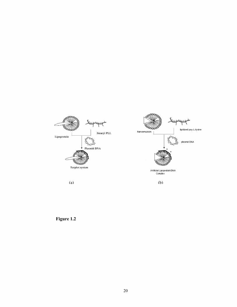
Figur
(a) (b)
e 1.2
20
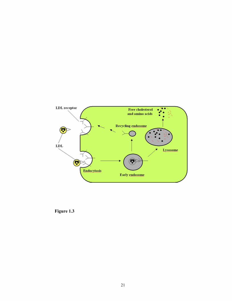
Figure 1.3
21
REFERENCES
1. Nakano M. Places of emulsions in drug delivery. Adv. Drug Deliv. Rev. 45:1-4
(2000).
2. Bangham A. D., Standish M. M., and Watkins J. C. Diffusion of univalent ions
across the lamellae of swollen phospholipids. J. Mol. Biol. 13:238-252 (1965).
3. Mahato R. I., Smith L. C., and Rolland A. Pharmaceutical perspectives of
nonviral gene therapy. Adv. Genet. 41:95-156 (1999).
4. Tomii Y. Lipid formulation as a drug carrier for drug delivery. Current Pharm.
Design 8:467-474 (2002).
5. Charman W. Lipids, lipophilic drugs, and oral drug delivery- some emerging
concepts. J. Pharm. Sci. 89(8):967-978 (2000).
6. Mizushima Y. Lipid microsphere (lipid emulsion) as a drug carrier- an overview.
Adv. Drug Deli. Rev. 20:113-115 (1996).
7. Constantinides P. P. Lipid microemulsions for improving drug dissolution and
oral absorption: physical and biopharmaceutical aspects. Pharm. Res.
12(11):1561-1572 (1995).
8. Ritschel W. A. Microemulsions for improved peptide absorption form the
gastrointestinal tract. Meth. Find. Exp. Clin. Pharmacol. 13:205-220 (1993).
9. Constantinides P. P., Scalart J. P., Lancaster C., Marcello J., Marks G., Ellens H.
and Smith P. L. Formulation and intestinal absorption enhancement evaluation of
water-in-oil microemulsions incorporating medium-chain glycerides. Pharm. Res.
11:1385-1390 (1994).
22
10. Constantinides P. P., Lancaster C. M., Marcello J., Chiossone D., Orner D.,
Hidalgo I., Smith P. L., Sarkahian A. B., Yiv S.H. and Owen A. J. Enhanced
intestinal absorption of an RGD peptide form water-in-oil microemulsions of
different composition and particle size. J. Control. Rel. 34:109-116 (1995).
11. Fevrie F., Bobin M. F., Lafforgue C., and Martii M. C. Advances in
microemulsions and transepidermal penetration of tyrosine. STP Pharm. Sci. 1:60
(1991).
12. Kriwet K. and Muller-Goymann C. C. Diclofenac release from phospholipid drug
systems and permeation through excised human stratum corneum. Int. J. Phar.
125:231-242 (1995).
13. Ktistis, G. Effect of polysorbate 80 and sorbitol concentration on in vitro release
of domethacin from microemulsions. J. Disp. Sci. 18(1):49 (1997)
14. Osborne, D. W., Ward A. J., and O’Neill K. Microemulsion as delivery vehicles:
1. Characterization of a model system. Drug Del. Ind. Pharm. 14:1203 (1988).
15. Friedman D. and Benita S. A mathematical model for drug release form o/w
emulsions: Application to controlled release morphine emulsions. Drug Dev. Ind.
Pharm. 13:2067 (1987).
16. Thevenin M. A., Grossiord J. L. and Poelman M. C. Sucrose esters/cosurfactant
microemulsion systems for transdermal penetration of tyrosine. STP Pharm. Sci.
1:60 (1991).
17. Arumugam R., Soriano H. E., Scheimann A. O., Reid B. S., Gopalakrishna G. S.,
Barakat O., Ozaki C. F., Wood P. R. Immunosuppressive therapy with
23
microemulsion cyclosporin A shortens the hospitalization of pediatric liver
transplant recipients. Clin. Transpl. 12:588-592 (1998).
18. Shinoda K., Araki M., Sadaghiani A., Khan A. and Lindman B. Lecithin-based
microemulsion: Phase behavior and microstructure. J. Phys. Chem. 95:989
(1991).
19. Trotta M., Cavalli R. E., Ugazio E. and Gasco M. R. Phase behavior of
microemulsion systems containing lecithin and lysolecithin as surfactants. Int. J.
Pharm. 143:67 (1998).
20. von Corswant C., Engstrom S. and Soderman O. Microemulsion based on
soybean phosphotidylcholine and triglycerides: Phase behavior and
microstructure. Langmuir 13:5061 (1997).
21. Block L. H. Medical applications. In: Remington: The Science and Practice of
Pharmacy, 19th ed. (A. R. Gennaro, et.), Mack, Easton, pp.1577-1597 (1995).
22. Ghanem A.-H., Higuchi W. I., and Simonelli A. P. Interfacial barriers in
interphase transport: retardation of the transport of diethylphthalate across the
hexadecane-water interface by an adsorbed gelatin film. J. Pharm. Sci. 58:165-
174 (1969).
23. Davis S. S. Washington C., West P., Illum L., Liversidge G., Sternson L., and
Kirsh R. Lipid emulsions as drug delivery systems. Ann. N. Y. Acad. Sci. 507:75-
88 (1987).
24. Muller H. C. Colloidal Carriers for Controlled Drug Delivery and Targeting:
Modification, Characterization and In Vivo Distribution, CRC Press, Boca Raton,
pp. 175-176 (1991).
24
25. Lasic D. D. Liposomes: from physics to applications. Amsterdam: Elsevier, 1993.
26. Roerdink, F. H., Daemen T., Bakker-Woudenberg I. A. J. M., Storm G.,
Crommelin D. J. A., and Scherphof G. L. Therapeutic Utility of Liposomes. In:
Drug Delivery Systems: Fundamentals and Techniques. Johnson P. and Lloyd-
Jones J. G. eds. Chichester, England: Ellis Horwood Ltd, pp. 66-80 (1987).
27. Ahmad et al. Antibody-targeted delivery of doxorubicin entrapped in sterically
stabilized liposomes can eradicate lung cancer in mice. Cancer Res. 53:1484-1488
(1993).
28. Mayhew F. J., Goldrosen R., and Vaage J. Effects of Liposome-entrapped
doxorubicin on liver metastases of mouse colon carcinoma 26 and 38. J. Natl.
Cancer Inst. 78:707-713 (1987).
29. Alving C. R. et al. Therapy of Leishmaniasis: Superior efficacies of liposome-
encapsulated drugs. Proc. Natl. Acad. Sci. USA 75:2959-2963 (1978).
30. Lopez-Berestein G. et al. Liposomal amphotericin B for the treatment of systemic
fungal infections in patients with cancer: a preliminary study. J. Infect. Dis.
51:704-710 (1985).
31. Gabizon A., Dagan A., Goren D., Barenholz Y., and Fuks Z. Liposomes as in
vivo carriers of adriamycin: reduced cardiac uptake and preserved anti-tumor
activity in mice. Cancer Res. 42:4734-4739 (1982).
32. Olson F., Mayhew E., Maslow D., Rustum Y., and Szoka F. Characterization,
toxicity, and therapeutic efficacy of adriamycin encapsulated in liposomes. Eur. J.
Cancer Clin. Oncol. 18:167-175 (1982).
25

33. Conley B. A., Egorin M. J., Whitacre M. Y., Carter D. C., Zuhowski E. G., Van
Echo D. A. Phase I and pharmacokinetic trial of liposome-encapsulated
doxorubicin. Cancer Chem. Pharm. 33:102-112 (1993).
34. Cowens J. W., Creaven P. J., Greco W. R., Brenner D. E., Tung Y., Ostro M.,
Pilkiewicz F., Gindberg R., Petrelli N. Initial clinical (phase I) trial of TLC D-99
(doxorubicin encapsulated in liposomes). Cancer Res. 53:2796-2802 (1993).
35. Woodle M. C., Newman M. S. and Cohen J. A. Sterically stabilized liposomes:
physical and biological properties. J. Drug Targeting 2:397-403 (1994).
36. Marjan M. J. Allen T. M. Long circulating liposomes: past, present and future.
Biotechnol. Adv. 14:151-175 (1996).
37. Leenders A. C. A. P. and De Marie S. The use of lipid formulation of
amphotericin B for systemic fungal infections. Leukemia 10:1570-1575 (1996).
38. Hiemenz J. W. and Walsh T. J. Lipid formulations of amphotericin B: recent
progress and future directions. Clin. Infect. Dis. 22(Suppl 2):S133-144 (1996).
39. Harding C. V., Collins D. S., Kanagawa O., Unanue E. R. Liposome-encapsulated
antigens engender lysosomal processing for class II MHC presentation and
cytosolic processing for class I presentation. J. Immunol. 147:2860-2863 (1991).
40. Collins D. S., Findlay K., Harding C. V. Processing of exogenous liposome-
encapsulated antigens in vivo generates class I MHC-restricted T cell responses.
J. Immunol. 148:3336-3341 (1992).
41. Walker C., Selby M., Erickson A., Cataldo D., Valensi J., Van Nest G. Cationic
lipids direct a viral glycoprotein into the class I major histocompatibility complex
antigen presentation pathway. Proc. Natl. Acad. Sci. USA 89:7915-7918 (1992).
26
42. Wassef N. M., Alving C. R., Richard R. L. Liposomes as carriers for vaccines.
Immuno. Methods 4:217-222 (1994).
43. Allison A. C. and Gregoriadis G. Liposomes as immunological adjuvants. Nature
252:252 (1974).
44. Gregoriadis G. and Allison A. C. Entrapment of proteins in liposomes prevents
allergic reactions in pre-immunized mice. FEBS Lett. 45:71-74 (1974).
45. Garcon N. M. J. and Six H. M. Universal vaccine carrier. Liposomes that provide
T-dependent help to weak antigens. J. Immunol. 146:3679-3702 (1991).
46. Ahmad I. et al. Antibody-targeted delivery of doxorubicin entrapped in sterically
stabilized liposomes can eradicate lung cancer in mice. Cancer Res. 53:1484-1488
(1993).
47. Brun B., Benchalal M., Lebas C., Piedbois P., Lin M., and Lebourgeois J. P.
Response to second-line chemotherapy in patients with metastasis breast
carcinoma previously responsive to first-line treatment: prognostic factors. Cancer
79:2137-2146 (1997).
48. Dunton C. J. New options for the treatment of advanced ovarian cancer. Semin
Oncol. 24:S2-11 (1997).
49. Wick B., and Groner B. Evaluation of cell surface antigens as potential targets for
recombinant tumor toxins. Cancer Lett. 118:161-172 (1997).
50. Hellstrom K. and Hellstrom I. Tumor antigens. In J. R. Bertino (Ed.),
Encyclopedia of Cancer, Vol. 1 (pp. 1810-1817). San Diego, CA: Academic
Press.
27
51. Jinno H., Ueda M., Enomoto K., Ikeda I., Kyriakos P. and Kitajima M.
Effectiveness of an adriamycin immunoconjugate that recognize the C-erb-2
product on breast cancer cell lines. Surg. Today 26:501-507 (1996).
52. Mukherjee S., Ghosh R. N., and Maxfield F. R. Endocytosis. Physiol. Rev.
77:759-803 (1997).
53. Lehr C. M. The transcytosis approach. In A. G. de Boer (ed.), Drug Absorption
Enhancement: Concepts, Possibilities, Limitations and Trends. Harwood,
Switzerland, pp. 325-365, 1994.
54. Hirt R. P., Hughes G. J., Frutiger S., Michetti P., Perregaux C., Poulain-Godefroy
O., Jeanguenat N., Neutra M. R., and Kraehenbuhl J. P. Transcytosis of the
polymeric Ig receptor requires phosphorylation of serine 664 in the absence but
not the presence of dimeric IgA. Cell 74:245-55 (1993).
55. Schwartz A. L. Receptor cell biology: receptor-mediated endocytosis. Pediatr.
Res. 38:835-843 (1995).
56. Nash R. A. Pharmaceutical Suspensions. In: Lieberman H., A, Rieger M. M., and
Banker G. S. (eds), Pharmaceutical Dosage Forms: Disperse Systems. Vol. 2,
pp.1-46 (1996).
57. Block L. H. Pharmaceutical Emulsions and Microemulsions. In: Lieberman H., A,
Rieger M. M., and Banker G. S. (eds), Pharmaceutical Dosage Forms: Disperse
Systems. Vol. 2, pp.47-109, Marcel Dekker (1996).
58. Gregoriadis G. Liposome research in drug delivery and targeting: thoughts of an
early participant. In: Lasic D. D. and Papahadjopoulos (ed), Medical Applications
of Liposomes, pp. 9-13, Elsevier (1998)
28
59. X. Gao, L. Huang. Potentiation of cationic liposome-mediated gene delivery by
polycations, Biochemistry 35(3): 1027-1036 (1996).
60. X. Zhou, L. Huang. DNA transfection mediated by cationic liposomes containing
lipopolylysine: characterization and mechanism of action, Biochim. Biophys.
Acta 1189(2):195-203 (1994).
61. J. Zabner, A. J. Fasbender, T. Moninger, K. A. Poellinger, M.J. Welsh. Cellular
and molecular barriers to gene transfer by a cationic lipid, J. Biol. Chem.
270:18997-19007 (1995).
62. Y. Xu, F. C. Szoka Jr. Mechanism of DNA release from cationic liposome/DNA
complexes used in cell transfection, Biochemistry 35(18):5616-5623 (1996).
63. O. Zelphati, F. C. Szoka Jr. Mechanism of oligonucleotide release from cationic
liposomes, Proc. Natl. Acad. Sci. USA 93(21):11493-11498 (1996).
64. Y. Tan, F. Liu, Z. Li, L. Huang. Sequential injection of cationic liposome and
plasmid DNA delivery gene effectively to the lung with minimal inflammatory
toxicity, Mol. Ther. 3(5):673-682 (2001).
65. Hara T., Tan Y., and Huang L. In vivo gene delivery to the liver using
reconstituted chylomicron remnants as a novel nonviral vector. Proc. Natl. Acad.
Sci. USA 94:14547-14552 (1997).
66. J.-S. Kim, A. Maruyama, T. Akaike, S. W. Kim. In vitro gene expression on
smooth muscle cells using a terplex delivery system, J. Control. Release 47:51-59
(1997).
67. J.-S. Kim, B.-I. Kim, A. Maruyama, T. Akaike, S. W. Kim. A new non-viral
DNA delivery vector: the terplex system, J. Control. Release 53:175-182 (1998).
29
68. Brown M. S. and Goldstein J. L. A receptor-mediated pathway for cholesterol
homeostasis. Science 232:34-47 (1986).
69. Vitols S., Gahrton G., Ost A. and Peterson C. Elevated low density lipoprotein
receptor activity in leukemia cells with monocytic differentiation. Blood,
63:1186-1193 (1984).
70. Vitols S., Gahrton, G., Bjorkholm M. and Peterson C. Hypocholesterolaemia in
malignancy due to elevated low-density-lipoprotein-receptor activity in tumor
cells: evidence from studies in patients with leukemia. Lancet, 2:1150-1153
(1985).
71. Vitols S., Peterson C., Larsson O., Holm P., and Aberg, B. Elevated uptake of low
density lipoprotein by human lung cancer tissue in vivo. Cancer Res. 52:6244-
6247 (1992).
72. Rudling M. J., Angelin B., Peterson C. O., and Collins V. P. Low density
lipoprotein receptor activity in human intracranial tumors and its relation to the
cholesterol requirement. Cancer Res. 50:483-487 (1990).
73. Jung-Testas I., Weintraub H., Dupuis D., Eychenne B., Baulieu D-E., and Robel
P. Low density lipoprotein-receptor in primary cultures of rat glial cells. J. Steroid
Biochem. Mol. Biol. 42:597-605 (1992).
74. Maletinska L., Blakely E. A. Bjornstad K. A., Deen D. F., Knoff L. J. and Forte
T. M. Human glioblastoma cell lines: levels of low-density lipoprotein receptor
and low-density lipoprotein receptor-related protein. Cancer Res. 60:2300-2303
(2000).
30
75. Ji B., Peacock G., and Lu D. R. Synthesis of cholesterol-carborane conjugate for
targeted drug delivery. Bioorg. Med. Chem. Lett. 12:2455-2459 (2002).
76. Lee R. J. and Low P. S. Delivery of liposomes into cultured KB cells via folate
receptor-mediated endocytosis. J. Biol. Chem. 269:3198-3204 (1994).
77. Watson-Clark R. A., Banquerigo M. L., Shelly K., Hawthorne M. F., and Brahn
E. Model studies directed toward the application of boron neutron capture therapy
to rheumatoid arthritis: boron delivery by liposomes in rat collagen-induced
arthritis. Proc. Natl. Acad. Sci. USA 95:2531-2534 (1998).
31
CHAPTER 2
IN VITRO GENE TRANSFECTION IN HUMAN GLIOMA CELLS
USING A NOVEL AND LESS CYTOTOXIC ARTIFICIAL LIPOPROTEIN
DELIVERY SYSTEM1
1Guangliang Pan, Mohannad Shawer, Svein Øie, D. Robert Lu. Published on
Pharmaceutical Research, 20(5):738-744 (2003).
32
ABSTRACT
Purpose. To develop and evaluate a novel artificial lipoprotein delivery system for in
vitro gene transfection in human glioma cells. Methods. A nanoemulsion was formulated
with lipid compositions similar to natural lipoproteins. The oil phase of the nanoemulsion
was composed of triolein (70%), egg phosphatidylcholine (22.7%),
lysophosphatidylcholine (2.3%), cholesterol oleate (3.0%) and cholesterol (2.0%). To
replace the surface protein as in natural lipoprotein, poly-L-lysine was modified to add
palmitoyl chains at a basic pH and was incorporated onto the nanoemulsion particles
through hydrophobic interaction. A model plasmid DNA, pSV-β-Gal containing a
reporter gene for β-galactosidase was carried by the nanoemulsion/poly-L-lysine
particles. The charge variation of the complex was examined by agarose gel
electrophoresis and zeta potential measurement. In vitro transfection was conducted on
human SF-767 glioma cell line using this new system. After standard X-Gal staining,
transfected cells were observed under light microscope. The effect of chloroquine on the
transfection was examined and, finally, the cytotoxicity of this new system was evaluated
in comparison with commercial Lipofectamine gene transfection system. Results. The
plasmid DNA was effectively carried by this artificial lipoprotein delivery system and the
reporter gene was expressed in the glioma cells. Transfection efficiency was significantly
increased by the treatment of chloroquine, indicating that endocytosis possibly was the
major cellular uptake pathway. Compared to Lipofectamine system, this new delivery
system demonstrated similar transfection efficiency but a much lower cytotoxicity. In the
experiment, the cell viability was up to 75% using this system compared to only 24%
using Lipofectamine system. Conclusion. A new artificial lipoprotein delivery system
was developed for in vitro gene transfection in tumor cells. The new system showed
similar transfection efficiency but a much lower cytotoxicity compared with the
commercial Lipofectamine system.
33

KEY WORDS: Gene delivery, transfection, glioma, palmitoyl poly-L-lysine,
nanoemulsion
ABBREVIATION: PLL, poly-L-lysine; p-PLL, palmitoyl poly-L-lysine; MTT, 3-(4,5-
dimethylthiazol-2-yl)-2,5-diphenyl tetrazolium bromide; ONPG, o-nitrophenyl-β-D-
galactopyranoside; X-Gal, 5-bromo-4-chloro-3-indoyl-β-D-galactoside; LDL, low-
density lipoprotein; PBS, phosphate-buffered saline
34
INTRODUCTION
Gene transfection can be defined as the delivery to and subsequent expression of
functional genetic material in specific cells to manipulate their intrinsic genetic profiles.
During the last decade, research involving gene transfection has been expanding very
rapidly and many gene delivery systems have been developed to efficiently transfect
various cells in both in vitro and in vivo experimental conditions. To be an effective gene
delivery system, it must be able to carry sufficient amount of genetic material and express
the genetic information in specific cells resulting in a significant changes in genetic
profiles. In general, genetic materials can be carried and expressed in specific cells by
either viral vector systems or non-viral vector systems. The viral vector systems,
including retrovirus, adenovirus, adeno-associated virus, herpes simplex virus and
lentivirus, have been extensively investigated owing to their high transfection efficiency.
However, their applications are limited by their complicated handling procedures for in
vitro experiments and poor safety profiles for in vivo studies (1-2). Compared to the viral
vector systems, the non-viral vector systems are easy to handle and have better safety
profiles. Consequently, the development of effective non-viral gene delivery systems has
become the the aim of many research laboratories (3-5).
Since lipids are the main components of cell membranes, most non-viral vectors
are lipid-based so that the vectors can be effectively incorporated into the cell membranes
and facilitate the delivery of genetic materials into specific cells. Among these non-viral
vectors, cationic liposomes, which carry positive charge and electrostatically interact with
negatively charged DNA to form complexes, are the most widely studied (6-11).
However, the success of using cationic liposomes for gene transfection is partly
35
hampered by the cytotoxicity of the cationic lipids. Polymer-based non-viral vectors have
also been widely investigated, including poly-L-lysine, polyethenimine, polyamidoamine
dendrimer, and chitosan (12-20). One main disadvantage of these systems is the low
efficiency of transfection. Recently, Kim et al. developed a new gene delivery system
called Terplex system, which is based on a complex formed by natural low-density
lipoprotein (LDL) and stearyl-poly-L-lysine (21-22). Through hydrophobic interaction,
stearyl-poly-L-lysine can be incorporated into the LDL particles. The assembled complex
possesses a positive charge and was able to carry negatively charged DNA and
successfully deliver the DNA into vascular smooth muscle cells.
In this paper, we report the development and evaluation of a novel artificial
lipoprotein delivery system that can carry DNA materials for effective in vitro gene
transfection in tumor cells. Similar to the structure of natural lipoproteins, this artificial
lipoprotein delivery system consists of nanoemulsion cores made of natural lipids and
surface lipidized poly-L-lysine, which replaces the surface protein as in natural
lipoproteins. With the proper weight ratio of poly-L-lysine to the lipids in the
nanoemulsion, the artificial lipoprotein delivery system efficiently carried plasmid DNA
containing β-galactosidase gene and transfected human SF-767 glioma tumor cells. Our
experiments showed that because the lipids used in the system are all natural substances,
the cytotoxicity of this delivery system could be significant lower than the commercial
gene transfection systems using cationic liposomes. The benefit associated with the low
cytotoxicity makes it especially useful as an alternative to Lipofectamine or other
commercial gene transfection systems. Another advantage of this system is that it can be
readily assembled using commercial available materials including phospholipids,
36
cholesterol and poly-L-lysine. The chemical composition, particle size and type of
surface poly-peptide or surface protein can be controlled and optimized allowing widely-
diversified gene or drug delivery applications.
MATERIALS AND METHODS
Materials
Triolein (99%), egg yolk phosphatidylcholine (99%), cholesterol (99%), poly-L-
lysine hydrobromide (MW 57900 Dalton based on viscosity), chloroquine (99%), o-
nitrophenyl-β-D-galactopyranoside (ONPG), and 3-(4,5-dimethylthiazol-2-yl)-2,5-
diphenyl tetrazolium bromide (MTT) were purchased from Sigma (St. Louis, MO, USA).
L-α-lysophosphatidylcholine (99%) was purchased from Avanti (Alabaster, AL, USA).
Cholesterol oleate (99%) was obtained from Acros (Pittsburgh, PA). 5-bromo-4-chloro-3-
indoyl-β-D-galactoside (X-Gal) was from Life Technologies (Rockville, MD, USA).
Electrophoretic grade agrose was purchased from FMC Bioproducts (Rockland, ME,
USA). All other chemicals were of analytical grade obtained from Sigma or J.T. Baker
(Phillipsburg, NJ, USA).
Preparation of the nanoemulsion
The oil phase of the emulsion was composed of triolein (70%), egg
phosphatidylcholine (22.7%), lysophosphatidylcholine (2.3%), cholesterol oleate (3.0%)
and cholesterol (2.0%). The lipid components were dissolved in chloroform individually
and then mixed thoroughly. The chloroform was then removed completely by a stream of
nitrogen gas. In each 100 mg of lipid mixture, 10 ml of 2.4 M NaCl solution was added.
The mixture was sonicated under nitrogen flow for 30 min using Model 450 Sonifier
(Branson Ultrasonics Corporation, Danbury, CT) with a duty cycle dial setting of 90% at
37
output of 40 watts. The temperature of the mixture was maintained at 55ºC during
sonication. The prepared emulsion was then passed to times through an Emulsiflex B3
device (Avestin, Ontario, Canada) at a pressure of 70 psi to reduce the particle size to
nanometer level. The emulsion was dialyzed against phosphate-buffered saline (PBS)
using Spectra/Por 2 molecularporous membrane tubing with molecular weight cut-off
of 6000-8000 dalton (Spectrum Medical Industries, Inc. Houston, Texas). The emulsion
particle size distribution was measured by Submicron Particle Sizer Autodiluter Model
370 (NICOMP Particle Sizing Systems, San Barbara, CA). In addition, the nanoemulsion
was stored at room temperature and the particle size distribution was measured in 2, 4, 8,
16 weeks, respectively, to examine the stability of the nanoemulsion particles.
Lipidization of poly-L-lysine
Lipidization of poly-L-lysine was performed as described by Kim et al. (21) with
slight modification. Briefly, poly-L-lysine hydrobromide (30 mg) was dissolved in 2 ml
DMSO in a 50 ml round-bottom flask. After triethylamine (10 µl) was added, palmitoyl
chloride (20 mg) was added to the mixture to react with the amino group of the lysine
residues in poly-L-lysine. The mixture was allowed to react at room temperature for 2 hrs
and filtered. Acetone was added to the filtrate to precipitate the lipidized polymer,
palmitoyl poly-L-lysine or abbreviated as p-PLL. The product was dissolved in methanol,
re-precipitated by acetone, and dried under vacuum overnight. The modified polymer was
characterized by proton NMR.
Incorporation of p-PLL into nanoemulsion particles
In each 1.5 ml microcentrifuge tube, nanoemulsion (50 µl) was diluted with 0.2
ml PBS solution and incubated with various amount of p-PLL at 37ºC based on the
38

weight ratio of p-PLL to triolein in nanoemulsion. The weight ratios of p-PLL to triolein
in the mixture were 0.125:1, 0.25:1, 0.5:1, and 1:1, respectively. After incubation for 1
hour, the mobility of the nanoemulsion particles in electric field was examined by
agarose gel electrophoresis using Nile Red as the fluorescent dye. Agarose gel (0.4%)
was prepared in TAE buffer (40 mM Tris-acetic acid, 1 mM EDTA, pH 8.0). Five µl of
Nile Red solution in acetone (100 µg/ml) was dried out in test tube and redissolved in 30
µl of the incubation mixture as described above. In each sample, 6 µl of glycerine was
added to increase the density of the sample and the sample (30 µl) was loaded in each
sample well of the agarose gel. Electrophoresis was conducted for 1 hr at 70 volts at
room temperature using Horizontal Mini-gel System (CBS Scientific Company Inc. Del
Mar, CA, USA). The mobility of the particles in an electric field was visualized by an
Eagle Eye II Video System (Stratagene, CA, USA).
Amplification and purification of plasmid DNA
Plasmid DNA, pSV-β-Galactosidase Control Vector (Figure 2.1), was purchased
from Promega (Madison, WI) and was introduced into Epicurian Coli XL1-Blue MRF’
(Stratagene, CA) by using standard transformation protocol. The transformed E. coli
strain was maintained in Luria-Bertani (LB) medium containing 15% of glycerol at –
80ºC. To amplify the plasmid DNA, the E. coli strain was cultured in LB medium
containing 100 unit/ml of ampicillin at 37ºC overnight and the cells were harvested by
centrifugation. Plasmid DNA in the cells was extracted and purified using Wizard Plus
SV Minipreps DNA Purification System (Promega, Madison, WI). The purity of plasmid
DNA was confirmed by determining the ratio of optical absorbance at 260 nm and 280
nm (>=1.8) and further by 0.6% agarose gel electrophoresis. The agarose gel was stained
39
with ethidium bromide (0.5 µg /ml) for 15 minutes and destained with deionized water
for 10 minutes. DNA bands in the agarose gel were visualized by the Eagle Eye II Video
System. The concentration of plasmid DNA was determined by spectrophotometer at
wavelength of 260 nm (1 OD260 ≈ 50 µg/ml).
Assembly of the complex of nanoemulsion, p-PLL and plasmid DNA
Nanoemulsion (50 µl) in 0.2 ml PBS was mixed with various amounts of p-PLL
in the same way as described above. The weight ratios of p-PLL to triolein in the
nanoemulsion were 0.0625:1, 0.125:1, 0.25:1, 0.5:1 and 1:1, respectively. After
incubation at 37ºC for 1 hour, DNA (2 µg) was added and incubated at room temperature
for 15 minutes. Samples were then loaded into 0.4% agarose gel and the electrophoresis
was performed as described above. Zeta potential and mobility of the assembled particles
were measured by Submicron Particle Size Analyzer 90Plus (Brookhaven Instrument
Corporation, Holtsville, NY, USA). Before they were measured for zeta potential, the
samples were diluted with sodium nitrate solution (1mM, pH 7.4) until the count of
particles in the sample reached 100-300 kilo-counts per second (KCPS). Water and
solutions used in zeta potential measurement were filtered with 0.1 µm Supor Acrodisc
(Gelman Sciences, Ann Arbor, MI, USA). The particle size, zeta potential and mobility
was recorded by the built-in PC computer system.
Gene transfection experiment
Human glioma cell line SF-767 was obtained from the tissue bank of Brain
Tumor Research Center (University of California-San Francisco, San Francisco, CA,
USA) and used in our transfection experiment because of its characteristics of aggressive
growth. The cells were grown at 37oC in 5% CO2 with Eagle’s Minimal Essential
40
Medium (EMEM) medium supplemented with 10% fetal bovine serum (BioCell
Laboratories, Rancho Dominguez, CA, USA), 100 units/ml penicillin, and 100 µg/ml
streptomycin. Culture was passaged twice a week to maintain the cells in exponential
growth. The transfection was conducted in 6-well (35-mm in diameter) culture plates. SF-
767 cells were seeded with 3 x 10⁵ cells in each well 24 hours before the transfection.
During the day of transfection, nanoemulsion (50 µl) in 0.2 ml PBS was mixed with p-
PLL in the ratios as described above, and incubated at 37ºC. After 1 hour of incubation, 2
µg of plasmid DNA was added and incubated at room temperature for 15 minutes to
obtain the complex of nanoemulsion/p-PLL/DNA. Cells were washed with PBS buffer
for three times and 1 ml of EMEM (without serum and antibiotics) was added to each
well. The nanoemulsion/p-PLL/DNA complex was added to each well and mixed with
the medium completely by swirling. The cells were then incubated at 37ºC in 5% CO2 for
12 hours before 1 ml EMEM medium containing 20% fetal bovine serum was
supplemented. The cells were incubated for additional 24 hours. Both the
nanoemulsion/p-PLL complex and the naked DNA were used, respectively, as the
negative controls. As the positive control, Lipofectamine™ reagent purchased from
Invitrogen (Carlsbad, CA, USA) was incubated with the DNA and the transfection
experiment was performed at the same condition.
Detection of β-galactosidase by X-Gal staining and enzymatic assay
After 24 hours of transfection incubation, the cells in each well were washed
twice with PBS buffer and then fixed with 2 ml fixing solution (2% formaldehyde and
0.2% glutarldehyde in PBS buffer) for 15 minutes at room temperature. After the cells
were washed for three times with PBS, 1.5 ml of staining solution (5 mM potassium
41
ferricyanide, 5 mM potassium ferrocyanide, 1 mM MgSO4, 1 mg/ml X-Gal from 20
mg/ml stock in dimethyl formamide) was added. The cells were incubated at 37ºC in 5%
CO2 for 30 hours before the transfection was evaluated by light microscope. β-
Galactosidase activity of cells was determined by β-Galactosidase Enzyme Assay System
(Promega, Madison, WI, USA). In brief, the cells from each well of the plate were
trypsinized and collected by centrifugation. They were disrupted by mixing with 200 µl
of Lysis Buffer and incubating for 30 minutes at room temperature. The protein
concentration of the cell lysate was determined by the Bradford method (23). Cell lysate
(100 µl) was mixed with 100 µl of ONPG solution in 2X Assay Buffer (1.33 mg/ml) and
incubated in water bath at 37ºC for 5 hours. The enzymatic reaction was terminated by
adding 300 µl of 1 M sodium carbonate solution. After the reaction mixture was diluted,
the absorbance at 420 nm was read in a spectrophotometer (Baush & Lomb Spectronic
2000, Rochester, NY, USA). The enzymatic activity unit was defined in a similar way as
described by Kim et al. (21).
Effect of chloroquine on the transfection efficiency
In order to demonstrate the effect of chloroquine, which is a lysomotropic agent,
on the transfection efficiency, chloroquine solution in PBS was added to the cell culture
(80% confluent) with a final concentration of 100 µM. After 30 minutes of incubation at
37ºC and 5% CO2, the culture medium was removed and the cells were washed with PBS
for three times. Fresh medium was supplemented before the transfection experiment was
started. The complex of nanoemulsion (50 µl in 0.2 ml PBS) with p-PLL (p-PLL:triolein
= 0.25:1) was used as the carrier for 2 µg of plasmid DNA in the transfection experiment.
42

After transfection incubation, the cells were trypsinized and collected by centrifugation.
The β-galactosidase activity was measured by the method as described above.
Cytotoxicity comparison of nanoemulsion/p-PLL complex and Lipofectamine
Cellular toxicity of the nanoemulsion system was tested according to the MTT
method reported by Mosmann (24). A complex of nanoemulsion (50 µl in 0.2 ml PBS)
with p-PLL (p-PLL:triolein = 0.25:1) was prepared in similar way as described above. In
a 96-well microplate, SF-767 cells were seeded at 2 x 10⁴ cells in each well containing
0.1 ml of EMEM medium. After 24 hours of incubation at 37ºC and in 5% CO2, the
nanoemulsion/p-PLL complex or Lipofectamine reagent was added to the cell culture.
The amount of nanoemulsion/p-PLL complex or Lipofectamine reagent added to the
cell culture was determined such that similar transfection efficiency could be obtained
based on the transfection experiments. The cell culture grown on EMEM medium
without nanoemulsion/p-PLL complex or Lipofectamine reagent was used as the control.
Since only living cells are able to cleave the tetrazolium ring to produce dark blue
crystals, which can be measured colorimetrically, the viability of cells after additional 1,
2, 3, and 4 days of growth was determined by measuring the ability of the cells to
degrade tetrazolium salt MTT. Briefly, 25 µl of MTT solution in PBS buffer (0.5 mg/ml)
was added to each well of culture and incubated at the same condition for additional 4
hours. The medium was then removed and 150 µl of DMSO was added to each well and
mixed thoroughly until all the dark blue crystals were dissolved. The plate was read on an
OPTImax Tunable Microplate Reader (Molecular Devices Corporation, Sunnyvale, CA,
USA) at wavelength of 550 nm. The cell viability was calculated and expressed as
(ODtrt/ODctrl ) x100%, where ODtrt is the optical absorbance from the culture treated with
43
either nanoemulsion/p-PLL complex or Lipofectamine reagent, and ODctrl is the
optical absorbance from the culture control.
RESULTS
Preparation of the nanoemulsion and its size distribution
With the proper control of temperature (55ºC) during sonication, the obtained
emulsion appeared to be homogeneous. The number weighted mean size of the emulsion
particle was 110.2 ± 42.9 nm. Since the emulsification was conduced by metal probe
sonication, chelating agent (0.1 mM EDTA) was added to remove the free iron ion before
it is dialyzed against PBS overnight (PBS changed every 6 hours). Followed by 10 cycles
of size reduction by Emusiflex B3 device (Avestin, Ottawa, Canada), the number
weighted mean particle size was reduced to 48.9 ± 19.8 nm. The particle size distribution
of the emulsion is shown in Figure 2.2.
The physical stability of the nanoemulsion can be investigated by measuring the
change in size distribution of the particles. The size distribution of the nanoemulsion
particles was measured at 0, 2, 4, 8, 16 weeks after the preparation and results are shown
in Figure 2.3. The size distribution did not change significantly upon storage at room
temperature. After 16 weeks of storage at room temperature, the mean size of the
emulsion particles was 78.9 ± 14.6 nm, indicating that the nanoemulsion particles are
rather stable.
Incorporation of p-PLL into nanoemulsion particles
Since p-PLL is positively charged, the incorporation of p-PLL molecules into
nanoemulsion particles will result in a change in surface charge of the particles. The
change can be clearly seen in the picture of agarose electrophoresis (Figure 2.4).
44
Nanoemulsion particles moved to anode since they were negatively charged (Lane 1).
The negative surface charge of the nanoemulsion particles was also confirmed by the zeta
potential and mobility, which was – 42.28 ± 2.3 mV and –3 ± 0.06 (m/s)/(V/cm),
respectively. The incorporation of p-PLL neutralized the surface charge (Lane 2 to Lane
4) and resulted in the retardation of movement in the electric field. When they were
incubated with sufficient amount of p-PLL, the surface charge of particles was reversed
to be positive and moved towards opposite direction in the electric field (Lane 5). The
results indicated that p-PLL could be incorporated into the nanoemulsion particles.
Interaction of p-PLL associated nanoemulsion with DNA
After incorporation of a sufficient amount of p-PLL molecules into the
nanoemulsion particles, the complex carried a positive charge and could electrostatically
interact with negatively charged DNA molecules. As indicated in Figure 2.5, plasmid
DNA (Lane 1) migrated towards the positive anode. When DNA plasmid was incubated
with p-PLL (Lane 2), no DNA migration was observed, possibly because DNA molecules
were bound by p-PLL and thus the ethidium bromide molecules could not intercalate into
the DNA molecules resulting in no fluorescence emission. Lane 3 to Lane 7 showed the
change in DNA carrying capability of the complex resulted from different ratios of p-
PLL to nanoemulsion (i.e. the p-PLL to triolein ratio). At a high ratio of p-PLL to
nanoemulsion, DNA was tightly held by the complex and thus no DNA migration band
appeared (Lane 3 to Lane 6). When the ratio of p-PLL to nanoemulsion became
sufficiently low (0.0625:1 as the p-PLL to triolein ratio), plasmid DNA started to escape
from the complex and a free DNA band (Lane 7) appeared in the agarose gel.
45
Since the surface charge of the nanoemulsion/p-PLL/DNA complex is very
important to transfection, the zeta potential and mobility of these complexes were
measured and the results are shown in Figure 2.6 and Figure 2.7. With fixed amount of
plasmid DNA (2 µg), the increased amount of p-PLL led to an increase in zeta potential
of the particles.
Transfection of glioma cell line SF-767 by the complex of nanoemulsion/p-PLL/DNA
Most of the positively charged nanoemulsion/p-PLL/DNA complexes (with
varying ratios of nanoemulsion/p-PLL/DNA) used in the experiments were found to
transfect the glioma SF-767 cells, but with different transfection efficiency. The complex
containing nanoemulsion and p-PLL (p-PLL:triolein = 0.25:1) and 2 µg DNA, which had
a zeta potential of 8.47 ± 1.85 mV and a loading capacity of 1 µg DNA per 0.25 mg of
lipid (or per 50 µl of formulation), had the highest transfection efficiency. Its efficiency
was comparable to that by Lipofectamine reagent (Figure 2.8). Under the microscope,
those cells that expressed active β-galactosidase appeared to be blue-green (as dark spots
in Figure 2.8) by X-Gal staining and the extent of transfection appeared to be comparable
for the nanoemulsion complex (Figure 2.8, C) to that by Lipofectamine™ reagent (Figure
2.8, A). However, Lipofectamine™ reagent appeared to be much more toxic than the
nanoemulsion complex, as indicated by the significant difference in cell counts.
Effect of chloroquine on the transfection
Cellular uptake of particles via endocytosis will result in the particles being
processed by endosomal-lysosomal pathway. This pathway will lead to the degradation of
the carried plasmid DNA and greatly lower the transfection efficiency. Chloroquine is a
weakly basic, lysomotropic drug, which will interfere with endosomal acidification and
46
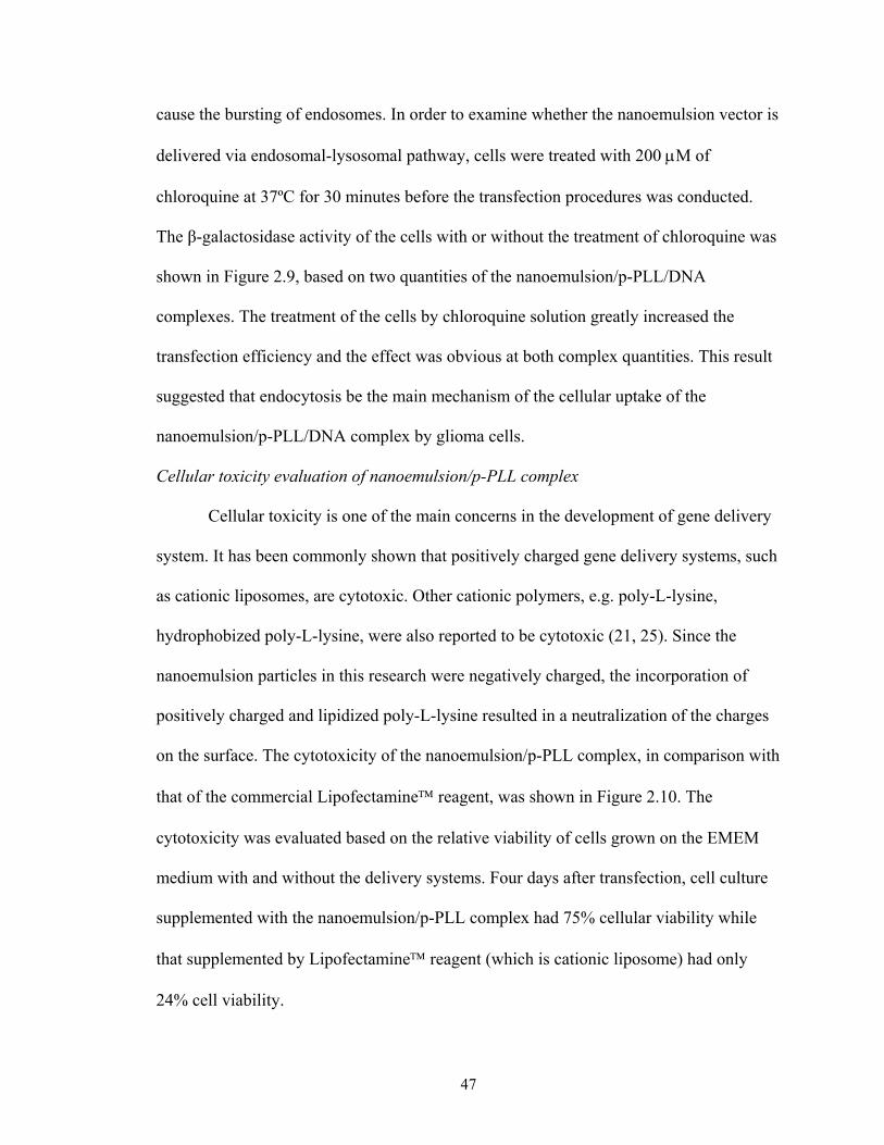
cause the bursting of endosomes. In order to examine whether the nanoemulsion vector is
delivered via endosomal-lysosomal pathway, cells were treated with 200 µM of
chloroquine at 37ºC for 30 minutes before the transfection procedures was conducted.
The β-galactosidase activity of the cells with or without the treatment of chloroquine was
shown in Figure 2.9, based on two quantities of the nanoemulsion/p-PLL/DNA
complexes. The treatment of the cells by chloroquine solution greatly increased the
transfection efficiency and the effect was obvious at both complex quantities. This result
suggested that endocytosis be the main mechanism of the cellular uptake of the
nanoemulsion/p-PLL/DNA complex by glioma cells.
Cellular toxicity evaluation of nanoemulsion/p-PLL complex
Cellular toxicity is one of the main concerns in the development of gene delivery
system. It has been commonly shown that positively charged gene delivery systems, such
as cationic liposomes, are cytotoxic. Other cationic polymers, e.g. poly-L-lysine,
hydrophobized poly-L-lysine, were also reported to be cytotoxic (21, 25). Since the
nanoemulsion particles in this research were negatively charged, the incorporation of
positively charged and lipidized poly-L-lysine resulted in a neutralization of the charges
on the surface. The cytotoxicity of the nanoemulsion/p-PLL complex, in comparison with
that of the commercial Lipofectamine reagent, was shown in Figure 2.10. The
cytotoxicity was evaluated based on the relative viability of cells grown on the EMEM
medium with and without the delivery systems. Four days after transfection, cell culture
supplemented with the nanoemulsion/p-PLL complex had 75% cellular viability while
that supplemented by Lipofectamine reagent (which is cationic liposome) had only
24% cell viability.
47
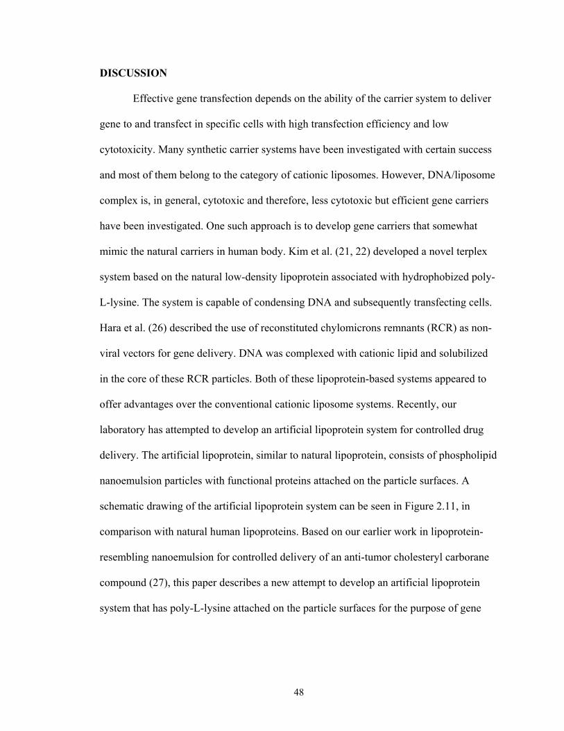
DISCUSSION
Effective gene transfection depends on the ability of the carrier system to deliver
gene to and transfect in specific cells with high transfection efficiency and low
cytotoxicity. Many synthetic carrier systems have been investigated with certain success
and most of them belong to the category of cationic liposomes. However, DNA/liposome
complex is, in general, cytotoxic and therefore, less cytotoxic but efficient gene carriers
have been investigated. One such approach is to develop gene carriers that somewhat
mimic the natural carriers in human body. Kim et al. (21, 22) developed a novel terplex
system based on the natural low-density lipoprotein associated with hydrophobized poly-
L-lysine. The system is capable of condensing DNA and subsequently transfecting cells.
Hara et al. (26) described the use of reconstituted chylomicrons remnants (RCR) as non-
viral vectors for gene delivery. DNA was complexed with cationic lipid and solubilized
in the core of these RCR particles. Both of these lipoprotein-based systems appeared to
offer advantages over the conventional cationic liposome systems. Recently, our
laboratory has attempted to develop an artificial lipoprotein system for controlled drug
delivery. The artificial lipoprotein, similar to natural lipoprotein, consists of phospholipid
nanoemulsion particles with functional proteins attached on the particle surfaces. A
schematic drawing of the artificial lipoprotein system can be seen in Figure 2.11, in
comparison with natural human lipoproteins. Based on our earlier work in lipoprotein-
resembling nanoemulsion for controlled delivery of an anti-tumor cholesteryl carborane
compound (27), this paper describes a new attempt to develop an artificial lipoprotein
system that has poly-L-lysine attached on the particle surfaces for the purpose of gene
48

delivery. The cytotoxicity of such a system has been specifically investigated in
comparison with commercial Lipofectamine reagent.
The lipoprotein-resembling particles were made of commercially available lipids
and lipidized poly-L-lysine. The lipidization of poly-L-lysine was achieved through N-
alkylation of the free ε-amino groups with palmitoyl chloride and confirmed via proton
NMR (data not shown). The reaction condition was controlled to only lipidize about 25%
of the lysine residues preserving sufficient amount of free ε-amino groups for
maintaining the ability of poly-L-lysine to condense DNA. The nanoemulsion particles
carried negative surface charge as shown by agarose gel electrophoresis in Figure 2.4.
When nanoemulsion particles are combined with unlipidized poly-L-lysine, immediate
precipitation was observed indicating the formation of large aggregates due to charge
neutralization. When appropriate amount of p-PLL was incubated with the particles, no
precipitation or change in the turbidity was observed. These results indicate that with
lipidized poly-L-lysine the interaction was not merely through electrostatic interaction,
but also through hydrophobic interaction between the palmitoyl chains of p-PLL and the
phospholipid of nanoemulsion particles. The charge of the nanoemulsion particles
became reversed when sufficient amount of p-PLL was added (Figure 2.4).
The surface charge of the complex formed by nanoemulstion and p-PLL depends
on their relative ratio. In order to carry DNA molecules, which are negatively charged,
the carrier needs to be positive. On the other hand, the surface charge of the complex
after DNA is incorporated is also critical. Since the cell surface is negatively charged, a
positively charged nanoemulsion/p-PLL/DNA complex is essential for successful
transfection. The surface charge of these particles can be monitored by agarose gel
49

electrophoresis qualitatively or by zeta potential and mobility measurement quantitatively
(Figure 2.5, 2.6 and 2.7). Our studies showed that most of the positively charged
complexes formed by varying the ratios of nanoemulsion, p-PLL and DNA can transfect
the glioma cells to a certain extent. The complex containing nanoemulsion, p-PLL and
DNA with a zeta potential of 8.47 ± 1.85 mV achieved the highest transfection efficiency
indicating the proper charge balance among these components was important for
transfection. This observation is consistent with that reported by Kim et al. (21) when the
terplex carrier system involving natural LDL was employed for gene delivery.
Chloroquine has been widely used to investigate the cellular uptake mechanism
(28-29). It will interact with endosome inside the cell. A positive correlation of
chloroquine level with the transfection efficiency indicates that the DNA is taken up
through endocytosis and, furthermore, the endosomal-lysosomal pathway. Through
chloroquine treatment, we have shown that the endocytosis appears the major cellular
uptake pathway for the lipoprotein-resembling gene carrier.
Cytotoxicity is an important consideration for developing novel gene delivery
systems. Using new gene carriers that mimic the nature substance such as human
lipoproteins, we can significantly reduce the cytotoxicity associated with the delivery
systems. It is known that poly-L-lysine is very toxic to cells (Morgan, 1989). Its complex
with the phospholipid nanoemulsion particles and plasmid DNA, however, has low
cytotoxicity as indicated by our experiments in comparison with the Lipofectamine
system. The nature of phospholipids, the neutralization of the positive charge of poly-L-
lysine and the proper balance among nanoemulsion, p-PLL and DNA apparently
contribute to the reduced cytotoxicity for this new gene delivery system.
50

In conclusion, a novel artificial lipoprotein system has been developed for in vitro
gene transfection to tumor cells. The system mimics natural lipoprotein in composition
but contains lipidized poly-L-lysine (instead of surface protein) to carry genetic materials.
Such a system can be conveniently formulated from natural lipids, with the ability to
control the size and surface charge. With proper ratios among its components, the new
gene delivery system shows similar transfection efficiency but a lower cytotoxicity
compared with the commercial Lipofectamine gene transfection system, making it
especially useful as an alternative to these commercial gene transfection systems.
51
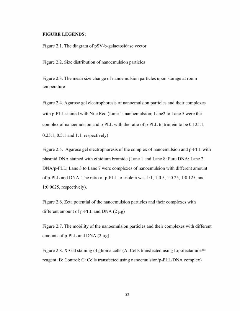
FIGURE LEGENDS: Figure 2.1. The diagram of pSV-b-galactosidase vector Figure 2.2. Size distribution of nanoemulsion particles
Figure 2.3. The mean size change of nanoemulsion particles upon storage at room
temperature
Figure 2.4. Agarose gel electrophoresis of nanoemulsion particles and their complexes
with p-PLL stained with Nile Red (Lane 1: nanoemulsion; Lane2 to Lane 5 were the
complex of nanoemulsion and p-PLL with the ratio of p-PLL to triolein to be 0.125:1,
0.25:1, 0.5:1 and 1:1, respectively)
Figure 2.5. Agarose gel electrophoresis of the complex of nanoemulsion and p-PLL with
plasmid DNA stained with ethidium bromide (Lane 1 and Lane 8: Pure DNA; Lane 2:
DNA/p-PLL; Lane 3 to Lane 7 were complexes of nanoemulsion with different amount
of p-PLL and DNA. The ratio of p-PLL to triolein was 1:1, 1:0.5, 1:0.25, 1:0.125, and
1:0.0625, respectively).
Figure 2.6. Zeta potential of the nanoemulsion particles and their complexes with
different amount of p-PLL and DNA (2 µg)
Figure 2.7. The mobility of the nanoemulsion particles and their complexes with different
amounts of p-PLL and DNA (2 µg)
Figure 2.8. X-Gal staining of glioma cells (A: Cells transfected using Lipofectamine
reagent; B: Control; C: Cells transfected using nanoemulsion/p-PLL/DNA complex)
52
Figure 2.9. The effect of chloroquine on transfection by the nanoemulsion gene delivery
system (white bar – untreated with chloroquine; gray bar – treated with chloroquine)
Figure 2.10. Cytotoxicity comparison between nanoemulsion/p-PLL and Lipofectamine:
control (♦, top), nanoemulsion/p-PLL (■, middle) and Lipofectamine (▲, bottom). Figure 2.11. (A) Artificial lipoproteins (20-100 nm) and (B) natural human lipoproteins (those in 20-100 nm range)
53
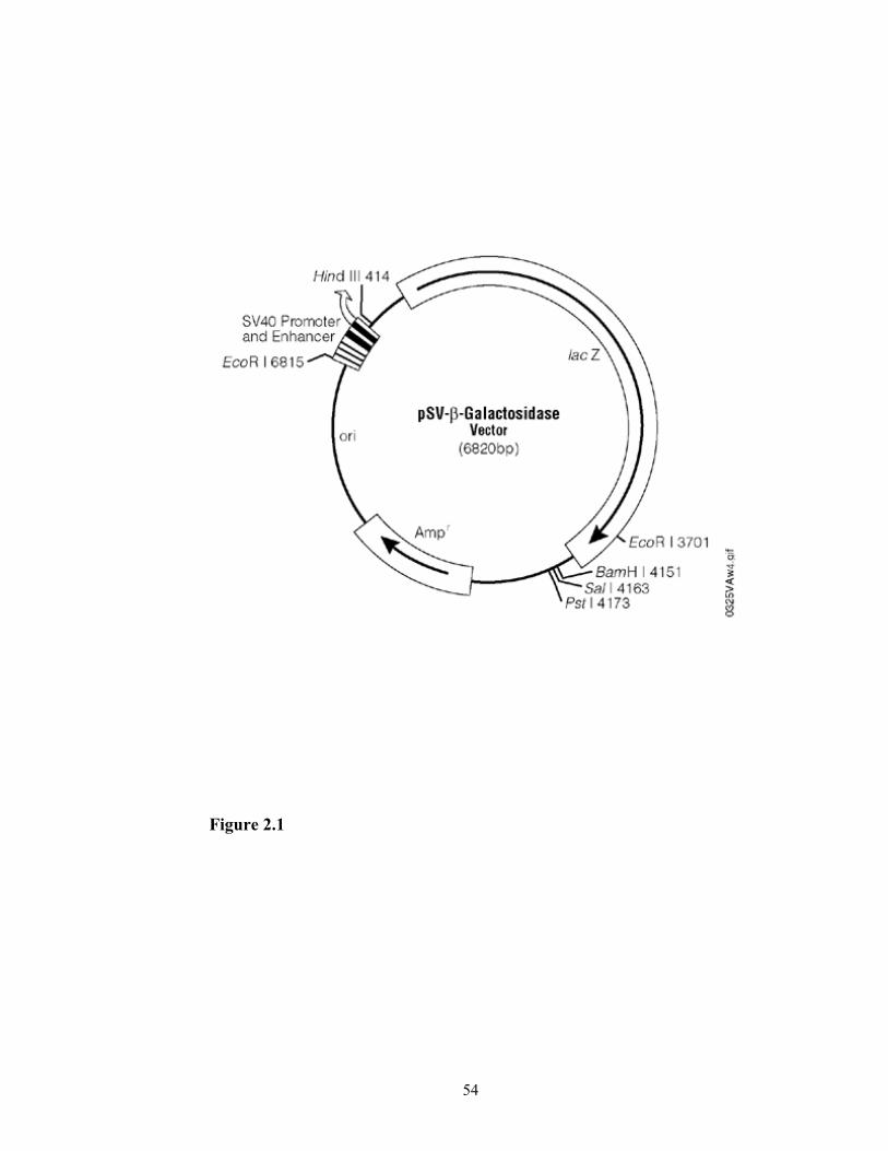
Figure 2.1
54
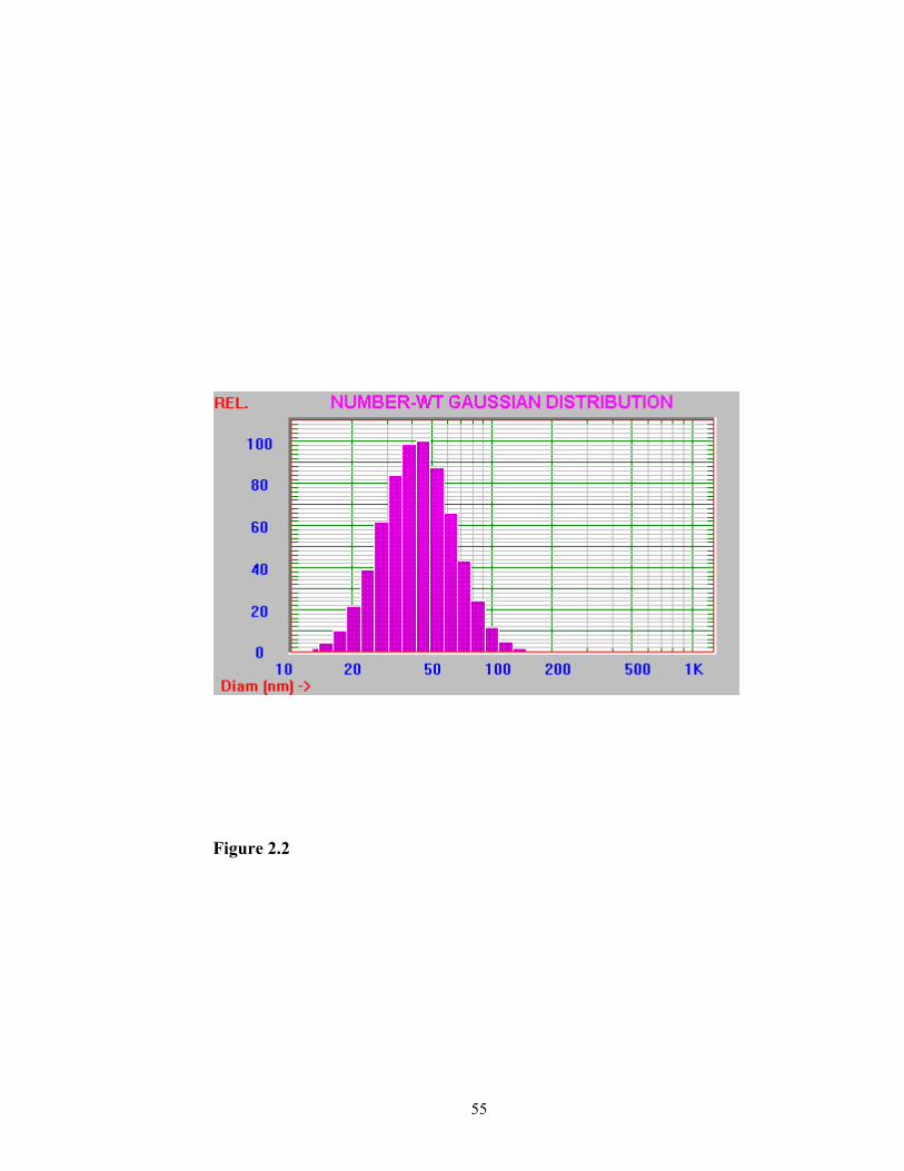
Figure 2.2
55
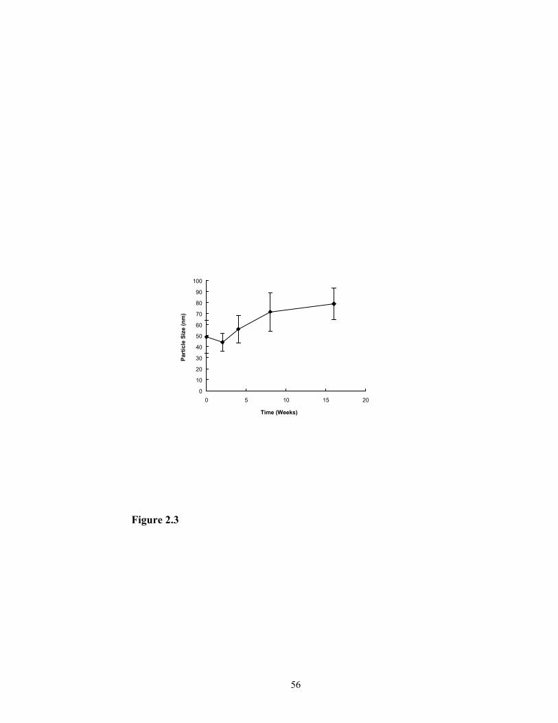
0
10
20
30
40
50
60
70
80
90
100
0 5 10 15 20
Time (Weeks)
Part
icle
Siz
e (n
m)
Figure 2.3
56

Figure 2.4
57
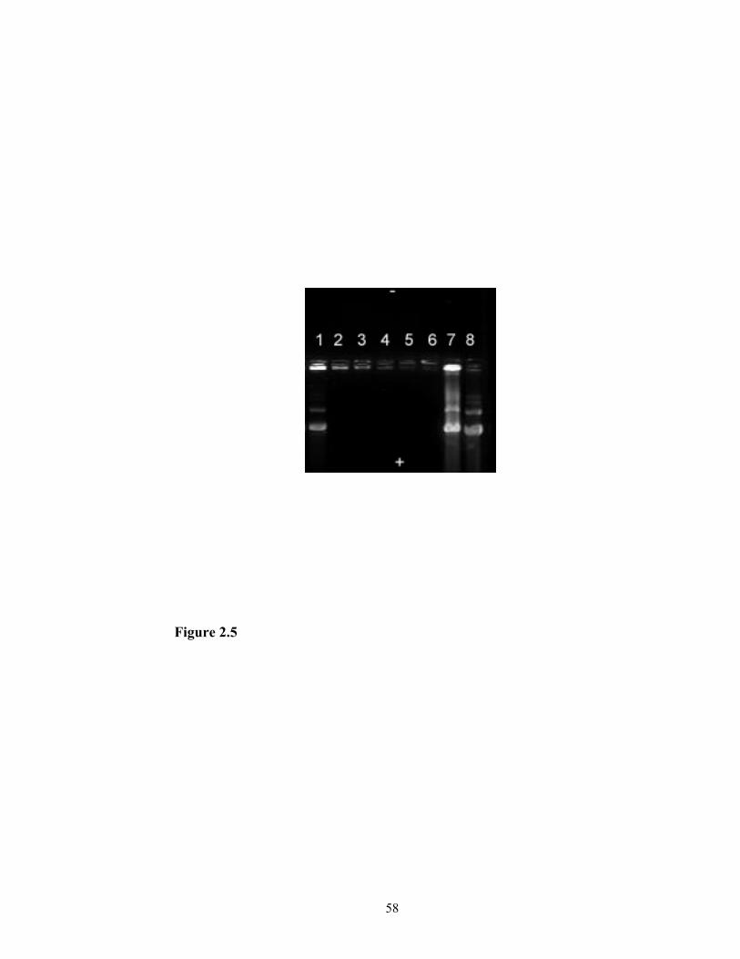
Figure 2.5
58

-50-40-30
-20-10
01020
304050
0 0.2 0.4 0.6 0.8 1
Nanoemulsion/p-PLL
Zeta
Pot
entia
l (m
V)
Figure 2.6
59
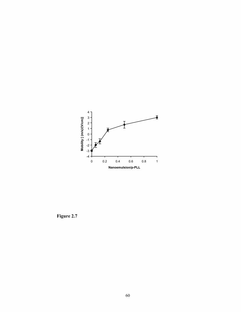
-4
-3
-2
-1
0
1
2
3
4
0 0.2 0.4 0.6 0.8 1
Nanoemulsion/p-PLL
Mob
ility
[ (m
/s)/(
V/cm
)]
Figure 2.7
60

Figure 2.8
61
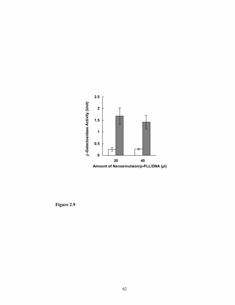
0
0.5
1
1.5
2
2.5
20 40Amount of Nanoemulsion/p-PLL/DNA (µl)
β-G
alac
tosi
dase
Act
ivity
(Uni
t)
Figure 2.9
62
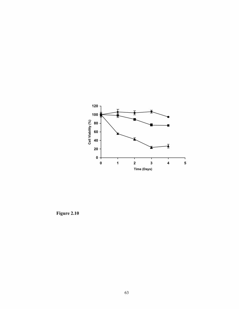
0
20
40
60
80
100
120
0 1 2 3 4 5Time (Days)
Cel
l Via
bilit
y (%
)
Figure 2.10
63

Phospholipid Lipidized-protein
Attached fatty chains
Vahydrsubs
Phospholipid Apolipoprotein
Fi
A
Cholesterol
Triglycerides(Triolein )
riousophobic tances
gure 2.11
64
B
Cholesteryl esterCholesterol
Triglycerides
REFERENCES
1. S. Lehrman. Virus treatment questioned after gene therapy death, Nature
401:517-518 (1999).
2. E. Marshall. Gene therapy death prompts review of adenovirus vector,
Science 286: 2244-2245 (1999).
3. W. T. Godbey, A. G. Mikos. Recent progress in gene delivery using non-viral
transfer complexes, J. Control. Release 72:115-125 (2001).
4. F. Liu, L. Huang. Development of non-viral vectors for systemic gene
delivery, J. Control. Release 78:259-266 (2002).
5. S. Han, R. I. Mahato, Y. K. Sung, S. W. Kim. Development of biomaterials
for gene therapy, Molecular Therapy 2(4):302-317 (2000).
6. X. Gao, L. Huang. Potentiation of cationic liposome-mediated gene delivery
by polycations, Biochemistry 35(3): 1027-1036 (1996).
7. X. Zhou, L. Huang. DNA transfection mediated by cationic liposomes
containing lipopolylysine: characterization and mechanism of action,
Biochim. Biophys. Acta 1189(2):195-203 (1994).
8. J. Zabner, A. J. Fasbender, T. Moninger, K. A. Poellinger, M.J. Welsh.
Cellular and molecular barriers to gene transfer by a cationic lipid, J. Biol.
Chem. 270:18997-19007 (1995).
9. Y. Xu, F. C. Szoka Jr. Mechanism of DNA release from cationic
liposome/DNA complexes used in cell transfection, Biochemistry
35(18):5616-5623 (1996).
65

10. O. Zelphati, F. C. Szoka Jr. Mechanism of oligonucleotide release from
cationic liposomes, Proc. Natl. Acad. Sci. USA 93(21):11493-11498 (1996).
11. Y. Tan, F. Liu, Z. Li, L. Huang. Sequential injection of cationic liposome and
plasmid DNA delivery gene effectively to the lung with minimal
inflammatory toxicity, Mol. Ther. 3(5):673-682 (2001).
12. H. Lee, J. H. Jeong, T. G. Park. A new gene delivery formulation of
polyethylenimine/DNA complexes coated with PEG conjugated fusogenic
peptide, J. Control. Release 76:183-192 (2001).
13. W. Guo, R. J. Lee. Efficient gene delivery via non-covalent complexes of
folic acid and polyethylenimine, J. Control. Release 77:131-138 (2001).
14. W. T. Godbey, K. K. Wu, A. G. Mikos. Poly(ethylenimine) and its role in
gene delivery, J. Control. Release 60(2-3):149-160 (1999).
15. D. Fischer, T. Bieber, Y. Li, H. P. Elsasser, T. Kissel. A novel non-viral
vector for DNA delivery based on low molecular weight, branched
polyethylenimine: effect of molecular weight on transfection efficiency and
cytotoxicity, Pharm. Res. 16(8):1273-1279 (1999).
16. D. Putnam, R. Langer. Poly(4-hydroxy-L-proline ester): low-temperature
polycondensation and plasmid DNA complexation, Macromolecules 32:3658-
3662 (1999).
17. K. W. Leong, H. Q. Mao, V. L. Truong-Le, K. Roy, S. M. Walsh, J. T.
August. DNA-polycation nanospheres as non-viral gene delivery vehicles, J.
Control. Release 53(1-3):183-193 (1998).
66
18. R. Puls, R. Minchin. Gene transfer and expression of a non-viral polycation-
based vector in CD4+ cells, Gene Ther. 6(10):1174-1178 (1999).
19. D. Oupicky, C. Konak, K. Ulbrich, M. A. Wolfert, L. W. Seymour. DNA
delivery system based on complexes of DNA with synthetic polycations and
their copolymers, J. Control. Release 65(1-2):149-171 (2000).
20. H. Gonzalez, S. J. Hwang, M. E. Davis. New class of polymers for the
delivery of macromolecular therapeutics, Bioconjug. Chem. 10(6):1068-1074
(1999).
21. J.-S. Kim, A. Maruyama, T. Akaike, S. W. Kim. In vitro gene expression on
smooth muscle cells using a terplex delivery system, J. Control. Release
47:51-59 (1997).
22. J.-S. Kim, B.-I. Kim, A. Maruyama, T. Akaike, S. W. Kim. A new non-viral
DNA delivery vector: the terplex system, J. Control. Release 53:175-182
(1998).
23. M. M. Bradford. A rapid and sensitive method for the quantitation of
microgram quantities of protein utilizing the principle of protein-dye binding,
Anal. Biochem. 72:248-254 (1976).
24. T. Mosmann. Rapid colorimetric assay for cellular growth and survival:
application to proliferation and cytotoxicity assays, J. Immunol. Methods
65:55-63 (1983).
25. D. M. L. Morgan, V. L. Lavin, and J. D. Pearson. Biochemical
characterization of polycation-induced cytotoxicity to human vascular
endothelial cells, J. Cell. Sci. 94:553-559 (1989).
67
26. T. Hara, Y. Tan, and L. Huang. In vivo gene delivery to the liver using
reconstituted chylomicron remnants as a novel nonviral vector, Proc. Natl.
Acad. Sci. USA 94:14547-14552 (1997).
27. M. Shawer, P. Greenspan, S. Øie and D. R. Lu. VLDL-resembling
phospholipid-submicron emulsion for cholesterol-based drug targeting, J.
Pharm. Sci. 91:1405-1413 (2002).
28. H. Luthman and G. Magnusson. High efficiency polyoma DNA transfection
of chloroquine treated cells, Nucl. Acids Res. 11:1295-1308 (1983).
29. C.-W. Cho, Y.-S. Cho, H.-K. Lee, Y. II Yeom, S.-N. Park, and Y. Yoon.
Improvement of receptor-mediated gene deliver to HepG2 cells using an
amphiphilic gelling agent, Biotech. Appl. Biochem. 32:21-26 (2000).
68

CHAPTER 3
IN VITRO CELLULAR UPTAKE OF A NEW CHOLESTERYL CARBORANE
ESTER COMPOUND BY HUMAN NORMAL NEURON CELLS AND
GLIOMA CELLS1
1Guangliang Pan, Svein Øie, D. Robert Lu. Submitted to Journal of
Pharmaceutical Sciences (2003).
69

ABSTRACT
The cellular uptake of a new cholesteryl carborane ester compound, cholesteryl
1,12-dicarba-closo-dodecaboranel-carboxylate (BCH), by one human normal neuron cell
line, HCN-1a, and two human glioma cell lines, glioblastoma multiforme SF-763 and SF-
767, was evaluated. BCH, which is an extremely hydrophobic compound, was formulated
into liposomes and incubated with the cells at 37 oC and 5% CO2 for 16 hours. The
amount of BCH uptake by the cells was measured by high performance liquid
chromatography (HPLC). In addition, BCH uptake by tumor cells was examined in the
presence and absence of lipoprotein in the culture medium. It was found that the amount
of BCH taken in by the glioma cell lines was much more (up to 14 times) than that by the
normal neuron cell line. The cellular uptake of BCH by SF-763 and SF-767 cells after 16
hours of incubation was 283.3 ± 38.9 µg boron/g cells and 264.0 ± 36.5 µg boron/g cells,
respectively. When the normal serum was replaced by lipoprotein deficient serum, the
cellular uptake of BCH by the tumor cells was reduced. In conclusion, the cellular
uptake of BCH by glioma cells was about 14 times higher than by normal neuron cells.
The uptake in glioma cells was up to 10 times higher than that required for successful
cancer treatment. Lipoprotein appeared to play an important role in the BCH uptake by
glioma cells.
KEYWORDS: Cholesteryl conjugate; liposomes; boron; tumor; cellular uptake;
lipoprotein
70

INTRODUCTION
Glioblastoma multiforme brain tumors, similar to many other type of malignant
tumors, are characterized by very aggressive cell growth and tend to be resistant to
conventional methods of treatment, including surgical resection of tumors, radiation
therapy, chemotherapy and combinations of these treatments (1). Among several
approaches that may enhance the efficacy of chemotherapy, targeted drug delivery that
produces a high concentration of therapeutic compounds in tumor cells and relatively low
concentrations in neighboring normal cells remains attractive. Recently, Maletinska, et al.
reported that seven human glioblastoma multiforme cell lines had very high numbers of
low-density lipoprotein (LDL) receptors per cell, indicating that the LDL pathway may
provide a method for selectively targeting compounds to glioblastoma multiforme cells
(2).
Rapidly dividing cells, such as malignant tumor cells, have high cholesterol
requirements because cholesterol is utilized to form the new cell membrane. Cells can
obtain cholesterol either by taking up plasma LDL (containing about 1500 cholesteryl
esters per LDL particle) via receptor-mediated endocytosis or by de novo synthesis.
However, it is known that up to 90% of cholesterol is obtained from receptor-mediated
endocytosis involving the degradation of LDL (3, 4). Many types of cancer cells have
been reported to take up more LDL than the corresponding normal cells in order to
absorb more cholesterol (5-10). These literature data suggest the potential of utilizing
cholesterol-drug conjugates to interact with lipoproteins and to follow through the LDL
pathway for targeted drug delivery.
71
Recently, our laboratory has designed and synthesized a cholesteryl carborane
ester compound mimicking native cholesteryl esters for targeted drug delivery to tumor
cells expressing a high degree of LDL receptors. The compound, cholesteryl 1,12-
dicarba-closo-dodecaboranel-carboxylate (BCH), is a mimic of native cholesteryl ester
containing carborane as an anti-tumor unit for boron neutron capture therapy (BNCT).
Unlike the utilization of other prodrugs, BNCT does not require the release of the
antitumor unit in cells. The boron-rich carborane unit, however, must be delivered to the
tumor cell preferentially, compared to surrounding normal cells (11). Similar to the native
cholesteryl esters, BCH is extremely hydrophobic requiring a suitable pharmaceutical
carrier. Since liposomes have been widely studied for the delivery of various boron
compounds (12, 13), we have formulated the compound in liposomes for cell culture
studies.
The objectives of the present cell culture study were to determine: 1) whether the
tumor cells could take up a sufficient amount of BCH; 2) whether the tumor cells took up
more BCH than normal brain cells; 3) whether the uptake process is related to the
presence of lipoproteins in culture medium. Correspondingly, the cellular uptake of BCH
in two human glioblastoma multiforme cell lines (SF-763 and SF-767) was evaluated.
These two tumor cell lines were known to have elevated level of LDL receptors on the
cell surface (2). A human normal brain neuron cell line (HCN-1a) was also examined to
compare its uptake with that by the tumor cell lines. In addition, the effect on BCH
uptake by tumor cells with a depletion of lipoprotein in culture medium was examined.
72

MATERIALS AND METHODS
Materials
BCH (Figure 3.1) was synthesized in our laboratory as described earlier (14). The
phospholipid DL-α-dipalmitoyl phosphatidylcholine (DPPC) and cholesterol were
purchased from Sigma Chemicals (St. Louis, MO). Human glioblastoma multiforme
(glioma) cells, SF-763 and SF-767, were obtained from the tissue bank of the Brain
Tumor Research Center (University of California-San Francisco, San Francisco, CA).
Human neuron cell line HCN-1a was purchased from American Type Culture Collection
(Manassas, VA). Eagle’s Minimum Essential Media (EMEM), Dulbecco’s phosphate-
buffered saline (PBS), trypsin-EDTA, and gentamicin solution were obtained from Fisher
Scientific Products (Suwanee, GA). Fetal bovine serum (FBS) and lipoprotein deficient
serum (LPDS) were obtained from BioCell Laboratories (Rancho Dominguez, CA).
Analytical grade chloroform, methanol, isopropanol, and other chemicals were obtained
from J.T. Baker (Phillipsburg, NJ).
Preparation of BCH in liposome formulation
The BCH liposomal formulation was prepared by the solvent evaporation method.
Lipids and BCH were mixed in organic solvent (methanol/chloroform in a ratio of 2:1) in
a round bottom flask and evaporated to a thin film. Liposomes were prepared by
hydration of the lipid film with phosphate buffered saline (PBS) solution at 55°C in a
water bath shaken at 120 rpm. Specifically, 52 mg DPPC, 10 mg cholesterol and 2.5 mg
BCH were dissolved in approximately 6 ml organic solvent in a round bottom flask. The
solvent was evaporated under vacuum in a rotary evaporator (Buchi Rotavap, RE121,
Brinkmann Instruments, NY). The dried lipids on the flask wall were hydrated with 10 ml
73
PBS that had been heated to 55°C. The flask was shaken mechanically for 5 hours at
55°C. Size reduction of the multilamellar vesicles was carried out using the Emulsiflex
B3 device (Avestin, Ontario, Canada) and running the vesicles for 10 cycles. The size
distribution of the resulting small liposomes was measured using a Nicomp submicron
particle sizer (Model 370, Nicomp, CA).
Cell culture studies
Human glioma cells (SF-763 and SF-767) were grown in 150 cm2 plastic cell
culture flasks containing 26 ml EMEM with 10% FBS, 50 U/ml penicillin, and 50 µg/ml
gentamicin. After three passages from the stock culture, the flasks were seeded with
approximately 2 x 10⁶ cells placed in a humidified 5% CO2 incubator at 37°C for 48
hours prior to incubation with BCH liposomal formulation. Human neuron cells (HCN-
1a) were grown in the same condition except that one month was required because they
grew more slowly. Each flask of cells was then incubated with a specified quantity of
BCH in the liposome formulation. After a pre-determined incubation period, the culture
medium was removed; the cells were rinsed 3 times with PBS, then harvested with
trypsin-EDTA and counted under a microscope. The cells were centrifuged at 3500 rpm
for 10 minutes. The cell pellets were weighed and stored at –20 °C until analysis.
Cellular uptake of BCH in liposome formulation by HCN-1a, SF-763 and SF-767
cell lines
To compare the BCH uptake by the normal cell line with the tumor cell lines,
HCN-1a, SF-763 and SF-767 cells were prepared and grown as described above. One ml
of BCH liposomal formulation (154.5 µg BCH/ml, equal to 30 µg boron/ml) was added
74
and incubated with each cell line for 16 hours. Cells were harvested and the BCH content
in the cells was determined by HPLC.
The effect of lipoprotein on cellular uptake of BCH in liposome formulation by SF-
763 and SF-767 cell lines
To determine the effect of lipoprotein on BCH uptake, 5%, 7%, 8%, 9%, and 10%
of delipidized human serum was supplemented in the culture medium to replace part of
the human normal serum to make up a total of 10 % serum. Cells were grown in 150 cm2
flasks at the same condition and the amount of BCH uptake by the tumor cell lines was
measured by HPLC.
Sample analyses
All the samples were analyzed by a high-performance liquid chromatography
(HPLC) method which was specific for BCH and the details of the HPLC method are
described elsewhere. Briefly, samples were prepared by sonication, evaporation to
dryness and reconstitution with 1 ml mobile phase (50:50 methanol-isopropanol).
Cholesteryl heptadecanoate was used as the internal standard. The analysis was
performed using a Waters model 2690 separations module equipped with a column heater
and Hewlett-Packard ZORBAX Stable Bond C-18 (5µm, 150 x 4.6mm) analytical
column. Since the cell division rates between normal neuron cells and the glioma cells
were different, the cellular BCH uptake was calculated with the consideration of cell
number counts and expressed as µg boron/g cells.
75
RESULTS
Characterization of BCH liposomal formulation
After size reduction using Emulsiflex, the size distribution of the resulting
liposomes was examined by photon correlation spectroscopy and a number-weighted
bimodal distribution was observed. Approximately 90 % of the vesicles had a mean
diameter of approximately 51 nm ± 7.3 nm and the remaining 10% of the vesicles had a
mean diameter of 165.1 ± 25.8 nm. Based on the HPLC analysis, the average BCH
content was 154.5 µg/ml (equal to 30 µg boron/ml) and entrapment efficiency was 61.8
%.
BCH uptake by three different cell lines The addition of 1 ml BCH liposomal formulation to 26 ml culture medium
resulted in a final concentration of 1.2 µg boron/ml in each medium. After 16 hours of
incubation, the amount of BCH uptake by HCN-1a, SF-763, and SF-767 cells is shown in
Table I. The amount of BCH uptake by the two tumor cell lines was much more (up to 14
times more) than that by the normal neuron cell line (p<0.05). It should be noted that
there was a small difference in total surface area per gram of cells between the tumor
cells and the normal cells. The ratio of the total surface area per gram of tumor cells over
that of normal cells was equal to 1.32. Since the cellular BCH uptake of tumor cells and
normal cells was significantly distinct, the small difference in surface area appeared not
to be a major consideration.
76
Effect of lipoprotein on the cellular uptake of BCH in liposome formulation by the
glioma cell lines
Although the inhibition of cellular uptake BCH in liposome formulation by the
presence of anti-LDL receptor antibody suggested the involvement of LDL receptor-
mediated pathway, we were not clear yet how the BCH in liposome formulation
interacted with this pathway. One possible interaction might be the transfer of the BCH in
the liposome formulation to the LDL particles by contact or fusion. This interaction could
be examined by the depletion of lipoprotein from the serum, which was supplemented to
the cell culture medium. During the uptake experiment of BCH in liposome formulation,
a combination of normal human serum and lipoprotein deficient serum was supplemented
in the culture medium, with a total serum to be 10% in the medium. The uptake of BCH
in liposome formulation by the glioma cells was determined and the result is shown in
Figure 3.2. The more the lipoproteins depleted from the culture medium, the less BCH
was taken up the cells. It appeared that lipoprotein is a very important factor for the
cellular uptake of BCH in the liposome formulation.
DISCUSSION
Effective chemotherapy for most malignant tumors requires preferential
accumulation and retention of the therapeutic compounds in tumor cells. Similarly,
BNCT requires preferential accumulation and retention of boron compounds in tumor
cells prior to the neutron irradiation. Laster et al. constructed boronated LDL by replacing
the cholesterol ester core with carborane carboxylic acid esters of fatty alcohols (4). Their
studies in cell culture indicated that resulting boron concentrations in tumor cells was 10
times higher than that required for BNCT and the amount of uptake was consistent with a
77

receptor-mediated binding mechanism. The boron concentration in tumor cells, about 240
µg boron/g cells, was significantly higher than that obtained with any other boron
compound previously evaluated for clinical applications (4).
To develop a cholesterol-based drug targeting approach by synthesizing new
cholesteryl carborane ester compounds to mimic the native cholesteryl esters present in
the hydrophobic core of LDL, our laboratory has recently synthesized a cholesteryl
carborane ester, cholesteryl 1,12-dicarba-closo-dodecaboranel-carboxylate (BCH). Our
results from the present experiments indicate that the cholesteryl carborane ester, carried
by liposomes, resulted in more than adequate cellular uptake of BCH by the tumor cell
lines. The uptake by the normal neuron cell line was, however, much lower (Table I)
indicating the cellular uptake appeared to be related to the rapidly growing rate of the
tumor cells. For the tumor cells, the boron uptake was significantly higher than that
required for successful BNCT and was similar to that reported by Laster et al. when
carborane carboxylic acid esters of fatty alcohols were used to boronate LDL and CHO
and V-79 Chinese hamster cells were used to examine the boron uptake (4). It can be
seen in Table 3.1, for the SF-763 cells, the boron uptake reached up to 283.3 µg boron/g
cells, about 11 times higher than the required boron level (≥ 20-25 µg boron per gram
cells) for successful BNCT. For SF-767 cells, the boron uptake reached 264 µg boron/g
cells, about 10 times higher than the required boron level. It has been noticed that the
degree of cell confluence affected BCH cellular uptake in tumor cells. Since it was very
difficult to precisely control the cell confluence, the batch-to-batch difference in the
amount of BCH uptake can be seen throughout our experiments. However, high BCH
uptake in the tumor cells was consistently observed with a reasonable variation.
78
The uptake, in both tumor cell lines, was dependent on the BCH concentration in
the incubation medium and the relationship between concentration and uptake appeared
to be linear within this concentration range. The studies, however, were limited as to the
maximum concentration of BCH in the culture medium because of the low aqueous
solubility of BCH and the potential toxicity from the high concentration of lipids with a
larger amount of the liposomal formulation. A high concentration of lipids in the
incubation medium may result in toxic effects as indicated in our earlier rat cell culture
studies (15). Our result also showed that the BCH uptake by SF-767 tumor cells in the
presence of lipoprotein was about 4 times higher than that in the absence of lipoprotein.
This suggested that the BCH uptake process involved lipoprotein. The amount of BCH
taken up by the human glioma cells was about 6-7 % of the total amount, indicating a
significant amount of BCH (in liposomal formulation) remained in the culture medium
and was washed out before the cells were collected.
In our earlier study involving 9L rat glioma cell lines, it was observed that BCH,
formulated in conventional and PEG liposomes, produced marginally sufficient levels of
boron in the rat tumor cells (about 50 µg boron/g of cells) (15). When the cellular BCH
uptake in the two human glioma cell lines was compared with the cellular uptake from
our previous studies conducted on the 9L rat glioma cell line with the same experimental
conditions, the cellular uptake in the two human glioma cell lines was much higher. This
disparity in uptake is obviously due to the differences between the rat glioma cell line and
the human glioma cell lines, possibly in the expression and activity of the LDL receptors.
When each glioma cell line was exposed to the BCH liposomal formulation for
different incubation times the uptake at 2 hours was equal to approximately 40% of the
79

uptake at 16 hour for the SF-763 cells and approximately 30 % of the uptake at 16 hour
for the SF-767 cells. The results indicated that the uptake for both cell lines was related to
the incubation time. The uptake at 48 hours of incubation was virtually the same as at 16
hours indicating that 16 hours of incubation would be a suitable experimental condition
for the subsequent cell culture studies.
In addition to the requirement for the cells to obtain adequate amount of BCH, it
is also essential for BCH to remain in the cells for a sufficient time period to allow the
subsequent neutron radiation. The results from our retention experiment indicated that a
significant amount of BCH taken up in the human glioma cells was retained in the cells
after the subsequent 24-hour incubation without the presence of BCH formulation.
In conclusion, BCH formulated in liposomes appears to be effectively taken up by
and retained in SF-763 and SF-767 human glioma cells. The cellular concentration was
about 10 to 11 times higher than that required for successful cancer treatment. The BCH
uptake in human normal neuron cells, however, was significantly lower indicating the
cellular uptake was related to the rapidly-growing rate of the tumor cells. The uptake
process also appeared related to the lipoprotein presence in the culture medium. At
present, the mechanism of cellular uptake of this type of cholesteryl carborane ester
compounds in glioma tumor cells is not clear. Further studies are needed to provide
detailed evidence to illustrate the uptake mechanism.
80

Table 3.1. Cellular uptake of BCH by different cell lines
Cell line Cellular BCH uptake (µg boron/g cell)
Normal neuron cell, HCN-1a 19.9 ± 2.9
Glioma tumor cell, SF-763 240.1 ± 22.3
Glioma tumor cell, SF-767 283.4 ± 48.1
81

FIGURE LEGEND
Figure 3.1. The chemical structure of cholesteryl 1,12-dicarba-closo-dodecaboranel-
carboxylate (BCH).
Figure 3.2. The effect of lipoprotein on the cellular uptake of BCH in liposome
formulation by glioma cell line SF-763 and SF-767 (Gray bar – with FBS; White bar –
without LPDS).
82
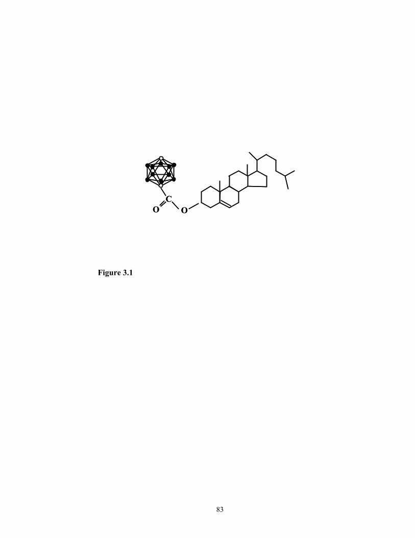
OC
O OC
O
Figure 3.1
83

0
100
200
300
400
500
600
BC
H ( µ
g/g
cell)
Figure 3.2
SF-763
Cell line
84
SF-767
s
REFERENCES
1. S. A. Leibel, Primary and metastatic brain tumors in adults. 1998. In S. A.
Leibel and T. L. Phillips (eds.), Textbook of Radiation Oncology, W.B.
Saunders Company, Philadelphia, PA, pp. 293-323.
2. L . Maletinska, E. A. Blakely, K. A. Bjornstad, D. F. Deen, J. J. Knoff, T.
M. Forte. 2000. Human glioblastoma cell lines: Levels of low-density
lipoprotein receptor and low-density lipoprotein receptor-related protein.
Cancer Res. 60:2300-2303.
3. Y. K. Ho, R. G. Smith, M. S. Brown, J. L. Goldstein. 1978. Low-density
lipoprotein (LDL) receptor activity in acute myelogenous leukemia cells.
Blood 52:1099-1114.
4. B. H. Laster, S. B. Kahl, E. A. Popenoe, D. W. Pate, R. G. Fairchild. 1991.
Biological efficacy of boronated low-density lipoprotein for boron neutron
capture therapy as measured in cell culture. Cancer Res. 51:4588-4593.
5. R. A. Firestone. 1994. Low-density lipoprotein as a vehicle for targeting
antitumor compounds to cancer cells. Bioconjugate Chem. 5:105-113.
6. S. Vitols, G. Gahrton, and C. Peterson. 1984. Significance of the low-
density lipoprotein receptor pathway for the in vitro accumulation of AD-
32 incorporated into LDL in normal and leukemic white blood cells.
Cancer Treat Rep. 68:515-520.
7. S. Vitols, B. Angelin, S. Ericsson, G. Gahrton, G. Juliusson, M.
Masquelier, C. Paul, C. Peterson, M. Rudling, K. Soderberg-Reid, and U.
Tidefelt. 1990. Uptake of low density lipoproteins by human leukemic
85

cells in vivo: relation to plasma lipoprotein levels and possible relevance
for selective chemotherapy. Proc Natl Acad Sci USA, 87:2598-2602.
8. S. Vitols, C. Peterson, O. Larsson, P. Holm, and B. Aberg. 1987. Elevated
uptake of low-density lipoproteins by human lung cancer tissue in vivo.
Cancer Res. 47:4105-4108.
9. M. J. Rudling, B. Angelin B, C. O. Peterson, V. P. Collins. 1990. Low-
density lipoprotein receptor activity in human intracranial tumors and its
relation to the cholesterol requirement. Cancer Res. 50:483-487.
10. M. J. Rudling, V. P. Collins, and C. O. Peterson. 1983. Delivery of
aclacinomycin A to human glioma cells in vitro by the low-density
lipoprotein pathway. Cancer Res. 43:4600-4605.
11. R. L. Gutman, G. Peacock, D. R. Lu. 2000. Targeted Drug Delivery for
Brain Cancer Treatment. J Controlled Release 65:31-41.
12. K. Shelly, D. A. Feakes, M. F. Hawthorne, P. G. Schmidt, T.A. Krisch,
and W. F. Bauer. 1992. Model studies directed toward the boron neutron-
capture therapy of cancer: boron delivery to murine tumors with
liposomes. Proc Natl Acad Sci U S A. 89:9039-43.
13. M. F. Hawthorne, K. Sherry. 1997. Liposomes as drug delivery vehicles
for boron agents. J Neuro-Oncol. 33:53-8.
14. B. Ji, G. Peacock, and D. R. Lu. 2002. Synthesis of cholesterol-carborane
conjugate for targeted drug delivery. Bioorganic & Medicinal Chemistry
Letters, 12:2455-2458 (2002)
86
15. Peacock, G., Ji, B., Wang, C. K. and Lu, D. R.: Cell culture studies of a
carborane cholesteryl ester with conventional and PEG liposomes. Drug
Delivery, 2002 (In press)
87
CHAPTER 4
UPTAKE OF CARBORANE DERIVATIVE OF CHOLESTERYL ESTER BY
GLIOMA CANCER CELLS IS MEDIATED THROUGH LDL RECEPTORS1
1Guangliang Pan, Svein Øie, D. Robert Lu. To be submitted to Biochimica
et Biophysica Acta.
88
ABSTRACT
Purpose. To elucidate the mechanism of cellular uptake of BCH in a liposomal
formulation by the glioma cell lines SF-763 and SF-767. Methods. BCH, an extremely
hydrophobic cholesteryl carborane ester, was synthesized in our laboratory. It was
formulated into liposomes by solvent evaporation and hydration method. After the cells
were grown to about 80% confluence, the BCH formulation was added to the culture in
the presence of different amount of monoclonal anti-LDL receptor antibody. The cells
were incubated at 37 oC and 5% CO2 for 4 hours. After repeated wash with PBS, the cells
were trypsinized and harvested by centrifugation. The amount of BCH in the cells was
measured by high performance liquid chromatography (HPLC). At the same time, the
effect of lipoprotein deficient serum (LPDS) on the cellular uptake of BCH in liposome
formulation was examined by substituting different amount of human normal serum in
the medium with LPDS. The effect of divalent calcium ion and low temperature on
uptake of BCH in liposome formulation was also investigated. Results. The cellular
uptake of BCH in liposome formulation by glioma cell lines SF-763 and SF-767 was
inhibited by monoclonal anti-LDL receptor antibody. The inhibition effect reached the
maximum when the concentration of monoclonal anti-LDL receptor antibody was 0.1
mg/ml. The BCH uptake by both cell lines was positively correlated with the amount of
normal human serum. The BCH uptake by glioma cells was significantly reduced by the
complete substitution of normal human serum by LPDS. The cellular uptake of BCH in
liposome formulation in the presence of calcium ion is about 2.7 times as much as that in
the absence of calcium ion for SF-763. For the SF-767, the uptake of BCH in liposome
formulation in the presence of calcium ion is about 1.5 times as much as that in the
89

absence of calcium ion. At 4oC, only small amount of BCH was taken up by both glioma
cell lines. Conclusions. The cellular uptake of BCH in liposomal formulation by both
glioma cell lines was inhibited by monoclonal anti-LDL receptor antibody and reduced
by the substitution of normal human serum with lipoprotein deficient serum. Calcium ion
had a positive effect on the uptake of BCH in liposome formulation by both glioma cell
lines. At low temperature, no significant BCH uptake occurred. These results suggested
that LDL receptors play a very important role in the cellular uptake of BCH in liposome
formulation by the glioma cell lines SF-763 and SF-767.
Keywords: BCH, cellular uptake, glioblastoma, LDL receptor, monoclonal anti-LDL
receptor antibody, LPDS, calcium ion, low temperature binding
Abbreviations: BCH, cholesteryl 1,12-dicarba-closo-dodecaboranel-carboxylate; FBS,
fetal bovine serum; ICP, inductively coupled plasma; LPDS, lipoprotein deficient serum;
BNCT, boron neutron capture therapy; DPPC, DL-α-dipalmitoyl phosphatidylcholine;
EMEM, Eagle’s Minimum Essential Medium; SDS-PAGE, sodium dodecyl sulfate-
polyacrylamide gel
90

INTRODUCTION
Cellular targeting requires site-specific drug deliveries that targets to a specific
cell type within a tissue. Two important steps that are involved in this process are the
recognition and interaction of drugs or drug carriers with specific target cells and delivery
of the therapeutics into the target cells with reduced uptake by the non-target cells (Poste,
1983). Such a process is either cell-surface receptor mediated by ligand-receptor
interaction or cell-surface epitope mediated by antigen-antibody interaction. The cellular
targeting is thus largely dependent on the specificity of the target cell surface proteins. It
is known that many cancer cells over-express certain types of cell surface receptors,
including transferrin receptor (Plant et. al. 1989; Huwyler et. al. 1996), folate receptor
(Garin-Chesa et. al. 1993; Ross et. al. 1994; Reddy et. al. 1998), and LDL receptor
(Vitols et. al, 1984, 1985, 1992; Rudling et. al. 1990; Jung-Testas et. al, 1992; Maletinska
et. al., 2000), to meet the increased cell proliferation and growth requirement. One
strategy to develop targeted drug delivery technique for cancer therapy is to take
advantage of these over-expressed cell surface receptors. By incorporating the
corresponding ligands for the cancer cells surface receptors, anticancer drugs can be
specifically delivered to the cancer site.
Among various ligands for the over-expressed receptors in cancer cells,
low-density lipoprotein (LDL) is unique. It is an endogenous lipoprotein complex, which
has an approximate size of 22-25 nm in diameter and serves as a natural carrier of
cholesterol and cholesterol ester in blood circulation. Composed of about 1500 molecules
of cholesteryl esters, the core of one LDL particle is surrounded by about 500 molecules
of cholesterol and 800 molecules of phospholipids in its polar shell. Apolipoprotein,
91
which contains unique binding site for LDL receptor, is incorporated into the particles by
hydrophobic interaction (Havel et al., 1980). For normal cell growth, the cholesterol level
is balanced by LDL receptor-mediated uptake and subcellular degradation of LDL.
However, because of the increased growth and proliferation of cancer cells, high turnover
of cellular cholesterol for membrane growth and metabolism is required. As a result,
over-expressed LDL receptor level has been found in many cancer cells (Vitols et. al,
1984, 1985, 1992; Rudling et. al. 1990; Jung-Testas et. al, 1992; Maletinska et. al., 2000).
Owing to its natural carrier capability and targeting specificity, LDL has been
extensively studied as anti-cancer carriers for hydrophobic drugs or prodrugs (Firestone,
1994; Kader and Pater, 2002, Sarkar and Lu, 2002). Recently, our laboratory has
designed and synthesized an anti-cancer cholesterol-carborane conjugate, cholesteryl
1,12-dicarba-closo-dodecaboranel-carboxylate (BCH), to mimic the structure of
cholesteryl esters located in the hydrophobic core of LDL (Ji and Lu, 2002). Because
these anti-cancer cholesteryl ester mimics have similar structures of natural cholesteryl
esters, they have the potential to interact with lipoproteins and to follow through the LDL
pathway for targeted drug delivery. The preformulation of BCH and its in vitro cellular
uptake by two cancer cell lines, Glioblastoma multiforme SF-763 and SF-767, have been
previously studied (Shawer et al., 2002; Peacock et al., 2003). The results indicated that
the cellular uptake of BCH by glioma cells was about 14 times higher than by normal
neuron cells and the uptake in glioma cells was up to 10 times higher than that required
for successful cancer treatment. However, the mechanism of BCH uptake in cancer cells
and the involvement of LDL receptor-mediated process are not clear.
92

In this research, we seek to elucidate the mechanism of BCH uptake by glioma
cells. In order to determine whether the BCH uptake is LDL receptor mediated, BCH
uptake by the glioma cells in the presence of monoclonal anti-LDL receptor antibody was
measured. The involvement of LDL during the uptake process was studied by uptake
measurement while supplementing the culture medium with a series of combinations of
lipoprotein deficient serum and normal serum and by electrophoresis for the interactions
between BCH and lipoproteins. In addition, the effect of calcium iron, a required divalent
metal ion for LDL receptor-mediated endocytosis, and temperature on the BCH uptake
was also investigated.
MATERIALS AND METHODS BCH was synthesized in our laboratory as described previously (Ji and Lu, 2002).
DL-α-dipalmitoyl phosphatidylcholine (DPPC) and cholesterol were purchased from
Sigma Chemicals (St. Louis, MO). Human Glioblastoma multiforme (glioma) cells, SF-
763 and SF-767, were obtained from the tissue bank of the Brain Tumor Research Center
(University of California-San Francisco, San Francisco, CA). Eagle’s Minimum Essential
Media (EMEM), Dulbecco’s phosphate-buffered saline (PBS), trypsin-EDTA, and
gentamicin solution were obtained from Fisher Scientific Products, Inc. (Suwanee, GA).
Fetal bovine serum (FBS), human normal serum, human lipoprotein deficient serum
(LPDS) was obtained from BioCell Laboratories, Inc. (Rancho Dominguez, CA).
Analytical grade chloroform, methanol, isopropanol, and other chemicals were obtained
from J.T. Baker (Phillipsburg, NJ).
93
Preparation of BCH liposomal formulation
Similar to the native cholesteryl esters, BCH is extremely hydrophobic and thus it
was formulated in liposomes as used in the previous studies (Peacock et al., 2003). The
BCH liposomal formulation was prepared by the solvent evaporation method. In brief, 52
mg DPPC, 10 mg cholesterol, and 2.5 mg BCH were dissolved in approximately 6 ml
chloroform in a round bottom flask. The chloroform was evaporated under vacuum in a
rotary evaporator (Buchi Rotavap, RE121, Brinkmann Instruments, NY). The dried lipid
film formed on the flask wall was hydrated with 10 ml PBS that had been pre-heated to
55°C. The flask was shaken in water bath at 55°C and 120 rpm for 5 hours. Size
reduction of the resulting multilamellar vesicles was carried out using Emulsiflex B3
device (Avestin, Ontario, Canada) for 10 cycles. The size distribution of the resulting
small liposomes was measured using a Nicomp Submicron Particle Sizer (Model 370,
Nicomp, CA). The liposome was stored at room temperature for short term before use.
Cell culture Human Glioblastoma multiforme SF-763 and SF-767 cells were routinely grown
in 75 cm2 plastic cell culture flasks (Corning Inc., Corning, NY, USA) containing 10 ml
EMEM supplemented with 10% FBS and 50 unit/ml penicillin and 50 µg/ml
streptomycin. The flasks were seeded with approximately 1 x 10⁶ cells and placed in a
humidified 5% CO2 incubator at 37°C. The cultures were passaged twice a week to
maintain the cells in exponential growth phase.
Determination of BCH in cell samples by high performance liquid chromatography
Cells containing BCH compound were dried by a freeze dry system (Labconco
Corporation, Kansas City, MO). BCH in the cells was extracted by a solvent mixture of
94
methanol and isopropanol (50:50 % v/v) and concentrated by Savant Speed Vac
Concentrator System (Albertville, Minnesota). A Zorbax Stable Bond C-18 column (4.6 x
150 mm; particle size, 5 µm) coupled to a Waters 2690 HPLC system was used to
analyze the BCH in samples. The column was equilibrated with the mobile phase
consisting of 50 % methanol and 50 % isopropanol at a flow rate of 0.5 ml/min. The
bound BCH was eluted isocraticly by the mobile phase and measured at 220 nm.
Monoclonal anti-LDL receptor antibody production and purification
Cell line ATCC CRL-1691 (C7, Mouse hybridoma) was grown in Dulbecco’s
Modified Eagle’s Medium supplemented with 4mM L-glutamine, 4.5 g/L glucose, 1.5
g/L sodium bicarbonate, and 10 % fetal bovine serum (FBS) at 37oC and 5% CO2. Two
liters of culture supernatant was harvested by high-speed centrifugation at 4oC. The
antibody-containing supernatant was mixed with a saturated ammonium sulfate solution
at 50% v/v ratio. Precipitation of antibody was conducted at 4oC and the precipitate was
harvested by high-speed centrifugation. The precipitate was resuspended in PBS and
dialyzed against PBS at 4 oC overnight. The antibody was further purified by affinity
liquid chromatography using Affi-Gel® Protein A MAPS® II Kit (Bio-Rad Laboratories,
Hercules, CA). Briefly, 1 ml of Affi-Gel Protein A agarose was packed into a 1 x 10 cm
Econo-Column chromatography column and equilibrated with 5 bed volumes of binding
buffer so that the effluent pH equals to the pH of the binding buffer (pH 9.0). Antibody
sample was loaded to the column and then washed with 15 bed volume of binding buffer.
The antibody was eluted with 5 bed volume of elution buffer. The column was
regenerated with 5 bed volume of regeneration buffer. The purity of the antibody was
95
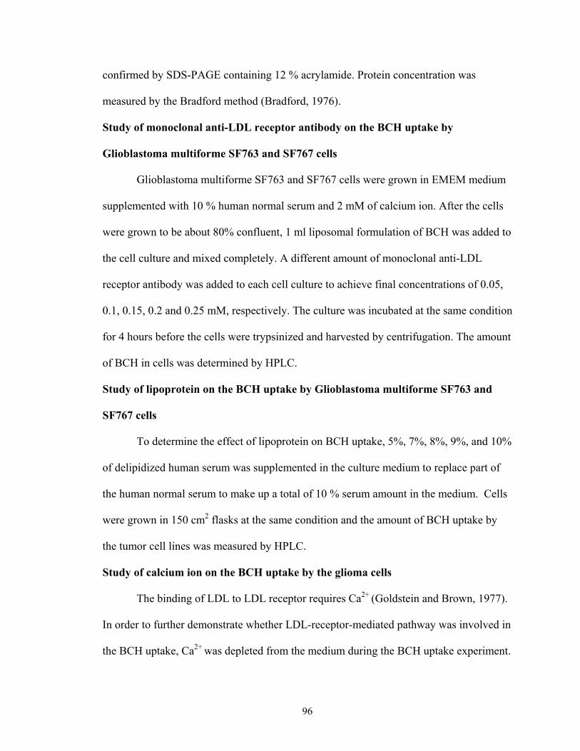
confirmed by SDS-PAGE containing 12 % acrylamide. Protein concentration was
measured by the Bradford method (Bradford, 1976).
Study of monoclonal anti-LDL receptor antibody on the BCH uptake by
Glioblastoma multiforme SF763 and SF767 cells
Glioblastoma multiforme SF763 and SF767 cells were grown in EMEM medium
supplemented with 10 % human normal serum and 2 mM of calcium ion. After the cells
were grown to be about 80% confluent, 1 ml liposomal formulation of BCH was added to
the cell culture and mixed completely. A different amount of monoclonal anti-LDL
receptor antibody was added to each cell culture to achieve final concentrations of 0.05,
0.1, 0.15, 0.2 and 0.25 mM, respectively. The culture was incubated at the same condition
for 4 hours before the cells were trypsinized and harvested by centrifugation. The amount
of BCH in cells was determined by HPLC.
Study of lipoprotein on the BCH uptake by Glioblastoma multiforme SF763 and
SF767 cells
To determine the effect of lipoprotein on BCH uptake, 5%, 7%, 8%, 9%, and 10%
of delipidized human serum was supplemented in the culture medium to replace part of
the human normal serum to make up a total of 10 % serum amount in the medium. Cells
were grown in 150 cm2 flasks at the same condition and the amount of BCH uptake by
the tumor cell lines was measured by HPLC.
Study of calcium ion on the BCH uptake by the glioma cells
The binding of LDL to LDL receptor requires Ca2+ (Goldstein and Brown, 1977).
In order to further demonstrate whether LDL-receptor-mediated pathway was involved in
the BCH uptake, Ca2+ was depleted from the medium during the BCH uptake experiment.
96
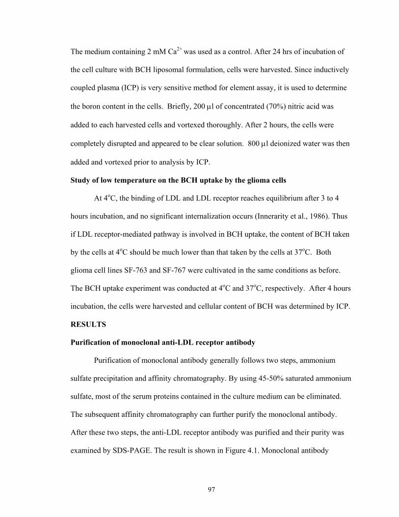
The medium containing 2 mM Ca2+ was used as a control. After 24 hrs of incubation of
the cell culture with BCH liposomal formulation, cells were harvested. Since inductively
coupled plasma (ICP) is very sensitive method for element assay, it is used to determine
the boron content in the cells. Briefly, 200 µl of concentrated (70%) nitric acid was
added to each harvested cells and vortexed thoroughly. After 2 hours, the cells were
completely disrupted and appeared to be clear solution. 800 µl deionized water was then
added and vortexed prior to analysis by ICP.
Study of low temperature on the BCH uptake by the glioma cells
At 4oC, the binding of LDL and LDL receptor reaches equilibrium after 3 to 4
hours incubation, and no significant internalization occurs (Innerarity et al., 1986). Thus
if LDL receptor-mediated pathway is involved in BCH uptake, the content of BCH taken
by the cells at 4oC should be much lower than that taken by the cells at 37oC. Both
glioma cell lines SF-763 and SF-767 were cultivated in the same conditions as before.
The BCH uptake experiment was conducted at 4oC and 37oC, respectively. After 4 hours
incubation, the cells were harvested and cellular content of BCH was determined by ICP.
RESULTS
Purification of monoclonal anti-LDL receptor antibody
Purification of monoclonal antibody generally follows two steps, ammonium
sulfate precipitation and affinity chromatography. By using 45-50% saturated ammonium
sulfate, most of the serum proteins contained in the culture medium can be eliminated.
The subsequent affinity chromatography can further purify the monoclonal antibody.
After these two steps, the anti-LDL receptor antibody was purified and their purity was
examined by SDS-PAGE. The result is shown in Figure 4.1. Monoclonal antibody
97
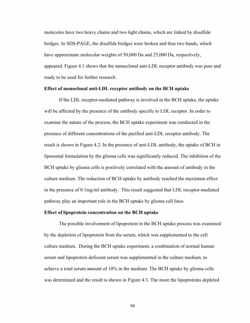
molecules have two heavy chains and two light chains, which are linked by disulfide
bridges. In SDS-PAGE, the disulfide bridges were broken and thus two bands, which
have approximate molecular weights of 50,000 Da and 25,000 Da, respectively,
appeared. Figure 4.1 shows that the monoclonal anti-LDL receptor antibody was pure and
ready to be used for further research.
Effect of monoclonal anti-LDL receptor antibody on the BCH uptake
If the LDL receptor-mediated pathway is involved in the BCH uptake, the uptake
will be affected by the presence of the antibody specific to LDL receptor. In order to
examine the nature of the process, the BCH uptake experiment was conducted in the
presence of different concentrations of the purified anti-LDL receptor antibody. The
result is shown in Figure 4.2. In the presence of anti-LDL antibody, the uptake of BCH in
liposomal formulation by the glioma cells was significantly reduced. The inhibition of the
BCH uptake by glioma cells is positively correlated with the amount of antibody in the
culture medium. The reduction of BCH uptake by antibody reached the maximum effect
in the presence of 0.1mg/ml antibody. This result suggested that LDL receptor-mediated
pathway play an important role in the BCH uptake by glioma cell lines.
Effect of lipoprotein concentration on the BCH uptake
The possible involvement of lipoprotein in the BCH uptake process was examined
by the depletion of lipoprotein from the serum, which was supplemented to the cell
culture medium. During the BCH uptake experiment, a combination of normal human
serum and lipoprotein deficient serum was supplemented in the culture medium, to
achieve a total serum amount of 10% in the medium. The BCH uptake by glioma cells
was determined and the result is shown in Figure 4.3. The more the lipoproteins depleted
98
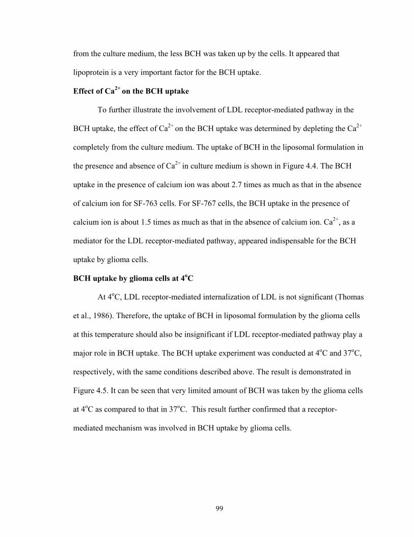
from the culture medium, the less BCH was taken up by the cells. It appeared that
lipoprotein is a very important factor for the BCH uptake.
Effect of Ca2+ on the BCH uptake
To further illustrate the involvement of LDL receptor-mediated pathway in the
BCH uptake, the effect of Ca2+ on the BCH uptake was determined by depleting the Ca2+
completely from the culture medium. The uptake of BCH in the liposomal formulation in
the presence and absence of Ca2+ in culture medium is shown in Figure 4.4. The BCH
uptake in the presence of calcium ion was about 2.7 times as much as that in the absence
of calcium ion for SF-763 cells. For SF-767 cells, the BCH uptake in the presence of
calcium ion is about 1.5 times as much as that in the absence of calcium ion. Ca2+, as a
mediator for the LDL receptor-mediated pathway, appeared indispensable for the BCH
uptake by glioma cells.
BCH uptake by glioma cells at 4oC
At 4oC, LDL receptor-mediated internalization of LDL is not significant (Thomas
et al., 1986). Therefore, the uptake of BCH in liposomal formulation by the glioma cells
at this temperature should also be insignificant if LDL receptor-mediated pathway play a
major role in BCH uptake. The BCH uptake experiment was conducted at 4oC and 37oC,
respectively, with the same conditions described above. The result is demonstrated in
Figure 4.5. It can be seen that very limited amount of BCH was taken by the glioma cells
at 4oC as compared to that in 37oC. This result further confirmed that a receptor-
mediated mechanism was involved in BCH uptake by glioma cells.
99
DISCUSSION
Cellular uptake of drugs is affected by many factors such as cell types, growth
stage of the cells and surrounding conditions. In our previous studies, the BCH uptake in
brain cancerous cells, SF-763, SF-767 cells, and brain normal cells, HCN-1a cells, were
compared. The cell culture was scheduled in an appropriate way such that all three cell
lines were in a similar stage before the BCH in liposomal formulation was added to the
culture medium. After 16 hours incubation with the BCH liposomal formulation, the cells
were harvested by trypsinization and centrifugation. The amount of boron uptake by cell
lines HCN-1a, SF-767, and SF-763 was determined by HPLC and the results are shown
in Table I. The amount of BCH uptake by the tumor cell lines was up to 14 times as high
as that by the normal human neuron cell line. The amount of BCH accumulated in the
glioma tumor cells was also much higher than the required amount of boron for
successful boron neutron capture therapy (BNCT). BCH is a carborane derivative of
cholesteryl esters, which is naturally located in the oily core of lipoproteins. The uptake is
thus potentially mediated by the lipoprotein pathway and possibly through the LDL
receptors. To address this question, the present research focuses on the elucidation of the
involvement of the LDL receptors during the uptake process.
If the cellular uptake of BCH is LDL receptor-mediated, the difference of BCH
uptake between the glioma cells and normal neuron cells becomes self-explanatory,
because glioma cell lines SF-763 and SF-767 overexpress LDL receptors on the cell
surface as compared with the normal cells (Maletinska et al., 2000). Since the
internalization of natural LDL by cells is LDL receptor-mediated (Brown and Goldstein,
1984; 1986), the binding of the LDL receptors by anti-LDL receptor antibodies could
100

significantly reduce LDL particle uptake by the cells. Our results have demonstrated the
reduction of BCH content in the cells when monoclonal anti-LDL receptor antibody was
present in the culture medium. This suggested the possibility of the involvement of the
LDL and LDL receptor in the uptake of BCH in liposome formulation by the glioma
cells.
On the other hand, if LDL and LDL receptor are involved in the cellular uptake
of BCH in liposome formulation, the depletion of LDL from the culture medium would
lead to the reduction of cellular uptake of BCH. Our result showed that the cellular
uptake of BCH in the liposome formulation by glioma cells was positively correlated
with the amount of LDL present in the culture medium. This result further inferred that
LDL functioned as a carrier of BCH during the cellular uptake of BCH in liposome
formulation by the glioma cells.
The specific binding of LDL and LDL receptor, which is the first step of
receptor-mediated pathway, requires divalent calcium ion (Innerarity et al., 1986). By
depleting the calcium ion in the culture medium, cellular uptake of BCH in liposome
formulation was reduced. Furthermore, the receptor-mediated endocytosis is temperature
dependent, and at 4oC, LDL is only bound to the cell surface and the internalization is not
significant (Innerarity et al., 1986). Therefore, the determination of BCH uptake at 4oC
and 37oC would help to confirm whether a receptor-mediated mechanism was involved.
When the cellular uptake experiment was conducted at 4oC, it was found that much
smaller amount of BCH was detected in the cells as compared to that at 37oC.
In conclusion, the inhibition of cellular uptake of BCH in liposome formulation
by the presence of anti-LDL receptor antibody and the positive correlation of uptake of
101
BCH in liposome formulation with the amount of LDL in the culture medium, together
with the effect of calcium ion and temperature on the uptake of BCH in liposome
formulation, have strongly suggested the involvement of an LDL receptor-mediated
pathway in the uptake of BCH in liposome formulation. However, we were not clear yet
how the BCH in liposome formulation interacted with this pathway. One possible
interaction might be the transfer of the BCH in liposome formulation to the LDL particles
by contact or fusion. However, this need to be further clarified in future experiments.
102

Table 4.1. Cellular uptake of BCH by different cell lines and the effect of lipoprotein on BCH uptake
Cell line Cellular BCH uptake (µg boron/g cell)
Normal neuron cell, HCN-1a 19.9 ± 2.9
Glioma tumor cell, SF-763 240.1 ± 22.3
Glioma tumor cell, SF-767 283.4 ± 48.1
103
Figure Legends:
Figure 4.1. SDS-PAGE (12% acrylamide) of the purified monoclonal anti-LDL receptor
antibody
Figure 4.2. The effect of monoclonal anti-LDL receptor antibody on the uptake of BCH
in liposome formulation by glioma cell lines SF-763 and SF-767
Figure 4.3. The effect of serum on the uptake of BCH in liposome formulation by glioma
cell lines SF-763 and SF-767
Figure 4.4. The effect of Ca2+ on the uptake of BCH in liposome formulation by glioma
cell lines SF-763 and SF-767 (white bar- with 2 mM Ca2+; gray bar-without Ca2+)
Figure 4.5. The effect of temperature on the uptake of BCH in liposome formulation by
glioma cell lines SF-763 and SF-767 (white bar-4oC; gray bar-37oC)
104

Figure 4.1
105
40
80
120
160
200
0 0.05 0.1 0.15 0.2 0.25
Anti-LDL receptor antibody (mg/ml)
BC
H u
ptak
e ( µ
g/g
cells
) SF763SF767
Figure 4.2
106
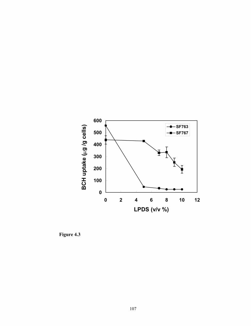
0
100
200
300
400
500
600
0 2 4 6 8 10 12
LPDS (v/v %)
BC
H u
ptak
e ( µ
g /g
cel
ls) SF763
SF767
Figure 4.3
107
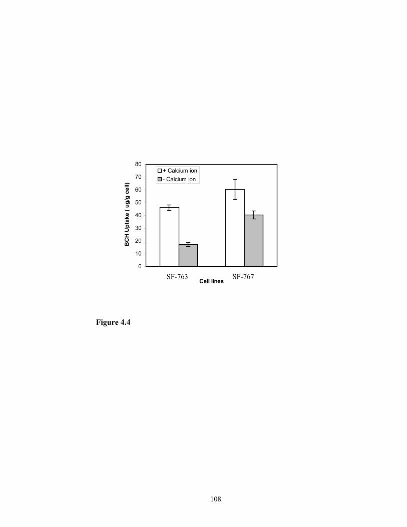
0
10
20
30
40
50
60
70
80
BC
H U
ptak
e ( u
g/g
cell)
+ Calcium ion- Calcium ion
73
Figure 4.4
SF-76
Cell lines108
SF-76
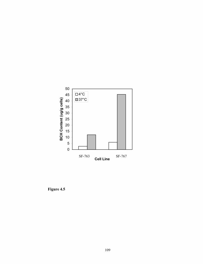
05
101520253035404550
Cell Line
BC
H C
onte
nt (u
g/g
cells
)
4°C37°C
SF-767 SF-763
Figure 4.5
109
REFERENCES
1. Alanazi, F., Halpern, D. S., and Lu, D. R.: Development of cholesterol-based
conjugates for targeted drug delivery. STP Pharmaceutical Sciences, 13:27-35
(2003).
2. Alanazi, F., Li, H., Halpern, D.S., Øie, S. and Lu, D. R. Synthesis, preformulation
and liposomal formulation of cholesteryl carborane esters with various fatty
chains. Int. J. Pharm. 255:189-197 (2003)
3. Brown MS, Goldstein JL. How LDL receptors influence cholesterol and
atherosclerosis. Scientific American 251:58-66 (1984).
4. Brown MS, Goldstein JL. A receptor-mediated pathway for cholesterol
homeostasis. Science 232:34-47 (1986).
5. Firestone RA. Low-density lipoproteins as a vehicle for targeting antitumor
compounds to cancer cells. Bioconjugate Chem 1994;5:105-113.
6. Garin-Chesa P., Campbell I., Saigo P. E., Lewis J. L. J., Old L. J. and Rettig W. J.
Trophoblast and ovarian cancer antigen LK26: Sensitivity and specificity in
immunopathology and molecular identification as a folate-binding protein. Am. J.
Pathol. 142:557-56 (1993).
7. Havel R. J., Goldstein J. L., and Brown M. S. In: Metabolic Control and Diseases,
Bondy P. K., Rosenberg L. E., ed. 8th ed., Chapter 7, p.393. Saunders, New York,
1980.
8. Huwyler J., Wu D., Pardridge W. M. Brain drug delivery of small molecules
using immunoliposomes. Proc. Natl. Acad. Sci. USA 93:14164 (1996)
110

9. Innerarity T. L., Pitas R. E., and Mahley R. W. Lipoprotein-receptor interaction.
Methods in Enzymology 120:542-565 (1986)
10. Ji, B., Peacock, G. and Lu, D.R.: Synthesis of cholesterol-carborane conjugate for
targeted drug delivery. Bioorganic & Medicinal Chem. Lett. 12:2455-2458
(2002).
11. Jung-Testas I., Weintraub H., Dupuis D., Eychenne B., Baulieu D-E., and Robel
P. Low density lipoprotein-receptor in primary cultures of rat glial cells. J. Steroid
Biochem. Mol. Biol. 42:597-605 (1992).
12. Kader A, Pater A. Loading anticancer drugs into HDL as well as LDL has little
affect on properties of complexes and enhances cytotoxicity to human carcinoma
cells. J Control Rel 2002;80:29-44.
13. Maletinska L., Blakely E. A. Bjornstad K. A., Deen D. F., Knoff L. J. and Forte
T. M. Human glioblastoma cell lines: levels of low-density lipoprotein receptor
and low-density lipoprotein receptor-related protein. Cancer Res. 60:2300-2303
(2000).
14. Pan G., Shawer M., Oie S., Lu R. D. In vitro gene transfection in human glioma
cells using a novel and less cytotoxic artificial lipoprotein delivery system. Pharm
Res. 2003;20(5):738-733.
15. Plant A. L., Brizgys M. V., Lacasio-Brown L., Durst RA. Generic liposome
reagent for immunoassays. Anal. Biochem. 176:420 (1989).
16. Poste G. Drug targeting in cancer therapy. In “Receptor-Mediated Targeting of
Drugs” edited by G. Gregoriadis, G. Poste, J. Senior and A. Trouet, pp. 427-475.
1983. Plenum Press.
111
17. Reddy J. A. and Low P. S. Folate-mediated targeting of therapeutic and imaging
agents to cancers, Crit. Rev. Ther. Drug Carrier Syst. 15:587 (1998).
18. Ross J. F., Chaudhuri P. K. and Ratnam M. Differential regulation of folate
receptor isoform in normal and malignant tissues in vivo and in established cell
lines, Cancer, 73:2432-2443 (1994).
19. Rudling M. J., Angelin B., Peterson C. O., and Collins V. P. Low density
lipoprotein receptor activity in human intracranial tumors and its relation to the
cholesterol requirement. Cancer Res. 50:483-487 (1990).
20. Sarkar, R., Halpern, D. S., Jacobs, S. K. and Lu, D. R.: LDL-receptor mediated
drug targeting to malignant tumors. in Biomedical Aspects of Drug Targeting
(V.R. Muzykantov and V.P. Torchilin, Eds.) Kluwer Academic Publisher, 327-
345 (2002).
21. Shawer, M., Greenspan, P., Øie, S. and Lu, D. R.: VLDL-resembling
phospholipid-submicron emulsion for cholesterol-based drug targeting. J. Pharm.
Sci., 91:1405-1413 (2002).
22. Thomas L. I., Pitas R. E., and Mahley R. W. Lipoprotein-receptor interactions. In:
Ablers J., Segrest J. P., ed. Methods in Enzymology, Academic Press, pp. 543-
566 (1986).
23. Vitols S., Gahrton G., Ost A. and Peterson C. Elevated low-density lipoprotein
receptor activity in leukemic cells with monocytic differentiation. Blood,
63:1186-1193 (1984).
24. Vitols S., Gahrton, G., Bjorkholm M. and Peterson C. Hypocholesterolaemia in
malignancy due to elevated low-density-lipoprotein-receptor activity in tumor
112
cells: evidence from studies in patients with leukemia. Lancet, 2:1150-1153
(1985).
25. Vitols S., Peterson C., Larsson O., Holm P., and Aberg, B. Elevated uptake of low
density lipoprotein by human lung cancer tissue in vivo. Cancer Res. 52:6244-
6247 (1992).
113
CHAPTER 5
IN VITRO EVALUATION OF THE ANTICANCER EFFECT OF
A METHOTREXATE-CHOLESTEROL CONJUGATE ON GLIOMA
CELL LINE SF-7671
1Guangliang Pan, Hengguang Li, Svein Øie, D. Robert Lu. To be submitted to
International Journal of Pharmaceutics (2003).
114
ABSTRACT
Objective. To evaluate and compare the in vitro cytotoxicity of anticancer drug
methotrexate and its derivative methotrexate-cholesterol conjugate to Glioblastoma
multiforme cell line SF-767. Methods. Methotrexate-cholesterol was synthesized by a
three step chemical reaction. The cytotoxicity of methotrexate and methotrexate-
cholesterol on Glioblastoma multiforme cell line SF-767 was evaluated by determining
the cell viability using MTT (3-(4,5-dimethylthiazol-2-yl)-2,5-diphenyl tetrazolium
bromide) after the incubation of the cells with the compounds. The effect of the
concentration of methotrexate and methotrexate-cholesterol conjugate on the cytotoxicity
to SF-767 was examined. In addition, the cell viability after 24, 48, and 72 hours of
incubation with methotrexate-cholesterol at a concentration of 0.125 mM were
determined. Results. The cytotoxicity of methotrexate-cholesterol to glioma cell SF-767
was higher than that of methotrexate. After 24 hours of incubation with the drug at a
concentration of 0.25 mM, only 2.6 % of glioma cells were viable when methotrexate-
cholesterol was added, while 15.4% of glioma cells were viable when methotrexate was
added. The cytotoxicity of methotrexate-cholesterol was concentration and time
dependent. When the glioma cells were treated with 0.125 mM methotrexate-cholesterol,
only 2.4 % cells were viable after three days. The cytotoxicity of the compounds to
glioma cell SF-767 at different concentrations and times was further demonstrated by the
microscopic pictures of the cells. Conclusion. The cholesteryl ester derivative of
methotrexate demonstrated increased cytotoxicity as compared methotrexate. Further
studies are needed to elucidate the reason of such increase in cytotoxicity.
115
Keywords Methotrexate; Methotrexate-cholesterol conjugate; Anticancer; Cytotoxicity;
Cell viability; MTT; Cellular uptake:
INTRODUCTION
The growth of human cells requires cholesterol for the bioconstruction of plasma
membrane. Cells obtain cholesterol either by de novo synthesis from acetyl-CoA or by
taking up plasma LDL which contains about 1500 cholesteryl esters per LDL particle.
The rate of de novo synthesis of cholesterol is relatively low and about 90% of
cholesterol in the cell is obtained from LDL receptor-mediated endocytosis of LDL
particles (Ho et al., 1978; Laster et al., 1991). However, there is a significant difference
in LDL consumption between human normal cells and cancer cells (Firestone, 1994;
Rudling et al., 1983,1990; Vitols et al., 1984,1987,1990). Most cancer cells are rapidly
dividing cells and thus have a higher cholesterol requirement for new cell membranes.
Correspondingly, these cells express significantly high numbers of cell surface LDL
receptors (Jung-Testas et al., 1992; Maletinska et al., 2000; Vitols, 1984, 1987, 1990).
This difference between the normal cells and cancer cells provides a fundamental drug
targeting strategy of utilizing cholesterol-drug conjugates to interact with lipoproteins and
to follow through the LDL pathway to selectively deliver a higher amount of anticancer
agents to cancer cells.
In our laboratory, a series of cholesterol-drug conjugates aimed at targeted drug
delivery to cancer cells have been designed and synthesized (Ji et al. 2002; Alanzi et al.,
2003a, 2003b). As an example, cholesteryl 1,12-dicarba-closo-dodecaboranel-carboxylate
(BCH), is a mimic of natural cholesteryl ester but contains carborane as an anti-cancer
unit for boron neutron capture therapy (BNCT) (Gutman et al., 2000; Ji et al., 2002;
116

Peacock, 2003). Cell culture studies in our laboratory indicated that BCH was taken up
extensively by human glioma cancer cells and the uptake was significantly higher in the
glioma cells than that in normal neuron cells (Peacock et al., 2003). However, the
anticancer activity of these boron-containing cholesterol-drug conjugates can not be
demonstrated because of the unavailability of the neutron radiation facility required by
BNCT.
In order to further demonstrate whether this type of compounds can be beneficial
for drug targeting and therapeutic effectiveness, cholesterol conjugates to other
chemotherapeutic agents was studied. Methotrexate is an antimetabolite used in the
treatment of certain neoplastic diseases, severe psoriasis, and adult rheumatoid arthritis
by inhibiting dihydrofolate reductase (Freeman et al., 1983; Weinblatt et al., 1985;
Roenigk et al., 1988; Jolivet et al., 1983). Actively proliferating tissues such as malignant
cells, bone marrow, fetal cells, buccal and intestinal mucosa are more sensitive to
methotrexate. Since cellular growth and proliferation in malignant tissues is greater than
in most normal tissues, methotrexate may impair malignant cell growth without
significant irreversible damage to normal tissues. Since the methotrexate contains a
carbonyl group, it can form an ester bond with the hydroxyl group in the cholesterol
molecule. In this research, methotrexate-cholesterol conjugate was synthesized by a three
step chemical reaction. The cytotoxicity of the methotrexate-cholesterol conjugate to
glioma cell line SF-767 was evaluated and compared with that of methotrexate.
117
MATERIALS AND METHODS
Materials
Human glioblastoma multiforme (glioma) SF-767 cell line was obtained from the
tissue bank of the Brain Tumor Research Center (University of California-San Francisco,
San Francisco, CA). Eagle’s Minimum Essential Media (EMEM), Dulbecco’s phosphate-
buffered saline (PBS) and trypsin-EDTA were obtained from Fisher Scientific Products
(Suwanee, GA). Fetal bovine serum (FBS) was obtained from BioCell Laboratories
(Rancho Dominguez, CA). Methotrexate, DMSO, 3-(4,5-dimethylthiazol-2-yl)-2,5-
diphenyl tetrazolium bromide (MTT), and penicillin/gentamycin solution was purchased
from Sigma (St. Louis, MO). N, N’-dicyclohexylcarbodiimide (DCC) was ordered from
Fisher Scientific (Pittsburgh, PA). Carbonyldiimidazole (CDI) was purchased from
Lancaster Synthesis (Windham, NH). Analytical grade solvents and other chemicals were
obtained from J.T. Baker (Phillipsburg, NJ).
Synthesis of Methotrexate-Cholesterol Conjugate
The chemical structure of methotrexate-cholesterol and its chemical synthesis are
shown in Figure 5.1 and Figure 5.2. Methotrexate (0.01 mmol) was first dissolved in 2
ml of water and 5 drops of triethylamine at room temperature. To the solution, 3.3 mg
(0.015 mmol) of Di-tert-butyl dicarbonate was added and the mixture was stirred for 6
hours at room temperature. The reaction mixture was then condensed under vacuum to
get product 1. In 3 ml dimethyl formamide (DMF), 6.65 mg of product 1 and 3.86 mg
(0.01 mmol) cholesterol were dissolved. To the mixture, 1.58 mg (0.01 mmol) of
carbonyldiimidazole was added with stirring at room temperature. After 5 minutes, 3.86
mg of cholesterol (0.01 mmol) and 2.0 mg DCC (0.01 mmol) were added and the mixture
118
was stirred for 6 hours. The reaction mixture was filtered and purified via silica column
chromatography eluted with petroleum ether and ethyl alcohol to get product 2. The
product 2 was then dissolved in 10 ml of dichloromethene. To the solution, three drops of
trifluoroacetic acid was added and stirred at room temperature for 2 hours. The mixture
was condensed under vacuum and the residue was further purified by silica column
chromatography eluted with ethyl alcohol to get product 3, which was methotrexate-
cholesterol conjugate.
Cell culture studies
Human glioma SF-767 cell line was grown and maintained in 75 cm2 culture flask
containing 10 ml of EMEM with 10% FBS, 50 µg/ml gentamycin and 50 U/ml penicillin.
After three passages from the stock culture, the cells were sub-cultured to 96-well cell
culture plate with approximately 8 x 10³ cells in 0.1 ml culture medium in each well. The
plates were placed in a humidified 5% CO2 incubator at 37°C for 24 hours prior to
incubation with the compunds. The stock solutions of methotrexate and methotrexate-
cholesterol were prepared by dissolving them in DMSO and were stored in a refrigerator
before use. When the cell culture was ready, different amount of methotrexate and
methotrexate-cholesterol (0.125, 0.25, 0.375 mM) were added to each well and mixed.
The culture was incubated at the same condition for 24 hours and the cell viability was
determined. In order to examine the cytotoxicity of methotrexate-cholesterol after
different time of incubation, 0.125 mM of methotrexate-cholesterol was incubated with
SF-767 cells for 24, 48, and 72 hours and the cell viability was determined. Microscopic
pictures of cell cultures were taken at various stage of the incubation with the compunds.
119

Cell viability determination after the incubation with methotrexate and
methotrexate-cholesterol
Cell viability was examined after incubation of the cells with different
concentration of the test compounds by the standard MTT method which measures the
ability of the cells to degrade tetrazolium salt MTT. The cells incubated with DMSO only
was used as the control. After the incubation, 20 µl of MTT solution in PBS buffer (0.5
mg/ml) was added to each well of culture and incubated at the same condition for an
additional 4 hours. The medium was then removed and 150 µl of DMSO was added to
each well and mixed thoroughly until all the dark blue crystals were dissolved. The plate
was read on an OPTImax Tunable Microplate Reader (Molecular Devices Corporation,
Sunnyvale, CA, USA) at wavelength of 550 nm. The cell viability was calculated and
expressed as (ODtrt/ODctrl ) x100%, where ODtrt is the optical absorbance from the culture
treated with the tested compounds, and ODctrl is the optical absorbance from the culture
incubated with DMSO only.
RESULTS
Comparison of the cytotoxicity of methotrexate and methotrexate-cholesterol
conjugate
The chemical linkage between methotrexate and cholesterol was successfully
obtained through the reactions shown in Fig. 2. The introduction of the cholesterol unit
greatly increased the hydrophobicity of the whole molecule. DMSO was used as the
solvent for both methotrexate and methotrexate-cholesterol. The anticancer activity of
these two compounds was then examined through the comparison of the cytotoxicity of
methotrexate and methotrexate-cholesterol conjugate.
120
Figure 5.3 showed that the cytotoxicity of methotrexate-cholesterol was higher
than methotrexate. At low concentration (0.125 mM), although methotrexate-cholesterol
had higher cytotoxicity than methotrexate, the difference in cytotoxicity was not
significant. However, when the concentrations of the test compunds were increased to
0.25 mM, only 2.6 % of glioma cells were viable when methotrexate-cholesterol was
added while 15.4% of glioma cells were viable when methotrexate was added. The
cytotoxicity of both compounds to glioma cells was also demonstrated by the cell culture
pictures (Figure 5.4). In the presence of either compunds, cells lost the capability to
attach to the wall of the flask and started to float in the medium. When methotrexate-
cholesterol was applied, no viable cells could be observed when the drug concentration
reached to 0.375 mM. For both compounds, as the concentration in the cell culture
increased, the cell viability decreased.
Dependence of the cytotoxicity on incubation time
In order to examine the effect of incubation time on cell death caused by
methotrexate-cholesterol, SF-767 cells were incubated with methotrexate-cholesterol at
concentration of 0.125 mM for 24, 48, and 72 hours. The low concentration of the test
compound was used because our previous result showed that the dependence of
cytotoxicity on the drug concentration was linear at low concentration. Figure 5.5 shows
that the cell viability was continuously reduced as the incubation time was prolonged.
Within the first 24 hours, about 75% of the cells died. After 72 hours incubation, only
2.4% of glioma cells were viable. The death of glioma cells were also reflected by the
changes on the cell culture picture (Figure 5.6). After 24 hours, most cells showed a
round shape as they lost the ability to attach to the plastic surface of the culture plate.
121
After 48 and 72 hours, most cells floated in the culture and aggregated to form clumps as
shown in picture 3 and picture 4.
DISCUSSION
Effective chemotherapy for most malignant cancers requires preferential
accumulation and retention of the therapeutic compounds in cancer cells. Since many
cancer cells overexpress surface receptors, such as transferrin receptor (Plant et. al. 1989;
Huwyler et. al. 1996), folate receptor (Ross et. al. 1994; Reddy et. al. 1998), and LDL
receptor (Vitols et. al, 1984, 1985, 1992; Rudling et. al. 1990; Maletinska et. al., 2000),
targeted drug delivery systems can be designed to utilize the overexpression for effective
cancer treatment. Among the different overexpressed receptors, LDL receptor is unique
because its ligand, LDL, is a natural carrier in blood circulation containing cholesterol
and cholesteryl esters. Therefore, it is possible to develop drug-conjugated cholesteryl
esters that can follow the LDL pathway for targeted drug delivery to cancer cells.
Experiments in our laboratory has shown that a carborane-conjugated cholesteryl ester,
BCH, can be taken up by glioma cancer cells in significantly higher amount than that by
normal nuron cells (Peacock et al., 2003). The mechanistic study involving anti-LDL
receptor antibody and lipoprotein deficient serum indicated that the uptake was through
the LDL receptors (Chapter 4).
In order to test whether such a strategy could be applied to other
chemotherapeutic units, our laboratory has recently synthesized a methotrexate-
conjugated cholesteryl ester. Methotrexate and its analogs are potent inhibitors of
hydrofolate reductase, which is required for the reduction of dihydrofolates to
tetrahydrofolate (THF). THF is essential carrier for one-carbon groups in the biosynthesis
122

of methionine, purines, and the pyrimidine thymine. Since growing cells, especially those
cells with rapid growth and proliferation have a high requirement for purine and thymine,
methotrexate and its analogs have been used as effective blockers of cancer growth.
Since the methotrexate molecule contains carbonyl groups, it can form an ester bond with
the hydroxyl group in the cholesterol molecule. Thus it was chosen as the model
chemotherapeutic agent for cholesteryl conjugate synthesis.
By comparing the cytotoxicity of methotrexate and methotrexate-cholesterol
conjugate, our results has shown that the cytotoxicity of cholesteryl derived methotrexate
increased significantly as compared to methotrexate. Methotrexate, the
pharmacologically effective part, was not changed signicantly after conjugating with the
cholesterol molecule. So it may still keep the binding ability to the dihydrofolate
reductase and interfere with the nornmal metabolism of the cancer cells. The increased
cytotoxicity of the conjugate may be caused by different reasons. For example, it may be
caused by the increased uptake of the methotrexate after the derivatization with the
cholesterol, whose cellular uptake pathway is involved in LDL receptor, because this is
true in the case of cellular uptake of BCH. However, further studies are required to
elucidate the mechanism of the increase of anticancer potency.
123

FIGURE LEGENDS:
Figure 5.1: The chemical structure of methotrexate-cholesterol conjugate
Figure 5.2: Steps of synthesis of methotrexate-cholesterol conjugate
Figure 5.3: Cytotoxicity of methotrexate and methotrexate-cholesterol conjugate on
glioma cell line SF-767 at different concentration
Figure 5.4: Microscopic pictures of cell culture incubated with methotrexate (A) and
methotrexate-cholesterol conjugate (B) at concentration of 0, 0.125, 1.25, and 0.375 mM.
Figure 5.5: Cytotoxicity of methotrexate-cholesterol conjugate at concentration of 0.125
mM after different time of incubation
Figure 5.6. Microscopic pictures of cell culture of SF-767 after incubation with
methotrexate-cholesterol conjugate at 0.125 mM after 0, 1, 2, 3 days.
124

N
N
N
N
N
NH
O
NH2
NH2
CO2HO
O
CH3
Figure 5.1
125
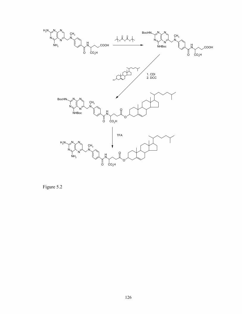
N
NH2
NH2
N
N
N
N
NH
O
COOH
CO2H
CH3
O
O
O O
O
N
N
N
N
N
NH
O
COOHNHBoc
BocHN
CO2H
CH3
OH
N
N
N
N
N
NH
O
NHBoc
BocHN
CO2HO
O
CH3
N
N
N
N
N
NH
O
NH2
NH2
CO2HO
O
CH3
TFA
1. CDI2. DCC
Figure 5.2
126

0
20
40
60
80
100
120
140
0 0.1 0.2 0.3 0.4
Drug Concentration (mM)
Cel
l Via
bilit
y (%
)
MTMT-Chol
Figure 5.3
127
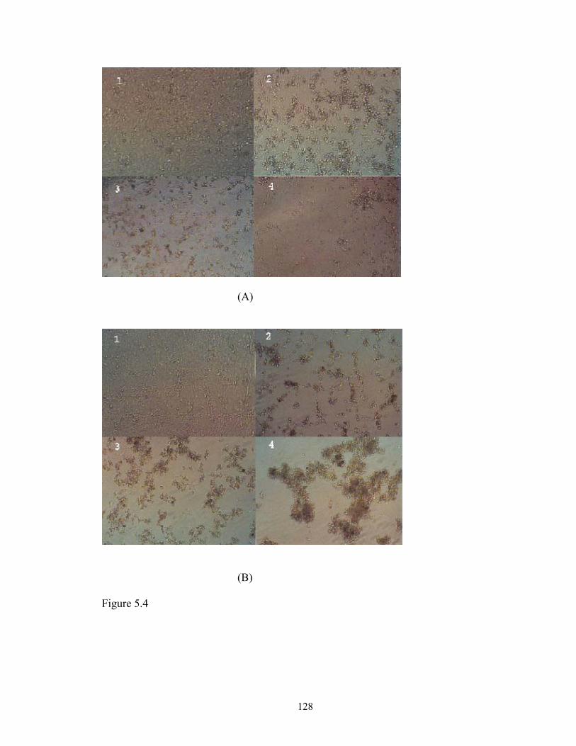
(A)
(B)
Figure 5.4
128
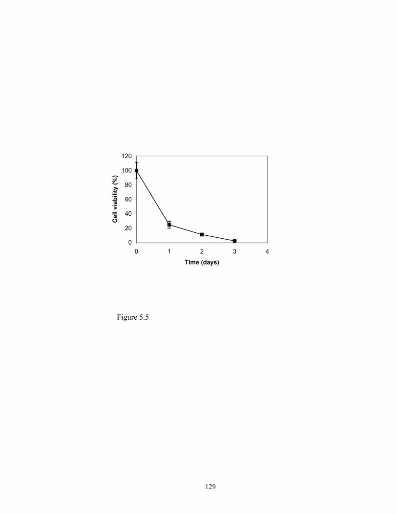
0
20
40
60
80
100
120
0 1 2 3 4
Time (days)
Cel
l via
bilit
y (%
)
Figure 5.5
129
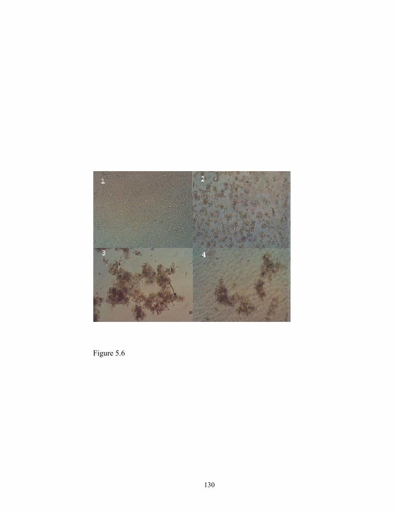
Figure 5.6
130
REFERENCES
1. Alanazi, F., Halpern, D. S., and Lu, D. R.: Development of cholesterol-based
conjugates for targeted drug delivery. STP Pharmaceutical Sciences, 2002 (In
press).
2. Alanazi, F., Li, H., Halpern, D.S., Øie, S. and Lu, D. R. Synthesis, preformulation
and liposomal formulation of cholesteryl carborane esters with various fatty
chains. Int. J. Pharm. 255:189-197 (2003).
3. Firestone R. A. 1994. Low-density lipoprotein as a vehicle for targeting antitumor
compounds to cancer cells. Bioconjugate Chem. 5:105-113.
4. Freeman A. I., Weinberg V., Brecher M. L. et al. Comparison of intermediate
dose methotrexate with cranial irradiation for the post-induction treatment of
acute lymphocytic leukemia in children. N. Engl. J. Med. 308:477-484 (1983).
5. Gutman R. L., Peacock G., Lu D. R. 2000. Targeted Drug Delivery for Brain
Cancer Treatment. J Controlled Release 65:31-41.
6. Ho Y. K., Smith R. G., Brown M. S., Goldstien J. L. 1978. Low-density
lipoprotein (LDL) receptor activity in acute myelogenous leukemia cells. Blood
52:1099-1114.
7. Huwyler J., Wu D., Pardridge W. M. Brain drug delivery of small molecules
using immunoliposomes. Proc. Natl. Acad. Sci. USA 93:14164 (1996)
8. Ji B., Peacock G., and Lu D. R. 2002. Synthesis of cholesterol-carborane
conjugate for targeted drug delivery. Bioorganic & Medicinal Chemistry Letters,
12:2455-2458 (2002).
131
9. Jolivet et al. The pharmacology and clinical use of methotrexate. N. Engl. J. Med.
309:1094-1104 (1983).
10. Jung-Testas I., Weintraub H., Dupuis D., Eychenne B., Baulieu D-E., and Robel
P. Low density lipoprotein-receptor in primary cultures of rat glial cells. J. Steroid
Biochem. Mol. Biol. 42:597-605 (1992).
11. Laster B. H., Kahl S. B., Popenoe E. A., Pate D.W., Fairchild R.G. 1991.
Biological efficacy of boronated low-density lipoprotein for boron neutron
capture therapy as measured in cell culture. Cancer Res. 51:4588-4593.
12. Maletinska L., Blakely E. A., Bjornstad K. A., Deen D. F., Knoff J. J., Forte T. M.
2000. Human glioblastoma cell lines: Levels of low-density lipoprotein receptor
and low-density lipoprotein receptor-related protein. Cancer Res. 60:2300-2303.
13. Peacock, G., Ji, B., Wang, C. K. and Lu, D. R.: Cell culture studies of a carborane
cholesteryl ester with conventional and PEG liposomes. Drug Delivery, 2002 (In
press).
14. Peacock G., Sidwell R., Pan G., Oie S., and Lu D. R. In vitro uptake of a new
cholesteryl carborane ester compound by human glioma cell lines. J. Phar. Sci.
(2003) (in press).
15. Plant A. L., Brizgys M. V., Lacasio-Brown L., Durst RA. Generic liposome
reagent for immunoassays. Anal. Biochem. 176:420 (1989).
16. Reddy J. A. and Low P. S. Folate-mediated targeting of therapeutic and imaging
agents to cancers, Crit. Rev. Ther. Drug Carrier Syst. 15:587 (1998).
17. Roenigk HH, Auerback R, Maibach HI, et al. Methotroxate in psoriasis: revised
guidelines. J. Am. Acad. Dermatol. 19:145-156 (1988).
132
18. Ross J. F., Chaudhuri P. K. and Ratnam M. Differential regulation of folate
receptor isoform in normal and malignant tissues in vivo and in established cell
lines, Cancer, 73:2432-2443 (1994).
19. Rudling M. J., Angelin B., Peterson C. O., and Collins V. P. Low density
lipoprotein receptor activity in human intracranial tumors and its relation to the
cholesterol requirement. Cancer Res. 50:483-487 (1990).
20. Rudling M. J., Collins V. P., and Peterson C. O. 1983. Delivery of aclacinomycin
A to human glioma cells in vitro by the low-density lipoprotein pathway. Cancer
Res. 43:4600-4605.
21. Vitols S., Gahrton G., and Peterson C. 1984. Significance of the low-density
lipoprotein receptor pathway for the in vitro accumulation of AD-32 incorporated
into LDL in normal and leukemic white blood cells. Cancer Treat Rep. 68:515-
520.
22. Vitols S., Peterson C., Larsson O., Holm P., and Aberg B. 1987. Elevated uptake
of low-density lipoproteins by human lung cancer tissue in vivo. Cancer Res.
47:4105-4108.
23. Vitols S., Angelin B., Ericsson S., Gahrton G., Juliusson G., Masquelie M., Paul
C., Peterson C., Rudling M., Soderberg-Reid K., and Tidefelt U. 1990. Uptake of
low density lipoproteins by human leukemic cells in vivo: relation to plasma
lipoprotein levels and possible relevance for selective chemotherapy. Proc Natl
Acad Sci USA, 87:2598-2602.
133
24. Weinblatt ME, Coblyn JS, Fox DA, Fraser PA, Holdsworth DE, Glass DN, and
Trentham DE. Efficacy of low-dose methotrexate in rheumatoid arthritis. N. Engl.
J. Med. 312:818-822 (1985).
134
CHAPTER 6
CONCLUSIONS
Malignant glioma represents a very difficult therapeutic challenge. One new
therapeutic strategy is gene therapy, which involves the delivery to and expression of the
therapeutic genes in the cancer cells. In this research, a novel artificial lipoprotein system
has been developed and evaluated for in vitro gene delivery to glioma cell line SF-767.
The system mimics the natural lipoprotein in composition and structure but contains
lipidized poly-L-lysine to replace the apolipoproteins in the surface of lipoproteins. Such
a system can be conveniently formulated from natural lipids, with the ability to control
the size and surface charge. With proper ratios among its components, this delivery
system can carry genetic materials and deliver them into glioma cells successfully. It
demonstrated similar transfection efficiency but a lower cytotoxicity as compared with
the commercial Lipofectamine gene transfection system. By incorporating other
surface proteins or peptides into this system, it may be used for targeted gene or drug
delivery. In addition, it could be extended to deliver DNA vaccines.
Since many cancer cells overexpress some cell surface receptors as compared to
their normal counterparts, one strategy for cancer therapy is to utilize these receptors to
develop targeted drug delivery system. As one of the major circulatory complexes in
blood circulation system, LDL is unique in that it is nanoscale particles and it is the
carrier of cholesterol and cholesteryl ester in the body. By mimicking the composition
and structure of cholesteryl ester, BCH, a carborane-cholesterol conjugate, was
synthesized in our laboratory. When formulated into liposomes and incubated with
135
normal neuron cells and glioma cells, it was found that the cellular uptake of BCH by
human glioma cell lines was significantly higher than that by human normal neuron cell
line. If the cellular uptake of BCH in liposome formulation is LDL receptor-mediated, the
big difference of BCH uptake between the glioma cells and normal neuron cells becomes
self-explanatory, since each glioma cell has much higher number of LDL receptors in
than that in each normal cell. Thus the elucidation of the mechanism for the uptake of
BCH in liposome formulation by the glioma cells turned to be a very important research
project for the further development of BCH as a targeted delivery system.
The results from this research have demonstrated that the uptake of BCH in
liposome formulation by both glioma cell line was inhibited by monoclonal anti-LDL
receptor antibody. In addition, by substituting the normal human serum with lipoprotein
deficient serum in the culture medium, the cellular uptake of BCH was also reduced.
Divalent metal ions, e.g., calcium ion, are required for the binding of LDL and LDL
receptor. By depleting the calcium ion from the culture medium, cellular uptake of BCH
was reduced. At low temperature, no significant BCH uptake occurred. These results
suggested that LDL receptor play a very important role in the uptake of BCH in liposome
formulation by the glioma cell lines SF-763 and SF-767.
In order to demonstrate further whether the cholesterol-drug conjugate can be
beneficial for the drug targeting, methotrexate- cholesterol conjugate was synthesized.
The cytotoxicity of the methotrexate-cholesterol conjugate to glioma cell line SF-767 was
evaluated and compared with that of methotrexate. It was found that the cytotoxicity of
methotrexate-cholesterol was higher than that of methotrexate.
136
APPENDIX
BIOLOGICAL PROTEIN NANOSTRUCTURES AND TARGETED
DRUG DELIVERY1
1Guangliang Pan, Svein ∅ie, and D. Robert Lu. Submitted to “Cellular
Drug Delivery: Principle and Practice” by D. Robert Lu and Svein ∅ie (eds), Humana
Press.
137

1. Introduction
Targeted drug delivery refers to the site-specific drug delivery that directs drugs
mainly to certain cell types within a tissue and to certain molecular complexes or
organelles within a cell while avoiding drug loading in non-targeted cells. Targeted
delivery of drugs to specific cells involves specific interactions between drugs or drug
carriers and cell surface proteins through ligand-receptor interactions or antigen-antibody
interactions. Targeted drug delivery to specific molecular complexes or organelles within
a cell requires specific interactions of drug with the targeted complexes to lead to the
therapeutic effect. In biological systems, these interactions generally occur on various
types of biological nanostructures of protein origin and understanding and utilization of
the biological nanostructures could lead to significant improvement in drug targeting and
drug carriers.
The biological protein nanostructures primarily include protein-lipid, protein-
protein, protein-carbohydrate, and protein-nucleic acid complexes. Proteins, one group of
the most important biological macromolecules in cells, are smaller nanoscale molecules
with typical size range between 1 and 20 nm (1). Through sophisticated interactions with
other biomolecules, these protein nanostructures are formed and widely distributed in
human body. For example, low-density lipoproteins (LDL), with a diameter of 25-28 nm,
are protein-lipid complexes. They are the major circulatory nanostructures in the blood.
When used as a drug carrier, these protein-lipid complexes offer certain advantages of
being endogenous nanostructures that do not trigger immunological response. They can
also escape recognition and elimination by the reticuloendothelial system (RES). On the
other hand, glycoproteins, i.e. protein-carbohydrate complexes, are vital structural and
138
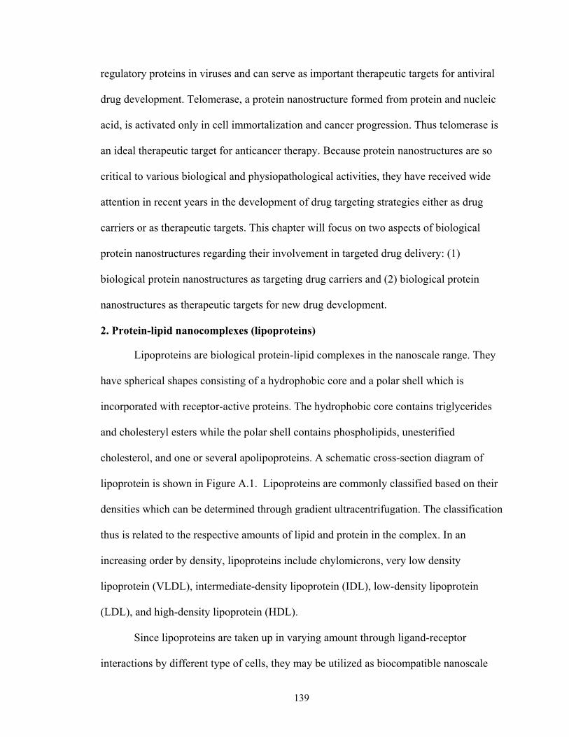
regulatory proteins in viruses and can serve as important therapeutic targets for antiviral
drug development. Telomerase, a protein nanostructure formed from protein and nucleic
acid, is activated only in cell immortalization and cancer progression. Thus telomerase is
an ideal therapeutic target for anticancer therapy. Because protein nanostructures are so
critical to various biological and physiopathological activities, they have received wide
attention in recent years in the development of drug targeting strategies either as drug
carriers or as therapeutic targets. This chapter will focus on two aspects of biological
protein nanostructures regarding their involvement in targeted drug delivery: (1)
biological protein nanostructures as targeting drug carriers and (2) biological protein
nanostructures as therapeutic targets for new drug development.
2. Protein-lipid nanocomplexes (lipoproteins) Lipoproteins are biological protein-lipid complexes in the nanoscale range. They
have spherical shapes consisting of a hydrophobic core and a polar shell which is
incorporated with receptor-active proteins. The hydrophobic core contains triglycerides
and cholesteryl esters while the polar shell contains phospholipids, unesterified
cholesterol, and one or several apolipoproteins. A schematic cross-section diagram of
lipoprotein is shown in Figure A.1. Lipoproteins are commonly classified based on their
densities which can be determined through gradient ultracentrifugation. The classification
thus is related to the respective amounts of lipid and protein in the complex. In an
increasing order by density, lipoproteins include chylomicrons, very low density
lipoprotein (VLDL), intermediate-density lipoprotein (IDL), low-density lipoprotein
(LDL), and high-density lipoprotein (HDL).
Since lipoproteins are taken up in varying amount through ligand-receptor
interactions by different type of cells, they may be utilized as biocompatible nanoscale
139
carriers for targeted drug delivery. For example, in hepatocytes, remnant receptor and
asialoglycoprotein receptor can recognize chylomicrons and lactosylated HDL,
respectively, in a molecular-specific manner. LDL receptors on cell surfaces can
specifically recognize LDL and its expression can be up-regulated or down-regulated
depending on the type and state of these cells. By incorporating bioactive molecules into
lipoproteins or modified lipoproteins, targeted drug delivery may be achieved resulting in
more bioactive molecules taken up by a select type of cells, e.g. cancer cells. Each class
of lipoproteins has its unique biological property and thus can be utilized individually for
targeted drug delivery.
2.1 Chylomicron for drug targeting
Chylomicrons are the largest lipoprotein complexes (80-500 nm) in human body.
Their main function is to transport dietary lipids from the intestine to the liver and
adipose tissue. Assembled in the intestine from the absorbed dietary lipids and the
apolipoproteins synthesized by the intestinal epithelium, they are transported out of the
epithelial cells to the tissue fluid and further carried by lymphatic system for general
circulation. When they enter the blood stream, their compositions of phospholipids and
proteins are changed greatly through the hydrolysis of triglycerides and the component
exchange with other lipoproteins in the plasma to form chylomicron remnants.
Chylomicron remnants are mainly taken up by parenchymal cells in liver (2). When they
are oxidized, chylomicrons can be taken up by liver endothelial cells and Kupffer cells
(3). The uptake of chylomicron remnants by various cells is LDL receptor-mediated,
which requires apoE protein as the ligand on the chylomicron remnant particles (3,4).
140
When associated with chylomicrons, many lipophilic drugs and xenobiotics can
be absorbed via the intestinal lymphatic system (5). This route can circumvent the first
pass effect in the intestine and, more importantly, it can be used for drug targeting to liver
cells because the liver is the destination of chylomicrons. Targeted drug delivery to the
liver can help treat many critical diseases such as alcohol-induced liver disorders, chronic
liver diseases and cirrhosis, virus-induced liver diseases (heptatitis), liver tumor, familial
hypercholesterolemia, and type III hyperlipoproteinemia. To effectively treat viral
infection, antiviral drugs or prodrugs can be incorporated into chylomicrons and targeted
to the liver. For example, by incorporating a nucleoside analogue iododeoxyuridine into
recombinant chylomicrons, the antiviral drug was selectively delivered to liver
parenchymal cells (6). The liver is also an excellent target for gene therapy for diseases
caused by metabolic defects. Gene therapy involving viral vector is in general limited by
the high immunogenicity and poor safety profile. Utilizing the hydrophobic core of the
chylomicrons, Hara et al. incorporated a hydrophobic DNA complex into reconstituted
chylomicron remnants and the DNA was successfully delivered to liver cells (7-8).
2.2 Very low density lipoprotein (VLDL) for drug targeting
VLDL particles have a size range of 30-80 nm. They are assembled in the
endoplasmic reticulum and matured in Golgi apparatus of hepatocytes before secretion
(9). After entering into the plasma, VLDL particles are catabolized by a series of
biochemical actions including apolipoprotein exchange with apoC-I, apoC-II, apoC-III,
and apoE, lipolysis by triglyceride lipase and cell-surface receptor-mediated uptake by
cells. As lipolysis proceeds, VLDL particles become smaller and smaller and eventually
are converted to IDL. Some of the IDL particles are rapidly taken up by hepatocytes via
141
a receptor-mediated mechanism and others undergo further hydrolysis before being
converted to LDL.
The catabolism cascade of VLDL suggests the possibility of using VLDL as a
drug carrier for targeted delivery. Because some cancer cells overexpress the receptors
for apo-E, a protein ligand present on the surface of VLDL, VLDL can potentially serve
as an antineoplastic drug carrier. In vitro experiments demonstrated that VLDL could
effectively incorporate cytotoxic drugs, 5-fluorouracil (5-FU), 5-iododeoxyuridine
(IudR), doxorubicin (Dox), and vindesine, and the resultant complex showed effective
cytotoxicity to human carcinoma cells (10). By mimicking the composition and structure
of VLDL, Shawer et al. developed a VLDL-resembling phospholipid nanoemulsion
system which could carry a new antitumor boron compound for targeted delivery to
cancer cells (11).
2.3 Low-density lipoprotein (LDL) for drug targeting
LDL (25-30 nm) is not directly synthesized in human body. Instead, most of them
are formed through the VLDL pathway. LDL is the major circulatory lipoprotein for the
transport of cholesterol and cholesteryl esters. Cholesterol required for cell membrane
construction is mainly obtained from LDL. LDL can be internalized by cells via LDL
receptor-mediated endocytosis, a process that is determined by the availability of LDL
receptors (12). Apolipoprotein apoB-100 is the major ligand in LDL for recognition and
binding by LDL receptor (13). Once it is internalized, LDL is distributed to lysosomes in
which cholesteryl esters are hydrolyzed (Figure A.2). Since cholesterol is required for
cell growth and LDL is the main carrier for cholesterol in blood circulation, most cells
can take up LDL through a receptor-mediated mechanism. It is estimated that 60-80% of
142

LDL can be cleared from plasma by LDL receptor-mediated pathways (14-15). As
compared to chylomicron, VLDL, and IDL, LDL has a longer serum half-life of 2 to 4
days (16). Thus, among various lipoproteins, LDL has a distinctive advantage to be used
as drug carrier for targeted delivery and has been widely studied.
2.3.1. LDL for anticancer drug targeting
It has been demonstrated that many tumor cells overexpress LDL receptors for the
uptake of LDL particles to meet their increased requirement for cholesterol in cell
membrane construction (17- 22). Subsequently, a significant amount of work has been
carried out to examine LDL as a candidate for antitumor drug carriers. Many lipophilic
anticancer drugs have been incorporated into LDL particles for the purpose of drug
targeting to various tumors (10, 23). When the antineoplastic drugs, methotrexate and
floxuridine (FdUrd), were oleyl derivatized and incorporated into LDL particles, they
were effectively delivered into the hepatocellular carcinoma cell line Hep G2 (24). The
serum half-life of these drugs carried by LDL particles was considerably prolonged as
compared to the free drug. Photodynamic therapy (PDT) of tumors is a recently
developed therapeutic approach. It is based on the generation of highly cytotoxic oxygen
species through the irradiation of photosensitizer such as porphyrins, chlorins, and
phthalocyanines at selected wavelength. The efficacy of this therapy is dependent on the
specific uptake of these photosensitizers by tumor cells. Using LDL as a carrier,
photosensitizers were successfully targeted to tumor cells (25-27).
2.3.2. LDL for brain drug targeting
The blood-brain barrier (BBB) is a semi-permeable barrier that allows certain
types of molecules to pass through but not others, depending on the lipophilicity,
143
molecular size and electric charge. It is a significant barrier for many drugs such as
antibiotics, neuropeptides, and antineoplastic agents. In order to overcome this barrier, a
number of methods have been employed including the use of prodrugs, antibody and drug
carrier systems such as liposomes (28-30). Because the brain involves a variety of
receptor-mediated transport systems to control the entry and exit of hydrophilic
molecules and macromolecules, such systems can be utilized for brain drug targeting and
transport. It is known that LDL receptors exist on endothelial cells of brain capillaries for
LDL endocytosis (31-33). Thus LDL can potentially be used as carriers for those drugs
that are unable to pass through BBB freely.
2.3.3. Acetylated LDL for drug targeting
When chemically altered lipoproteins appear in the circulation of the human
body, the RES system is activated to remove these altered lipoproteins if they are
recognized as foreign substances. The process involves the scavenger receptors on the
cell surface of human macrophage (34). Unlike T4 lymphocytes, which lead to collapse
of the immune system when they are infected by human immunodeficiency virus (HIV),
HIV infected macrophages allow HIV to replicate for a long period of time. Macrophages
play a very important role in HIV dissemination to various organs and to other parts of
the immune system (35-36). Experiments have shown that when antiviral drugs, e.g.
AZT, are incorporated into chemically altered LDL, such as acetylated LDL, the HIV
infected macrophages can be targeted (37).
2.3.4. Lactosylated LDL for drug targeting
In the liver, Kupffer cells play a critical role during inflammation through
enhanced expression of adhesion molecules, often resulting in the harmful infiltration of
144

neutrophils into the liver. In addition, the production of inflammation mediators, such as
interleukins and tumor necrosis factors by Kupffer cells, causes a cascade of events that
are related to serious physiological problems (38). Because only Kupffer cells express
galactose particle receptors in the liver, lactosylated LDL became a good candidate for
drug targeting to Kupffer cells (39). A cholesterol-conjugated oligonucleotide, which is a
potent inhibitor to the gene expression of intercellular adhesion molecule-1, was
associated with lactosylated LDL and the antisense oligonucleotide was efficiently
delivered into Kupffer cells (40), indicating the specific uptake of the encapsulated
content by Kupffer cells.
2.3.5. Oxidized LDL for drug targeting
Atherosclerosis is responsible for more deaths than any other disease in Western
countries. One important hallmark of this disease is the appearance of lipid-loaded
macrophages in the vessel wall. Current available therapies such as percutaneous
angioplasty and bypass surgery are limited by recurrence or worsening of the
atherosclerotic process. Photodynamic therapy involving various photosensitizers was
considered as a promising new therapy in recent years (41). One obstacle to this therapy
is how to efficiently deliver photosensitizers into macrophage cells. It is known that a
high level of scavenger receptors is expressed on the cell surface of macrophages with the
atheroscelosis plaque (34). These scavenger receptors can be good candidates for targeted
delivery. It has been shown that photosensitizer aluminum phthalocyanine chloride
associated with oxidatively modified LDL (OxLDL) was selectively delivered to
macrophages (42).
145

2.3.6. Surface modified LDL for gene delivery
The success of gene therapy is dependent on a safe and efficient gene delivery
system. Most of the current gene therapy protocols are based on viral gene delivery
vectors, which may cause long term safety problems (43). Although many non-viral gene
delivery vectors have been widely investigated, most of them were limited by low
transfection efficiency. Lipoprotein has been used to construct a new gene delivery
system to increase the safety and efficiency. Kim et al. developed a Terplex system,
which had a diameter about 100 nm. The Terplex system was formed through the
balanced hydrophobic and electrostatic interactions among LDL, lipidized poly(L-lysine),
and plasmid DNA (44-45). This system has demonstrated its efficiency by delivering
both plasmid DNA and antisense oligonucleotide to smooth muscle cells and lung
fibroblasts. As an endogenous nanoparticle, LDL played a key role in the internalization
of the Terplex system into the target cells via LDL receptor-mediated endocytosis.
2.4. High-density lipoprotein (HDL) for drug targeting
HDL is the smallest lipoprotein with a diameter of 7-11 nm. It shares common
structural characteristics with other lipoproteins. However, its polar shell contributes
more than 80% of the total mass. Newly synthesized HDL hardly contains any
cholesteryl ester molecules. Cholesteryl esters are gradually added to the particles via
enzymatic reaction by lecithin:cholesterol acyltransferase (LCAT), a 59-kD glycoprotein
associated with HDLs. The cholesteryl esters in HDL can also be transferred to VLDL
and LDL via another associated protein, cholesteryl ester transfer protein. The uptake of
HDL into cells appears to occur in a similar way as for LDL. However, cholesterol
uptake from HDL also involves more selective means than wholesale uptake as its
146
cholesteryl esters can be transferred into the cells (46). Although the function of HDL in
human body is not well defined, in general it transports excess cholesterol and cholesteryl
esters from various tissue cells to the liver. The major advantage of utilizing HDL for
drug delivery and targeting is their small size and fast internalization by tumor cells.
Among various lipoproteins, HDL has the smallest size. This makes it easier to pass
through the vascular pores to reach the target tissue and quicker to be internalized by the
cells. HDL has mainly been used for the incorporation of water insoluble anticancer
drugs for targeting (47-48). When the anticancer drug Taxol was incorporated into HDL,
stable complexes were formed for cancer cell targeting (48).
2.5. Artificial lipoprotein for drug delivery and targeting
Endogenous lipoprotein for drug delivery has usually been purified from plasma
by gradient ultracentrifugation. These lipoproteins are limited in availability and loading
additional transport or gene transfection enhancers has been problematic. In order to
overcome such limitations, the concept of artificial lipoprotein can be utilized.
Previously, several research groups have attempted to develop artificial lipoproteins (49-
52). However, these studies have primarily been focused on the incorporation of natural
apo-B protein into lipid microemulsion for biochemical and metabolism research, and
few of them on drug delivery and targeting.
By mimicking the structure of natural lipoproteins, artificial lipoproteins have
been extensively investigated in our laboratory to incorporate different lipidized proteins
or peptides for a diversified drug delivery and targeting strategy. The artificial
lipoproteins consist of two structural portions, a hydrophobic core and a polar shell
containing surface proteins. The hydrophobic core is mainly composed of triolein and
147

cholesterol oleate. The polar shell is composed of egg phosphatidylcholine,
lysophosphatidylcholine, cholesterol, and lipidized protein or peptide. The fatty chains on
the lipidized proteins and peptides serve as anchors to interact with the phospholipid
chains and to form stable protein-lipid nanocomplexes. An early trial of such a system for
drug delivery and targeting was through the constitution of a lipidized poly-L-lysine onto
phospholipid nanoemulsion particles for gene delivery to tumor cells (53). The
incorporation of sufficient amount of palmitoyl poly-L-lysine (p-PLL) molecules onto the
nanoemulsion particles led to positively charged complexes that were able to
electrostatically interact with negatively charged DNA molecules. As demonstrated by
Figure A.3, plasmid DNA migrated towards the positive anode because they were
negatively charged (Lane 1). When plasmid DNA was incubated with p-PLL (Lane 2), no
DNA migration band was observed. The binding of DNA molecules by p-PLL could
block the intercalation of ethidium bromide molecules into the DNA molecules and thus
no fluorescence emission occurred. When different ratios of p-PLL to nanoemulsion (i.e.
the p-PLL to triolein ratio) were incubated, they demonstrated different DNA carrying
capability (Lane 3 to Lane 7). A high ratio of p-PLL to nanoemulsion could tightly bind
all the DNA molecules and no free DNA migration band appeared (Lane 3 to Lane 6).
When the ratio of p-PLL to nanoemulsion became sufficiently low (0.0625:1 as the p-
PLL to triolein ratio), plasmid DNA started to escape from the complex and free DNA
bands (Lane 7) appeared on the agarose gel. Since the cell surface is normally negatively
charged, the uptake of exogenous particles is largely affected by the surface charge of the
particles. Positively charged particles appeared to be required for the cellular uptake of
the particles. However, excess positive charge on the particles could induce cellular
148
toxicity and limit its use as DNA carrier. Thus particles with a properly balanced charge
are required for the cellular uptake. The surface charge of the nanoemulsion/p-PLL/DNA
complexes were measured and their zeta potentials are shown in Figure A.4. The zeta
potentials of the particles increased with the increase of the amount of p-PLL when a
fixed amount of DNA was used. Among the nanoemulsion/p-PLL/DNA complexes
examined for the gene transfection, the complex with the zeta potential of 8.47 ± 1.85
mV resulted in the highest transfection efficiency. Such complexes demonstrated similar
transfection efficiency as the Lipofectamine, a commercial gene transfection product.
The artificial lipoprotein complex, however, had much lower toxicity (Figure A.5).
In recent years, cancer drug delivery and targeting have become a very active
research area. Many cancer cells overexpress specific receptor proteins or peptides,
which can recognize and bind specific ligands. For example, during the development of a
tumor, angiogenic endothelial cells overexpress αv integrin, which can specifically
recognize cyclic peptides containing Arg-Gly-Asp motif (54). Many other specific
ligand-receptor binding has also been found in tumor cells (55-57). In order to direct
anticancer drug to the specific cellular site, an effective delivery system becomes
critically important. The nanoscale size of the artificial lipoproteins and their capability
of incorporating different recognition protein ligands may present a practical solution for
anticancer drug targeting and effective gene delivery.
3. Protein-protein complexes and drug targeting
Protein-protein interactions are critical events for a wide range of physiological
and pathological processes. The biological formation of protein nanostructures through
protein-protein interactions must be controlled in a precise manner in order to function
149
properly. In viruses, virtually all the cellular processes including the formation of protein
nanostructures during replication and assembly involve protein-protein interactions (58).
In the human immune system, the proper interaction between CD4 and CD8, the cell
surface proteins expressed on T-cells, with T-cell receptor (TCR) and major
histocompatibility complex (MHC) Class I or II is required to activate T-cells (59). The
importance of such protein-protein interactions, as in the examples of viral replication
and assembly and immune activation in human body makes these nanocomplexes to be
ideal therapeutic targets for new drug developments.
There are many types of nanoscale protein-protein complexes including
homodimers, heterodimers, antigen-antibody complexes, enzyme-inhibitor complexes,
and multicomponents such as viral coat protein and ribosomes (60). The formation of
these protein nanostructures via protein-protein interactions generally involves large and
relatively flat surface areas with numerous contact sites, making it difficult to use small
drug molecules to block such processes. However, these interactions require precise
control in order to form pathophysiologically functional complexes. This presents an
opportunity for therapeutic drug design and development. In herpes virus (HSV), HSV
ribonucleotide reductase (RR) is a tetramer (α2β2) that consists of two large R1 subunits
and two small R2 subunits (61-62). The formation of an intact tetramer through proper
interactions is important for the survival of HSV. When a synthetic peptide
YAGAVVNDL was introduced into cells, the activity of RR was inhibited without
causing significant side effect in the host cells (63-64). The proper formation of other
protein nanocomplexes in HSV, such as DNA polymerase (heterodimer) and helicase-
150
primase complex, are also essential for the virus. Therefore, these nanocomplexes are
also being considered as potential therapeutic targets.
In HIV, protease, intergrase, and reverse transcriptase are all homodimer
nanostructures formed by protein-protein interactions. Protease has been one of the
primary therapeutic targets for AIDS chemotherapy being critical for viral maturation.
All successful inhibitors of HIV-1 protease to date are peptide mimetics that bind to the
active site of the protease. Because of specific mutation within the HIV-1 genome (65),
drug-resistant proteases appeared in many HIV-1 strains. A good alternative to this
therapeutic strategy would be agents that can block dimeriazation of protease (66-67).
The assembly of other important protein nanostructures, such as intergrase and reverse
transcriptase, can also be inhibited by many peptides at the dimeric interface (68-69).
These types of protein-protein interactions are also widely observed in other types of
viruses and have been considered as therapeutic target for drug development (70-71)
Although the formation of protein nanostructures through protein-protein
interactions is critical to many physiopathological processes, it has been difficult to
develop effective drug compounds to inhibit these processes. Recent design and
screening strategies include rational structure-based drug design, peptide display
technology, and in vivo genetic selection systems (72-74). However, many issues relating
to drug delivery, such as cell permeability, intracellular localization and physicochemical
stability, must be resolved. Subsequently, various systems including scrape loading,
electroporation, and delivery systems have been investigated. Among the delivery
systems, liposomes, polycationic peptides, viruses and proteins of eukaryotic, bacterial or
viral origin have been studied (75-78). A good example of utilizing protein as delivery
151
vehicle is the B subunit of E. coli heat-labile enterotoxin, which is able to deliver
bioactive peptide to the cells to disrupt viral protein-protein complex (79).
4. Protein-carbohydrate complexes and drug targeting
As one of the major groups of biological molecules, carbohydrates are unique in
that they can have many branches and their monomeric units can connect to each other in
different linkages in contrast to proteins and nucleic acids, which are exclusively linear
and have only one type of linkage (amide linkage in proteins and 3’-5’ phosphodiester
linkage in nucleic acids). Most carbohydrates exist as nanoscale complexes with proteins
(glycoproteins) or lipids (glycolipids). The complex sugar chains of glycoproteins and
glycolipids play very important roles in the control of cellular functions and cell-cell
recognitions, and therefore extensive investigations into the assembly of carbohydrate
complexes may yield important information for drug targeting development.
Glycoproteins are one of the major components in the outer surface of
mammalian cells. They play critical roles in many important biological processes such as
cell growth, fertilization, cell adhesion, immune responses, bacterial and viral infections,
degradation of blood clot, and inflammation. The majority of glycoproteins are formed by
covalent attachment of carbohydrates to the nitrogen atom (provided by asparagines
residue) or oxygen atom (provided by serine or threonine residue) in proteins. The
proteins are glycosylated as they move through the lumen of endoplasmic reticulum (ER)
and Golgi apparatus in the cells, mostly by glycosidase and glycotransferase. The type
and extent of glycosylation is dependent on the type and nature of proteins, cells and
tissues (80).
The fusion of HIV envelope with host cell membranes is a critical step for HIV to
enter the cells. The envelope glycoproteins of HIV are highly glycosylated. HIV-1 gp120
152
contains 20-25 glycosylation sites and the carbohydrates contribute about 50% of the
apparent molecular weight. Blocking of the protein glycosylation can significantly
interfere with the normal life cycle of HIV (81). Many sugar analogues have been
screened for the anti-HIV activity in vitro. One of these analogues, N-
butyldeoxygalactonojirimycin (NB-DNJ) has been shown to be a potent inhibitor of
infection with minimal cytotoxicity. In hepatitis B virus, although there are only two
glycosylation sites on the glycoprotein, the viral replication and assembly was inhibited
by the treatment of NB-DNJ. Gp41, another HIV envelope glycoprotein, also plays an
important role in the fusion of HIV envelope with host cell membranes. Corresponding
peptide and non-peptide inhibitors to gp41 have been developed (82). Knowledge about
the HIV-1 envelope glycoprotein has provided insight into the possibilities for design of
novel HIV vaccines (83). Protein-carbohydrate nanostructures in HIV-1 have currently
become the most important therapeutic targets for the development for anti-HIV drugs.
The molecular targets for new anticancer agents include inducers of cell
differentiation, cell cycle arrest, apoptosis, and signaling pathways for growth factors and
cytokines. Because the protein glycosylation pathways are ubiquitous in cancer cells,
they provide excellent opportunities for anticancer drug targeting. For example, alkaloid
swainsonine, a Golgi alpha-mannosidase II inhibitor, is the first inhibitor to be selected
for clinical test (84). Because p-glycoproteins are multidrug transporters that result in
multidrug resistance in cancer chemotherapy, inhibitors targeting this protein have been
developed (85). Protein glycosylation pathways are not only the ideal targets for drug
development to treat cancers, but are also excellent targets in other diseases. For example,
platelets play an important role in the pathophysiology of certain diseases such as acute
153
myocardial infarction and diabetes mellitus. Platelet activation and aggregation is caused
by the activation of the glycoprotein IIb/IIIa receptor. Thus glycoprotein IIb/IIIa has been
considered as a therapeutic target in these diseases (86-87).
5. Protein-nucleic acid complexes and drug targeting
Nucleic acids (DNA and RNA) are linear polymers of nucleotides with linkages
of 3’ to 5’ by phosphodiester bridges. The genetic information for making all functional
macromolecules are stored by the cellular DNA and accessed through the transcription of
information into RNA. A typical DNA double helix has a diameter of 2 nm with varying
length depending on organism. RNA occurs in various forms with different important
biological functions such as messenger RNA (mRNA), ribosomal RNA (rRNA), transfer
RNA (tRNA), and small nuclear RNA (snRNA).
The protein-nucleic acid nanostructures resulting from the interactions between
protein and nucleic acids are critical to almost all aspects of genetic activity within an
organism including DNA replication, transcription, packaging, rearrangement, and repair
(88). Based on the structural motif for DNA binding, there are four types of major
protein-DNA complexes, i.e. Helix-Turn-Helix (HTH), Zinc Finger (ZF), Basic Leucine
Zipper (B-Zip), and Basic Helix-Loop-Helix (B-HLH). Although the proteins in protein-
DNA complexes are very diversified, the basic goal is to achieve a precise
complementarity of the molecular shapes. This requires specific chemical recognition
between proteins and their particular DNA targets. Thus it has been proven possible to
design proteins with novel recognition specificities for the purpose of breaking the
normal protein-DNA binding (89). On the other hand, a specific DNA or DNA complex
can also be designed to bind the protein. For example, peptide nucleic acid (PNA), a
DNA mimic in which the nucleobases are attached to a pseudopeptide backbone, was
154
used to arrest transcription within a gene sequence and to provide an artificial open
complex to promote transcription (90).
Certain potent drug molecules interfere with DNA transcription by binding to the
transcription factors and thus obstructing the specific DNA binding (91). For example,
doxorubicin can bind to NIL2A, a basic leucine-zipper transcription factor, to inhibit the
formation of NIL2A/DNA complex. Echinomycin can bind to EGR1, a Cys2His2 zinc
finger transcription factor, and inhibit the formation of EGR1/DNA complex.
Intercalators such as nogalamycin and hedamycin, G/C-rich minor groove binding drugs
such as chromomycin A3, and A/T-specific drugs such as pluramycin can effectively
inhibit the transcription factor-DNA complex formation (91-92).
Interaction between protein and RNA plays very important roles in post-
transcriptional RNA processing and protein biosynthesis. For example, spliceosomes,
complexes of small nuclear RNAs and different proteins, are responsible for the precise
formation of messenger RNAs. Ribosomes, the complexes of proteins with RNAs, are the
agents for protein synthesis. For these complexes of proteins and RNAs, either
component can be the target for chemotherapy. For example, peptide nucleic acid (PNA)
can be targeted to mRNA to block protein synthesis in an antisense strategy. PNA can
also be targeted to the RNA components of ribonucleoproteins (RNPs) to inhibit their
enzymatic activities (90).
One of the most interesting protein-RNA complexes for drug targeting is
telomerase, a protein-RNA complex that elongates telomeric DNA and appears to play an
important role in cellular immortalization (93). Telomeres are nucleoprotein structures at
the end of human chromosomes. They play a fundamental role in the regulation of
155
cellular lifespan (94). The tandemly repeated DNA sequence of telomeres is specified
and controlled by telomerase, which is tightly repressed in the vast majority of normal
cells but becomes activated during cell immortalization and in cancers (94-95).
Telomerase has received much attention as a novel and potentially highly specific target
for the development of new anticancer therapeutics (96-97).
6. Concluding remarks
Nanoscale protein complexes in the biological system, including protein-lipid,
protein-protein, protein-carbohydrate, and protein-nucleic acid complexes, are ubiquitous
in living systems. They play essential roles in various biochemical and genetic activities
in cells and viruses. As a result, they become very important objects in pharmaceutical
and biomedical research and development, especially in the development of targeted drug
delivery systems. The understanding of the formation, structure, and function of these
protein nanostructures are essential for the development of targeted therapeutic delivery
systems either using these nanostructures as drug carriers or treating these nanostructures
as the therapeutic targets. With the rapid advancement in life sciences, pharmaceutical
and biomedical sciences, and nanoscience and nanotechnology, more and more efficient
targeted drug delivery strategies based on protein nanostructues can be developed.
156
Figure legends: Figure A.1. Structure of lipoprotein
Figure A.2. LDL receptor-mediated endocytosis
Figure A.3. 0.4% agarose gel electrophoresis of plasmid DNA and its complexes with
nanoemulsion and p-PLL stained with ethidium bromide (Lane 1: Pure DNA; Lane 2:
DNA/p-PLL; Lane 3 to Lane 7 were complexes of nanoemulsion with different amount
of p-PLL and DNA. The ratio of p-PLL to triolein was 1:1, 1:0.5, 1:0.25, 1:0.125, and
1:0.0625, respectively).
Figure A.4. Zeta potential of the nanoemulsion particles and their complexes with
different amount of p-PLL and DNA (2 µg)
Figure A.5. X-Gal staining of glioma cells (A: Cells transfected using Transfectamine
Reagent; B: Control; C: Cells transfected using nanoemulsion/p-PLL/DNA complex)
157
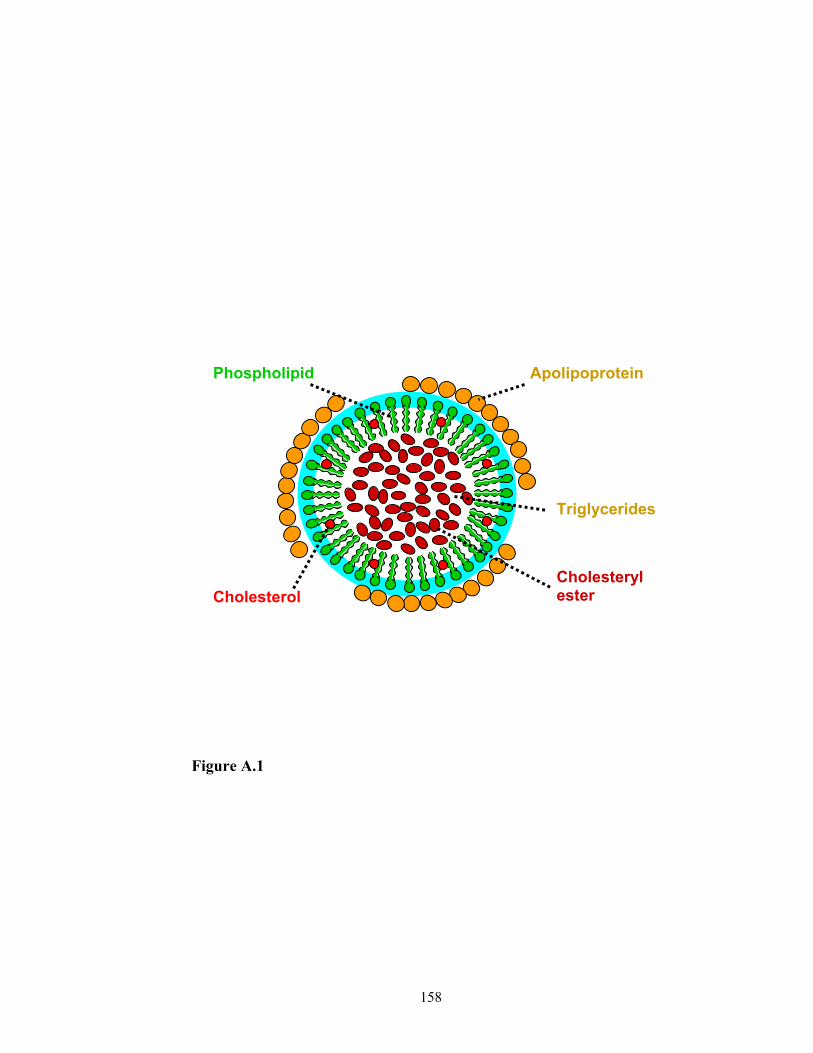
Triglycerides
Cholesterol
Apolipoprotein
Cholesteryl ester
Phospholipid
Figure A.1
158
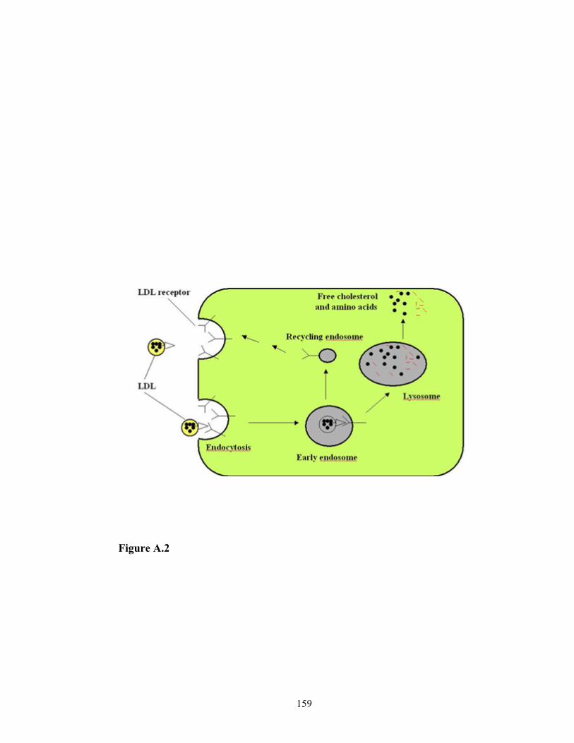
Figure A.2
159

Figure A.3
160
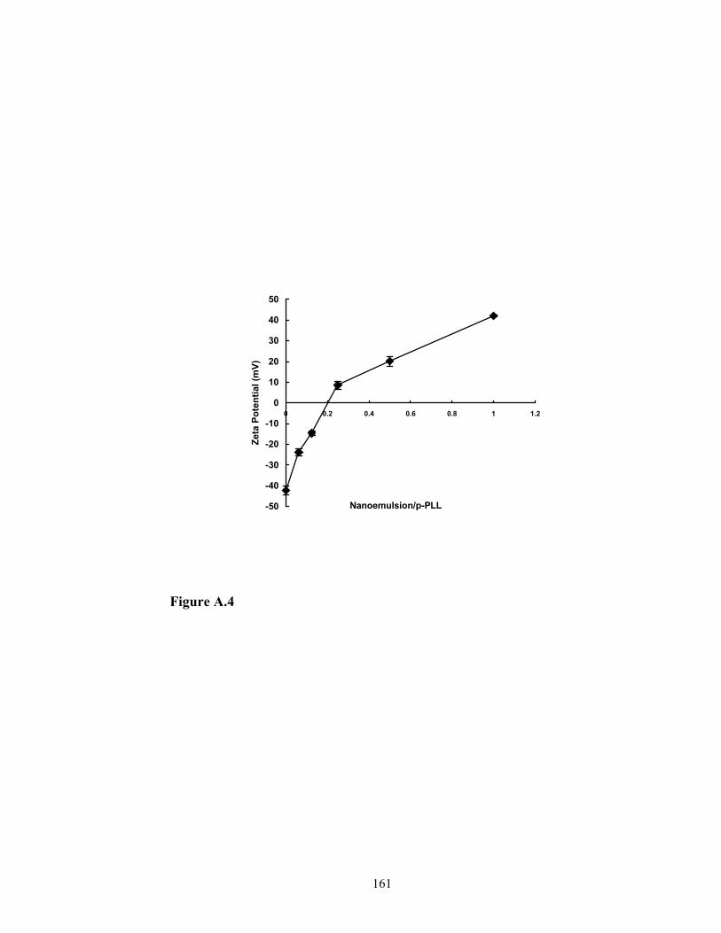
-50
-40
-30
-20
-10
0
10
20
30
40
50
0 0.2 0.4 0.6 0.8 1 1.2
Nanoemulsion/p-PLL
Zeta
Pot
entia
l (m
V)
Figure A.4
161
Figure A.5
162
REFERENCES
1. Roco MC. Nanotechnology – A frontier for engineering education. Int J Eng Edu
2002; 18(5):488-497.
2. Jones AL, Hradek GT, Hornick C, Renaud G, Windler EET, Havel RJ. Uptake
and processing of remnants of chylomicrons and very low density lipoproteins by
rat liver. J Lipid Res 1984;25:1151-8.
3. Umeda Y, Redgrave TG, Mortimer B-C, Mamo JCL. Kinetics and uptake
oxidatively modified lymph chylomicrons. American J Physiol 1995;268:G709-
16.
4. Soued M, Mansbach II CM. Chylomicron remnant uptake by enterocytes is
receptor dependent. American J Physiol 270 1996;(1 Pt 1):G203-212.
5. Porter CJH, Charman WH. Uptake of drugs into the intestinal lymphatic after oral
administration. Adv Drug Deliv Rev 1997;25:71-89.
6. Rensen PCN, De Vrueh RLA, van Berkel TJC. Targeting hepatitis B therapy to
the liver: clinical pharmacokinetic considerations. Clin Pharmacokinet
1996;31:131-155.
7. Hara T, Tan Y, Huang L. In vivo gene delivery to the liver using reconstituted
chylomicron remnants as a novel nonviral vector. Proc Natl Acad Sci USA
1997;94:14547-14552.
8. Hara T, Liu F, Liu DX, Huang L. Emulsion formulations as a vector for gene
delivery in vitro and in vivo. Adv Drug Deliv Rev 1997;24:265-271.
9. Olofsson, SO, Bjursell G, Bostrom K et al. Apolipoprotein B: structure,
biosynthesis and role in the lipoprotein assembly process. Atherosclerosis
1987;68:1-17.
163
10. Kader A, Pater A. Loading anticancer drugs into HDL as well as LDL has little
affect on properties of complexes and enhances cytotoxicity to human carcinoma
cells. J Control Rel 2002;80:29-44.
11. Shawer M, Greenspan P, ∅ie S, DR Lu. VLDL-resembling phospholipid-
submicron emulsion for cholesterol-based drug targeting. J Phar Sci
2002;91:1405-1413.
12. Brown MS, Goldstein JL. How LDL receptors influence cholesterol and
atherosclerosis. Scientific American 1984;251:58-66.
13. Brown, MS, Goldstein JL. A receptor-mediated pathway for cholesterol
homeostasis. Science 1986;232:34-47.
14. Shepherd J, Packard CJ, Bicker S, Lawrie TD, Morgan HG. Cholestyramine
promotes receptor-mediated low-density-lipoprotein catabolism. New England J
Med 1980; 302:1219-22.
15. Kesaniemi YA, Witztum JL, Steibrecher UP. Receptor-mediated catabolism of
low density lipoprotein in man: Quantitation using glucosylated low density
lipoprotein. J Clin Invest 1983;71:950-9.
16. Kader A, Davis PJ, Kara M, Liu H. Drug targeting using low density lipoprotein
(LDL): physicochemical factors affecting drug loading into LDL particles. J
Control Rel 1998; 55:231-243.
17. Vitols S, Gahrton G, Ost A, Peterson C. Elevated low density lipoprotein
receptor activity in leukemic cells with monocytic differentiation. Blood
1984;63:1186-1193.
164

18. Vitols S, Gahrton G, Bjorkholm M, Peterson C. Hypocholesterolaemia in
malignancy due to elevated low-density-lipoprotein-receptor activity in tumor
cells: evidence from studies in patients with leukemia. Lancet 1985;2:1150-1153.
19. Vitols S, Peterson C, Larsson O, Holm P, Aberg B. Elevated uptake of low
density lipoprotein by human lung cancer tissue in vivo. Cancer Res
1992;52:6244-6247.
20. Rudling MJ, Angelin B, Peterson CO, Collins VP. Low density lipoprotein
receptor activity in human intracranial tumors and its relation to the cholesterol
requirement. Cancer Res 1990;50:483-487.
21. Jung-Testas I, Weintraub H, Dupuis D, Eychenne B, Baulieu D-E, Robel P. Low
density lipoprotein-receptor in primary cultures of rat glial cells. J. Steroid
Biochem. Mol Biol 1992;42:597-605.
22. Maletinska L, Blakely EA, Bjornstad KA, Deen DF, Knoff LJ, Forte TM. Human
glioblastoma cell lines: levels of low-density lipoprotein receptor and low-density
lipoprotein receptor-related protein. Cancer Res 2000;60:2300-2303.
23. Firestone RA. Low-density lipoproteins as a vehicle for targeting antitumor
compounds to cancer cells. Bioconjugate Chem 1994;5:105-113.
24. de Smidt PC, van Berkel TJ. Prolonged serum half-life of antineoplastic drugs by
incorporation into the low density lipoprotein. Cancer Res 1990;50:7476-7482.
25. Allison BA, Pritchard PH, Levy JG. Evidence for low-density lipoprotein
receptor-mediated uptake of benzoporphyrin derivatives. Br J Cancer
1994;69:833-839.
165
26. Soncin M, Polo L, Reddi E, Jori G, Kenney ME, Cheng G, Rodgers MAJ. Effect
of the delivery system on the biodistribution of Ge(IV)octabutoxy-
phthalocyanines in tumor-bearing mice. Cancer Lett 1995;89:101-106.
27. Jori G, Reddi E. The role of lipoproteins in the delivery of tumor-targeting
photosensitizers. Int J Biochem 1993;25:1369-1375.
28. Pardridge WM, Buciak JL, Friden PM. Selective transport of an anti-transferrin
receptor antibody through the blood-brain barrier in vivo. J Pharmacol Exp Ther
1991;259:66-70.
29. Chen D, Lee KH. Biodistribution of calcitonin encapsulated in liposomes in mice
with particular reference to the central nerve system. Biochem Biophys Acta
1993;1158:244-250.
30. Huwyler J, Wu D, Pardridge WM. Brain drug delivery of small molecules using
immunoliposomes. Proc Natl Acad Sci USA 1996;93:14164-14169.
31. Meresse S, Delbart C, Fruchart JC, Cecchelli R. Low-density lipoprotein receptor
on endothelium of brain capillaries. J Neurochem 1989;53(2):340-345.
32. Lucarelli M, Gennarelli M, Cardelli P, Novelli G, Scarpa S, Dallapiccola B,
Strom R. Expression of receptors for native and chemically modified low-density
lipoprotein in brain microvessels. FEBS Lett 1997;401(1):53-58.
33. Dehouck B, Fenart L, Dehouck M, Pierce A, Torpier G, Cecchelli R. A new
function for the LDL receptor: transcytosis of LDL across the Blood Brain
Barrier. J Cell Biol 1997; 138(4):877-889.
34. Matsumoto A, Naito M, Itakura H, Ikemoto S, Asaoka H, Hayakawa I, Kanamori
H. Human macrophage scavenger receptors: Primary structure, expression, and
166
localization in atherosclerotic lesions. Proc Natl Acad Sci USA 1990;87:9133-
9137.
35. Meltzer MS, Gendelman HE. Mononuclear phagocytes as targets, tissue
reservoirs, and immunoregulatory cells in human immunodeficiency virus
disease, Curr Top Microbiol Immunol 1992;181:239-263.
36. Gendelman HE, Orenstein JM, Baca LM, Weiser B, Burger H, Kalter DC,
Meltzer MS. The macrophage in the persistence and pathogenesis of HIV
infection. AIDS 1989; 3(8):475-495.
37. Hu J, Liu H, Wang L. Enhanced delivery of AZT to macrophages via acetylated
LDL. J Control Rel 2000;69:327-335.
38. Jaeschke H, Smith CW, Clemens MG, Ganey PE, Roth RA. Mechanisms of
inflammatory liver injury: adhesion molecules and cytotoxicity of neutrophiles.
Toxicol Appl Pharmacol 1996;139:213-26.
39. Bijsterbosch MK, Ziere GJ, van Berkel TJ. Lactosylated low density lipoprotein:
a potential carrier for the site specific delivery of drugs to Kupffer cells. Mol
Pharmacol 1989;36:484-9.
40. Bijsterbosch MK, Manoharan M, Dorland R, Waarlo IHE, Biessen EAL, van
Berkel TJ. Delivery of cholesteryl-conjugated phosphorothioate
oligodeoxynucleotides to Kupffer cells by lactosylated low-density lipoprotein.
Biochem Pharmacol 2001;62:627-633.
41. Nyamekye I, Buonaccorsi G, McEwan J, MacRobert A, Bown S, Bishop C.
Inhibition of intimal hyperplasia in balloon injured arteries with adjunctive
167
phthalocyanine sensitized photodynamic therapy. Eur J Vasc Endovasc Surg
1996;11:19-28.
42. de Varies HE, Moor ACE, Dubbelman TMAR, van Berkel TJC, Kuiper J.
Oxidized low-density lipoprotein as a delivery system for photosensitizers:
Implications for photodynamic therapy of atherosceloris. J Pharmacol Exper
Therap 1999;289:528-534.
43. Phillips AJ. The challenge of gene therapy and DNA delivery. J Pharm Pharmcol
2001;53:1169-1174.
44. Kim J-S, Maruyama A, Akaike T, Kim SW. In vitro gene expression on smooth
muscle cells using a terplex delivery system. J Control Rel 1997;47:51-59.
45. Kim J-S, Kim B-I, Maruyame A, Akaike T, Kim SW. A new non-viral DNA
delivery vector: the terplex system. J Control Rel 1998;53:175-182.
46. Steinberg D. A docking receptor for HDL cholesterol esters. Science
1996;271:460-461.
47. Rensen PC, de Vrueh RL, Kuiper J, Bijsterbosch MK, Biessen EA, van Berkel TJ.
Recombinant lipoproteins: lipoprotein-like lipid particles for drug targeting.
Adv Drug Deliv Rev. 2001;47(2-3):251-76.
48. Lacko AG, Nair M, Paranjape S, Johnson S, Mcconathy WJ. High density
lipoprotein complexes as delivery vehicles for anticancer drugs. Anticancer
Res2002;22:2045-9.
49. Reisinger RE, Atkinson D. Phospholipid/cholesteryl ester microemulsion
containing unesterified cholesterol: model systems for low density lipoproteins. J
Lipid Res 1990; 31:849-858.
168
50. Chun PW, Brumbauge EE, Shiremann RB. Interaction of human low density
lipoprotein and apolipoprotein B with ternary lipid microemulsion. Biophys Chem
1986;25:223-241.
51. Hirata RDC, Hirata MH, Mesquita CH, Cesar TB, Maranhao RC. Effects of
apolipoprotein B-100 on the metabolism of a lipid microemulsion model in rats.
Biochim Biophy Acta 1999;1437:53-62.
52. Maranhao RC, Cesar TB, Pedross-Mariani SR, Hirata MH, Mesquita CH.
Metabolic behavior in rats of a nonprotein microemulsion resembling low-density
lipoprotein. Lipids 1993;28:691-695.
53. Pan G., Shawer M., Oie S., Lu R. D. In vitro gene transfection in human glioma
cells using a novel and less cytotoxic artificial lipoprotein delivery system. Pharm
Res. 2003;20(5):738-733.
54. Arap W, Pasqualini R, Ruoslahti E. Cancer treatment by targeted drug delivery to
tumor vasculature in a mouse model. Science 1998;279:377-380.
55. Croix BS, Rago C, Velculescu V, Traverso G, Romans KE, Montgomery E, Lal
A, Riggins GJ, Lengauer C, Vogelstein B, Kinzler K. Genes expressed in human
tumor endothelium. Science 2000;289:1197-1202.
56. Hong FD, Clayman GL. Isolation of a peptide for targeted drug delivery into
human head and neck solid tumors. Cancer Res 2000;60:6551-6556.
57. Molema G. Tumor vasculature directed drug targeting: applying new technologies
and knowledge to the development of clinically relevant therapies. Pharm Res
2002;19(9):1251-1258.
169

58. Loregian A, Marsden HS, Palu G. Protein-protein interactions as targets for
antiviral chemotherapy. Rev Med Virol 2002;12: 239-262.
59. Huang Z. Structural chemistry and therapeutic intervention of protein-protein
interactions in immune response, human immunodeficiency virus entry, and
apoptosis. Pharmacol Therap 2000;86:201-215.
60. Jones S, Thornton JM. Principles of protein-protein interactions. Proc Natl Acad
Sci USA 1996;93:13-20.
61. Cohen GH. Ribonucleotide reductase activity of synchronized KB cells infected
with herpes simplex virus. J Virol 1972;9:408-418.
62. Dutia BM. Ribonucleotide reductase induced by herpes simplex virus has a virus-
specified constituent. J Gen Virol 1983;64:513-521.
63. Paradis H, Gaudreau P, Brazeau P, Langelier Y. Mechanism of inhibition of
herpes simplex virus (HSV) ribonucleotide reductase by a nonapeptide
corresponding to the carboxy-terminus of its subunits. J Biol Chem
1988;263:16045-16050.
64. McClements W, Yamanaka G, Garsky V. Oligonucleotides inhibit the
ribonucleotide reductase of herpes simplex virus by causing subunit separation.
Viology 1988;162: 270-273.
65. Pillay D, Taylor S, Richman DD. Incidence and impact of resistance against
approved antiretroviral drugs. Rev Med Virol 2000;10: 231-253.
66. Zutshi R, Franciskovich J, Slultz M, Schweitzer B, Bishop P, Wilson M,
Chmielewski J. Targeting the dimerization interface of HIV-1 protease: inhibition
with cross-linked interfacial peptides. J Am Chem Soc 1997;119:4841-4845.
170

67. Bouras A, Boggetto N, Benatalah Z, de Rosny E, Sicsic S, Reboud-Ravaux M.
Design, synthesis, and evaluation of conformationally constrained tongs, new
inhibitors of HIV-1 protease dimerization. J Med Chem 1999;42:957-962.
68. Morris MC, Robert-Hebmann V, Chaloin L, Mery J, Heitz F, Devaux C, Goody
RS, Divita G. A new potent HIV-1 reverse transcriptase inhibitor. J Biol Chem
1999;274: 24941-24946.
69. Maroun RG, Krebs D, Roshani M, Porumb H, Auclair C, Troalen F, Fermandjian
S. Conformational aspects of HIV-1 integrase inhibition by a peptide derived
from the enzyme central domain and by antibodies raised against this peptide. Eur
J Biochem 1999;260:145-155.
70. Kasukawa H, Howley PM, Benson JD. A fifteen-amino-acid peptide inhibits
human papilloma E1-E2 interaction and human papillomavirus DNA replication
in vitro. J Virol 1998;72: 8166-8173.
71. Huang H, Chopra R, Verdine GL, Harrision SC. Structure of a covalently trapped
catalytic complex of HIV-1 reverse transcriptase: implication for drug resistance.
Science 1998;282: 1669-1675.
72. Bundell TL. Structure-based drug design. Nature 1996;384: 23-26.
73. Johnsson K, Ge L. Phage display of combinatorial peptide and protein libraries
and their application in biology and chemistry. Curr Top Microbiol Immunol
1999;243: 87-105.
74. Shih HM, Goldman PS, Demaggio AJ, Hollenberg SM, Goodman RH, Hoekstra
MF. A positive genetic selection for disrupting protein-protein interactions:
171

identification of CREB mutations that prevent association with the coactivator
CBP. Proc Natl Acad Sci USA 1997;94: 13396-13401.
75. Weiner AL. Liposomes for protein delivery selecting manufacture and
development processes. Immunomethods 1994;4:201-209.
76. Blanke SR, Milne JC, Benson EL, Collier RJ. Fused polycationic peptide
mediated delivery of diphtheria toxin A chain to the cytosol in the presence of
anthrax protective antigen. Proc Natl Acad Sci USA 1996;93: 8437-8442.
77. Elliot G, O’Hare P. Intracellular trafficking and protein delivery by a herpes
structural protein. Cell 1997;88:223-233.
78. Fawell S, Seery J, Daikh Y, Moore C, Chen LL, Pepinsky B, Barsoum J. Tat-
mediated delivery of heterologous protein into cells. Proc Natl Acad Sci USA
1994;91:664-668.
79. Loregian A, D’Acunto MR, Battistutta R, Tossi A, Palù G, Zanotti G. Crystal
structure of the B subunit of Eshcherichia coli heat-labile enterotoxin carrying
peptides with anti-herpes simplex virus type 1 activity. J Biol Chem
1999;274:8764-8769.
80. Dwek R. Glycobiology: Toward understanding the function of sugars. Chem Rev
1996; 96: 683-720.
81. Karpas A, Fleet GWJ, Dwek RA, Petrusson S, Namgoong SK, Ramsden NG,
Jacob GS, Rademacher TW. Aminosugar derivatives as potential anti-human
immunodeficiency virus agents. Proc Natl Acad Sci USA 1988;85: 9229-9233.
82. Jiang S, Zhao Q, Debnath AK. Peptide and non-peptide HIV fusion inhibitors.
Curr Phar Des 2002;8(8): 563-80.
172
83. D’Souza MP, Cairns JS, Plaeger SF. Current evidence and future direction for
targeting HIV entry: therapeutic and prophylactic strategies. JAMA 2000;284(2):
215-22.
84. Goss PE, Baker MA, Carver JP, Dennis JW. Inhibitors of carbohydrate
processing: A new class of anticancer agents. Clin Cancer Res 1995;1(9):935-44.
85. Lehne G. P-glycoprotein as a drug target in the treatment of multidrug resistant
cancer. Curr Drug Targets 2000;1(1): 85-99.
86. Campbell KR, Ohman EM, Cantor W, Lincoff AM. The use of glycoprotein
IIb/IIIa inhibitor therapy in acute ST-segment elevation myocardial infarction:
current practice and future trends. Am J Cardiol 2000; 85(8A): 32C-8C.
87. Meier-Ewert HK, Nesto RW. Targeting the use of glycoprotein IIb/IIIa
antagonists--- the diabetic patient. Rev Cardiovasc Med 2002;3(Suppl 1): S20-7.
88. Luscombe NM, Austin SE, Berman HM, Thornton JM. An overview of the
structure of protein-DNA complexes. Genome Biol 2000;1(1):1-10.
89. Rhodes D, Schwabe JW, Chapman L, Fairall L. Towards an understanding of
protein-DNA recognition. Philos Trans R Soc Lond Biol Sci 1996;351(1339):501-
9.
90. Good L, Nielsen PE. Progress in developing PNA as a gene-targeted drug.
Antisense Nucleic Acid Drug Dev 1997;7(4):431-7.
91. Welch JJ, Rauscher FJ, Beerman TA. Targeting DNA-binding drugs to sequence-
specific transcription factor-DNA complexes. Differential effects of intercalating
and minor groove binding drugs. J Biol Chem 1994;269(49):31051-8.
173
92. Henderson D, Hurley H. Specific targeting of protein-DNA complexes by DNA-
reactive drugs (+)-CC-1065 and pluramycin. J Mol Recognit 1996;9(2):75-87.
93. Kondo S, Kondo Y, Li G, Silverman RH, Cowell JK. Targeted therapy of human
malignant glioma in a mouse model by 2-5A antisense directed against telomerase
RNA. Oncogene 1998;16(25):3323-30.
94. Stewart SA, Hahn WC. Prospects for anti-neoplastic therapies based on telomere
biology. Curr Cancer Drug Targets 2001;2(1):1-17.
95. Blackburn EH. The telomere and telomerase: nucleic acid-protein complexes
acting in a telomere homeostasis system: a review. Biochem (Mosc)
1997;62(11):1196-1201.
96. Neidle S, Kelland LR. Telomerase as an anti-cancer target: current status and
future prospects. Anticancer Drug Des 1999;14(4):341-7.
97. Perry PJ, Arnold JR, Jenkins TC. Telomerase inhibitors for the treatment of
cancer: the current perspective. Expert Opin Investig Drugs 2001;10(12):2141-56.
174