Embed Size (px)
Citation preview
Int.J.Curr.Microbiol.App.Sci (2015) 4(5): 274-288
274
Original Research Article
In Vitro Antimicrobial Activity and Cytotoxicity Test of Native South Dakota Plant Extracts
Gitanjali NandaKafle1*, Kristen De Berg1, R. Neil Reese1, Radhey Shyam Kaushik1,2 and David Francis2
1Department of Biology and Microbiology, South Dakota State University, Brookings, SD, USA 2Department of Veterinary and Biomedical Science, South Dakota State University,
Brookings, SD, USA
*Corresponding author
A B S T R A C T
Introduction
Around the middle of 20th century, major advances in antibacterial drug development and other means of infection control helped human beings triumph over many infectious diseases. Development of penicillin in the early 1940s changed the situation radically with respect to bacterial infection (Tenover, 2006). However, the attainment of complete
eradication of infectious diseases is almost impossible. Bacteria respond to antibiotics by developing various modes of resistance as fast as we use antibiotics. Antimicrobial resistance in bacteria is one of the biggest global concerns at present (Siddiqi et al., 2011). Multidrug resistant isolates in several infectious agents are a cause for serious
ISSN: 2319-7706 Volume 4 Number 5 (2015) pp. 274-288 http://www.ijcmas.com
In vitro antimicrobial activity of 28 extracts of plants native to South Dakota were screened and evaluated against Escherichia coli. A disk diffusion assay was used for initial screening and antimicrobial activities of potential plant extracts were further assessed by a broth dilution method to determine their MIC values. Out of 28 plant extracts five plant extracts from Clematis ligusticifolia, Monarda fistulosa, Rhus aromatica, Centaurea stoebe and Onosmodium molle showed significant antimicrobial activity. In disk diffusion assay the percentage of inhibition was maximum for C. ligusticifolia.The minimum inhibitory concentration (MIC) for C. ligustifolia was 0.031gm /ml, for C. stoebe was 0.062gm /ml, for O. molle and M. fistulosa it was 0.125gm /ml and for R. aromatica 0.25gm /ml. In vitro cytotoxicity tests indicated that C. ligustifolia and R. aromatic showed some toxicity to porcine intestinal epithelial jejunum-2 cells (IPEC-J2) whereas C. stoebe did not have impact on cell growth compared to controls. However O. molle and M. fistulosa increased the growth of IPEC-J2 cell. Based on our findings it can be concluded that these five plants have antimicrobial activity against E. coli. Further investigation to determine which of their secondary metabolites (phytochemicals) are biologically active will be pursued.
K e y w o r d s
Antimicrobial activity, Escherichia coli, Methanol extract, Minimum Inhibitory Concentration, Cytotoxicity
Int.J.Curr.Microbiol.App.Sci (2015) 4(5): 274-288
275
concerns. Escherichia coli isolates from environmental, animal and human sources have been identified as resistant to antibiotics (Heike and Reinhard, 2005). This in part is the consequence of improper use of antibiotics as medicine. Pathogenic organisms have acquired some degree of resistance toward antimicrobials through various mechanisms (Wose Kinge et al., 2010). Three types of antibacterial resistance strategies have been suggested. A drug efflux pump has been observed in several bacteria which prevent accumulation of antibiotics. A second mechanism is deactivation or destruction of the antibiotics by hydrolytic enzymes in the periplasmic space and a third strategy for antibiotic resistance appears to be reprogramming of the target macromolecules to reduce the affinity of antibiotics for their RNA (Walsh, 2000; Tenover, 2006; Alviano and Alviano, 2009). Resistance has led to need for development of alternate sources of antibiotics to control E. coli infections.
Ancient herbal traditions are the source of origin for numerous contemporary medicines (Al-Bakri and Afifi, 2007). For the past few decades researchers have been trying to identify novel antibiotics from plant products. Medicinal plants used in folk medicine may provide guidance for developing new antibiotics. Higher plants produce large numbers of secondary metabolites with different biological activities. Medicinal plants have been used for centuries to treat various diseases all over the world (Vaghasiya and Chanda, 2007). So far, only a small percent of traditionally prescribed plant species on the earth have been studied for their therapeutic value. Plant phytochemicals could provide alternative classes of antibiotics having different target sites than current antibiotics, which may be effective against drug resistant pathogens. The drug industry is
exploring phytochemicals extensively to develop new antibiotics which can overcome resistant pathogens without any toxic effect to their host (Oskay and
Sar ,
2007). However, recent events show that drug companies are abandoning antibiotic research as Pfizer exit for the market suggests the need for academic research where knowledge - not profit are still important. Plants contain a large number of secondary compounds like alkaloids, phenolic compounds and flavonoids having antimicrobial properties.
There are several reports presented on the antibacterial activity of organic and aqueous plant extracts. Casearia sylvestris is a popular medicinal plant of South America and is being used against several diseases; diarrhea, gastric ulcer, inflammation, and herpes. Furthermore it was found that the ethanol extracts from leaves of this plant have potential antimicrobial activity (da Silva et al., 2008). In a recent study methanol extracts of Globularia alypum have shown significant effects on the growth of E. coli (Bogdadi et al., 2007). Berberis asiatia, a plant thatis traditionally used in Nepal and India to treat wounds, contains alkaloids that show strong activity against gram negative bacteria (Bhandari et al., 2000). Antimicrobial activity of native plants of Jordan; Gundelia tournefortii L. and Piminella anisum L. have shown demonstrated activity against antibiotic resistant strains of E. coli whereas, Origanum syriacum L, Eruca sativa Mill extracts have synergistic effects when used with the antibiotic clarithromycin (Darwish and Aburjai, 2010). Leaf extracts of three different species of Aloe; Aloe barbadensis, A. chabaudii and A. arborescens, used in folklore veterinary medicine in Zimbabwe were evaluated for their antimicrobial activity and the study showed that E. coli are sensitive to all three extracts (Mbanga et al.,
Int.J.Curr.Microbiol.App.Sci (2015) 4(5): 274-288
276
2010). Dubey et al. (2009) screened medicinal plants of India for their antimicrobial activity against E. coli and found four plants Terminalia catappa, Syzygium cumini, Eucalyptus hybrid and Holarrhena antidysentrica have significant antimicrobial activities.
Native Americans have used herbs medicinally for thousands of years as a part of their holistic approach to good health (Moerman, 1998). Anthony (2001) in his book has described a number of herbal remedies used by Native Americans to treat illness and heal injuries. Borchardt et al. (2008) found seven plant species that showed some effect against E. coli from Minnesota and Wisconsin. In South Dakota there are many varieties of plants that have been used as folk medicine, only few of them have been studied scientifically for their antimicrobial properties. Our main objective in this study was to identify South Dakota medicinal plants having significant antimicrobial activity that could be exploited as a source of antimicrobial agents. We also evaluated their potential for treating bacterial infection without significant toxicity to mammalian cells. In this article we have identified five medicinal plants having antimicrobial activity against E. coli.
Material and Methods
Selection of plants: Plants were selected (Table 1) based on their traditional use by Native Americans (Moerman, 2009).
Sample collection: Plant specimens for our study were collected from the Northern Great Plain from June through August 2010. Plants were collected from different locations weighed and stored at -80°C if not used immediately. Identification of the plants species was made based upon the USDA plant database nomenclature.
Voucher specimens were prepared and stored in the herbarium.
Extract preparation: Plant samples were homogenized in a blender with methanol at a ratio of 25gm fresh weight/ 250ml methanol. Plant materials were extracted with methanol for about 24 hours in dark. The plant samples were then vacuum filtered using VWR grade 415 qualitative filter paper. Methanol was removed from the sample using a rotary evaporator under vacuum. The residue was solubilized in 5 ml of 70% ethanol with concentration representing 5 g fresh plant weight per ml of ethanol in the final plant extract.
Bacterial strains and growth conditions
Escherichia coli ATCC # 25922 were obtained from the South Dakota Veterinary Diagnostic Laboratory at SDSU. The assay medium for E. coli was Trypton Soy Broth (TSB) or Agar (TSB, TSA, Oxoid Ltd. Hamsphire, UK). Bacterial cultures for antimicrobial testing were prepared by inoculating 25ml of TSB from fresh culture plates. Cultures were grown overnight in a shaker incubator at 200rpm and 37ºC (Klancnik et al. 2010). For antibacterial activity assay the CFU is adjusted to 106-107/ml. Stock culture were maintained in 70% glycerol and stored at -80ºC.
Antimicrobial testing methods
Disk Diffusion Assay (DDA): The disk diffusion assay was performed following the protocols of National Committee for Clinical Laboratory Standards (NCCLS, 2003) a modified Kirby-Bauer disk diffusion method (Bauer et al., 1966). One hundred µl of bacterial culture was evenly spread on a Mueller Hinton agar medium in a petri dish. Sterile6mm diameter paper disks were impregnated with 20µlplant extract. The
Int.J.Curr.Microbiol.App.Sci (2015) 4(5): 274-288
277
disks were allowed to completely air dry. Disks were then placed on the inoculated agar plates. Each extract was screened with three replicates. One disk of gentamicin (10µg per disk) was placed on each plate as positive control and one disk of ethanol dried as above, as negative control. The inoculated petri dishes were incubated for 18-24 hours at 37°C. After incubation the diameter of the inhibition zones were measured in mm.
Broth Microdilution method for Minimum Inhibitory Concentration (MIC): The MIC value of the plant extracts that were active against E. coli in the disk diffusion assay was then measured. For the MIC value the method described by OrdoñEz et al., 2003 and Sherlock Orla et al., 2010 were followed with some modifications.
Using 96- well plate (Corning, NY), columns 1 and 2 were filled with 50 µl sterile water and column 3 with 50 µl of 70% ethanol. In the remaining columns, the first row was filled with 90 µl of sterile water. The remaining wells were filled with 50 µl of sterile water. Then, 10 µl of each extract was mixed with the 90 µl of water in the first row. To obtain a twofold serial dilution of each extract ranging from 0.5 g/ml to 0.0039 g/ml of plant extract in fresh weight, 50 µl of the first row were aspirated with a micropipette and mixed with the second row. This process was repeated till the last row with remaining 50 µl was discarded resulting in all the wells containing a total of 50 µl of diluted extract.
Then, 50 µl of TSB with bacteria (106
CFU/ml) was added to all the wells except those in the first column, which served as blanks. Fifty µl of TSB without bacteria was added to column one. Column 2 was negative control and column 3 was positive
control. Finally the 96 well plate was incubated at 37°C for 18 hours. The bacterial growth was observed by taking absorbance at 600 nm. All assays were performed in triplicate, with absorbance of extract only and blanks subtracted to adjust for background absorbance.
The MIC value was termed as the minimum concentration of plant extract that inhibited growth of E. coli to a level < 0.05 at 600nm.Growth at this level cannot be observed through microscope (OrdoñEz et al., 2003).
The minimum bactericidal concentration was also determined by using the plate streaking method. In this method a loopful of the content of the well was streaked onto a sterile tryptic soy agar plate and allowed to incubate for an additional 24 hours, if there was bacterial growth within 24 hours it was concluded that extract acted as a bacteriostatic. If after 24 hour there no visual sign of growth the extract was determined to be bactericidal.
Cytotoxicity test
DNA
based proliferation assay: The DNA based proliferation assay was performed as per the instruction provided by Roche Molecular Biochemicals (Catalog Number: 11669915001) briefly, undifferentiated 7500 porcine intestinal epithelial cells (IPEC-J2) that were derived from one day old pig jejunum (Koh et al., 2008) were cultured in 100 µl media in a 96 well flat bottom plate and incubated for 24 hours at 37ºC. Ten µl of media was replaced with 10µl of plant extract with a final volume 100µl in each well. Cells were incubated for 18- 24 hours at 37°C. All cells in 96 well plate were labeled with BrdU labeling solution (10µl per well). The plate was incubated for 18 hours at 37° C, the
Int.J.Curr.Microbiol.App.Sci (2015) 4(5): 274-288
278
media was aspirated, and 200µl of FixDenat solution was added to each well and incubated for 30 min at room temperature. FixDenat solution was removed and 100ul anti-BrdU-POD working solution was added and incubated for 90 min at room temp, followed by washing and the addition of substrate solution. Plates were incubated for 20 min and quenched with H2SO4. Absorbance was measured at 450 and 690 nm (back ground correction).
Result and Discussion
Twenty eight plants species were collected for our study (Table 1). The antimicrobial activity of these plant extracts were screened by disk diffusion assay. Out of twenty eight plants five plants showed significant antimicrobial effect on E. coli. Centaurea stoeba, Rhus aromatica, Monarda fistulosa and Onosmodium molle showed moderate inhibition to E. coli growth whereas Clematis ligusticifolia showed maximum inhibition (Fig. 1). C. ligusticifolia was found to be highly effective against E. coli having zone of inhibition of 24.75 mm which was even greater than gentamicin (Fig. 1). R. aromatic and C. stoebe showed zone of inhibition that was more than 50% as large as that caused by gentamicin. M. fistulosa and O. molle showed inhibition distances less than 50% of that of gentamicin.
The disk diffusion assay provides an initial screening method showing that extracts from these 5 plant species have the potential for use as antimicrobials. To further assess these plants we determine their MIC value by broth dilution method and evaluate whether the effect of these extracts were bactericidal or bacteriostatic.
Broth Microdilution Method
The minimum inhibitory concentration
(MIC) is the lowest concentration at which an antimicrobial substance inhibits microbial growth under specified conditions. This concentration is bacteriostatic as it inhibits the growth but does not kill the bacteria completely. C. ligusticifolia showed the lowest MIC value, 0.031 g/ml whereas the highest MIC value was for R. aromatica 0.25 g/ml (Table 2).
The minimum bactericidal concentration (MBC) is defined as the lowest concentration at which the test compounds kill the bacteria. The MBC values for all five plants were found to be 0.5gm/ml (Table 2).
Cytotoxicity Test
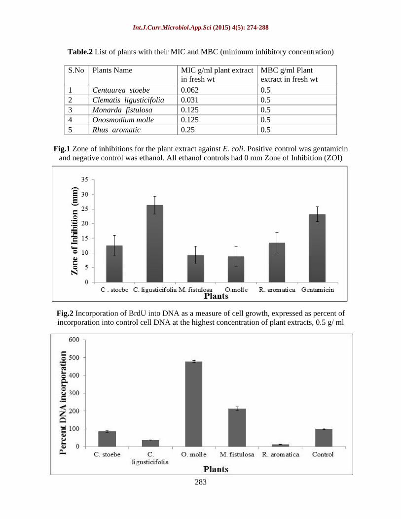
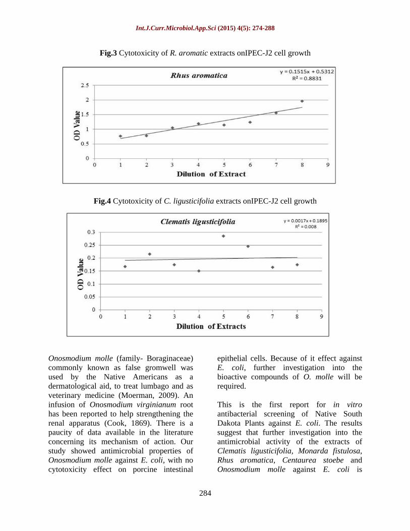
To determine the toxic effect of these plant extract on animals we performed cytotoxicity test using DNA- based proliferation assay. The result from this assay showed a wide range of effect of the plant extracts on the animal cell (IPEC-J2) (Fig. 2). Clematis ligusticifolia and Rhus aromatica both showed cytotoxicity to the IPECJ2 cells. A total of eight different concentrations had been examined from 0.0039 gm/ml to 0.5gm fresh weight /ml. C. ligusticifolia did not show any change in its toxic effect even at the lowest concentration whereas Rhus aromatica decreased in cytotoxicity as the concentration of the extract decreased (Fig. 3 and 4).
The extract of C. stoebe did not alter the growth of IPEC-J2 cells, when compared to the control. The extracts of M. fistulosaand O. molle appeared to increase the growth of IPEC-J2 cells at their higher concentration. These extracts significantly increased incorporation of BrdU by greater than two fold compare to control (Fig. 2). R. aromatica and C. ligusticifolia reduce the cell growth below 50% compare to control.
Int.J.Curr.Microbiol.App.Sci (2015) 4(5): 274-288
279
All plants tested have been used traditionally by the American Indians. Many of these species were utilized alone or in combination with other plants to treat a wide range of ailments. Only five plants out of 28 were effective against E. coli. The results of MIC assay showed that some of these plant extracts may provide potential sources of new antimicrobials. The BrdU assay indicated that only C. ligusticifolia was the most toxic to cultured IPEC-J2 cells R. aromatica also showed some toxicity against IPEC-J2 cells. Tetracyclines, which are broad spectrum antibiotics and widely used for various bacterial infections, have also been shown to induce cytotoxic effects in human blood lymphocytes in vitro (Celikand Eke, 2011). Erythromycin and its chemical derivatives are used to treat variety of human infections. These antibiotics have also been reported to cause cytotoxicity in human liver cell lines when treated in vitro (Viluksela et al., 1996). Additionally antibiotics ciprafoxacin, clyndamicin and metronidazole were tested on human gingival fibroblast cell cultures and showed cytotoxicity effect in a dose dependent manner (Ferreira et al., 2010). Though these antibiotics showed toxicity in vitro test, still they are in use to treat bacterial infections. To confirm the safety use of C. ligusticifolia and R. aromatica further in vivo studies are necessary.
The genus Clematis (Family
Ranunculaceae) includes 350 species which grow worldwide (Dong et al., 2010). Pharmacological studies have shown that extracts of many species of this genus demonstrate antimicrobial activity (Buzzini and Pieroni, 2003). Khan et al. (2001) reported that Clematis papuasica leaves and stem bark have broad spectrum antimicrobial activity. Buzzini and Pieroni (2003) studied antimicrobial activity of Clematis vitalba and Kyung et al. (2007) reported the antimicrobial activity of
Clematis apfifolia DC. Bioactive components of different species were identified and isolated by researchers. Phytochemical researches carried out on Clematis mandshurica revealed the presence of triterpenoidsaponins (Dong et al., 2010), lignans, alkaloids (Shi et al., 2006b), macrocyclic glycoside (Shi et al., 2007) and phenolic glycosides (Shi et al., 2006a). Wu et al. (2010) studied the therapeutic action of Clematis chinensis. However the nature and chemical constituents of Clematis ligusticifolia have not been extensively studied. The Native American used an infusion of leaves on horses for wounds, treatment of skin disease, ulcers and colds (Sweet.M, 1998). C. ligusticifoila extract inhibited E. coli even more than gentamicin (positive control). It also inhibited cell growth in the IPEC J2 culture across a range of serial dilutions. Although this extract was highly effective against E. coli, additional research needs to be performed to determine how these extracts can be used as antibiotics and which of the compounds in the extracts are responsible for their activity.
Antimicrobial effect of M. fistulosa (Family- Lamiaceae) has been studied by various researchers. Zhilyakova et al. (2009) studied the essential oils of M. fistulosa and showed that they have antibacterial activity against Gram negative bacteria. GC-MS analysis of Monarda oil revealed that thymoquinone (simple monoterpene quinine) is the major constituent and that it shows antimicrobial activity against Escherichia coli (Inouye et al., 2006). Johnson et al (1998) reported the isolation of two bioactive monterpenesthymoquinone and thymol and also observed that the reduction of thymoquinone to thymohydroquinone reduced its activity. Our results showed that it is toxic to E. coli but not toxic to the animal cell line and therefore it may be used as a potential antibiotic to control E. coli infections.
Int.J.Curr.Microbiol.App.Sci (2015) 4(5): 274-288
280
The genus Centaurea (Family
Asteraceae)
includes over 500 species that are found all over the world (Tekeli et al., 2011).Various species of Centaurea have been used traditionally to treat diseases such as hemmorhoids, abscesses and common colds. Twelve Centaurea species has been studied in Turkey for their antimicrobial activity. Eight of these species (C. balsamita, C. calolepis, C.cariensis subsp. maculiceps, C. cariensis subsp. microlepis, C. kotschyi var. kotschyi, C. solsitialis subsp.solsitialis, C. urvillei subsp. urvillei and C. virgata) demonstrated significant antimicrobial activity against different microorganisms (Tekeli et al., 2011). Guven et al. (2005) reported that ethyl acetate extracts of C.odyssei and C. kurdica have significant antimicrobial effect. The chemical composition of C.austro-anatolica has been determined by GC-MS and the major components they found were caryophyllene oxide, spthulenol, n-tricosanol and geranyl isovalerate(Ugur et al., 2009). The main classes of components are oxygenated monterpene hydrocarbons, sesquiterpene hydrocarbons, oxygenated sesquiterpines and aromatic alcohols (Ugur et al., 2009). The key compound of essential oil in C. sessilis and C. armena was found to be -eudesmol and was shown to have antimicrobial activity against both gram positive and gram negative bacteria (Yayli et al., 2005). Many species of Centaurea have been found to be effective against a broad spectrum of microorganism, however there is little published research concerning the chemical constituents of Centaurea stoebe. Further investigation to isolate the bioactive compounds of C. stoebe will be necessary.
Genus Rhus contains 250 species in the family of Anacardiacea found in temperate and tropical region worldwide. Various species have been used by the native people
for medicine (Rayne and Mazza, 2007). Gundidza et al. (2008) reported the presence of -pinene (86.95%) in the essential oil of Rhus lancea having antimicrobial activity against E. coli. Rhus coriaria is used as a spice in Turkey and has been shown to have antimicrobial activity against E. coli 0157: H7 (food borne pathogen) (Nasar-Abbas and Halkman, 2004). Shabana et al. (2008) reported that the major constituents of essential oil of Rhus coraria fruits are thymol, caryophyllene and embrene which are the key components with antimicrobial activity.
It is also reported that Rhus coraria had significant antimicrobial activity against several gram positive and gram negative bacteria including E. coli (Fazeli et al., 2007). Mossa et al (1996) described the presence of free flavonoids such as persicogenin, velutin, (2S) 5,3 ,4 -trihydroxy 7-methoxyflavonone and homoeriodictyol in Rhus retinorrhoea. Leaves, stems, barks, roots, fruits and the galls on R. chinensis have been shown to have therapeutic value for treating diarrhea, dysentery, rectal and intestinal cancer, diabetes mellitus, sepsis, oral disease and inflammation (Djakpo and Yao, 2010). Very little information is available about the phytochemical constituents and antimicrobial activity of Rhus aromatica the fragrant sumac which is native to the Northern Great Plains and used by the Native American for various medicinal purposes. Reichling et al. (2009) have reported that the aqueous extract of R. aromatica has antiviral potency against herpes simplex virus type 1 and type 2. In our research we demonstrated the antimicrobial activity of Rhus aromatica against E. coli. Further research is required to identify its active compounds.
Int.J.Curr.Microbiol.App.Sci (2015) 4(5): 274-288
281
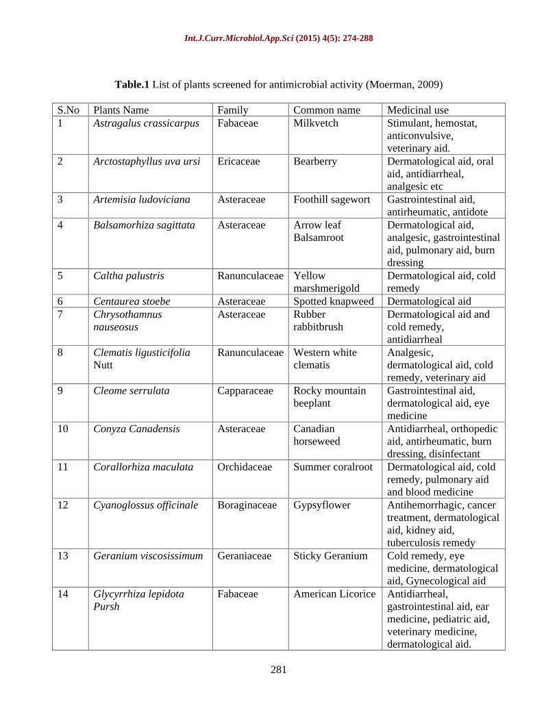
Table.1 List of plants screened for antimicrobial activity (Moerman, 2009)
S.No Plants Name Family Common name Medicinal use 1 Astragalus crassicarpus Fabaceae Milkvetch Stimulant, hemostat,
anticonvulsive, veterinary aid.
2 Arctostaphyllus uva ursi Ericaceae Bearberry Dermatological aid, oral aid, antidiarrheal, analgesic etc
3 Artemisia ludoviciana Asteraceae Foothill sagewort Gastrointestinal aid, antirheumatic, antidote
4 Balsamorhiza sagittata Asteraceae Arrow leaf Balsamroot
Dermatological aid, analgesic, gastrointestinal aid, pulmonary aid, burn dressing
5 Caltha palustris Ranunculaceae
Yellow marshmerigold
Dermatological aid, cold remedy
6 Centaurea stoebe Asteraceae Spotted knapweed Dermatological aid 7 Chrysothamnus
nauseosus Asteraceae Rubber
rabbitbrush Dermatological aid and cold remedy, antidiarrheal
8 Clematis ligusticifolia Nutt
Ranunculaceae
Western white clematis
Analgesic, dermatological aid, cold remedy, veterinary aid
9 Cleome serrulata Capparaceae Rocky mountain beeplant
Gastrointestinal aid, dermatological aid, eye medicine
10 Conyza Canadensis Asteraceae Canadian horseweed
Antidiarrheal, orthopedic aid, antirheumatic, burn dressing, disinfectant
11 Corallorhiza maculata Orchidaceae Summer coralroot Dermatological aid, cold remedy, pulmonary aid and blood medicine
12 Cyanoglossus officinale Boraginaceae Gypsyflower Antihemorrhagic, cancer treatment, dermatological aid, kidney aid, tuberculosis remedy
13 Geranium viscosissimum Geraniaceae Sticky Geranium Cold remedy, eye medicine, dermatological aid, Gynecological aid
14 Glycyrrhiza lepidota Pursh
Fabaceae American Licorice Antidiarrheal, gastrointestinal aid, ear medicine, pediatric aid, veterinary medicine, dermatological aid.
Int.J.Curr.Microbiol.App.Sci (2015) 4(5): 274-288
282
15 Melilotus officinalis Fabaceae Yellow
sweetclover Dermatological aid, cold remedy
16 Monarda fistulosa Lamiceae Wildbergamot beebalm
Dematological aid, ough medicine, throat aid, toothache remedy, gastrointestinal aid, eye medicine for sore eye.
17 Oenothera biennis Onagraceae Evening primerose Dermatological aid, hemorrhoid remedy
18 Onosmodium molle Boraginaceae Smooth onosmodium
Veterinary aid, dermatological aid, antirheumatic
19 Pediomelum argophyllum Fabaceae SilverleafScrufpea Used for fever, veterinary aid, used as laxative, dermatological aid
20 Perideridia gairdneri (Hook and Arn) Mathias (root)
Apiaceae Giardner sYampah
Antidiarrheal, cough medicine, dermatological aid, veterinary aid, diuretic and laxative
21 Physialis virginiana Solanaceae Virginia groundcherry
Used as stimulant
22 Plantago rugelii Plantaginaceae
Blackseed plantain Dermatological aid: poultice of fresh leaves applied to burn or any inflammation
23 Psoralea argophylla Fabaceae 24 Quercus macrocarpa Fagaceae Bur oak Antidiarrheal,
dermatological aid, pulmonary aid, gastrointestinal aid
25 Rhus aromatic Anacardiaceae Fragrant Sumac Dermatological aid, urinary aid, antidiarrheal, cold remedy, oral aid, pediatric aid
26 Senecio rapifolius Asteraceae Openwoods ragwort
27 Symphocarpus albus Caprifoliaceae Common snowberry
Diuretic and venereal aid, dermatological aid
28 Tanacetum vulgare Asteraceae Common tansy Anthelmintic, dermatological aid, antidiarrheal, cold remedy
Int.J.Curr.Microbiol.App.Sci (2015) 4(5): 274-288
283
Table.2 List of plants with their MIC and MBC (minimum inhibitory concentration)
S.No Plants Name MIC g/ml plant extract in fresh wt
MBC g/ml Plant extract in fresh wt
1 Centaurea stoebe 0.062 0.5 2 Clematis ligusticifolia 0.031 0.5 3 Monarda fistulosa 0.125 0.5 4 Onosmodium molle 0.125 0.5 5 Rhus aromatic 0.25 0.5
Fig.1 Zone of inhibitions for the plant extract against E. coli. Positive control was gentamicin and negative control was ethanol. All ethanol controls had 0 mm Zone of Inhibition (ZOI)
Fig.2 Incorporation of BrdU into DNA as a measure of cell growth, expressed as percent of incorporation into control cell DNA at the highest concentration of plant extracts, 0.5 g/ ml
Int.J.Curr.Microbiol.App.Sci (2015) 4(5): 274-288
284
Fig.3 Cytotoxicity of R. aromatic extracts onIPEC-J2 cell growth
Fig.4 Cytotoxicity of C. ligusticifolia extracts onIPEC-J2 cell growth
Onosmodium molle (family- Boraginaceae) commonly known as false gromwell was used by the Native Americans as a dermatological aid, to treat lumbago and as veterinary medicine (Moerman, 2009). An infusion of Onosmodium virginianum root has been reported to help strengthening the renal apparatus (Cook, 1869). There is a paucity of data available in the literature concerning its mechanism of action. Our study showed antimicrobial properties of Onosmodium molle against E. coli, with no cytotoxicity effect on porcine intestinal
epithelial cells. Because of it effect against E. coli, further investigation into the bioactive compounds of O. molle will be required.
This is the first report for in vitro antibacterial screening of Native South Dakota Plants against E. coli. The results suggest that further investigation into the antimicrobial activity of the extracts of Clematis ligusticifolia, Monarda fistulosa, Rhus aromatica, Centaurea stoebe and Onosmodium molle against E. coli is
Int.J.Curr.Microbiol.App.Sci (2015) 4(5): 274-288
285
warranted. It is also important to identify the secondary metabolites of these plants and their major active components having antimicrobial activity. Although monoterpene is the bioactive components in M. fistulosa, there is a paucity of information concerning the influence of growth environment and time of harvest on their antibiotic potential.
In this paper we showed that M. fistulosa and O. molle have inhibitory effect on E. coli but they are not toxic to the IPECJ2 cells. These two plants necessitate further study into their phytochemical inventory and their compounds that are active for treating infections. C. ligusticifolia and R. aromatica have promising antibacterial activity against E. coli but they also showed toxicity to the IPECJ2 cells in vitro. The application of these two plants should be tested in vivo to understand the toxicity effect for further medicinal use of these plants. The medicinal plants screened in this study may have metabolites which are effective for antibacterial activity and it needs to be further investigated.
Acknowledgement
We thank South Dakota Agricultural Experiment Station and Biology/ Microbiology Department of SDSU for the financial and technical support provided to complete this project.
References
Al-Bakri, A.G., Afifi, F.U. 2007. Evaluation of antimicrobial activity of selected plant extracts by rapid XTT colorimetry and bacterial enumeration. J. Microbiol. Meth., 68: 19 25.
Alviano, D.S., Alviano, C.S. 2009. Plant extracts: search for new alternatives
to treat microbial diseases. Curr. Pharm. Biotech., 10: 106 121.
Anthony, J.C. 2001. Secrets of native American herbal remedies (Book Review). Total Health, 23, 12. New York.
Bauer, A., Kirby, W., Sherris, J.A., Turck, M. 1966. Antibiotic susceptibility testing by a standardized single disk method. Am. J. Clin. Path., 45: 493496.
Bhandari, D.K., Nath, G., Ray, A.B., Tewari, P.V. 2000. Antimicrobial activity of crude extracts from Berberis asiatica stem bark. Pharm. Biol., 38: 254 257.
Bogdadi, H.A.A., Kokoska, L., Havlik, J., Kloucek, P., Rada, V., Vorisek, K. 2007. In vitro antimicrobial activity of some Libyan medicinal plant extracts. Pharm. Biol., 45: 386 391.
Borchardt Joy, R., Donad, L.W., Craig, C.S., Kendra, L.K., Fulcher, G.R., Nancy, J.E., Biesboer, D.D., Russell, F.B. 2008. Antimicrobial activity of native and naturalized plants of Minnesota and Wisconsin. J. Med. Plants Res., 2(5): 098 110.
Buzzini, P., Pieroni, A. 2003. Antimicrobial activity of extracts of Clematis vitalba towards pathogenic yeast and yeast-like microorganisms. Fitoterapia, 74: 397 400.
Celik, A., Eke, D. 2011. The Assessment of cytotoxicity and genotoxicity of tetracycline antibiotic in human blood lymphocytes using CBMN and SCE analysis, in Vitro. Int. J. Hum. Genet., 11(1): 23 29.
Cook, H.W.M. 1869. The physio-medical dispensatory, Cincinnati, OH.
Coutinho, H.D.M., Costa, J.G.M., Lima, E.O., Falcao-Silva, V.S., Siqueira, J.P. Jr. 2008. Enhancement of the antibiotic activity against a multiresistant Escherichia coli by
Int.J.Curr.Microbiol.App.Sci (2015) 4(5): 274-288
286
Mentha arvensis L. and chlorpromazine. Chemotherapy, 54: 328 330.
Da Silva, S.L., Chaar, J.D.S., Damico, D.C.S., Figueiredo, P.D.M.S., Yano, T. 2008. Antimicrobial activity of ethanol extract from leaves of Casearia sylvestris. Pharm. Biol., 46: 347 351.
Darwish, R.M., Aburjai, T.A. 2010. Effect of ethnomedicinal plants used in folklore medicine in Jordan as antibiotic resistant inhibitors on Escherichia coli. BMC Complement Altern. Med., 10.
Djakpo, O., Yao, W. 2010. Rhus chinensis and Galla chinensis - folklore to modern evidence: review. Phytother. Res., 24: 1739 1747.
Dong, F-Y., Cui, G-H., Zhang, Y-H., Zhu, R-N., Wu, X-J., Sun, T-T., Wang, W. 2010. Clematomandshurica saponin E, a new triterpenoid saponin from Clematis mandshurica. J. Asian Nat. Prod. Res., 12: 10611068.
Dubey, M., Kumar, A., Mahour, K., Dwivedi, D., Vihan, V.S. 2009. Screening of antibacterial activity of some Indian plants with their phytochemical analysis. J. Pure Appl. Microbiol., 3: 211 214.
Fazeli, M.R., Amin, G., Attari, M.M.A., Ashtiani, H., Jamalifar, H., Samadi, N. 2007. Antimicrobial activities of Iranian sumac and avishan-e shirazi. (Zataria multiflora) against some food-borne bacteria. Food Control, 18: 646 649.
Ferreira, M.B., Myiagi, S., Nogales, G.C., Campos, S.M., Lage-Marques, J.L. 2010. Time and concentration dependent cytotoxicity of antibiotics used in endodontic therapy. J. Appl. Oral Sci., 18(3).
Gundidza, M., Gweru, N., Mmbengwa, V., Ramalivhana, N.J., Magwa, Z., Samie, A. 2008. Phytoconstituents and biological activities of essential Oil from Rhus lancea L. F. Afr. J. Biotech., 7: 2787 2789.
Guven, K., Celik, S., Uysal, I. 2005. Antimicrobial activity of Centaurea Species. Pharm. Biol, 43: 67 71.
Heike, V.B., Reinhard, M. 2005. Antimicrobial resistance of Escherichia coli and therapeutic implications. Int. J. Med. Microbiol., 295: 503 511.
Inouye, S., Uchida, K., Takizawa, T., Yamaguchi, H., Abe, S. 2006. Evaluation of the effect of terpenoids quinones on Trichophyton mentagrophytes by solution and vapor contact. J. Infect. Chemo., 12: 100-104.
Johnson, H.A., Rogers, L.L., Alkire, M.L., McCloud, T.G., McLaughlin, J.L. 1998. Bioactive Monoterpenes from Monarda fistulosa (LAMIACEAE). Nat. Prod. Lett., 11: 241 250.
Khan, M.R., Kihara, M., Omoloso, A.D. 2001. Antimicrobial activity of Clematis papuasica and Nauclea obversifolia. Fitoterapia, 72: 575578.
Koh, S.Y., George, S., Brozel, V., Moxley, R., Francis, D., Kaushik, R.S. 2008. Porcine intestinal epithelial cell lines as a new in vitro model for studying adherence and pathogenesis of enterotoxigenic Escherichia coli. Vet. Microbiol., 130: 191 197.
Kyung, K.H., Woo, Y.H., Kim, D.S., Park, H.J., Kim, Y.S. 2007. Antimicrobial activity of an edible wild plant, apiifolia Virgin's Bower (Clematis apiifolia DC). Food Sci. Biotechnol., 16: 1051 1054.
Mbanga, J., Mangoma, N., Said, B. 2010. An evaluation of the antimicrobial
Int.J.Curr.Microbiol.App.Sci (2015) 4(5): 274-288
287
activities of Aloe barbadensis, A. chabaudii and A. arborescens leaf extracts used in folklore veterinary medicine in Zimbabwe. J. Anim. Vet. Adv., 9: 2918 2923.
Moerman, D.E. 1998. Native American medicinal plants: an ethnobotanicl dictionary. Timber Press, Portland, Or.
Moerman, D.E. 2009. Native American medicinal plants: an ethnobotanicl dictionary. Timber Press, Portland, Or.
Mossa, J.S., Sattar, E.A., Aboushoer, M., Galal, A.M. 1996. Free flavonoids from Rhus retinorrhoea Steud, ex Olive. Int. J. Pharmacogn., 34: 198201.
Nasar-Abbas, S.M., Halkman, A.K. 2004. Antimicrobial effect of water extract of sumac (Rhus coriaria L.) on the growth of some food borne bacteria including pathogens. Int. J. Food Microbiol., 97: 63 69.
NCCLS (ed.), 2003. Determination of minimum inhibitory concentrations (MICs) of antibacterial agents by broth dilution. Wiley-Blackwell.
Ordoñez, A.A.L., Gomez, J.D., Cudmani, N.M., Vattuone, M.A., Lsla, M.I. 2003. Antimicrobial activity of nine extracts of Sechium edule (Jacq.) Swartz. Microb. Ecol. Health Dis., 15: 33.
Oskay, M., Sari, D. 2007. Antimicrobial screening of some Turkish medicinal plants. Pharm Biol., 45: 176 181.
Rayne, S., Mazza, G. 2007. Biological activities of extracts from sumac (Rhus spp.): A review. Plant Foods Hum. Nutr., 62: 165 175.
Reichling, J., Neuner, A., Sharaf, M., Harkenthal, M., Schnitzler, P. 2009. Antiviral activity of Rhus aromatica (fragrant sumac) extract aginst two types of herpes simplex viruses in
cell culture. Die Pharmazie, 64: 538 541.
Shabana, M.M., El Sayed, A.M., Yousif, M.F., El Sayed, A.M., Sleem, A.A. 2008. Phytochemical and biological study of sumac. Egypt J. Pharma Sci., 49: 83 101.
Sherlock, O., Dolan, A., Athman, R., Power, A., Gethin, G., Cowman, S., Hilary, H. 2010. Comparison of the antimicrobial activity of Ulmo honey from Chile and Manuka honey against methicillin-resistant Staphylococcus aureus, Escherichia coli and Pseudomonas aeruginosa. BMC Complement Altern. Med., 10.
Shi, S-P., Dong, C-X., Jiang, D., Tu, P-F. 2007. Macrocyclic glucosides from Clematis mandshurica and Clematis hexapetala. Biochem. Sys. Ecol., 35: 57 60.
Shi, S-P., Jiang, D., Dong, C-X., Tu, P-F. 2006. New phenolic glycosides from Clematis mandshurica. Helv Chimica Acta, 89: 1023 1029.
Shi, S-P., Tu, P-F., Dong, C-X., Jiang, D. 2006. Alkaloids from Clematis manshurica Rupr. J. Asian Nat. Prod. Res., 8.
Siddiqi, R., Naz, S., Ahmad, S., Sayeed, S.A. 2011. Antimicrobial activity of the polyphenolic fractions derived from Grewia asiatica, Eugenia jambolana and Carissa carandas. Int. J. Food Sci. Tech., 46: 250 256.
Sweet, M. 1998. Common edible and useful plant of the West. Naturegraph Publisher, Inc., Happy Camp, CA.
Tekeli, Y., Zengin, G., Aktumsek, A., Sezgin, M., Torlak, E. 2011. Antibacterial activities of extracts from twelve Centaurea species from Turkey. Arch. Biol. Sci., 63: 685690.
Tenover, F.C. 2006. Mechanisms of antimicrobial resistance in bacteria.
Int.J.Curr.Microbiol.App.Sci (2015) 4(5): 274-288
288
Am. J. Med., 119: S3 10; discussion S62-70.
Ugur, A., Sarac, N., Ceylan, O., Duru, M.E. 2009. Chemical composition of endemic Centaurea austro-anatolica and studies of its antimicrobial activity against multi-resistant bacteria. Acta Pharmaceutica, 59: 463 472.
Vaghasiya, Y., Chanda, S.V. 2007. Screening of methanol and acetone extracts of fourteen Indian medicinal plants for antimicrobial activity. Turk. J. Biol., 31: 243 248.
Viluksela, M., Vainio, P.J., Tuominen, R.K. 1996. Cytotoxicity of macrolide antibiotics in a cultured human liver cell line. J. Antimicrob. Chemother., 38(3): 465 473.
Walsh, C. 2000. Molecular mechanisms that confer antibacterial drug resistance. Nature, 406: 775 781.
Wose-Kinge, C.N., Ateba, C.N., Kawadza, D.T. 2010. Antibiotic resistance profiles of Escherichia coli isolated from different water sources in the mmabatho locality, north-west province, South Africa. South Afr. J. Sci., 106: 44 49.
Wu, W., Xu, X., Dai, Y., Xia, L. 2010. Therapeutic effect of the saponin fraction from Clematis chinensis Osbeck roots on osteoarthritis induced by monosodium iodoacetate through protecting articular cartilage. Phytother. Res., 24: 538 546.
Yayli, N., Ya ar, A., Güleç, C., Usta, A., Kolayl , S., Co kunçelebi, K., Karao lu, . 2005. Composition and antimicrobial activity of essential oils from Centaurea sessilis and Centaurea armena. Phytochemistry, 66: 1741 1745.
Zhilyakova, E.T., Novikov, O.O., Naumenko, E.N., Krichkovskaya, L.V., Kiseleva, T.S., Timoshenko, E.
Yu, Novikova, M., Yu, Litvinov, S.A. 2009. Study of Monarda fistulosa essential oil as a prospective antiseborrheic agent. Bull. Experiment Biol. Med., 148(4): 414.