Embed Size (px)
Citation preview

In tobacco leaf epidermal cells, the integrity of protein exportfrom the endoplasmic reticulum and of ER export sitesdepends on active COPI machinery
Giovanni Stefano1,†, Luciana Renna1,†, Laurent Chatre2,†,‡, Sally L. Hanton1, Patrick Moreau2, Chris Hawes3 and
Federica Brandizzi1,*
1Department of Biology, 112 Science Place, University of Saskatchewan, Saskatoon, SK S7N 5E2, Canada,2Laboratoire de Biogenese Membranaire, UMR5200, CNRS, Universite de Bordeaux 2, Bordeaux, France, and3Research School of Biological and Molecular Sciences, Oxford Brookes University, Oxford OX3 0BP, UK
Received 5 October 2005; revised 2 December 2005; accepted 14 December 2005.*For correspondence (fax þ1 306 966 4461; e-mail [email protected]).†These authors have contributed equally.‡Present address: Department of Biology, 112 Science Place, University of Saskatchewan, Saskatoon, SK S7N 5E2, Canada.
Summary
Trafficking of secretory proteins between the endoplasmic reticulum (ER) and the Golgi apparatus depends on
coat protein complexes I (COPI) and II (COPII) machineries. To date, full characterization of the distribution and
dynamics of these machineries in plant cells remains elusive. Furthermore, except for a presumed linkage
between COPI and COPII for the maintenance of ER protein export, the mechanisms by which COPI influences
COPII-mediated protein transport from the ER in plant cells are largely uncharacterized. Here we dissect the
dynamics of COPI in intact cells using live-cell imaging and fluorescence recovery after photobleaching
analyses to provide insights into the distribution of COPI and COPII machineries and the mechanisms by which
COPI influences COPII-mediated protein export from the ER. We found that Arf1 and coatomer are dynamically
associated with the Golgi apparatus and that the COPII coat proteins Sec24 and Sec23 localize at ER export
sites that track with the Golgi apparatus in tobacco leaf epidermal cells. Arf1 is also localized at additional
structures that originate from the Golgi apparatus but that lack coatomer, supporting the model that Arf1 also
has a coatomer-independent role for post-Golgi protein transport in plants. When ER to Golgi protein transport
is inhibited by mutations that hamper Arf1-GTPase activity without directly disrupting the COPII machinery for
ER protein export, Golgi markers are localized in the ER and the punctate distribution of Sec24 and Sec23 at the
ER export sites is lost. These findings suggest that Golgi membrane protein distribution is maintained by the
balanced action of COPI and COPII systems, and that Arf1-coatomer is most likely indirectly required for
forward trafficking out of the ER due to its role in recycling components that are essential for differentiation of
the ER export domains formed by the Sar1-COPII system.
Keywords: COPI, anterograde transport, endoplasmic reticulum export sites.
Introduction
In plant cells, proteins destined for the early secretory
pathway attain a steady-state localization by cycling be-
tween the endoplasmic reticulum (ER) and the Golgi
apparatus (Brandizzi et al., 2002; Denecke et al., 1992; Han-
ton et al., 2005). Our understanding of the mechanisms
regulating protein cycling at the ER/Golgi interface in plants
is limited. Emerging evidence indicates that anterograde ER/
Golgi protein transport is mediated by COPII vectors (An-
dreeva et al., 2000; Bar-Peled and Raikhel, 1997; Phillipson
et al., 2001; Ritzenthaler et al., 2002; daSilva et al., 2004;
Takeuchi et al., 2000; Yang et al., 2005), although their
morphology is still undetermined. It is assumed that retro-
grade Golgi/ER protein transport is mediated by COPI
machinery. The existence of COPI components such as
coatomer and the GTPase Arf1, which is responsible for
coatomer recruitment (Teal et al., 1994), has been proved in
plants (Contreras et al., 2000; Couchy et al., 2003; Movafeghi
et al., 1999; Pimpl et al., 2000; Takeuchi et al., 2002), and
ª 2006 The Authors 95Journal compilation ª 2006 Blackwell Publishing Ltd
The Plant Journal (2006) 46, 95–110 doi: 10.1111/j.1365-313X.2006.02675.x

COPI-coated vesicles have been observed predominantly at
cis and medial Golgi cisternae (Pimpl et al., 2000). However,
a full characterization of the dynamics of COPI at the Golgi
apparatus in living plant cells has yet to emerge.
It has been shown that Arf1 localizes at the Golgi
apparatus (Takeuchi et al., 2002) with the coatomer (Mova-
feghi et al., 1999; Pimpl et al., 2000; Ritzenthaler et al., 2002).
However, a distribution of Arf1 on non-Golgi structures has
also been verified (Xu and Scheres, 2005), and it has been
shown that Arf1 has a role in the sequence-specific vacuolar
sorting route to the lytic vacuole in tobacco (Pimpl et al.,
2003). This raises the question of whether Arf1 requires
coatomer at other organelles besides the Golgi apparatus, or
whether Arf1 interacts with different proteins in its non-
Golgi locations.
Mutations that hamper the GTPase activity of Arf1 have
been used to manipulate anterograde ER to Golgi protein
transport (Lee et al., 2002; Pimpl et al., 2003; Ritzenthaler
et al., 2002; Saint-Jore et al., 2002; daSilva et al., 2004;
Takeuchi et al., 2002). Dominant negative mutants of Arf1
that are impaired in GTP/GDP exchange affect the secretion
of soluble markers (Pimpl et al., 2003), and, along with
brefeldin A (BFA)-induced inhibition of Arf1, cause a re-
absorbance of Golgi membrane proteins into the ER (Bran-
dizzi et al., 2002; Lee et al., 2002; Ritzenthaler et al., 2002;
Saint-Jore et al., 2002; daSilva et al., 2004; Takeuchi et al.,
2000). The information available on the mechanisms by
which COPI may regulate COPII-mediated protein export
from the ER in plant cells is in general speculative, based on
studies in vitro and in non-plant systems (Hanton et al.,
2005). It remains to be shown in vivo how perturbation of
COPI-mediated traffic affects protein export from the ER in
plant cells.
Protein export from the ER occurs at the ER export sites
(ERES), the subcellular distribution of which has been
recently investigated in plants (daSilva et al., 2004; Yang
et al., 2005). It has been demonstrated that ERES differen-
tiation can be induced upon co-expression of the COPII
initiator Sar1 and Golgi membrane markers in tobacco leaf
epidermal cells. In this system, ERES tracked with the
Golgi bodies in close proximity (daSilva et al., 2004). It has
also been shown that a fluorescent protein fusion of a
COPII component, Sec13, labelled stationary punctate
structures on the surface of the ER in the absence of
Golgi markers in tobacco BY-2 cells (Yang et al., 2005).
These structures considerably outnumbered the Golgi
stacks, although some were seen to associate with the
rims of Golgi stacks (Yang et al., 2005). These data suggest
that the Golgi apparatus is not continually linked to a
single ERES. On the basis of these observations, it cannot
be excluded that in tobacco leaf epidermal cells COPII coat
markers may have a different distribution in comparison
to cargo-induced Sar1 punctae. Therefore, an examination
of COPII coat distribution is needed to determine
unambiguously the distribution of ERES in tobacco leaf
epidermal cells.
The drug BFA, which blocks the activation of Arf1 (Robi-
neau et al., 2000), prevents cargo-induced recruitment of a
fluorescent fusion of Sar1 at the ERES in tobacco leaf
epidermal cells (daSilva et al., 2004). In contrast, BFA does
not have an effect on the distribution of ERES labelled by
Sec13-GFP in tobacco BY-2 cells (Yang et al., 2005). Thus, it
is important to define whether the integrity of ERES depends
on active COPI machinery in the absence of cargo-induced
ERES formation in order to complete the characterization of
the requirements for differentiation of ERES in tobacco leaf
epidermal cells.
Here, we have analyzed the dynamic distribution of COPI
and COPII machineries in living tobacco leaf epidermal cells.
In particular, we focused on the manipulation of Arf1-
mediated protein transport to address the relevance of an
intact COPI machinery in control of the integrity of ER export
sites and of efficient ER protein export in vivo. Our results
indicate that Arf1 and coatomer are dynamically associated
with Golgi bodies but that Arf1 also binds to Golgi-derived
structures that lack coatomer, and that COPII components,
such as Sec23 and Sec24, localize at punctate structures that
track with Golgi stacks. We also found that blockage of Arf1
activity results in either inhibition or retardation of the
assembly of COPI machinery, in disruption of the integrity of
the ER export sites, and in the collapse of protein export
from the ER. Thus, our data suggest that the Arf1–coatomer
system is required for forward protein trafficking out of the
ER due to its role in recycling essential components to the ER
that are needed to differentiate ER export domains formed
by the COPII system.
Results
Arf1 localizes at the Golgi apparatus and Golgi-derived
structures
To determine the distribution of Arf1 in tobacco leaf epi-
dermal cells, a green fluorescent fusion (GFP) of an Ara-
bidopsis Arf1 (Phillipson et al., 2001; Arf1-GFP) was used
for confocal microscopy analysis. Arf1-GFP was distributed
on punctate structures of heterogeneous size (£1 lm in
diameter; Figure 1a,b). The larger Arf1-GFP structures
corresponded to Golgi bodies (Figure 1a), as shown by co-
expression experiments with a yellow fluorescent protein
(YFP) fusion to the Golgi marker ERD2 (ERD2-YFP; Bran-
dizzi et al., 2002). To establish the origin of the smaller
Arf1-labelled structures, we monitored the distribution of
Arf1 with respect to the Golgi apparatus over time in cells
co-expressing Arf1-GFP and ERD2-YFP. We observed that
small Arf1-structures formed on a Golgi stack and even-
tually detached from it (Figure 1b and Supplementary
data 1).
96 Giovanni Stefano et al.
ª 2006 The AuthorsJournal compilation ª 2006 Blackwell Publishing Ltd, The Plant Journal, (2006), 46, 95–110

Our localization data support additional roles of Arf1
besides regulation of COPI assembly (Pimpl et al., 2003). To
provide further evidence for this, we investigated whether
coatomer was distributed on the non-Golgi structures
labelled by Arf1. Co-expression experiments using either
ERD2-GFP or Arf1-GFP with a YFP fusion to an Arabidopsis
eCOP (eCOP-YFP), a component of the COPI coatomer (Kreis
et al., 1995), demonstrated that coatomer co-localized with
the Golgi marker (Figure 2a) but not with the additional Arf1-
GFP structures (Figure 2b). This suggests that COPI coatom-
er components only appear to play a role in intra-Golgi and
Golgi to ER transport, although Arf1 may have additional
roles besides COPI assembly at the Golgi apparatus.
Arf1, coatomer and protein cargo exhibit different dynamics
at the Golgi apparatus
To provide evidence of a dynamic cycle of COPI on and off
the Golgi membranes in living plant cells and to correlate the
residence time of COPI on Golgi membranes with the ex-
change of protein cargo between the ER and Golgi appar-
atus, we used fluorescence recovery after photobleaching
(FRAP) analysis on fluorescent fusions of eCOP, Arf1 or ERD2
expressed in tobacco leaves. FRAP allows measurement of
the rate of movement of a fluorescent marker towards and
from defined areas of a cell. A significant fluorescence
recovery indicates that the bleached molecules are ex-
changing with fluorescent pools of the protein localized in
other parts of the cell, assuming that chimaeric protein
levels in the bleached area had reached a steady state at the
start of each experiment (Lippincott-Schwartz and Patterson,
2003). By photobleaching eCOP-YFP fluorescence, we cal-
culated that coatomer associates and dissociates from Golgi
membranes with a half time of 20.7 � 3.0 sec (Figure 3a,b).
Photobleaching of Arf1-GFP fluorescence on the Golgi
bodies (Figure 3a) was followed by a rapid fluorescence
recovery (half time ¼ 12.1 � 2.1 sec; Figure 3a,b), which
was significantly faster than that of eCOP-YFP (P < 0.05).
FRAP experiments on Golgi bodies labelled with ERD2-YFP
(Figure 3a), which is known to distribute all over the Golgi
stack (Boevink et al., 1998), indicated a half-time recovery of
fluorescence of 123.0 � 14.8 sec (Figure 3b), much slower
than the half-time values measured for coatomer and Arf1.
These data show that not only does a Golgi membrane
protein move to and from the Golgi apparatus, as described
earlier and for other Golgi proteins, such as ST-GFP and
GONST1-GFP (Brandizzi et al., 2002; daSilva et al., 2004), but
also reveal that cytosolic COPI components exchange con-
tinuously at the surface of the Golgi apparatus, providing
evidence for constant remodelling of the membranous and
soluble components of this organelle.
GTPase-defective mutants of Arf1 disrupt Golgi protein
distribution to different extents
To analyze in vivo whether interference with the normal
cycling of Arf1 on and off Golgi membranes would influence
Golgi membrane protein distribution, we examined the ef-
fects of active and inactive forms of Arf1 on the distribution
of a fluorescent fusion of ERD2, which is exported from the
ER by COPII carriers and cycles continuously between ER
and Golgi at the same rate as other Golgi membrane
Figure 1. Arf1 localizes at the Golgi apparatus and at additional structures that originate from the Golgi apparatus.
(a) A confocal image of a tobacco leaf epidermal cell co-expressing Arf1-GFP and the Golgi marker ERD2-YFP shows that Arf1-GFP is localized at Golgi bodies and at
smaller additional structures (inset in merged image, arrowhead). Scale bar ¼ 5 lm.
(b) Sequence of images showing a cell co-transformed with ERD2-YFP (pseudo-coloured magenta) and Arf1-GFP (pseudo-coloured green). Co-localization of the two
markers appears white. Time (sec) of acquisition of frames is indicated at the top left corner. The arrowheads indicate Arf1-GFP structures that are detached from the
Golgi apparatus. Scale bar ¼ 4 lm.
COPI influences ER protein export 97
ª 2006 The AuthorsJournal compilation ª 2006 Blackwell Publishing Ltd, The Plant Journal, (2006), 46, 95–110

markers (Brandizzi et al., 2002; daSilva et al., 2004). There-
fore, we co-expressed this marker with either wild-type Arf1
or Arf1 sequences bearing point mutations that create
dominant negative mutants of the GTPase by enhancing its
affinity for either GDP or GTP [T31N or Q71L, respectively
(Teal et al., 1994)]. To identify cells expressing untagged
Arf1 or its mutant forms, we subcloned their coding se-
quences in a bi-cistronic vector encoding a cyan fluorescent
protein (CFP) spliced to the tripeptide serine-lysine-leucine
(CFP-SKL) for targeting to peroxisomes (Sparkes et al., 2003;
see also Supplementary data 2). We then co-transformed
tobacco leaf epidermal cells with these bi-cistronic vectors
and with a plasmid encoding ERD2-YFP. To produce com-
parable results, we used the same optical density of Agro-
bacterium containing the different constructs for tobacco
leaf transient transformation, and we analyzed samples by
confocal microscopy at the same time after transformation.
In the presence of wild-type Arf1, we observed a typical
punctate distribution of the ERD2-YFP (Figure 4a; cf. Fig-
ure 1), in agreement with reports that over-expression of
Arf1 does not affect the distribution of Golgi markers (Lee
et al., 2002; Takeuchi et al., 2002; Xu and Scheres, 2005), and
has no effect on protein secretion (Pimpl et al., 2003). Co-
expression of Arf1GDP with ERD2-YFP caused re-absorb-
ance of the Golgi marker into the ER in the majority (95%) of
cells co-expressing the mutant and ERD2-YFP (Figure 4a).
However, the fluorescence of the marker was mostly distri-
buted in the Golgi in the majority of cells co-expressing
Arf1GTP and ERD2-YFP (85% of the cells co-expressing the
mutant and the Golgi marker; Figure 4a). The ER mem-
branes appeared enlarged in those cells where Golgi mem-
branes were re-absorbed into the ER in comparison to
control cells. This is probably due to accumulation of a Golgi
pool of fluorescent ERD2 whose export is inhibited by the
mutants (see also Lee et al., 2002).
To determine whether Arf1 mutants could have a similar
effect on other Golgi markers, we co-expressed wild-type
Arf1 and its mutants with YFP fusions of either a rat sialyl-
transferase transmembrane domain (ST-YFP; Brandizzi
et al., 2002) or an Arabidopsis b1,2-xylosyltransferase cyto-
solic tail and transmembrane domain (Dirnberger et al.,
2002; Xylo-YFP). Under these conditions, we verified that the
effect of Arf1 mutants on the distribution of these proteins is
similar to that of ERD2-YFP (Supplementary data 3).
These data show that disruption of the COPI machinery at
the Golgi apparatus mediated by Arf1 mutants is followed by
collapse of the Golgi membranes into the ER. As the Golgi
markers used in these experiments cycle continuously in and
out of the Golgi apparatus, most likely from and to the ER
(Brandizzi et al., 2002; daSilva et al., 2004), the redistribution
Figure 2. Coatomer localizes at the Golgi apparatus but not at additional Arf1 structures.
(a) A confocal image of a cell co-expressing ERD2-GFP and eCOP-YFP shows that coatomer localizes at the Golgi apparatus (inset).
(b) A confocal image of a tobacco leaf epidermal cell co-expressing Arf1-GFP and eCOP-YFP shows co-localization of the two markers at the Golgi apparatus, and the
presence of smaller additional Arf1-GFP structures which lack eCOP-YFP labelling (inset in merged image, arrowhead). Scale bars ¼ 5 lm.
98 Giovanni Stefano et al.
ª 2006 The AuthorsJournal compilation ª 2006 Blackwell Publishing Ltd, The Plant Journal, (2006), 46, 95–110
of the Golgi markers into the ER suggests an interruption
of protein exchange between the ER and the Golgi
apparatus.
To confirm further that Arf1GDP has a stronger negative
effect on protein export from the ER than does Arf1GTP, we
co-transfected tobacco leaf protoplasts with the secretory
marker a-amylase (Phillipson et al., 2001) and a dilution
series of untagged Arf1, and Arf1GDP or Arf1GTP mutants.
Arf1 did not affect a-amylase secretion (Figure 4b), but the
Arf1GDP and Arf1GTP mutants exhibited a negative effect
on the secretion of a-amylase (see also Pimpl et al., 2003).
Arf1GDP exhibited a stronger effect than Arf1GTP on
secretion of the soluble marker. These data mirror our
microscopy results showing that Arf1GDP affected the ER
export of a Golgi marker more strongly than Arf1GTP under
the same experimental conditions, and suggest that the two
mutants may block ER export by different means.
Differences in the cycling of the Arf1 mutants on and off
Golgi membranes is the basis of their effect on Golgi
membrane protein distribution
To determine whether the Arf1 mutants perturbed the dis-
tribution of membrane proteins at the Golgi apparatus with
different mechanisms, we first aimed to determine the
subcellular localizations of their fluorescent protein fusions
by confocal microscopy (Figure 5). The activity of these
proteins was also tested by a biochemical assay (Supple-
mentary data 4).
GFP fusions of the inactive and active mutants of Arf1
(Arf1GDP-GFP and Arf1GTP-GFP, respectively) were ex-
pressed in tobacco leaf epidermal cells using the same
Agrobacterium optical density and confocal microscope
imaging settings to compare the levels of expression
between cells (see also Experimental procedures). We
hypothesized that Arf1GDP should be localized in the
cytosol, as although it should be able to bind to membranes
via its interaction with a guanine nucleotide exchange factor
(GEF), its inability to exchange GDP for GTP would be
followed by fast release from the membranes into the
cytosol, causing its association with Golgi membranes to be
undetectable. Consistent with our hypothesis, permanently
inactive Arf1 (Arf1GDP-GFP) was localized in the cytosol
(Figure 5a) at any level of expression (see also Xu and
Scheres, 2005). The GTP-trapped mutant was instead distri-
buted to punctate structures in most cells (Figure 5b),
corresponding well with observations by Xu and Scheres
(2005). To determine the identity of the structures highligh-
ted by Arf1GTP-GFP, we co-expressed the fusion with ERD2-
YFP. We found that Arf1GTP-GFP mostly co-localized with
the Golgi marker but also highlighted additional structures
(Figure 6a, inset in merged image, arrowhead), which coin-
cided with those marked by wild-type Arf1-YFP (data not
shown). We also detected a cytosolic distribution of
Arf1GTP-GFP in those cells that were over-expressing the
mutant (Figures 5c and 6b). In these cells, ERD2-YFP was
distributed in the ER (Figure 6b).
These data suggest that the Arf1 mutants may affect COPI
distribution via different mechanisms. In fact, the cytosolic
(a)
(b)
Figure 3. COPI machinery and Golgi enzymes cycle to and from the Golgi
apparatus continuously.
(a) Qualitative FRAP experiment on a cortical section of tobacco leaf
epidermal cells expressing eCOP-YFP, Arf1-GFP or ERD2-YFP treated with
the actin depolymerizing agent, latrunculin B (Brandizzi et al., 2002) to stop
Golgi movement. In the ERD2-YFP panel, the fluorescence of all Golgi stacks
was photobleached but only two Golgi stacks are indicated by arrowheads.
Time from the photobleaching event (bleach) is expressed in seconds at the
bottom left of the two subsequent frames. Scale bars ¼ 2 lm.
(b) Histograms of the time required for the fluorescence in the photobleached
region to recover to 50% of the recovery asymptote (half-time) measured in
FRAP experiments in cells expressing either eCOP-YFP (n ¼ 15), Arf1-GFP
(n ¼ 12) or ERD2-YFP (n ¼ 11). n, number of bleached Golgi stacks. The
difference between the groups was significant at P < 0.05 (student’s test).
COPI influences ER protein export 99
ª 2006 The AuthorsJournal compilation ª 2006 Blackwell Publishing Ltd, The Plant Journal, (2006), 46, 95–110

distribution of Arf1GDP indicates that this mutant is not able
to initiate the assembly of COPI machinery at the Golgi
membranes. Golgi-associated Arf1GTP may be capable of
initiating the assembly of COPI machinery on the Golgi. To
explore these hypotheses further, we co-expressed un-
tagged Arf1GDP with either Arf1-YFP or eCOP-YFP. This
caused a cytosolic distribution of both markers (Fig-
ure 7b,d), in comparison with the controls (Figure 7a,c; see
also Supplementary data 5). These results suggest that Arf1-
GDP most likely titrates out any Arf1-GEFs needed for the
activation of wild-type Arf1 at the Golgi apparatus, and
consequently impedes the association of coatomer to the
Golgi membranes. In the presence of Arf1GTP bound
mutant, coatomer was capable of cycling on and off Golgi
membranes (Supplementary data 6). This result prompted
us to test the extent to which protein exchange between ER
and Golgi could be affected by this mutant. Therefore, we
performed FRAP analysis on Golgi membranes labelled by
ERD2-YFP. As this marker cycles between the ER and Golgi
apparatus (Brandizzi et al., 2002), a difference in the recovery
(a)
(b)
Figure 4. Arf1 is determinant for the integrity of Golgi membranes.
(a) Merged confocal images of tobacco leaf epidermal cells co-expressing ERD2-YFP (pseudo-coloured magenta) with untagged Arf1, Arf1GDP and Arf1GTP
mutants. The presence of the untagged GTPase in cells is revealed by the visualization of peroxisomes (pseudo-coloured green) with imaging settings for CFP. The
percentage values correspond to the number of cells with punctate ERD2-YFP distribution divided by the total number of observed cells and multiplied by 100. The
total number was the sum of the cells with punctate fluorescence distribution of ERD2-YFP plus the number of cells with ERD2-YFP re-distributed exclusively in the
ER. Sample size ¼ 100 cells for each experiment with one Arf1 protein. Scale bars ¼ 5 lm.
(b) Secretion assay using tobacco leaf protoplasts. Histogram showing the secretion index of a-amylase [ratio of extracellular and intracellular activities, Phillipson
et al. (2001)] in protoplasts expressing untagged Arf1, Arf1GDP [Arf1(T31N)], and Arf1GTP [Arf1(Q71L)]. Concentrations of DNA (lg) for each sample are indicated
along the x-axis. Error bars ¼ standard error of the mean for three independent repetitions.
Figure 5. The subcellular localization of Arf1 depends on its GTPase status.
Confocal images of tobacco leaf epidermal cells expressing GFP fusions of Arf1GDP (a) or Arf1GTP (b,c). Arf1GDP-GFP is cytosolic (a), while Arf1GTP-GFP localizes
at punctate structures (b), which are lost in over-expressing cells (c). Note that for presentation purposes the settings for confocal imaging (laser output and detector
gain) to acquire image (c) were lowered with respect to image (b) in order not to over-saturate the image (see also Experimental procedures). Scale bars ¼ 5 lm.
100 Giovanni Stefano et al.
ª 2006 The AuthorsJournal compilation ª 2006 Blackwell Publishing Ltd, The Plant Journal, (2006), 46, 95–110

of fluorescence of ERD2 in the Golgi apparatus in comparison
to a control would indicate that the Arf1GTP mutant may
affect protein cycling between the ER and the Golgi appar-
atus. Photobleaching of ERD2-YFP fluorescence in the pres-
ence of the mutant was followed by recovery (half-
time ¼ 204.3 � 40.6 sec; Figure 8b,c), indicating that ER to
Golgi protein transport was occurring. However, comparison
with the recovery rate of the fluorescence of the same
Golgi marker in control cells (half-time ¼ 123.0 � 14.8 sec;
Figures 3 and 8a,c) indicated that the mutant significantly
reduced the recovery of fluorescence of ERD2-YFP into the
Golgi apparatus. Therefore, constitutive activation of Arf1 on
Golgi membranes most likely retards protein cargo exchange
at the ER/Golgi interface.
Figure 6. Constitutively active Arf1 localizes at Golgi membranes as long as the integrity of Golgi bodies is maintained.
(a) Confocal image of a cell co-expressing ERD2-YFP (pseudo-coloured magenta) and Arf1GTP-GFP (pseudo-coloured green), showing that the active form of Arf1
localizes at the Golgi apparatus and additional structures (inset in merged image, arrowhead).
(b) Confocal image of a cell co-expressing ERD2-YFP and high levels of Arf1GTP-GFP. Note the distribution of ERD2-YFP fluorescence in the ER and that the
fluorescence of Arf1GTP-GFP is cytosolic. Scale bars ¼ 5 lm.
Figure 7. When Arf1 is inactive, the COPI machinery is cytosolic.
Confocal images of tobacco leaf epidermal cells expressing either Arf1-YFP alone as a control (a), or (b) Arf1-YFP (pseudo-coloured magenta) and untagged Arf1GDP
encoded in a bi-cistronic vector bearing the CFP-SKL sequence for labelling peroxisomes (pseudo-coloured green) for identification of cells co-expressing the
mutant protein.
(c) Control cell expressing eCOP-YFP alone.
(d) Confocal image of a cell co-expressing eCOP-YFP and untagged Arf1GDP in a bi-cistronic vector as in (b). Co-expression of CFP-SKL with Arf1-YFP did not affect
the distribution of the Arf1-YFP in comparison to the control (Supplementary data 5). Scale bar ¼ 5 lm.
COPI influences ER protein export 101
ª 2006 The AuthorsJournal compilation ª 2006 Blackwell Publishing Ltd, The Plant Journal, (2006), 46, 95–110

The slower recovery of ERD2-YFP fluorescence at the
Golgi apparatus suggested that GTPase activity of the
Arf1GTP mutant was not entirely abolished. To test this,
we bleached the fluorescence of Arf1GTP-GFP on Golgi
stacks. Upon photobleaching Arf1GTP-GFP, the fluores-
cence recovered with a half-time of 35.7 � 3.4 sec (n ¼ 11),
which is threefold slower than the Arf1-GFP half-time
(12.1 � 2.1 sec, n ¼ 12, see Figure 3a,b) in accordance with
results in mammalian cells and in vitro (Teal et al., 1994;
Vasudevan et al., 1998).
Taken together, our results indicate that inactive Arf1
prevents COPI formation at the Golgi apparatus while the
active Arf1 mutant allows COPI formation and COPI-medi-
ated protein transport with a reduced activity in comparison
with wild-type Arf1.
The differentiation of ER export sites depends on active
retrograde protein export
We next aimed to test whether impaired COPI activity affects
the integrity of the ER export machinery. For this, we used a
YFP fusion of an Arabidopsis Sec24, a COPII coat component
(YFP-Sec24), to label the ERES. To ensure that fluorescent
Sec24 retained its functionality, we tested its ability to
interact with another COPII coat component Sec23 and form
a heterodimeric complex (Antonny and Schekman, 2001) in
a glutathione–agarose affinity chromatography assay based
on the interaction of a recombinant GST-Sec23 with plant-
expressed YFP-Sec24 (Figure 9). YFP-Sec24 was found to
interact with GST-Sec23 (Figure 9, lane 5) but not with GST
alone (Figure 9, lane 3). Therefore, YFP-Sec24 is a functional
marker capable of interacting with Sec23 as shown in
mammalian cells (Stephens et al., 2000). At a subcellular
level, YFP-Sec24 was localized at punctate structures (Fig-
ure 10a, arrowhead) and in the cytosol (Figure 10a). Co-
expression experiments using YFP-Sec24 with ERD2-GFP
indicated that these structures localized at the peri-Golgi
area, with the exception of additional rare bright YFP-Sec24
structures of variable size that did not localize at the
Figure 8. In the presence of Arf1GTP, ER protein export to the Golgi is impaired.
(a,b) Half-time recovery curves of ERD2-YFP fluorescence in cells expressing ERD2-YFP alone (a) or in the presence of Arf1GTP (b) and treated with latrunculin B to
depolymerize actin. In (b), the presence of untagged Arf1GTP was ensured by the visualization of peroxisomes labelled with CFP-SKL.
(c) Histogram of the half-times of recovery of YFP fluorescence after photobleaching in cells expressing ERD2-YFP alone (ERD2-YFP, n ¼ 11) or in the presence of
Arf1GTP (ERD2-YFP þ Arf1GTP; n ¼ 11). The difference between the groups was significant at P < 0.05 (Student’s t-test).
Figure 9. YFP-Sec24 is capable of interaction with the COPII component
Sec23
Extracts of tobacco leaves expressing Sec24-YFP were incubated with
recombinant GST-Sec23 bound onto glutathione–agarose beads. Proteins
retained by the GST-Sec23 agarose beads were eluted and then boiled in SDS
sample buffer for immunoblot analysis with anti-GFP serum. Sec24-YFP (lane
5) was retained by GST-Sec23. Negative controls: extracts of untransformed
tobacco leaves [()); lane 1]; GST beads alone did not retain YFP (lane 2) nor
Sec24-YFP (lane 3); Sec23-GST did not interact with YFP (lane 4).
102 Giovanni Stefano et al.
ª 2006 The AuthorsJournal compilation ª 2006 Blackwell Publishing Ltd, The Plant Journal, (2006), 46, 95–110
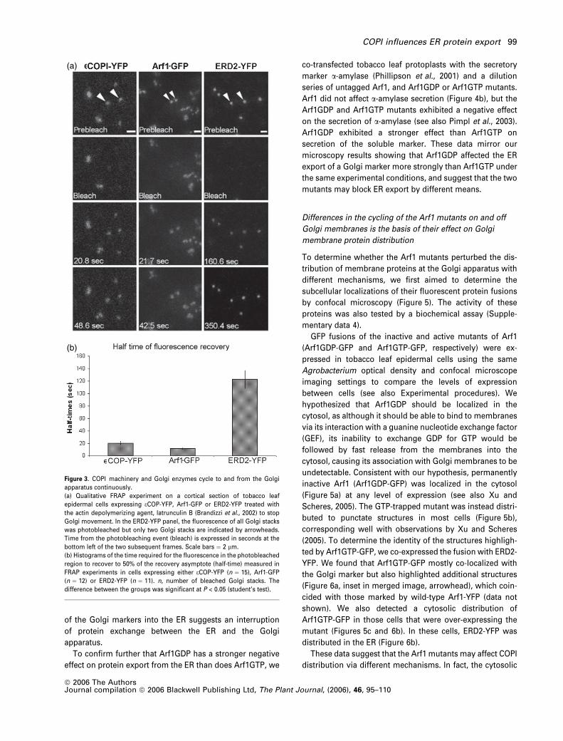
peri-Golgi area (6.7% of YFP-Sec24-labelled structures; Fig-
ure 10b–d). We also verified that the YFP-Sec24-labelled
ERES tracked together with Golgi stacks (Figure 10e–g; see
also Supplementary data 7a–c). Furthermore, the expres-
sion of a Golgi marker did not appear to be necessary for the
movement of YFP-Sec24 punctate structures as these were
motile in cells expressing the Sec24 construct alone (Sup-
plementary data 7d). Similar results were achieved with a
fluorescent fusion of another COPII component, Sec23 (YFP-
Sec23; Supplementary data 7d and 8, and data not shown),
although the percentage of Sec23 structures that did not co-
localize was lower than that of Sec24 (3.7%). These data re-
veal that in tobacco leaf epidermal cells COPII markers can
label ERES without over-expression of Golgi marker pro-
teins, and support previous findings that ERES are in close
association with the Golgi stacks (daSilva et al., 2004).
To explore the influence of COPI machinery on the
differentiation of ERES, we co-expressed YFP-Sec24 with
fluorescent fusions of wild-type and mutant Arf1 proteins. In
the presence of Arf1-GFP, YFP-Sec24 was distributed at the
peri-Golgi area but not at the additional smaller structures
labelled by Arf1-GFP (Figure 11a, inset arrowhead). How-
ever, YFP-Sec24 was distributed into the cytosol in co-
expression with Arf1GDP-GFP (Figure 11b). In the presence
of the active Arf1 mutant, the punctate appearance of the
ERES at the Golgi area labelled by YFP-Sec24 was main-
tained in most cells (Figure 11c). In cells that were highly
over-expressing Arf1GTP-GFP, YFP-Sec24 fluorescence was
distributed in the cytosol (Figure 11d). Similar results were
obtained with the other COPII marker, YFP-Sec23 (Supple-
mentary data 8). These data indicate that collapse of COPI-
mediated protein transport perturbs differentiation of ER
export sites and distribution of membrane proteins at the
Golgi apparatus. Taken together, our findings also suggest
that ERES maintain their integrity as long as COPI-mediated
protein transport takes place.
Discussion
The association of COPI at membranes is highly dynamic
and implies additional functions for Arf1 beside ER/Golgi
protein transport
In this work, localization analyses of Arf1-GFP mutants have
indicated that active Arf1 associates with Golgi membranes
while inactive Arf1 resides in the cytosol, consistent with
cycles of binding and release of the GTPase to and from
Golgi membranes demonstrated by FRAP experiments.
FRAP experiments also indicated a transient association of
coatomer with Golgi bodies with a slower turnover than
Figure 10. ER export sites and Golgi bodies behave as single mobile secretory units.
(a)–(d) Confocal images of tobacco leaf epidermal cells expressing either the ER export site marker YFP-Sec24 alone (a), or YFP-Sec24 (b) with a Golgi marker, ERD2-
GFP (c). (d) Merged image of (b) and (c). Note that YFP-Sec24 punctate structures localize mostly at the peri-Golgi area. Rare additional punctate structures are
indicated (arrowheads).
(e)–(g) High-magnification time-lapse of a cell co-expressing YFP-Sec24 and ERD2-GFP presented in vertical display as single channel for ERD2-GFP (e), for YFP-
Sec24 (f) and as merged images (g).
Scale bar in (a), (b) ¼ 5 lm; (e) ¼ 2 lm.
COPI influences ER protein export 103
ª 2006 The AuthorsJournal compilation ª 2006 Blackwell Publishing Ltd, The Plant Journal, (2006), 46, 95–110

Arf1, suggesting that coatomer dissociation does not coin-
cide with Arf1 GTP hydrolysis. These results imply that
several cycles of binding and release of Arf1 may be required
to form a COPI domain at the ER/Golgi interface and to
determine COPI uncoating, analogous to findings in mam-
malian cells (Presley et al., 2002).
Arf1-GFP was found to localize at the Golgi apparatus with
coatomer, and also at additional structures that lack coa-
tomer and originate from the Golgi apparatus. These
structures may represent the trans Golgi network (TGN) or
TGN-derived structures that detach from the Golgi appar-
atus, analogous to findings in mammalian cells (Waguri
et al., 2003). A separate nature of the plant TGN from the
Golgi apparatus has also been suggested in a study of the
localization of TGN SNAREs in Arabidopsis cells (Uemura
et al., 2004). These structures showed heterogeneous size
and it cannot be excluded that a population of larger ones
may be composed of groups of smaller structures. It has
been suggested that the non-Golgi Arf1 structures may be
endocytic compartments in Arabidopsis and onion cells
based on labelling with a Rab5 homologue, ARA7-GFP (Xu
and Scheres, 2005), although this protein has also been
shown to identify pre-vacuolar compartments in tobacco
leaf cells (Kotzer et al., 2004). As we followed the formation
of the structures only at the Golgi apparatus, we cannot
exclude the co-existence of Arf1-associated endocytic struc-
tures, and it is also possible that the Golgi-derived structures
may eventually mature to become, or merge with, endo-
somal or pre-vacuolar structures. The distribution of Arf1
verified in this study and by Xu and Scheres (2005) may be
linked to the subcellular location of various GEFs for Arf1
activation that are present in the Arabidopsis genome
Figure 11. The integrity of the ERES depends on a functional COPI-mediated protein transport.
Confocal images of cells co-expressing YFP-Sec24 and GFP fusions of Arf1 (a), Arf1 GDP mutant (b), or Arf1GTP at lower (c) and higher expression (d). The
arrowhead in the inset in the merged image in (a) indicates an Arf1-GFP structure that lacks YFP-Sec24 labelling.
(c,d) Confocal images of cells expressing Arf1GTP-GFP. In cells where the punctate appearance of Arf1GTP-GFP was maintained, YFP-Sec24 localized at the Golgi
area but not at the additional Arf1GTP-GFP structures (inset, arrowhead), analogously to wild-type Arf1-GFP (a). Note that the punctate distribution of YFP-Sec24 is
lost in cells expressing high levels of Arf1GTP-GFP (d, arrowhead). Scale bars ¼ 5 lm.
104 Giovanni Stefano et al.
ª 2006 The AuthorsJournal compilation ª 2006 Blackwell Publishing Ltd, The Plant Journal, (2006), 46, 95–110

database, the best characterized being the EMB30/GNOM
gene product (Geldner et al., 2003; Grebe et al., 2000;
Memon, 2004; Shevell et al., 2000). The lack of distribution
of coatomer on the non-Golgi organelles supports the
findings that Arf1 has cellular functions besides intra-Golgi
transport and Golgi/ER protein transport in plant cells (Pimpl
et al., 2003). These functions may also include the formation
of clathrin-coated vesicles as shown in non-plant systems
(Puertollano et al., 2001; Robinson and Kreis, 1992; Stamnes
and Rothman, 1993).
The distribution of fluorescent fusions of components of the
COPII coat confirms that ER export sites move with the Golgi
apparatus in tobacco leaf epidermal cells
To provide further evidence of the subcellular distribution of
the ERES in tobacco leaf epidermal cells, we used active
fluorescent fusions of the COPII coat components Sec24 and
Sec23, which are known ERES markers in non-plant systems
(Stephens, 2003; Stephens et al., 2000). YFP fusions of Sec24
and Sec23 localize at punctate structures that tracked with
Golgi stacks. We have also verified the existence of rare
additional bright YFP-Sec24 and -Sec23 structures, which do
not localize to nor track with Golgi bodies. A similar result
was obtained with a fluorescent fusion of Sar1 and of an
active mutant of Sar1 (daSilva et al., 2004). These structures
may represent embryonic ERES where new Golgi bodies
may differentiate.
Our findings confirm that the distribution of ERES shown
by Golgi-cargo-induced accumulation of Sar1 (daSilva et al.,
2004) is not artefactual, and provides further support for the
model which suggests that Golgi stacks and associated
ERES function as mobile secretory units in this system. The
dimer Sec23/24 may accumulate at ERES in the absence of
over-expression of Golgi proteins for the suggested involve-
ment of Sec24 in cargo selection for incorporation into COPII
vectors (Miller et al., 2002). We cannot exclude the possibil-
ity that co-expression of a Golgi marker may induce higher
accumulation of these COPII components at the ERES in
comparison to cells expressing these COPII markers alone. It
is possible that basal levels of cargo export from the ER are
sufficient to make Sec24, and consequently Sec23, accumu-
late visibly at the ERES. It may also be that case that Sec24
and Sec23 are more visible than Sar1 at the ER export sites
as they may cycle on and off the ERES more slowly than
Sar1, analogous to the dynamics of eCOP and Arf1 at the
Golgi membranes shown in this paper.
Recently, it has been proposed that ERES associate
intermittently with Golgi bodies in tobacco BY-2 cells (Yang
et al., 2005), although it was not possible to equate the
Sec13-GFP structures with ERES in the absence of correla-
tive imaging data on the export of membrane or lumenal ER
cargo (Yang et al., 2005). It cannot be excluded that the
different dynamics of ERES observed in BY-2 cells and
tobacco leaf epidermal cells may be linked to the different
experimental systems (see also Yang et al., 2005, for similar
discussion).
The close spatial association of Golgi with ERES in
tobacco leaf epidermal cells is comparable to that observed
in Pichia pastoris (Mogelsvang et al., 2003; Rossanese et al.,
1999), Trypanosoma brucei (He et al., 2004), and Drosophila
melanogaster (Herpers and Rabouille, 2004; Kondylis and
Rabouille, 2003), although the exact organization of plant
ERES has yet to be determined at an ultrastructural level.
Biochemical studies have shown that Sar1 binds to the ER
and not to the Golgi membranes by virtue of its N-terminal
hydrophobic domain, and that the subsequent assembly of
the multi-subunit COPII complex occurs after Sar1 recruit-
ment (Bar-Peled and Raikhel, 1997; Barlowe, 2002a,b; Bi
et al., 2002; Matsuoka et al., 2001). It has been suggested
that, in tobacco leaf epidermal cells, the Golgi and ER may
be physically linked although the extent of this association is
unknown (Brandizzi et al., 2002; Hawes and Satiat-Jeune-
maitre, 2005). We cannot exclude the possibility that COPI
and COPII may create domains at the ER/Golgi interface that
allow separation of the direction of transport. Future studies
utilizing electron microscopy will elucidate which model
applies to the ER/Golgi interface in tobacco leaf epidermal
cells.
The GTPase activity of Arf1 influences ER protein export to
the Golgi apparatus
Here we have shown that impaired GTPase activity of Arf1
disrupts the distribution of membrane markers in the Golgi
apparatus. FRAP experiments on these markers have dem-
onstrated that these proteins cycle in and out of the Golgi
apparatus (this paper, Brandizzi et al., 2002; daSilva et al.,
2004; Brandizzi, unpublished results), and most likely
between this organelle and the ER (see daSilva et al., 2004,
for a discussion). Therefore, a disruption of the localization
of membrane proteins in the Golgi operated by Arf1 mutants
is most likely linked to a block in protein export from the ER.
It cannot be excluded that Arf1 and COPI components are
directly required for successful construction or functioning
of the ERES. It has been shown that in mammalian cells COPI
may be required at a pre-Golgi step in transport from the ER
(Stephens and Pepperkok, 2002). Although our data do not
allow us to distinguish whether the Arf1 mutants act directly
by blocking anterograde protein movement or retrograde
protein transport, Arf1 and coatomer have been localized on
the Golgi apparatus (Movafeghi et al., 1999; Pimpl et al.,
2000; Ritzenthaler et al., 2002; Takeuchi et al., 2002). There-
fore, it is reasonable to suggest that Arf1 mutants may block
the retrograde pathway. The interference with the antero-
grade transport of membrane proteins to the Golgi appar-
atus may then be an indirect effect mediated by Arf1 mutants
on protein export from the ER.
COPI influences ER protein export 105
ª 2006 The AuthorsJournal compilation ª 2006 Blackwell Publishing Ltd, The Plant Journal, (2006), 46, 95–110

We have shown that impaired retrograde transport from
the Golgi apparatus mediated by Arf1 mutants affects the
differentiation of the ER export sites and Golgi protein
distribution, although inactive and active Arf1 mutants
appear to have different mechanisms. Inactive Arf1 gener-
ally stabilizes the cytosolic pool of the GTPase, inhibits
association of the coatomer with the Golgi bodies and
disrupts the distribution of Golgi markers and the integrity of
ERES (see also Pimpl et al., 2000; Ritzenthaler et al., 2002;
daSilva et al., 2004; Xu and Scheres, 2005). In contrast, in
conditions of low expression, the Arf1GTP mutant binds to
Golgi membranes, Golgi bodies remain intact, the move-
ment of a Golgi marker in and out of the Golgi apparatus is
reduced and the integrity of ER export sites is unaffected.
However, at a higher expression of this mutant, the mutant is
cytosolic, and Golgi membranes and ER export sites are
disrupted. FRAP experiments on Arf1GTP-GFP show that
this protein is capable of exchanging between cytosol and
membranes but at a slower rate than the wild-type Arf1.
FRAP experiments have also indicated that COPI assembly
and dissociation at the Golgi can still occur in the presence
of active Arf1 mutant but that Arf1 mutant-associated
membranes accumulate before fusion with acceptor mem-
branes, probably due to a slow shedding of the COPI coat.
Such retardation of fusion of COPI vectors with acceptor
membranes results in a loss of Golgi integrity in conditions
of over-expression of the Arf1GTP mutant, possibly caused
by a loss of other essential components that normally
recycle from the Golgi body back to the ER, but are now
titrated out in fusion-incompetent COPI vectors. Therefore,
we suggest that the slower GTPase activity of the mutant
allows recycling of COPII components at the ERES from the
Golgi apparatus, while complete blockage of COPI coatomer
assembly at the Golgi membranes mediated by the Arf1GDP
mutant inhibits this recycling completely. This effect may
explain how, under the same conditions of expression,
Arf1GDP has a stronger effect on protein export from the ER
than Arf1GTP does.
Our data on the dependence of ERES differentiation on a
functional COPI machinery highlight an important difference
between the organization of the ERES in tobacco leaf
epidermal cells and in mammalian cells. In vertebrate cells
treated with BFA, the COPII coat on the ERES dynamically
exchanges on and off membranes in cells. Vertebrate ERES
maintenance is dependent on ER export activities, as COPII
labelling of ERES was lost in the presence of an inactive
mutant of Sar1 (Ward et al., 2001). Furthermore, it has been
shown that, in the presence of BFA, ERES recruit some
membrane proteins (e.g. p58, GRASP65, and GM130) but not
others (e.g. Golgi enzymes and secretory cargo such as
vesicular stomatitis virus G protein) (Ward et al., 2001). This
has been interpreted to mean that the differentiation of ERES
requires the activity of Sar1–COPII prior to activity of the
Arf1–COPI system to enable recruitment of the diverse array
of secretory proteins (Ward et al., 2001). Our data instead
suggest that events consequent to an active retrograde
transport operating at ERES are crucial for their differenti-
ation and, as a consequence, for protein cargo movement to
the Golgi apparatus. Therefore, we propose a model that, in
tobacco leaf epidermal cells, COPII and COPI transport
routes that control the trafficking of proteins between the
ER and the Golgi strictly depend on each other, most likely
because of the recycling of necessary transport machinery
for ER protein export. Inhibition of one transport route leads
to the collapse of its matching retrograde route and vice
versa, and a sequential activity of the Sar1–COPII and Arf1–
coatomer systems jointly serves to form and maintain ERES
and Golgi structures, whose components continuously
circulate through the ER. Without the joint activities of both
Sar1–COPII and Arf1–coatomer, forward trafficking into the
Golgi cannot occur. In this hypothesis, Arf1–coatomer is
required for forward trafficking out of the ER due to its role in
differentiating ER export domains formed by the Sar1–COPII
system. This model suggests that the Golgi apparatus is an
outgrowth of the ER whose identity depends on the active
process of secretion and whose positioning is influenced by
the localization of the ER export sites (Hawes and Satiat-
Jeunemaitre, 2005).
Experimental procedures
Molecular cloning
Standard molecular techniques were used for subcloning. Thefluorescent proteins used in this study were based on fusions witheither mGFP5 (Haseloff et al., 1997), ECFP or EYFP (Clontech Inc.,Palo Alto, CA, USA). The spectral properties of mGFP5 allow effi-cient spectral separation from YFP (Brandizzi et al., 2002). As aGolgi marker, we used the H/KDEL receptor (ERD2, Lee et al., 1993)fused to GFP (Boevink et al., 1998) or YFP (Brandizzi et al., 2002),ST-YFP (Brandizzi et al., 2002) and Xylo-YFP. For the Xylo-YFPconstruct, the DNA sequence encompassing the cytoplasmic tailand transmembrane region of an Arabidopsis b1,2-xylosyltransf-erase (DNA kindly provided by H. Steinkellner, Zentrum furAngewandte Genetik, Universitat fur Bodenkultur Wien, Austria)that have been shown to target the Golgi apparatus in Nicotianabenthamiana as a GFP fusion (Dirnberger et al., 2002) was ampli-fied by PCR and subcloned into pVKH18En6 upstream of a YFPsequence using XbaI and SalI sites of the binary vector. For coa-tomer labelling, we generated a YFP fusion with the Arabidopsishomologue of eCOP, a component of the COPI coatomer (Kreiset al., 1995). The coding sequence for eCOP (Genbank accessionnumber AF370325) was obtained from RIKEN and amplified byPCR with primers containing the XbaI and SalI sites for subcloningupstream of YFP in the binary vector pVKH18-En6. To generatefluorescent fusions of Arf1 proteins (Pimpl et al., 2003), we usedthe DNA of the GTPases kindly provided by J. Denecke (Universityof Leeds, Leeds, UK) and spliced it upstream of a fluorescentprotein sequence in the binary vector pVKH18-En6 with the XbaIand SalI sites. For labelling of ER export sites, the Arabidopsishomologues of Sec24 (locus At3g07100) and Sec23 (locusAt3g23660) were obtained as ABRC clones and fused to theN-terminus of a YFP using the BamHI and SacI sites of the binary
106 Giovanni Stefano et al.
ª 2006 The AuthorsJournal compilation ª 2006 Blackwell Publishing Ltd, The Plant Journal, (2006), 46, 95–110

vector pVKH18-En6. For Agrobacterium tumefaciens transientexpression of untagged Arabidopsis Arf1 and its GDP and GTPrestricted mutants (Pimpl et al., 2003), we used a modified binaryvector pVKH18-En6 bearing two 35S-pNOS reading cassettes indirect orientation (see also Supplementary material 2). In onecassette we subcloned the DNA coding sequence of the proteinswithin the unique XbaI and SacI sites, and in the other cassette wesubcloned a CFP-SKL fusion to ensure visualization of peroxi-somes. The primer sequences used for the subcloning indicatedabove are available upon request. For a-amylase experiments,plasmids bearing the sequences encoding the wild-type Arf1,Arf1GDP and GTP mutants described in Pimpl et al. (2003) wereused.
Plant material and transient expression systems
Four-week-old Nicotiana tabacum (cv. Petit Havana) greenhouseplants grown at 25�C were used for Agrobacterium tumefaciens(strain GV3101)-mediated transient expression (Batoko et al., 2000).The bacterial optical density (OD600) used for plant transformationwas 0.02–0.05 for tagged and untagged versions of Arf1 and itsmutants, and 0.2 for ERD2-, ST- and Xylo-constructs.
For transient expression in protoplasts, Nicotiana tabacum plants(cv. Petit Havana) were grown in Murashige and Skoog medium and2% sucrose in a controlled room at 25�C with a 16 h light/8 h darkregime at a light irradiance of 200 mE m)2 sec)1. Tobacco leafprotoplast preparation and subsequent DNA transfection via elec-troporation were performed as described by Phillipson et al. (2001),and the plasmid concentrations used are given in Figure 4(b). Forthese experiments we used untagged Arf1 proteins subcloned inexpression vectors described by Pimpl et al. (2003) and tagged Arf1proteins (Supplementary data 4) that were subcloned into thebinary vector pVKH18-En6. After incubation for 24 h in the dark,the protoplast suspension was spun for 5 min at 100 g in a swing-out centrifuge (4K15; Sigma, Oakville, Canada), which results in thefloating of the cells. Using an extra-fine Pasteur pipette, 1 ml of clearsupernatant from below the floating cell layer was removed. Theremainder of the suspension (1 ml) was brought to 10 ml with250 mM NaCl and mixed gently by inverting the tube twice. After asecond spin of 5 min at 100 g, the supernatant was removed with aperistaltic pump, and the cell pellet was placed on ice. The cellswere extracted in a final volume of 250 ll. Equal volumes of cellextract and culture medium were analysed by protein gel blotting orby enzymatic analysis.
a-Amylase assay
Protoplasts were extracted in a-amylase extraction buffer (Croftset al., 1999) via sonication for 5 sec. The extracts were centri-fuged for 10 min at 25 000 g at 4�C and the supernatant wasrecovered. The culture medium was also spun for 10 min at25 000 g at 4�C to remove residual cell debris. The a-amylaseassays and calculation of the secretion index were performed asdescribed previously (Phillipson et al., 2001). The secretion indexrepresents the ratio between the extracellular and intracellularactivity (Phillipson et al., 2001).
Sampling, imaging and spot fluorescence recovery after
photobleaching (FRAP) analysis
Transformed leaves were analysed 44–48 h after infection of thelower epidermis. Imaging was performed using an upright Zeiss
Laser Scanning Confocal Microscope LSM510 META (Zeiss, Jena,Germany), and a 63· water immersion objective. For imagingexpression of either GFP constructs or YFP constructs or both, weused imaging setting as described by Brandizzi et al. (2002) with a3 lm pinhole diameter. Time-lapse scanning was acquired withimaging system software of the microscope. Comparison of differ-ent levels of expression between cells expressing tagged Arf1 mu-tants was carried out by visualizing cells with the same imagingsettings of the confocal microscope (i.e. laser intensity, pinholediameter and settings of the imaging detectors) as described bydaSilva et al. (2004). For each image, we used the palette function ofthe microscope software, which measures the fluorescence inten-sity value for each image pixel. Differences in the number of sat-urated pixels between cells were an indication of higher or lowerconcentrations of a GFP fusion. Subsequently, only for presentationpurposes in this paper, the images may have been acquired withdifferent settings. Spot FRAP experiments for fluorochrome pho-tobleaching and half-time computation were performed as des-cribed by Brandizzi et al. (2002). Significance was determined usinga Student two-tailed t-test for two samples assuming equal vari-ance. For FRAP experiments, a steady-state protein distribution atthe Golgi bodies was assumed. Such an assumption is reasonableas FRAP experiments have not produced appreciable differenceswhen performed at either 2 or 3 days after transformation when thelevels of expression are highest.
In vitro expression
Production of GST-Sec23 subcloned in pGEX vector was induced inEsherichia coli BL21(DE3) lysogens. Positive clones were selectedfor low-scale protein production. A single colony was inoculatedinitially into 5 ml of Luria Bertani containing ampicillin (100 ll ml)1),further expanded into a 100 ml shaker culture in 250 ml flasks. Thecells were incubated with shaking at 30�C until an OD600 ofapproximately 1.0 was reached. Protein production was induced bythe addition of 1 mM IPTG and further incubation of the culture for5 h at 30�C. Cells were then pelleted and lysed according to theinstructions provided by the manufacturer of the glutathione resincolumns (BD Biosciences, Mississauga, Canada) for binding of GST-tagged proteins. Protein binding, removal of endogenous proteinsand elution of GST-tagged proteins were performed according tothe manufacturer’s instructions.
Glutathione–agarose affinity chromatography of leaf
extracts
A sample (1 g) of leaves transformed with YFP-Sec24 was sub-jected to protein extraction in 1.25 ml of NE buffer (20 mM HEPES,pH 7.5, 100 mM NaCl, 10 mM EDTA, 5 mM MgCl2) with proteaseinhibitor cocktail for plant cell extracts (Sigma) in liquid N2. Theresulting suspension was then centrifuged at 4�C, 14 000 g for15 min. An aliquot (1 ml) of the supernatant was added to 150 llof a glutathione–agarose beads suspension (see below) [72% inNS buffer (20 mM HEPES, pH 7.5, 100 mM NaCl, 5 mM MgCl2)]previously mixed with bacterial lysates containing GST-Sec23 andwashed from unbound proteins. The mix was kept for 3 h at 4�Cwith gentle rotation. The beads were centrifuged at 4�C, 500 g for1 min and then washed three times with NS buffer. Bound pro-teins were eluted from the beads with an appropriate volume of5x SDS–PAGE sample buffer [0.225 M Tris-HCl, pH 6.8; 50%glycerol; 5% SDS; 0.05% bromophenol blue; 0.25 M DTT (QIAGEN-QIAexpressionist kit; Qiagen, Mississauga, Canada) in a
COPI influences ER protein export 107
ª 2006 The AuthorsJournal compilation ª 2006 Blackwell Publishing Ltd, The Plant Journal, (2006), 46, 95–110

proportion sample:buffer ¼ 1:0.4, respectively] and run on a 10%SDS–PAGE gel.
Western blot analysis
Western blot analysis of proteins was conducted on protoplast ex-tracts after sonication of protoplasts in a-amylase extraction buffer,and on leaf extracts in NE buffer. Protein samples were resuspendedin a-amylase extraction buffer, and were loaded in equal volumesafter twofold dilution with 2x SDS loading buffer prior to boiling(Crofts et al., 1999). Proteins in SDS–polyacrylamide gels weretransferred onto a nitrocellulose membrane and then blocked withPBS, 0.5% Tween-20, and 5% milk powder for 1 h. The filter wasthen incubated in blocking buffer with primary antibody at a dilutionof 1:2000 for the anti-GFP serum (AbCam) and 1:5000 for the anti-IgG antibody. All the antisera were from rabbits, and further stepswere performed as described by Crofts et al. (1999).
Acknowledgements
We are grateful to Dr E.L. Snapp (Einstein College of Medicine, NewYork, NY, USA) for valuable discussion. We acknowledge forfinancial support the University of Saskatchewan and the Depart-ment of Biology, U of S, CFI and Canada Research Chair (CRC) grantsto F.B for the development of this work. S.H. is supported by a CRCProvincial Operating Fund and a Department of Biology Post-Doc-toral Award. CRC Provincial Operating Fund and Graduate CollegeStudies Award are acknowledged for the support of G.S. For herMSc studentship, L.R. is indebted to a University of SaskatchewanNew Faculty Award. L.C. is recipient of a Government of CanadaAward CIEC-ICCS, International Council for Canadian Studies, spentin F.B. laboratory. We are grateful to Dr J. Denecke (Centre for PlantSciences, School of Biology, University of Leeds, Leeds, UK) for thegenerous gift of Arf1 DNA, and Dr H. Steinkellner (Zentrum furAngewandte Genetik, Universitat fur Bodenkultur Wien, Austria) forthe generous gift of the Arabidopsis b1,2-xylosyltransferase DNA.
Supplementary Material
The following supplementary material is available for this articleonline:Figure S1. Arf1-GFP localizes at the Golgi apparatus and atadditional structures that originate from this organelle.Figure S2. Generation of a bi-cistronic vector that allows visualiza-tion of cells expressing an untagged protein via monitoring thepresence of CFP-SKL, a fluorescent protein fusion targeted to theperoxisomes.Figure S3. Effect of Arf1 proteins on the subcellular distribution ofST-YFP or Xylo-YFP.Figure S4. Tagged Arf1 proteins have a similar effect on thesecretion of a soluble cargo in comparison to the untaggedcounterparts, and are present in cells as intact fusions.Figure S5. The peroxisomal marker CFP-SKL does not have an effecton the subcellular distribution of Arf1-YFP.Figure S6. FRAP experiments on eCOPI-YFP and Arf1GTP-GFP showYFP fluorescence recovery in the presence of Arf1GTP-GFP.Figure S7. Time-lapse microscopy on Sec24- and Sec23-labelledERES shows that these structures are highly motile.Figure S8. The subcellular distribution of fluorescent Sec23 isinfluenced by the integrity of COPI.This material is available as part of the online article from http://www.blackwell-synergy.com
References
Andreeva, A.V., Zheng, H., Saint-Jore, C.M., Kutuzov, M.A., Evans,
D.E. and Hawes, C.R. (2000) Organization of transport fromendoplasmic reticulum to Golgi in higher plants. Biochem. Soc.Trans. 28, 505–512.
Antonny, B. and Schekman, R. (2001) ER export: public trans-portation by the COPII coach. Curr. Opin. Cell Biol. 13, 438–443.
Bar-Peled, M. and Raikhel, N.V. (1997) Characterization of AtSEC12and AtSAR1. Proteins likely involved in endoplasmic reticulumand Golgi transport. Plant Physiol. 114, 315–324.
Barlowe, C. (2002a) COPII-dependent transport from the endoplas-mic reticulum. Curr. Opin. Cell Biol. 14, 417–422.
Barlowe, C. (2002b) Three-dimensional structure of a COPII pre-budding complex. Dev Cell, 3, 467–468.
Batoko, H., Zheng, H.Q., Hawes, C. and Moore, I. (2000) A rab1GTPase is required for transport between the endoplasmic reti-culum and Golgi apparatus and for normal Golgi movement inplants. Plant Cell, 12, 2201–2218.
Bi, X., Corpina, R.A. and Goldberg, J. (2002) Structure of the Sec23/24-Sar1 pre-budding complex of the COPII vesicle coat. Nature,419, 271–277.
Boevink, P., Oparka, K., Santa Cruz, S., Martin, B., Betteridge, A. and
Hawes, C. (1998) Stacks on tracks: the plant Golgi apparatustraffics on an actin/ER network. Plant J. 15, 441–447.
Brandizzi, F., Snapp, E.L., Roberts, A.G., Lippincott-Schwartz, J.
and Hawes, C. (2002) Membrane protein transport between theendoplasmic reticulum and the Golgi in tobacco leaves is energydependent but cytoskeleton independent: evidence from select-ive photobleaching. Plant Cell, 14, 1293–1309.
Contreras, I., Ortiz-Zapater, E., Castilho, L.M. and Aniento, F. (2000)Characterization of Cop I coat proteins in plant cells. Biochem.Biophys. Res. Commun. 273, 176–182.
Couchy, I., Bolte, S., Crosnier, M.T., Brown, S. and Satiat-Jeune-
maitre, B. (2003) Identification and localization of a beta-COP-likeprotein involved in the morphodynamics of the plant Golgiapparatus. J. Exp. Bot. 54, 2053–2063.
Crofts, A.J., Leborgne-Castel, N., Hillmer, S., Robinson, D.G., Phil-
lipson, B., Carlsson, L.E., Ashford, D.A. and Denecke, J. (1999)Saturation of the endoplasmic reticulum retention machineryreveals anterograde bulk flow. Plant Cell, 11, 2233–2248.
Denecke, J., De Rycke, R. and Botterman, J. (1992) Plant andmammalian sorting signals for protein retention in the endo-plasmic reticulum contain a conserved epitope. EMBO J. 11,2345–2355.
Dirnberger, D., Bencur, P., Mach, L. and Steinkellner, H. (2002) TheGolgi localization of Arabidopsis thaliana beta1,2-xylosyltransf-erase in plant cells is dependent on its cytoplasmic and trans-membrane sequences. Plant Mol. Biol. 50, 273–281.
Geldner, N., Anders, N., Wolters, H., Keicher, J., Kornberger, W.,
Muller, P., Delbarre, A., Ueda, T., Nakano, A. and Jurgens, G.
(2003) The Arabidopsis GNOM ARF-GEF mediates endosomalrecycling, auxin transport, and auxin-dependent plant growth.Cell, 112, 219–230.
Grebe, M., Gadea, J., Steinmann, T., Kientz, M., Rahfeld, J.U.,
Salchert, K., Koncz, C. and Jurgens, G. (2000) A conserved do-main of the Arabidopsis GNOM protein mediates subunit inter-action and cyclophilin 5 binding. Plant Cell, 12, 343–356.
Hanton, S.L., Bortolotti, L.E., Renna, L., Stefano, G. and Brandizzi, F.
(2005) Crossing the divide – transport between the endoplasmicreticulum and Golgi apparatus in plants. Traffic, 6, 267–277.
Haseloff, J., Siemering, K.R., Prasher, D.C. and Hodge, S. (1997)Removal of a cryptic intron and subcellular localization of green
108 Giovanni Stefano et al.
ª 2006 The AuthorsJournal compilation ª 2006 Blackwell Publishing Ltd, The Plant Journal, (2006), 46, 95–110
fluorescent protein are required to mark transgenic Arabidopsisplants brightly. Proc. Natl Acad. Sci. USA, 94, 2122–2127.
Hawes, C. and Satiat-Jeunemaitre, B. (2005) The plant Golgiapparatus – going with the flow. Biochim. Biophys. Acta, 1744,466–480.
He, C.Y., Ho, H.H., Malsam, J., Chalouni, C., West, C.M., Ullu, E.,
Toomre, D. and Warren, G. (2004) Golgi duplication in Trypano-soma brucei. J. Cell Biol. 165, 313–321.
Herpers, B. and Rabouille, C. (2004) mRNA localization and ER-based protein sorting mechanisms dictate the use of transitionalendoplasmic reticulum-Golgi units involved in gurken transportin Drosophila oocytes. Mol. Biol. Cell, 15, 5306–5317.
Kondylis, V. and Rabouille, C. (2003) A novel role for dp115 in theorganization of tER sites in Drosophila. J. Cell Biol. 162, 185–198.
Kotzer, A.M., Brandizzi, F., Neumann, U., Paris, N., Moore, I. and
Hawes, C. (2004) AtRabF2b (Ara7) acts on the vacuolar traffickingpathway in tobacco leaf epidermal cells. J. Cell Sci. 117, 6377–6389.
Kreis, T.E., Lowe, M. and Pepperkok, R. (1995) COPs regulatingmembrane traffic. Annu. Rev. Cell Dev. Biol. 11, 677–706.
Lee, H.I., Gal, S., Newman, T.C. and Raikhel, N.V. (1993) The Ara-bidopsis endoplasmic reticulum retention receptor functions inyeast. Proc. Natl Acad. Sci. USA, 90, 11433–11437.
Lee, M.H., Min, M.K., Lee, Y.J., Jin, J.B., Shin, D.H., Kim, D.H., Lee,
K.H. and Hwang, I. (2002) ADP-ribosylation factor 1 of Arabidopsisplays a critical role in intracellular trafficking and maintenance ofendoplasmic reticulum morphology in Arabidopsis. Plant Phys-iol. 129, 1507–1520.
Lippincott-Schwartz, J. and Patterson, G.H. (2003) Developmentand use of fluorescent protein markers in living cells. Science,300, 87–91.
Matsuoka, K., Schekman, R., Orci, L. and Heuser, J.E. (2001) Surfacestructure of the COPII-coated vesicle. Proc. Natl Acad. Sci. USA,98, 13705–13709.
Memon, A.R. (2004) The role of ADP-ribosylation factor and SAR1 invesicular trafficking in plants. Biochim. Biophys. Acta, 1664, 9–30.
Miller, E., Antonny, B., Hamamoto, S. and Schekman, R. (2002)Cargo selection into COPII vesicles is driven by the Sec24p sub-unit. EMBO J. 21, 6105–6113.
Mogelsvang, S., Gomez-Ospina, N., Soderholm, J., Glick, B.S. and
Staehelin, L.A. (2003) Tomographic evidence for continuousturnover of Golgi cisternae in Pichia pastoris. Mol. Biol. Cell, 14,2277–2291.
Movafeghi, A., Happel, N., Pimpl, P., Tai, G.H. and Robinson, D.G.
(1999) Arabidopsis Sec21p and Sec23p homologs. Probable coatproteins of plant COP-coated vesicles. Plant Physiol. 119, 1437–1446.
Phillipson, B.A., Pimpl, P., daSilva, L.L., Crofts, A.J., Taylor, J.P.,
Movafeghi, A., Robinson, D.G. and Denecke, J. (2001) Secretorybulk flow of soluble proteins is efficient and COPII dependent.Plant Cell, 13, 2005–2020.
Pimpl, P., Movafeghi, A., Coughlan, S., Denecke, J., Hillmer, S. and
Robinson, D.G. (2000) In situ localization and in vitro induction ofplant COPI-coated vesicles. Plant Cell, 12, 2219–2236.
Pimpl, P., Hanton, S.L., Taylor, J.P., Pinto-daSilva, L.L. and Denecke,
J. (2003) The GTPase ARF1p controls the sequence-specific vac-uolar sorting route to the lytic vacuole. Plant Cell, 15, 1242–1256.
Presley, J.F., Ward, T.H., Pfeifer, A.C., Siggia, E.D., Phair, R.D. and
Lippincott-Schwartz, J. (2002) Dissection of COPI and Arf1dynamics in vivo and role in Golgi membrane transport. Nature,417, 187–193.
Puertollano, R., Randazzo, P.A., Presley, J.F., Hartnell, L.M. and
Bonifacino, J.S. (2001) The GGAs promote ARF-dependentrecruitment of clathrin to the TGN. Cell, 105, 93–102.
Ritzenthaler, C., Nebenfuhr, A., Movafeghi, A., Stussi-Garaud, C.,
Behnia, L., Pimpl, P., Staehelin, L.A. and Robinson, D.G. (2002)Reevaluation of the effects of brefeldin A on plant cells usingtobacco Bright Yellow 2 cells expressing Golgi-targeted greenfluorescent protein and COPI antisera. Plant Cell, 14, 237–261.
Robineau, S., Chabre, M. and Antonny, B. (2000) Binding site ofbrefeldin A at the interface between the small G protein ADP-ribosylation factor 1 (ARF1) and the nucleotide-exchange factorSec7 domain. Proc. Natl Acad. Sci. USA, 97, 9913–9918.
Robinson, M.S. and Kreis, T.E. (1992) Recruitment of coat proteinsonto Golgi membranes in intact and permeabilized cells: effectsof brefeldin A and G protein activators. Cell, 69, 129–138.
Rossanese, O.W., Soderholm, J., Bevis, B.J., Sears, I.B., O’Connor, J.,
Williamson, E.K. and Glick, B.S. (1999) Golgi structure correlateswith transitional endoplasmic reticulum organization in Pichiapastoris and Saccharomyces cerevisiae. J. Cell Biol. 145, 69–81.
Saint-Jore, C.M., Evins, J., Batoko, H., Brandizzi, F., Moore, I. and
Hawes, C. (2002) Redistribution of membrane proteins betweenthe Golgi apparatus and endoplasmic reticulum in plants isreversible and not dependent on cytoskeletal networks. Plant J.29, 661–678.
Shevell, D.E., Kunkel, T. and Chua, N.H. (2000) Cell wall alterationsin the Arabidopsis emb30 mutant. Plant Cell, 12, 2047–2060.
daSilva, L.L., Snapp, E.L., Denecke, J., Lippincott-Schwartz, J.,
Hawes, C. and Brandizzi, F. (2004) Endoplasmic reticulum exportsites and Golgi bodies behave as single mobile secretory units inplant cells. Plant Cell, 16, 1753–1771.
Sparkes, I.A., Brandizzi, F., Slocombe, S.P., El-Shami, M., Hawes, C.
and Baker, A. (2003) An Arabidopsis pex10 null mutant is embryolethal, implicating peroxisomes in an essential role during plantembryogenesis. Plant Physiol. 133, 1809–1819.
Stamnes, M.A. and Rothman, J.E. (1993) The binding of AP-1clathrin adaptor particles to Golgi membranes requires ADP-ri-bosylation factor, a small GTP-binding protein. Cell, 73, 999–1005.
Stephens, D.J. (2003) De novo formation, fusion and fission ofmammalian COPII-coated endoplasmic reticulum exit sites.EMBO Rep. 4, 210–217.
Stephens, D.J. and Pepperkok, R. (2002) Imaging of procollagentransport reveals COPI-dependent cargo sorting during ER-to-Golgi transport in mammalian cells. J. Cell Sci. 115, 1149–1160.
Stephens, D.J., Lin-Marq, N., Pagano, A., Pepperkok, R. and Pac-
caud, J.P. (2000) COPI-coated ER-to-Golgi transport complexessegregate from COPII in close proximity to ER exit sites. J. CellSci. 113, 2177–2185.
Takeuchi, M., Ueda, T., Sato, K., Abe, H., Nagata, T. and Nakano, A.
(2000) A dominant negative mutant of sar1 GTPase inhibits proteintransport from the endoplasmic reticulum to the Golgi apparatusin tobacco and Arabidopsis cultured cells. Plant J. 23, 517–525.
Takeuchi, M., Ueda, T., Yahara, N. and Nakano, A. (2002) Arf1GTPase plays roles in the protein traffic between the endoplasmicreticulum and the Golgi apparatus in tobacco and Arabidopsiscultured cells. Plant J. 31, 499–515.
Teal, S.B., Hsu, V.W., Peters, P.J., Klausner, R.D. and Donaldson,
J.G. (1994) An activating mutation in ARF1 stabilizes coatomerbinding to Golgi membranes. J. Biol. Chem. 269, 3135–3138.
Uemura, T., Ueda, T., Ohniwa, R.L., Nakano, A., Takeyasu, K. and
Sato, M.H. (2004) Systematic analysis of SNARE molecules inArabidopsis: dissection of the post-Golgi network in plant cells.Cell Struct. Funct. 29, 49–65.
Vasudevan, C., Han, W., Tan, Y., Nie, Y., Li, D., Shome, K., Watkins,
S.C., Levitan, E.S. and Romero, G. (1998) The distribution andtranslocation of the G protein ADP-ribosylation factor 1 in livecells is determined by its GTPase activity. J. Cell Sci. 111, 1277–1285.
COPI influences ER protein export 109
ª 2006 The AuthorsJournal compilation ª 2006 Blackwell Publishing Ltd, The Plant Journal, (2006), 46, 95–110
Waguri, S., Dewitte, F., Le Borgne, R., Rouille, Y., Uchiyama, Y.,
Dubremetz, J.F. and Hoflack, B. (2003) Visualization of TGN toendosome trafficking through fluorescently labeled MPR and AP-1 in living cells. Mol. Biol. Cell, 14, 142–155.
Ward, T.H., Polishchuk, R.S., Caplan, S., Hirschberg, K. and Lippin-
cott-Schwartz, J. (2001) Maintenance of Golgi structure andfunction depends on the integrity of ER export. J. Cell Biol. 155,557–570.
Xu, J. and Scheres, B. (2005) Dissection of Arabidopsis ADP-RI-BOSYLATION FACTOR 1 function in epidermal cell polarity. PlantCell, 17, 525–536.
Yang, Y.D., El Amawi, R., Bubeck, J., Pepperkok, R., Ritzenthaler, C.
and Robinson, D.G. (2005) Visualization of COPII and Golgidynamics in Nicotiana tabacum BY-2 cells provides evidence fortransient association of Golgi stacks with ER exit sites. Plant Cell,17, 1513–1531.
110 Giovanni Stefano et al.
ª 2006 The AuthorsJournal compilation ª 2006 Blackwell Publishing Ltd, The Plant Journal, (2006), 46, 95–110


![Leaf Epidermal Anatomy of some Species of the Family ... · โลกจ านวน 8 เผ่า 60 สกุล ประมาณ 2,200 ชนิด [13, 14] ในประเทศไทยมีการรายงานไว้](https://img.pdfslide.us/doc/110x75/5e031d6bd9e2ea2f2041d381/leaf-epidermal-anatomy-of-some-species-of-the-family-aaaa-aaaa.jpg)