Embed Size (px)
Citation preview


IN THE NAME OF
ALLAH
THE MOST BENEFICENT THE MOST MERCIFUL

SALT RESISTANCE OF HALOPEPLIS PERFOLIATA: A COASTAL SALT MARSH STEM SUCCULENT HALOPHYTE
SARWAT GHULAM RASOOL
INSTITUTE OF SUSTAINABLE HALOPHYTE UTILIZATION
UNIVERSITY OF KARACHI
KARACHI, PAKISTAN
January, 2016

SALT RESISTANCE OF HALOPEPLIS PERFOLIATA: A COASTAL
SALT MARSH STEM SUCCULENT HALOPHYTE
THESIS
Submitted to the Faculty of Science, University of Karachi
in Fulfillment of the Requirement for the Degree of
DOCTOR OF PHILOSOPHY IN BOTANY
By
SARWAT GHULAM RASOOL
INSTITUTE OF SUSTAINABLE HALOPHYTE UTILIZATION
UNIVERSITY OF KARACHI
KARACHI, PAKISTAN
January, 2016

SALT RESISTANCE OF HALOPEPLIS PERFOLIATA: A COASTAL
SALT MARSH STEM SUCCULENT HALOPHYTE
THESIS APPROVED
RESEARCH SUPERVISOR: __________________________ (PROF. DR. BILQUEES GUL)
EXTERNAL EXAMINER: ____________________________

Acknowledgments
First of all, I am thankful to Almighty Allah for showering his blessings upon me in every step of my life. I would like to thank my advisor Dr. Bilquees Gul (Professor and Director, Institute of Sustainable Halophyte Utilization - ISHU) for urging me to join her lab to do my PhD work. I cannot forget her continuous support and encouragement in various ways over the years towards my struggle for higher education. Special thanks to Dr. Ajmal Khan (Professor and former Director, Institute of Sustainable Halophyte Utilization - ISHU) for collecting the seeds of Halopeplis perfoliata from Saudi Arabia for my research and also for providing a conducive work environment, constant support, and advice whenever needed for my research.
I am thankful to Dr. Abdul Hameed for his guidance, encouragement and help throughout my lab work, dissertation and manuscripts writing. Thanks to Dr. Salman Gulzar who helped in taking photosynthesis data with Licor and PAM, in data analyses and dissertation writing. I would also like to thank Dr. Irfan Aziz and Dr. Zaheer Ahmed for their thought provoking ideas, helpful discussions and critical comments during my work. I am also thankful to Dr. Raziuddin Ansari for reviewing my thesis. Many thanks to all of my dear friends and colleagues who contributed a lot during various stages of my research, particularly to Dr. Ayesha Rasheed for her generous friendship and continuous guidance, Ms. Hina Asrar for Rubisco quantification, Ms. Sadaf Tauqeer, Ms. Farah Nisar and Mr. Zaheer Shah for their moral support, Ms. Erum Shoukat and Ms. Shazia Anjum Qadri for their help during lab work, Mr. Irfanuddin Taimuri and Mr. Noman Masood for their assistance in computer work and of course to sweet Ms. Mishal Birgees Khan for her kind friendship.
I would like to extend my gratitude to the Hon. Prime Minister of Pakistan, Mr. Mian Muhammad Nawaz Sharif and the Higher Education Commission (HEC) Islamabad for providing me a laptop through the Prime Minister’s LapTop Scheme which was instrumental during dissertation writing. This thesis was partially supported with funds from Higher Education Commission, Islamabad through the Pak-US collaborative research grant to Dr. Bilquees Gul. Help of University of Karachi for providing facilities and logistic support is gratefully acknowledged. I would like to express my appreciation for Prof. Dr. Hans-Werner Koyro, Giessen University, Germany for his thought provoking lectures and discussions during his visits at ISHU in better understanding of halophyte eco-physiology. Thanks are also due to Drs. Gerald Edwards and Ray Lee at Washington State University for their kind help in analyzing carbon and nitrogen isotope ratios on plant samples.
Last but not least, thanks to my family. My heartfelt thanks to my beloved mother Mrs. Farooqa Ghulam Rasool who always supported me and made it possible to successfully complete this doctorate degree program. I am also thankful to my dear brothers and sisters for their kindness, generosity, encouragement and support. Finally, I pray for my father Mr. Ghulam Rasool (late) for all his love and support throughout my educational carrier.

VII
Table of Contents
Page No.
List of Tables IX
List of Figures XI
List of Abbreviations XVI
Summary in Urdu 1
Summary in English 2
Chapter 1 General Introduction 4
References 13
Chapter 2 Effects of environmental factors, seed storage conditions, and exogenous chemical treatments in modulating seed germination of Halopeplis perfoliata
21
Abstract 22
Introduction 23
Materials and Methods 26
Results 29
Discussion 40
References 45
Chapter 3 Comparison of osmotic and ionic effects of various salts on germination inhibition and recovery responses of Halopeplis perfoliata seeds
52
Abstract 53
Introduction 54
Materials and Methods 55
Results 56
Discussion 63
References 66

VIII
Chapter 4 Growth and physio-chemical adaptations of Halopeplis perfoliata under saline condition
72
Abstract 73
Introduction 74
Materials and Methods 77
Results 83
Discussion 99
References 105
Chapter 5 General Conclusions 116

IX
List of Tables
Page No.
Table 2.1: Characteristic features of Halopeplis perfoliata seeds
collected from a coastal salt marsh in Jizan, Saudi Arabia.
29
Table 2.2: Three-way ANOVA indicating effects of photoperiod
(Phot.), temperature (Temp.), salinity (Salt) and their
interactions on mean final germination (MFG), rate of
germination (GRate), recovery from salinity (SR) and
recovery from dark (DR) of Halopeplis perfoliata seeds.
Numbers are F-values and asterisks in superscript are
significance levels at P < 0.05 (*** = P < 0.001).
30
Table 2.3: Two-way ANOVA indicating effects of storage time (Days)
temperature (Temp.), hyper-salinity (Salt), and their
interactions on mean final germination (MFG), recovery
from salinity (SR), viability (Viab.) and mortality (Mort.) of
Halopeplis perfoliata seeds. Numbers are F-values and
asterisks in superscript are significance levels at P < 0.05 (ns
= non-significant, * = P < 0.05, ** = P < 0.01 and *** = P <
0.001).
33
Table 2.4: Four-way ANOVA showing effects of dry storage (Stor.)
photoperiod (Phot.), temperature (Temp.), salinity (Salt) and
their interactions on mean final germination (MFG) of
Halopeplis perfoliata seeds. Numbers are F-values and
asterisks in superscript are significance levels at P < 0.05 (*
= P < 0.05 and *** = P < 0.001).
35
Table 2.5: Three-way ANOVA showing effects of dormancy regulating
chemicals (DRCs), photoperiod (Phot.), salinity (Salt) and
their interactions on mean final germination (MFG) of
Halopeplis perfoliata seeds. Numbers are F-values and
asterisks in superscript are significance levels at P < 0.05 (**
37

X
= P < 0.01 and *** = P < 0.001).
Table 3.1: One-way ANOVA of the effects of salinity on growth, water
and ion relations, pigments, photosynthesis, isotope ratios,
stress markers, antioxidant enzymes and substrates of
Halopeplis perfoliata grown under salinity treatments (0,
1.50, 0.30 and 0.60 mol L-1 NaCl) for 30 days. Numbers are
F-values and asterisks in superscript are significance levels at
P < 0.05 (ns = non-significant, * = P < 0.05, ** = P < 0.01
and *** = P < 0.001).
58
Table 4.1: One-way ANOVA of the effect of salinity on different
parameters of shoot and root of Halopeplis perfoliata.
Numbers are the F-values with the level of significance (ns
= non-significant, * = P < 0.05, ** = P < 0.01 and *** = P <
0.001). Details of each parameter given below are
mentioned in Materials and Methods section.
84
Table 4.2: Photosynthetic pigments [chlorophyll a (Chl. a), chlorophyll
b (Chl. b), carotenoids (CAR.), total chlorophyll (Chl. a+b)
and chl. a/b ratio of Halopeplis perfoliata grown in different
salinity treatments (0, 1.5, 0.3 and 0.6 mol L-1 NaCl)
measured on (A) fresh weight (FW) (B) dry weight (DW) and
(C) leaf area basis. Values represent means ± S.E. Different
alphabets show significant difference among the salinity
treatments within each parameter; Bonferroni test; P < 0.05.
90
Table 4.3: Net photosynthetic rate (PN), respiration (RD), stomatal conductance (gS), transpiration (E), water use efficiency (WUE), and intercellular CO2 concentration (Ci) measured on photosynthetic shoots of Halopeplis perfoliata in response to NaCl treatments (0, 1.5, 0.3 and 0.6 mol L-1 NaCl). Values represent means ± S.E. Different alphabets show significant differences among salinity treatments. Bonferroni test; P < 0.05.
91

XI
List of Figures
Page No.
Figure 1.1:
Halopeplis perfoliata A) plants and B) in the natural environmental
conditions.
11
Figure 2.1:
Morphology of a) un-germinated and b) germinated seeds of
Halopeplis perfoliata as seen under a light microscope.
29
Figure 2.2: Germination and recovery percentages of Halopeplis perfoliata seeds
after different salinity treatments (0, 0.1, 0.2 0.3, 0.4, 0.5 and 0.6 mol
L-1 NaCl); temperatures (10/20, 15/25, 20/30 and 25/35 oC) and light
(12-h photoperiod and complete darkness) regimes. Vertical bars are
means ± S.E. Different alphabets show significant differences among
salinity treatments; Bonferroni test; P < 0.05.
31
Figure 2.3: Rate of germination responses of Halopeplis perfoliata seeds after
different salinity treatments (0, 0.1, 0.2 0.3, 0.4, 0.5 and 0.6 mol L-1
NaCl); temperatures (10/20, 15/25, 20/30 and 25/35 oC) and light
(12-h photoperiod and 24- darkness) regimes. Vertical bars are means
± S.E. Different alphabets show significant differences among
salinity treatments; Bonferroni test; P < 0.05.
32
Figure 2.4: Germination, recovery, viability and mortality percentages of
Halopeplis perfoliata seeds after prolonged exposure (20, 40 and 60
days) in hyper-saline (0.5, 1.0, 1.5 and 2.0 mol L-1 NaCl) treatment,
temperatures (20/30 and 25/35 oC); and light (12-h photoperiod)
regimes. Values are means of 4 replicates.
34
Figure 2.5: Comparison germination percentages of fresh and 1-year stored
seeds of Halopeplis perfoliata in salinity treatments (0, 0.1, 0.2 0.3,
0.4, 0.5 and 0.6 mol L-1 NaCl); temperatures (10/20, 15/25, 20/30
and 25/35 oC) and light (12-h photoperiod and 24- darkness)
regimes. Different alphabets show significant differences among
salinity treatments; Bonferroni test; P < 0.05. Asterisks (*) shows
36

XII
significant differences in fresh and stored seeds by Student’s t-test;
P < 0.05.
Figure 2.6: Germination percentages of Halopeplis perfoliata treated with
different dormancy regulating chemicals (DRCs) under different
salinity treatments (0.3 and 0.6 mol L-1 NaCl) at 20/30 oC temperature
and 12-h photoperiod. Vertical bars are means ± S.E. Asterisks (*)
show significant differences among salinity treatments within each
chemical treatment by Student’s t-test; P < 0.05.
38
Figure 2.7: Germination percentages of Halopeplis perfoliata treated with
different dormancy regulating chemicals (DRCs) under different
salinity treatments (0.3 and 0.6 mol L-1 NaCl) at 20/30 oC temperature
and complete darkness. Vertical bars are means ± S.E. Asterisks (*)
show significant differences among salinity treatments within each
chemical treatment by Student’s t-test; P < 0.05.
39
Figure 2.8: Regulation of seed germination in Halopeplis perfoliata by light,
temperature, salinity and chemicals (DRCs).
44
Figure 3.1: Effects of NaCl, Na2SO4, KCl, K2SO4, MgCl2, MgSO4, CaCl2 and
sea-salt on mean final germination (MFG) and germination recovery
in distilled water (SR) at 20/30 oC temperature and 12-h
photoperiod. Vertical bars are means ± S.E. Different alphabets
show significant differences among isotonic treatments within each
parameter; Bonferroni test; P < 0.05.
59
Figure 3.2: Effects of NaCl, Na2SO4, KCl, K2SO4, MgCl2, MgSO4, CaCl2 and
sea-salt on rate of germination at 20/30 oC temperature and 12-h
photoperiod. Vertical bars are means ± S.E. Different alphabets show
significant differences among isotonic treatments within indiviual
salt; Bonferroni test; P < 0.05.
60
Figure 3.3: Effects of isotonic treatments of NaCl, Na2SO4, KCl, K2SO4,
MgCl2, MgSO4, CaCl2 and sea-salt on mean final germination
(MFG), recovery from Salinity (SR) and recovery from Dark (DR)
61

XIII
at 20/30 oC temperature and complete darkness. Vertical bars are
means ± S.E. Different alphabets show significant differences
among isotonic treatments within each paramerter of individual
salt; Bonferroni test; P < 0.05.
Figure 3.4: Comparative effects of NaCl and PEG on mean final germination
(MFG), Germination rate (GRate); recovery from salinity (SR) and
recovery from dark (DR) under 12-h photoperiod at 20/30 oC
temperature and complete darkness. Vertical bars are means ± S.E.
Different alphabets show significant differences among sainity
treatments within each parameter; Bonferroni test; P < 0.05. Asterisks
(*) show significant differences in germination within an isotonic
treatment of NaCl and PEG by student’s t-test; P < 0.05.
62
Figure 3.5: Differential inhibitory effects of iso-osmotic treatments with NaCl,
seasalt, and various chloride (Cl-) and sulphate (SO4 --) salts on seed
germination of Halopeplis perfoliata.
66
Figure 4.1: Scheme showing salinity treatment regimes administered for growth
experiment on Halopeplis perfoliata under greenhouse conditions.
78
Figure 4.2: Comparison of Halopeplis perfoliata grown under various salinity
treatments (0, 0.5, 0.3 and 0.6 mol L-1NaCl) for 30 days under
greenhouse conditions.
85
Figure 4.3: Growth analysis of Halopeplis perfoliata in salinity treatments (0,
0.15, 0.3 and 0.6 mol L-1 NaCl). Shoot FW (A), Root FW (B), Shoot
DW (C) Root DW (D), Shoot succulence (E) Root succulence (F).
Vertical bars are means ± S.E. Different alphabets are significantly
differences among salinity treatments within each parameter;
Bonferroni test; P < 0.05.
86
Figure 4.4: Shoot osmotic potential (A) and relative water content (B) in
response to salinity treatments (0, 0.15, 0.3 and 0.6 mol L-1 NaCl).
Vertical bars are means ± S.E. Different alphabets are significantly
differences among salinity treatments within each parameter;
87

XIV
Bonferroni test; P < 0.05.
Figure 4.5: Na+ (A), K+ (B), Ca++ (C), Mg++ (D), Na+/K++ ratio (E), and K+ shoot
to root ratio in response to salinity treatment (0, 0.15, 0.3 and 0.6 mol
L-1 NaCl). Vertical bars are means ± standard error. Different
alphabets show significant differences among the salinity treatments
within each parameter; Bonferroni test; P < 0.05.
88
Figure 4.6: Relative levels of Rubisco quantified on total protein basis where 50
µg protein was applied based on SDS-PAGE gel electrophoresis on
photosynthetic shoot of Halopeplis perfoliata plants grown under
different salinity treatments (0, 0.15, 0.3 and 0.6 mol L-1 NaCl).
92
Figure 4.7: Carbon and Nitrogen isotopes discrimination in salinity treatments (0,
0.15, 0.3 and 0.6 mol L-1 NaCl). Vertical bars are means ± standard
error. Different alphabets show significant differences among salinity
treatments within each parameter; Bonferroni test; P < 0.05.
93
Figure 4.8: Maximum quantum efficiency (Fv/Fm), actual yield (PSII), electron
transport rate (ETR), photochemical (qL) and non-photochemical
quenching (NPQ) on photosynthetic shoot of H. perfoliata under a
range of irradiance and in salinity treatments (0, 0.15, 0.3 and 0.6 mol
L-1 NaCl). Values are mean of 3 replicates with means ± S.E.
94
Figure 4.9: Hydrogen peroxide in shoot (A) and root (B), Malondialdehyde in
shoot (C) and root (D) of Halopeplis perfoliata in salinity treatments
(0, 0.15, 0.3 and 0.6 mol L-1 NaCl). Vertical bars are means ± S.E.
Different alphabets show significant differences among the salinity
treatments within each parameter; Bonferroni test; P < 0.05.
95
Figure 4.10: Antioxidant enzyme activities in shoots and roots of Halopeplis
perfoliata in response to salinity treatments (0, 0.15, 0.3 and 0.6 mol
L-1 NaCl). Vertical bars are means ± S.E. Different alphabets show
significant differences salinity treatments within each enzyme
activity data; Bonferroni test; P < 0.05.
96

XV
Figure 4.11: Reduced glutathione (GSH) in shoot (A), and root (B), ascorbic acid
(ASA) in shoot (C) and root (D) of Halpeplis perfoliata in response
to salinity treatments (0, 0.15, 0.3 and 0.6 mol L-1 NaCl). Vertical
bars are means ± S.E. Different alphabets show significant
differences among salinity treatments within each parameter;
Bonferroni test; P < 0.05.
97
Figure 4.12: Total soluble sugars (TSS) in response to salinity treatments (0, 0.15,
0.3 and 0.6 mol L-1 NaCl). Vertical bars are means ± S.E. Different
alphabets show significant differences among salinity treatments in
shoot and root of Halopeplis perfoliata; Bonferroni test; P < 0.05.
98
Figure 4.13:
Eco-physiological adaptations of water and ion relations,
photosynthesis and antioxidant defense system of Halopeplis
perfoliata under high salinity concentrations. (~ = similar response; ↑
= increased response).
104
Figure 5.1:
Summary of the stage-specific eco-physiological responses of
Halopeplis perfoliata in response to environmental factors.
118
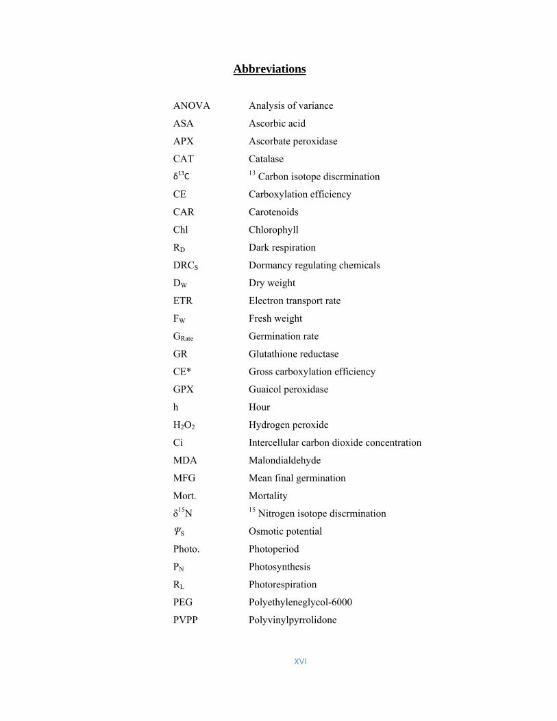
XVI
Abbreviations
ANOVA Analysis of variance
ASA Ascorbic acid
APX Ascorbate peroxidase
CAT Catalase
δ13C 13 Carbon isotope discrmination
CE Carboxylation efficiency
CAR Carotenoids
Chl Chlorophyll
RD Dark respiration
DRCS Dormancy regulating chemicals
DW Dry weight
ETR Electron transport rate
FW Fresh weight
GRate Germination rate
GR Glutathione reductase
CE* Gross carboxylation efficiency
GPX Guaicol peroxidase
h Hour
H2O2 Hydrogen peroxide
Ci Intercellular carbon dioxide concentration
MDA Malondialdehyde
MFG Mean final germination
Mort. Mortality
δ15N 15 Nitrogen isotope discrmination
ΨS Osmotic potential
Photo. Photoperiod
PN Photosynthesis
RL Photorespiration
PEG Polyethyleneglycol-6000
PVPP Polyvinylpyrrolidone

XVII
ROS Reactive oxygen species
DR Recovery of germination from dark
SR Recovery of germination from salinity
GSH Reduced glutathione
RWC Relative water content
SDS Sodiumdodecylsulfate
gs Stomatal conductance
SOD Superoxide dismutase
Temp. Temperature
T. Chl Total chlorophyll
TSS Total soluble sugars
E Transpiration
TCA Trichloroacetic acid
TW Turgid weight
Viab Viability
WUE Water use efficiency

1

2
Summary
Halopeplis perfoliata Forssk. is a C3 perennial halophyte found in coastal salt
marshes of Saharo-Sindian, Mediterranean and Western Irano-Turanian regions.
The plant has several ecological and economic usages but little is known about its
salt resistance strategies. Therefore, salt tolerance mechanism at seed germination
and growth stages of H. perfoliata were investigated. Increases in salinity
concentration decreased seed germination at all temperature regimes, while some
seeds could still germinate in 0.6 mol L-1 NaCl (equivalent to seawater salinity).
Hyper-saline conditions and complete darkness induced conditional dormancy.
Among salts tested, sea-salt inhibited seed germination more than isotonic NaCl
treatments. Among anions, chloride salts inhibited germination more than
isotonic sulfate salts. Dry storage of seeds for 1-year at room temperature
increased their tolerance to moderate salinities (0.2-0.3 mol L-1 NaCl) and
complete darkness, but decreased seed tolerance to higher temperature (25/35 oC)
as compared to freshly collected seeds. All dormancy regulating chemicals
(except for proline) alleviated dark-enforced dormancy under non-saline
conditions. Halopeplis perfoliata grew optimally at 0.15 mol L-1 NaCl treatment
and with increasing salinity (0.3 and 0.6 mol L-1 NaCl) treatments plant biomass
was comparable to non-saline controls. Lower (more negative) shoot osmotic
potentials and Na+ accumulation indicated capacity for osmotic adjustment with
increasing salinity. In general, photosynthetic CO2 fixation, light harvesting
ability of PSII and cation (K+, Ca++ and Mg++) concentrations were unchanged
with increasing salinity treatments. Higher CAT activity and H2O2 and the less
negative carbon isotope ratios in photosynthetic shoots indicated high rates of
photorespiration under saline conditions. Halopeplis perfoliata displayed unique
adaptations at seed germination and growth to survive under saline conditions.

3
Halopeplis perfoliata

4
Chapter 1
General Introduction

5
Introduction
Salt marshes are highly productive yet threatened ecosystems
Coastal salt marshes are transition zones between marine and terrestrial ecosystems
(Teixeira et al., 2014). They inundated diurnally by seawater therefore marsh sediments
have high salinity (equivalent to > 0.6 mol L-1 NaCl) (Aziz et al., 2001; Khan et al.,
2005). Salt marshes in sub-tropical regions often lack sufficient rainfall which leads to
even higher soil salinity (Böer, 1996; Boorman, 2003). Most, salt marshes are
characterized by dense stands of few highly salt-tolerant species (Anjum et al., 2014). In
marshy habitats, halophytes perform key role in carbon cycling (Gribsholt et al., 2003;
Sousa et al., 2010) by serving as a net sinks of carbon from atmosphere and produce
between 100-1000 g carbon m−2 y-1 (McLusky and Elliot, 2004; Sousa et al., 2010)
which makes the marsh environment highly productive. However, these habitats face
various natural and anthropogenic threats (Yasseen and Al-Thani, 2007; Ramadan et al.,
2013). According to a recently published t of United Nations report, the global climate
changes are causing global warming and mean sea level which could have drastic effects
on these habitats (IPCC, 2014). In addition, intense construction activities for resorts,
hotels and picnic points in coastal areas are also a serious threat to these important
habitats. Hence, coastal marshes and their vegetation require special attention both in
research and conservation/protection activities.
Salt marsh vegetation of coastal areas is under serious threat
Common plant species of warm coastal salt marshes can be categorized into perennials,
succulents, shrubs, stolonifers and hemicrytophytes (Ghazanfar, 2006) and include many
highly salt tolerant succulent species such as Arthrocnemum macrostachyum,
Halocnemum strobilaceum, Halopeplis perfoliata, Haloxylon persicum, Salicornia
europea, Zygophyllum mandavielli, Salsola imbricata, Sesuvium verrucosum, Suaeda
maritima and Suaeda vermiculata. (Gairola et al., 2015). These salt marsh plants provide
habitat for the coastal fauna, beside many other ecological and economic benefits
(Barbier, 2013). However, anthropogenic activities such as installation of reverse
osmosis facilities, construction of excursion points and buildings are posing serious
threat to these salt marshes. Therefore, salt marshes have received special attention both

6
from researchers and Government, which is evident from a growing body of research
works (Mahmoud et al., 1983, Böer, 1991, Al-Zaharani and Hajar, 1998; Ghazanfar,
2006; Anjum et al., 2015 and Meirland et al., 2015). Most of these research deals with
species composition, edaphology and threats to salt marshes. However, information on
eco-physiology and salt resistance mechanisms of the plants is missing.
Multiple factors regulate seed germination responses of marsh halophytes
Seed germination is the most sensitive stage in the life cycle of halophytes and is
influenced by a number of environmental factors, of which salinity is considered
foremost (Ungar 1995; Katembe et al., 1998; Gul et al., 2013). Insufficient rainfall in
sub-tropical regions results in high salinity in salt marsh sediments (Böer, 1996;
Boorman, 2003). Therefore, salinity of upper soil layers in such salt marshes may reach
44 ppt (equivalent 70 dS m-1) (Mandora et al., 1987). Seeds of most of the coastal plants
deposited in highly saline soils, hence they have to endure highly hostile conditions
(Ungar, 1978; Gul and Khan, 1998; Gul et al., 2013).
Seeds of most succulent halophytes of salt marshes are generally high salt
tolerant and can germinate in salinities higher than seawater salinity e.g. Salicornia
herbacea (1.7 mol L-1; Chapman 1960), Salicornia europaea (0.6 mmol L-1 NaCl;
Keiffer and Ungar, 1997), Arthrocnemum macrostachyum (1.0 mol L-1 NaCl; Khan and
Gul, 1998), Halosarica pergranulata (0.8 mol L-1 NaCl; Short and Colmer, 1999),
Halocnemum salicornicum (0.8-0.9 mmol L-1 NaCl; El-Keblawi and Al- Shamsi, 2008)
and Sarcocornia ambigua (45 g NaCl L-1 ~ 770 mmol L-1 NaCl; Freitas and Costa,
2014). In addition, seeds of these succulent halophytes can endure hypersaline conditions
by undergoing enforced dormancy (yet maintaining their viability) and show high
recovery of seed germination when salinity of the salt marshes is reduced following
rainfall (Ungar, 1995; Khan and Ungar, 1997; Gul et al., 2013). Seeds of most non-
succulent salt marsh halophytes such as the Atriplex, Crithmum and Limonium species on
the other hand could germinate at or below seawater salinity (Katembe et al., 1998; Zia
and Khan, 2008; Atia et al., 2009).
Seed germination of marsh halophytes is not only influenced by total salinity, but
also by different salts (Ryan et al., 1975; Khan and Gul, 2006). For example, germination
of stem succulent salt marsh halophytes Arthrocnemum macrostachyum, Halocnemum
strobilaceum, Sarcocornia fruticosa and Salicornia ramosissima varied under similar

7
levels of Cl- and SO4-- salts, and was linked to differential osmotic rather than ionic
effects of these salts (Pujol et al., 2000). Furthermore, comparison of NaCl and sea-salt
on salt marsh halophyte Arthrocnemum indicum indicated that NaCl reduced germination
more than the sea-salt (Saeed et al., 2011). However, little information is available on the
effects of different salts and seawater on salt marsh halophytes in comparison to the
effects of NaCl.
Beside salinity, many other factors such as light, temperature, storage, and levels
of phyto-hormones also influence seed germination of halophytes (Baskin and Baskin,
1994; Khan and Gul, 2006). According to Baskin and Baskin (1998) out of 91
halophytes, seed of 23 species were germinated in the presence of light. Similar
responses were observed for various other stem succulent halophytes like Salicornia
pacifica (Khan and Weber, 1986), Allenrolfea occidentalis (Gul et al., 2000).
Arthrocnemum indicum (Saeed et al., 2011), Halocnemum strobilaceum and Halopeplis
perfoliata (El-Keblawy and Bhatt, 2015). In addition, temperature also affects seed
germination of halophytes (Bewley and Black 1994; Gul and Weber 1999; Khan et al.,
2001; Zheng et al., 2004, 2005). Seeds of salt marsh stem succulent halophytes generally
germinate best at the temperature 20/30 oC. For example Allenrolfea occidentalis (Gul
and Weber, 1999), Salicornia rubra (Khan et al., 2000) and Arthrocnemum indicum
(Saeed et al., 2011) germinated best at 20/30 oC. According to Mahmoud et al. (1983)
germination success in salt marshes of Arabian Peninsula depends on temperature.
However, this kind of information on the marsh halophytes of Arabian-Peninsula is
limited to just few studies (Gul et al., 2013).
Growth hormones are reported to enhance the seed germination under salinity
stress condition (Ahmed et al., 2014; Li et al., 2014). Alternation in endogenous
dormancy regulating compounds at the appropriate salinity, temperature and light could
facilitate seed germination (Ahmed and Khan, 2010; El-Kablawy et al., 2010). It has
been reported that ethephon, fusicoccin, GA3 and kinetin enhanced germination in stem
succulent halophytes such as Salicornia utahensis (Khan and Weber, 1986; Gul and
Khan, 2003), Allenrolfia occidentalis (Gul and Weber, 1998), Arthrocnemum indicum
(Khan et al., 1998) and Salicornia rubra (Khan et al., 2002). However, this information
on the salt marsh halophytes of Arabian-Peninsula is largely missing.

8
Most salt marsh halophytes have “obligate” salt requirement for optimal growth
Succulent halophytes particularly those of Salicornoideae are reported to dominate
coastal salt marshes (McKee et al., 2012). Halophytes of Salicornoideae are supposed to
have higher salt tolerance than other halophytes. For example Arthrocnemum
macrostachyum (up to 1.0 mol L-1 NaCl, Khan et al., 2005), Salicornia europaea and S.
persica (up to 0.6 mmol L-1 NaCl, Aghaleh et al., 2009) and Tecticornia pergranulata
(syn. Halosarcia pergranulata) (up to 0.8 mol L-1 NaCl, Colmer et al., 2009). A number
of Salicornoideae members are in fact “obligate halophytes” (Flower and Colmer, 2008;
Rozema and Schat, 2013) which require some salt for their optimal growth (Flowers and
Colmer, 2008). For instance, Sarcocornia natalensis (300 mmol L-1 NaCl, Naidoo and
Rughunanan, 1990), Salicornia bigellovii (0.2 mol L-1 NaCl, Ayala and O’Leary, 1995),
Salicornia persica (Aghaleh et al., 2009) and Salicornia dolichostachya (0.3 mol L-1
NaCl, Katschnig et al., 2013) required salt for optimal growth. Hence, salt marsh
halophytes are well-adapted for surviving highly hostile marsh conditions. Owing to
their obligate halophyte nature, they could be interesting organisms for studying salt
tolerance mechanisms.
Salient features of salt tolerance mechanisms in salt marsh halophytes
Salt marsh halophytes are mostly “includers” which accumulate large amounts of salt in
their tissues and show tissue tolerance to increasing salinity (Aziz and Khan, 2001). Salt
tolerance is a highly complex phenomenon that incorporates a number of
cellular/molecular as well as whole plant level processes (Flowers and Colmer, 2008).
Generally, we can classify salt tolerance mechanisms of halophytes into four groups: 1)
osmotic adjustment, 2) ion homeostasis, 3) protection of metabolic activities such as
photosynthesis and 4) oxidative stress management (Jitesh et al., 2006; Flowers and
Colmer, 2015). Osmotic adjustment in halophytes helps to absorb and retain water in tissues
under hypersaline conditions (Shabala and Mackay, 2011; Flowers and Colmer, 2015).
This is achieved by compartmentalizing salts (mostly as Na+ and Cl-) in the apoplast and
cell vacuoles, with concomitant accumulation of organic osmolytes such as glycine
betaine, proline, free amino acids, sugars and sugar alcohols in cell cytosols (Flowers
and Colmer, 2008; Slama et al., 2015). Sequestration of salts in vacuoles is energetically
less costly than synthesis of organic osmolytes as vacuoles occupy the bulk of the cells’

9
internal volume (Flowers and Colmer, 2008). Organic osmolytes in cytoplasm provide
additional advantage by protecting macromolecules under stress, also referred as
‘osmoprotection’ (Slama et al., 2015). Plant growth may be compromised for osmotic
adjustment with salinity increments due to the high energy cost of organic osmolytes
(Flowers and Colmer, 2008). Salt accumulation in halophytes for osmotic adjustment is a tightly regulated
multi-facted phenomenon (Shabala and Mackay, 2011). Salt exclusion at the level of
root, restrict its entry into the xylem stream (Flowers and Colmer, 2015) and most of the
woody halophytes of tidal marshes are reported to exclude 90 to 99% of the external Na+
(Reef and Lovelock 2015). Stomatal size and density as well as number reduces
transpiration rates which may also help in restricting Na+ entry into the shoots/leaves
(Shabala and Mackay, 2011). Some non-succulent salt marsh halophytes particularly
grasses get-rid of excess salts by secreting them via salt glands/hairs (Flowers et al.,
2010; Céccoli et al., 2015). At the cellular level, using a complex machinery of
membrane transporter proteins, halophytes keep Na+ and Cl- within tolerable ranges
along with efficient acquisition of K+ and Mg++ (Mansour, 2014; Flowers and Colmer,
2015). High external salinity may however results in ion imbalance which leads to
metabolic disruption causing tissue injury. Effective osmotic adjustment and ion-
homeostasis for plant survival under saline conditions would require the ability to
maintain significant photosynthetic CO2 gain at minimum water loss (Álvarez et al.,
2012; Naidoo et al., 2012).
Salinity is known to affect photosynthesis due to stomatal limitation (Lawlor and
Cornic, 2002; Flexas et al. 2004; Lambers et al., 2008). A decrease in stomatal
conductance reduces the influx of CO2 which ultimately reduces photosynthetic rate
(Maricle et al., 2007). However, many succulent halophytes of salt marshes are reported
to maintain significant photosynthetic rates under high salinities (Maricle et al., 2007).
Unlike photosynthetic CO2 assimilation, light-harvesting mechanisms of halophytes are
generally more resilient. For example, photochemistry was generally unaffected by
salinity in succulent halophytes Suaeda salsa (James et al., 2002) and Sarcocornia
fruticosa (Redondo-Gómez et al, 2006). Under decreased CO2 assimilation such as in
response to salinity, use of alternate electron sinks become essential for the survival of
plants (Taiz and Zeiger, 2010). Non-photochemical quenching (NPQ), which is the
dissipation of surplus energy mainly by heat production is considered an important route

10
in plants (Taiz and Zeiger, 2010; Lambrev et al., 2012). For example, NPQ increased
with increases in salinity in Atriplex centralasiatica (Qiu et al., 2003) and Cakile
maritima (Megdiche et al., 2008). Rise in NPQ is a photo-protective mechanism that
minimizes the incidence of electron flow from photosystems to oxygen (O2) which
otherwise accelerates the formation of cytotoxic reactive oxygen species (ROS) (Jithesh
et al., 2006; Taiz and Zeiger, 2010; Demidchik, 2015). Accumulation of ROS can cause
oxidative damage to cell components such as membrane lipids, proteins, chlorophyll and
nucleic acids (Müller et al., 2001, Jithesh et al., 2006; Møller et al., 2007; Hameed and
Khan, 2011; Demidchik, 2015). Therefore, the antioxidant defense system, which
comprises of both enzymatic and non-enzymatic components, plays an important role in
defending cell components from oxidative damage (Jithesh et al., 2006). Superoxide
dismutase (SOD), catalase (CAT), and enzymes of Foyer-Halliwell-Asada pathway are
important antioxidant enzymes, while ascorbate (ASA) and glutathione (GSH) are key
antioxidant substances, which are implicated in protecting cells from salinity-induced
oxidative stress (Jithesh et al., 2006). However, efficacy of antioxidant system in
protecting plants from salinity effects may vary considerable with species and the
magnitude of salinity imposed, among other factors (Hameed and Khan, 2011).
Halopeplis perfoliata is a common halophyte of coastal salt marshes
Halopeplis perfoliata Forssk. is a succulent C3 perennial shrub found in coastal salt
marshes of Arabian peninsula (Böer, 1996; Zahran and Ansari, 1999; Al-Oudat and
Qadir, 2011), saline desert habitats along Arabian Gulf coast (El-Keblawy and Bhatt,
2015) and also occupies the first zone (near coast) of the many Red Sea salt marshes
(Migahid, 1978). There are many ecological and economic usages of this halophyte. For
example, H. perfoliata is used for soap manufacture by local communities (Al-Oudat and
Qadir, 2011). It can also be used for sand dune stabilization (Zreik, 1990). Halopeplis
perfoliata is an important primary producer of intertidal coastal zones and provides
habitat for wild life (Pilcher et al., 2003). However, little is known about the seed
germination (Mahmoud et al., 1983; El-Keblawy and Bhatt, 2015) and growth (Al-
Zaharani and Hajar, 1998) of this halophyte. Furthermore, underlying mechanisms for
salt tolerance of halophytes from the Arabian Peninsula have seldom been studied.
Halopeplis perfoliata is an interesting plant, owing to its naturally high salt tolerance and
economic potentials.

11
A
B
Fig. 1.1. Halopeplis perfoliata A) plants and B) in the natural environmental conditions.

12
Research questions The following question were addressed in the present study:
1. What is the salt tolerance limit of Halopeplis perfoliata during seed germination
stage under various photoperiod, temperature, and storage environments?
2. Can salinity-induced seed germination inhibition be mitigated by the exogenous
application of various dormancy regulating chemicals?
3. Are seeds of Halopeplis perfoliata equally tolerant to all types of salts?
4. What is the salt tolerance limit of Halopeplis perfoliata at growth stage?
5. What are different physiological and biochemical adaptations which enable
Halopeplis perfoliata to tolerate high salinity?

13
References: Aghaleh, M., Niknam, V., Ebrahimzadeh, H., and Razavi, K. (2009). Salt stress effects
on growth, pigments, proteins and lipid peroxidation in Salicornia persica and S.
europaea. Biologia Plantarum, 53, 243-248. Ahmed, M. Z., and Khan, M. A. (2010). Tolerance and recovery responses of playa
halophytes to light, salinity and temperature stresses during seed germination.
Flora, 205, 764-771.
Ahmed, M. Z., Gulzar, S., and Khan, M. A. (2014). Role of dormancy regulating
chemicals in alleviating the seed germination of three playa halophytes. Ekoloji,
23, 1-7.
Al-Oudat, M., and Qadir, M. (2011). The halophytic flora of Syria. International Center
for Agricultural Research in the Dry Areas (ICARDA), Aleppo, Syria, 8, 186.
Álvarez, S., Gómez-Bellot, M. J., Castillo, M., Banón, S., and Sánchez-Blanco, M. J.
(2012). Osmotic and saline effect on growth, water relations, and ion uptake and
translocation in Phlomis purpurea plants. Environmental and Experimental
Botany, 78, 138-145.
Al-Zaharani, H. S., and Hajar, A. S. (1998). Salt tolerance in the halophyte Halopeplis
perfoliata Forsk. Effect of NaCl salinity on growth and ion uptake. Indian
Journal of Plant Physiology, 3, 32-35.
Anjum, N. A., Ahmed, I., Válega, M., Mohmood, I., Gill, S. S., Tuteja, N., Duarte, A. C.,
and Pereira, E. (2014). Salt marsh halophytes services to metal-metalloid
remediation: Assessment of the processes and underlying mechanisms. Critical
Reviews in Environmental Sciences and Technology, 44, 2038-2106.
Atia, A., Debez, A., Barhoumi, Z., Smaui, A., and Abdelly, C. (2009). ABA, GA3 and
nitrate may control seed germination of Crithmum maritimum (Apiaceae) under
saline conditions. Comptes Rendus Biologies, 332, 704-710.
Ayala, F., and O’Leary, J. W. (1995). Growth and physiology of Salicornia bigellovii
Torr. at suboptimal salinity. International Journal of Plant Science, 156, 197-
205.
Aziz, I., and Khan, M. A. (2001). Experimental assessment of salinity on the growth,
ionic composition and water relations of Ceriops tagal from MianiHor, Pakistan.
Aquatic Botany, 70, 259-268.

14
Barbier, E. B. (2013). Valuing ecosystem services for Coastal Wetland protection and
restoration: progress and challenges. Resources, 2, 213-230.
Baskin, C. C., and Baskin, J. M. (1994). Deep complex morpho-physiological dormancy
in seeds of the mesic woodland herb Delphinium tricorne (Ranunculaceae).
International Journal of Plant Sciences, 155, 738-743.
Baskin, C. C., and Baskin, J. M. (1998). Seeds: Ecology, Biography and Evolution of
Dormancy and Germination. Sen Diego: Academic press.
Bewley, J. D., and Black, M. (1994). Seeds: physiology of development and germination.
(2nd ed. pp. 1-33). Verlag, USA: Springer.
Böer, B. (1991). Trial planting of mangroves (Avicennia marina) and salt-marsh plants
(Salicornia europaea) in oil-impacted soil in the Jubail area, Saudi Arabia. A
Marine Wildlife Sanctuary for the Arabian Gulf, Environmental Research and
Conservation following the Gulf War Oil Spill, 186-192.
Böer, B. (1996). Plants as soil indicators along the Saudi coast of the Arabian Gulf.
Journal of Arid Environments, 33, 417-423.
Boorman, L. A. (2003). Salt marsh review. An overview of coastal saltmarshes, their
dynamic and sensitivity characteristics for conservation and management. Joint
Nature Conservation Committee. JNCC: Peterborough. Report, 334, 1-116.
Céccoli, G., Ramos, J., Pilatti, V., Dellaferrera, I., Tivano, J. C., Taleisnik, E., and
Vegetti, A. C. (2015). Salt glands in the poaceae family and their relationship to
salinity tolerance. The Botanical Review, 81, 162-178.
Chapman, V. J. (1960). Salt marshes and salt deserts of the world. New York:
Interscience publications.
Colmer, T. D., Vos, H., and Pedersen, O. (2009). Tolerance of combined submergence
and salinity in the halophytic stem-succulent Tecticornia pergranulata. Annals of
Botany, 103, 303-312.
Demidchik, V. (2015). Mechanisms of oxidative stress in plants: from classical
chemistry to cell biology. Environmental and Experimental Botany, 109, 212-
228.
El-Keblawy, A., and Al-Shamsi, N. (2008). Salinity, temperature and light affect seed
germination of Haloxylon salicornicum, a common perennial shrub of the
Arabian deserts. Seed Science and Technology, 36, 679-688.

15
El-Keblawy, A., Al-Ansari, F., and Al-Shamsi, N. (2010). Impact of dormancy
regulating chemicals on salinity induced dormancy in Lasiurus scindicus and
Panicum turgidum: two desert glycophytic grasses. Plant Growth Regulation, 62,
163-170.
El-Keblawy, A. A., and Bhatt, A. (2015). Aerial seed bank affects germination in two
small-seeded halophytes in Arab Gulf desert. Journal of Arid Environments, 117,
10-17.
Flexas, J., Bota, J., Loreto, F., Cornic, G., and Sharkey, T. D. (2004). Diffusive and
metabolic limitations to photosynthesis under drought and salinity in C3 plants.
Plant Biology, 6, 269-279.
Flowers, T. J., and Colmer, T. D. (2008). Salinity tolerance in halophytes. New
Phytologist, 179, 945- 963.
Flowers, T. J., Galal, H. K., and Bromham, L. (2010). Evolution of halophytes: multiple
origins of salt tolerance in land plants. Functional Plant Biology, 37, 604-612.
Flowers, T. J., and Colmer, T. D. (2015). Preface: part of a special issue on halophytes
and saline adaptations preface: part of a special issue on halophytes and saline
adaptations, plant salt tolerance: adaptations in halophytes. Annals of Botany, 11,
327-331.
Freitas, R. F., and Costa, C. S. (2014). Germination responses to salt stress of two
intertidal populations of the perennial glasswort Sarcocornia ambigua. Aquatic
Botany, 117, 12-17.
Gairola, S., Bhatt, A., and El-Keblawy. A. (2015). A perspective on potential use of
halophytes for reclamation of salt-affected lands. Wulfenia, 22, 88-97.
Ghazanfar, S. A. (2006). Saline and alkaline vegetation of NE Africa and the Arabian
Peninsula: An overview. Biosaline Agriculture and Salinity Tolerance in Plants
(Öztürk, M., Waisel, Y., Khan, M. A., and Görk, G. ed. pp. 101-108). Birkhäuser
Basel.
Gribsholt, B., Kostka, J. E., and Kristensen, E. (2003). Impact of fiddler crabs and plant
roots on sediment biogeochemistry in a Georgia salt marsh. Marine Ecology
Progress Series, 259, 237-251.
Gul, B., and Khan, M. A. (1998). Population characteristics of the coastal halophyte
Arthrocnemum macrostachyum. Pakistan Journal of Botany, 30, 189-197.

16
Gul, B., and Weber, D. J. (1998). Effect of Dormancy relieving compounds on the seed
germination of non-dormant Allenrolfea occidentalis under salinity stress. Annals
of Botany, 82, 555-560.
Gul, B., and Weber, D. J. (1999). Effect of salinity, light and thermoperiod on the seed
germination of Allenrolfea occidentalis. Canadian Journal of Botany, 77, 1-7.
Gul, B., Khan, M. A., and Weber, D. J. (2000). Alleviation salinity and dark-enforced
dormancy in Allenrolfea occidentalis seeds under various thermoperiods.
Australian Journal of Botany, 48, 745-752.
Gul, B., and Khan, M. A. (2003). Effect of growth regulators and osmotica in alleviating
salinity effects on the germination of Salicornia utahensis. Pakistan Journal of
Botany, 35, 885-894.
Gul, B., Ansari, R., Flowers, T. J., and Khan, M. A. (2013). Germination strategies of
halophyte seeds under salinity. Environmental and Experimental Botany, 92, 4-
18.
Hameed, A., and Khan, M. A. (2011). Halophytes: Biology and Economic Potentials.
Karachi University Journal of Science, 39, 40-44.
James, R. A., Rivelli, A. R., Munns, R., and von Caemmerer, S. (2002). Factors affecting
CO2 assimilation, leaf injury and growth in salt-stressed durum wheat. Functional
Plant Biology, 29, 1393-1403.
Jithesh, M. N., Prashanth, S. R., Sivaprakash, K. R., and Parida, A. K. (2006). Anti-
oxidative response mechanisms in halophytes: their role in stress defense.
Journal of Genetics, 85, 237-254.
Katembe, W. J., Ungar, I. A., and Mitchell, J. P. (1998). Effect of salinity on germination
and seedling growth of two Atriplex species (Chenopodiaceae). Annals of Botany,
82, 167-175.
Katschnig, D., Broekman, R., and Rozema, J. (2013). Salt tolerance in the halophyte
Salicornia dolichostachya Moss: Growth, morphology and physiology.
Environmental and Experimental Botany, 92, 32-42.
Keiffer, C. H., and Ungar, I. A. (1997). The Effect of extended exposure to hypersaline
conditions on the germination of five inland halophyte species. American Journal
of Botany, 84, 104-111.
Khan, M. A., and Weber, D. J. (1986). Factors influencing seed germination in
Salicornia pacifica var. utahensis. American Journal of Botany, 73, 1163-1167.

17
Khan, M. A., and Ungar, I. A. (1997). Germination responses of the subtropical annual
halophyte Zygophyllum simplex. Seed Science and Technology, 25, 83-92.
Khan, M. A., and Gul, B. (1998). High salt tolerance in germinating dimorphic seeds of
Arthrocnemum indicum. International Journal of Plant Sciences, 159, 826-832.
Khan, M. A., Ungar, I. A., and Gul, B. (1998). Action of compatible osmotica and
growth regulators in alleviating the effect of salinity on the germination of
dimorphic seeds of Arthrocnemum indicum L. International Journal of Plant
Sciences, 159, 313-317.
Khan, M. A., Gul, B., and Weber, D. J. (2000). Germination responses to Salicornia
rubra to temperature and salinity. Journal of Arid Environments, 45, 207-214.
Khan, M. A., Gul, B., and Weber, D. J. (2001). Effect of salinity and temperature on the
germination of Kochia scoparia. Wetland Ecology and Management, 9, 483-
489.
Khan, M. A., Gul, B., and Weber, D. J. (2002). Improving seed germination of
Salicornia rubra (Chenopodiaceae) under saline conditions using germination-
regulating chemicals. Western North American Naturalist, 62, 101-105.
Khan, M. A., Ungar, I. A., and Showalter, A. M. (2005). Salt stimulation and tolerance in
an intertidal stem-succulent halophyte. Journal of Plant Nutrition, 28, 1365-1374.
Khan, M. A., and Gul, B. (2006). Halophyte seed germination. Ecophysiology of high
salinity tolerant plants (pp. 11-30). Netherlands: Springer.
Lambers, H., Chapin III, F. S., and Pons, T. L. (2008). Mineral nutrition (pp. 255-320).
New York: Springer.
Lambrev, P. H., Miloslavina, Y., Jahns, P., and Holzwarth, A. R. (2012). On the
relationship between non-photochemical quenching and photoprotection of
Photosystem II. Biochimica et Biophysica Acta (BBA)-Bioenergetics, 1817, 760-
769.
Lawlor, D. W., and Cornic, G. (2002). Photosynthetic carbon assimilation and associated
metabolism in relation to water deficits in higher plants. Plant, Cell and
Environment, 25, 275-294.
Li, W., Khan, M. A., Yamaguchi, S., and Liu, X. (2014). Hormonal and environmental
regulation in salt cress (Thellungiella halophila). Plant Growth Regulation, 76,
41-49.

18
Mahmoud, A., El Sheikh, A. M., and Abdul Baset, S. (1983). Germination of two
halophytes: Halopeplis perfoliata and Limonium aillare from Saudi Arabia.
Journal of Arid Environments, 6, 87-98.
Mansour, M. M. F. (2014). The plasma membrane transport systems and adaptation to
salinity. Journal of Plant Physiology, 171, 1787-1800.
Mandura, A. S., Saifullah, S. M., and Khafaji, A. K. (1987). Mangrove Ecosystem of
Southern Red Sea Coast of Saudi Arabia. Proceedings of the Saudi Biological
Society, 10, 165-193.
Maricle, B. R., Lee, R. W., Hellquist, C. E., Kiirats, O., and Edwards, G. E. (2007).
Effects of salinity on chlorophyll fluorescence and CO2 fixation in C4 estuarine
grasses. Photosynthetica, 45, 433-440.
McKee, K., Rogers, K., and Saintilan, N. (2012). Response of salt marsh and mangrove
wetlands to changes in atmospheric CO2, climate, and sea level. Global change
and the function and distribution of wetlands (pp. 63-96). Netherlands: Springer.
McLusky, D. S., and Elliott, M. (2004). The Estuarine Ecosystem: Ecology, Threats and
Management (Elliott. M, ed. p. 216). Oxford: Oxford University Press.
Megdiche, W., Hessini, K., Gharbi, F., Jaleel, C. A., Ksouri, R., and Abdelly, C. (2008).
Photosynthesis and photosystem 2 efficiency of two salt-adapted halophytic
seashore Cakile maritima ecotypes. Photosynthetica, 46, 410-419.
Meirland, A., Gallet-Moron, E., Rybarczyk, H., Dubois, F., and Chabrerie, O. (2015).
Predicting the effects of sea level rise on salt marsh plant communities: does
vegetation age matter more than sea level? Plant Ecology and Evolution, 148, 5-
18.
Migahid, A. M. (1978). Flora of Saudi Arabia (2nd ed.). Riyadh University Press.
Møller, I. M., Jensen, P. E., and Hansson, A. (2007). Oxidative modifications to cellular
components in plants. Annual Review of Plant Biology, 58, 459-481.
Müller, P., Li, X. P., and Niyogi, K. K. (2001). Non-photochemical quenching. A
response to excess light energy. Plant Physiology, 125, 1558-1566.
Naidoo, G. R., and Rughunanan, R. (1990). Salt tolerance in the succulent halophyte,
Sarcocornia natalensis. Journal of Experimental Botany, 41, 497-502.
Naidoo, G., Naidoo, Y., and Achar, P. (2012). Ecophysiological responses of the salt
marsh grass Spartina maritima to salinity. African Journal of Aquatic Science,
37, 81-88.

19
Pilcher, N. J., Phillips, R. C., Aspinall, S., Al-Madany, I., King, H., Hellyer, P., Beech,
M., Gillespie, C., Wood, S., Schwarze, H., Al Dosary, M., Al Farraj, E., Khalifa,
A., and Böer, B. (2003). Hawar Islands Protected Area (Kingdom of Bahrain).
Management Plan (1st Draft. p. 30). UNESCO, Doha.
Pujol, J. A., Calvo, J. F., and Ramirez-Diaz, L. (2000). Recovery of germination from
different osmotic conditions by four halophytes from southeastern Spain. Annals
of Botany, 85, 279-286.
Qiu, N., Lu, Q., and Lu, C. (2003). Photosynthesis, photosystem II efficiency and the
xanthophyll cycle in the salt‐adapted halophyte Atriplex centralasiatica. New
Phytologist, 159, 479-486.
Ramadan, H. A-Z., and Bantan, R. A. (2013). Hypersaline benthic foraminifera from the
Shuaiba Lagoon, eastern Red Sea, Saudi Arabia: Their environmental controls
and usefulness in sea-level reconstruction. Marine Micropaleontology, 103, 51-
67.
Redondo‐Gómez, S., Wharmby, C., Castillo, J. M., Mateos‐Naranjo, E., Luque, C. J., De
Cires, A., Luque, T., Davy, A. J., and Figueroa, M. E. (2006). Growth and
photosynthetic responses to salinity in an extreme halophyte, Sarcocornia
fruticosa. Physiologia Plantarum, 128, 116-124.
Reef, R., and Lovelock, C. E. (2015). Regulation of water balance in mangroves. Annals
of Botany, doi: 10.1093/aob/mcu174.
Rozema, J., and Schat, H. (2013). Salt tolerance of halophytes, research questions
reviewed in the perspective of saline agriculture. Environmental and
Experimental Botany, 92, 83-95.
Ryan, J., Miyamoto, S., and Stroehlein, J. L. (1975). Salt and specific ion effects on
germination of four grass. Journal of Range Management, 28, 61-64.
Saeed, S., Gul, B., and Khan, M. A. (2011). Comparative effects of NaCl and sea salt on
seed germination of Arthrocnemum indicum. Pakistan Journal of Botany, 43,
1091-1103.
Shabala, S., and Mackay, A. (2011). Ion transport in halophytes. Advances in Botanical
Research (Kader, J. C., Delseny, M. eds. Vol. 57, pp. 151-199). Elsevier.
Short, D. C., and Colmer, T. D. (1999). Salt tolerance in the halophyte Halosarcia
pergranulata sub sp. pergranulata. Annals of Botany, 83, 207-213.

20
Slama, I., Abdelly, C., Bouchereau, A., Flowers, T., and Savouré, A. (2015). Diversity,
distribution and roles of osmo-protective compounds accumulated in halophytes
under abiotic stress. Annals of Botany, doi: 10.1093/aob/mcu239.
Sousa, A. I., Lillebø, A. I., Pardal, M. A., and Caçador, I. (2010). Productivity and
nutrient cycling in salt marshes: contribution to ecosystem health. Estuarine,
Coastal and Shelf Science, 87, 640-646.
Taiz, L., and Zeiger, E. (2010). Plant Physiology (5th ed.). USA: Sinauer Associates.
Teixeira, A., Duarte, B., and Caçador, I. (2014). Salt Marshes and Biodiversity. Sabkha
Ecosystems: Cash Crop Halophyte and Biodiversity Conservation (Vol. 4, pp.
283-298). Netherlands: Springer.
Ungar, I. A. (1978). Halophyte seed germination. The Botanical Review, 44, 233-264.
Ungar, I. A. (1991). Ecophysiology of Vascular Halophytes (pp. 9-48). Boca Raton: CRC
press.
Ungar, I. A. (1995). Seed germination and seed-bank ecology in halophytes. Seed
Development and Germination (Kigel, J., and Galili, G. eds. pp. 629-644). New
York: Marcel Dekker.
Yasseen, B. T., and Al-Thani, R. F. (2007). Halophytes and associated properties of
natural soils in the Doha area, Qatar. Aquatic Ecosystem Health and
Management, 10, 320-326.
Zahran, M. A., and Al-Ansari, F. M. (1999). The Ecology of Al-Samaliah Island, UAE.
Estuarine, Coastal and Shelf Science, 49, 11-19.
Zheng, Y., Gao, Y., An, P., Shimizu, H., Rimmington, G. M. (2004). Germination
characteristics of Agriophyllim squarrosum. Canadian Journal of Botany, 82,
1662-1670.
Zheng, Y., Rimmington, G. M., Gao, Y., Jiang, L., Xing, X., An, P., El-Sidding, K., and
Shimizu, H. (2005). Germination characteristics of Atremisia ordosica
(Asteraceae) in relation to ecological restoration in northern China. Canadian
Journal of Botany, 83, 1021-1028.
Zia, S., and Khan, M. A. (2008). Seed germination of Limonium stocksii under saline
conditions. Pakistan Journal of Botany, 40, 683-695.
Zreik, R. (1990). The domestication and economic cultivation of halophytes. Developing
world agriculture (pp. 74-79). London: Grosvenor Press.

21
Chapter 2
Effects of environmental factors, seed storage conditions, and exogenous chemical treatments in modulating seed germination of
Halopeplis perfoliata

22
Abstract Halopeplis perfoliata is a coastal marsh halophyte with several ecological and economic
usages. Information about seed germination ecology of this plant is scanty. Therefore
this study was conducted to investigate the germination, recovery and viability responses
of H. perfoliata seeds to photoperiod (light/dark), temperature, salinity, hyper-salinity,
long-term storage and dormancy regulating chemicals (DRCs). Seeds were small and
non-dormant, and displayed highest germination in distilled water irrespective of
incubation temperatures. Seeds germinated better in light (12-h photoperiod) than in 24-h
dark. Increases in salinity decreased seed germination; however some seeds could
germinate in 0.6 mol L-1 NaCl (equivalent to seawater salinity) under 12-h photoperiod.
High salinity (> 0.3 mol L-1 NaCl) and darkness imposed conditional dormancy in seeds
and they showed high germination recovery when transferred to distilled water and light
respectively. Seeds of H. perfolaita could endure hyper salinity (> 1.0 mol L-1 NaCl) by
entering into a state of conditional dormancy. However, high incubation temperatures
resulted in higher seed mortality under hyper-saline condition. Dry storage of seeds for
one year at 25 oC increased their tolerance to moderate salinity and dark, but
substantially decreased tolerance to high temperature compared to fresh seeds. All DRCs
(except proline) alleviated dark-enforced dormancy but were generally ineffective in
reversing inhibitory effects of salinity. Hence it appears that individual effects of
photoperiod, salinity and temperature influenced seed germination more than their
interactive effects.

23
Introduction
Arabian Peninsula salt marshes are facing various natural and anthropogenic threats
Coastal salt marshes are among the most productive ecosystems of the world and harbor
unique halophytic vegetation bearing specialized adaptations to produce enormous
biomass despite high seawater salinity (Teixeira et al., 2014). Their location at the
interface of sea and land makes them ecologically important. These habitats serve as an
important carbon and nitrogen sink, thereby help in CO2 sequestration and prevention of
eutrophication in coastal areas, beside many other ecosystem services (Seitzinger, 1988;
Valiela and Cole, 2002; Teixeira et al., 2014). However, these habitats are facing various
natural and anthropogenic threats (Yasseen and Al-Thani, 2007; Ramadan et al., 2013).
Sub-tropical salt marshes especially those along the coastline of Arabian Peninsula often
face lack of sufficient rainfall which leads to increased sediment salinity in salt marshes
(Böer, 1996; Boorman, 2003). Salinity of upper soil layer in such salt marshes could be
as high as 44 ppt (equivalent 70 dS m-1) (Mandora et al., 1987) which could be
detrimental for the prevailing vegetation (Ungar, 1991; Böer, 1996). Since seeds of most
coastal plants are deposited in top soil layers, which have substantially higher salinity
than lower layers, therefore conditions for seeds are harsher in comparison to mature
plants (Ungar, 1978; Gul and Khan, 1998; Gul et al., 2013).
Seed germination responses of salt marsh halophytes are highly variable
Salinity is one of the main factors influencing vegetation zonation in coastal salt marshes
mainly via its effects on seed germination (Bertness et al., 1992; Pujol et al., 2000;
Hameed et al., 2006; Elsey-Quirk et al., 2009; Gul et al., 2013). Seed germination of
halophytes generally decreases with increases in salinity (Khan and Gul, 2006).
However, seeds of most salt marsh halophytes could germinate in as high as seawater (>
0.6 mol L-1 NaCl) or even higher salinity (Gul et al., 2013). For instance, seeds of salt
marsh halophytes Arthrocnemum indicum (1.0 mol L-1 NaCl, Khan and Gul, 1998;
Ruppia tuberosa (90 g L-1 sea-salt or ~1.5 mol L-1 NaCl, Kim et al., 2013) and
Sarcocornia ambigua (0.77 mol L-1 NaCl, Freitas and Costa, 2014) showed high salinity
tolerance during germination. Seeds of most salt marsh halophytes can even endure
hyper-saline conditions (Gul et al., 2013) in which they although do not germinate but
maintain their viability and hence quickly germinate (germination recovery, sensu Khan
and Ungar, 1997) following sufficient rains which dilute soil/sediment salinity (Khan

24
and Gul, 2006). Salinity tolerance of halophyte seeds also varies with changes in ambient
temperature and presence/absence of light (Gul et al., 2013). Sub/supra-optimal
temperatures and dark (mainly due to burial) decrease both germination as well as
salinity tolerance of halophyte seeds (Baskin and Baskin, 2001; Zia and Khan, 2004; El-
Keblawy and Al-Rawai, 2006; Hameed et al., 2013). However, large variations exist in
responses of the seeds of salt marsh halophytes to temperature and photoperiod changes.
For example, seeds of only four out of eight salt marsh halophytes responded to changes
in temperature and light/dark during germination (Noe and Zedler, 2000). Seeds of
another salt marsh halophyte Arthrocnemum indicum responded to changes in
temperature but not to light/dark (Saeed et al., 2011). Changes in temperature and
light/dark also influence seed viability and dormancy of halophytes (Gulzar et al., 2013;
Hameed et al., 2013). However, information about the seed germination of salt marsh
halophytes is limited (Saeed et al., 2011; Gul et al., 2013).
Salinity tolerance of halophyte seeds, as discussed above, is a variable trait and is
also influenced by storage conditions and duration (El-Kablawy, 2013a; Cao et al.,
2014). Recently El-Keblawy (2013a) studied effect of various storage conditions on
seed germination of two succulent halophytes Haloxylon salicornicum and Salsola
imbricata. He found that the seed storage for three months significantly increased
germination but longer than nine month storage at room and warm temperatures caused
significant reduction or complete inhibition in the germination of the aforementioned
halophytes. While seeds of some halophytes such as Allenrolfea occidentalis (Gul and
Weber, 1999), Salicornia rubra (Khan et al., 2000), Kochia scoparia (Khan et al., 2001)
and Salsola iberica (Khan et al., 2002) could maintain viability and germinablity after 20
days of high salinity (1.0 mol L-1 NaCl) exposure. In another study, Keiffer and Ungar
(1997) found variable viability and germination responses of the seeds of Atriplex
prostrata, Hordeum jubatum, Salicornia europaea, Spergularia marina, and Suaeda
calceoliformi to storage under high salinity for different time periods. Hence, these data
indicate that seed responses of halophytes to storage under high salinity could be variable
and species specific.
Dormancy regulating compounds can improve seed germination under stress conditions
Germination and salinity tolerance of the seeds of halophytes could be improved by
exogenous application of certain chemicals (Mehrun-Nisa et al., 2007; Atia et al., 2009;

25
Ahmed et al., 2014). GA3, kinetin and thiourea are widely used chemicals, which can
mitigate salinity effects and improve seed germination of halophytes (Khan and Gul,
2006; Gul and Khan 2008; El-Keblawy et al., 2010). Recently, Ahmed et al. (2014)
reported that thiourea could significantly improve seed germination of three salt playa
halophytes under saline condition, while GA3 and kinetin had species-specific effects.
Hence, information about effects of chemicals on salinity tolerance and germination of
halophyte seeds appears inconclusive and could be species and/or chemical specific (Gul
and Khan 2008; El-Keblawy et al., 2010; Ahmed et al., 2014).
Seed germination eco-physiology of H. perfoliata is largely unstudied
Halopeplis perfoliata Forssk. is a succulent halophyte from family Amaranthaceae, that
occupies coastal salt marshes of Arabian peninsula (Al-Oudat and Qadir, 2011). This
species has many ecological and potential economic uses. For instance, it can be utilized
for sand dune stabilization in coastal deserts (Zreik, 1990) and also in soap industries
(Al-Oudat and Qadir, 2011). This plant is an important intertidal/terrestrial producer and
provides habitat structure for wild life (Pilcher et al., 2003). Seeds of this plant could
germinate up to 0.25 mol L-1 NaCl treatment (Mahmoud et al., 1983). However, little is
known about the seed germination eco-physiology or the two other members of genus
Halopeplis. Therefore detailed studies on salinity tolerance mechanisms of H. perfloiata
were performed to understand how its seeds withstand highly saline salt marsh
conditions. The objectives of this research were to find out: 1) salinity tolerance limit and
optimal germination conditions (temperature and light/dark regimes) for H. perfoliata
seeds, 2) viability and germination response of H. perfoliata seeds to storage under
hyper-saline conditions, 3) germination characteristics after long-term (1-year) dry-
storage in laboratory condition, and 4) salinity tolerance and germination responses to
exogenous application of various dormancy regulating chemicals (DRCs).

26
Materials and Methods
Study site and Species description
Mature inflorescence of Halopeplis perfoliata were collected from the coast of Jizan,
Saudi Arabia in 2012. Study site was a salt marsh with frequent inundation, where mean
ambient temperature reportedly ranges between 36 and 21 oC (Alfarhan et al., 2005).
Mean annual rainfall of the area is 169 mm and humidity ranges between 39 and 86%
(Alfarhan et al., 2005). Seeds were collected from large number of plants randomly to
ensure adequate representation of the genetic diversity. Seeds were separated from their
inflorescence husk manually and brought to the laboratory in Pakistan. Seeds were
surface sterilized using 1% bleach solution for 1 min, followed by thorough rinsing with
distilled water and air-drying. Sterilized seeds were then used in experiments.
Seed characteristics
Fresh weight (FW) of 1000 seeds was measured using an analytical balance (0.0001 g).
Dry weight (DW) of the seeds was determined by placing 1000 seeds in an oven at 105 oC for 48-h. Moisture content was then calculated as difference between FW and DW. Ash
or inorganic content of the seeds was determined by igniting oven dried seeds in a
furnace at 550 oC for 3-h. Size of the seeds was measured using photographs of about
randomly chosen 100 seeds with the help of ImageJ software
(http://imagej.nih.gov/ij/images/). Seed texture was determined by observing seeds under
a mini-microscope (Dini-Lite digital Microscope, ANMO Electronics Corporation). Seed
color was matched against the catalog of Ralcolors (www.ralcolors.com).
Experiment. 1: salinity tolerance limit and optimal germination conditions
Germination was carried out in 50 x 9 mm (Gelman No. 7232) tight-fitting plastic petri-
plates with 5 ml of test solution. Seven concentrations of NaCl solution (0, 0.1, 0.2, 0.3,
0.4, 0.5 and 0.6 mol L-1) were used. Petri plates were placed in a germination chambers
set at temperature regimes of 10/20, 15/25, 20/30 and 25/35 oC, where low temperature
coincided with 12-h dark and high temperature with 12-h light (~25 µmol m-2 s-1, 400-
750 nm, Philips cool-white fluorescent lamps). Under similar temperature and salinity
treatments seeds were also incubated in complete darkness (24-h dark) by using dark
photographic envelops. Four replicates with twenty-five seeds per petri-plate were used
for each treatment. Petri-plates placed under 12-h photoperiod were monitored at 2 days

27
intervals for 20 days and percent seed germination were noted. Seeds were considered to
be germinated with the emergence of the radical (Bewley and Black, 1994). After 20
days of incubation, rate of germination was estimated by using a modified Timson’s
index of germination velocity given below:
Where G is the percentage of seed germination at 2d intervals and t is the total
germination period (Khan and Ungar, 1985). The greater the value, the more rapid was
germination. While, germination of seeds incubated in dark was noted once after 20
days. After 20 days light exposed (12-h) un-germinated seeds were transferred to
distilled water, while un-germinated seeds from dark incubation were exposed to light in
order to study the recovery of germination. Seed germination during recovery
experiment was recorded at 2 days intervals for another 20 days period. The recovery
percentage was calculated by using following formula given below:
Where, A is total number of seeds germinated after being transferred to distilled
water, B is the number of seeds germinated in saline solution and C is total number of
seeds. The seeds which did not germinate were further tested for their viability using 1%
(w/v) 2,3,5-triphenyle-tetrazolium chloride solution (MacKay, 1972; Bradbeer, 1998).
Experiment. 2: germination and viability responses of seeds to hyper-saline storage
To study the tolerance of H. perfoliata seeds to hyper-saline conditions, seeds were
exposed to four high salinity treatments (0.5, 1.0, 1.5 and 2.0 mol L-1 NaCl) at two
temperature regimes (20/30 and 25/35 oC) under 12-h light: 12-h dark photoperiod for
different time periods (20, 40 and 60 days). Seeds were also incubated for 24-h in dark.
Germination, recovery and viability of the seeds were examined according to the
methods described above.

28
Experiment. 3: germination characteristics after long-term dry storage
Seeds of H. perfoliata were stored at room temperature (~25 oC) for 1-year and then
tested for germination, recovery and viability responses to salinity, temperature and
light/dark treatments, as described in experiment 1.
Experiment. 4: salinity tolerance and germination responses to dormancy regulating chemicals
To study if salinity tolerance of H. perfoliata seeds could be improved using different
dormancy regulating chemicals (DRCs), seeds were germinated in different salinity
treatments (0, 0.3 and 0.6 mol L-1 NaCl) in light (12-h photoperiod) as well as dark under
optimal temperature (20/30 oC) in presence and absence of different DRCs. Betaine (1
mmol L-1), ethaphon (10 mmol L-1), fusicoccin (5 µmol L-1), kinetin (0.05 mmol L-1),
proline (0.1 mmol L-1) and thiourea (10 mmol L-1) were used. Germination and recovery
were noted, as described above.
Statistical analyses
Germination data were arcsine transformed before statistical analysis. Analyses of
variance (ANOVAs) were used to determine if treatments (photoperiod, temperature,
salinity, hyper-saline storage, dry storage and DRCs) had significant effect on seed
parameters. While, a Post-hoc Bonferroni test was carried out to compare individual
mean values for significant (P < 0.05) differences. SPSS Version 11.0 (SPSS, 2011) was
used for data analysis. Student t-test (P < 0.05) was performed to compare mean values
for experiment 3 and 4.

29
Results
Seed characteristics
Seeds of H. perfoliata were ellipsoid, small (0.28 mm Ф) and rough textured (Fig. 2.1).
Fresh weight of 1000 seeds was 92.86 mg and they had about 4.88% moisture. There
were no perianth or hairs attached to the fully mature seeds.
Fig. 2.1 Morphology of a) un-germinated and b) germinated seeds of Halopeplis perfoliata as seen under a light microscope. Table 2.1. Characteristic features of Halopeplis perfoliata seeds collected from a coastal
salt marsh in Jizan, Saudi Arabia.
Characteristic Description
Color Terra Brown*
Texture Rough
Shape Ellipsoid
Size (ф, mm) 0.28
FW (mg 1000-1 seeds) 92.86
DW (mg 1000-1 seeds) 88.33
Moisture (% of FW) 4.88
Ash (% of FW) 3.87
*RAL-8028, www.ralcolors.com
B A

30
Experiment. 1: salinity tolerance limit and optimal germination conditions
Threee way Analysis of variance (ANOVAs) indicated significant (P < 0.001) effect of
temperature, photoperiod, salinity and their interaction on mean final germination
(MFG), rate of germination (GRate), recovery of germination from salinity (SR) and
recovery of germination from dark (DR) of H. perfoliata seeds (Table 2.2).
Table 2.2. Three-way ANOVA indicating effects of photoperiod (Phot.), temperature
(Temp.), salinity (Salt) and their interactions on mean final germination (MFG), rate of
germination (GRate), recovery from salinity (SR) and recovery from dark (DR) of
Halopeplis perfoliata seeds. Numbers are F-values and asterisks in superscript are
significance levels at P < 0.05 (*** = P < 0.001).
Seeds were non-dormant and germinated maximally (~90%) in distilled water
irrespective of incubation temperature (Fig. 2.2) however increase in salinity (NaCl
concentration) decreased MFG at all temperature regimes and there was > 10% MFG in
0.6 mol L-1 NaCl treatment. Comparatively higher MFG and GRate values were observed
in > 0.5 mol L-1 NaCl treatments at 20/30 oC (Fig. 2.2 and 2.3). There was substantially
low MFG in dark in comparison to 12-h photoperiod in all salinity and temperature
treatments (Fig. 2.2). Salinity and temperature treatments had a similar effect on GRate as
in case of MFG (Fig. 2.3).
When un-germinated seeds from salinity and 12-h photoperiod were transferred
to distilled water and incubated at the respective temperature for another 20 days, almost
Factor MFG GRate SR DR
Phot. 1746.58*** - - -
Temp. 80.37*** 87.85*** 24.80*** 24.70***
Salt 404.20*** 656.80*** 284.46*** 62.43***
Phot. * Temp. 41.94*** - - -
Phot. * Salt 215.50*** - - -
Temp. * Salt 14.28*** 19.34*** 7.790*** 5.08***
Phot. * Temp. * Salt 12.03*** - - -

31
25/35oC
0.0 0.1 0.2 0.3 0.4 0.5 0.6
20/30oC
NaCl (mol L-1)
15/25oC
10/20oC
Recovery
Dark
aa a a
b
a
bc
a a
a
aa
a
b b
a a a a
bbab
b b b
b
cc
c cc
ccc cc
c c c c
b
c
a
b
a a
a
b
a
b
a
c
a
25/35oC
0.0 0.1 0.2 0.3 0.4 0.5 0.60
20406080
100
20/30oC
020406080
100
15/25oC
020406080
100
10/20oC
Ger
min
atio
n (%
)
020406080
100
a
b
cc
cc
d
a
b
d
a
d
c
c
e
d db
c
Germination
Light
a
a
e
f
c
a a
b b
cc
a b
bbb
a a
a
c
b b
c
b
dbb
a
bb
b
a
b b
d
c
Fig. 2.2. Germination and recovery percentages of Halopeplis perfoliata seeds after
different salinity treatments (0, 0.1, 0.2 0.3, 0.4, 0.5 and 0.6 mol L-1 NaCl); temperatures
(10/20, 15/25, 20/30 and 25/35 oC) and light (12-h photoperiod and complete darkness)
regimes. Vertical bars are means ± S.E. Different alphabets show significant differences
among salinity treatments; Bonferroni test; P < 0.05.

32
10/20oC
0
10
20
30
40
50
25/35oC
NaCl (mol L-1)
0.0 0.1 0.2 0.3 0.4 0.5 0.6
20/30oC
0.0 0.1 0.2 0.3 0.4 0.5 0.6
Ger
min
atio
n R
ate
0
10
20
30
40
50
15/25oCa
aa
aa
a a
ab
c
bb
b
c
b
cc
d
e
f
b
c c
d d d
c c c
all seeds were germinated (SR), hence total germination (MFG under saline condition +
SR) approach to the level of MFG in distilled water. Likewise, when un-germinated seeds
from dark incubation were exposed to 12-h photoperiod for 20 days, they germinated
(DR) and total germination (MFG in dark + DR) was comparable to MFG in respective
salinity treatments under 12-h photoperiod (Fig. 2.2).
Fig. 2.3. Rate of germination responses of Halopeplis perfoliata seeds after different
salinity treatments (0, 0.1, 0.2 0.3, 0.4, 0.5 and 0.6 mol L-1 NaCl); temperatures (10/20,
15/25, 20/30 and 25/35 oC) and light (12-h photoperiod and 24- darkness) regimes.
Vertical bars are means ± S.E. Different alphabets show significant differences among
salinity treatments; Bonferroni test; P < 0.05.

33
Experiment. 2: germination and viability responses of seeds to hyper-saline storage
A three-way ANOVA indicated significant individual effects of duration, temperature,
hyper-salinity and their interaction on MFG, SR, viability and mortality of the seeds of H.
perfoliata (Table 2.3). Maximum (85%) germination occurred in distilled water within
20 days and no seed germinated after this period (Fig. 2.4). Some seeds were germinated
(> 20%) was noted in 0.5 mol L-1 NaCl treatment and no seed could germinate in hyper-
salinity treatments (1.0, 1.5 and 2.0 mol L-1 NaCl) even after 60 days (Fig. 2.4).
Comparatively higher MFG was found in distilled water and 0.5 mol L-1 NaCl treatments
at 20/30 oC than at 25/30 oC. About 70% of the un-germinated seeds from hyper-salinity
germinated when transferred to distilled water (SR) at 20/30 oC, even after 60 days of
exposure. Exposure of seeds to hyper-salinity for 60 days caused significant (P < 0.05)
reduction in SR value at warmer incubation temperature 25/35 oC (Fig. 2.4). About 10-
15% seeds were found dead in all salinity treatments at 20/30 oC after various exposure
times. High temperature (25/35 oC) treatments resulted in mortality of about 50% seeds
after 60 days under saline condition (Fig. 2.4).
Table 2.3. Two-way ANOVA indicating effects of storage time (Days) temperature
(Temp.), hyper-salinity (Salt), and their interactions on mean final germination (MFG),
recovery from salinity (SR), viability (Viab.) and mortality (Mort.) of Halopeplis
perfoliata seeds. Numbers are F-values and asterisks in superscript are significance
levels at P < 0.05 (ns = non-significant, * = P < 0.05, ** = P < 0.01 and *** = P <
0.001).
Factor MFG SR Viab. Mort.
Days 2.127ns 8.45*** 25.32*** 8.70*** Temp. 8.55** 58.41*** 35.99*** 26.07*** Salt 416.19*** 143.92*** 6.48*** 1.85ns Days * Temp. 2.13ns 1.493ns 9.01*** 2.50ns Temp. * Salt 3.30* 9.70*** 5.41** 3.04* Days * Salt 0.83ns 3.00* 1.52ns 1.04ns Temp. * Days *Salt 3.28** 1.70ns 2.27* 1.04ns

34
NaCl (mM)
0.0 0.5 1.0 1.5 2.00
20
40
60
80
100
NaCl (mol L-1)0.0 0.5 1.0 1.5 2.0
0
20
40
60
80
100
20/30oC 25/35oC
60 Days
Germination % Recovery % Viability % Mortality%
20 Days
Ger
min
atio
n (%
)
0
20
40
60
80
10040 Days
0.0 0.5 1.0 1.5 2.0
60 Days
40 Days
20 Days
40 Days
Fig. 2.4. Germination, recovery, viability and mortality percentages of Halopeplis
perfoliata seeds after prolonged exposure (20, 40 and 60 days) in hyper-saline (0.5, 1.0,
1.5 and 2.0 mol L-1 NaCl) treatment, temperatures (20/30 and 25/35 oC); and light (12-h
photoperiod) regimes. Values are means of 4 replicates.

35
Experiment. 3: germination characteristics after dry storage
A three- way ANOVA showed significant individual effect of storage, photoperiod,
temperature, salinity, and their interaction on seed germination of H. perfoliata (Table
2.4). Maximum germination of both fresh and stored seeds occurred in distilled water
and increase in salinity decreased germination of both fresh and stored seeds at all
temperature regimes under 12-h photoperiod (Fig. 2.5). Relatively higher germination
was observed in stored seeds as compared to fresh ones under moderately saline (0.30-
0.50 mol L-1 NaCl) conditions at 10/20, 15/25 and 20/30 oC. However, stored seeds did
not germinate at 25/35 oC under 12-h photoperiod). There was no difference in
germination of fresh and stored seeds under dark at low (10/20 and 15/25 oC) and warm
(25/35 oC), while stored seeds germinated better under dark at moderate (20/30 oC)
temperature (Fig. 2.5).
Table 2.4. Four-way ANOVA showing effects of dry storage (Stor.) photoperiod
(Phot.), temperature (Temp.), salinity (Salt) and their interactions on mean final
germination (MFG) of Halopeplis perfoliata seeds. Numbers are F-values and asterisks
in superscript are significance levels at P < 0.05 (* = P < 0.05 and *** = P < 0.001).
Factor MFG Stor. 21.07*** Phot. 2302.68*** Temp. 303.30*** Salt 240.28*** Stor. * Phot. 5.80* Stor. * Temp. 44.70*** Phot. * Temp. 125.28*** Stor. * Salt 6.86*** Phot. * Salt 100.60*** Temp. * Salt 19.40*** Stor. * Phot. * Temp. 20.91*** Stor. * Phot. * Salt 27.00*** Stor. * Temp. * Salt 4.98*** Phot. * Temp. * Salt 16.97*** Stor. * Phot. * Temp. * Salt 4.49***

36
25/35oC
0.0 0.1 0.2 0.3 0.4 0.5 0.6
20/30oC
NaCl (mol L-1)
15/25oC
10/20oC
Stored
Dark
b
a
cc
d
ab
c
a a
a
aa
a
b b
a a ab bb
abb b b
a
c
c cc
bc c c c cc
d d d de e
b aa a b a b a b a b a
25/35oC
0.0 0.1 0.2 0.3 0.4 0.5 0.60
20
40
6080
100
20/30oC
0
20
4060
80
100
15/25oC
0
20
40
60
80
100
10/20oC
Ger
min
atio
n (%
)
0
20
40
60
80
100
a
b
cc
eed
a
b
b
a
b
c
c
cd d
a a
Fresh
Light
aa a a ab
d
c
a a
b b
cc
a a
aaaa a
a
c
b b
c
b
d
a
b
a a a a a aa
* *
** *
* *
*
*
*
* *
*
**
*
Fig. 2.5. Comparison germination percentages of fresh and 1-year stored seeds of
Halopeplis perfoliata in salinity treatments (0, 0.1, 0.2 0.3, 0.4, 0.5 and 0.6 mol L-1
NaCl); temperatures (10/20, 15/25, 20/30 and 25/35 oC) and light (12-h photoperiod
and 24- darkness) regimes. Different alphabets show significant differences among
salinity treatments; Bonferroni test; P < 0.05. Asterisks (*) shows significant
differences in fresh and stored seeds by Student’s t-test; P < 0.05.

37
Experiment. 4: salinity tolerance and germination responses to dormancy regulating chemicals
There was a significant effect of dormancy regulating chemicals (DRCs), photoperiod,
salinity and their interaction on MFG of H. perfoliata (Table 2.5). Ethephon improved
MFG in high salinity (0.6 mol L-1 NaCl) only; kinetin was inhibitory, while betaine,
fusicoccin, proline and thiourea had no effect on MFG of H. perfoliata in distilled
water as well as in saline condition under 12-h photoperiod (Fig. 2.6). However, all
DRCs (except proline) improved MFG under complete darkness in distilled water as
well as under saline condition (Fig. 2.7). There was no effect of DRCs on recovery
(both SR and DR) responses of seeds (data not given).
Table 2.5. Three-way ANOVA showing effects of dormancy regulating chemicals
(DRCs), photoperiod (Phot.), salinity (Salt) and their interactions on mean final
germination (MFG) of Halopeplis perfoliata seeds. Numbers are F-values and asterisks
in superscript are significance levels at P < 0.05 (** = P < 0.01 and *** = P < 0.001).
Factor MFG
DRCS 4.07**
Phot. 1119.73***
Salt 470.31***
DRCS * Phot. 15.59***
DRCS * Salt 3.65 ***
Phot. * Salt 105.73***
DRCS * Phot. * Salt 3.78***

38
NaCl (mol L-1)
0.0 0.3 0.60
20406080
1000
20406080
100
0.0 0.3 0.6
020406080
100
NaCl Thiourea
NaCl Fusicoccin
NaCl Proline NaCl Betaine
NaCl Ethephon
NaCl Kinetin
*
**
Ger
min
atio
n (%
)
Fig. 2.6. Germination percentages of Halopeplis perfoliata treated with different
dormancy regulating chemicals (DRCs) under different salinity treatments (0.3 and 0.6
mol L-1 NaCl) at 20/30 oC temperature and 12-h photoperiod. Vertical bars are means ±
S.E. Asterisks (*) show significant differences among salinity treatments within each
chemical treatment by Student’s t-test; P < 0.05.

39
Fig. 2.7. Germination percentages of Halopeplis perfoliata treated with different
dormancy regulating chemicals (DRCs) under different salinity treatments (0.3 and 0.6
mol L-1 NaCl) at 20/30 oC temperature and complete darkness. Vertical bars are means ±
S.E. Asterisks (*) show significant differences among salinity treatments within each
chemical treatment by Student’s t-test; P < 0.05.
0.0 0.3 0.6
NaCl (mol L-1)0.0 0.3 0.6
020406080
100
()
020406080
1000
20406080
100
NaCl Betain
NaCl Fusicoccin
NaCl KinetinNaCl Thiourea
NaCl Ethephon
NaCl Proline
*
*
**
*
*
*

40
Discussion
Seed size and moisture content of Halopeplis perfoliata Seeds of H. perfoliata were small (> 1 mm in diameter) like H. amplexicaulis and H.
pygmaea seeds (Shepherd et al., 2005; Anonymous, 2014) and also had low (> 5%)
moisture like many other halophytes such as Limonium stocksii and Suaeda fruticosa
(Hameed et al., 2014). In absence of hairs and perianth, small size could be an effective
adaptation for dispersal and also reduces their predation (Fenner, 1985; Leishman et al.,
2000; Shepherd et al., 2005). Stromberg and Boudell (2013) found that small seed size
was associated with adaption to disturbance and wet environments. Low moisture
content on the other hand is reportedly essential for prolonged longevity of the seeds
(Tompsett, 1986; Ellis, 1991; Rajjou and Debeaujon, 2008). Hence, small size and low
moisture content of the seeds are important adaptations of H. perfolaita for survival in
salt marsh, as reported for another subtropical halophyte Arthrocnemum macrostahyum
(Gul and Khan, 1998).
Seed germination responses of H. perfoliata are influenced by salinity, temperature and photoperiod Innate dormancy is generally absent in perennial halophytes (Gul et al., 2013), which
may be an adaptive strategy to take advantage of water availability after rain (Song et al.,
2005). Similarly, mature seeds of H. perfoliata were non-dormant and germinated
maximally (> 90%) in distilled water under 12-h photoperiod irrespective of incubation
temperature. Increases in salinity decreased both rate and final seed germination of H.
perfoliata, however a few seeds could also germinate in 0.6 mol L-1 NaCl (equivalent to
seawater salinity) under 12-h photoperiod. Some other Salicornioideae species such as
Arthrocnemum indicum (1.0 mol L-1 NaCl, Khan and Gul, 1998) and Sarcocornia
ambigua (0.77 mol L-1 NaCl, Freitas and Costa, 2014) also showed high salinity
tolerance during seed germination. When transferred to distilled water from saline
solutions, ungerminated seeds showed a high recovery of germination, as reported for
most other halophytes (Gul et al., 2013). This enforced or conditional dormancy
(inability to germinate yet maintaining their viability) due to salinity is an important
adaptive feature of halophyte seeds that distinguishes them from glycophytes and is a
key to constitute persistent soil seed bank so that chance for seedling recruitment only
after sufficient rain (Khan and Ungar, 1984; Khan and Gul, 2006; Cao et al., 2014).

41
Changes in temperature affect a number of processes regulating the seed
germination such as membrane permeability, activity of membrane-bound proteins and
cytosol enzymes (Gul and Weber, 1999; Bewley et al., 2013). Change in temperature
changes may also act as a germination timing sensor (Ekstam et al., 1999). Hence,
changes in incubation temperature also influence germination and salinity tolerance of
halophyte seeds (Hameed et al., 2013; Gul et al., 2013). However, seeds of some
halophytes are more sensitive to temperature changes than others (Khan and Gul, 2006).
In this study, relatively higher germination and salinity tolerance of H. perfolaita seeds
was observed at 20/30 oC in comparison to other temperature regimes used. Recently Gul
et al. (2013) reviewed that seed of most subtropical halophytes germinate better at 20/30 oC, which resembles the post-rain ambient temperature in many subtropical areas. Higher
(25/35 oC) temperature was more inhibitory for the seed germination of H. perfoliata
than lower (10/20 and 15/25 oC) temperatures, as reported for another salt marsh
halophyte Arthrocnemum indicum (Saeed et al., 2011). Changes in temperature also
influence recovery of germination of halophyte seeds (Khan and Ungar, 1997). Recovery
of germination was also inhibited significantly at higher temperature regime, as in case
of A. indicum (Saeed et al., 2011). Hence, higher temperature and salinity appear to act
as regulatory factors by preventing germination of H. perfolaita in summer when there is
high salinity in marsh sediments owing to no/low precipitation and high
evapotranspiration due to high ambient temperatures.
Light has also been recognized as an important factor regulating seed germination
of many plant taxa including most halophytes (Pons, 2000; Baskin and Baskin, 2001;
Flores et al., 2006; Kettenring et al., 2006; Gul et al., 2013). Halophytes vary in their
light requirement for seed germination (Gul et al., 2013). Baskin and Baskin (1995)
reported that out of 41 halophytes, seed germination of 20 species was promoted by
light, 10 geminated in dark, and 11 species germinated equally well in light or dark.
However, most light requiring (positive photoblastic) seeds are small (Grime et al.,
1981). Seeds of H. perfolaita are also small and germinated better in presence of light
(12-h photoperiod) than in dark. In addition un-germinated (conditionally dormant) seeds
from dark treatment showed recovery of germination when exposed to 12-h photoperiod,
thus indicating their positive photoblastic nature. Milberg et al. (2000) suggested that
light requirement for seed germination is a co-evolved adaptation of small seeds to
ensure germination only when close to the soil surface. Light requirement is important

42
for small seeds because they contain too little food reserve to support seedling
emergence from deep burial in soil (Winn, 1985; Bonfil, 1998; Seiwa, 2000). In our test
species, light requirement would favor germination of seeds at/near soil surface, to
ensure light access to nascent seedlings, thereby maximizing their survival in marsh
environment.
Seeds of Halopeplis perfoliata can endure hyper-salinity
Seeds of most halophytes can even withstand hyper salinity (equivalent to 1.0 mol L-1
NaCl or higher) by undergoing conditional dormancy, in which although they do not
germinate but remain viable and germinate quickly when salinity is diluted/alleviated
(Keiffer and Ungar, 1997; Gul et al., 2013). Seeds of the salt marsh halophyte H.
perfolaita also exhibited tolerance to hyper salinity by entering into a state of conditional
dormancy and showed high (80%) recovery when transferred to distilled water after 60
days of exposure to 2.0 mol L-1 NaCl at 20/30 oC. Similarly, seeds of another chenopod
halophyte Salsola affinis showed 37% recovery of germination, after 14 days of exposure
to 2.0 mol L-1 NaCl salinity (Wei et al., 2008). Seeds of some other halophytes Suaeda
depressa, Salicornia europaea, Suaeda linearis and Spergularia marina could also
withstand 0.85 mol L-1 NaCl treatment for 30 days in state of conditional dormancy and
showed substantial recovery in distilled water (Ungar, 1995). Hence, germination
inhibition in halophytes including our test species appears a consequence of osmotic
effect rather than ionic toxicity. However, high (25/35 oC) incubation temperature was
detrimental to seed viability and about 50% seeds of H. perfoliata seeds died after 60
days of exposure to 2.0 mol L-1 NaCl at 25/35 oC as compared to 20/30 oC, at which
there was only ~20% seed mortality. High storage temperatures reportedly reduce
longevity of seeds, especially under moist condition (Egley, 1990; Rajjou and
Debeaujon, 2008).
Storage influences seed germination of Halopeplis perfoliata
Long term storage of seeds is often reported to affect their germination and viability
(Kibinza et al., 2006; Rajjou and Debeaujon, 2008; Zhang et al., 2011). Studies on crop
seeds showed that germination requirements may become less specific after dry storage
(Probert, 2000). However, little is known about effects of long term storage on stress
tolerance of the seeds of non-crops such as halophytes (El-Keblawy, 2013a; Gul et al.,
2013). Dry storage of the seeds of two succulent halophytes Haloxylon salicornicum and
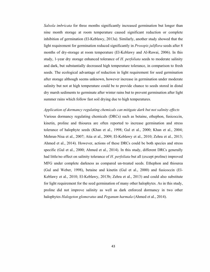
43
Salsola imbricata for three months significantly increased germination but longer than
nine month storage at room temperature caused significant reduction or complete
inhibition of germination (El-Keblawy, 2013a). Similarly, another study showed that the
light requirement for germination reduced significantly in Prosopis juliflora seeds after 8
months of dry-storage at room temperature (El-Keblawy and Al-Rawai, 2006). In this
study, 1-year dry storage enhanced tolerance of H. perfoliata seeds to moderate salinity
and dark, but substantially decreased high temperature tolerance, in comparison to fresh
seeds. The ecological advantage of reduction in light requirement for seed germination
after storage although seems unknown, however increase in germination under moderate
salinity but not at high temperature could be to provide chance to seeds stored in distal
dry marsh sediments to germinate after winter rains but to prevent germination after light
summer rains which follow fast soil drying due to high temperatures.
Application of dormancy regulating chemicals can mitigate dark but not salinity effects
Various dormancy regulating chemicals (DRCs) such as betaine, ethephon, fusicoccin,
kinetin, proline and thiourea are often reported to increase germination and stress
tolerance of halophyte seeds (Khan et al., 1998; Gul et al., 2000; Khan et al., 2004;
Mehrun-Nisa et al., 2007; Atia et al., 2009; El-Keblawy et al., 2010; Zehra et al., 2013;
Ahmed et al., 2014). However, actions of these DRCs could be both species and stress
specific (Gul et al., 2000; Ahmed et al., 2014). In this study, different DRCs generally
had little/no effect on salinity tolerance of H. perfoliata but all (except proline) improved
MFG under complete darkness as compared un-treated seeds. Ethephon and thiourea
(Gul and Weber, 1998), betaine and kinetin (Gul et al., 2000) and fusicoccin (El-
Keblawy et al., 2010; El-Keblawy, 2013b; Zehra et al., 2013) and could also substitute
for light requirement for the seed germination of many other halophytes. As in this study,
proline did not improve salinity as well as dark enforced dormancy in two other
halophytes Halogeton glomeratus and Peganum harmala (Ahmed et al., 2014).

44
Conclusions
Halopeplis perfoliata seeds were non-dormant and germinated quickly in distilled water
irrespective of incubation temperature but required light for germination. Seeds showed
high tolerance to salinity and some seeds could germinate in as high as seawater salinity
(0.6 mol L-1 NaCl) under 12-h photoperiod. High salinity (> 0.3 mol L-1 NaCl) and dark
imparted enforced/conditional dormancy in seeds, as they showed high recovery of
germination when transferred to distilled water and light. Seeds of our test species could
withstand hyper salinity (> 1.0 mol L-1 NaCl) by entering a state of enforced dormancy.
However, high incubation temperature enhanced seed mortality under hyper-saline
condition. Dry storage for 1-year increased tolerance of H. perfoliata seeds to moderate
salinity and dark, but substantially decreased high temperature tolerance, in comparison
to fresh seeds. Whereas, dormancy regulating chemicals could compensate for light
requirement but had no effect on salinity-enforced dormancy. Hence, it appears that
salinity enforced dormancy has osmotic, while dark enforced dormancy has biochemical
basis.
Fig. 2.8. Regulation of seed germination in Halopeplis perfoliata by light, temperature,
salinity and chemicals (DRCs).
45
References:
Ahmed, M. Z., Gulzar, S., and Khan, M. A. (2014). Role of dormancy regulating chemicals in alleviating the seed germination of three playa halophytes. Ekoloji, 23, 1-7.
Alfarhan, A. H., Al-Turki, T. A., and Basahy, A. Y. (2005). Flora of Jizan Region. King
Abdulaziz City for Science and Technology, Riyadh. Saudi Arabia, 1, 1-523.
Al-Oudat, M., and Qadir, M. (2011). The halophytic flora of Syria. International Center
for Agricultural Research in the Dry Areas (ICARDA), Aleppo. Syria, 8, 186.
Anonymous. (2014).http://www.efloras.org/florataxon.aspx?flora_id=2&taxon_id= 2000
06870.
Atia, A., Debez, A., Barhoumi, Z., Smaoui, A., and Abdelly, C. (2009). ABA, GA3, and
nitrate may control seed germination of Crithmum maritimum (Apiaceae) under
saline conditions. Comptes Rendus Biologies, 332, 704-710.
Baskin, C. C., and Baskin, J. M. (1995). Dormancy types and dormancy-breaking and
germination requirements in seeds of halophytes. Biology of salt tolerant plants.
(Khan, M. A. and Ungar, I. A. eds. pp. 23-30). Department of Botany, University
of Karachi, Karachi, Pakistan.
Baskin, C. C., and Baskin, J. M. (2001). Seeds: ecology, biogeography, and evolution of
dormancy and germination. San Diego: Academic Press.
Bertness, M. D., Gough, L., and Shumway, S. W. (1992). Salt tolerances and the
distribution of fugitive salt marsh plants. Ecology, 73, 1842-1851.
Bewley, J. D., and Black, M. (1994). Seeds: physiology of development and germination.
(2nd ed.). Verlag, US: Springer.
Bewley, J. D., Bradford, K. J., Hilhorst, H. W., and Nonogaki, H. (2013). Mobilization
of stored reserves. Seeds (pp. 183-246). New York: Springer.
Böer, B. (1996). Plants as soil indicators along the Saudi coast of the Arabian Gulf.
Journal of Arid Environments, 33, 417-423.
Bonfil, C. (1998). The effect of seed size, cotyledon reserves, and herbivory on seedling
survival and growth in Quercus rugosa and Q. laurina (Fagaceae). American
Journal of Botany, 85, 79-87.
Boorman, L. A. (2003). Salt marsh review. An overview of coastal saltmarshes, their
dynamic and sensitivity characteristics for conservation and management. Joint
Nature Conservation Committee. JNCC: Peterborough. Report, 334, 1-116.

46
Bradbeer, J. W. (1998). Seed Dormancy and Germination. New York, USA: Chapman
and Hall.
Cao, D., Baskin, C. C., Baskin, J. M., Yang, F., and Huang, Z. (2014). Dormancy cycling
and persistence of seeds in soil of a cold desert halophyte shrub. Annals of
Botany, 113, 171-179.
Egley, G. H. (1990). High temperature effects on seed germination and survival of weed
seeds in soil. Weed Science, 3, 429-435.
Ekstam, B., Johannesson, R., and Milberg, P. (1999). The effects of light and number of
diurnal temperature fluctuations on germination of Phragmites australis. Seed
Science Research, 9, 165-170.
El-Keblawy, A., and Al-Rawai, A. (2006). Effects of seed maturation time and dry
storage on light and temperature requirements during germination in invasive
Prosopis juliflora (Sw.). Flora- Morphology, Distribution, Functional Ecology of
Plants, 201, 135-143.
El-Keblawy, A., Al-Ansari, F., and Al-Shamsi, N. (2010). Impact of dormancy
regulating chemicals on salinity induced dormancy in Lasiurus scindicus and
Panicum turgidum: two desert glycophytic grasses. Plant Growth Regulation, 62,
163-170.
El-Keblawy, A. (2013a). Effects of seed storage on germination of two succulent desert
halophytes with little dormancy and transient seed bank. Acta Ecologica Sinica,
33, 338-343.
El-Keblawy, A. (2013b). Impacts of dormancy-regulating chemicals on innate and
salinity-induced dormancy of four forage grasses native to Arabian deserts. Grass
and Forage Science, 68, 288-296.
Ellis, R. H. (1991). The longevity of seeds. Hort Science, 26, 1119-1124.
Elsey-Quirk, T., Middlet, B. A., and Proffitt, C. E. (2009). Seed flotation and
germination of salt marsh plants: the effects of stratification, salinity, and/or
inundation regime. Aquatic Botany, 91, 40-46.
Fenner, M. (1985). Seed Ecology. The University of Michigan: Springer.
Flores, J., Jurado, E., and Arredondo, A. (2006). Effect of light on germination of seeds
of cactaceae from the Chihuahuan Desert, México. Seed Science Research, 16,
149-155.

47
Freitas, R. F., and Costa, C. S. (2014). Germination responses to salt stress of two
intertidal populations of the perennial glasswort Sarcocornia ambigua. Aquatic
Botany, 117, 12-17.
Grime, J. P., Mason, G., Curtis, A. V., Rodman, J., and Band, S. R. (1981). A
comparative study of germination characteristics in a local flora. The Journal of
Ecology, 69, 1017-1059.
Gul, B., and Khan, M. A. (1998). Population characteristics of a coastal halophyte
Arthrocnemum macrostachyum. Pakistan Journal of Botany, 30, 189-197.
Gul, B., Weber, D. J. (1998). Effect of dormancy relieving compounds on the seed
germination of non-dormant Allenrolfea occidentalis under salinity stress. Annals
of Botany, 82, 555-560.
Gul, B., and Weber, D. J. (1999). Effect of salinity, light and thermo period on the seed
germination of Allenrolfea occidentalis. Canadian Journal of Botany, 77, 1-7.
Gul, B., Khan, M. A., and Weber, D. J. (2000). Alleviation salinity and dark-enforced
dormancy in Allenrolfea occidentalis seeds under various thermo-periods.
Australian Journal of Botany, 48, 745-752.
Gul, B., and Khan, M. A. (2008). Effect of compatible osmotica and plant growth
regulators in alleviating salinity stress on the seed germination of Allenrolfea
occidentalis. Pakistan Journal of Botany, 40, 1957-1964.
Gul, B., Ansari, R., Flowers, T. J., and Khan, M. A. (2013). Germination strategies of
halophyte seeds under salinity. Environmental and Experimental Botany, 92, 4-
18.
Gulzar, S., Hameed, A., Alatar, A. A., Hegazy, A. K., and Khan, M. A. (2013). Seed
germination ecology of Cyperus arenarius - a sand binder from Karachi coast.
Pakistan Journal of Botany, 45, 493-496.
Hameed, A., Ahmed, M. Z., and Khan, M. A. (2006). Comparative effects of NaCl and
sea-salt on seed germination of coastal halophytes. Pakistan Journal of Botany,
38, 1605-1612.
Hameed, A., Ahmed, M. Z., Gulzar, S., Gul, B., Alam, J., Hegazy, A. K., Alatar, A. R.
A., and Khan, M. A. (2013). Seed germination and recovery responses of Suaeda
heterophylla to abiotic stresses. Pakistan Journal of Botany, 45, 1649-1656.
Hameed, A., Rasheed, A., Gul, B., and Khan, M. A. (2014). Salinity inhibits seed
germination of perennial halophytes Limonium stocksii and Suaeda fruticosa by

48
reducing water uptake and ascorbate dependent antioxidant system.
Environmental and Experimental Botany, 107, 32-38.
Image J. (2012). Image J image processing and analysis in Java: ImageJ/Fiji 1.46: http://
imagej.nih.gov.
Keiffer, C, H., and Ungar, I. A. (1997). The Effect of extended exposure to hypersaline
conditions on the germination of five inland halophyte species. American Journal
of Botany, 84, 104-111.
Kettenring, K. M., Gardner, G., and Galatowitsch, S. M. (2006). Effect of light on seed
germination of eight wetland Carex species. Annals of Botany, 98, 869-874.
Khan, M. A., and Ungar, I. A. (1984). Effects of salinity and temperature on the
germination and growth of Atriplex triangularis Wild. American Journal of
Botany, 71, 481-489.
Khan, M. A., and Ungar, I. A. (1985). The role of hormone in regulating germination of
polymorphic seeds and early seedling growth of Atriplex triangularis under
saline conditions. Physiologia Plantarum, 63, 109-113.
Khan, M. A., and Ungar, I. A. (1997). Effects of light, salinity, and thermo period on the
seed germination of halophytes. Canadian Journal of Botany, 75, 835-841.
Khan, M. A., and Gul, B. (1998). High salt tolerance in germinating dimorphic seeds of
Arthrocnemum indicum. International Journal of Plant Sciences, 159, 826-832.
Khan, M. A., Ungar, I. A., and Gul, B. (1998). Action of compatible osmotica and
growth regulators in alleviating the effect of salinity on the germination of
dimorphic seeds of Arthrocnemum indicum L. International Journal of Plant
Sciences, 159, 313-317.
Khan, M. A., Gul, B., and Weber, D. J. (2000). Germination responses to Salicornia
rubra to temperature and salinity. Journal of Arid Environments, 45, 207-214.
Khan, M. A., Gul, B., and Weber, D. J. (2001). Effect of salinity and temperature on the
germination of Kochia scoparia. Wetland Ecology and Management, 9, 483-489.
Khan, M. A., Gul, B., and Weber, D. J. (2002). Seed germination in the Great Basin
halophyte Salsola iberica. Canadian Journal of Botany, 80, 650-655.
Khan, M. A., Gul, B., and Weber, D. J. (2004). Action of plant growth regulators and
salinity on the seed germination of Ceratoides lanata. Canadian Journal of
Botany, 82, 37-42.

49
Khan, M. A., and Gul, B. (2006). Halophyte seed germination. Ecophysiology of High
Salinity Tolerant Plants (Khan, M. A., and Weber, D. J. pp. 11-30). Springer.
Kim, D. H., Aldridge, K. T., Brookes, J. D., and Ganf, G. G. (2013). The effect of
salinity on the germination of Ruppia tuberosa and Ruppia megacarpa and
implications for the Coorong: A coastal lagoon of southern Australia. Aquatic
Botany, 111, 81-88.
Leishman, M. R., Wright, I. J., Moles, A. T., and Westoby, M. (2000). The evolutionary
ecology of seed size. Seeds: The Ecology of Regeneration in Plant Communities,
2, 31-57.
MacKay, D. B. (1972). The measurement of viability. Viability of Seeds (pp. 172-208).
Netherlands: Springer.
Mahmoud, A., El Sheikh, A. M., and Abdul Baset, S. (1983). Germination of two
halophytes: Halopeplis perfoliata and Limonium aillare from Saudi Arabia.
Journal of Arid Environments, 6, 87-98.
Mandura, A. S., Saifullah, S. M., and Khafaji, A. K. (1987). Mangrove Ecosystem of
Southern Red Sea Coast of Saudi Arabia. Proceedings of the Saudi Biological
Society, 10, 165-193.
Mehrun-Nisa, Khan, M. A., and Weber, D. J. (2007). Dormancy, germination and
viability of Salsola imbricata seeds in relation to light, temperature and salinity.
Seed Science and Technology, 35, 595-606.
Milberg, P., Andersson, L., and Thompson, K. (2000). Large seeded species are less
dependent on light for germination than small seeded ones. Seed Sciences, 10, 99-
104.
Noe, G. B., and Zedler, J. B. (2000). Differential effects of four a-biotic factors on the
germination of salt marsh annuals. American Journal of Botany, 87, 1679-1692.
Pilcher, N. J., Phillips, R. C., Aspinall, S., Al-Madany, I., King, H., Hellyer, P., Beech,
M., Gillespie, C., Wood, S., Schwarze, H., Al Dosary, M., Al Farraj, E., Khalifa,
A., and Böer, B. (2003). Hawar islands protected area (Kingdom of Bahrain).
Management Plan (1st Draft. p. 30). UNESCO, Doha.
Pons, T. L. (2000). Seed responses to light. Seeds: The Ecology of Regeneration in Plant
Communities (Fenner, M. ed. pp. 237-260). Wallingford, London: CAB
International.

50
Probert, R. J. (2000). The role of temperature in the regulation of seed dormancy and
germination. Seeds: The Ecology of Regeneration in Plant Communities, 2, 261-
292.
Pujol, J. A., Calvo, J. F., Ramirez-Diaz, L. (2000). Recovery of germination from
different osmotic conditions by four halophytes from southeastern Spain. Annals
of Botany, 85, 279-286.
Rajjou, L., and Debeaujon, I. (2008). Seed longevity: survival and maintenance of high
germination ability of dry seeds. Compete Rendues Biologies, 331, 796-805.
Ramadan, H. A-Z., Bantan, R. A. (2013). Hypersaline benthic foraminifera from the
Shuaiba Lagoon, eastern Red Sea, Saudi Arabia: Their environmental controls
and usefulness in sea-level reconstruction. Marine Micropaleontology, 103, 51-
67.
Saeed, S., Gul, B., and Khan, M. A. (2011). Comparative effects of NaCl and sea salt on
seed germination of Arthrocnemum indicum. Pakistan Journal of Botany, 43,
1091-1103.
Seitzinger, S. P. (1988). Denitrification in freshwater and coastal marine ecosystems:
ecological and geochemical significance. Limnology and Oceanography, 33, 702-
724.
Seiwa, K. (2000). Effects of seed size and emergence time on tree seedling
establishment: importance of developmental constraints. Oecologia, 123, 208-
215.
Shepherd, K. A., Macfarlane, T. D., and Colmer, T. D. (2005). Morphology, anatomy
and histo-chemistry of Salicornioideae (Chenopodiaceae) fruits and seeds.
Annals of Botany, 95, 917-933.
Song, J., Feng, G. U., Tian, C., and Zhang, F. (2005). Strategies for adaptation of Suaeda
physophora, Haloxylon ammodendron and Haloxylon persicum to a saline
environment during seed-germination stage. Annals of Botany, 96, 399-405.
SPSS. (2011). IBM SPSS statistics base 20. Chicago, IL: SPSS Inc.
Stromberg, J. C., and Boudell, J. A. (2013). Floods, drought, and seed mass of riparian
plant species. Journal of Arid Environments, 97, 99-107.
Teixeira, A., Duarte, B., and Caçador, I. (2014). Salt Marshes and Biodiversity. Sabkha
Ecosystems: Cash Crop Halophyte and Biodiversity Conservation (Vol. 4, pp.
283-298). Netherlands: Springer.

51
Tompsett, P. B. (1986). The effect of desiccation on the viability of dipterocarp seed.
Seed Problems under Stressful Conditions (pp. 181-201). Proceedings of
the IUFRO Symposium. Vienna.
Ungar, I. A. (1978). Halophyte seed germination. The Botanical Review, 44, 233-264.
Ungar, I. A. (1991). Ecophysiology of vascular halophytes. USA: CRC press.
Ungar, I. A. (1995). Seed germination and seed-bank ecology of halophytes. Seed
Development and Germination (Kigel, J., and Galili, G. eds.). New York: Marcel
and Dekker.
Valiela, I., Cole, L. M. (2002). Comparative evidence that salt marshes and mangroves
may protect seagrass meadows from land-derived nitrogen loads. Ecosystems, 5,
92-102.
Wei, Y., Dong, M., Huang, Z. Y., and Tan, D. Y. (2008). Factors influencing seed
germination of Salsola affinis (Chenopodiaceae), a dominant annual halophyte
inhabiting the deserts of Xinjiang, China. Flora-Morphology, Distribution,
Functional Ecology of Plants, 203, 134-140.
Winn, A. A. (1985). The effect of seed size and microsite on seedling emergence in four
population of Prunella vulgaris. Journal of Ecology, 73, 831-840.
Yasseen, B. T., and Al-Thani, R. F. (2007). Halophytes and associated properties of
natural soils in the Doha area, Qatar. Aquatic Ecosystem Health and
Management, 10, 320 -326.
Zehra, A., Gul, B., Ansari, R., Alatar, A. R. A., Hegazy, A. K., and Khan, M. A. (2013).
Action of plant growth regulators in alleviating salinity and temperature effects
on the germination of Phragmites karka. Pakistan Journal of Botany, 45, 1919-
1924.
Zhang, Y., Wang, Y., and Li, S. (2011). Effects of different storage conditions and
durations on seed viability and germination of five desert plants in West
Erdos. Scientia Silvae Sinicae, 47, 36-41.
Zia, S., and Khan, M. A. (2004). Effect of light, salinity, and temperature on seed
germination of Limonium stocksii. Canadian Journal of Botany, 82, 151-157.
Zreik, R. (1990). The domestication and economic cultivation of halophytes. Developing
World Agriculture (pp. 74-79). London: Grosvenor Press.

52
Chapter 3
Comparison of osmotic and ionic effects of various salts on germination inhibition and recovery responses of
Halopeplis perfoliata seeds
53
Abstract
Salinity affects seed germination of halophytes by inducing ionic toxicity, osmotic
constraint or both. Information about the effects of salinity on seed germination of a
large number of halophytes exists, but little is known about the basis of this salinity-
induced germination inhibition. In order to partition salinity effects, we studied seed
germination and recovery responses of a coastal salt marsh halophyte Halopeplis
perfoliata to different isotonic treatments (ΨS: -0.5, -1.0, -1.5, -2.0 and -2.5, MPa) of
various salts and polythylene glycol (PEG) under two light regimes (12-h light
photoperiod and 24-h complete darkness). Highest seed germination was observed in
distilled water under 12-h light photoperiod and reduction in osmotic potential of the
solution decreased seed germination. However, some seeds of H. perfoliata could
germinate in as low as -2.5 MPa (~0.6 mol L-1 NaCl), which is equivalent to seawater
salinity. Sea-salt treatment was more inhibitory than isotonic NaCl at the lowest osmotic
potential (ΨS -2.5 MPa). Generally, chloride salts inhibited germination more than the
isotonic sulfate salts. Comparable germination responses of seeds in NaCl and isotonic
PEG treatments as well as high recovery of germination in un-germinated seeds after
alleviation of NaCl salinity indicated prevalence of osmotic constraint. Hence seed
germination inhibition in H. perfoliata under different salts is a combination of osmotic
constraint and their differential ionic effects.

54
Introduction
Seeds of salt marsh halophytes are under direct influence of seawater
Coastal salt marshes are transition between marine and terrestrial ecosystems,
characterized by highly salt tolerant halophyte vegetation and offer a number of
ecological and economic services to mankind (Böer, 1996; Pennings and Bertness 2001;
Lee et al., 2006; Gedan et al., 2009; Teixeira et al., 2014). These habitats are however
under threat due to various natural and anthropogenic reasons (Yasseen and Al-Thani,
2007; Gedan et al., 2011; Ramadan et al., 2013). Therefore, salt marsh ecosystems
warrant special attention in research, which would eventually help in their protection and
conservation. Establishment of plants in salt marshes is reportedly dependent on the seed
germination responses which are influenced by a number of factors especially salt
concentrations in the marsh sediments (Pujol et al., 2000; Khan et al., 2001; Hameed et
al., 2006; Gul et al., 2013). However, data on seed germination responses of succulent
halophytes of subtropical salt marshes is scanty (Saeed et al., 2011; Gul et al., 2013).
Seeds of salt marsh halophytes are directly influenced by seawater owing to diurnal and
seasonal inundations (Gul and Khan, 1998; Gul et al., 2013; Teixeira et al., 2014). NaCl
is although dominant but other chloride and sulfate salts and their interactions may also
play a significant role in germination, radical emergence and seedling growth of coastal
halophytes (Gul and Khan, 1998; Khan, 2003; Zia and Khan, 2008). Most research on
the seed germination of halophytes has focused on responses to NaCl (reviewed by Gul
et al., 2013), with few studies on comparison of the effects of NaCl and sea-salt (Zia and
Khan, 2002; Atia et al., 2006; Hameed et al., 2006; Liu et al., 2006; Saeed et al., 2011;
Shaikh et al., 2013) and even fewer studies on effects of different salts (Ungar, 1991;
Egan et al., 1997; Panuccio et al., 2014). Likewise, investigations to partition osmotic
and ionic constraints of various salts (Munns et al., 1995) by comparing their effects on
seed germination with germination responses in non-ionic solutes such as polyethylene
glycol (PEG) are also limited and inconclusive (Tobe et al., 2000; Sosa et al., 2005;
Hameed et al., 2013).
Salt marsh halophyte Halopeplis perfoliata is largely unstudied
Halopeplis perfoliata Forssk. (Amaranthaceae) is a succulent halophyte of coastal salt
marshes along Arabian Peninsula (Al-Oudat and Qadir, 2011), where it acts as an
important primary producer and provides shelter to marine life (Pilcher et al., 2003). A

55
previous report showed that the seeds of H. perfoliata can germinate in up to 0.25 mol L-
1 NaCl treatment (Mahmoud et al., 1983). This study was therefore designed to
comparatively investigate the effects of 1) sea-salt 2) various chloride and sulfate salts
and 3) polyethylene glycol 6000 (PEG-6000) on germination and recovery of the seeds
of H. perfoliata.
Materials and methods
Study site and seed collection
Seeds of Halopeplis perfoliata were collected from the coast of Jizan, Saudi Arabia in
2012. Seeds were separated from inflorescence husk manually; surface sterilized using
1% bleach solution for 1 min, followed by rinsing with distilled water and air-drying in
the laboratory.
Treatments and experimental conditions
Germination experiments were carried out in clear-lid plastic petri-plates (50 mm
diameter x 15 mm depth) with 5 ml of test solution. Seeds were immersed in distilled
water (0 MPa) and isotonic solutions of various salts (NaCl, Na2SO4, KCl, K2SO4,
MgCl2, MgSO4, CaCl2 and Sea-salt) and polyethylene glycol 6000 (PEG). Five isotonic
levels (ΨS: -0.5, -1.0, -1.5, -2.0 and -2.5, MPa) were used. The osmotic potential of the
above solutions was measured with a vapor pressure osmo-meter (5520 Wescor, Inc.).
There were four replicates of 25 seeds each per treatment. The petri-plates were placed in
programmed incubators maintained at 20/30 °C (optimum temperature for the seed
germination of sub-tropical halophytes, Gul et al., 2013), where low temperature
coincided with 12-h dark and high temperature with 12-h light (~25 µ mol m-2 s-1, 400-
750 nm, Philips cool -white fluorescent lamps). Seed germination percentage (radicle
emergance, Bewley and Black, 1994) was recorded at 2 days intervals for 20 days. Rate
of germination (GRate) was estimated by using a modified Timson index of germination
velocity given below:
Where G is the percentage of seed germination at 2 days intervals and t is the
total germination period (Khan and Ungar, 1985). The greater the value, the more rapid
is germination. After 20 days un-germinated seeds were transferred to distilled water for

56
another 20 days and recovery of germination (SR) from various osmotic treatments of
solutes (salts and PEG) was recorded. The recovery percentage was determined by
counting the recovered seeds from total number of seeds. The recovery percentage was
calculated by using given formula:
Where, A is total number of seeds germinated after being transferred to distilled
water, B is the number of seeds germinated in saline solution and C is total number of
seeds. A parallel set of experiment was conducted by placing petri-plates in dark-
envelops under aforementioned temperature and osmotic treatments for 20 days.
Germination of this experiment was noted once after 20 days. After 20 days un-
germinated seeds were exposed to light in order to study the recovery of germination
from dark (DR).
Statistical analyses
Data were statistically analyzed using SPSS version 11.0 (SPSS, 2011). Analysis of
variance (ANOVA) was performed to determine if various treatments affected the seed
germination parameters significantly. A Bonferroni post-hoc was conducted to indicate
significant (P < 0.05) differences among mean values.
Results
A four-way ANOVA indicated significant effects of osmotic potential (ΨS), photoperiod
anions and but not of cations on mean final germination (MFG) of H. perfoliata seeds
(Table 3.1).
Comparitive effects of NaCl and Sea-salt
Seeds were non-dormant and germinated maximally (~90%) in distilled water under 12-h
photoperiod (Fig. 3.1). Effects of NaCl and sea-salt were comparable in up to -1.0 MPa,
wherein there was maximal (~90%) MFG as in distilled water under 12-h photoperiod
(Fig. 3.1). While, in lowest (-2.5 MPa) ΨS treatment, sea-salt (MFG = ~10%) was found
more inhibitory than NaCl (MFG = ~25%). Rate of germination in -2.5 MPa was also
lowest in sea-salt compared to NaCl (Fig. 3.2). Un-germinated seeds from low ΨS
treatments of both NaCl and sea-salt, showed recovery (SR) when transferred to distilled
57
water and almost all un-germinated seeds recovered (Fig. 3.1). Dark inhibited seed
germination of test species in both NaCl and sea-salt treatments (Fig. 3.3). When un-
germinated seeds from dark were transferred to 12-h photoperiod for another 20 days, a
significant recovery (DR) was seen in either salt type. Still un-germinated seeds from low
ΨS treatments of both NaCl and sea-salt showed complete recovery (SR) when transferred
to distilled water (Fig. 3.3).
Comparison of the effects of different salts
A decrease of up to -1.0 MPa (equivalent to 0. 215 mol L-1 NaCl) for chloride salts and -
1.5 MPa (equivalent to 0.4 mol L-1 NaCl) for sulfate salts had no inhibitory effect on
seed germination of test species (Fig. 3.1). K2SO4 was less inhibitory, in which over 70%
seeds germinated in lowest (-2.5 MPa) ΨS treatment. On the other hand, CaCl2 and NaCl
reduced MFG to ~50% in -1.5 MPa treatments (Fig. 3.1). Inhibitory effects of chloride
salts were in following order: CaCl2 > NaCl > KCl > MgCl2. While, MFG inhibition
among sulfate salts was as follows: Na2SO4 > MgSO4 > K2SO4. Rate of germination also
decreased with decreases in ΨS of salts (Fig. 3.2). However, there was a significant
decrease in rate of germination in -1.0 MPa for chloride salts and in -1.5 MPa for sulfate
salts. While similar to MFG, NaCl and CaCl2 had higher inhibition and K2SO4 caused
less inhibition to the rate of germination (Fig. 3.2). Almost all un-germinated seeds
showed recovery of germination (SR) when transferred to distilled water and generally
highest SR was observed from the lowest (-2.5 MPa) ΨS treatments of all salts (Fig. 3.1).
Dark caused substantial inhibition to the MFG of H. perfoliata, irrespective of the salt
and ΨS treatments (Fig. 3.3). When un-germinated seeds from dark were exposed to 12-h
photoperiod after 20 days, a significant recovery of germination (DR) was observed. The
DR was highest in distilled water and there was decrease in DR in solution of low ΨS (Fig.
3.3). When still un-germinated seeds from low ΨS treatments were transferred to distilled
water for another 20 days, most of them showed recovery of germination (SR),
irrespective of salt used (Fig. 3.3).
Comparison of the effects of NaCl and PEG
Effects of NaCl and PEG on MFG and germination rate were generally comparable at
higher osmotic levels and these germination parameters decreased with decreases in ΨS
under 12-h photoperiod (Fig. 3.4 A and 4 C). Un-germinated seeds from low ΨS

58
treatments of both NaCl and PEG showed high recovery (SR; Fig. 3.4 B) complete
darkness caused substantial inhibition to MFG (Fig. 3.4. D). Un-germinated seeds from
the dark showed high recovery (DR) upon transfer to 12-h photoperiod condition (Fig.
3.4 E). However, relatively higher DR was observed in moderately low (-1.0 to -1.5 MPa)
PEG compared to isotonic NaCl treatments. Remaining un-germinated seeds from low
ΨS treatments of both NaCl and PEG germinated (SR) when transferred to distilled water
(Fig. 3.4 F). Table 3.1. One-way ANOVA of the effects of salinity on growth, water and ion
relations, pigments, photosynthesis, isotope ratios, stress markers, antioxidant enzymes
and substrates of Halopeplis perfoliata grown under salinity treatments (0, 1.50, 0.30
and 0.60 mol L-1 NaCl) for 30 days. Numbers are F-values and asterisks in superscript
are significance levels at P < 0.05 (ns = non-significant, * = P < 0.05, ** = P < 0.01 and
*** = P < 0.001).
Factors MFG GRate Rec.
Phot. 4341.52*** 372.55*** - ΨS 98.50*** 4.02** 500.63*** Anion 13.63*** 0.53ns 28.65*** Cat. 2.16ns 1.96ns 5.71** Phot. * ΨS 97.76*** 189.41*** - Phot.* Ani 14.39*** 45.74*** - Phot.* Cat. 2.35ns 9.08*** - ΨS*Ani 4.65*** 5.45*** 9.31*** ΨS *Cat. 0.78ns 1.58ns 4.29*** Ani* Cat. 2.46ns 9.20*** 10.58*** Phot.*Ani* Cat. 2.26ns 3.51* - Phot.* ΨS *Ani 4.50** 3.13** - Phot.* ΨS *Cat. 0.74ns 1.45ns - ΨS *Ani* Cat. 2.91** 2.91** 16.72*** Phot.* ΨS*Ani*Cat. 2.95** 1.99* -

59
SeaSalt
0.0 0.5 1.0 1.5 2.0 2.50.0 0.5 1.0 1.5 2.0 2.5
20
40
60
80
100
MgSO4
20
40
60
80
100Ger
min
atio
n (%
)
20
40
60
80
100
20
40
60
80
100
NaCl
KCl
MgCl2
CaCl2
K2SO4
a
a
a
b
bb b
b
a
aaabab
a a
c
a
a
aa
a a
b
b
b
aa
b
b
a
a a
d
c
c
d
d
b
b
aa
b
b
aa
ab
a
aa
a
a
a
aa
bb
a
a
a
bc
b
c
c
c
a
a
aa
a
a a
a
a a a
b
Germination Recovery
ψs (-MPa)
b
b
aa
a aa
b b
Na2SO4
Fig. 3.1. Effects of NaCl, Na2SO4, KCl, K2SO4, MgCl2, MgSO4, CaCl2 and sea-salt on
mean final germination (MFG) and germination recovery in distilled water (SR) at 20/30 oC temperature and 12-h photoperiod. Vertical bars are means ± S.E. Different alphabets
show significant differences among isotonic treatments within each parameter;
Bonferroni test; P < 0.05.

60
Sea-salt
0.0 0.5 1.0 1.5 2.0 2.5
MgSO4
K2SO4
CaCl2
0.0 0.5 1.0 1.5 2.0 2.50
1020304050
MgCl2
01020304050
KCl
01020304050
NaClG
erm
inat
ion
Rat
e
01020304050 Na2SO4
ψs (-MPa)
a a
aa
c
b
b
c
d
c
b
b
a a a
a
b
b
a aaab
c
a a a
b
c
d
a a a
b
d
c
d
a aab
b b
c
a a
bb
cc
Fig. 3.2. Effects of NaCl, Na2SO4, KCl, K2SO4, MgCl2, MgSO4, CaCl2 and sea-salt on
rate of germination at 20/30 oC temperature and 12-h photoperiod. Vertical bars are
means ± S.E. Different alphabets show significant differences among isotonic treatments
within indiviual salt; Bonferroni test; P < 0.05.

61
Sea-salt
0.0 0.5 1.0 1.5 2.0 2.5
MgSO4
K2SO4
CaCl2
0.0 0.5 1.0 1.5 2.0 2.50
20406080
100
MgCl2
020406080
100
KCl
Ger
min
atio
n (%
)
020406080
100
NaCl
020406080
100Na2SO4
MFG SR DR
a
bb
cd
ea a
b
b
cbc
ca
a a
a a
a a
ab b
ca
b
c
a
a
aab
a a
bb
c
bb
c
b
aa
aba a
b
c
da a
b
cc
a
a
aa
ab
b
b ba
ab
b
c a
a
a
aa
a
bb
a
b
c
b
c
a a
aaa
a
ab
a
ab
bc
ca
a
a a
a
bab
b
c
bb
bb
ψs (-MPa)
Fig. 3.3. Effects of isotonic treatments of NaCl, Na2SO4, KCl, K2SO4, MgCl2,
MgSO4, CaCl2 and sea-salt on mean final germination (MFG), recovery from Salinity
(SR) and recovery from Dark (DR) at 20/30 oC temperature and complete darkness.
Vertical bars are means ± S.E. Different alphabets show significant differences among
isotonic treatments within each paramerter of individual salt; Bonferroni test; P <
0.05.

62
DR (%
)
Ger
min
atio
n (%
)
Ger
min
atio
n (%
)S R
(%)
ψs (-MPa)
2D Graph 1
0.0 0.5 1.0 1.5 2.0 2.50
20
40
60
80
100
0.0 0.5 1.0 1.5 2.0 2.5
Ger
min
atio
n R
ate
0
10
20
30
40
500
20
40
60
80
100
Dark
0
20
40
60
80
100
S R (%
)
0
20
40
60
80
100
0
20
40
60
80
100
aab
a
a
a bb
ccc
dd
aa
b
bb
a a a a
b
c
aa
bb
b
a a a
b
c a aa
a
b
bb
cd
ec c
a aa
a
b
c
c
b
b
c
bcc
*
*
**
*
* **
*
A
B
C
E
F
Light
NaCl
PEG
aa
D
Fig. 3.4. Comparative effects of NaCl and PEG on mean final germination (MFG),
Germination rate (GRate); recovery from salinity (SR) and recovery from dark (DR) under
12-h photoperiod at 20/30 oC temperature and complete darkness. Vertical bars are
means ± S.E. Different alphabets show significant differences among sainity treatments
within each parameter; Bonferroni test; P < 0.05. Asterisks (*) show significant
differences in germination within an isotonic treatment of NaCl and PEG by student’s t-
test; P < 0.05.

63
Discussion
Sea-salt is more inhibitory for germination than NaCl
Seeds of H. perfoliata were non-dormant and showed maximum germination (~90%) in
distilled water under 12-h photoperiod, as reported for many other marsh halophytes
such as Aeluropus lagopoides (Khan and Gulzar, 2003), Limonium stocksii (Zia and
Khan, 2004) and Arthrocnemum indicum (Saeed et al., 2011). Recently, Gul et al. (2013)
reviewed that lack of innate dormancy in most subtropical perennial halophytes is
probably a common adaptation to take advantage of brief water availability after rains. In
addition, seeds of H. perfoliata displayed a high salt tolerance, as some seeds could
germinate in as high as seawater (-2.5 MPa NaCl) salinity. Seeds of many other
Salicornioideae halophytes such as Arthrocnemum indicum (Khan and Gul, 1998) and
Sarcocornia ambigua (Freitas and Costa, 2014) also showed tolerance to seawater or
higher salinity during seed germination. Furthermore, un-germinated seeds of H.
perfoliata showed a high recovery of germination when transferred to distilled water
from saline solutions like many other perennial halophytes, indicating that salinity
treatments resulted in enforced/conditional dormancy and were no detrimental to seed
viability (Khan and Gul, 2006; Cao et al., 2014). Hence, it appears that seeds of H.
perfoliata are well-adapted for saline environment. However, whether these seed
responses to NaCl based salinity are comparable to seawater salinity, have seldom been
studied. Most studies on effects of salinity on seed germination of halophytes are based
on NaCl treatments and generally little is known about effects of seawater on seed
germination of halophytes (Zia and Khan, 2002; Atia et al., 2006; Hameed et al., 2006;
Liu et al., 2006; Saeed et al., 2011; Shaikh et al., 2013). In this study, effects of NaCl and
sea-salt were comparable in up to -1.0 MPa but at lowest (-2.5 MPa) ΨS treatment sea-
salt was more inhibitory than isotonic NaCl. Many studies showed that the sea-salt
inhibits seed germination more in comparison to NaCl. For instance, higher
concentrations (or lower ΨS treatments) of sea-salt were more inhibitory for the seed
germination of Limonium stocksii (Zia and Khan, 2002), Aeluropus lagopoides,
Desmostachya bipinnata and Suaeda fruticosa in comparison to NaCl (Hameed et al.,
2006). However, NaCl inhibited seed germination of Hipophae rhamnoides (Tirmizi et
al., 1993) and Crithmum maritimum (Atia et al., 2006) more as compared to sea-salt.
Effects of NaCl and sea-salt on seed germination of Haloxylon stocksii comparable

64
(Hameed et al., 2006) also indicated that the effects of NaCl and seawater on seed
germination of halophytes are species-specific.
Sulfate salts are less detrimental than chloride salts for seed germination of Halopeplis
perfoliata
Seawater is composed of many salts, of which NaCl is the major (> 85%) fraction
(Murray, 2004; Khan and Gul, 2006). Other notable cations of seawater in order of
magnitude are Mg++ > Ca++> K+ and SO4-- is second major anion after Cl- (Murray,
2004). Typically, six ions constitute > 99% of the seawater (http://oceanplasma.org
/documents/chemistry.html). Therefore, I examined the effects of these ions on seed
germination of coastal marsh halophyte H. perfoliata found significant effect of anions
but not of cations on seed germination of test species. A decrease of up to -1.0 MPa
(equivalent to ~0.2 mol L-1 NaCl) for chloride salts and -1.5 MPa (equivalent to 0.4 mol
L-1 NaCl) for sulfate salts had no inhibitory effect on seed germination of test species but
a further decrease in ΨS of solutions was inhibitory. Likewise, seed germination of three
perennial salt marsh halophytes Arthrocnemum macrostachyum, Juncus acutus and
Schoenus nigricans was inhibited more in chloride than sulfate salts of sodium and
magnesium (Vicente et al., 2007). Seed germination characteristics of Kalidium
capsicum (Tobe et al., 2002) Haloxylon ammodendron (Tobe et al., 2004) and
Halostachys caspica (Assareh et al., 2011) were also more sensitive to chloride than
sulfate salts. These data including the present study indicate that sulfate salts are
comparatively less detrimental to seed germination of most halophytes than isotonic
chloride salts. However eco-physiological significance of this differential effect is yet to
be determined by further research.
Halopeplis perfoliata seeds show differential response to isotonic NaCl and PEG
treatments
Salinity affects seed germination either by reducing soil water potential (i.e.
compromising imbibition an osmotic effect) and/or by excessive entry of Na+ which is
toxic to metabolism (ionic toxicity) (Khan and Gul, 2006; Kranner and Seal, 2013;
Hameed et al., 2014). Uptake of salt could lower seed’s water potential thereby
facilitating imbibition, however ionic toxicity may overshadow such beneficial osmotic
effects (Collis-George and Sands, 1962; Khan et al., 1987; Song et al., 2005; Gul et al.,
2013). In order to partition osmotic and ionic components of salinity, often seed

65
germination response in a salt solution is compared with that in an isotonic solution of a
non-ionic solute such as polyethylene glycol (Hardegree and Emmerich, 1990; Bajji et
al., 2002; Song et al., 2005; Hameed et al., 2013). In this study, effects of NaCl and PEG
on seed germination of H. perfoliata were generally comparable, especially under high
osmotic levels. Similar effects of NaCl and non-ionic solutes (PEG or mannitol) were
observed on seed germination of Chrysothamnus nauseosus (Dodd and Donovan, 1999)
and Atriplex halimus (Bajji et al., 2002). Greater incidence of osmotic constraint of
salinity on seed germination than ionic toxicity was also observed in this study. High
recovery of un-germinated seeds in this study after alleviation of salinity also supports
this assumption. On the other hand, salinity caused ionic toxicity in some species like
Artemesia ordosica (Tobe et al., 1999), Aristida adscensionis and Prosopis strombulifera
(Sosa et al., 2005) and Suaeda heterophylla (Hameed et al., 2013).
Conclusions
Halopeplis perfoliata seeds appear highly tolerant to salinity, as some seeds could
germinate in NaCl treatments as low as -2.5 MPa (~0.6 mol L-1), which is equivalent to
seawater salinity. However, a sea-salt treatment of -2.5 MPa inhibited seed germination
more than isotonic NaCl treatment (Fig. 3.5). Furthermore, chloride salts were generally
more inhibitory to seed germination than isotonic sulfate salts (Fig. 3.5). Un-germinated
seeds from low osmotic potential treatments of all salts showed high recovery of
germination when transferred to distilled water, indicating osmotic rather than ionic
effect of salts (Fig. 3.5). Similar germination response of H. perfolaita seeds in isotonic
NaCl and PEG treatments at higher osmotic levels also indicate osmotic effect rather
than ionic effect of NaCl salinity.

66
Fig. 3.5. Differential inhibitory effects of iso-osmotic treatments with NaCl, seasalt, and
various chloride (Cl-) and sulphate (SO4 --) salts on seed germination of Halopeplis
perfoliata.
References: Al-Oudat, M., and Qadir, M. (2011). The halophytic flora of Syria. International Center
for Agricultural Research in the Dry Areas (ICARDA), Aleppo, Syria, 8, 186.
Assareh, M. H., Rasouli, B., and Amiri, B. (2011). Effects of NaCl and Na2SO4 on
germination and initial growth phase of Halostachys caspica. Desert, 15, 119-
125.
Atia, A., Hamed, K. B., Debez, A., and Abdelly, C. (2006). Salt and seawater effects on
the germination of Crithmum maritimum. Biosaline agriculture and salinity
tolerance in plants (Ozturk, M., Waisel, Y., Khan, M. A., Gork, G., and Verlag,
B. eds. pp. 29-33). Switzerland.
Bajji, M., Kinet, J. M., and Lutts, S. (2002). Osmotic and ionic effects of NaCl on
germination, early seedling growth, and ion content of Atriplex halimus
(Chenopodiaceae). Canadian Journal of Botany, 80, 297-304.

67
Bewley, J. D., and Black, M. (1994). Seeds: physiology of development and germination.
(2nd ed. pp. 1-33). Verlag, USA: Springer.
Böer, B. (1996). Plants as soil indicators along the Saudi coast of the Arabian Gulf
Journal of Arid Environments, 33, 417-423.
Cao, D., Baskin, C. C., Baskin, J. M., Yang, F., and Huang, Z. (2014). Dormancy cycling
and persistence of seeds in soil of a cold desert halophyte shrub. Annals of
Botany, 113, 171-179.
Collis-George, N., and Sands, J. E. (1962). Comparison of the effect of physical and
chemical component of soil water energy on seed germination. Australian
Journal of Botany, 13, 575-584.
Dodd, G. L., and Donovan, L. A. (1999). Water potential and ionic effects on
germination and seedling growth of two cold desert shrubs. American Journal of
Botany, 86, 1146-1153.
Egan, U. I., and Meekins, J. F. (1997). The effects of NaCl, KCl, Na2SO4 and K2NO4 on
the germination of Atriplex prostrata (Chenopodiaceae). Journal of Plant
Nutrition, 20, 1723-1730.
Freitas, R. F., and Costa, C. S. (2014). Germination responses to salt stress of two
intertidal populations of the perennial glasswort Sarcocornia ambigua. Aquatic
Botany, 117, 12-17.
Gedan, K. B., Silliman, B. R., and Bertness, M. D. (2009). Centuries of human-driven
change in salt marsh ecosystems. Annual Review of Marine Science, 1, 117-141.
Gedan, K. B., Altieri, A. H., and Bertness, M. D. (2011). Uncertain future of New
England salt marshes. Marine Ecology Progress Series, 434, 229-237.
Gul, B., and Khan, M. A. (1998). Population characteristics of the coastal halophyte
Arthrocnemum macrostachyum. Pakistan Journal of Botany, 30, 189-197.
Gul, B., Ansari, R., Flowers, T. J., and Khan, M. A. (2013). Germination strategies of
halophyte seeds under salinity. Environmental and Experimental Botany, 92, 4-
18.
Hameed, A., Ahmed, M. Z., and Khan, M. A. (2006). Comparative effects of NaCl and
seasalt on seed germination of coastal halophytes. Pakistan Journal of Botany,
38, 1605-1612.

68
Hameed, A., Ahmed, M. Z., Gulzar, S., Gul, B., Alam, J., Hegazy, A. K., Alatar, A. R.
A., and Khan, M. A. (2013). Seed germination and recovery responses of Suaeda
Heterophylla to abiotic stresses. Pakistan Journal of Botany, 45, 1649-1656.
Hameed, A., Rasheed, A., Gul, B., and Khan, M. A. (2014). Salinity inhibits seed
germination of perennial halophytes Limonium stocksii and Suaeda fruticosa by
reducing water uptake and ascorbate dependent antioxidant system.
Environmental and Experimental Botany, 107, 32-38.
Hardegree, S. P., and Emmerich, W. E. (1990). Partitioning water potential and specific
salt effects on seed germination of four grasses. Annals of Botany, 66, 587-595.
Kranner, I., and Seal, C. E. (2013). Salt stress, signalling and redox control in seeds.
Functional Plant Biology, 40, 848-859.
Khan, M. A., and Ungar, I. A. (1985). The role of hormone in regulating germination of
polymorphic seeds and early seedling growth of Atriplex triangularis under
saline conditions. Physiologia Plantarum, 63, 109-113.
Khan, M. A., Weber, D. J., and Hess, W. M. (1987). Elemental compartmentalization in
seeds of Atriplex triangularis and Atriplex confertifolia. The Great Basin
Naturalist, 47, 91-95.
Khan, M. A., and Gul, B. (1998). High salt tolerance in germinating dimorphic seeds of
Arthrocnemum indicum. International Journal of Plant Sciences, 159, 826-832.
Khan, M. A., Gul, B., and Weber, D. J. (2001). Influence of salinity and temperature on
the germination of Kochia scoparia. Wetlands Ecology and Management, 9, 483-
489.
Khan, M. A. (2003). Halophyte seed germination: Success and Pitfalls. Optimum
Resource Utilization in Salt Affected Ecosystems in Arid and Semi-arid Regions
(Hegazi, A. M., El-Shaer, H. M., El-Demerdashe, S., Guirgis, R. A., Metwally,
A. S. A., Hasan, F. A., and Khashaba, H. E. eds. pp. 346-358). International
symposium on Desert Research Centre, Cairo.
Khan, M. A., and Gulzar, S. (2003). Light, salinity, and temperature effects on the seed
germination of perennial grasses. American Journal of Botany, 90, 131-134.
Khan, M. A., and Gul, B. (2006). Halophyte seed germination. Eco-physiology of High
Salinity Tolerant Plants (Khan, M. A., and Weber, D. J. eds. pp. 11-30.).
Springer.

69
Lee, S. Y., Dunn, R. J. K., Young, R. A., Connolly, R. M., Dale, P. E. R., Dehayr, R.,
Lemckert, C. J., Mckinnon, S., Powell, B., Teasdale, P. R., and Welsh, D. T.
(2006). Impact of urbanization on coastal wetland structure and function. Austral
Ecology, 31, 149-163.
Liu, X, H., Qiao, W. Li., Tadano, T., Khan, M. A., Yamaguchi, S., and Kamiya, Y.
(2006). Comparative effect of NaCl and seawater on seed germination of Suaeda
salsa and Atriplex centralasiatica. Biosaline Agriculture and Salinity Tolerance
in Plants (Ozturk, M., Waisel, Y., Khan, M. A., Gork, G. eds. pp. 45-54), Berlin:
Birkhauser.
Mahmoud, A., El Sheikh, A. M., and Abdul Baset, S. (1983). Germination of two
halophytes: Halopeplis perfoliata and Limonium aillare from Saudi Arabia.
Journal of Arid Environments, 6, 87-98.
Munns, R., Schachtman, D. P., and Condon, A. G. (1995). The significance of a two-
phase growth response to salinity in wheat and barley. Functional Plant Biology,
22, 561-569.
Murray, J. (2004). Major ions of seawater. http:// www.ocean.washington.edu /courses
/oc400/ Lecture_Notes / CHPT4.pdf.
Panuccio, M. R., Jacobsen, S. E., Akhtar, S. S., and Muscolo, A. (2014). Effect of saline
water on seed germination and early seedling growth of the halophyte quinoa.
AoB plants, 6, doi: 10.1093/aobpla/plu047.
Pennings, S. C., and Bertness, M. D. (2001). Salt marsh communities. Marine
Community Ecology (Bertness, M. D., Gaines, S. D., and Hay, M. E. eds. pp.
289-316). Sunderland, Massachusetts, USA: Sinauer Associates.
Pilcher, N. J., Phillips, R. C., Aspinall, S., Al-Madany, I., King, H., Hellyer, P., Beech,
M., Gillespie, C., Wood, S., Schwarze, H., Al Dosary, M., Al Farraj, E., Khalifa,
A., and Böer, B. (2003). Hawar islands protected area (Kingdom of Bahrain).
Management Plan (1st Draft. p. 30). UNESCO, Doha.
Pujol, J. A., Calvo, J. F., and Ramirez-Diaz, L. (2000). Recovery of germination from
different osmotic conditions by four halophytes from southeastern Spain. Annals
of Botany, 85, 279-286.
Ramadan, H. A-Z., Bantan, R. A. (2013). Hypersaline benthic foraminifera from the
Shuaiba Lagoon, eastern Red Sea, Saudi Arabia: Their environmental controls

70
and usefulness in sea-level reconstruction. Marine Micropaleontology, 103, 51-
67.
Saeed, S., Gul, B., and Khan, M. A. (2011). Comparative effects of NaCl and sea salt on
seed germination of Arthrocnemum indicum. Pakistan Journal of Botany, 43,
1091-1103.
Shaikh, F., Gul, B., Ansari, R., Alatar, A. A., Hegazy, A. K., and Khan, M. A. (2013).
Comparative effects of NaCl and sea salt on the seed germination of two
halophytic grasses under various light and temperature regimes. Pakistan Journal
of Botany, 45, 743-754.
Song, J., Feng, G. U., Tian, C., and Zhang, F. (2005). Strategies for adaptation of Suaeda
physophora, Haloxylon ammodendron and Haloxylon persicum to a saline
environment during seed-germination stage. Annals of Botany, 96, 399-405.
Sosa, L., Llanes, A., Reinoso, H., Reginato, M., and Luna, V. (2005). Osmotic and
specific ion effects on the germination of Prosopis strombulifera. Annals of
Botany, 96, 261-267.
SPSS. (2011). IBM SPSS statistics base 20. Chicago, IL: SPSS Inc.
Teixeira, C., Almeida, C. M. R., da Silva, M. N., Bordalo, A. A., and Mucha, A. P.
(2014). Development of autochthonous microbial consortia for enhanced
phytoremediation of salt-marsh sediments contaminated with cadmium. Science
of the Total Environment, 493, 757-765.
Tirmizi, S. A. S., Khan, K. M., and Qadir, S. A. (1993). Study on salt tolerance of
Hippophae rhamnoides L. during germination. Pakistan Journal of Scientific and
Industrial Research, 36, 252-257.
Tobe, K., Zhang, L., and Omasa, K. (1999). Effects of NaCl on seed germination of five
non-halophytic species from a Chinese desert environment. Seed Science and
Technology, 27, 851-863.
Tobe, K., Li, X., and Omasa, K. (2002). Effects of sodium, magnesium and calcium salts
on seed germination and radicle survival of a halophyte, Kalidium caspicum
(Chenopodiaceae). Australian Journal of Botany, 50, 163-169.Tobe, K., Li, X.,
and Omasa, K. (2004). Effects of five different salts on seed germination and
seedling growth of Haloxylon ammodendron (Chenopodiaceae). Seed Science
Research, 14, 345-353.

71
Ungar, I. A. (1991). Ecophysiology of vascular halophytes. USA: CRC press.Vicente, M.
J., Conesa, E., Álvarez-Rogel, J., Franco, J. A., and Martínez-Sánchez, J. J.
(2007). Effects of various salts on the germination of three perennial salt marsh
species. Aquatic Botany, 87, 167-170.
Yasseen, B. T., and Al-Thani, R. F. (2007). Halophytes and associated properties of
natural soils in the Doha area, Qatar. Aquatic Ecosystem Health and
Management, 10, 320-326.
Zia, S., and Khan, M. A. (2002). Comparative effect of NaCl and seawater on seed
germination of Limonium stocksii. Pakistan Journal of Botany, 34, 345-350.
Zia, S., and Khan, M. A. (2004). Effect of light, salinity, and temperature on seed
germination of Limonium stocksii. Canadian Journal of Botany, 82, 151-157.
Zia, S., and Khan, M. A. (2008). Seed germination of Limonium stocksii under saline
conditions. Pakistan Journal of Botany, 40, 683-695.

72
Chapter 4
Growth and physio-chemical adaptations of Halopeplis perfoliata under saline conditions

73
Abstract
Halopeplis perfoliata, a C3 salt marsh halophyte with succulent stems, was grown in a
greenhouse to study its adaptive responses for ion regulation, antioxidant defense system,
photosynthesis and growth after one month exposure to saline conditions. Plants were
grown in sand culture with and without 0.15, 0.3 and 0.6 mol L-1 NaCl with Hoagland’s
nutrient solution and sampled for measurements of biomass, osmotic adjustment, cations,
photosynthetic pigments, RubisCo content, CO2 gas exchange, chlorophyll fluorescence
and antioxidant enzymes and substrates. After four weeks of exposure to salinity,
biomass was highest in the 0.15 mol L-1 NaCl treatment but similar to non-saline controls
at higher salinities. There was little difference in the effect of salinity on the water
relations, as indicated by the constitutively high shoot succulence, osmotic potential and
relative water content except for the highest salinity (0.6 mol L-1 NaCl) treatment where
decreased succulence and more negative values of ΨS, indicated signs of water stress.
Plants were able to maintain adequate K+ levels even at 0.6 mol L-1 NaCl where Na+
accumulation was highest. Photosynthetic rates were unaffected by salinity whereas,
transpiration (E) rates and stomatal conductance (gs) increased in low and moderate
salinity treatments. Decreased E and gs induced higher WUE in the 0.6 mol L-1 NaCl
treatment. The larger fall in the value of carbon isotope discrimination measured in
photosynthetic shoots at 0.6 mol L-1 NaCl also indicated greater transpiration efficiency
when exposed to high saline conditions. Chlorophyll fluorescence measurements failed
to indicate damage to photochemical pathways. However, decreased ETR/(PN+RL+RD)
ratios indicated a down regulation of photochemistry under saline conditions and little
need for alternative electron sinks for ROS detoxification. Increased H2O2 production
and CAT activity, higher CO2 compensation point and lower carbon isotope
discrimination indicated increased photorespiration rates at the highest salinity to avoid
photo-inhibition. These data indicate that H. perfoliata is an obligate halophyte that can
grow up to seawater salinities by down regulating its photochemistry and growth, high
osmotic adjustment, an efficient ion homeostasis and antioxidant defense system.

74
Introduction
Growth
Halophytes distributed in salt marsh environments have developed specialized
adaptations for growing in high light, salinity and inundation regimes (Flowers et al.,
2015; Flower and Colmer, 2015). Extreme variations in these abiotic conditions results
in additional stress for plants. Characterization of the salt resistance traits could help in
predicting and improving plant performance under increasing salinity. Succulent eu-
halophytes such as Arthrocnemum macrostachyum and Suaeda fruticosa are reported to
endure up to 1.0 mol L-1 NaCl salinity during vegetative growth (Gul and Khan, 2006;
Hameed et al., 2012). Growth of these succulent halophytes appears to be promoted
between 0.2-0.4 mol L-1 NaCl in contrast to succulent xero-halophytes such as Haloxylon
recurvum syn. Haloxylon stocksii (Khan et al., 2000), Atriplex halimus (Khedr et al.,
2011) and Zygophyllum xanthoxylum (Yue et al., 2012). Little is known about the eco-
physiology of salt resistant Chenopods (Amaranthaceae) of the warm Saharo-Sindian
region which may vary considerably among species (Moinuddin et al., 2014).
Water and ion relations
Salinity affects the plant growth via osmotic or ionic stress. Osmotic stress affects the
plants growth by limiting the water availability. Halophytes adapt to seawater or higher
salinity through osmotic adjustment via ion compartmentalization in cell vacuoles, by
accumulating compatible organic solutes, increased succulence, and by means of salt-
secreting glands or bladders (reviewed by Shabala, 2013). Flowers and Colmer (2015)
also highlighted the role of ion exclusion besides excretion from photosynthetic tissues
which would otherwise result in the accumulation of salts to precipitate in the xylem
stream in a plant growing under seawater salinity. Ion exclusion from the xylem involves
membrane transporter proteins to maintain tissue levels of Na+ and Cl- within tolerable
range with efficient acquisition of K+ and Mg++ (Mansour, 2014; Flowers and Colmer,
2015). In addition, loading of excess salt in old senescing leaves also contribute to
management of excess ions (mainly Na+ and Cl-) at the whole-plant level (Flower and
Colmer, 2008; Shabala, 2013; Reef and Lovelock, 2015). Na+ and Cl- retained in
growing plant tissues are actively compartmentalized in cell vacuoles which minimize
metabolic toxicity and contribute towards osmoregulation to acquire and retain water.
Accumulation of organic solutes such as glycine betaine, proline and sugars in the

75
cytoplasm may be involved in osmotic adjustment and osmoprotection (Munns and
Tester, 2008; Hameed and Khan, 2011). Higher amount of organic solutes were mostly
found in Chenopods such as Atriplex nummularia, Halocnemum strobilaceum and
Salicornia europaea, (reviewed by Lokhande and Suprasanna, 2012).
Photosynthesis and photorespiration
At the leaf level, growth is driven by CO2 fixation which is more sensitive to salinity
than light harvesting ability of photosynthetic machinery (Osmond 1994; Maricle et al.,
2007; Taiz and Zeiger, 2010). Increasing salinity generally decreases CO2 assimilation
by reducing mesophyll CO2 availability as a result of stomatal closure to minimize water
loss (Álvarez et al., 2012; Naidoo et al., 2012). Stomatal closure in salt-stressed leaves
has been shown to result in enhanced rates of photorespiration as reviewed by Wingler et
al. (2000). At the biochemical level, photosynthetic efficiency could be limited under
saline conditions due to reduced rubisCo activity, rubisCo activase, or chlorophyll
content (Rivelli et al., 2002; Koyro et al., 2013). At higher leaf Na+ accumulation under
saline conditions, C3 wheat responded to reduced internal CO2 concentration by
increasing water use efficiency (lower carbon isotope discrimination) (Rivelli et al.,
2002).
δ13C values can indicate whether C3 or C4 pathways are used for carbon fixation
(Zhu and Meinzer, 1999). The use of C-isotope discrimination in plant biomass is an
efficient tool for determining changes in carboxylation efficiency of photosynthetic
machinery under salinity stress (Michener and Lajtha, 2008). In C3 plants, δ13C
enrichment of biomass with increasing salinity treatments is attributed to either low Ci
values by stomatal closure and/or increased rates of photorespiration (higher
oxygenation/carboxylation) (Ivlev et al., 2013). Under both the above conditions, δ13C
values tend to become less negative (greater 13C enrichment in biomass) (Michener and
Lajtha, 2008; Ivlev et al., 2013). Atmospheric nitrogen has a δ15N value of zero. High
δ15N values are found in salt marsh halophytes such as Spartina sp. (6%) (Wigand et al.,
2007). A number of pot culture studies have reported significant discrimination between
plant tissues and the N in solution, there is general agreement that discrimination is only
observed when plant N demand is low compared with N supply. Proteins are generally
δ15N enriched relative to the bulk δ15N of the plant cell, while secondary products such

76
as chlorophyll, lipids, amino sugars and alkaloids are depleted in δ15N (Michener and
Lajtha, 2008).
Chlorophyll Fluorescence
Although salinity stress causes damage to the light harvesting machinery of most plants,
many halophytes show no evidence of photo-inhibition (Qiu et al., 2003). Growth under
higher salinity could ultimately lead to over-reduction of photosynthetic electron
transport chains resulting in higher demands for alternative sinks to prevent photo-
inhibition of PSII (Demmig-Adams and Adams, 1992). Dissipation of excess energy in
form of heat via xanthophyll cycle at PSII acts as an efficient front-line defense in this
regard (Demmig-Adams and Adams, 1996; Müller et al., 2001; Terashima et al., 2009).
Whereas, water-water cycle contributes in protection of PSI by quenching reactive
oxygen species (ROS) produced by electron leakage to di-oxygen from over-reduced
PSII (Mehler, 1957; Asada, 1999). However, the magnitude and efficiency of these
mechanisms in protecting photosynthetic machinery may vary among species (Driever
and Baker, 2011; García-Plazaola et al., 2012).
Oxidative stress
Halophytes possess an efficient antioxidant system that keeps cellular levels of ROS
within a range, which is essential for their signaling and other roles (Jithesh et al., 2006;
Ozgur et al., 2013; Uzilday et al., 2015). Antioxidant system is composed of various
antioxidant enzymes such as superoxide dismutases (SODs), catalases (CATs) and
Foyer-Halliwell-Asada pathway enzymes and different low-molecular weight antioxidant
substances such as ascorbate (ASA) and glutathione (GSH) (Sharma et al., 2012; Ozgur
et al., 2013). These enzymatic and non-enzymatic antioxidants quench excess ROS;
thereby prevent oxidative damages to important cell components such as membrane
lipids, proteins, chlorophyll and nucleic acids (Sharma et al., 2012). However, under
highly saline conditions these protective activities become inadequate, thereby oxidative
damages results on species.
Halopeplis perfoliata Forssk. is a stem succulent C3 perennial halophyte from
family Amaranthaceae, which is commonly found in the coastal salt marshes and coastal
salt deserts of Arabian gulf coast (Boer and Saenger, 2006; Al-Oudat and Qadir, 2011;
El-Keblawy and Bhatt, 2015). An earlier study showed that this plant can tolerate as high
77
as ~ 0.5 mol L-1 NaCl salinity by maintaining Na+ and Cl- contents to low levels (Al-
Zaharani and Hajar, 1998). Besides, nothing is known about the salt tolerance
mechanisms of this halophyte. The aim of this study was therefore to understand the
effects of increasing NaCl salinity on growth, water relations, ion relations,
photosynthesis and the antioxidant system of H. perfoliata seedlings.
It has been hypothesized that (1) Halopeplis perfoliata will tolerate near seawater
concentrations of NaCl treatments; (2) Halopeplis perfoliata will accumulate high shoot
Na+ concentrations with increasing salinity; (3) Shoot succulence will increase with
increasing shoot Na+ accumulation; (4) Plants will maintain K+ ion homeostasis across
all salinity treatments; (5) Light harvesting efficiency of PSII will be unaffected in
comparison with gas exchange processes; (6) Halopeplis perfoliata will maintain an
efficient constitutive antioxidant defense system under saline conditions.
Materials and Methods
Seed collection site
Halopeplis perfoliata inflorescence were collected from a salt marsh of Jizan Island,
Saudi Arabia (160 20` N to 170 40` N and 410 55` E to 430 20` E) in 2012. Study area is
frequently inundated by seawater and mean ambient temperature ranges between 36 and
21 oC (Alfarhan et al., 2005). Mean annual rainfall of the area is 169 mm, while humidity
ranges between 39 and 86% (Alfarhan et al., 2005). Seeds were collected from large
number of plants randomly to ensure adequate representation of the genetic diversity.
Seeds were separated from inflorescence husk manually and brought to laboratory in
Pakistan.
Growth experiment
Seedlings were raised in plastic pots (19 cm Ф x 25 cm depth) containing sandy soil
through sub-irrigated with half strength Hoagland’s nutrient solution (Epstein, 1972) in a
green net house (PAR ~600 umol m-2 s-1) under semi-ambient environmental conditions.
Temperature during the day-time ranged between 32 to 40 oC, while relative humidity
ranged from 40 to 60%.When seedlings attained a height of ~10 cm salinity (0, 0.15, 0.3
and 0.6 mol L-1 NaCl) was introduced gradually (at the rate of 0.05 mol L-1) NaCl after
every 12-h to avoid osmotic shock) in such a manner that all final salinity concentrations
were achieved on the same day. There were four replicate pots of one plant each per

78
treatment. Plants were harvested 5 weeks after final salinity concentrations achieved.
Plant fresh weight (FW) were recorded immediately after harvest. Dry weight (DW) was
determined after drying vegetative parts in an oven at 60 oC for 48-h.
Fig. 4.1. Scheme showing salinity treatment regimes administered for growth experiment
on Halopeplis perfoliata under greenhouse conditions.

79
Water and ion relations
Shoot succulence
Succulence was measured on a dry weight basis by using the following formula:
Succulence (H2O g g-1 DW) = (FW – DW)/DW
Relative water content (RWC)
Shoot RWC was calculated with the help of the following formula:
RWC (%) = (FW – DW)/(TW – DW)×100
Where, turgid weight (TW) of the shoot was determined after rehydration in distilled
water for 24-h at room temperature (25 oC).
Osmotic potential (ΨS)
Shoot osmolality was measured in pressed sap with the help of vapor pressure
osmometer (VAPRO-5520; Wescor Inc., Logan, UTAH, USA) (Gucci et al., 1991) and
osmotic potential (ΨS) was calculated by using van’t Hoff equation (Kramer and Boyer,
1995).
Cations regulation
Soluble Na+, K+, Ca++ and Mg++ in shoot and root sap were determined by using atomic absorption spectrometry (AA-700; Perkin Elmer, Santa Clara, CA, USA). Photosynthetic system
Shoot Photosynthetic pigments
Photosynthetic pigments (chlorophyll a, b and carotenoid) were extracted from freshly
harvested shoots in 80% (v/v) ice-cold acetone and estimated with the method of
Lichtenthaler (1987).
CO2 Gas-Exchange Measurements
Net photosynthesis rate (PN) on intact green shoots was estimated with the help of a Li-
Cor 6400XT portable gas exchange system (Li-Cor Biosciences Inc., Lincoln, Nebraska,
U.S.A.) by providing [CO2] of 400 µmol m-2 with a flow rate of 300 µmol m-2 s-1 and leaf
chamber relative humidity between 60-80%. A CO2 cartridge was used, and a 6400-01
CO2 Injector system controlled the CO2 partial pressure entering the cuvette. For all

80
salinity treatments of H. perfoliata, measurements were made on one photosynthetic
shoot on three different plants (n = 3). In general, shoots in the same position as those
sampled for other biochemical tests in this experiment were chosen. The mid to distal
portion of individual branch was inserted in the conifer chamber for gas-exchange
measurements. All the readings were recorded between 10:00 am and 2:00 pm standard
time in a room with air temperature around 35 oC. Digital images of overlapping
projected area of photosynthetic shoots were used to calculate the complex leaf area
assuming that photosynthesis reading are predominantly dependent on surface area to
exposed incident light. The ImageJ 1.43 u software (NIH, U.S.A) was used to calculate
the projected shoot area. Carboxylation efficiency (CE) and gross Carboxylation
efficiency (CE*) were calculated from photosynthetic gas exchange data as:
CE = PN / PPFD
CE* = PN / PPFD * Leaf Absorbance
The risk of oxidative damage was indicated by the ratio of gross ETR (ETR*)
(from chlorophyll fluorescence data) and the sum of photosynthesis, photorespiration and
dark respiration (from gas exchange data)
= ETR*/(PN + RL +RD)
Where PN = rate of photosynthesis; E = rate of transpiration; F = flow rate; Cs =
sample CO2 concentration; Cr = reference CO2 concentration; Ca = ambient CO2
concentration; k = diffusion effect; S = leaf area.
Quantification of Rubisco
Rubisco (Larege Sub Unit) in photosynthetic shoots was extracted by grinding 0.05 g
plant material in liquid nitrogen and adding 0.05 g polyvinyl-polypyrrolidon (PVPP) in a
buffer (100 mmol L-1 Imidazol; 1.25 mmol L-1 EDTA; pH 7.8). Proteins were denatured
with sodiumdodecylsulfate (SDS) and protein content calculated on fresh weight basis
(Bradford, 1976). Proteins were separated by polyacrylamide gel electrophoresis
(agarose 6% (g/g) for stacking, 12.5% (g/g) for separation) according to Laemmli (1970).
Rubisco content to the total protein content was calculated using the integrals of the
signal strength on the gel using the image processing and analysis software ImageJ

81
(National Institute of Health, Maryland, USA). Rubisco total amount was calculated with
the protein analysis mentioned above.
δ13C and δ15N values
Shoot δ13C values were determined relative to VPDB (Vienna Pee Dee Belemnite) and
δ15N values were determined relative to atmospheric N2 (Ehleringer and Osmond, 1989).
Three photosynthetic shoots were sampled each from a different potted plant 30 days
after salinity treatments were initiated. Shoot samples were oven dried at 60 oC for 48-h,
and ground to a fine powder in a mixer mill (Retch GmbH, Haan, Germany).
Chlorophyll Fluorescence Measurements
Chlorophyll fluorescence of intact green shoots was measured with a PAM-2500
flourometer (Heinz Walz GmbH, Effeltrich, Germany) under conditions similar to those
used for the gas exchange data. Maximum quantum efficiency of PSII (Fv/Fm) and
effective Quantum yield of photosystem II (PSII) was estimated by the method of Genty
et al. (1989) respectively. Non-photochemical fluorescence quenching (NPQ) was
estimated according to Bilger and Björkman (1990) and the coefficient of photochemical
fluorescence quenching (qL) as formulated by (Oxborough and Baker, 1997). ETR was
calculated according to the method of Krall and Edwards (1992) by using the shoot
absorbance data with a Licor Li-1800-12 integrating sphere (Li-Cor Biosciences Inc.,
Lincoln, Nebraska, U.S.A.).
Oxidative stress markers
Trichloroacetic acid (TCA) extracts were prepared by homogenizing finely ground plant
samples (0.5 g) in 5 ml of 3% (w/v) ice-cold TCA, followed by centrifugation at
12000×g for 15 min at 4 oC. Supernatant was stored at -80 oC in form of aliquots prior to
biochemical assays, which were conducted at 25 oC. Hydrogen peroxide (H2O2) was
determined by the method of Loreto and Velikova (2001) with minor modifications.
TCA extract (0.5 ml) was mixed with 0.5 ml of 0.5 mol L-1 potassium phosphate buffer
(pH 7.0) and 1.0 ml of 1 mol L-1 potassium iodide (KI). Mixture was incubated at room
temperature (25 oC) for 10 min and absorbance was recorded at 390 nm.
Malondialdehyde (MDA) contents were quantified with slightly modified method
of Heath and Packer (1968). TCA extract (0.5 ml) was mixed with 0.5 ml of 20% (w/v)
TCA containing 0.5% (w/v) 2-thiobarbituric acid and heated at 95 oC for 30 min in a

82
shaking water bath. The reaction was terminated in an ice bath, followed by
centrifugation at 12000×g for 10 min at 4 oC. Absorbance of the supernatant was
measured at 532 nm and 600 nm (Heath and Packer, 1968).
Antioxidant enzyme activities
Antioxidant enzymes were extracted by the method of Polle et al. (1994). Finely ground
plant samples (0.5 g) were homogenized in 5 ml of ice-chilled 0.1 M potassium
phosphate buffer (pH 7.0) containing 2% (w/v) polyvinyl-pyrrolidone, 5 mmol L-1
disodium ethylene-diamine-tetra-acetic acid and 1 mmol L-1 L-ascorbic acid.
Homogenate was then centrifuged at 12000×g for 20 min at 4 oC. Aliquots of the
supernatants were stored at -80 oC prior to antioxidant enzyme assays, which were
carried out at 25 oC. Superoxide dismutase (SOD, EC 1.15.1.1) activity was assayed at
560 nm according to Beauchamp and Fridovich (1971), catalase (CAT, EC 1.11.1.6)
activity was measured at 240 nm by the method of Abey (1984), guaicol peroxidase
(GPX, EC1.11.17) activity was determined at 470 nm using the method Zaharieva et al.
(1999); ascorbate peroxidase (APX, EC 1.11.1.11) activity was estimated at 290 nm by
ascorbic acid oxidation method of Nakano and Asada (1981), and glutathione reductase
(GR, EC 1.6.4.2) activity was assayed at 340 nm by NADPH oxidation method of Foyer
and Halliwell (1976). Enzyme activities were defined as in original methods and
expressed as units of enzyme activity/mg protein which was determined according to the
method of Bradford (1976).
Antioxidant substances
Reduced glutathione (GSH) was determined in TCA extracts according to Guri (1983).
Briefly, TCA extracts (0.5 ml) were mixed with 0.5 ml of potassium phosphate buffer
(pH 7.0, 200 mmol L-1) and 0.1 ml of 5,5-dithiobis-(2-nitrobenzoic acid) (DTNB) and
incubated at room temperature (25 oC) for 30 min. Absorbance was measured at 412 nm.
Concentrations were estimated by using a standard curve prepared from analytical grade
GSH. Reduced ascorbate (ASA) was determined in TCA extracts by following the
method of Luwe et al. (1993). TCA extracts (0.2 ml) were mixed with potassium
phosphate buffer (pH 7.4, 150 mmol L-1) containing 10% TCA, 44% H3PO4, 4%
bipyridyl and 3% FeCl3 then incubated at 37 oC for 1-h and absorbance was measured at
525 nm. Concentrations were estimated by using a standard curve. Total soluble sugars
(TSS) in tissue sap were analyzed by using anthrone reagent according to Yemm and

83
Willis (1954). Sap of different treatments were homogenized with anthrone reagent and
incubated in a water bath at 100 ºC for 11 min. The reaction was immediately stopped in
an ice bath and absorbance was noted at 630 nm (Yemm and Willis, 1954).
Statistical Analyses
Statistical analysis of the data was carried out by using SPSS Version 11.0 (SPSS, 2011).
One-way analysis of variance (ANOVA) was used to determine whether salinity had
significant effects on growth and different physio-chemical parameters. A Bonferroni
post-hoc test was carried out to determine if significant (P < 0.05) differences occurred
between individual treatments.
Results
Growth
One-way analysis of variance indicated significant effects of salinity on fresh weight
(FW) and dry weight (DW) of shoot (P < 0.05) and root (P < 0.01) and on succulence in
shoot (P < 0.001) and root (P < 0.01) tissues of H. perfoliata (Table 4.1). Shoot biomass
and succulence were highest at 0.15 mol L-1 NaCl compared to the other saline and non-
saline treatments (Fig. 4.3 A, B, C, D, E). However, root succulence increased
progressively with increasing salinity (Fig. 4.3 F).

84
Table 4.1. One-way ANOVA of the effect of salinity on different parameters of shoot
and root of Halopeplis perfoliata. Numbers are the F-values with the level of
significance (ns = non-significant, * = P < 0.05, ** = P < 0.01 and *** = P < 0.001).
Details of each parameter given below are mentioned in Materials and Methods section.
Parameters Shoot Root Parameters Shoot Root
Growth CO2 Gas-Exchange FW 8.68* 20.90** PN 0.428ns - DW 9.12* 15.90** DR 12.12*** - Succulence 104.86*** 15.85** gs 18.24*** - Height 1.54ns 1.63ns Ci 1.38ns - Water and ion relations E 10.68*** - ΨS 32.60*** - WUE 131.44*** RWC 0.27ns - Isotopes Cations regulation δ13C 21.14*** - Na+ 1157.73*** 58.29*** δ15N 12.11*** - K+ 7.03* 0.86ns Stress markers Mg++ 8.94* 14.23*** H2O2 10.52*** 14.02** Ca++ 3.95ns 0.75ns MDA 6.57** 14.34** Na/K ratio 108.35*** 30.77*** Antioxidant enzymes N L/R 13.80*** - SOD 7.04* 4.43ns K L/R 0.51ns - APX 15.24** 4.90ns Shoot Photosynthetic Pigments CAT 7.13* 4.02ns Chl. a 24.13*** - GPX 10.19* 7.44* Chl. b 24.19*** - GR 0.48ns 2.52ns CAR. 22.27*** - Antioxidant Substances T. Chl. 24.34*** - GSH 25.58*** 51.88*** Chl. a/b ratio 3.63ns - ASA 96.26*** 37.63*** TSS 20.025*** 19.20***

85
Fig. 4.2. Comparison of Halopeplis perfoliata plants grown under various salinity treatments (0, 0.15, 0.3 and 0.6 mol L-1
NaCl for 30 days uder greenhouse conditions.

86
0 0.15 0.3 0.60
2
4
6
0
1
2
3
NaCl (mol L-1)
0 0.15 0.3 0.60
2
4
6
0
1
2
3
Shoot
0
8
16
24Root
0
8
16
24
F
W
(g p
lant
-1)
Succ
ulen
ce (H
2O g
g-1
dry
wt)
D
W
(g p
lant
-1)
a
a
b
a
ab
a ab
a
b
a
a
ab ab
a
a
ba
c
aab b
c
A B
C D
E F
F
W
(g p
lant
-1)
D
W
(g p
lant
-1)
Succ
ulen
ce (H
2O g
g-1
dry
wt)
Fig. 4.3. Growth analysis of Halopeplis perfoliata in salinity treatments (0, 0.15, 0.3 and
0.6 mol L-1 NaCl). Shoot FW (A), Root FW (B), Shoot DW (C) Root DW (D), Shoot
succulence (E) Root succulence (F). Vertical bars are means ± S.E. Different alphabets
show significant differences among salinity treatments within each parameter;
Bonferroni test; P < 0.05.

87
NaCl (mol L-1)
0 0.15 0.3 0.6
RW
C (%
)
020406080
100120-10
-8
-6
-4
-2
0
aa a
b
A
B
ψs (
MPa
)
Water and ion relations
Osmotic potential and Relative Water Content
One-way ANOVA showed significant (P < 0.001) effect of salinity on shoot sap solute
potential (ΨS) (Table 4.1). There was a two-fold decrease in Ψs (P < 0.001) under 0.6
mol L-1 treatment compared to non-saline control (Fig. 4.4 A). RWC of shoots was
unaffected by increases in salinity (Fig. 4.4 B).
Fig. 4.4. Shoot osmotic potential (A) and relative water content (B) in response to
salinity treatments (0, 0.15, 0.3 and 0.6 mol L-1 NaCl). Vertical bars are means ± S.E.
Different alphabets show significant differences among salinity treatments within each
parameter; Bonferroni test; P < 0.05.
Cation regulation
Increase in salinity had significant effect on Na+, Mg++ and Na+/K+ ratio but not on K+
and Ca++ content in shoots and roots of H. perfoliata (ANOVA; Table 4.1). Shoot Na+
increased linearly with increases in salinity, whereas root Na+ increased only in the 0.60
mol L-1 NaCl treatment. Shoot Na+ was substantially higher than in root (Fig. 4.5 A). K+
in both shoots and roots remained largely unaffected by salinity increments; however in
shoots it was substantially higher than roots (Fig. 4.5 B). Ca++ in both shoots and roots

88
0 0.15 0.3 0.6K
Sho
ot/R
oot
0
2
4
6
8
10
Mg++
(mm
ol /
L-1)
15
30
45
60
75
NaCl (mol L-1)
0 0.15 0.3 0.6
Na+ / K
+
0
4
8
12
16
Ca++
(mm
ol /
L-1)
0
15
30
45
60
75
N
a+
(mm
ol /
L-1)
200400600800
100012001400
Shoot Root
K
+ (m
mol
/ L-1
)
20406080100120
aa
aa a a
b
aa
a
b
ab
b b
aa
a
a
a aaa
a a a a
A
E
C D
b
c
a
a aa
a
b
a
a a
b aa
a
a
a
B
F
also remained unaltered by salinity, but roots had significantly (P < 0.05) higher Ca++
than shoots (Fig. 4.5 C). Mg++ in shoots increased at 0.6 mol L-1 NaCl compared to other
treatments, but was generally lower in roots under saline conditions compared to no
salinity control (Fig. 4.5 D) treatments (Fig. 4.5 E) while shoot to root K+ remained
unaffected by salinity (Fig. 4.5 F).
Fig. 4.5. Na+ (A), K+ (B), Ca++ (C), Mg++ (D), Na+/K++ ratio (E), and K+ shoot to root
ratio in response to salinity treatment (0, 0.15, 0.3 and 0.6 mol L-1 NaCl). Vertical bars
are means ± standard error. Different alphabets show significant differences among the
salinity treatments within each parameter; Bonferroni test; P < 0.05.

89
Salinity effects on Photosynthesis
Shoot Photosynthetic Pigments
ANOVA indicated significant effects of salinity on contents of chlorophyll a (Chl. a),
chlorophyll b (Chl. b), carotenoids (CAR.) and total chlorophyll (T. Chl.) (Table 4.1). On
fresh weight and dry weight basis contents of photosynthetic pigments were decreased
under high salinity (Table 4.2 A and B). While on leaf area basis no significant
difference was found in photosynthetic pigments between salinity treatments (Table 4.2
C).
CO2 Gas-Exchange
Net photosynthesis (PN), measured on incident area basis, was unaffected by increases in
salinity (Table 4.1; Table 4.3). Dark respiration (RD) increased with increases in salinity
(Table 4.3). Stomatal conductance (gs) and transpiration rate (E) increased in up to 0.3
mol L-1 NaCl and declined afterwards (Table 4.3). A transient decrease in water use
efficiency (WUE) was observed under low (0.15 mol L-1 NaCl) and moderate (0.30 mol
L-1 NaCl) salinity treatments compared to no and high (0.6 mol L-1 NaCl) salinity
treatments. Salinity had no effect on intercellular carbon dioxide (Ci) concentration
(Table 4.3). However carboxylation efficiency (CE) and gross carboxylation efficiency
(CE*) increased at optimal salinity (0.15 mol L-1) as compared to other treatments.
However, oxidative risk indicated by the ratio of ETR and sum of photosynthesis (PN),
photorespiration (RL) and dark respiration (RD) was highest under non-saline control and
lowest at optimal salinity level (0.15 mol L-1 NaCl) (Fig. 4.3).

90
Table 4.2 Photosynthetic pigments [chlorophyll a (Chl. a), chlorophyll b (Chl. b),
carotenoids (CAR.), total chlorophyll (Chl. a+b) and chl. a/b ratio of Halopeplis
perfoliata grown in different salinity treatments (0, 1.5, 0.3 and 0.6 mol L-1 NaCl)
measured on (A) fresh weight (FW) (B) dry weight (DW) and (C) leaf area basis. Values
represent means ± S.E. Different alphabets show significant difference among the
salinity treatments within each parameter; Bonferroni test; P < 0.05.
Parameters 0 0.15 0. 3 (mol L-1)
0.6
(A) Fresh weight basis (mg g-1 FW) Chl. a 0.23±0.03a 0.20±0.01ab 0.14±0.01b 0.14±0.03b Chl. b 0.09±0.01a 0.07±0.00b 0.04±0.00c 0.04±0.01c CAR. 0.06±0.01a 0.05±0.01ab 0.04±0.00b 0.04±0.00b T. Chl. (a + b) 0.32±0.07a 0.25±0.01b 0.2±0.01b 0.23±0.01b Chl. a/b ratio 2.64±0.11a 3.00±0.1ab 3.7±0.2b 3.08±0.19b (B) Dry weight basis (mg g-1 DW) Chl. a 7.41±0.30a 9.45±2.20a 3.58±0.02b 0.59±0.01c Chl. b 2.90±0.02a 3.12±0.04a 1.10±0.06b 0.20±0.04b CAR. 2.06±0.05a 2.72±0.64a 0.98±0.02b 0.22±0.01c T. Chl. (a + b) 10.316±0.50a 8.14±1.50a 4.70±0.04b 0.78±0.11c Chl. a/b ratio 2.645±0.11a 2.98±0.07a 3.45±0.25a 3.08±0.11a
(C) Shoot area basis (g m-2) Chl. a 0.32±0.01a 0.31±0.04a 0.23±0.03a 0.29±0.05a Chl. b 0.12±0.01a 0.08±0.01ab 0.07±0.07b 0.01±0.01ab CAR. 0.09±0.00a 0.09±0.01a 0.07±0.07a 0.01±0.01a T. Chl. (a + b) 0.44±0.02a 0.41±0.05a 0.30±0.04a 0.40±0.07a Chl. a/b ratio 2.75±0.04a 3.03±0.00ac 3.70±0.20b 3.30±0.10c

91
Table 4.3. Net photosynthetic rate (PN), respiration (RD), stomatal conductance (gS),
transpiration (E), water use efficiency (WUE), and intercellular CO2 concentration (Ci)
measured on photosynthetic shoots of Halopeplis perfoliata in different salinity
treatments (0, 1.5, 0.3 and 0.6 mol L-1 NaCl). Values represent means ± S.E. Different
alphabets show significant differences among salinity treatments within each parameter;
Bonferroni test; P < 0.05.
Parametrs 0 0.15 0.3 0.6 (mol L-1)PN (µmol m-2 s-1) 5.99±1.71a 7.51±1.25a 6.71±1.08a 5.51±1.19a
RD (mol m-2 s-1) -1.49±0.01a -2.40±0.02b -2.71±0.07bc -3.21±0.04c
gS (mol m-2 s-1) 0.06±0.00a 0.11±0.00b 0.13±0.01b 0.08±0.01c
E (mol m-2 s-1) 1.43±0.05a 2.30±0.07b 2.78±0.44b 1.46±0.10a
Ci (µmol mol-1) 286.88±18.00a 301.18±17.90a 295.89±17.71a 274.85±26.08a
WUE 69.91±3.57a 41.31±1.51b 34.22±1.78b 89.35±1.17c
CE 0.01±0.00a 0.02±0.00a 0.02±0.00a 0.02±0.00a
CE* 0.01±0.00a 0.03±0.00a 0.02±0.00a 0.02±0.00a
ETR/(PN+RD+RL) 5.06±0.88a 3.46±0.64a 4.16±0.54a 3.51±0.83a

92
NaCl (mol L-1)
0 0.15 0.3 0.60
500
1000
1500
2000
2500
3000R
ubis
co c
onte
nt (r
elat
ive
unit)
Quantification for Rubisco
Rubisco content peaked at 0.15 mol L-1 NaCl with substantially lower values in all other
treatments (0, 0.3 and 0.6 mol L-1NaCl) used (Fig. 4.6).
Fig. 4.6. Relative levels of Rubisco quantified on total protein basis where 50 µg protein
was applied based on SDS-PAGE gel electrophoresis on photosynthetic shoots of
Halopeplis pefoliata plants grown under different salinity treatments (0, 0.15, 0.3 and
0.6 mol L-1 NaCl).
δ13C and δ15N values
One-way ANOVA showed that significant (P < 0.001) effects of salinity on δ13C and
δ15N in shoot tissues (Table 4.1). H. perfoliata exhibit a similar pattern of δ13C values to
other C3 species. Lower values of δ13C were determined under salinity compared to
control (non-saline) (Fig. 4.7 A). However δ15N values increased at 0.15 mol L-1 and 0.3
mol L-1 NaCl treatments than in the non-saline control and 0.6 mol L-1 NaCl treatment
(Fig. 4.7 B).

93
NaCl (mol L-1)
0 0.15 0.3 0.6
δ15N
(‰)
0
5
10
15
δ13C
(‰)
-36
-34
-32
-30
aa
a
b
b
bb
b
Fig. 4.7. Carbon and Nitrogen isotopes discrimination in salinity treatments (0, 0.15, 0.3
and 0.6 mol L-1 NaCl). Vertical bars are means ± standard error. Different alphabets
show significant differences among salinity treatments within each parameter;
Bonferroni test; P < 0.05.
Chlorophyll fluorescence
Maximum efficiency of PSII (Fv/Fm) was not affected by low or high irradiance and
salinity and it values ranged from 0.68 to 0.71 (Fig. 4.8 A). Actual yield of PSII was
largely unaffected by salinity but increases in light decreased it (Fig. 4.8 B). ETR*
values increased with increasing irradiance with little variation between treatments
however, high salinity (0.6 mol L-1) had lower electron transport rates than others (Fig.
4.8 C). Photochemical quenching (qL) gradually decreased with increasing irradiance

94
YII
0.1
0.2
0.3
0.4
0.5
ETR
*
0
10
20
30
40
50
60
NPQ
PAR
60 107
185
300
632
900
1298
1852
0.0
0.5
1.0
1.5
2.0
qL
60 107
185
300
632
900
1298
1852
0.0
0.2
0.4
0.6
0.8
Fv/F
m
0.68
0.70
0.72
0.74
PAR
A B
C D
E
however qL values were higher in non-saline control and 0.15-0.3 mol L-1 but lower at
0.6 mol L-1 (Fig. 4.8 D). Non-photochemical quenching (NPQ) increased at high salinity
treatments by a factor of 0.4 - 2.0 in comparison to the control at high irradiance (~1850
µmol m-2 s-1) (Fig. 4.8 E).
Fig. 4.8. Maximum quantum efficiency (Fv/Fm), actual yield (PSII), electron transport
rate (ETR), photochemical (qL) and non-photochemical quenching (NPQ) on
photosynthetic shoot of Halopeplis perfoliata under a range of irradiance and in salinity
treatments [0 (ο), 0.15 (∆), 0.3 (◊) and 0.6 (□) mol L-1 NaCl]. Values are mean of 3
replicates with means ± S.E.
Oxidative stress markers
One-way ANOVA showed significant effects of salinity on endogenous H2O2 and MDA
content (Table 4.1). In the shoot, endogenous H2O2 and MDA were higher in 0.3 and 0.6
mol L-1 NaCl treatments in comparison with control (Fig. 4.9 A and C). Endogenous

95
NaCl (mol L-1)0 0.15 0.3 0.6
0
10
20
30
40
50
0 0.15 0.3 0.6
MD
A
(nm
ol g
-1D
W)
0
10
20
30
40
Root
0
5
10
15
20
25Shoot
H2O
2
( µm
ol g
-1 D
W)
MD
A
(nm
ol g
-1D
W)
a ab
bcb
a
b
a
c
a
b
a
b
a
a
b b
A B
C D
a a
b bH2O
2
( µm
ol g
-1 D
W)
0
5
10
15
20
a a
b b
A
H2O2 and MDA contents of the roots decreased under high salinity compared to other
NaCl treatments (Fig. 4.9 B and D).
Fig. 4.9. Hydrogen peroxide in shoot (A) and root (B), Malondialdehyde in shoot (C)
and root (D) of Halopeplis perfoliata in salinity treatments (0, 0.15, 0.3 and 0.6 mol L-1
NaCl). Vertical bars are means ± S.E. Different alphabets show significant differences
among the salinity treatments within each parameter; Bonferroni test; P < 0.05.
Antioxidant enzymes
Salinity had significant effects on antioxidant enzyme activities (one-way ANOVA;
Table 4.1). In shoots, activities of SOD (Fig. 4.10 A) and APX (Fig. 4.10 G) decreased,
that of CAT (Fig. 4.10 C) increased, while those of GPX (Fig. 4.10 E) and GR (Fig. 4.10
I) remained unaffected under saline conditions as compared to non- saline controls.
While in roots, SOD activity showed a transient decrease under moderately saline
conditions (Fig. 4.10 B), CAT activity decreased in 0.6 mol L-1 NaCl (Fig. 4.10 D), GPX
activity increased with increases in salinity (Fig. 4.10 F), while activities of APX and GR
transiently increases in 0.15 mol L-1 NaCl (Fig. 4.10 H and J).

96
Fig. 4.10. Antioxidant enzyme activities in shoots and roots of Halopeplis perfoliata in
response to salinity treatments (0, 0.15, 0.3 and 0.6 mol L-1 NaCl). Vertical bars are
means ± S.E. Different alphabets show significant differences among salinity treatments
within each enzyme activity data; Bonferroni test; P < 0.05.
0 0.15 0.3 0.6
SOD
(u
nits
/mg
prot
ein)
0.0
0.1
0.2
0.3
0.4
CA
T (u
nits
/mg
prot
ein)
0.000.010.020.030.040.05
GR
(uni
ts/m
g pr
otei
n)
0.0
0.5
1.0
1.5
2.0
GPX
(uni
ts/m
g pr
otei
n)
0.00.20.40.60.81.01.21.41.6
NaCl (mol L-1)0 0.15 0.3 0.6
SOD
(uni
ts/m
g pr
otie
n)
0.0
0.1
0.2
0.3
0.4
CA
T (u
nits
/mg
prot
ein)
0.000.010.020.030.040.05
GR
(u
nits
/mg
prot
ein)
0.0
0.5
1.0
1.5
2.0
GPX
(u
nits
/mg
prot
ein)
0.000.010.020.031.001.201.401.60
Shoot
APX
(uni
ts/m
g pr
otei
n)1
2
3
Root
APX
(u
nit/m
g pr
otei
n)
1
2
3
ab b b
a
a
a a
a a a a a
a
bab
aa
a
a
a
a
a a
a
b
cc
a
a a
a
ab
b b
a
b
c
abca
A B
D
E F
G H
I J
C

97
NaCl (mol L-1)0 0.15 0.3 0.6
0
5
10
15
20
0 0.15 0.3 0.6
AsA
(µ
mol
g-1
DW
)
0
5
10
15
200.0
0.5
1.0
1.5
2.0
0.0
0.5
1.0
1.5
2.0
GSH
(µ
mol
g-1
DW
)
GSH
(µ
mol
g-1
DW
)
ba a
c
a
b
c
b
0.3 0.60.0000.0070.0140.0210.028
ab
c
d b
a
ccA B
C D
AsA
(µ
mol
g-1
DW
)
Shoot Root
Antioxidant substrates
Analysis of variance indicated significant effects of salinity on reduced glutathione
(GSH) and reduced ascorbate (ASA) contents of the shoots and roots of H. perfoliata
(Table 4.1). Shoot GSH content increased under saline conditions with highest values in
0.3 mol L-1 NaCl (4.11 A). While, root GSH content decreased with increasing salinity
treatments and levels were substantially lower in 0.3 and 0.6 mol L-1 NaCl (Fig. 4.11 A
and B). Shoot ASA content increased with increases in salinity (Fig. 4.11. C), while root
ASA increased in up to 0.3 mol L-1 NaCl followed by a substantial decline in 0.6 mol L-1
NaCl (Fig. 4.11 D).
Fig. 4.11. Reduced glutathione (GSH) in shoot (A), and root (B), ascorbic acid (ASA) in
shoot (C) and root (D) of Halopeplis perfoliata in response to salinity treatments (0,
0.15, 0.3 and 0.6 mol L-1 NaCl). Vertical bars are means ± S.E. Different alphabets show
significant differences among treatments within each parameter; Bonferroni test; P <
0.05.

98
Root
NaCl (mol L-1)0 0.15 0.3 0.6
0
10
20
30
0
10
20
30
40Shoot
TSS
(mm
ol L
-1Pl
ant w
ater
) a
b b
b
a
b
a
b
Soluble Sugars
ANOVA table showed that salinity has significant effect on soluble sugars on shoots (P
< 0.001) and roots (P < 0.01) (Table 4.1). Sugars were significantly higher in 0.3 mol L-
1 and 0.6 mol L-1 NaCl treatments in shoots (Fig. 4.12. A). While lower in roots at the
same concentrations than control and 0.15 mol L-1 of NaCl treatments (Fig. 4.12 B).
Fig. 4.12. Total soluble sugars (TSS) in response to salinity treatments (0, 0.15, 0.3 and
0.6 mol L-1 NaCl). Vertical bars are means ± S.E. Different alphabets show significant
differences among salinity treatments in shoot and root of Halopeplis perfoliata;
Bonferroni test; P < 0.05.

99
Discussion
Growth experiment indicated that Halopeplis perfoliata is an obligate halophyte
Succulent eu-halophytes in the subfamily Salicornioideae (Amaranthaceae) could
optimally grow optimally in the presence of salinity (Flower and Colmer, 2008; Rozema
and Schat, 2013). For example, Sarcocornia natalensis (0.3 mol L-1 NaCl, Naidoo and
Rughunanan, 1990); Salicornia bigellovii (0.2 mol L-1 NaCl, Ayala and O’Leary, 1995);
Halocnemum strobilaceum (0.34 mol L-1 NaCl, Pujol et al., 2001); Arthrocnemum
macrostachyum (0.4 mol L-1 NaCl, Khan et al., 2005); Salicornia utahensis 0.6 mol L-1
NaCl, Gul et al., 2009)and Salicornia dolichostachya (0.3 mol L-1 NaCl, Katschnig et al.,
2013) showed optimal growth under saline conditions. Inhibition of growth above
optimal salinity appears to compromise strategy to ensure survival by investing in
mechanisms of salt resistance and was referred to as a “life insurance” policy under salt
stress by Pujol et al. (2001). In this study, optimal growth of Halopeplis perfoliata at
0.15 mol L-1 NaCl coincided with increased shoot succulence. Succulence could help
plants to avoid physiological drought, reduce ion toxicity (Sánchez et al., 2004;
Touchette et al., 2009; Rozema and Schat, 2013) and buffer thermal changes (Medina,
1999).
Osmotic adjustment and succulence maintain sufficient tissue hydration for growth under saline conditions
Succulent halophytes such as Sueada fruticosa could constitutively maintain low tissue
osmotic potentials for water uptake under saline conditions (Hameed et al., 2012).
Halopeplis perfoliata also maintained low osmotic potentials up to 0.3 mol L-1 NaCl
which was not coupled with turgidity loss (high RWC) indicating high ability for
osmotic adjustment. Decreased succulence and a sudden decrease in ΨS at 0.6 mol L-1
NaCl treatment were suggestive of water stress. Vacuolar Na+ sequestration contributes
significantly in osmotic adjustment accounting for ~50% of the sap Op (Shabala and
Mackay, 2011) witha parallel accumulation of organic solutes (Hussin et al., 2013;
Jogaiah et al., 2014; Flowers and Colmer, 2015). Similar results were reported for
Halosarcia pergranulata (syn. Tecticornia pergranulata) (Short and Colmer, 1999)
Salicornia dolichostachya (Katschnig et al., 2013). Compromised K+ acquisition from
soil and replacement of K+ by Na+ in plant tissues under saline condition and is
deleterious for plant metabolism. However, highly tolerant halophytes of Salicornioideae

100
such as Tecticornia pergranulata (Colmer et al., 2009) and Tecticornia indica (English
and Colmer, 2013) appeared to maintain K+ homeostasis under saline conditions which
has several important roles in plants metabolism (Munns and Tester, 2008; Benito et al.,
2013; Hussin et al., 2013). Several membrane-embedded transport proteins such as
K+/H+ symporters, outward rectifying K+ channels (KOR), members of high affinity K+
transporter family (KUP/HAK/KT) and inward rectifying K+ channels (KIR) are
responsible for K+ homeostasis in plants (Mansour, 2014). H. perfoliata also maintained
sufficient tissue K+ under saline conditions in all salinity treatments. Two to four fold
higher shoot to root K+ indicated that K+ transport to shoot was also not compromised by
salinity increments. Ca++ is also known to be involved in SOS pathway mediated Na+
efflux (Ji et al., 2013), inhibition of voltage independent cation channel (VIC) for Na+
influx (Maathius and Amtmann, 1999; White and Davenport, 2002). In addition, Ca++
regulates stress signaling (Spalding and Harper, 2011), enzyme activation (Plieth, 2005)
membrane and cell wall integrity (Marschner, 1995). Shoot and root Ca++ of H.
perfoliata remained unaffected by salinity increments, as reported for Ocimum Basilicum
(Ning et al., 2015). Magnesium (Mg++) is also required for activation of many enzymes,
ribosome aggregation and chlorophyll synthesis (Shaul, 2002). At 0.6 mol L-1 NaCl peak
shoot Mg++ levels compared to root in H. perfoliata compared to saline and non-saline
treatments could be linked to higher transport to shoots. Similar rise in Mg++ under saline
condition were observed in Salicornia europaea (McNulty, 1985) and Limonium stocksii
(Zia et al., 2008). In sunflower leaves this rise was due to dehydration stress imposed by
low water potential treatment (Rao et al., 1987). Peak shoot Mg++ levels at the highest
NaCl treatment in H. perfoliata could be a consequence of water stress and/or
chlorophyll degradation (Stocking and Ongun, 1962).
Down regulation of photosynthetic machinery helps to maintain the photosynthetic
efficiency
Chlorophyll content may not always be linked with growth reduction or inhibition with
increasing substrate salinity and may either increase, decrease or remain unchanged
(Moinuddin et al., 2014). NaCl-induced reduction in photosynthetic pigments could be
related to decrease in size and number of chloroplasts (Grigore et al., 2014).
Photosynthetic pigments decreased in response to increasing salinity in members of
Salicornioideae such as Salicornia persica, S. europaea (Aghaleh et al., 2009),

101
Arthrocnemum macrostachyum (Redondo-Gómez et al., 2010) and Sarcocornia fruticosa
(Duarte et al., 2013). In H. perfoliata, changes in shoot photosynthetic pigments
appeared to be masked by on a leaf area basis by variations in shoot succulence with
increasing salinity. However, pigment levels decreased on fresh weight and particularly
on dry weight basis indicating either chlorophyll degradation or decreased allocation of
plant resources toward synthesis.
Members of Salicornioideae family utilize C3 mode of CO2 assimilation and have
the ability to maintain (Salicornia europaea, Kuramoto and Brest, 1979) or enhance
(Sarcocornia fruticosa; Redondo-Gómez et al., 2006 and Arthrocnemum
macrostachyum; Redondo-Gómez et al., 2010) photosynthetic rates under saline
conditions. This could be related to efficient storage of salt in the vacuole and dilution
effect which protects the chloroplast (Kuramoto and Brest, 1979). CO2 compensation
points of H. perfoliata indicated C3 function. Maintenance of CO2 assimilation rates (PN)
and internal carbon dioxide (Ci) concentrations of H. perfoliata were similar to
Tecticornia pergranulata with increasing salinity (Colmer et al., 2009). Decrease in
WUE under moderately low and moderate salinity (0.15 and 0.3 mol L-1 NaCl) could
indicate fixed response similar to natural salt marsh conditions with no water stress
whereas high salinity (0.6 mol L-1 NaCl) stimulated stomatal closure along with
decreased shoot succulence could be a preemptive protection against further dehydration.
Dark respiration (RD) in H. perfoliata increased with increasing salinity such as reported
by (Burchett et al., 1989; Koyro, 2006; Colmer et al., 2009), supporting the view that
salinity tolerance increases energy requirements such as for ion transport processes (Yeo,
1983). Reduction in biomass alongside sustained photosynthesis in H. perfolaita also
supports this assumption that most photosynthetic carbon gain was consumed in salt
tolerance processes. Decreased photosynthetic rates were attributed to inhibition
(synthesis or activity) of rubisco by salinity (Eisa et al., 2012; Koyro et al., 2013).
Decrease in rubisco levels (per protein basis) of H. perfoliata did not correlate well with
photosynthetic rates (leaf area basis) but could explain lack of plant growth at more than
optimal salinity (0.15 mol L-1 NaCl).

102
Carbon and Nitrogen isotope discrimination values indicate variations in photosynthetic
efficiency and nutrient uptake
C3 plants generally show a linear correlation of carbon isotope composition vs. salinity
or close to it particularly under controlled laboratory studies for example Puccinellia
nutelliana and Salicornia europeae sub sp. rubra (Ivlev et al., 2013). Similar results
were obtained in the present study indicating a progressively higher δ13C enrichment of
biomass with increasing salinity treatments apparently due to increased rates of
photorespiration rather than due to stomatal closure. In H. perfoliata, a sharp increase
δ15N values up to 0.30 mol L-1 NaCl could be attributed to a higher demand for biomass
production at 0.15 mol L-1 NaCl and for nitrogen to produce osmotically active
molecules at 0.30 mol L-1 NaCl, as well as to a decrease in nitrate uptake at 0.60 mol L-1
NaCl (Song et al., 2009).
Chlorophyll fluorescence data (Fv/Fm) for H. perfoliata indicated little influence
of salinity on the integrity or function of light harvesting system (PSII) as reported in
Sarcocornia fruticosa (Redondo-Gómez et al., 2006); Salicornia veneta (Pagliano et al.,
2009) and Arthrocnemum macrostachyum (Redondo-Gómez et al., 2010). Decrease in
ETR* under saline conditions indicates dynamic photo-inhibition, a down-regulation of
the PSII activity to reduce the need for alternative electron sinks and to avoid oxidative
stress (Qiu et al., 2003).
In C3 plants, stress caused due to high light and temperature and low CO2 and
water results in increased photorespiration to avoid photo-inhibition of the
photosynthetic machinery (Taiz and Zeiger, 2010). Salt stress is also known to increase
photorespiration at initial stages due to stomatal reasons (similar to drought effects) i.e.,
by decreased stomatal conductance (lower Ci) or at later stages by non-stomatal factors,
such as high Cl- levels in the photosynthetic tissues (Wingler et al., 2002). In addition,
zeaxanthin may also be involved in dissipating additional photon energy to avoid photo-
inhibition at moderate light under saline conditions (Sharma and Hall, 1992; Duarte et
al., 2013). Elevated CO2 compensation point H. perfoliata indicated increased rates of
photorespiration at 0.6 mol L-1 NaCl possibly linked with high shoot accumulation of
salts (high osmotic potential). Higher rates of photorespiration under salt stress may
contribute in increased H2O2 production and lower CAT activity (Wingler et al., 2002).

103
Antioxidative defense system protects Halopeplis perfoliata from salt stress
ROS accumulation generally leads to oxidative damages to important cell components
such as to membrane lipids (Jithesh et al., 2006). In this study, H2O2 production
gradually increased with increasing salinity in photosynthetic shoots similar to previous
studies such as in Arabidopsis thaliana (Ellouzi et al., 2011) and in some other plants
(Corpas et al., 1993; Noctor et al., 2002; Jithesh et al., 2006; Ozgur et al., 2013). Increase
in MDA content (an indicator of membrane lipids’ peroxidation) with increasing NaCl
was also observed in shoots of H. perfoliata. Similarly, MDA content increased under
salinity in many other halophytes (Jithesh et al., 2006; Ozgur et al., 2013; Hameed et al.,
2015). The relationship between MDA levels and plant performance under stress is
unresolved (Hameed et al., 2012; Alhdad et al., 2013), however, some stress signaling
roles of MDA have been reported (Weber et al., 2004). Higher MDA concentrations in
Suaeda fruticosa were associated with improved growth (Hameed et al., 2012). In this
study, MDA contents under high salinity were not too high and absence of visible
necrosis indicated that they might be involved in stress signaling.
Halophytes employ several enzymatic and non-enzymatic antioxidants to avoid
oxidative damage (Jithesh et al., 2006; Ozgur et al., 2013). In this study, activity of
antioxidant enzyme superoxide dismutase (SOD) declined and of ascorbate peroxidase
(APX) and glutathione reductase (GR) remained unchanged under saline conditions.
Unaltered photosynthesis rates under saline condition also indicate little incidence of
ROS production via over-reduction of photosynthetic electron transport chain, hence
enhanced activities of water-water cycle antioxidant enzyme (SOD and APX) were not
needed in H. perfolaita. Furthermore, unchanged photosynthesis alongside higher CAT
activity under saline condition, indicate that the rise in shoot H2O2 might be associated
with photorespiration rather than over-reduction of photosynthetic electron transport
chain. Shoot antioxidant substrates ascorbate (ASA) and glutathione (GSH) increased
with increasing salinity in H. perfoliata as in Sphaerophysa kotschyana (Yildiztugay et
al., 2013) and Limonium stocksii (Hameed et al., 2015). Accumulation of ASA and GSH
are suggestive of an efficient antioxidant system (Foyer and Halliwell, 1976; Jithesh et
al., 2006; Ozgur et al., 2013) besides their roles in photosynthesis (Foyer and Shigeoka,
2011). Increasing shoot soluble sugars could be linked with higher ASA biosynthesis
under saline conditions in H. perfoliata (Nishikawa et al., 2005).

104
Conclusions
Halopeplis perfolaita could grow optimally at 0.15 mol L-1 NaCl treatment while with
increasing salinity (0.3 and 0.6 mol L-1 NaCl) treatments, it could still produce biomass
comparable to non-saline controls (Fig. 4. 13). Lower tissue osmotic potentials along
with Na+ accumulation indicated higher capacity for osmotic adjustment with increasing
salinity (Fig. 4. 13). In general, photosynthetic CO2 fixation and light harvesting ability
of PSII remained unaltered with increasing salinity treatments (Fig. 4. 13). Higher CAT
activity and H2O2 levels and also the less negative carbon isotope ratios in photosynthetic
shoot tissues indicate changes in rates of photorespiration under saline conditions.
Further investigations into the molecular arguments for these responses could reveal
opportunities for identification of these traits.
Fig. 4.13. Ecophysiological adaptations of water and ion relations, photosynthesis and
antioxidant defense system of Halopeplis perfoliata under high salinity concentrations.
(~ = similar response; ↑ = increased response across all salinity treatments).

105
References:
Abey, H. (1984). Catalase in Vitro. Methods Enzymol, 105, 121-126.
Aghaleh, M., Niknam, V., Ebrahimzadeh, H., and Razavi, K. (2009). Salt stress effects
on growth, pigments, proteins and lipid peroxidation in Salicornia persica and S.
europaea. Biologia Plantarum, 53, 243-248.
Alfarhan, A. H., Al-Turki, T. A., and Basahy, A. Y. (2005). Flora of Jizan Region. King
Abdulaziz City for Science and Technology, Riyadh. Saudi Arabia, 1, 1-523.
Alhdad, G. M., Seal, C. E., Al-Azawi, M. J., and Flowers, T. J. (2013). The effect of
combined salinity and waterlogging on the halophyte Suaeda maritima: the role
of antioxidants. Environmental and Experimental Botany, 87, 120-125.
Al-Oudat, M., and Qadir, M. (2011). The halophytic flora of Syria. International Center
for Agricultural Research in the Dry Areas (ICARDA), Aleppo, Syria, 8, p. 186.
Álvarez, S., Gómez-Bellot, M. J., Castillo, M., Banón, S., and Sánchez-Blanco, M. J.
(2012). Osmotic and saline effect on growth, water relations, and ion uptake and
translocation in Phlomis purpurea plants. Environmental and Experimental
Botany, 78, 138-145.
Al-Zaharani, H. S., and Hajar, A. S. (1998). Salt tolerance in the halophyte Halopeplis
perfoliata Forsk. Effect of NaCl salinity on growth and ion uptake. Indian
Journal of Plant Physiology, 3, 32-35.
Asada, K. (1999). The water-water cycle in chloroplasts: scavenging of active oxygen
and dissipation of excess photons. Annual Review of Plant Biology, 50, 601-639.
Ayala, F., and O'Leary, J. W. (1995). Growth and physiology of Salicornia bigelovii
Torr. at suboptimal salinity. International Journal of Plant Sciences, 156, 197-
205.
Beauchamp, C., and Fridovich, I. (1971). Superoxide dismutase: improved assays and an
assay applicable to acrylamide gels. Analytical Biochemistry, 44, 278-287.
Benito, B., Haro, R., Amtmann, A., Cuin, T. A., and Dreyer, I. (2014). The twins K+ and
Na+ in plants. Journal of Plant Physiology, 171, 723-731.
Bilger, W., and Björkman, O. (1990). Role of the xanthophyll cycle in photoprotection
elucidated by measurements of light-induced absorbance changes, fluorescence
and photosynthesis in leaves of Hedera canariensis. Photosynthesis
Research, 25, 173-185.

106
Böer, B., and Saenger, P. (2006). The biogeography of the coastal vegetation of the Abu
Dhabi Gulf Coast. Sabkha Ecosystems (pp. 31-36). Netherlands: Springer.
Bradford, M. M. (1976). A rapid and sensitive method for the quantitation of microgram
quantities of protein utilizing the principle of protein-dye binding. Analytical
Biochemistry, 72, 248-254.
Burchett, M. D., Clarke, C. J., Field, C. D., and Pulkownik, A. (1989). Growth and
respiration in two mangrove species at a range of salinities. Physiologia
Plantarum, 75, 299-303.
Colmer, T. D., Vos, H., and Pedersen, O. (2009). Tolerance of combined submergence
and salinity in the halophytic stem-succulent Tecticornia pergranulata. Annals of
Botany, 103, 303-312.
Corpas, F. J., Gómez, M., Hernández, J. A., and Luis, A. (1993). Metabolism of
activated oxygen in peroxisomes from two Pisum sativum L. cultivars with
different sensitivity to sodium chloride. Journal of Plant Physiology, 141, 160-
165.
Demmig-Adams, B., and Adams, W. W. (1992). Photoprotection and other responses of
plants to high light stress. Annual Review of Plant Biology, 43, 599-626.
Demmig-Adams, B., and Adams, W. W. (1996). The role of xanthophyll cycle
carotenoids in the protection of photosynthesis. Trends in Plant Science, 1, 21-26.
Driever, S. M., and Baker, N. R. (2011). The water–water cycle in leaves is not a major
alternative electron sink for dissipation of excess excitation energy when CO2
assimilation is restricted. Plant, Cell and Environment, 34, 837-846.
Duarte, B., Santos, D., Marques, J. C., and Caçador, I. (2013). Ecophysiological
adaptations of two halophytes to salt stress: photosynthesis, PS II photochemistry
and anti-oxidant feedback–implications for resilience in climate change. Plant
Physiology and Biochemistry, 67, 178-188.
Ehleringer, J. R., and Osmond, C. B. (1989) Stable isotopes. Plant Physiological
Ecology Field Methods and Instrumentations (Pearcy, R. W., Ehleringer, J. R.,
Mooney, H. A., Rundel, P. W. eds. pp. 281-300). New York: Chapman and Hall.
Eisa, S., Hussin, S., Geissler, N., and Koyro, H. W. (2012). Effect of NaCl salinity on
water relations, photosynthesis and chemical composition of Quinoa
(Chenopodium quinoa Wild.) as a potential cash crop halophyte. Australian
Journal of Crop Science, 6, 357-368.
107
El-Keblawy, A. A. and Bhatt, A. (2015). Aerial seed bank affects germination in two
small-seeded halophytes in Arab Gulf desert. Journal of Arid Environments, 117,
10-17.
Ellouzi, H., Hamed, K. B., Cela, J., Munné-Bosch, S., and Abdelly, C. (2011). Early
effects of salt stress on the physiological and oxidative status Cakile maritima
(halophyte) and Arabidopsis thaliana (glycophyte). Physiologia Plantarum, 142,
128-143.
English, J. P., and Colmer, T. D. (2013). Tolerance of extreme salinity in two stem-
succulent halophytes (Tecticornia species). Functional Plant Biology, 40, 897-
912.
Epstein, E. (1972). Mineral Nutrition in Plants: Principles and Perspectives. New York,
USA: John Wiley and Sons,
Flowers, T. J., and Colmer, T. D. (2008). Salinity tolerance in halophytes. New
Phytologist, 179, 945-963.
Flowers, T. J., and Colmer, T. D. (2015). Plant salt tolerance: adaptations in
halophytes. Annals of Botany, 115, 327-331.
Flowers, T. J., Munns, R., and Colmer, T. D. (2015). Sodium chloride toxicity and the
cellular basis of salt tolerance in halophytes. Annals of Botany, 115, 419-431.
Foyer, C. H., and Halliwell, B. (1976). The presence of glutathione and glutathione
reductase in chloroplasts: a proposed role in ascorbic acid metabolism. Planta,
133, 21-25.
Foyer, C. H., and Shigeoka, S. (2011). Understanding oxidative stress and antioxidant
functions to enhance photosynthesis. Plant Physiology, 155, 93-100.
García-Plazaola, J. I., Esteban, R., Fernández-Marín, B., Kranner, I., and Porcar-Castell,
A. (2012). Thermal energy dissipation and xanthophyll cycles beyond the
Arabidopsis model. Photosynthesis Research, 113, 89-103.
Genty, B., Briantais, J. M., and Baker, N. R. (1989). The relationship between the
quantum yield of photosynthetic electron transport and quenching of chlorophyll
fluorescence. Biochimica et Biophysica Acta (BBA)-General Subjects, 990, 87-
92.
Grigore, M. N., Ivanescu, L., and Toma, C. (2014). Halophytes: An Integrative
Anatomical Study. Springer.

108
Gucci, R., Xiloyannis, C., and Flore, J. A. (1991). Gas exchange parameters, water
relations and carbohydrate partitioning in leaves of field‐grown Prunus domestica
following fruit removal. Physiologia Plantarum, 83, 497-505.
Gul, B., and Khan, M. A. (2006). Role of calcium in alleviating salinity effects in coastal
halophytes. Ecophysiology of High Salinity Tolerant Plants (pp. 107-114).
Netherlands: Springer.
Gul, B., Ansari, R., and Khan, M. A. (2009). Salt tolerance of Salicornia utahensis from
the great basin desert. Pakistan Journal of Botany, 41, 2925-2932.
Guri, A. (1983). Variation in glutathione and ascorbic acid content among selected
cultivars of Phaseolus vulgaris prior to and after exposure to ozone. Canadian
Journal of Plant Science, 63, 733-737.
Hameed, A., and Khan, M. A. (2011). Halophytes: biology and economic potentials.
Karachi University Journal of Science, 39, 40-44.
Hameed, A., Hussain, T., Gulzar, S., Aziz, I., Gul, B., and Khan, M. A. (2012). Salt
tolerance of a cash crop halophyte Suaeda fruticosa: biochemical responses to
salt and exogenous chemical treatments. Acta Physiologiae Plantarum, 34, 2331-
2340.
Hameed, A., Gulzar, S., Aziz, I., Hussain, T., Gul, B., and Khan, M. A. (2015). Effects
of salinity and ascorbic acid on growth, water status and antioxidant system in a
perennial halophyte. AoB plants, plv004, doi:10.1093/aobpla/plv004.
Heath, R. L., and Packer, L. (1968). Photoperoxidation in isolated chloroplasts: I.
Kinetics and stoichiometry of fatty acid peroxidation. Archives of Biochemistry
and Biophysics, 125, 189-198.
Hussin, S., Geissler, N., Koyro, H. W. (2013). Effect of NaCl salinity on Atriplex
nummularia (L.) with special emphasis on carbon and nitrogen metabolism. Acta
Physiologiae Plantarum, 35, 1025-1038.
Ivlev, A. A., Pichouzkin, V. I., and Tarakanov, I. G. (2013). Soil salinity effect on carbon
isotope composition of plant biomass. Advanced Studies in Biology, 5, 223-234.
Ji, H., Pardo, J. M., Batelli, G., Van Oosten, M. J., Bressan, R. A., and Li, X. (2013). The
salt overly sensitive (SOS) pathway: established and emerging roles. Molecular
Plant, 6, 275-286.

109
Jithesh, M. N., Prashanth, S. R., Sivaprakash, K. R., and Parida, A. K. (2006). Anti-
oxidative response mechanisms in halophytes: their role in stress defense.
Journal of Genetics, 85, 237-254.
Jogaiah, S., Ramteke, S. D., Sharma, J., and Upadhyay, A. K. (2014). Moisture and
salinity stress induced changes in biochemical constituents and water relations of
different grape rootstock cultivars. International Journal of Agronomy,
doi.org/10.1155/2014/789087.
Katschnig, D., Broekman, R., and Rozema, J. (2013). Salt tolerance in the halophyte
Salicornia dolichostachya Moss: growth, morphology and physiology.
Environmental and Experimental Botany, 92, 32-42.
Khan, M. A., Ungar, I. A., and Showalter, A. M. (2000). Effects of sodium chloride
treatments on growth and ion accumulation of the halophyte Haloxylon
recurvum. Communications in Soil Science and Plant Analysis, 31, 2763-2774.
Khan, M. A., Ungar, I. A., and Showalter, A. M. (2005). Salt stimulation and tolerance in
an intertidal stem-succulent halophyte. Journal of Plant Nutrition, 28, 1365-1374.
Khedr, A. H. A., Serag, M. S., Nemat-Alla, M. M., Abo-Elnaga, A. Z., Nada, R. M.,
Quick, W. P., and Abogadallah, G. M. (2011). A DREB gene from the xero-
halophyte Atriplex halimus is induced by osmotic but not ionic stress and shows
distinct differences from glycophytic homologues. Plant Cell, Tissue and Organ
Culture, 106, 191-206.
Koyro, H. W. (2006). Effect of salinity on growth, photosynthesis, water relations and
solute composition of the potential cash crop halophyte Plantago coronopus (L.).
Environmental and Experimental Botany, 56, 136-146.
Koyro, H. W., Hussain, T., Huchzermeyer, B., and Khan, M. A. (2013). Photosynthetic
and growth responses of a perennial halophytic grass Panicum turgidum to
increasing NaCl concentrations. Environmental and Experimental Botany, 91, 22-
29.
Krall, J. P., and Edwards, G. E. (1992). Relationship between photosystem II activity and
CO2 fixation in leaves. Physiologia Plantarum, 86, 180-187.
Kramer, P. J., and Boyer, J. S. (1995). Water Relations of Plants and Soils. London:
Academic press.

110
Kuramoto, R.T. and Brest, D.E. (1979) Physiological response to salinity by four salt
marsh plants. Botanical Gazette, 140, 295-298.
Laemmli, U. K. (1970). Cleavage of structural proteins during the assembly of the head
of bacteriophage T4. Nature, 227, 680-685.
Lichtenthaler, H. K. (1987). Chlorophylls and Carotenoids: Pigments of Photosynthetic
Biomembranes. Methods in Enzymology, 148, 350-382.
Lokhande, V. H., and Suprasanna, P. (2012). Prospects of halophytes in understanding
and managing abiotic stress tolerance. Environmental Adaptations and Stress
Tolerance of Plants in the Era of Climate Change (pp. 29-56). New York:
Springer.
Luwe, M. W., Takahama, U., and Heber, U. (1993). Role of ascorbate in detoxifying
ozone in the apoplast of spinach (Spinacia oleracea L.) leaves. Plant
Physiology, 101, 969-976.
Mansour, M. M. F. (2014). The plasma membrane transport systems and adaptation to
salinity. Journal of Plant Physiology, 171, 1787-1800.
Maricle, B. R., Lee, R. W., Hellquist, C. E., Kiirats, O., and Edwards, G. E. (2007).
Effects of salinity on chlorophyll fluorescence and CO2 fixation in C4 estuarine
grasses. Photosynthetica, 45, 433-440.
Marschner, H. (1995). Mineral nutrition of higher plants (2nd ed.). London: Academic
Press.
McNulty, I. B. (1985). Rapid osmotic adjustment by a succulent halophyte to saline
shock. Plant Physiology, 78, 100-103.
Medina, E. (1999). Mangrove physiology: the challenge of salt, heat, and light stress
under recurrent flooding, (Yáñez-Arancibia, A., and Lara-Domínguez, A. L. eds.
pp. 109-126).
Mehler, A. H. (1957). Introduction to Enzymology. New York: Academic Press.
Michener, R., and Lajtha, K. (Eds.). (2008). Stable isotopes in Ecology and
Environmental Science (2nd ed.). UK: John Wiley and Sons.
Moinuddin, M., Gulzar, S., Ahmed, M. Z., Gul, B., Koyro, H. W., and Khan, M. A.
(2014). Excreting and non-excreting grasses exhibit different salt resistance
strategies, doi: 10.1093/aobpla/plu038.

111
Müller, P., Li, X. P., and Niyogi, K. K. (2001). Non-photochemical quenching. A
response to excess light energy. Plant Physiology, 125, 1558-1566.
Munns, R., and Tester, M. (2008). Mechanisms of salinity tolerance. Annual Review of
Plant Biology, 59, 651-681.
Nakano, Y., and Asada, K. (1981). Hydrogen peroxide is scavenged by ascorbate-
specific peroxidase in spinach chloroplasts. Plant and Cell Physiology, 22, 867-
880.
Naidoo, G., and Rughunanan, R. (1990). Salt tolerance in the succulent, coastal
halophyte, Sarcocornia natalensis. Journal of Experimental Botany, 41, 497-502.
Naidoo, G., Naidoo, Y., and Achar, P. (2012). Ecophysiological responses of the salt
marsh grass Spartina maritima to salinity. African Journal of Aquatic
Science, 37, 81-88.
Noctor, G., Veljovic‐Jovanovic, S., Driscoll, S., Novitskaya, L., and Foyer, C. H. (2002).
Drought and oxidative load in the leaves of C3 plants: a predominant role for
photorespiration? Annals of Botany, 89, 841-850.
Ning, J. F., Cui, L. H., Yang, S. H., Ai, S. Y., Li, M. J., Sun, L. L., Chen, Y., Wang, R.
H., and Zeng, Z. B. (2015). Basil ionic responses to seawater stress and the
identification of gland salt secretion. Journal of Animal and Plant Sciences, 25,
131-138.
Osmond, C. B. (1994). What is photoinhibition? Some insights from comparisons of
shade and sun plants. Photoinhibition of photosynthesis from molecular
mechanisms to the field. http://ci.nii.ac.jp/naid/10014634221/.
Oxborough, K., and Baker, N. R. (1997). Resolving chlorophyll a fluorescence images of
photosynthetic efficiency into photochemical and non-photochemical
components–calculation of qP and Fv-/Fm-; without measuring Fo.
Photosynthesis Research, 54, 135-142.
Ozgur, R., Uzilday, B., Sekmen, A. H., and Turkan, I. (2013). Reactive oxygen species
regulation and antioxidant defense in halophytes. Functional Plant Biology, 40,
832-847.
Pagliano, C., La Rocca, N., Andreucci, F., Deák, Z., Vass, I., Rascio, N., and Barbato, R.
(2009). The extreme halophyte Salicornia veneta is depleted of the extrinsic
PsbQ and PsbP proteins of the oxygen-evolving complex without loss of
functional activity. Annals of Botany, 103, 505-515.

112
Plieth, C. (2005). Calcium: just another regulator in the machinery of life? Annals of
Botany, 96, 1-8.
Polle, A., Otter, T., and Seifert, F. (1994). Apoplastic peroxidases and lignification in
needles of Norway spruce (Picea abies L.). Plant Physiology, 106, 53-60.
Pujol, J. A., Calvo, J. F., and Ramírez-Díaz, L. (2001). Seed germination, growth, and
osmotic adjustment in response to NaCl in a rare succulent halophyte from
southeastern Spain. Wetlands, 21, 256-264.
Qiu, N., Lu, Q., and Lu, C. (2003). Photosynthesis, photosystem II efficiency and the
xanthophyll cycle in the salt‐adapted halophyte Atriplex centralasiatica. New
Phytologist, 159, 479-486.
Rao, I. M., Sharp, R. E., and Boyer, J. S. (1987). Leaf magnesium alters photosynthetic
response to low water potentials in sunflower. Plant Physiology, 84, 1214-1219.
Reef, R., and Lovelock, C. E. (2015). Regulation of water balance in mangroves. Annals
of Botany, 115, 385-395.
Redondo‐Gómez, S., Wharmby, C., Castillo, J. M., Mateos‐Naranjo, E., Luque, C. J., De
Cires, A., Luque T., Davy A. J., and Figueroa, E. M. (2006). Growth and
photosynthetic responses to salinity in an extreme halophyte, Sarcocornia
fruticosa. Physiologia Plantarum, 128, 116-124.
Redondo‐Gómez, S., Mateos‐Naranjo, E., Figueroa, M. E., and Davy, A. J. (2010). Salt
stimulation of growth and photosynthesis in an extreme halophyte,
Arthrocnemum macrostachyum. Plant Biology, 12, 79-87.
Rivelli, A. R., James, R. A., Munns, R., and Condon, A. T. (2002). Effect of salinity on
water relations and growth of wheat genotypes with contrasting sodium uptake.
Functional Plant Biology, 29, 1065-1074.
Rozema, J., and Schat, H. (2013). Salt tolerance of halophytes, research questions
reviewed in the perspective of saline agriculture. Environmental and
Experimental Botany, 92, 83-95.
Sánchez, F. J., De Andres, E. F., Tenorio, J. L., and Ayerbe, L. (2004). Growth of
epicotyls, turgor maintenance and osmotic adjustment in pea plants (Pisum
sativum L.) subjected to water stress. Field Crops Research, 86, 81-90.
Shabala, S. N., and Mackay, A. S. (2011). Ion transport in halophytes. Advances in
Botanical Research, 57, 151-187.

113
Shabala, S. (2013). Learning from halophytes: physiological basis and strategies to
improve abiotic stress tolerance in crops (Invited Review). Annals of Botany, doi:
10.1093/aob/mct205.
Sharma, P. K., and Hall, D. O. (1992). Changes in carotenoid composition and
photosynthesis in sorghum under high light and salt stresses. Journal of Plant
Physiology, 140, 661-666.
Sharma, P., Jha, A. B., Dubey, R. S., and Pessarakli, M. (2012). Reactive oxygen
species, oxidative damage, and antioxidative defense mechanism in plants under
stressful conditions. Journal of Botany, doi:10.1155/2012/217037.
Shaul, O. (2002). Magnesium transport and function in plants: the tip of the iceberg. Bio
Metals, 15, 309-323.
Short, D. C., and Colmer, T. D. (1999). Salt Tolerance in the Halophyte Halosarcia
pergranulata subsp. pergranulata. Annals of Botany, 83, 207-213.
Spalding, E. P., and Harper, J. F. (2011). The ins and outs of cellular Ca2+
transport. Current Opinion in Plant Biology, 14, 715-720.
SPSS. (2011). IBM SPSS statistics base 20. Chicago, IL: SPSS Inc.
Stocking, C. R., and Ongun, A. (1962). The intracellular distribution of some metallic
elements in leaves. American Journal of Botany, 49, 284-289.
Taiz, L., and Zeiger, E. (2010).Plant Physiology (5th ed. pp. 112-169). USA: Sinauer
Associates.
Terashima, I., Fujita, T., Inoue, T., Chow, W. S., and Oguchi, R. (2009). Green light
drives leaf photosynthesis more efficiently than red light in strong white light:
revisiting the enigmatic question of why leaves are green. Plant and Cell
Physiology, 50, 684-697.
Touchette, B. W., Smith, G. A., Rhodes, K. L., and Poole, M. (2009). Tolerance and
avoidance: two contrasting physiological responses to salt stress in mature marsh
halophytes Juncus roemerianus Scheele and Spartina alterniflora Loisel. Journal
of Experimental Marine Biology and Ecology, 380, 106-112.
Uzilday, B., Ozgur, R., Sekmen, A. H., Yildiztugay, E., and Turkan, I. (2015). Changes
in the alternative electron sinks and antioxidant defence in chloroplasts of the
extreme halophyte Eutrema parvulum (Thellungiella parvula) under salinity.
Annals of Botany, 115, 449-463.

114
Weber, H., Chételat, A., Reymond, P., and Farmer, E. E. (2004). Selective and powerful
stress gene expression in Arabidopsis in response to malondialdehyde. The Plant
Journal, 37, 877-888.
White, P. J., and Davenport, R. J. (2002). The voltage-independent cation channel in the
plasma membrane of wheat roots is permeable to divalent cations and may be
involved in cytosolic Ca2+ homeostasis. Plant Physiology, 130, 1386-1395.
Wigand, C., McKinney, R. A., Cole, M. L., Thursby, G. B., and Cummings, J. (2007).
Varying stable nitrogen isotope ratios of different coastal marsh plants and their
relationships with wastewater nitrogen and land use in New England, USA.
Environmental Monitoring and Assessment, 131, 71-81.
Wingler, A., Lea, P. J., Quick, W. P., and Leegood, R. C. (2000). Photorespiration:
metabolic pathways and their role in stress protection. Philosophical
Transactions of the Royal Society B: Biological Sciences, 355, 1517-1529.
Yemm, E. W., and Willis, A. J. (1954). The estimation of carbohydrates in plant extracts
by anthrone. Biochemical Journal, 57, 508-514.
Yeo, A. R. (1983). Salinity resistance: physiologies and prices. Physiologia
Plantarum, 58, 214-222.
Yildiztugay, E., Ozfidan-Konakci, C., and Kucukoduk, M. (2013). Sphaerophysa
kotschyana, an endemic species from Central Anatolia: antioxidant system
responses under salt stress. Journal of Plant Research, 126, 729-742.
Yue, L. J., Li, S. X., Ma, Q., Zhou, X. R., Wu, G. Q., Bao, A. K., Zhang, J. L., and
Wang, S. M. (2012). NaCl stimulates growth and alleviates water stress in the
xerophyte Zygophyllum xanthoxylum. Journal of Arid Environments, 87, 153-
160.
Zaharieva, T., Yamashita, K., and Matsumoto, H. (1999). Iron deficiency induced
changes in ascorbate content and enzyme activities related to ascorbate
metabolism in cucumber roots. Plant and Cell Physiology, 40, 273-280.
Zhu, J., and Meinzer, F. C. (1999). Efficiency of C4 photosynthesis in Atriplex
lentiformis under salinity stress. Australian Journal of Plant Physiology, 26, 79-
86.

115
Zia, S., Egan, T. P., and Khan, M. A. (2008). Growth and selective ion transport of
Limonium stocksii Plumbaginacea under saline conditions. Pakistan Journal of
Botany, 40, 697-709.

116
Chapter 5
General conclusions
117
Eco-physiological investigations into novel salt tolerance mechanisms among
halophytic plants growing under saline conditions are essential for sustainable
utilization of such species for conventional and non-conventional agriculture. The
results obtained in this study indicate that Halopeplis perfoliata is an obligate halophyte
with high salt tolerance at both the germination and mature vegetative stages of its life
cycle. Most freshly collected seeds germinated in distilled water in the presence of light
and in moderate temperature regimes indicating their non-dormant nature and their
preference for less stressful environmental conditions. Non-deep conditional dormancy
under hyper-saline conditions and in complete darkness was completely reversed upon
transfer to non-saline conditions in the presence of light. Isotonic treatments of NaCl,
PEG and sea-salt on seed germination revealed primarily an osmotic effect rather than
toxic effect of salts. Among various chemicals used in this study, chloride salts proved
to be more inhibitory for germination compared to isotonic sulfate salts. All seeds kept
in dry storage for one year at room temperature (25 oC) were viable and showed
enhanced tolerance to moderate salinities and darkness, but had lower tolerance to
higher temperatures in comparison to freshly collected seeds. All dormancy regulating
chemicals (except proline) alleviated dark-enforced dormancy under non-saline
conditions but were generally ineffective in reversing inhibitory effects of high salinity
(except for betaine, kinetin and thiourea at 0.3 mol L-1 NaCl). Optimum growth of H.
perfoliata in 0.15 mol L-1 NaCl treatment indicated its obligate salt requirement (Fig.
5.1) while at higher salinities (0.3 and 0.6 mol L-1 NaCl) biomass production was
comparable to non-saline controls. Plants appeared to maintain water uptake by
regulating succulence to achieve constant relative water content. Shoot ionic balance
(K+, Ca++ and Mg++) generally remained unaltered in response to increasing salinity.
Photosynthetic rates on leaf area basis were also unaffected by salinity which appear to
be more than compensated for by an increase in shoot area per plant and shoot
succulence per gram dry weight at low and moderate salinity treatments. This was
accompanied by higher transpiration rates (E) and stomatal conductance (gs) possibly
suggestive of adaptation for growth in a salt marsh environment. Decreased
transpiration rates in the 0.6 mol L-1 NaCl treatment resulted in higher WUE which was
also indicated by less negative carbon isotope discrimination values in photosynthetic
shoots. Chlorophyll fluorescence measurements ruled out any signs of damage to the
photochemical machinery while decreased ETR/(PN+RL+RD) ratios indicated a down

118
regulation of photochemistry under saline conditions and therefore reduced requirement
for alternative electron sinks and ROS detoxification. In conclusion, H. perfoliata
appears to be a high salt tolerant plant which is well adapted for growth in salt marsh
habitats.
Fig. 5.1. Summary of eco-physiological adaptatons of Halopeplis perfoliata during seed germination and vegetative growth.