Embed Size (px)
Citation preview
Invited Review
Functions and structure of roots and their contributions to salinity tolerancein plants
Ichirou Karahara1) and Tomoaki Horie*2)
1) Department of Biology, Faculty of Science, University of Toyama, Toyama 930-8555, Japan2) Division of Applied Biology, Faculty of Textile Science and Technology, Shinshu University, 3-15-1 Tokida, Ueda, Nagano 386-8567,
Japan
Soil salinity is an increasing threat to the productivity of glycophytic crops worldwide. The root plays vitalroles under various stress conditions, including salinity, as well as has diverse functions in non-stress soilenvironments. In this review, we focus on the essential functions of roots such as in ion homeostasis mediatedby several different membrane transporters and signaling molecules under salinity stress and describe recentadvances in the impacts of quantitative trait loci (QTLs) or genetic loci (and their causal genes, if applicable)on salinity tolerance. Furthermore, we introduce important literature for the development of barriers againstthe apoplastic flow of ions, including Na+, as well as for understanding the functions and components of thebarrier structure under salinity stress.
Key Words: salinity stress, osmotic stress, roots, quantitative trait loci, Na+ exclusion, Casparian strip,apoplastic transport barrier.
Introduction
Salinity stress induced by soil salinization is one of themajor constraints that limit crop production worldwide asmost of the major crops are glycophytes that have evolvedby adaptation to soils with low sodium levels (Cheeseman2015). Soils that accumulate excessive soluble salts in theroot zone are often labelled as salt-affected soils and gener‐ally divided into two major categories, saline and sodicsoils (Table 1; Horie et al. 2012, Tuyen et al. 2010). Salinesoils occur in estuaries and coastal fringes as well as aridregions and are defined to have an electrical conductivity(EC) of more than 4 dS/m, which is approximately 40 mMNaCl (Horie et al. 2012, Munns and Tester 2008). Sodicsoils are found in arid and semi-arid regions and are domi‐nated by Na+ at the exchangeable site of clay particles, withhigh concentrations of carbonate or bicarbonate anions,which leads to high pH of >8.5 (Horie et al. 2012, Tuyen etal. 2010). Saline-sodic soils, which show features of bothsoils, occasionally occur in salt-affected lands (CISEAU,IPTRID and AGLL, FAO 2005). The percentage of salt-affected soils is remarkably high in the region of Asia andthe pacific and Australia (Table 1), where important crops,including rice, wheat, barley, soybean, corn, and so on are
Communicated by Mikio NakazonoReceived September 8, 2020. Accepted December 15, 2020.First Published Online in J-STAGE on February 5, 2021.*Corresponding author (e-mail: [email protected])
widely cultivated.Breeding of salt-tolerant crop cultivars is thought to be
one of the means for addressing the problem of soil sali‐nization in arable lands (CISEAU, IPTRID and AGLL,FAO 2005). In the past few decades, numerous studies haveattempted to elucidate the underlying mechanisms of plantsalt tolerance at the molecular, physiological, and geneticlevels focusing on not only the model plant Arabidopsisthaliana but also crop plants including halophytes(Blumwald 2000, Deinlein et al. 2014, Ismail and Horie2017, Munns et al. 2020a, 2020b, Munns and Tester 2008,Shabala 2013, Teakle and Tyerman 2010, Zhu 2002).
The root, which generally develop underneath the sur‐face of the soil, is an indispensable organ of vascularplants. Roots play vital roles in the growth and reproduc‐tion of plants, such as uptake of water and nutrients; per‐ception of changes in soil environments, including soilsalinity; and even interaction with microorganisms. Undersalinity stress, roots encounter two major difficulties; (i)insufficient water absorption (or loss of water depending onthe extent of the stress) due to hyperosmosis-inducedreduction in soil water potential, and (ii) massive influx oftoxic ions such as Na+ and Cl–, which eventually over-accumulate in leaves and trigger ion toxicity (Horie et al.2012, Munns and Tester 2008). In the past few decades,many root-based quantitative trait loci (QTLs), which havea remarkable impact on salt tolerance, have been identifiedwith the development of genome sciences and bioinformat‐ics. This has led to several remarkable discoveries in the
Breeding Science 71: 89–108 (2021)doi: 10.1270/jsbbs.20123
89
molecular physiological mechanisms of Na+/K+ transportand plant salt tolerance (Bassil and Blumwald 2014, Horieet al. 2009, Ismail and Horie 2017, Zhu 2002, and refer‐ences therein). Furthermore, the impacts of the functions ofplant membrane transporters on stress tolerance mecha‐nisms, including salinity, have been identified and dis‐cussed (Schroeder et al. 2013). However, genes or locigoverning other key mechanisms such as as-yet-uncoveredion homeostasis, osmotic stress tolerance, management ofreactive oxygen species (ROS), and essential sensor or sig‐naling molecules under salinity stress remain to be identi‐fied.
In this review, we discuss the recent advances in thestudy of plant salt tolerance; the paper is divided into fourmajor sections. In the first two sections, QTLs or geneticloci and their responsible genes, if applicable, which gov‐ern ion homeostasis and osmotic adjustment under salinitystress, are referred, and the third section summarizes impor‐tant questions regarding Na+ homeostasis. In the last sec‐tion, we introduce recent findings on the structure of rootsand its relevance to salinity tolerance.
1. QTLs or loci associated with the functions andbiomass of roots under salinity stress
1.1. Overview: The basis of salinity stress-related QTLs orgenetic loci
In general, important agronomic traits are controlled bypolygenes, that is, quantitative trait loci (QTLs) (Ashikariand Matsuoka 2006). The development of DNA sequencingtechnologies has dramatically expanded genomic sequenceinformation and molecular markers, which has facilitatedQTL analysis in the field of plant science. Salinity toler‐ance is based on multifactorial inheritance, and such a poly‐genic nature has led to the identification of numeroussalinity stress-related QTLs. Narrowing down the QTLsidentified recently by using root-based indices such as ioncontents and biomass, many QTLs still can be found in var‐ious plant species including Arabidopsis (Kobayashi et al.2016, Roy et al. 2013), barley (Gill et al. 2017, 2019,Lonergan et al. 2009, Long et al. 2013, Nguyen et al. 2013,Rivandi et al. 2011, Shavrukov et al. 2010, Xue et al. 2009,2017), Brassica rapa (Basnet et al. 2015), Cotton (Oluoch
et al. 2016), grapevine (Henderson et al. 2018), maize (Caoet al. 2019, Zhang et al. 2018, 2019a), rice (Bonilla et al.2002, De Leon et al. 2017, Gimhani et al. 2016, Haq et al.2010, Li et al. 2020, Lin et al. 2004, Sabouri et al. 2009,Thomson et al. 2010, Tian et al. 2011, Wang et al. 2012a,2012b), tomato (Villalta et al. 2008), wheat (Lindsay et al.2004), Medicago truncatula (Arraouadi et al. 2012), soy‐bean (Zhang et al. 2014), and white clover (Wang et al.2010). Major QTLs/loci that have been identified by usingindices relevant to the function and biomass of roots undersalinity stress are summarized in Table 2.
Na+ is a major toxic element in both saline and sodicsoils, and glycophytic plants should preferably not over-accumulate Na+ in leaves under salinity stress. Therefore,restricting Na+ transfer from the roots to shoots can be anessential mechanism for the salinity tolerance of glyco‐phytes (Ismail and Horie 2017). Conversely, K+ is a posi‐tive element, which functions in maintaining a favorablecytosolic environment for cellular metabolism, includingthe regulation of membrane potential and playing a role insignaling against salinity stress (Hauser and Horie 2010,Rubio et al. 2020, Shabala 2017). Considering that K+/Na+
selectivity can be competitive at the level of membranetransport in plant cells (Blumwald 2000), the consequencetends to appear in tissue K+/Na+ contents. Therefore, Na+
and K+ contents and their ratios (K+/Na+ or Na+/K+) in tis‐sues often became target-indices for seeking loci that havea large impact on salinity stress tolerance (Table 2). Insome cases, significant correlation between such indicesand salinity tolerance or actual impacts of the alteration ofNa+/K+ homeostasis on salinity tolerance have been proven(ex. Cao et al. 2019, Do et al. 2016, Lin et al. 2004, Liu etal. 2016, Long et al. 2013, Munns et al. 2012, Xue et al.2009, Zhang et al. 2018, 2019a). Consequently, the respon‐sible genes for salinity stress-related QTLs or loci encodeeither a membrane transporter or a signaling protein thathas been shown to play a key role in salinity tolerance in aCa2+-dependent manner except in the case of a recent studyof alkali stress tolerance in rice (Table 2; Li et al. 2020).
Like Na+, Cl– is also a dominant toxic element in salinesoil, although it is an essential micronutrient for plantsunder non-stress conditions (Teakle and Tyerman 2010).Since most studies on salinity tolerance QTLs have focused
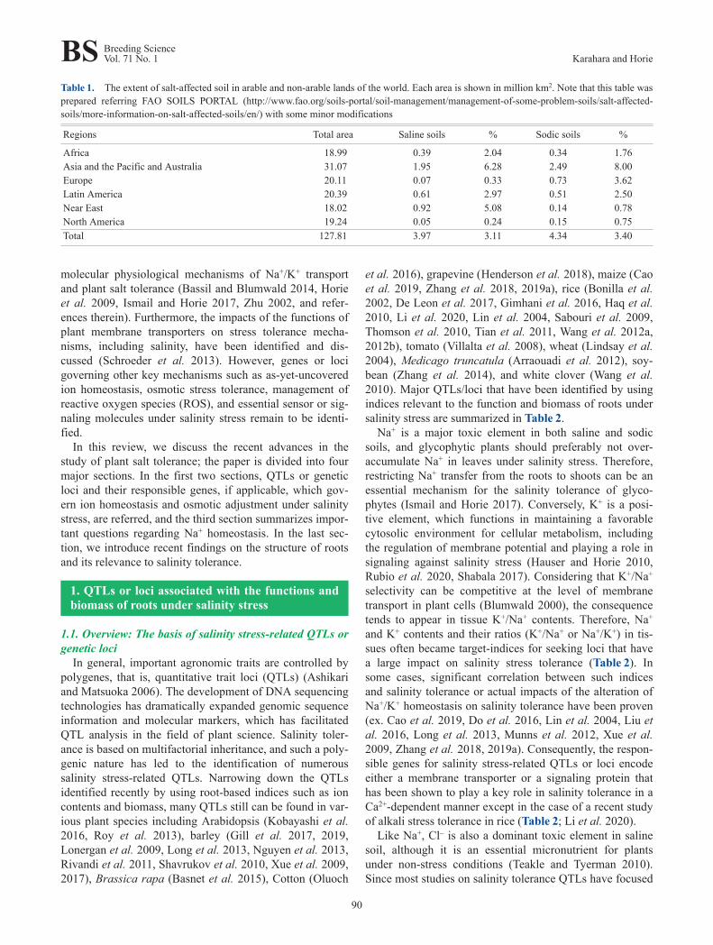
Table 1. The extent of salt-affected soil in arable and non-arable lands of the world. Each area is shown in million km2. Note that this table wasprepared referring FAO SOILS PORTAL (http://www.fao.org/soils-portal/soil-management/management-of-some-problem-soils/salt-affected-soils/more-information-on-salt-affected-soils/en/) with some minor modifications
Regions Total area Saline soils % Sodic soils %
Africa 18.99 0.39 2.04 0.34 1.76Asia and the Pacific and Australia 31.07 1.95 6.28 2.49 8.00Europe 20.11 0.07 0.33 0.73 3.62Latin America 20.39 0.61 2.97 0.51 2.50Near East 18.02 0.92 5.08 0.14 0.78North America 19.24 0.05 0.24 0.15 0.75Total 127.81 3.97 3.11 4.34 3.40
BS Breeding ScienceVol. 71 No. 1 Karahara and Horie
90
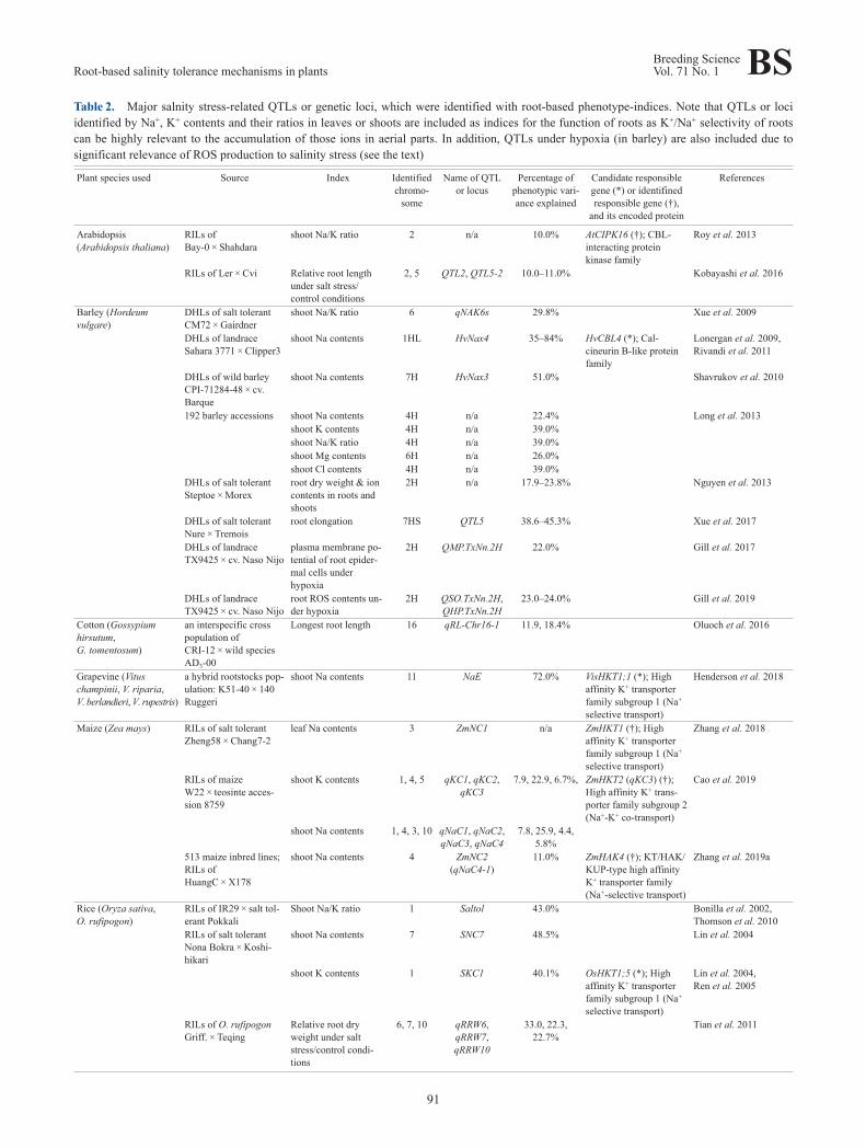
Table 2. Major salnity stress-related QTLs or genetic loci, which were identified with root-based phenotype-indices. Note that QTLs or lociidentified by Na+, K+ contents and their ratios in leaves or shoots are included as indices for the function of roots as K+/Na+ selectivity of rootscan be highly relevant to the accumulation of those ions in aerial parts. In addition, QTLs under hypoxia (in barley) are also included due tosignificant relevance of ROS production to salinity stress (see the text)
Plant species used Source Index Identifiedchromo‐
some
Name of QTLor locus
Percentage ofphenotypic vari‐ance explained
Candidate responsiblegene (*) or identifinedresponsible gene (†),
and its encoded protein
References
Arabidopsis(Arabidopsis thaliana)
RILs ofBay-0 × Shahdara
shoot Na/K ratio 2 n/a 10.0% AtCIPK16 (†); CBL-interacting proteinkinase family
Roy et al. 2013
RILs of Ler × Cvi Relative root lengthunder salt stress/control conditions
2, 5 QTL2, QTL5-2 10.0–11.0% Kobayashi et al. 2016
Barley (Hordeumvulgare)
DHLs of salt tolerantCM72 × Gairdner
shoot Na/K ratio 6 qNAK6s 29.8% Xue et al. 2009
DHLs of landraceSahara 3771 × Clipper3
shoot Na contents 1HL HvNax4 35–84% HvCBL4 (*); Cal‐cineurin B-like proteinfamily
Lonergan et al. 2009,Rivandi et al. 2011
DHLs of wild barleyCPI-71284-48 × cv.Barque
shoot Na contents 7H HvNax3 51.0% Shavrukov et al. 2010
192 barley accessions shoot Na contents 4H n/a 22.4% Long et al. 2013shoot K contents 4H n/a 39.0%shoot Na/K ratio 4H n/a 39.0%shoot Mg contents 6H n/a 26.0%shoot Cl contents 4H n/a 39.0%
DHLs of salt tolerantSteptoe × Morex
root dry weight & ioncontents in roots andshoots
2H n/a 17.9–23.8% Nguyen et al. 2013
DHLs of salt tolerantNure × Tremois
root elongation 7HS QTL5 38.6–45.3% Xue et al. 2017
DHLs of landraceTX9425 × cv. Naso Nijo
plasma membrane po‐tential of root epider‐mal cells underhypoxia
2H QMP.TxNn.2H 22.0% Gill et al. 2017
DHLs of landraceTX9425 × cv. Naso Nijo
root ROS contents un‐der hypoxia
2H QSO.TxNn.2H,QHP.TxNn.2H
23.0–24.0% Gill et al. 2019
Cotton (Gossypiumhirsutum,G. tomentosum)
an interspecific crosspopulation ofCRI-12 × wild speciesAD3-00
Longest root length 16 qRL-Chr16-1 11.9, 18.4% Oluoch et al. 2016
Grapevine (Vituschampinii, V. riparia,V. berlandieri, V. rupestris)
a hybrid rootstocks pop‐ulation: K51-40 × 140Ruggeri
shoot Na contents 11 NaE 72.0% VisHKT1;1 (*); Highaffinity K+ transporterfamily subgroup 1 (Na+
selective transport)
Henderson et al. 2018
Maize (Zea mays) RILs of salt tolerantZheng58 × Chang7-2
leaf Na contents 3 ZmNC1 n/a ZmHKT1 (†); Highaffinity K+ transporterfamily subgroup 1 (Na+
selective transport)
Zhang et al. 2018
RILs of maizeW22 × teosinte acces‐sion 8759
shoot K contents 1, 4, 5 qKC1, qKC2,qKC3
7.9, 22.9, 6.7%, ZmHKT2 (qKC3) (†);High affinity K+ trans‐porter family subgroup 2(Na+-K+ co-transport)
Cao et al. 2019
shoot Na contents 1, 4, 3, 10 qNaC1, qNaC2,qNaC3, qNaC4
7.8, 25.9, 4.4,5.8%
513 maize inbred lines;RILs ofHuangC × X178
shoot Na contents 4 ZmNC2(qNaC4-1)
11.0% ZmHAK4 (†); KT/HAK/KUP-type high affinityK+ transporter family(Na+-selective transport)
Zhang et al. 2019a
Rice (Oryza sativa,O. rufipogon)
RILs of IR29 × salt tol‐erant Pokkali
Shoot Na/K ratio 1 Saltol 43.0% Bonilla et al. 2002,Thomson et al. 2010
RILs of salt tolerantNona Bokra × Koshi‐hikari
shoot Na contents 7 SNC7 48.5% Lin et al. 2004
shoot K contents 1 SKC1 40.1% OsHKT1;5 (*); Highaffinity K+ transporterfamily subgroup 1 (Na+
selective transport)
Lin et al. 2004,Ren et al. 2005
RILs of O. rufipogonGriff. × Teqing
Relative root dryweight under saltstress/control condi‐tions
6, 7, 10 qRRW6,qRRW7,qRRW10
33.0, 22.3,22.7%
Tian et al. 2011
Root-based salinity tolerance mechanisms in plantsBreeding ScienceVol. 71 No. 1 BS
91
on Na+ but not Cl–, the information regarding QTLs linkedto Cl– is considerably less than that for Na+. Correlation ofCl– exclusion (i.e., low Cl– contents in leaves or shoots)with salinity tolerance was reported in several plant speciesincluding soybean, Hordeum marinum, and M. truncatula(Teakle and Tyerman 2010, and references therein). Inaddition, increased sensitivity to excessive amount of Cl–
was observed in some barley cultivars (Tavakkoli et al.2011). Analyses of barley accessions revealed a significantnegative correlation of Cl– content in the shoots and rootsto salinity tolerance and led to the identification of a QTLcontrolling the shoot Cl– content (Table 2; Long et al.2013). Thus, focusing on Cl–-related indices in the analysisof salinity QTLs might unravel novel mechanisms for Cl–
homeostasis during salinity stress.The number of QTLs based on root biomass under salin‐
ity stress are comparatively less and their responsible genesremain largely unknown (Table 2). A genome-wide associ‐ation study (GWAS) on the average relative root length ofmore than 200 natural A. thaliana accessions highlightedthe 10 most significant single nucleotide polymorphisms(SNPs) in two different NaCl conditions, which includedgenes of ATP-binding cassette B10 and vacuolar H+-ATPase subunit A isoform 2, implying the importance ofauxin efflux and regulation of vacuolar H+-ATPase on themaintenance of root growth under salinity stress(Kobayashi et al. 2016). Furthermore, a recent QTL map‐ping on the root length of rice under sodic alkali stress
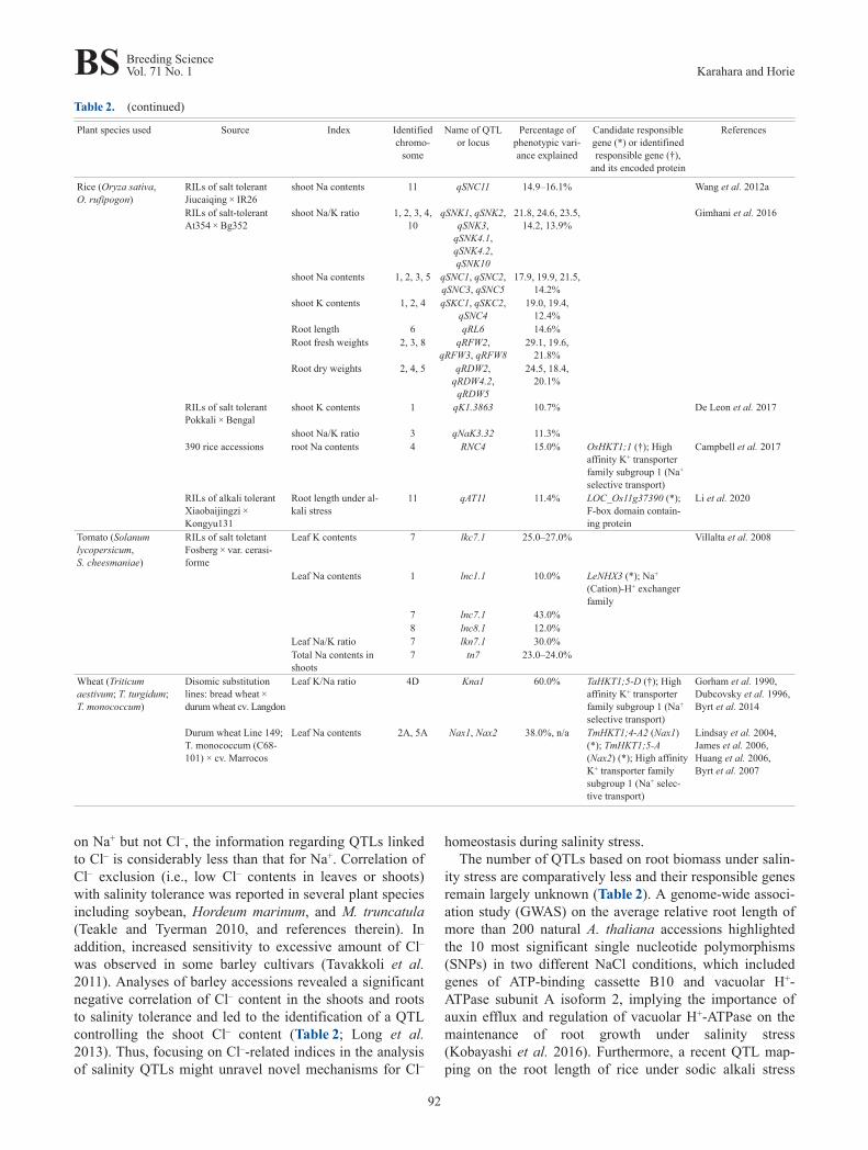
Table 2. (continued)
Plant species used Source Index Identifiedchromo‐
some
Name of QTLor locus
Percentage ofphenotypic vari‐ance explained
Candidate responsiblegene (*) or identifinedresponsible gene (†),
and its encoded protein
References
Rice (Oryza sativa,O. rufipogon)
RILs of salt tolerantJiucaiqing × IR26
shoot Na contents 11 qSNC11 14.9–16.1% Wang et al. 2012a
RILs of salt-tolerantAt354 × Bg352
shoot Na/K ratio 1, 2, 3, 4,10
qSNK1, qSNK2,qSNK3,
qSNK4.1,qSNK4.2,qSNK10
21.8, 24.6, 23.5,14.2, 13.9%
Gimhani et al. 2016
shoot Na contents 1, 2, 3, 5 qSNC1, qSNC2,qSNC3, qSNC5
17.9, 19.9, 21.5,14.2%
shoot K contents 1, 2, 4 qSKC1, qSKC2,qSNC4
19.0, 19.4,12.4%
Root length 6 qRL6 14.6%Root fresh weights 2, 3, 8 qRFW2,
qRFW3, qRFW829.1, 19.6,
21.8%Root dry weights 2, 4, 5 qRDW2,
qRDW4.2,qRDW5
24.5, 18.4,20.1%
RILs of salt tolerantPokkali × Bengal
shoot K contents 1 qK1.3863 10.7% De Leon et al. 2017
shoot Na/K ratio 3 qNaK3.32 11.3%390 rice accessions root Na contents 4 RNC4 15.0% OsHKT1;1 (†); High
affinity K+ transporterfamily subgroup 1 (Na+
selective transport)
Campbell et al. 2017
RILs of alkali tolerantXiaobaijingzi ×Kongyu131
Root length under al‐kali stress
11 qAT11 11.4% LOC_Os11g37390 (*);F-box domain contain‐ing protein
Li et al. 2020
Tomato (Solanumlycopersicum,S. cheesmaniae)
RILs of salt toletantFosberg × var. cerasi‐forme
Leaf K contents 7 lkc7.1 25.0–27.0% Villalta et al. 2008
Leaf Na contents 1 lnc1.1 10.0% LeNHX3 (*); Na+
(Cation)-H+ exchangerfamily
7 lnc7.1 43.0%8 lnc8.1 12.0%
Leaf Na/K ratio 7 lkn7.1 30.0%Total Na contents inshoots
7 tn7 23.0–24.0%
Wheat (Triticumaestivum; T. turgidum;T. monococcum)
Disomic substitutionlines: bread wheat ×durum wheat cv. Langdon
Leaf K/Na ratio 4D Kna1 60.0% TaHKT1;5-D (†); Highaffinity K+ transporterfamily subgroup 1 (Na+
selective transport)
Gorham et al. 1990,Dubcovsky et al. 1996,Byrt et al. 2014
Durum wheat Line 149;T. monococcum (C68-101) × cv. Marrocos
Leaf Na contents 2A, 5A Nax1, Nax2 38.0%, n/a TmHKT1;4-A2 (Nax1)(*); TmHKT1;5-A(Nax2) (*); High affinityK+ transporter familysubgroup 1 (Na+ selec‐tive transport)
Lindsay et al. 2004,James et al. 2006,Huang et al. 2006,Byrt et al. 2007
BS Breeding ScienceVol. 71 No. 1 Karahara and Horie
92
identified a strong QTL qAT11 on chromosome 11(Table 2; Li et al. 2020). Subsequent linkage mapping andGWAS led to the narrowing down of the candidates to threegenes and, among which the most significant candidate wasLOC_Os11g37390, which encodes an F-box domain con‐taining protein (Table 2; Li et al. 2020). Unlike the QTLsgoverning ion accumulation in tissues, one can expect moreto isolate key genes that contribute to hyperosmotic stresstolerance from root biomass-based salinity QTLs. In fact,as salinity-induced osmotic stress impairs water uptake anddistribution (Horie et al. 2012, Munns and Tester 2008), thecoping mechanisms can be partly expected to overlap withthose for drought stress tolerance. Therefore, to gain insightinto the coping mechanism of salinity-triggered osmoticstress, investigating the effect of QTLs for drought stresstolerance under salinity stress might be an effective strat‐egy. Indeed, some QTLs for drought stress tolerance inpearl millet were shown to confer salinity stress toleranceas well (Sharma et al. 2011, 2014). To promote detection ofhidden QTLs for osmotic adjustments under salinity, devel‐oping novel approaches such as a high-throughput imagingtechnology might be a key (Al-Tamimi et al. 2016). Yichieet al. (2018) successfully assessed the growth and water-use efficiency of wild rice accessions under salinity stressby using high-throughput imaging and phenotyping, which,for example, could have a potential to be extended to thescreening of osmotic QTLs.
1.2. Interaction of soil waterlogging-related QTLs andsalinity tolerance
Recent studies indicated the detection of QTLs that arehighly correlated with hypoxia tolerance (Table 2; Gillet al. 2017, 2019). Hypoxia is often caused by soilwaterlogged conditions and triggers a reduction in ATPproduction through respiration in root cells. Therefore,waterlogging stress damages roots and eventually impairsthe supply of water and nutrients from the roots to shoots.A significant reduction in ATP production has a largeimpact on broad cell metabolism, including membranepotential and pH homeostasis, by decreasing the activity ofH+-ATPase at the plasma membrane (Gill et al. 2017).Oxygen-deprived conditions facilitate the generation ofROS, which further reduce the viability of plants by dam‐aging biological molecules and reducing enzymatic activity(Bailey-Serres and Chang 2005). Salinity stress is known toinduce ROS, and the detoxification of ROS is one of thecritical mechanisms for salinity tolerance (Bose et al.2014). Waterlogging stress concurrently occurs with salin‐ity stress and salinity-induced damages tend to be exacer‐bated in combination with waterlogging (Ma et al. 2015).Indeed, a combination of salinity and waterlogging causedgreater damage on barley plants than with salinity stressalone (Ma et al. 2015). By using a double haploid (DH)population of salt-tolerant and salt-sensitive cultivars, Maet al. (2015) identified salinity tolerance QTLs at chromo‐somes 2H and 5H under salinity stress with or without
waterlogging. The same research group later applied dis‐tinct methods to identify QTLs related to salinity andwaterlogging stress in barley. In one method, a positiverelationship was found between the maintenance of highlynegative membrane potential (MP) and tolerance of theplant to salinity and waterlogging stress, based on which amajor QTL maintaining higher MP in root epidermal cellsunder hypoxia has been detected using a barley DH popula‐tion (Table 2; Gill et al. 2017). Subsequently, in anothermethod, the same QTL identified in the aforementionedMP-based screening was detected by measuring ROS con‐tent in the roots of the same barley DH population (Table 2;Gill et al. 2019). Interestingly, these QTLs were found tobe located at chromosome 2H, which was deduced to be thesame locus as that for the QTLs at chromosome 2Hreported by Ma et al. (2015). These findings reinstate thelink between salinity and waterlogging stress. Wang et al.(2019) recently proposed novel root-based high-throughputphenotyping methods to screen germplasm for oxidativestress tolerance, which can be expected to be utilized foridentifying salinity tolerance QTLs in different germplasmas well as different plant species.
2. Essential root-based mechanisms contributingto ion homeostasis and salinity tolerance
2.1. Maintenance of Na+ exclusion and K+ accumulationin shoots, mediated by HKT and HAK genes
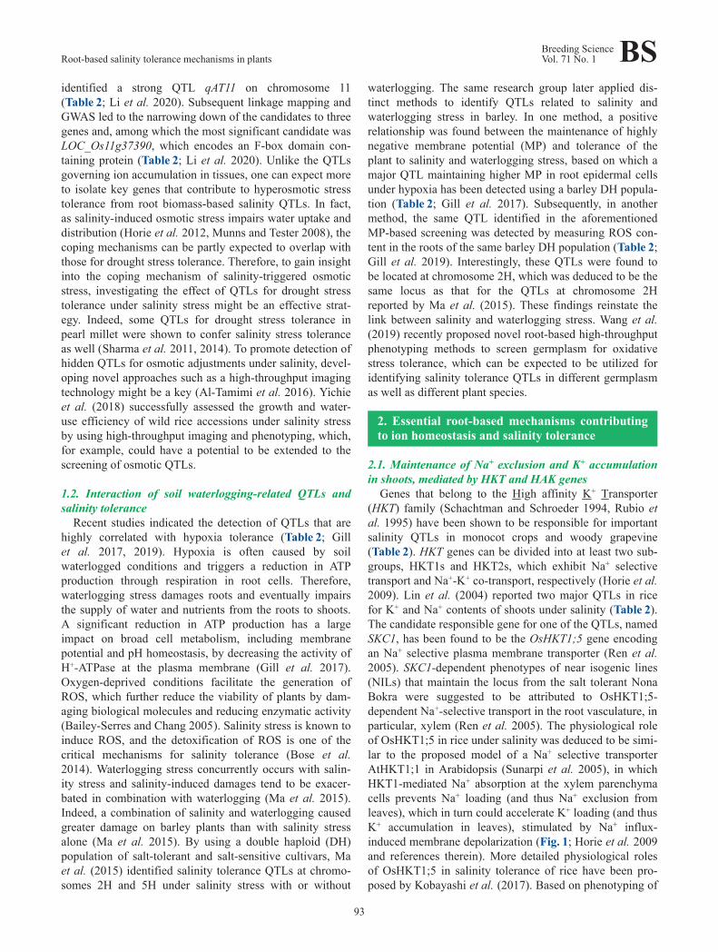
Genes that belong to the High affinity K+ Transporter(HKT) family (Schachtman and Schroeder 1994, Rubio etal. 1995) have been shown to be responsible for importantsalinity QTLs in monocot crops and woody grapevine(Table 2). HKT genes can be divided into at least two sub‐groups, HKT1s and HKT2s, which exhibit Na+ selectivetransport and Na+-K+ co-transport, respectively (Horie et al.2009). Lin et al. (2004) reported two major QTLs in ricefor K+ and Na+ contents of shoots under salinity (Table 2).The candidate responsible gene for one of the QTLs, namedSKC1, has been found to be the OsHKT1;5 gene encodingan Na+ selective plasma membrane transporter (Ren et al.2005). SKC1-dependent phenotypes of near isogenic lines(NILs) that maintain the locus from the salt tolerant NonaBokra were suggested to be attributed to OsHKT1;5-dependent Na+-selective transport in the root vasculature, inparticular, xylem (Ren et al. 2005). The physiological roleof OsHKT1;5 in rice under salinity was deduced to be simi‐lar to the proposed model of a Na+ selective transporterAtHKT1;1 in Arabidopsis (Sunarpi et al. 2005), in whichHKT1-mediated Na+ absorption at the xylem parenchymacells prevents Na+ loading (and thus Na+ exclusion fromleaves), which in turn could accelerate K+ loading (and thusK+ accumulation in leaves), stimulated by Na+ influx-induced membrane depolarization (Fig. 1; Horie et al. 2009and references therein). More detailed physiological rolesof OsHKT1;5 in salinity tolerance of rice have been pro‐posed by Kobayashi et al. (2017). Based on phenotyping of
Root-based salinity tolerance mechanisms in plantsBreeding ScienceVol. 71 No. 1 BS
93

stable OsHKT1;5 mutants and immuno-staining of rice tis‐sues, OsHKT1;5 was proposed to function in Na+ exclusionfrom leaves by mediating Na+ unloading from xylem ves‐sels in not only roots but also leaf sheaths, with a novelunexpected function in Na+ unloading from the phloem inbasal nodes (Fig. 1; Kobayashi et al. 2017).
Comparing the proposed functions of OsHKT1;5 in rice,it is interesting to note that the Na+ unloading function ofOsHKT1;5 in roots and sheaths well overlaps with that ofTmHKT1;4-A2, the candidate responsible gene of the Nax1QTL in durum wheat (Table 2; Huang et al. 2006, James etal. 2006, Lindsay et al. 2004). Interestingly, another impor‐tant QTL, Nax2, was also identified in durum wheat andfound to encode TmHKT1;5-A, which functions in Na+
unloading from the xylem only in roots (Table 2; Byrt et al.2007, James et al. 2006). The Nax2 locus was suggested tobe homoeologous to the Kna1 locus that has long beenknown to harbor a key gene controlling leaf Na+ exclusionand a high K+/Na+ ratio in the leaves of bread wheat undersalinity stress (Table 2; Byrt et al. 2007). Byrt et al. (2014)showed that TaHKT1;5-D, the product of which showsNa+-selective transport as TmHKT1;5-A, is most likely tobe the causal gene of Kna1 (Table 2). In fact, the Nax2-mediated leaf Na+ exclusion trait was shown to confer
salinity tolerance with a significant increase in yield to adurum wheat cultivar at the level of salt-affected fieldassessment (Munns et al. 2012).
Rice also has the OsHKT1;4 gene that encodes a plasmamembrane-localized Na+ selective transporter (Suzuki et al.2016). The expression of OsHKT1;4 in roots was relativelyweak, and reduction in OsHKT1;4 expression via RNAi didnot disturb both Na+ and K+ accumulation in any tissues ofa japonica rice cultivar in the vegetative growth stage(Campbell et al. 2017, Suzuki et al. 2016). However, arecent study on transgenic rice lines expressing artificialmicroRNA indicated the involvement of OsHKT1;4 in Na+
unloading from the xylem in roots under a wide range ofexternal Na+ concentrations (Khan et al. 2020). Furtherstudies are required to completely understand the functionand role of OsHKT1;4 in rice.
The mechanism of HKT1-mediated Na+ exclusion undersalinity stress in monocot plants shows a more complicatedview. A genetic study using diverse rice accessions led tothe discovery of a novel locus named RCN4 that governsroot Na+ contents and root Na+/K+ ratios under salinitystress (Table 2; Campbell et al. 2017). The causal gene forthe RCN4 locus was concluded to be OsHKT1;1, whichencodes an inward-rectified Na+ selective transporter
Fig. 1. Schematic drawings of OsHKT1-mediated Na+ exclusion in rice (The rice model). OsHKT1;5 was proposed to function in Na+ unload‐ing from the xylem in roots and leaf sheaths and from the phloem in basal nodes at the vegetative growth stage under salinity stress. Note thatthe phloem of the tissue in basal nodes where OsHKT1;5 functions appear to have an unusual structure: phloem companion cells next to sievetubes are lacking, but phloem parenchyma cells are retained (Kobayashi et al. 2017). A suggested function of OsHKT1;1-mediated Na+ recircu‐lation needs to be elucidated.
BS Breeding ScienceVol. 71 No. 1 Karahara and Horie
94
(Campbell et al. 2017). In fact, the involvement ofOsHKT1;1 in Na+ exclusion of rice under salinity has alsobeen reported by independent groups (Takagi et al. 2015,Wang et al. 2015). According to phenotype assessment ofthe oshkt1;1 mutant plant, OsHKT1;1 was suggested tomediate “Na+ recirculation from shoots to roots” via thephloem to exclude Na+ from leaves (Fig. 1; Wang et al.2015). Notably, HKT1-mediated Na+ recirculation has beenproposed in the AtHKT1;1-mediated salt tolerance mecha‐nism in Arabidopsis (Hauser and Horie 2010 and referencestherein). In barley, a molecular physiological study showedthat HvHKT1;1-mediated Na+ selective transport con‐tributes to the lowering of Na+ accumulation in both rootsand leaves of a Tibetan wild barley accession (Han et al.2018). In contrast, the role of the HKT1;5 transporter insalinity tolerance of barley plants is yet to be determined ascontrasting results have been reported (Huang et al. 2020,van Bezouw et al. 2019).
Studies of salinity tolerance in maize (Zea mays) also ledto the identification of important genes. A QTL namedZmNC1 was discovered by leaf Na+ content analysis of arecombinant inbred line (RIL; Table 2; Zhang et al. 2018).The ZmHKT1 gene was found to be localized within theQTL (Zhang et al. 2018). Phenotyping of CRISPR-Cas9-derived transgenic maize (ZmHKT1Crispr) mutants revealedthat ZmHKT1 mediates Na+ unloading from the xylem inroot stelar cells as other HKT1 transporters such asAtHKT1;1 and OsHKT1;5 (Fig. 1A; Horie et al. 2009).Moreover, an independent salinity QTL study using RILsof teosinte and maize identified several QTLs related toNa+ and K+ contents in shoots (Table 2; Cao et al. 2019).One of the QTLs, qKC3, was determined to be theZmHKT2 gene that encoded a Na+ and K+ co-transporterbased on the assessments of ZmHKT2Crispr mutants (Cao etal. 2019). In this case, the proposed mechanism is uniquesuch that the maize-derived ZmHKT2, which exhibits sig‐nificant reduction in the K+-transport activity comparedwith that from teosinte, unloads less K+ from xylem undersalinity stress, which accounts for an increase in the K+
content in the xylem sap and thus in leaves (Cao et al.2019). More recently, the same research group identifiedthe ZmNC2 locus by conducting a GWAS for Na+ contentusing 513 maize inbred lines (Zhang et al. 2019a). Thecausal gene for the ZmNC2 locus was deduced to encode aKT/HAK/KUP-type high affinity K+ transporter, ZmHAK4,which appears to function as a Na+-selective transporterunder salinity stress (Zhang et al. 2019a). Based on thephenotypes of ZmHAK4Crispr mutants and its expression inroot stelar cells, the authors proposed a model thatZmHAK4 mediates Na+ unloading to reduce Na+ accumu‐lation in the leaves of maize under salinity stress (Zhang etal. 2019a), similar to the proposed function of HKT1 trans‐porters in the root xylem (Fig. 1). These findings suggestthat Na+ unloading in roots is mediated by multiple trans‐porter families to circumvent the triggering of Na+ toxicityin leaves.
2.2. The Saltol QTL-mediated salt tolerance and its inter‐action with the SKC1 QTL (OsHKT1;5) in rice
A major QTL associated with salt tolerance of riceseedlings, named Saltol, has been identified on chromo‐some 1 by using RILs derived from salt-tolerant Pokkaliand salt-sensitive IR29 (Table 2; Bonilla et al. 2002,Thomson et al. 2010). This QTL had a major influence onNa+ exclusion from rice leaves. One highly salt-tolerantRIL (FL478) containing the Saltol QTL contributed toincreased salt tolerance of some cultivars as a donor inbreeding programs (for more details, see review by Ismailand Horie 2017). On the other hand, the effect of the SaltolQTL was found to be “not straightforward” (Thomson et al.2010), and its responsible gene(s) remain to be determined.In fact, by using a different rice RIL population, Haq et al.(2010) identified QTLs for Na+ contents and K+/Na+ ratioin shoots under salinity stress in an overlapping region withthe Saltol QTL. However, two QTL clusters were detectedin the region with one more relatively minor QTL immedi‐ately adjacent to the overlapping genomic region. Further‐more, transcriptome analysis based on the Affymetrix ricegenome array performed using FL478 and IR29 suggestedthat the source of the core of the Saltol QTL in FL478appears to be derived from the sensitive IR29 (Walia et al.2005). Note that the Saltol QTL includes the above-mentioned SKC1 (OsHKT1;5) locus and this gene isexpected to be at least one of the causal genes. However, arecent study on NIL-SKC1 plants indicated that the NILplants showed increased sensitivity to moderate salinity of80 mM NaCl with unexpected overexpression ofOsHKT1;5 in roots (Al Nayef et al. 2020). Furthermore,electrophysiological experiments using the microelectrodeion flux estimation (MIFE) technique indicated substantialreduction in the activity of Na+ reabsorption (i.e., Na+
unloading) in the xylem of NIL-SKC1 (Al Nayef et al.2020). These results altogether imply the involvement andcomplex interactions of several different genes, includingOsHKT1;5, in the mechanism of salinity tolerance medi‐ated by the QTLs, and further research is needed to eluci‐date the underlying molecular mechanisms.
2.3. Salinity QTLs in soybean and responsible genesHKT1-mediated Na+ exclusion in roots was shown to be
an important salinity tolerance mechanism even in woodygrapevine based on the analysis of the NaE QTL identifiedusing a hybrid rootstock population (Table 2; Henderson etal. 2018). However, the tolerance mechanism against salin‐ity so far looks distinct in the dicot soybean. Many salinityQTLs, including a major QTL for sodic-alkaline stress,have been identified by assessing the degree of toleranceand measuring the leaf chlorophyll content (Zhang et al.2019b and references therein, Tuyen et al. 2010). A majorQTL for salinity stress was repeatedly detected on chromo‐some 3 (Guan et al. 2014, and references therein). Guan etal. (2014) isolated the causal gene by map-based cloning,and it was found to encode a cation/H+ exchanger family
Root-based salinity tolerance mechanisms in plantsBreeding ScienceVol. 71 No. 1 BS
95
member protein, Glyma03g32900 and was namedGmSALT3 (for Glycine max salt tolerance-associated geneon chromosome 3). The expression of GmSALT3 in theendoplasmic reticulum membrane of roots was shown to behighly associated with Na+ exclusion from the shoots andgreater salt tolerance (Guan et al. 2014). Independent stud‐ies based on whole-genome de novo sequencing and a map-based cloning strategy have also isolated the same gene asthe causal gene (GmCHX1 and Ncl, respectively; Qi et al.2014, Do et al. 2016). Later GmSALT3 was shown to con‐tribute to Cl– exclusion from leaves as well as Na+ exclu‐sion (Do et al. 2016, Liu et al. 2016) and improve soybeanyield (higher seed weights) under saline field conditions(Liu et al. 2016). Detailed transport and exclusion mecha‐nisms mediated by GmSALT3/GmCHX1/Ncl have not yetbeen elucidated.
Recently, a new QTL named qST8 for the sensitivity tosalinity stress at the soybean germination stage was mappedon chromosome 8 (Zhang et al. 2019b). In combinationwith a GWAS analysis, the candidate responsible gene wasidentified (Glyma08g102000) and found to encode a cationdiffusion facilitator (CDF) family protein (GmCDF1).Transgenic hairy roots and haplotype analyses indicatedthat the function of GmCDF1 is negatively correlated withsalinity tolerance of soybean (Zhang et al. 2019b).
2.4. Important factors in signaling under salinity stressThe importance of a CIPK (CBL-interacting protein
kinase)-CBL (Calcineurin B-like Ca2+ sensor protein) com‐plex in the mechanism of plant salt tolerance has beenestablished by studies of the regulation on SOS1 (plasmamembrane Na+-H+ antiporter) by the SOS2 (CIPK24)-SOS3 (CBL4) complex in Arabidopsis (Zhu 2002). Evi‐dence of the involvement of a CIPK-CBL complex inresistant mechanisms to various biotic or abiotic stress hasbeen provided (Ma et al. 2020). A CIPK protein and a CBLCa2+ sensor were identified as candidate responsible genesfor the QTLs that control Na+/K+ homeostasis inArabidopsis and barley, respectively (Table 2). An impactof a key signaling molecule on plant salt tolerance andbreeding of tolerant cultivars has also been highlighted byTakagi et al. (2015). A salt-tolerant japonica rice mutanthst1, derived from an ethylmethanesulfonate (EMS)-mutagenized population, harbored a single recessive muta‐tion in the gene Os06g0183100, which encodes a B-typeresponse regulator OsRR22 (Takagi et al. 2015). OsRR22expression was shown to complement a loss-of-functionmutant of the Arabidopsis type-B RR family, which sug‐gested the involvement of OsRR22 in cytokinin signaling(Tsai et al. 2012). The OsHKT1;1 gene was identified asone of the highly expressed genes in the hst1 mutant incomparison with wild-type (Takagi et al. 2015). These find‐ings imply an important role of cytokinin in the regulationof Na+ homeostasis and salinity tolerance of rice. Recently,a salt-responsive Ca2+ sensor calmodulin in barley,HvCaM1 was shown to be a negative regulator of salt toler‐
ance in barley plants (Shen et al. 2020). RNAi-mediatedHvCaM1 knockdown rendered the barley lines more salttolerant with lower Na+ accumulation in shoots and higherexpression of the HvHKT1;1 gene, suggesting that, at leastin part, upregulated HvHKT1;1 conferred salt tolerance tothe transgenic lines (Shen et al. 2020).
3. Important questions remain to be elucidatedfor understanding the physiological functions ofroot under salinity stress
3.1. Na+ influx mediated by non-selective cation channelsand possible involvement of plasma membrane intrinsicproteins
Several important salt tolerance mechanisms and keygenes involved have been discovered over the past fewdecades. Nevertheless, some more questions in relation toNa+ homeostasis remain to be addressed. One of them is theplasma membrane protein which can be a major entry forNa+ under salinity stress. Previous electrophysiologicalstudies have implicated that non-selective cation channels(NSCCs) are likely to be the primary candidates that medi‐ate Na+ influx into roots subjected to salinity stress (seeDemidchik and Maathuis 2007). However, the molecularidentity of NSCCs still remains elusive. Cyclic nucleotidegated channels and glutamate receptors are thought to bepotential candidates (Ismail and Horie 2017, and referencestherein), but crucial evidence for the molecular identity islacking. Recently, water channels called plasma membraneintrinsic protein (PIP) in Arabidopsis, AtPIP2;1 andAtPIP2;2, were reported to show ion channel activity forNa+ and K+ when the external Ca2+ concentration is low(Byrt et al. 2017, Kourghi et al. 2017). Given the highlyabundant nature of the protein and the sensitivity ofAtPIP2;1-mediated Na+ channel activity in Xenopus laevisoocytes to low pH and external Ca2+ concentration, whichwas found to be similar to the features of Na+ currentsmediated by NSCCs in root protoplasts from Arabidopsis,AtPIP2;1 was considered as a candidate for NSCCs in plantroots (Byrt et al. 2017, McGaughey et al. 2018). Morerecently, like AtPIP2;1, HvPIP2;8 in barley was shown toexhibit ion channel activity for Na+ and K+ in a Ca2+ sensi‐tive manner (Tran et al. 2020). In addition, OsPIP1;3 in ricewas shown to mediate the transport of nitrate anions inmammalian HEK293 cells in addition to being a waterchannel (Liu et al. 2020). Whether PIP aquaporins fromdifferent plant species also show dual ion and water perme‐ability and whether these features are reminiscent of thenature of NSCCs in plant cells need to be elucidated in thefuture research.
3.2. Does futile Na+ cycling or rapid transmembrane Na+
cycling occur across the plasma membrane?When subjected to soil salinity, plants need to pay signif‐
icant energy costs for osmotic and ionic adjustments, whichhas a large influence on crop production in saline or sodic
BS Breeding ScienceVol. 71 No. 1 Karahara and Horie
96

soils (Munns and Gilliham 2015). Energy costs associatedwith plant salinity tolerance mechanisms have recentlybeen highlighted with extensive reviews (ex. See Munnset al. 2020a, 2020b, Shabala et al. 2020, Tyerman et al.2019).
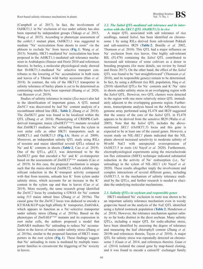
Under salinity stress, it is thought that a large amount ofNa+ rapidly and passively enters the epidermal and cortexcells of plant roots. Cytosolic Na+ concentrations are main‐tained low by extruding Na+ ions out of the cells across theplasma membrane against the gradient of electrochemicalpotential and by sequestering Na+ ions into vacuoles. Theextrusion of Na+ is exclusively carried out by the plasmamembrane transporter SOS1 owing to the electrochemicalH+ gradient (Zhu 2002), which is established by the H+-pump ATPase by using the energy from ATP hydrolysis(Fig. 2A). Previous studies have shown that Na+ efflux/influx ratios across the plasma membrane increase in accor‐dance with an increase in the external Na+ concentrationand eventually get to nearly 1, suggesting that the cyclingof Na+ across the plasma membrane is driven using a largeamount of energy (for details of the energy cost, see Munnset al. 2020a). These processes look at a glance futile andenergetically costly (Fig. 2A; Britto and Kronzucker 2006,2015). Such futile Na+ cycling is referred to as “RapidTransmembrane Sodium Cycling (RTSC)” (Britto andKronzucker 2015). The 24Na+ tracer experiments performedusing a salt-tolerant variety of rice Pokkali and a sensitivecultivar IR29 indicated that unidirectional influx and effluxof Na+ in IR29 were more than 4 times higher than those inPokkali at the external Na+ concentration of 25 mM(Malagoli et al. 2008). Furthermore, the estimation of therespiratory oxygen consumption rate at 25 mM Na+
revealed that the amount of O2 consumed in the roots ofIR29, but not of Pokkali, is insufficient for driving theactive Na+ efflux via the Na+/H+ antiporters at the plasmamembrane (Britto and Kronzucker 2015, Malagoli et al.2008). These phenomena lead to an assumption that anunknown transport mechanism might function in the rootNa+ extrusion. This mechanism would be coupled with thepassive fluxes of some ion and/or vesicular transport of Na+
(Fig. 2A; Flowers et al. 2019, Malagoli et al. 2008).Revealing the detailed mechanism of Na+ efflux out of theroot under salinity stress as well as understanding howplants manage the cost for this process are crucial questionsto be addressed.
3.3. The problem of energy cost for futile sodium leak invacuoles
The Na+/H+ antiport activity across the tonoplast viatransporters such as NHX-type cation/H+ antiporters is cru‐cial for plant salt tolerance since it promotes Na+ sequestra‐tion into vacuoles as has been extensively investigated andreviewed (e.g., see Bassil and Blumwald 2014, and refer‐ences therein, Bassil et al. 2019). A salinity QTL analysisperformed using tomato detected the lnc1.1 locus, thecausal gene of which was deduced to be LeNHX3, which
controls Na+ contents in leaves (Table 2; Villalta et al.2008). This result might imply that Na+ sequestration inroot cells could have a robust impact on the overall salt tol‐erance. Recent findings in barley support this possibility, inwhich a combination of techniques of electrophysiologicalMIFE and confocal fluorescence dye imaging on the
Fig. 2. Schematic drawings of the energy-consuming Na+ transportmechanisms in plants subjected to salinity stress, which need to beelucidated. (A) A hypothesis of futile Na+ cycling in the root epider‐mal cells under salinity stress, which is called Rapid TransmembraneSodium Cycling. “X” represents an unknown transporter(s) or mecha‐nisms such as vesicular transport, which mediate rapid Na+ efflux. (B)A futile Na+ leak from vacuoles via non-selective vacuolar channels,called slow-activating (SVc) and fast-activating (FVc) channels. PMrepresents plasma membrane.
Root-based salinity tolerance mechanisms in plantsBreeding ScienceVol. 71 No. 1 BS
97
assessment of barley accessions revealed a positive correla‐tion of the ability for root vacuolar Na+ sequestration, butnot for Na+ extrusion from roots, with the overall salt toler‐ance (Wu et al. 2019). There is, however, another problemof futile Na+ cycling in the mechanism of vacuolar Na+
sequestration and energy costs (see Shabala et al. 2020, andreferences therein). In brief, Na+ retention in vacuoles is notstable as two types of Na+-permeable non-selective vacuo‐lar ion channels, called “slow-activating (SV) channels”and “fast-activating (FV) channels”, mediate Na+ extrusionfrom the inside of vacuoles to the cytosol, that is, “back-leak of Na+ into cytosol” (Fig. 2B; Shabala et al. 2020).Since the Na+/H+ antiport activity across the tonoplastlargely depends on the activity of V-ATPase that hydrolyzesATP for H+ pumping (Fig. 2B), the futile cycle of “Na+
sequestration-Na+ back-leak” across the tonoplast undersalinity conditions can be very costly: According to themodel calculations, plants would need to retain only a verysmall number of SV channels open (0.1%) to avoid thefutile cycle (Shabala et al. 2020). However, electrophysio‐logical studies on SV and FV channels in halophytes haveprovided evidence that halophytes retain traits to signifi‐cantly reduce the number of open SV and FV channelsunder salinity stress (Bonales-Alatorre et al. 2013a, 2013b).The two-pore channel 1 (TPC1) protein appears to functionas an SV channel by forming two TPC protein subunits,which mainly mediate Ca2+ and Na+ transport (Guo et al.2017). The estimated number of TPCs in halophytes wasfound to be similar as that in glycophytes (Shabala et al.2020). Therefore, understanding the regulatory mechanismon the gating of TPC1 (SV) and FV channels as well asdetermining the molecular identity of the FV channel areprimary subjects to be elucidated. Such information wouldform the basis for breeding salt tolerant crops.
4. Development of the structural features of rootsin response to salinity stress
We mainly focused on advances in this field of researchover the last decade since we published a relevant review(Horie et al. 2012). Since then, omics and other emergingtechnologies have been introduced in this field, andremarkable progresses have been made. Herein, topicsrelated to structural or anatomical aspect are mainlyfocused, and relevant reviews are also introduced. First, webegin with the endodermis that plays an important role asan apoplastic barrier in younger parts of roots both in eudi‐cots and monocots.
4.1. Molecular mechanisms of endodermal Casparianstrip development
Among issues related to the endodermis, in the pastdecade, remarkable progress has been achieved for theCasparian strip research at the molecular level by usingArabidopsis. Attention has been paid by not only basicplant scientists but also cell biologists. Excellent reviews
dealing with these achievements have also been published.Nevertheless, we have briefly summarized the recentprogress regarding this topic while referring to some previ‐ous related, but uncommon studies.
In Arabidopsis, starting from endodermal differentiation,the cells at the cortex or endodermis initially undergoasymmetrical formative division to regenerate themselvesand produce daughter cells at the tip of the root having asimple structural organization. The daughter cells divide toproduce the progenitors of endodermal cells and of normalcortical cells (Liberman et al. 2015). Transcriptional factorsregulating the initial daughter cell division of the cortex orendodermis and those regulating endodermal cell-fate spec‐ification have been identified. SHORTROOT (SHR)expressed in the stele moves into the daughter cells and theendodermis and induces the expression of SCARECROW(SCR) (Nakajima et al. 2001). Next, SCR directly activatesthe expression of a transcription factor, MYB DOMAINPROTEIN 36 (MYB36; Liberman et al. 2015), which is themaster regulator of Casparian strip formation (Kamiyaet al. 2015).
The formation of Casparian strip is initiated by the local‐ization of CASPARIAN STRIP DOMAIN PROTEINs(CASPs) at the site of the Casparian strip (Casparian stripdomain, CSD; Roppolo et al. 2011). CASPs then recruitperoxidase 64 (PER64)—a respiratory burst oxidasehomolog F—and a dirigent-like protein Enhanced Suberin1 (ESB1), to polymerize lignin precursors (Kamiya et al.2015). A receptor-like kinase protein SCHENGEN3(SGN3/GASSHO1) has been shown to be necessary forlocalizing CASPs into an uninterrupted, ring-like domain(Pfister et al. 2014). Peptides named Casparian stripIntegrity Factors 1 and 2 (CIF1 and CIF2, respectively),which are expressed in the stele, specifically bind SGN3(Doblas et al. 2017b, Nakayama et al. 2017), mediating thesurveillance of root apoplastic barrier integrity (Doblas etal. 2017a). For more information on the recent Casparianstrip research, please refer to more detailed reviews by theauthors of the abovementioned publications, that is, byGeldner (2013) and Doblas et al. (2017a).
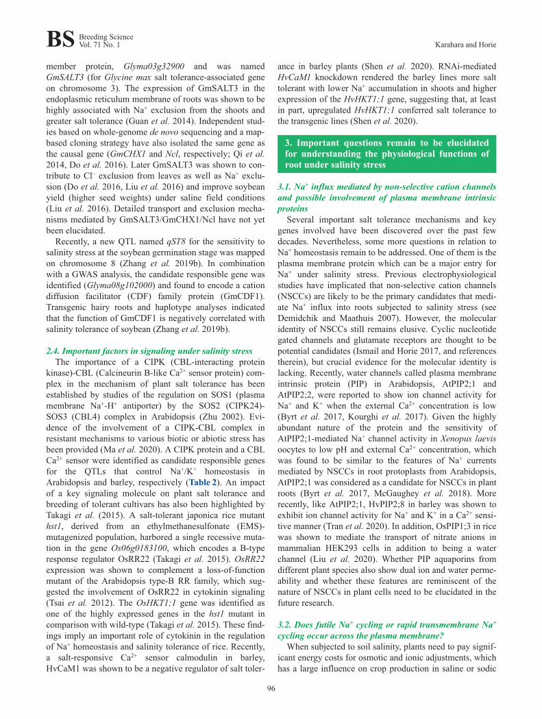
We would like to also present our viewpoint. A uniqueexperiment performed using surgical manipulation ofPisum sativum L. (pea) stems, affecting the physical envi‐ronment of endodermal cells, indicated that some positionalinformation is accumulated in the radial wall of endodermalcells that defines the future site of the formation of the stripand its width, i.e., CSD, at an early stage of endodermalcell development (Yokoyama and Karahara 2001). TheCasparian strip is occasionally, but naturally, observed intwo neighboring endodermal cells in the radial direction(Fig. 3). Considering this case, such positional informationhas been speculated to be accumulated even before the finalpericlinal division to form an endodermal cell. The sameexperimental system performed using surgical manipula‐tion also indicated the involvement of brefeldin A-sensitivesecretory transport for not only the modification of cell
BS Breeding ScienceVol. 71 No. 1 Karahara and Horie
98
wall but also the completion of the tight adhesion of theplasma membrane at the CSD, and that the former precedesthe latter (Karahara and Shibaoka 1998, Karahara 2012).
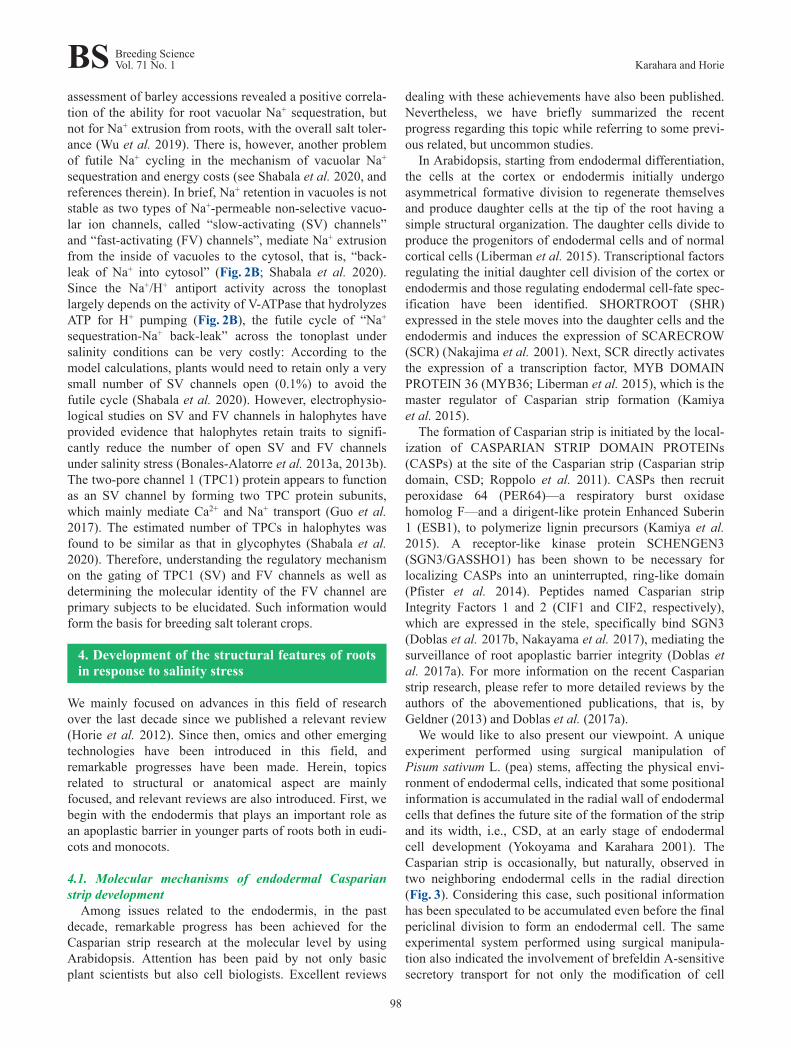
In fact, when the ultrastructure of endodermal cellsdeveloping Casparian strip was carefully observed in a pearoot, Golgi apparatuses and vesicles were found in thevicinity of the developing Casparian strip, and vesicles car‐rying electron dense materials were found to fuse with theplasma membrane at the CSD (Fig. 4; Karahara 1995). Theradial width of the Casparian strip, a morphological param‐eter that should be related to the effectiveness of the strip asa barrier, increased under salinity stress in Zea mays L.roots (Karahara et al. 2004).
A comparative study between salinity-sensitive IR24 andNipponbare and salinity-tolerant cultivars of rice NonaBokra and Pokkali showed that the Casparian strip matured
Fig. 3. The Casparian strip formed in two neighboring endodermalcells in the radial direction (arrows). A fluorescence cross-sectionalimage of a pea stem observed under UV light. Arrowheads: theCasparian strips formed in normal endodermal cells. This image isadapted from Karahara (2000).
Fig. 4. Ultrastructure of endodermal cells developing a Casparianstrip in a cross-section of a pea root cut at 15 mm from the root tip. Itappears as if the vesicles carrying electron-dense materials fuse withthe plasma membrane at CSD (arrows). G: Golgi apparatus. Bar,1 μm. This image is adapted from Karahara (1995).
closer to the root tip after high NaCl treatment in all culti‐vars than in the control (without NaCl treatment; Ferdoseet al. 2009). Determining whether salinity accelerated thedevelopment of the Casparian strip formation would beinteresting (Karahara et al. 2008). More interestingly, thedevelopment of the Casparian strip was more prominent inthe salinity-sensitive cultivars than in the salinity-tolerantcultivars, and the distance between the root tip to the lower‐most position of the endodermal Casparian strip was com‐paratively shorter in the salinity-sensitive cultivars than inthe salinity-tolerant ones. This result is consistent withthose obtained by the same group that Na+ exclusion acrossthe endodermis is more efficient in a sensitive cultivar thanin a resistant one (Tsuchiya et al. 1994). These results indi‐cate that the developmental regulation of the Casparianstrip responding to salinity stress might be more compli‐cated than was suggested elsewhere (Chen et al. 2011,Krishnamurthy et al. 2009).
Regarding the issue about the acceleration of the devel‐opment of the Casparian strip under salinity stress, salinitycan be thought to accelerate the formation of Casparianstrip when the distance from the root tip to the lowest posi‐tion of the strip decreases under salinity stress. However,because the distance from the root tip to the lowest positionof the Casparian strip depends on the cell division rate, cellelongation rate, and the time required for the formation ofthe band in individual cells, these factors need to be consid‐ered. This issue was tested in maize roots, and the esti‐mated time required for formation of the endodermalCasparian strip in an individual cell was found to not sub‐stantially change under salinity stress even when the dis‐tance from the root tip to the lowest position of the stripdecreased (Karahara et al. 2004). Furthermore, a uniqueintegrative method was established to monitor the changesin the developmental processes of a particular cell type inthe root, i.e., the rates of cell differentiation, production,and elongation (Karahara et al. 2008). A shortcoming ofthis method is that cell production rate needs to be mea‐sured, which is a laborious procedure, even solely to ana‐lyze cell differentiation. To overcome this difficulty, theauthors developed the unique “sandwich” method(Karahara et al. 2012), in which the roots are sandwichedbetween two different agar media, and are thereby unilater‐ally exposed to different environmental conditions. Byusing this method, the authors showed that the formation ofthe Casparian strip was not promoted under osmotic stress(Karahara et al. 2009).
4.2. Endodermal lignificationThe Casparian strip is formed in the, so called, state I
developmental stage of endodermal cells (Haas andCarothers 1975). One of the most interesting findings of theprevious apoplastic barrier research is that lignin polymerplays an important role in the function of the apoplasticbarrier of the Casparian strip in roots in state I endodermalcells (Naseer et al. 2012). This finding is surprising
Root-based salinity tolerance mechanisms in plantsBreeding ScienceVol. 71 No. 1 BS
99
because lignin itself has been considered to be morehydrophilic than suberin (Geldner 2013). Nonetheless, con‐sidering lignin as an apoplastic barrier component isbecoming important. Barbosa et al. (2019) even suggestedthat the Casparian strip can be used in lignin research.Lignin might be present in a specialized form in theCasparian strip (Nawrath et al. 2013). Conversely, whether,like in Arabidopsis, lignin solely functions as the apoplasticbarrier in other plant species needs to be determinedbecause the roots of Pisum sativum contain aliphaticsuberin besides lignin in the Casparian strip (Zeier et al.1999). Furthermore, the tight adhesion of the plasma mem‐brane to the CSD also needs to be considered as an impor‐tant feature of the apoplastic barrier of the Casparian strip(Karahara and Shibaoka 1992).
Recently, an interesting finding was reported thatLaccase3, which is involved in lignin monomer polymer‐ization, is indicated to provide possible positional informa‐tion for directing Casparian strip formation in Arabidopsisby pharmacological experiments (Zhuang et al. 2020). Thisfinding is surprising because an enzyme that acts at thefinal stage of lignin formation provides positional informa‐tion needed at the initial stage of the Casparian strip forma‐tion. However, further careful examination is needed forthis issue because peroxidases, rather than laccases, havebeen shown to be required in the Casparian strip lignifica‐tion by genetic experiments (Rojas-Murcia et al. 2020).
Regarding the relationship between lignin formation andsalinity stress, genes responsible for lignin biosynthesissuch as PHENYLALANINE AMMONIA LYASE, CAFFEICACID-O-METHYLTRANSFERASE, and CINNAMYLALCOHOL DEHYDROGENASE are upregulated in theroots of a loss-of-function rice mutant of a nuclear factorgene RICE SALT SENSITIVE3 (Toda et al. 2013a, 2013b),which shows severer reduction of root growth under salin‐ity stress than under normal conditions. This fact suggeststhat a complicated relationship exists between lignin forma‐tion and salinity stress when the recently revealed role oflignin in the apoplastic barrier at the Casparian strip is con‐sidered, although cell type specificity of the upregulatedexpression of these genes under salinity stress is still notknown.
4.3. Endodermal suberizationAfter the Casparian strip is developed, endodermal cell
walls deposit suberin, which is called state II of the endo‐dermal cells (Haas and Carothers 1975). The permeabilityof suberin to water and solutes has already been discussedwell in a review by Ranathunge et al. (2011b). Sincesuberin lamellae themselves are deposited between theplasma membrane and cell wall as a secondary wall com‐ponent, their role is to prevent the blocking of the apoplas‐tic diffusion between cells in the cell wall, but to limittransmembrane transport into cells at least in the state IIendodermal cells (Hosmani et al. 2013).
Although the mechanism regulating the transition from
state I to state II endodermal cell is still largely unknown, atranscription factor MYB41, the promoter of which is acti‐vated by abscisic acid (ABA) and salinity stress, is sug‐gested to be a candidate (Barberon 2017, Kosma et al.2014). In addition to transcription factors that positivelyregulate suberin synthesis, such as MYBs, some wererecently found to be negative regulators, such as ANAC46,which is expressed in the endodermis (Mahmood et al.2019).
In addition to transcription factors, an enzyme calleddocosanoic acid synthase, which is involved in the biosyn‐thesis of aliphatic suberin, has been identified in roots(Franke et al. 2009, Lee et al. 2009). Yadav et al. (2014)showed that the G subfamily of the ATP-binding cassette(ABCG) half-transporters are involved in the synthesis ofsuberin, and their gene expression is positively regulated byABA for endodermal barrier function in Arabidopsis roots.They also showed that a triple mutant of the genes of thesetransporters (abcg2-1, abcg6-1, and abcg20-1) still devel‐oped a functional Casparian strip. Regarding the regulationof suberization in response to environment changes, thedevelopment of endodermal suberin was shown to beinduced unilaterally in the root side exposed to the air or incontact with cadmium in maize (Líška et al. 2016), sug‐gesting the flexibility of suberization regulation.
The role of suberin in the function of a transport barrierwas elucidated by using modern biological tools such asmutants of suberin deposition, chemical analysis of suberincomposition, and ionome analysis. A combination of thesetools actually revealed that the enhanced suberin1 (esb1)mutant of Arabidopsis showed decreased accumulation ofCa, Mn, and Zn and increased accumulation of Na, S, K,As, Se, and Mo in the shoot (Baxter et al. 2009). They sug‐gested that the increase of Na concentration in the shoot ofthe esb1 mutant was attributed to a possible decrease in theapoplastic bypass flow for this element at low external Naconcentrations in the soil. Therefore, they also speculatedthat the esb1 mutant might be salinity tolerant when theexternal Na concentration is elevated. These suggestionsseem to be reasonable when facts that esb1 mutant showeddrought tolerance (Franke et al. 2012) and that rice culti‐vars having elevated suberin showed salinity tolerance(Krishnamurthy et al. 2009) are considered. Conversely,given that esb1 mutants form defective lignin-basedCasparian strips (Hosmani et al. 2013), another possiblecause, i.e., incomplete apoplastic barrier at the Casparianstrip, might be considered for the increase of Na concentra‐tion in the shoot of the esb1 mutants.
Recently, a transcriptional factor SUBERMAN(MYB39) was shown to positively regulate suberin lamel‐lae formation in root endodermal cells (Cohen et al. 2020).Interestingly, the rosette leaves of the SUBERMAN over‐expressor showed significant accumulation of elementssuch as Mg, P, S, K, Ca, Mn, and Fe, but not of Na (Cohenet al. 2020). However, the results of these two studies(Baxter et al. 2009, Cohen et al. 2020) were not concerning
BS Breeding ScienceVol. 71 No. 1 Karahara and Horie
100
Na, indicating that a careful evaluation about the differencein the degree, site, and timing of altered suberization inthese mutants is needed for the interpretation of theseresults. In addition, a combination of the manipulation ofsuberin deposition and ionome analysis raised the funda‐mental question of why the barrier function of suberin isdifferent between different elements.
4.4. ExodermisWhen the hypodermis, a cell layer beneath the root epi‐
dermis (rhizodermis), forms the Casparian strip, the celllayer is called exodermis (Peterson 1988). The exodermis isobserved in many species, including gramineous plantssuch as rice and maize. Apoplastic transport barriers inroots have been considered to exist even in the exodermis(Perumalla and Peterson 1986), and many studies havebeen providing evidence of this (Kreszies et al. 2018).
In the case of rice roots, a lignified sclerenchymatouscell layer develops beneath the exodermis in the outer partof the root (OPR). Since studies have been focusing on theimportance of lignin in the apoplastic barrier function ofthe endodermal Casparian strip, the contribution of scle‐renchyma to the barrier function of the OPR needs to befurther considered.
The barrier function of the exodermis is important as aradial oxygen loss (ROL) barrier (Shiono et al. 2014).When rice is grown in oxygen-deprived medium, the rootexodermis as a ROL barrier is strengthened to reduce oxy‐gen leakage from the root, and, in such roots, solute perme‐ability for NaCl was reduced (Ranathunge et al. 2011a),indicating that enhancement of the OPR is also effective asa barrier to NaCl.
In maize, interestingly, high salinity at 100 mM NaClinduced suberization over a large part of the cortex, but didnot notably influence the suberization of the endodermalcell wall (Shen et al. 2015). In contrast, osmotic stress aftertreatment with 20% (w/v) polyethylene glycol acceleratedsuberization both in the endodermis and exodermis, butsuberization induced by osmotic stress was limited to sev‐eral cell layers in the outer cortex (Shen et al. 2015). Thisindicates differences in roles between the endodermis andexodermis to salinity and osmotic stress, although carefulinterpretation is needed because salinity stress itself imposesosmotic stress.
An inspiring computer simulation modeling of siliconuptake in rice roots suggested that the double-layer struc‐ture of the Casparian strips (both in the endodermis andexodermis) is an important factor in the high silicon uptakeby rice (Sakurai et al. 2015). Such an in silico simulationmodeling might be applicable to sodium uptake or exclu‐sion as well. Actually, a mathematical approach is beingused to evaluate the effectiveness of the endodermalCasparian strip and suberin lamellae in the salinity stressresponse of plant roots (Foster and Miklavcic 2017).
Previous studies have been focusing on understandingthe molecular mechanisms of suberin or lignin formation in
the exodermis. Reduced suberin deposition was observed inthe root exodermis of RNAi plants of a gene encoding theABC half-transporter, ABCG1 (Landgraf et al. 2014). Flecket al. (2011) conducted histochemical analysis of rice rootstreated with silicon and found enhanced development ofendodermal and exodermal Casparian strip. They alsoshowed that the expression of genes of ABC transporters(including OsABCG25) was upregulated in the roots ofrice. Utilizing this experimental system, i.e., analyzing riceroot treated with silicon, in combination with gene over‐expression and knockout techniques, Hinrichs et al. (2017)revealed that an ABC transporter gene OsABCG25 con‐tributed to Casparian strip formation in the exodermis ofrice roots. Conversely, chemical analyses by Fleck et al.(2015) showed that suberin content, but not lignin content,in the outer part of roots decreased owing to a decrease inits aromatic components, and the silicon content increasedwhen silicon treatment promoted exodermal Casparian stripformation in rice and maize roots. They suggested thatsilicon-induced enhancement of the Casparian strip mightbe attributed to the chemical interaction of phenolic com‐pounds with silicon.
4.5. Bypass flow of solutesRegardless of the development of the apoplastic barrier
in roots, sodium ions leak in roots via the apoplast, which iscalled “apoplastic bypass flow”, and its possible locationhas been discussed to be either the OPR, root tip, or breakscreated by lateral root emergences (Horie et al. 2012).Since this issue has been thoroughly reviewed recently(Kreszies et al. 2018), we have only mentioned some newinteresting aspects regarding this.
Regarding the bypass flow at breaks created by lateralroot emergences, a newly characterized gene LOTR1,which is essential for Casparian strip formation inArabidopsis, was shown to be involved in the formation ofsuberin lamellae as an apoplastic barrier at sites of lateralroot emergence where Casparian strips are disrupted (Liet al. 2017).
The alleviation of salinity stress by the application ofsilicon possibly involves the reduction of the bypass flowof ions to the shoot, although the exact mechanism is notyet clear (Thorne et al. 2020). The blockage of salt mayinvolve the polymerization of silicic acid within the endo‐dermal apoplast, for example, via complexation with ligninand other phenolics (Thorne et al. 2020). This is impliedfrom the results of material science studies showing thatSiO2 can bind to lignin (Cabrera et al. 2016, Strzemieckaet al. 2016).
4.6. Cell wall components other than suberin and ligninrelated to barrier function and salinity stress
Suberin and lignin are not the only components of cellwall related to root function under salinity stress. Theeffects of abiotic stress on cell wall modification are gener‐ally categorized into two aspects: one is on the primary cell
Root-based salinity tolerance mechanisms in plantsBreeding ScienceVol. 71 No. 1 BS
101

walls and the other is on the secondary cell walls. In the lat‐ter, the modification of the secondary cell walls is mainlyrelated to the apoplastic barrier function in the case ofroots.
The visible effects of abiotic stresses such as salinitystress on plant organs are growth inhibition. Organ growthinhibition is caused by the inhibition of cell elongation,which is related to the modification of the primary cellwalls. Byrt et al. (2018) introduced an interesting viewpointthat, under salinity stress, Na+ can physically and directlyinteract with the cell wall components and change theirchemical properties. That is, Na+ could displace Ca2+ fromits binding sites of pectin, reducing pectin crosslinking(Byrt et al. 2018), which possibly leads to changes in cellelongation through pectin dynamics (Proseus and Boyer2012). Reviews by researchers such as Le Gall et al. (2015)and Byrt et al. (2018) also address this issue.
4.7. Hormone signaling in the endodermisAs mentioned above, the endodermis plays an important
role as an apoplastic barrier, and this function is regulatedthrough hormone signaling. In particular, endodermalsuberization is highly plastic in response to many nutri‐tional stress conditions, which exert their effect on suberinthrough ethylene and ABA hormonal pathways: ABA pro‐motes endodermal suberization, and ethylene induces thedisappearance of suberin from state II endodermal cells(Barberon et al. 2016).
In addition, Duan et al. (2013) indicated the importanceof the endodermis in regulating root growth under salinitystress through ABA signaling based on the result thatpostemergence growth of lateral roots is strongly sup‐pressed during salinity stress through endodermal ABAsignaling. Furthermore, many studies have revealed theimportance of endodermis in plant hormonal regulation ofroot growth (Dinneny 2014, Robbins et al. 2014). Ubeda-Tomás et al. (2008) showed that the endodermis representsthe primary gibberellin-responsive tissue that regulatesentire root growth. Zhang et al. (2011) and Heo et al.(2011) showed that the endodermis-expressedSCARECROW-LIKE 3, the promoter of which is directlyinduced by SCR and SHR heterodimer, mediatesgibberellin-promoted cell elongation in the root. InArabidopsis, osmotic stress via ABA signaling in meri‐stematic endodermal cells induces the differentiation of pro‐toxylem (Bloch et al. 2019). These facts raise the questionwhether the endodermis functioning as a communicationcenter during root responses to the external environment aswell as an important apoplastic barrier is a coincidence.
Conclusions
Recent advances in technologies for plant breeding,genomic science, and bioinformatics have accelerated todetect many important QTLs or genetic loci, which func‐tion in the mechanism of salinity tolerance of crops. Identi‐
fying the responsible genes for crop QTLs is not easy, butinformation of key genes for salinity tolerance is increas‐ing. In general, since Na+ toxicity has a larger impact on thesensitivity of herbaceous crops to salinity stress than Cl–
toxicity, the primary focus has been toward the copingmechanisms with Na+. However, the mechanism to circum‐vent Na+ toxicity is yet to be elucidated, and impacts of Cl–
toxicity on crop salinity tolerance need to be determined. Inaddition to the problem of ion toxicity, elucidation of thetolerance mechanisms to hyperosmotic conditions is essen‐tial as salinity-induced osmotic stress causes serious waterdeficiency and growth inhibition. Applying novel strate‐gies, including bioimaging, for screening genes and usinghalophytes as donors of genes may open a path toward theidentification of unknown genes that are indispensable tosalinity tolerance.
Regarding studies on the structural and developmentalfeatures of roots in response to salinity stress, omics andother emerging technologies have been introduced in thisfield of researches, and remarkable progresses have beenmade over the last decade. In particular, we gained a deeperunderstanding of the molecular mechanisms of the endo‐dermal Casparian strip formation as well as endodermal lig‐nification and suberization by using Arabidopsis as a modelplant. Furthermore, studies have been attempting to under‐stand the developmental features of exodermis, which isnot formed in Arabidopsis but in gramineous plants andalso plays an important role as an apoplastic transport bar‐rier. If more knowledge on the developmental and struc‐tural features of roots is obtained, computer simulationmodeling would allow the understanding of solute uptakemechanisms. Emergence of new technologies might pro‐vide interesting insights in this field of research.
Author Contribution Statement
TH and IK developed ideas for the structure of this review.TH prepared the text, figures, and tables for sections 1–3.IK prepared the text and figures for section 4.
Acknowledgments
This work was supported by JSPS KAKENHI (Grant Num‐bers, JP 17KK0152 and JP 18K05572) and a research grantfrom the 49th Naito Memorial Foundation to TH and 2020Front loading research grant funded by JAXA ISAS ExpertCommittee for Space Environment Utilization Science toIK.
Literature Cited
Al Nayef, M., C. Solis, L. Shabala, T. Ogura, Z. Chen, J. Bose, F.J.M.Maathuis, G. Venkataraman, K. Tanoi, M. Yu et al. (2020)Changes in expression level of OsHKT1;5 alters activity ofmembrane transporters involved in K+ and Ca2+ acquisition andhomeostasis in salinized rice roots. Int. J. Mol. Sci. 21: 4882.
BS Breeding ScienceVol. 71 No. 1 Karahara and Horie
102

Al-Tamimi, N., C. Brien, H. Oakey, B. Berger, S. Saade, Y.S. Ho,S.M. Schmöckel, M. Tester and S. Negrão (2016) Salinity toler‐ance loci revealed in rice using high-throughput non-invasive phe‐notyping. Nat. Commun. 7: 13342.
Arraouadi, S., M. Badri, C. Abdelly, T. Huguet and M.E. Aouani(2012) QTL mapping of physiological traits associated with salttolerance in Medicago truncatula Recombinant Inbred Lines.Genomics 99: 118–125.
Ashikari, M. and M. Matsuoka (2006) Identification, isolation andpyramiding of quantitative trait loci for rice breeding. Trends PlantSci. 11: 344–350.
Bailey-Serres, J. and R. Chang (2005) Sensing and signalling inresponse to oxygen deprivation in plants and other organisms.Ann. Bot. 96: 507–518.
Barberon, M., J.E.M. Vermeer, D. De Bellis, P. Wang, S. Naseer, T.G.Andersen, B.M. Humbel, C. Nawrath, J. Takano, D.E. Salt et al.(2016) Adaptation of root function by nutrient-induced plasticityof endodermal differentiation. Cell 164: 447–459.
Barberon, M. (2017) The endodermis as a checkpoint for nutrients.New Phytol. 213: 1604–1610.
Barbosa, I.C.R., N. Rojas-Murcia and N. Geldner (2019) Thecasparian strip—One ring to bring cell biology to lignification?Curr. Opin. Biotechnol. 56: 121–129.
Basnet, R.K., A. Duwal, D.N. Tiwari, D. Xiao, S. Monakhos, J.Bucher, R.G.F. Visser, S.P.C. Groot, G. Bonnema and C.Maliepaard (2015) Quantitative trait locus analysis of seed germi‐nation and seedling vigor in Brassica rapa reveals QTL hotspotsand epistatic interactions. Front. Plant Sci. 6: 1032.
Bassil, E. and E. Blumwald (2014) The ins and outs of intracellularion homeostasis: NHX-type cation/H+ transporters. Curr. Opin.Plant Biol. 22: 1–6.
Bassil, E., S. Zhang, H. Gong, H. Tajima and E. Blumwald (2019)Cation specificity of vacuolar NHX-Type cation/H+ antiporters.Plant Physiol. 179: 616–629.
Baxter, I., P.S. Hosmani, A. Rus, B. Lahner, J.O. Borevitz, B.Muthukumar, M.V. Mickelbart, L. Schreiber, R.B. Franke andD.E. Salt (2009) Root suberin forms an extracellular barrier thataffects water relations and mineral nutrition in Arabidopsis. PLoSGenet. 5: e1000492.
Bloch, D., M.R. Puli, A. Mosquna and S. Yalovsky (2019) Abioticstress modulates root patterning via ABA-regulated microRNAexpression in the endodermis initials. Development 146:dev177097.
Blumwald, E. (2000) Sodium transport and salt tolerance in plants.Curr. Opin. Cell Biol. 12: 431–434.
Bonales-Alatorre, E., I. Pottosin, L. Shabala, Z.H. Chen, F.R. Zeng,S.E. Jacobsen and S. Shabala (2013a) Differential activity ofplasma and vacuolar membrane transporters contributes to geno‐typic differences in salinity tolerance in a halophyte species,Chenopodium quinoa. Int. J. Mol. Sci. 14: 9267–9285.
Bonales-Alatorre, E., S. Shabala, Z.H. Chen and I. Pottosin (2013b)Reduced tonoplast fast-activating and slow-activating channelactivity is essential for conferring salinity tolerance in a facultativehalophyte, quinoa. Plant Physiol. 162: 940–952.
Bonilla, P., J. Dvorak, D. Mackill, K. Deal and G. Gregorio (2002)RFLP and SSLP mapping of salinity tolerance genes in chromo‐some 1 of rice (Oryza sativa L.) using recombinant inbred lines.Philipp. Agric. Sci. 85: 68–76.
Bose, J., A. Rodrigo-Moreno and S. Shabala (2014) ROS homeostasisin halophytes in the context of salinity stress tolerance. J. Exp.Bot. 65: 1241–1257.
Britto, D.T. and H.J. Kronzucker (2006) Futile cycling at the plasmamembrane: a hallmark of low-affinity nutrient transport. TrendsPlant Sci. 11: 529–534.
Britto, D.T. and H.J. Kronzucker (2015) Sodium efflux in plant roots:What do we really know? J. Plant Physiol. 186–187: 1–12.
Byrt, C.S., J.D. Platten, W. Spielmeyer, R.A. James, E.S. Lagudah,E.S. Dennis, M. Tester and R. Munns (2007) HKT1;5-like cationtransporters linked to Na+ exclusion loci in wheat, Nax2 and Kna1.Plant Physiol. 143: 1918–1928.
Byrt, C.S., B. Xu, M. Krishnan, D.J. Lightfoot, A. Athman, A.K.Jacobs, N.S. Watson-Haigh, D. Plett, R. Munns, M. Tester et al.(2014) The Na+ transporter, TaHKT1;5-D, limits shoot Na+ accu‐mulation in bread wheat. Plant J. 80: 516–526.
Byrt, C.S., M. Zhao, M. Kourghi, J. Bose, S.W. Henderson, J. Qiu, M.Gilliham, C. Schultz, M. Schwarz, S.A. Ramesh et al. (2017) Non-selective cation channel activity of aquaporin AtPIP2;1 regulatedby Ca2+ and pH. Plant Cell Environ. 40: 802–815.
Byrt, C.S., R. Munns, R.A. Burton, M. Gilliham and S. Wege (2018)Root cell wall solutions for crop plants in saline soils. Plant Sci.269: 47–55.
Cabrera, Y., A. Cabrera, F.H. Larsen and C. Felby (2016) Solid-state29Si NMR and FTIR analyses of lignin-silica coprecipitates.Holzforschung 70: 709–718.
Campbell, M.T., N. Bandillo, F.R.A. Al Shiblawi, S. Sharma, K. Liu,Q. Du, A.J. Schmitz, C. Zhang, A.A. Véry, A.J. Lorenz et al.(2017) Allelic variants of OsHKT1;1 underlie the divergencebetween indica and japonica subspecies of rice (Oryza sativa) forroot sodium content. PLoS Genet. 13: e1006823.
Cao, Y., X. Liang, P. Yin, M. Zhang and C. Jiang (2019) Adomestication-associated reduction in K+-preferring HKT trans‐porter activity underlies maize shoot K+ accumulation and salt tol‐erance. New Phytol. 222: 301–317.
Cheeseman, J.M. (2015) The evolution of halophytes, glycophytesand crops, and its implications for food security under saline con‐ditions. New Phytol. 206: 557–570.
Chen, T., X. Cai, X. Wu, I. Karahara, L. Schreiber and J. Lin (2011)Casparian strip development and its potential function in salt toler‐ance. Plant Signal. Behav. 6: 1499–1502.
CISEAU, IPTRID and AGLL, FAO (2005) Management of irrigation-induced salt-affected soils. http://www.fao.org/tempref/agl/agll/docs/salinity_brochure_eng.pdf
Cohen, H., V. Fedyuk, C. Wang, S. Wu and A. Aharoni (2020)SUBERMAN regulates developmental suberization of theArabidopsis root endodermis. Plant J. 102: 431–447.
Deinlein, U., A.B. Stephan, T. Horie, W. Luo, G. Xu and J.I.Schroeder (2014) Plant salt-tolerance mechanisms. Trends PlantSci. 19: 371–379.
De Leon, T.B., S. Linscombe and P.K. Subudhi (2017) Identificationand validation of QTLs for seedling salinity tolerance in introgres‐sion lines of a salt tolerant rice landrace ‘Pokkali’. PLoS ONE 12:e0175361.
Demidchik, V. and F.J. Maathuis (2007) Physiological roles of non‐selective cation channels in plants: from salt stress to signallingand development. New Phytol. 175: 387–404.
Dinneny, J.R. (2014) A gateway with a guard: how the endodermisregulates growth through hormone signaling. Plant Sci. 214: 14–19.
Do, T.D., H. Chen, V.T.T. Hien, A. Hamwieh, T. Yamada, T. Sato, Y.Yan, H. Cong, M. Shono, K. Suenaga et al. (2016) Ncl syn‐chronously regulates Na+, K+, and Cl– in soybean and greatlyincreases the grain yield in saline field conditions. Sci. Rep. 6:
Root-based salinity tolerance mechanisms in plantsBreeding ScienceVol. 71 No. 1 BS
103

19147.Doblas, V.G., N. Geldner and M. Barberon (2017a) The endodermis, a
tightly controlled barrier for nutrients. Curr. Opin. Plant Biol. 39:136–143.
Doblas, V.G., E. Smakowska-Luzan, S. Fujita, J. Alassimone, M.Barberon, M. Madalinski, Y. Belkhadir and N. Geldner (2017b)Root diffusion barrier control by a vasculature-derived peptidebinding to the SGN3 receptor. Science 355: 280–284.
Duan, L., D. Dietrich, C.H. Ng, P.M.Y. Chan, R. Bhalerao, M.J.Bennett and J.R. Dinneny (2013) Endodermal ABA signaling pro‐motes lateral root quiescence during salt stress in Arabidopsisseedlings. Plant Cell 25: 324–341.
Dubcovsky, J., G.S. María, E. Epstein, M.C. Luo and J. Dvořák(1996) Mapping of the K+/Na+ discrimination locus Kna1 inwheat. Theor. Appl. Genet. 92: 448–454.
Ferdose, J., M. Kawasaki, M. Taniguchi and H. Miyake (2009) Differ‐ential sensitivity of rice cultivars to salinity and its relation to ionaccumulation and root tip structure. Plant Prod. Sci. 12: 453–461.
Fleck, A.T., T. Nye, C. Repenning, F. Stahl, M. Zahn and M.K.Schenk (2011) Silicon enhances suberization and lignification inroots of rice (Oryza sativa). J. Exp. Bot. 62: 2001–2011.
Fleck, A.T., S. Schulze, M. Hinrichs, A. Specht, F. Waßmann, L.Schreiber and M.K. Schenk (2015) Silicon promotes exodermalCasparian band formation in Si-accumulating and Si-excludingspecies by forming phenol complexes. PLoS ONE 10: e0138555.
Flowers, T.J., E.P. Glenn and V. Volkov (2019) Could vesicular trans‐port of Na+ and Cl– be a feature of salt tolerance in halophytes?Ann. Bot. 123: 1–18.
Foster, K.J. and S.J. Miklavcic (2017) A comprehensive biophysicalmodel of ion and water transport in plant roots. I. Clarifying theroles of endodermal barriers in the salt stress response. Front.Plant Sci. 8: 1326.
Franke, R., R. Höfer, I. Briesen, M. Emsermann, N. Efremova, A.Yephremov and L. Schreiber (2009) The DAISY gene fromArabidopsis encodes a fatty acid elongase condensing enzymeinvolved in the biosynthesis of aliphatic suberin in roots and thechalaza‐micropyle region of seeds. Plant J. 57: 80–95.
Franke, R.B., I. Dombrink and L. Schreiber (2012) Suberin goesgenomics: use of a short living plant to investigate a long lastingpolymer. Front. Plant Sci. 3: 4.
Geldner, N. (2013) The endodermis. Annu. Rev. Plant Biol. 64: 531–558.
Gill, M.B., F. Zeng, L. Shabala, G. Zhang, Y. Fan, S. Shabala and M.Zhou (2017) Cell-based phenotyping reveals QTL for membranepotential maintenance associated with hypoxia and salinity stresstolerance in barley. Front. Plant Sci. 8: 1941.
Gill, M.B., F. Zeng, L. Shabala, G. Zhang, M. Yu, V. Demidchik, S.Shabala and M. Zhou (2019) Identification of QTL related to ROSformation under hypoxia and their association with waterloggingand salt tolerance in barley. Int. J. Mol. Sci. 20: 699.
Gimhani, D.R., G.B. Gregorio, N.S. Kottearachchi and W.L.G.Samarasinghe (2016) SNP‑based discovery of salinity‑tolerantQTLs in a bi‑parental population of rice (Oryza sativa). Mol.Genet. Genomics 291: 2081–2099.
Gorham, J., R.G.W. Jones and A. Bristol (1990) Partial characteriza‐tion of the trait for enhanced K+-Na+ discrimination in the Dgenome of wheat. Planta 180: 590–597.
Guan, R., Y. Qu, Y. Guo, L. Yu, Y. Liu, J. Jiang, J. Chen, Y. Ren, G.Liu, L. Tian et al. (2014) Salinity tolerance in soybean is modu‐lated by natural variation in GmSALT3. Plant J. 80: 937–950.
Guo, J., W. Zeng and Y. Jiang (2017) Tuning the ion selectivity of
two-pore channels. Proc. Natl. Acad. Sci. USA 114: 1009–1014.Haas, D.L. and Z.B. Carothers (1975) Some ultrastructural observa‐
tions on endodermal cell development in Zea mays roots. Am. J.Bot. 62: 336–348.
Han, Y., S.Y. Yin, L. Huang, X.L. Wu, J.B. Zeng, X.-H. Liu, L. Qiu,R. Munns, Z.H. Chen and G.P. Zhang (2018) A sodium transporterHvHKT1;1 confers salt tolerance in barley via regulating tissueand cell ion homeostasis. Plant Cell Physiol. 59: 1976–1989.
Haq, T.U., J. Gorham, J. Akhtar, N. Akhtar and K.A. Steele (2010)Dynamic quantitative trait loci for salt stress components on chro‐mosome 1 of rice. Funct. Plant Biol. 37: 634–645.
Hauser, F. and T. Horie (2010) A conserved primary salt tolerancemechanism mediated by HKT transporters: a mechanism forsodium exclusion and maintenance of high K+/Na+ ratio in leavesduring salinity stress. Plant Cell Environ. 33: 552–565.
Henderson, S.W., J.D. Dunlevy, Y. Wu, D.H. Blackmore, R.R.Walker, E.J. Edwards, M. Gilliham and A.R. Walker (2018) Func‐tional differences in transport properties of natural HKT1;1 vari‐ants influence shoot Na+ exclusion in grapevine rootstocks. NewPhytol. 217: 1113–1127.
Heo, J.-O., K.S. Chang, I.A. Kim, M.-H. Lee, S.A. Lee, S.-K. Song,M.M. Lee and J. Lim (2011) Funneling of gibberellin signaling bythe GRAS transcription regulator SCARECROW-LIKE3 in theArabidopsis root. Proc. Natl. Acad. Sci. USA 108: 2166–2171.
Hinrichs, M., A.T. Fleck, E. Biedermann, N.S. Ngo, L. Schreiber andM.K. Schenk (2017) An ABC transporter is involved in thesilicon-induced formation of casparian bands in the exodermis ofrice. Front. Plant Sci. 8: 671.
Horie, T., F. Hauser and J.I. Schroeder (2009) HKT transporter-mediated salinity resistance mechanisms in Arabidopsis andmonocot crop plants. Trends Plant Sci. 14: 660–668.
Horie, T., I. Karahara and M. Katsuhara (2012) Salinity tolerancemechanisms in glycophytes: An overview with the central focuson rice plants. Rice (N Y) 5:11.
Hosmani, P.S., T. Kamiya, J. Danku, S. Naseer, N. Geldner, M.L.Guerinot and D.E. Salt (2013) Dirigent domain-containing proteinis part of the machinery required for formation of the lignin-basedCasparian strip in the root. Proc. Natl. Acad. Sci. USA 110:14498–14503.
Huang, L., L. Kuang, L. Wu, Q. Shen, Y. Han, L. Jiang, D. Wu and G.Zhang (2020) The HKT transporter HvHKT1;5 negatively regu‐lates salt tolerance. Plant Physiol. 182: 584–596.
Huang, S., W. Spielmeyer, E.S. Lagudah, R.A. James, J.D. Platten,E.S. Dennis and R. Munns (2006) A sodium transporter (HKT7) isa candidate for Nax1, a gene for salt tolerance in durum wheat.Plant Physiol. 142: 1718–1727.
Ismail, A. and T. Horie (2017) Genomics, physiology, and molecularbreeding approaches for improving salt tolerance. Annu. Rev.Plant Biol. 68: 405–434.
James, R.A., R.J. Davenport and R. Munns (2006) Physiological char‐acterization of two genes for Na+ exclusion in durum wheat, Nax1and Nax2. Plant Physiol. 142: 1537–1547.
Kamiya, T., M. Borghi, P. Wang, J.M.C. Danku, L. Kalmbach, P.S.Hosmani, S. Naseer, T. Fujiwara, N. Geldner and D.E. Salt (2015)The MYB36 transcription factor orchestrates Casparian strip for‐mation. Proc. Natl. Acad. Sci. USA 112: 10533–10538.
Karahara, I. and H. Shibaoka (1992) Isolation of Casparian stripsfrom pea roots. Plant Cell Physiol. 33: 555–561.
Karahara, I. (1995) The Casparian strip: a tight juntion in plants.Kagaku To Seibutsu 33: 246–251 (in Japanese).
Karahara, I. and H. Shibaoka (1998) Effects of brefeldin A on the
BS Breeding ScienceVol. 71 No. 1 Karahara and Horie
104
development of the Casparian strip in pea epicotyls. Protoplasma203: 58–64.
Karahara, I. (2000) Structure of the Casparian strip: an artwork knit‐ted by cells. Plant Morphol. 12: 68–78 (in Japanese).
Karahara, I., A. Ikeda, T. Kondo and Y. Uetake (2004) Developmentof the Casparian strip in primary roots of maize under salt stress.Planta 219: 41–47.
Karahara, I., K. Matsuda and Y. Honma (2008) Effects of ethylene onthe production, elongation, and differentiation of endodermal cellsin maize primary root: An integrative analysis of the developmen‐tal process of a particular cell type. Plant Root 2: 29–37.
Karahara, I., Y. Ito, Y. Akai and J. Abe (2009) Effects of lateral man‐nitol treatment on the development of tissues in rice seminal roots.In: Himmelbauer, M. and W. Loiskandl (eds.) Short paperabstracts of the 7th International Society of Root Research sym‐posium: Root Research and Applications (RootRAP), Vienna,Austria, p. 51.
Karahara, I. (2012) The pea stem: A unique experimental system tostudy the development of the Casparian strip. Plant Signal. Behav.7: 1182–1184.
Karahara, I., K. Umemura, Y. Soga, Y. Akai, T. Bando, Y. Ito, D.Tamaoki, K. Uesugi, J. Abe, D. Yamauchi et al. (2012) Demon‐stration of osmotically dependent promotion of aerenchyma for‐mation at different levels in the primary roots of rice using a“sandwich” method and X-ray computed tomography. Ann. Bot.110: 503–509.
Khan, I., S. Mohamed, T. Regnault, D. Mieulet, E. Guiderdoni, H.Sentenac and A.-A. Véry (2020) Constitutive contribution by therice OsHKT1;4 Na+ transporter to xylem sap desalinization andlow Na+ accumulation in young leaves under low as high externalNa+ conditions. Front. Plant Sci. 11: 1130.
Kobayashi, N.I., N. Yamaji, H. Yamamoto, K. Okubo, H. Ueno, A.Costa, K. Tanoi, H. Matsumura, M. Fujii-Kashino, T. Horiuchiet al. (2017) OsHKT1;5 mediates Na+ exclusion in the vasculatureto protect leaf blades and reproductive tissues from salt toxicity inrice. Plant J. 91: 657–670.
Kobayashi, Y., A. Sadhukhan, T. Tazib, Y. Nakano, K. Kusunoki, M.Kamara, R. Chaffai, S. Iuchi, L. Sahoo, M. Kobayashi et al.(2016) Joint genetic and network analyses identify loci associatedwith root growth under NaCl stress in Arabidopsis thaliana. PlantCell Environ. 39: 918–934.
Kosma, D.K., J. Murmu, F.M. Razeq, P. Santos, R. Bourgault, I.Molina and O. Rowland (2014) At MYB 41 activates ectopicsuberin synthesis and assembly in multiple plant species and celltypes. Plant J. 80: 216–229.
Kourghi, M., S. Nourmohammadi, J.V. Pei, J. Qiu, S. McGaughey,S.D. Tyerman, C.S. Byrt and A.J. Yool (2017) Divalent cationsregulate the ion conductance properties of diverse classes of aqua‐porins. Int. J. Mol. Sci. 18: 2323.
Kreszies, T., L. Schreiber and K. Ranathunge (2018) Suberized trans‐port barriers in Arabidopsis, barley and rice roots: From the modelplant to crop species. J. Plant Physiol. 227: 75–83.
Krishnamurthy, P., K. Ranathunge, R. Franke, H. Prakash, L.Schreiber and M. Mathew (2009) The role of root apoplastic trans‐port barriers in salt tolerance of rice (Oryza sativa L.). Planta 230:119–134.
Landgraf, R., U. Smolka, S. Altmann, L. Eschen-Lippold, M.Senning, S. Sonnewald, B. Weigel, N. Frolova, N. Strehmel andG. Hause (2014) The ABC transporter ABCG1 is required forsuberin formation in potato tuber periderm. Plant Cell 26: 3403–3415.
Le Gall, H., F. Philippe, J.-M. Domon, F. Gillet, J. Pelloux and C.Rayon (2015) Cell wall metabolism in response to abiotic stress.Plants (Basel) 4: 112–166.
Lee, S.B., S.J. Jung, Y.S. Go, H.U. Kim, J.K. Kim, H.J. Cho, O.K.Park and M.C. Suh (2009) Two Arabidopsis 3‐ketoacyl CoA syn‐thase genes, KCS20 and KCS2/DAISY, are functionally redundantin cuticular wax and root suberin biosynthesis, but differentiallycontrolled by osmotic stress. Plant J. 60: 462–475.
Li, B., T. Kamiya, L. Kalmbach, M. Yamagami, K. Yamaguchi, S.Shigenobu, S. Sawa, J.M.C. Danku, D.E. Salt, N. Geldner et al.(2017) Role of LOTR1 in nutrient transport through organizationof spatial distribution of root endodermal barriers. Curr. Biol. 27:758–765.
Li, X., H. Zheng, W. Wu, H. Liu, J. Wang, Y. Jia, J. Li, L. Yang, L.Lei, D. Zou et al. (2020) QTL mapping and candidate gene analy‐sis for alkali tolerance in Japonica rice at the bud stage based onlinkage mapping and genome-wide association study. Rice (N Y)13: 48.
Liberman, L.M., E.E. Sparks, M.A. Moreno-Risueno, J.J. Petricka andP.N. Benfey (2015) MYB36 regulates the transition from prolifer‐ation to differentiation in the Arabidopsis root. Proc. Natl. Acad.Sci. USA 112: 12099–12104.
Lin, H.X., M.Z. Zhu, M. Yano, J.P. Gao, Z.W. Liang, W.A. Su, X.H.Hu, Z.H. Ren and D.Y. Chao (2004) QTLs for Na+ and K+ uptakeof the shoots and roots controlling rice salt tolerance. Theor. Appl.Genet. 108: 253–260.
Lindsay, M.P., E.S. Lagudah, R.A. Hare and R. Munns (2004) A locusfor sodium exclusion (Nax1), a trait for salt tolerance, mapped indurum wheat. Funct. Plant Biol. 31: 1105–1114.
Líška, D., M. Martinka, J. Kohanová and A. Lux (2016) Asymmetri‐cal development of root endodermis and exodermis in reaction toabiotic stresses. Ann. Bot. 118: 667–674.
Liu, S.Y., T. Fukumoto, P. Gena, P. Feng, Q. Sun, Q. Li, T.Matsumoto, T. Kaneko, H. Zhang, Y. Zhang et al. (2020) Ectopicexpression of a rice plasma membrane intrinsic protein (OsPIP1;3)promotes plant growth and water uptake. Plant J. 102: 779–796.
Liu, Y., L. Yu, Y. Qu, J. Chen, X. Liu, H. Hong, Z. Liu, R. Chang, M.Gilliham, L. Qiu et al. (2016) GmSALT3, which confers improvedsoybean salt tolerance in the field, increases leaf Cl– exclusionprior to Na+ exclusion but does not improve early vigor undersalinity. Front. Plant Sci. 7: 1485.
Lonergan, P.F., M.A. Pallotta, M. Lorimer, J.G. Paull, S.J. Barker andR.D. Graham (2009) Multiple genetic loci for zinc uptake and dis‐tribution in barley (Hordeum vulgare). New Phytol. 184: 168–179.
Long, N.V., O. Dolstra, M. Malosetti, B. Kilian, A. Graner, R.G.Visser and C.G. van der Linden (2013) Association mapping ofsalt tolerance in barley (Hordeum vulgare L.). Theor. Appl. Genet.126: 2335–2351.
Ma, X., Q.H. Li, Y.N. Yu, Y.M. Qiao, S.U. Haq and Z.H. Gong (2020)The CBL–CIPK pathway in plant response to stress signals. Int. J.Mol. Sci. 21: 5668.
Ma, Y., S. Shabala, C. Li, C. Liu, W. Zhang and M. Zhou (2015)Quantitative trait loci for salinity tolerance identified underdrained and waterlogged conditions and their association withflowering time in barley (Hordeum vulgare. L). PLoS ONE 10:e0134822.
Mahmood, K., V.V. Zeisler-Diehl, L. Schreiber, Y.-M. Bi, S.J.Rothstein and K. Ranathunge (2019) Overexpression ofANAC046 promotes suberin biosynthesis in roots of Arabidopsisthaliana. Int. J. Mol. Sci. 20: 6117.
Malagoli, P., D.T. Britto, L.M. Schulze and H.J. Kronzucker (2008)
Root-based salinity tolerance mechanisms in plantsBreeding ScienceVol. 71 No. 1 BS
105
Futile Na+ cycling at the root plasma membrane in rice (Oryzasativa L.): kinetics, energetics, and relationship to salinity toler‐ance. J. Exp. Bot. 59: 4109–4117.
McGaughey, S.A., J. Qiu, S.D. Tyerman and C.S. Byrt (2018) Regu‐lating root aquaporin function in response to changes in salinity.Ann. Plant Rev. 1: 1–36.
Munns, R. and M. Tester (2008) Mechanisms of salinity tolerance.Annu. Rev. Plant Biol. 59: 651–681.
Munns, R., R.A. James, B. Xu, A. Athman, S.J. Conn, C. Jordans,C.S. Byrt, R.A. Hare, S.D. Tyerman, M. Tester et al. (2012) Wheatgrain yield on saline soils is improved by an ancestral Na+ trans‐porter gene. Nat. Biotechnol. 30: 360–364.
Munns, R. and M. Gilliham (2015) Salinity tolerance of crops—whatis the cost? New Phytol. 208: 668–673.
Munns, R., D.A. Day, W. Fricke, M. Watt, B. Arsova, B.J. Barkla, J.Bose, C.S. Byrt, Z.H. Chen, K.J. Foster et al. (2020a) Energy costsof salt tolerance in crop plants. New Phytol. 225: 1072–1090.
Munns, R., J.B. Passioura, T.D. Colmer and C.S. Byrt (2020b)Osmotic adjustment and energy limitations to plant growth insaline soil. New Phytol. 225: 1091–1096.
Nakajima, K., G. Sena, T. Nawy and P.N. Benfey (2001) Intercellularmovement of the putative transcription factor SHR in root pattern‐ing. Nature 413: 307–311.
Nakayama, T., H. Shinohara, M. Tanaka, K. Baba, M. Ogawa-Ohnishiand Y. Matsubayashi (2017) A peptide hormone required forCasparian strip diffusion barrier formation in Arabidopsis roots.Science 355: 284–286.
Naseer, S., Y. Lee, C. Lapierre, R. Franke, C. Nawrath and N. Geldner(2012) Casparian strip diffusion barrier in Arabidopsis is made ofa lignin polymer without suberin. Proc. Natl. Acad. Sci. USA 109:10101–10106.
Nawrath, C., L. Schreiber, R.B. Franke, N. Geldner, J.J. Reina-Pintoand L. Kunst (2013) Apoplastic diffusion barriers in Arabidopsis.Arabidopsis Book 2013: e0167.
Nguyen, V.L., S.A. Ribot, O. Dolstra, R.E. Niks, R.G.F. Visser andC.G. van der Linden (2013) Identification of quantitative trait locifor ion homeostasis and salt tolerance in barley (Hordeum vulgareL.). Mol. Breed. 31: 137–152.
Oluoch, G., J. Zheng, X. Wang, M. Khan, Z. Zhou, X. Cai, C. Wang,Y. Wang, Y. Li, H. Wang et al. (2016) QTL mapping for salt toler‐ance at seedling stage in the interspecific cross of Gossypiumtomentosum with Gossypium hirsutum. Euphytica 209: 223–235.
Perumalla, C.J. and C.A. Peterson (1986) Deposition of Casparianbands and suberin lamellae in the exodermis and endodermis ofyoung corn and onion roots. Can. J. Bot. 64: 1873–1878.
Peterson, C.A. (1988) Exodermal Casparian bands: their significancefor ion uptake by roots. Physiol. Plant. 72: 204–208.
Pfister, A., M. Barberon, J. Alassimone, L. Kalmbach, Y. Lee, J.E.M.Vermeer, M. Yamazaki, G. Li, C. Maurel, J. Takano et al. (2014)A receptor-like kinase mutant with absent endodermal diffusionbarrier displays selective nutrient homeostasis defects. Elife 3:e03115.
Proseus, T.E. and J.S. Boyer (2012) Pectate chemistry links cellexpansion to wall deposition in Chara corallina. Plant SignalBehav. 7: 1490–1492.
Qi, X., M.W. Li, M. Xie, X. Liu, M. Ni, G. Shao, C. Son, A.K.Y. Yim,Y. Tao, F.L. Wong et al. (2014) Identification of a novel salt toler‐ance gene in wild soybean by whole-genome sequencing. Nat.Commun. 5: 4340.
Ranathunge, K., J. Lin, E. Steudle and L. Schreiber (2011a) Stagnantdeoxygenated growth enhances root suberization and lignifica‐
tions, but differentially affects water and NaCl permeabilities inrice (Oryza sativa L.) roots. Plant Cell Environ. 34: 1223–1240.
Ranathunge, K., L. Schreiber and R. Franke (2011b) Suberin researchin the genomics era—new interest for an old polymer. Plant Sci.180: 399–413.
Ren, Z.H., J.P. Gao, L.G. Li, X.L. Cai, W. Huang, D.Y. Chao, M.Z.Zhu, Z.Y. Wang, S. Luan and H.X. Lin (2005) A rice quantitativetrait locus for salt tolerance encodes a sodium transporter. Nat.Genet. 37: 1141–1146.
Rivandi, J., J. Miyazaki, M. Hrmova, M. Pallotta, M. Tester and N.C.Collins (2011) A SOS3 homologue maps to HvNax4, a barleylocus controlling an environmentally sensitive Na+ exclusion trait.J. Exp. Bot. 62: 1201–1216.
Robbins, N.E., C. Trontin, L. Duan and J.R. Dinneny (2014) Beyondthe barrier: communication in the root through the endodermis.Plant Physiol. 166: 551–559.
Rojas-Murcia, N., K. Hématy, Y. Lee, A. Emonet, R. Ursache, S.Fujita, D. De Bellis and N. Geldner (2020) High-order mutantsreveal an essential requirement for peroxidases but not laccases inCasparian strip lignification. Proc. Natl. Acad. Sci. USA 117:29166–29177.
Roppolo, D., B. De Rybel, V.D. Tendon, A. Pfister, J. Alassimone,J.E.M. Vermeer, M. Yamazaki, Y.-D. Stierhof, T. Beeckman andN. Geldner (2011) A novel protein family mediates Casparian stripformation in the endodermis. Nature 473: 380–383.
Roy, S.J., W. Huang, X.J. Wang, A. Evrard, S.M. Schmöckel, Z.U.Zafar and M. Tester (2013) A novel protein kinase involved in Na+
exclusion revealed from positional cloning. Plant Cell Environ.36: 553–568.
Rubio, F., W. Gassmann and J.I. Schroeder (1995) Sodium-drivenpotassium uptake by the plant potassium transporter HKT1 andmutations conferring salt tolerance. Science 270: 1660–1663.
Rubio, F., M. Nieves-Cordones, T. Horie and S. Shabala (2020) Doing‘business as usual’ comes with a cost: evaluating energy cost ofmaintaining plant intracellular K+ homeostasis under saline condi‐tions. New Phytol. 225: 1097–1104.
Sabouri, H., A.M. Rezai, A. Moumeni, A. Kavousi, M. Katouzi andA. Sabouri (2009) QTLs mapping of physiological traits related tosalt tolerance in young rice seedlings. Biol. Plant. 53: 657–662.
Sakurai, G., A. Satake, N. Yamaji, N. Mitani-Ueno, M. Yokozawa,F.G. Feugier and J.F. Ma (2015) In silico simulation modelingreveals the importance of the Casparian strip for efficient siliconuptake in rice roots. Plant Cell Physiol. 56: 631–639.
Schachtman, D.P. and J.I. Schroeder (1994) Structure and transportmechanism of a high-affinity potassium uptake transporter fromhigher plants. Nature 370: 655–658.
Schroeder, J.I., E. Delhaize, W.B. Frommer, M.L. Guerinot, M.J.Harrison, L. Herrera-Estrella, T. Horie, L.V. Kochian, R. Munns,N.K. Nishizawa et al. (2013) Using membrane transporters toimprove crops for sustainable food production. Nature 497: 60–66.
Shabala, S. (2013) Learning from halophytes: physiological basis andstrategies to improve abiotic stress tolerance in crops. Ann. Bot.112: 1209–1221.
Shabala, S. (2017) Signalling by potassium: another second messen‐ger to add to the list? J. Exp. Bot. 68: 4003–4007.
Shabala, S., G. Chen, Z.H. Chen and I. Pottosin (2020) The energycost of the tonoplast futile sodium leak. New Phytol. 225: 1105–1110.
Sharma, P.C., D. Sehgal, D. Singh, G. Singh and R.S. Yadav (2011)A major terminal drought tolerance QTL of pearl millet is alsoassociated with reduced salt uptake and enhanced growth under
BS Breeding ScienceVol. 71 No. 1 Karahara and Horie
106

salt stress. Mol. Breed. 27: 207–222.Sharma, P.C., D. Singh, D. Sehgal, G. Singh, C.T. Hash and R.S.
Yadav (2014) Further evidence that a terminal drought toleranceQTL of pearl millet is associated with reduced salt uptake.Environ. Exp. Bot. 102: 48–57.
Shavrukov, Y., N.K. Gupta, J. Miyazaki, M.N. Baho, K.J. Chalmers,M. Tester, P. Langridge and N.C. Collins (2010) HvNax3—a locuscontrolling shoot sodium exclusion derived from wild barley(Hordeum vulgare ssp. spontaneum). Funct. Integr. Genomics 10:277–291.
Shen, J., G. Xu and H.Q. Zheng (2015) Apoplastic barrier develop‐ment and water transport in Zea mays seedling roots under salt andosmotic stresses. Protoplasma 252: 173–180.
Shen, Q., L. Fu, T. Su, L. Ye, L. Huang, L. Kuang, L. Wu, D. Wu,Z.H. Chen and G. Zhang (2020) Calmodulin HvCaM1 negativelyregulates salt tolerance via modulation of HvHKT1s and HvCAM‐TA4. Plant Physiol. 183: 1650–1662.
Shiono, K., M. Ando, S. Nishiuchi, H. Takahashi, K. Watanabe, M.Nakamura, Y. Matsuo, N. Yasuno, U. Yamanouchi, M. Fujimotoet al. (2014) RCN1/OsABCG5, an ATP‐binding cassette (ABC)transporter, is required for hypodermal suberization of roots in rice(Oryza sativa). Plant J. 80: 40–51.
Strzemiecka, B., Ł. Klapiszewski, A. Jamrozik, T.J. Szalaty, D.Matykiewicz, T. Sterzyński, A. Voelkel and T. Jesionowski (2016)Physicochemical characterization of functional lignin–silicahybrid fillers for potential application in abrasive tools. Materials9: 517.
Sunarpi, T. Horie, J. Motoda, M. Kubo, H. Yang, K. Yoda, R. Horie,W.Y. Chan, H.Y. Leung, K. Hattori et al. (2005) Enhanced salt tol‐erance mediated by AtHKT1 transporter-induced Na+ unloadingfrom xylem vessels to xylem parenchyma cells. Plant J. 44: 928–938.
Suzuki, K., N. Yamaji, A. Costa, E. Okuma, N.I. Kobayashi, T.Kashiwagi, M. Katsuhara, C. Wang, K. Tanoi, Y. Murata et al.(2016) OsHKT1;4-mediated Na+ transport in stems contributes toNa+ exclusion from leaf blades of rice at the reproductive growthstage upon salt stress. BMC Plant Biol. 16: 22.
Takagi, H., M. Tamiru, A. Abe, K. Yoshida, A. Uemura, H. Yaegashi,T. Obara, K. Oikawa, H. Utsushi, E. Kanzaki et al. (2015)MutMap accelerates breeding of a salt-tolerant rice cultivar. Nat.Biotechnol. 33: 445–449.
Tavakkoli, E., F. Fatehi, S. Coventry, P. Rengasamy and G.K.McDonald (2011) Additive effects of Na+ and Cl− ions on barleygrowth under salinity stress. J. Exp. Bot. 62: 2189–2203.
Teakle, N.L. and S.D. Tyerman (2010) Mechanisms of Cl− transportcontributing to salt tolerance. Plant Cell Environ. 33: 566–589.
Thomson, M.J., M. de Ocampo, J. Egdane, M.A. Rahman, A.G.Sajise, D.L. Adorada, E. Tumimbang-Raiz, E. Blumwald, Z.I.Seraj, R.K. Singh et al. (2010) Characterizing the Saltol quantita‐tive trait locus for salinity tolerance in rice. Rice (N Y) 3: 148–160.
Thorne, S., S.E. Hartley and F. Maathuis (2020) Is silicon a panaceafor alleviating drought and salt stress in crops? Front. Plant Sci.11: 1221.
Tian, L., L. Tan, F. Liu, H. Cai and C. Sun (2011) Identification ofquantitative trait loci associated with salt tolerance at seedlingstage from Oryza rufipogon. J. Genet. Genomics 38: 593–601.
Toda, Y., M. Yoshida, T. Hattori and S. Takeda (2013a) RICE SALTSENSITIVE3 binding to bHLH and JAZ factors mediates controlof cell wall plasticity in the root apex. Plant Signal. Behav. 8:e26256.
Toda, Y., M. Tanaka, D. Ogawa, K. Kurata, K. Kurotani, Y. Habu, T.Ando, K. Sugimoto, N. Mitsuda, E. Katoh et al. (2013b) RICESALT SENSITIVE3 forms a ternary complex with JAZ and class-C bHLH factors and regulates jasmonate-induced gene expressionand root cell elongation. Plant Cell 25: 1709–1725.
Tran, S.T.H., T. Horie, S. Imran, J. Qiu, S. McGaughey, C.S. Byrt,S.D. Tyerman and M. Katsuhara (2020) A survey of barley PIPaquaporin ionic conductance reveals Ca2+-sensitive HvPIP2;8 Na+
and K+ conductance. Int. J. Mol. Sci. 21: 7135.Tsai, Y.C., N.R. Weir, K. Hill, W. Zhang, H.J. Kim, S.H. Shiu, G.E.
Schaller and J.J. Kieber (2012) Characterization of genes involvedin cytokinin signaling and metabolism from rice. Plant Physiol.158: 1666–1684.
Tsuchiya, M., M. Miyake and H. Naito (1994) Physiological responseto salinity in rice plant: III. A possible mechanism for Na+ exclu‐sion in rice root under NaCl-stress conditions. Jpn. J. Crop Sci. 63:326–332.
Tuyen, D.D., S.K. Lal and D.H. Xu (2010) Identification of a majorQTL allele from wild soybean (Glycine soja Sieb. & Zucc.) forincreasing alkaline salt tolerance in soybean. Theor. Appl. Genet.121: 229–236.
Tyerman, S.D., R. Munns, W. Fricke, B. Arsova, B.J. Barkla, J. Bose,H. Bramley, C.S. Byrt, Z.H. Chen, T.D. Colmer et al. (2019)Energy costs of salinity tolerance in crop plants. New Phytol. 221:25–29.
Ubeda-Tomás, S., R. Swarup, J. Coates, K. Swarup, L. Laplaze,G.T.S. Beemster, P. Hedden, R. Bhalerao and M.J. Bennett (2008)Root growth in Arabidopsis requires gibberellin/DELLA sig‐nalling in the endodermis. Nat. Cell Biol. 10: 625–628.
van Bezouw, R.F.H.M., E.M. Janssen, M. Ashrafuzzaman, R.Ghahramanzadeh, B. Kilian, A. Graner, R.G.F. Visser and C.G.van der Linden (2019) Shoot sodium exclusion in salt stressedbarley (Hordeum vulgare L.) is determined by allele specificincreased expression of HKT1;5. J. Plant Physiol. 241: 153029.
Villalta, I., A. Reina-Sánchez, M.C. Bolarín, J. Cuartero, A. Belver,K. Venema, E.A. Carbonell and M.J. Asins (2008) Genetic analy‐sis of Na+ and K+ concentrations in leaf and stem as physiologicalcomponents of salt tolerance in Tomato. Theor. Appl. Genet. 116:869–880.
Walia, H., C. Wilson, P. Condamine, X. Liu, A.M. Ismail, L. Zeng,S.I. Wanamaker, J. Mandal, J. Xu, X. Cui et al. (2005) Compara‐tive transcriptional profiling of two contrasting rice genotypesunder salinity stress during the vegetative growth stage. PlantPhysiol. 139: 822–835.
Wang, H., L. Shabala, M. Zhou and S. Shabala (2019) Developing ahigh‑throughput phenotyping method for oxidative stress tolerancein barley roots. Plant Methods 15: 12.
Wang, J., M.C. Drayton, J. George, N.O.I. Cogan, R.C. Baillie, M.L.Hand, G.A. Kearney, S. Erb, T. Wilkinson, N.R. Bannan et al.(2010) Identification of genetic factors influencing salt stress tol‐erance in white clover (Trifolium repens L.) by QTL analysis.Theor. Appl. Genet. 120: 607–619.
Wang, R., W. Jing, L. Xiao, Y. Jin, L. Shen and W. Zhang (2015) Therice high-affinity potassium transporter 1;1 is involved in salt tol‐erance and regulated by an MYB-type transcription factor. PlantPhysiol. 168: 1076–1090.
Wang, Z., J. Cheng, Z. Chen, J. Huang, Y. Bao, J. Wang and H. Zhang(2012a) Identification of QTLs with main, epistatic andQTL × environment interaction effects for salt tolerance in riceseedlings under different salinity conditions. Theor. Appl. Genet.125: 807–815.
Root-based salinity tolerance mechanisms in plantsBreeding ScienceVol. 71 No. 1 BS
107
Wang, Z., Z. Chen, J. Cheng, Y. Lai, J. Wang, Y. Bao, J. Huang andH. Zhang (2012b) QTL analysis of Na+ and K+ concentrations inroots and shoots under different levels of NaCl stress in rice(Oryza sativa L.). PLoS ONE 7: e51202.
Wu, H., L. Shabala, M. Zhou, N. Su, Q. Wu, T. Ul-Haq, J. Zhu, S.Mancuso, E. Azzarello and S. Shabala (2019) Root vacuolar Na+
sequestration but not exclusion from uptake correlates with barleysalt tolerance. Plant J. 100: 55–67.
Xue, D., Y. Huang, X. Zhang, K. Wei, S. Westcott, C. Li, M. Chen, G.Zhang and R. Lance (2009) Identification of QTLs associated withsalinity tolerance at late growth stage in barley. Euphytica 169:187–196.
Xue, W., J. Yan, G. Zhao, Y. Jiang, J. Cheng, L. Cattivelli and A.Tondelli (2017) A major QTL on chromosome 7HS controls theresponse of barley seedling to salt stress in the Nure × Tremoispopulation. BMC Genet. 18: 79.
Yadav, V., I. Molina, K. Ranathunge, I.Q. Castillo, S.J. Rothstein andJ.W. Reed (2014) ABCG transporters are required for suberin andpollen wall extracellular barriers in Arabidopsis. Plant Cell 26:3569–3588.
Yichie, Y., C. Brien, B. Berger, T.H. Roberts and B.J. Atwell (2018)Salinity tolerance in Australian wild Oryza species varies widelyand matches that observed in O. sativa. Rice (N Y) 11: 66.
Yokoyama, M. and I. Karahara (2001) Radial widening of theCasparian strip follows induced radial expansion of endodermalcells. Planta 213: 474–477.
Zeier, J., A. Goll, M. Yokoyama, I. Karahara and L. Schreiber (1999)Structure and chemical composition of endodermal and rhizoder‐
mal/hypodermal walls of several species. Plant Cell Environ. 22:271–279.
Zhang, M., Y. Cao, Z. Wang, Z. Wang, J. Shi, X. Liang, W. Song, Q.Chen, J. Lai and C. Jiang (2018) A retrotransposon in an HKT1family sodium transporter causes variation of leaf Na+ exclusionand salt tolerance in maize. New Phytol. 217: 1161–1176.
Zhang, M., X. Liang, L. Wang, Y. Cao, W. Song, J. Shi, J. Lai and C.Jiang (2019a) A HAK family Na+ transporter confers natural vari‐ation of salt tolerance in maize. Nat. Plants 5: 1297–1308.
Zhang, W., X. Liao, Y. Cui, W. Ma, X. Zhang, H. Du, Y. Ma, L. Ning,H. Wang, F. Huan et al. (2019b) A cation diffusion facilitator,GmCDF1, negatively regulates salt tolerance in soybean. PLoSGenet. 15: e1007798.
Zhang, W.J., Y. Niu, S.H. Bu, M. Li, J.Y. Feng, J. Zhang, S.X. Yang,M.M. Odinga, S.P. Wei, X.F. Li et al. (2014) Epistatic associationmapping for alkaline and salinity tolerance traits in the soybeangermination stage. PLoS ONE 9: e84750.
Zhang, Z.-L., M. Ogawa, C.M. Fleet, R. Zentella, J. Hu, J.-O. Heo, J.Lim, Y. Kamiya, S. Yamaguchi and T.-P. Sun (2011) Scarecrow-like 3 promotes gibberellin signaling by antagonizing mastergrowth repressor DELLA in Arabidopsis. Proc. Natl. Acad. Sci.USA 108: 2160–2165.
Zhu, J.K. (2002) Salt and drought stress signal transduction in plants.Annu. Rev. Plant Biol. 53: 247–273.
Zhuang, Y., D. Zuo, Y. Tao, H. Cai and L. Li (2020) Laccase3-basedextracellular domain provides possible positional information fordirecting Casparian strip formation in Arabidopsis. Proc. Natl.Acad. Sci. USA 117: 15400–15402.
BS Breeding ScienceVol. 71 No. 1 Karahara and Horie
108