Embed Size (px)
Citation preview
rXXXX American Chemical Society 218 DOI: 10.1021/jz101690f |J. Phys. Chem. Lett. 2011, 2, 218–222
pubs.acs.org/JPCL
ImprovedMonodispersity of Plasmonic Nanoantennas viaCentrifugal ProcessingTimothy P. Tyler,† Anne-Isabelle Henry,‡ Richard P. Van Duyne,*,‡ and Mark C. Hersam*,†,‡
†Department of Materials Science and Engineering and ‡Department of Chemistry, Northwestern University, Evanston,Illinois 60208-3108, United States
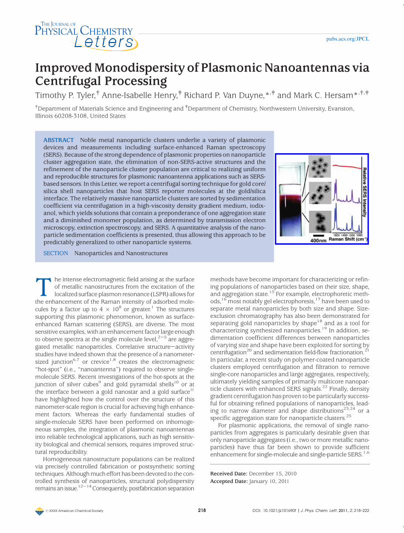
ABSTRACT Noble metal nanoparticle clusters underlie a variety of plasmonicdevices and measurements including surface-enhanced Raman spectroscopy(SERS). Because of the strong dependence of plasmonic properties on nanoparticlecluster aggregation state, the elimination of non-SERS-active structures and therefinement of the nanoparticle cluster population are critical to realizing uniformand reproducible structures for plasmonic nanoantenna applications such as SERS-based sensors. In this Letter, we report a centrifugal sorting technique for gold core/silica shell nanoparticles that host SERS reporter molecules at the gold/silicainterface. The relatively massive nanoparticle clusters are sorted by sedimentationcoefficient via centrifugation in a high-viscosity density gradient medium, iodix-anol, which yields solutions that contain a preponderance of one aggregation stateand a diminished monomer population, as determined by transmission electronmicroscopy, extinction spectroscopy, and SERS. A quantitative analysis of the nano-particle sedimentation coefficients is presented, thus allowing this approach to bepredictably generalized to other nanoparticle systems.
SECTION Nanoparticles and Nanostructures
T he intense electromagnetic field arising at the surfaceof metallic nanostructures from the excitation of thelocalized surface plasmon resonance (LSPR) allows for
the enhancement of the Raman intensity of adsorbed mole-cules by a factor up to 4 � 108 or greater.1 The structuressupporting this plasmonic phenomenon, known as surface-enhanced Raman scattering (SERS), are diverse. The mostsensitive examples,with anenhancement factor largeenoughto observe spectra at the single molecule level,2-5 are aggre-gated metallic nanoparticles. Correlative structure-activitystudies have indeed shown that the presence of a nanometer-sized junction6,7 or crevice1,8 creates the electromagnetic“hot-spot” (i.e., “nanoantenna”) required to observe single-molecule SERS. Recent investigations of the hot-spots at thejunction of silver cubes9 and gold pyramidal shells10 or atthe interface between a gold nanostar and a gold surface11
have highlighted how the control over the structure of thisnanometer-scale region is crucial for achieving high enhance-ment factors. Whereas the early fundamental studies ofsingle-molecule SERS have been performed on inhomoge-neous samples, the integration of plasmonic nanoantennasinto reliable technological applications, such as high sensitiv-ity biological and chemical sensors, requires improved struc-tural reproducibility.
Homogeneous nanostructure populations can be realizedvia precisely controlled fabrication or postsynthetic sortingtechniques. Althoughmucheffort has beendevoted to the con-trolled synthesis of nanoparticles, structural polydispersityremainsan issue.12-14Consequently, postfabricationseparation
methods have become important for characterizing or refin-ing populations of nanoparticles based on their size, shape,and aggregation state.15 For example, electrophoretic meth-ods,16 most notably gel electrophoresis,17 have been used toseparate metal nanoparticles by both size and shape. Size-exclusion chromatography has also been demonstrated forseparating gold nanoparticles by shape18 and as a tool forcharacterizing synthesized nanoparticles.19 In addition, se-dimentation coefficient differences between nanoparticlesof varying size and shape have been exploited for sorting bycentrifugation20 and sedimentation field-flow fractionation.21
In particular, a recent study on polymer-coated nanoparticleclusters employed centrifugation and filtration to removesingle-core nanoparticles and large aggregates, respectively,ultimately yielding samples of primarily multicore nanopar-ticle clusters with enhanced SERS signals.22 Finally, densitygradient centrifugation has proven to be particularly success-ful for obtaining refined populations of nanoparticles, lead-ing to narrow diameter and shape distributions23,24 or aspecific aggregation state for nanoparticle clusters.25
For plasmonic applications, the removal of single nano-particles from aggregates is particularly desirable given thatonly nanoparticle aggregates (i.e., two ormoremetallic nano-particles) have thus far been shown to provide sufficientenhancement for single-molecule and single-particle SERS.1,6
Received Date: December 15, 2010Accepted Date: January 10, 2011
rXXXX American Chemical Society 219 DOI: 10.1021/jz101690f |J. Phys. Chem. Lett. 2011, 2, 218–222
pubs.acs.org/JPCL
In particular, SERS nanoantennas consisting of aggregatedspherical gold cores encapsulated in a protective silica shellwith Raman reporter molecules adsorbed at the gold/silicainterface have been shown to be ideal SERS substrates,1,26
offering robustness and stability. The silica shell has theadded benefit that it directly enables dispersion in aqueoussolutions, thereby eliminating the need for additional chemicalfunctionalization.
Herewe report an effectivemethod for sorting SERS-activesilica-coated gold nanoparticle clusters by aggregation state ina high-viscosity density gradient. Whereas equilibrium iso-pycnic density gradient centrifugation techniques that arecommon for carbon-based nanomaterials27-32 are incompa-tible with high-density structures such as nanoparticle clus-ters, sorting can, in principle, be accomplished by sedimenta-tion coefficient in the transient centrifugal regime. However,in this study, the relatively large size of the gold cores (∼100 nmin diameter) combined with the silica shell (∼60 nm thick)makes sorting by transient motion challenging because thesehigh-mass structures will sediment significantly faster thansmaller nanoparticles. This issue is overcome by using thehigh-viscositydensity gradientmedium iodixanol,which slowsthe sedimentation of the gold/silica nanoparticle clusters tothe point where structurally distinct fractions can be col-lected following centrifugation. Through transmission elec-tron microscopy (TEM), extinction spectroscopy, and SERScharacterization, the collected fractions are found to possessa preponderance of one aggregation state and a diminishedmonomer population, thus yielding ideal SERS nanoanten-nas that can be directly employed in a variety of plasmonicapplications. In addition, the nanoparticle sedimentation co-efficients are quantitatively analyzed, which will facilitate theapplication of this approach to other nanoparticle systems.
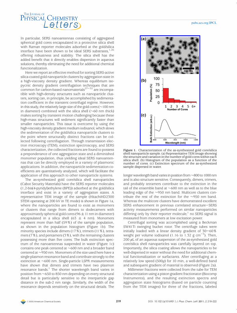
The as-synthesized gold core/silica shell nanoparticles(Cabot Security Materials) have the SERS reporter molecules(1,2-bis(4-pyridyl)ethylene (BPE)) adsorbed at the gold/silicainterface and exist in a variety of aggregation states. Arepresentative TEM image of the sample (HitachiHD2300STEM operating at 200 kV in TE mode) is shown in Figure 1a,where the nanoparticles are found to exist as monomersor clusters that range from dimers to dodecamers withapproximately spherical gold cores (96(11nm in diameter)encapsulated in a silica shell (63 ( 4 nm). Monomersrepresent more than half (59%) of the sample population,as shown in the population histogram (Figure 1b). Theminority species include dimers (17%), trimers (11%), tetra-mers (7%), and pentamers (3%), with the remaining clusterspossessing more than five cores. The bulk extinction spec-trum of the nanoantennas suspended in water (Figure 1c)contains one peak centered at∼600 nm and a broader bandcentered at∼950 nm.Monomers of the size used here have asingle plasmon resonanceband and contribute strongly to theextinction at ∼600 nm. Single-particle LSPR measurementshave shown that dimers and trimers have two plasmonresonance bands.1 The shorter wavelength band varies inposition from∼650 to 850 nmdepending on every structuraldetail but is particularly sensitive to the interparticle gapdistance in the sub-2 nm range. Similarly, the width of theresonance depends sensitively on the structural details. The
longerwavelengthbandvaries inposition from∼800 to1000nmand is also structure sensitive. Consequently, dimers, trimers,and probably tetramers contribute to the extinction in thetail of the ensemble band at ∼600 nm as well as to the blueleading edge of the ∼950 nm band. Multicore clusters con-tribute the rest of the extinction for the ∼950 nm band.Whereas the multicore clusters have demonstrated excellentSERS enhancement in previous correlated structure-SERSactivity measurements performed on similar nanoparticlesdiffering only by their reporter molecule,1 no SERS signal ismeasured from monomers at low excitation power.
Centrifugal sorting was accomplished using a BeckmanSW41Ti swinging bucket rotor. The centrifuge tubes wereinitially loaded with a linear density gradient of 30-60%weight per volume iodixanol (1.16 to 1.32 g cm-3). Then,200 μL of an aqueous suspension of the as-synthesized goldcore/silica shell nanoparticles was carefully layered on top.Importantly, the silica coating allows the nanoparticles to bewell-dispersed inwater without the need for additional chem-ical functionalization or surfactants. After centrifuging at arelatively low speed (500g) for 10 min, a well-defined bandand subsequent gradient of material is observed (Figure 2a).
Millimeter fractions were collected from the tube for TEMcharacterization using a piston gradient fractionator (BiocompInstruments), and the resulting extinction spectra andaggregation state histograms (based on particle countingfrom the TEM images) for three of the fractions, labeled
Figure 1. Characterization of the as-synthesized gold core/silicashell nanoparticle sample. (a) Representative TEM image showingthe structure and variation in the numberof gold coreswithin eachsilica shell. (b) Histogram of the population as a function of thenumber of cores. (c) Extinction spectrum of the as-synthesizedsample dispersed in water.
rXXXX American Chemical Society 220 DOI: 10.1021/jz101690f |J. Phys. Chem. Lett. 2011, 2, 218–222
pubs.acs.org/JPCL
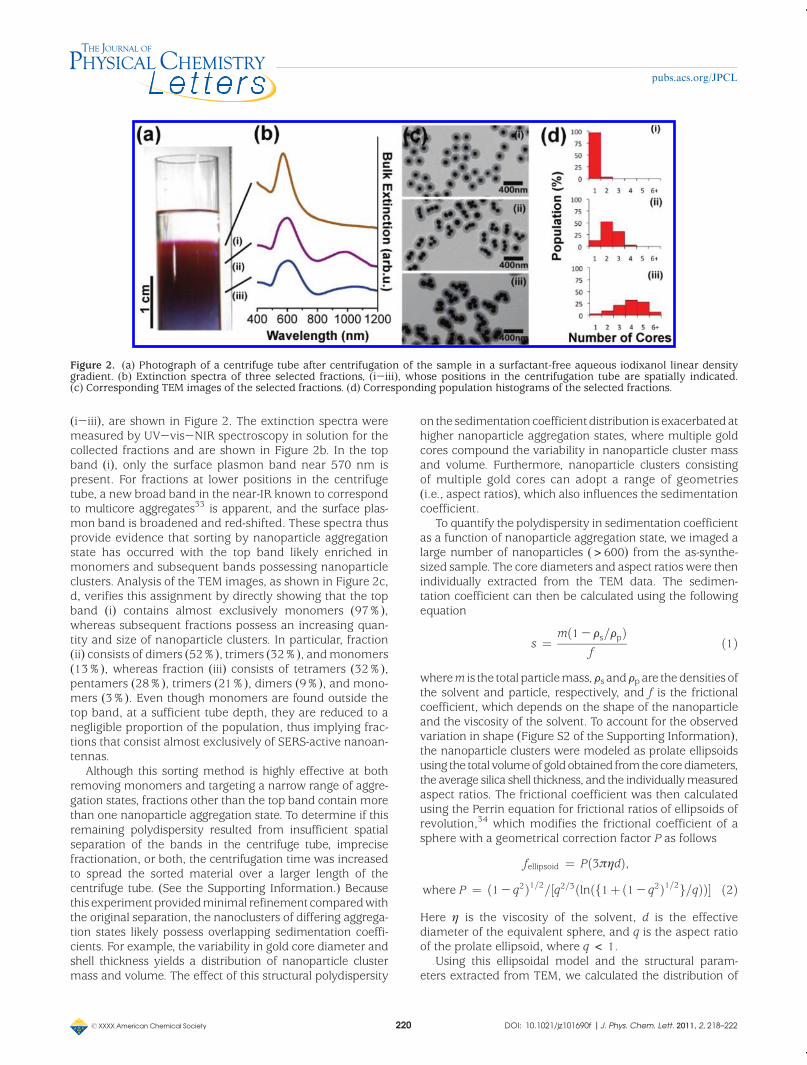
(i-iii), are shown in Figure 2. The extinction spectra weremeasured by UV-vis-NIR spectroscopy in solution for thecollected fractions and are shown in Figure 2b. In the topband (i), only the surface plasmon band near 570 nm ispresent. For fractions at lower positions in the centrifugetube, a new broad band in the near-IR known to correspondto multicore aggregates33 is apparent, and the surface plas-mon band is broadened and red-shifted. These spectra thusprovide evidence that sorting by nanoparticle aggregationstate has occurred with the top band likely enriched inmonomers and subsequent bands possessing nanoparticleclusters. Analysis of the TEM images, as shown in Figure 2c,d, verifies this assignment by directly showing that the topband (i) contains almost exclusively monomers (97%),whereas subsequent fractions possess an increasing quan-tity and size of nanoparticle clusters. In particular, fraction(ii) consists of dimers (52%), trimers (32%), andmonomers(13%), whereas fraction (iii) consists of tetramers (32%),pentamers (28%), trimers (21%), dimers (9%), and mono-mers (3%). Even though monomers are found outside thetop band, at a sufficient tube depth, they are reduced to anegligible proportion of the population, thus implying frac-tions that consist almost exclusively of SERS-active nanoan-tennas.
Although this sorting method is highly effective at bothremoving monomers and targeting a narrow range of aggre-gation states, fractions other than the top band contain morethan one nanoparticle aggregation state. To determine if thisremaining polydispersity resulted from insufficient spatialseparation of the bands in the centrifuge tube, imprecisefractionation, or both, the centrifugation time was increasedto spread the sorted material over a larger length of thecentrifuge tube. (See the Supporting Information.) Becausethis experimentprovidedminimal refinement comparedwiththe original separation, the nanoclusters of differing aggrega-tion states likely possess overlapping sedimentation coeffi-cients. For example, the variability in gold core diameter andshell thickness yields a distribution of nanoparticle clustermass and volume. The effect of this structural polydispersity
on the sedimentation coefficientdistribution is exacerbatedathigher nanoparticle aggregation states, where multiple goldcores compound the variability in nanoparticle cluster massand volume. Furthermore, nanoparticle clusters consistingof multiple gold cores can adopt a range of geometries(i.e., aspect ratios), which also influences the sedimentationcoefficient.
To quantify the polydispersity in sedimentation coefficientas a function of nanoparticle aggregation state, we imaged alarge number of nanoparticles (>600) from the as-synthe-sized sample. The core diameters and aspect ratios were thenindividually extracted from the TEM data. The sedimen-tation coefficient can then be calculated using the followingequation
s ¼ mð1-Fs=FpÞf
ð1Þ
wherem is the total particlemass,Fs andFp are thedensities ofthe solvent and particle, respectively, and f is the frictionalcoefficient, which depends on the shape of the nanoparticleand the viscosity of the solvent. To account for the observedvariation in shape (Figure S2 of the Supporting Information),the nanoparticle clusters were modeled as prolate ellipsoidsusing the total volumeofgold obtained fromthe corediameters,the average silica shell thickness, and the individuallymeasuredaspect ratios. The frictional coefficient was then calculatedusing the Perrin equation for frictional ratios of ellipsoids ofrevolution,34 which modifies the frictional coefficient of asphere with a geometrical correction factor P as follows
fellipsoid ¼ Pð3πηdÞ,where P ¼ ð1- q2Þ1=2=½q2=3ðlnðf1þð1- q2Þ1=2g=qÞÞ� ð2Þ
Here η is the viscosity of the solvent, d is the effectivediameter of the equivalent sphere, and q is the aspect ratioof the prolate ellipsoid, where q < 1.
Using this ellipsoidal model and the structural param-eters extracted from TEM, we calculated the distribution of
Figure 2. (a) Photograph of a centrifuge tube after centrifugation of the sample in a surfactant-free aqueous iodixanol linear densitygradient. (b) Extinction spectra of three selected fractions, (i-iii), whose positions in the centrifugation tube are spatially indicated.(c) Corresponding TEM images of the selected fractions. (d) Corresponding population histograms of the selected fractions.
rXXXX American Chemical Society 221 DOI: 10.1021/jz101690f |J. Phys. Chem. Lett. 2011, 2, 218–222
pubs.acs.org/JPCL
sedimentation coefficients for the as-synthesized sample at afixed point (40% w/v iodixanol, 1.21 g cm-3) in the densitygradient35 (Figure 3). This model reveals the presence of amonomer band at low sedimentation coefficients, followedby a gap in sedimentation coefficient, and finally overlappingaggregation states at higher sedimentation coefficients. Con-sequently, transient centrifugal sorting is expected to yieldhighly enriched monomers at the top of the centrifuge tubeand then increasing but overlapping levels of nanoparticleaggregation for subsequent fractions, in agreement with theexperimental results. This model thus holds promise forevaluating the feasibility of and refining the experimentalconditions for sorting other nanoparticle clusters via transientdensity gradient centrifugation techniques, assuming that theinitial nanoparticle structural parameters andpolydispersity havebeen determined. Conversely, the results of transient densitygradient centrifugation experiments can provide quantitativeinsight into the structural polydispersity of nanoparticle samples.
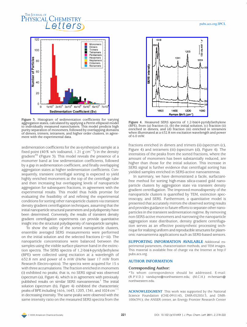
To show the utility of the sorted nanoparticle clusters,ensemble averaged SERS measurements were performedon the initial solution and the selected fractions (i-iii). Thenanoparticle concentrations were balanced between thesamples using the visible surface plasmon band in the extinc-tion spectra. The SERS spectra of 1,2-bis(4-pyridyl)ethylene(BPE) were collected using excitation at a wavelength of632.8 nm and power of 6 mW (HeNe laser 17 mW fromResearch Electro-optics). The spectra were acquired for 15 swith threeaccumulations. The fractionenriched inmonomers(i) exhibited no peaks; that is, no SERS signal was observed(spectrum (a), Figure 4), which is in agreement with previouslypublished results on similar SERS nanoantennas.1 The initialsolution (spectrum (b), Figure 4) exhibited the characteristicpeaks of BPE including1616, 1643, 1203, 1341, and1024cm-1
in decreasing intensity. The same peaks were observedwith thesame intensity ratio on themeasured SERS spectra from the
fractions enriched in dimers and trimers (ii) (spectrum (c),Figure 4) and tetramers (iii) (spectrum (d), Figure 4). Theintensities of the peaks from the sorted fractions, where theamount of monomers has been substantially reduced, arehigher than those for the initial solution. This increase inSERS signal is further evidence that centrifugal sorting hasyielded samples enriched in SERS-active nanoantennas.
In summary, we have demonstrated a facile, surfactant-free method for sorting high-mass silica-coated gold nano-particle clusters by aggregation state via transient densitygradient centrifugation. The improved monodispersity of thenanoparticle clusters is quantified by TEM, extinction spec-troscopy, and SERS. Furthermore, a quantitative model ispresented that accuratelymirrors the observed sorting resultsandprovides guidance to future efforts to separate other nano-particles in the transient sedimentation regime. By removingnon-SERS-activemonomers and narrowing the nanoparticleaggregation state distribution, density gradient centrifuga-tion serves as an effective postsynthetic processing tech-nique for realizinguniformand reproducible structures for plasm-onic nanoantenna applications such as SERS-based sensors.
SUPPORTING INFORMATION AVAILABLE Additional ex-perimental parameters, characterization methods, and TEM images.This material is available free of charge via the Internet at http://pubs.acs.org.
AUTHOR INFORMATION
Corresponding Author:*To whom correspondence should be addressed. E-mail:(R.P.V.D.) [email protected]; (M.C.H.) [email protected].
ACKNOWLEDGMENT This work was supported by the NationalScience Foundation (CHE-0911145, DMR-0520513, and DMR-1006391); the ANSER center, an Energy Frontier Research Center
Figure 3. Histogram of sedimentation coefficients for varyingaggregation states, calculated by applying a Perrin ellipsoid modelto individually measured nanoclusters. This model predicts highpurity separation of monomers, followed by overlapping domainsof dimers, trimers, tetramers, and higher order clusters, in agree-ment with the experimental data.
Figure 4. Measured SERS spectra of 1,2-bis(4-pyridyl)ethylene(BPE), from (a) fraction (i), (b) the initial solution, (c) fraction (ii)enriched in dimers, and (d) fraction (iii) enriched in tetramerswhen illuminated at a 632.8 nm excitation wavelength and powerof 6.0 mW.

rXXXX American Chemical Society 222 DOI: 10.1021/jz101690f |J. Phys. Chem. Lett. 2011, 2, 218–222
pubs.acs.org/JPCL
funded by the U.S. Department of Energy (DE-SC0001059); andAFSOR/DARPA Project BAA07-61 (FA9550-08-1-0221). We thankDr. R. Griff Freeman and Dr. Michael J. Natan from Cabot SecurityMaterials for providing the SERS nanoantenna samples. STEMexperiments were performed in the EPIC Facility of the NUANCECenter at Northwestern University. NUANCE is supported by theNSF-NSEC, NSF-MRSEC, Keck Foundation, State of Illinois, andNorthwestern University.
REFERENCES
(1) Wustholz, K. L.; Henry, A.-I.; McMahon, J. M.; Freeman, R. G.;Valley, N.; Piotti, M. E.; Natan, M. J.; Schatz, G. C.; Van Duyne,R. P. Structure-Activity Relationships in Gold NanoparticleDimers and Trimers for Surface-Enhanced Raman Spectros-copy. J. Am. Chem. Soc. 2010, 132, 10903–10910.
(2) Kneipp, K.; Wang, Y.; Kneipp, H.; Perelman, L. T.; Itzkan, I.;Dasari, R. R.; Feld, M. S. Single Molecule Detection UsingSurface-Enhanced Raman Scattering (SERS). Phys. Rev. Lett.1997, 78, 1667–1670.
(3) Nie, S. M.; Emory, S. R. Probing Single Molecules and SingleNanoparticles by Surface-Enhanced Raman Scattering.Science 1997, 275, 1102–1106.
(4) LeRu, E. C.; Meyer, M.; Etchegoin, P. G. Proof of Single-Molecule Sensitivity in Surface Enhanced Raman Scattering(SERS) byMeans of a Two-Analyte Technique. J. Phys. Chem. B2006, 110, 1944–1948.
(5) Dieringer, J. A.; Lettan, R. B., II; Scheidt, K. A.; VanDuyne, R. P.A Frequency Domain Existence Proof of Single-MoleculeSurface-Enhanced Raman Spectroscopy. J. Am. Chem. Soc.2007, 129, 16249–16256.
(6) Michaels, A. M.; Jiang, J.; Brus, L. Ag Nanocrystal Junctionsas the Site for Surface-Enhanced Raman Scattering of SingleRhodamine 6G Molecules. J. Phys. Chem. B 2000, 104, 11965–11971.
(7) Camden, J. P.; Dieringer, J. A.; Wang, Y.; Masiello, D. J.; Marks,L. D.; Schatz, G. C.; Van Duyne, R. P. Probing the Structure ofSingle-Molecule Surface-Enhanced Raman Scattering HotSpots. J. Am. Chem. Soc. 2008, 130, 12616–12617.
(8) Moskovits, M.; Jeong, D. H. Engineering Nanostructures forGiant Optical Fields. Chem. Phys. Lett. 2004, 397, 91–95.
(9) Rycenga, M.; Camargo, P. H. C.; Weiyang, L.; Moran, C. H.;Xia, Y. Understanding the SERS Effects of Single Nanoparti-cles and Their Dimers, One at a Time. J. Phys. Chem. Lett.2010, 1, 696–703.
(10) Stoerzinger, K. A.; Hasan,W.; Lin, J. Y.; Robles, A.; Odom, T.W.Screening Nanopyramid Assemblies to Optimize SurfaceEnhanced Raman Scattering. J. Phys. Chem. Lett. 2010, 1,1046–1050.
(11) Alvarez-Puebla, R.; Liz-Marz�an, L. M.; García de Abajo, F. J.Light Concentration at the Nanometer Scale. J. Phys. Chem.Lett. 2010, 1, 2428–2434.
(12) Jana, N. R.; Gearheart, L.; Murphy, C. J. Wet Chemical Synthe-sis of High Aspect Ratio Cylindrical Gold Nanorods. J. Phys.Chem. B 2001, 105, 4065–4067.
(13) Xia, Y.; Xiong, Y.; Lim, B.; Skrabalak, S. E. Shape-ControlledSynthesis of Metal Nanocrystals: Simple Chemistry MeetsComplex Physics? Angew. Chem., Int. Ed. 2009, 48, 60–103.
(14) Wiley, B.; Sun, Y.;Mayers, B.; Xia, Y. Shape-Controlled Synthe-sis of Metal Nanostructures: The Case of Silver. Chem.;Eur. J.2005, 11, 454–463.
(15) Liu, F.-K. Analysis and Applications of Nanoparticles in theSeparation Sciences: A Case of Gold Nanoparticles. J. Chro-matogr. A 2009, 1216, 9034–9047.
(16) Hanauer, M.; Pierrat, S.; Zins, I.; Lotz, A.; S€onnichsen, C.Separation ofNanoparticles byGel Electrophoresis Accordingto Size and Shape. Nano Lett. 2007, 7, 2881–2885.
(17) Surugau, N.; Urban, P. L. Electrophoretic Methods for Separa-tion of Nanoparticles. J. Sep. Sci. 2009, 32, 1889–1906.
(18) Wei, G. T.; Liu, F. K.; Wang, C. R. C. Shape Separation ofNanometer Gold Particles by Size-Exclusion Chromatogra-phy. Anal. Chem. 1999, 71, 2085–2091.
(19) Liu, F.-K. SEC Characterization of Au Nanoparticles Preparedthrough Seed-Assisted Synthesis. Chromatographia 2007, 66,791–796.
(20) Sharma, V.; Park, K.; Srinivasarao, M. Shape Separation ofGold Nanorods Using Centrifugation. Proc. Natl. Acad. Sci.U.S.A. 2009, 106, 4981–4985.
(21) Contado, C.; Argazzi, R. Size Sorting of Citrate Reduced GoldNanoparticles by Sedimentation Field-Flow Fractionation.J. Chromatogr. A 2009, 1216, 9088–9098.
(22) Braun, G. B.; Lee, S. J.; Laurence, T.; Fera, N.; Fabris, L.; Bazan,G. C.; Moskovits, M.; Reich, N. O. Generalized Approach toSERS-Active Nanomaterials via Controlled Nanoparticle Link-ing, Polymer Encapsulation, and Small-Molecule Infusion.J. Phys. Chem. C 2009, 113, 13622–13629.
(23) Sun, X.; Tabakman, S. M.; Seo, W.-S.; Zhang, L.; Zhang, G.;Sherlock, S.; Bai, L.; Dai, H. Separation of Nanoparticles in aDensity Gradient: FeCo@C and Gold Nanocrystals. Angew.Chem., Int. Ed. 2009, 48, 939–942.
(24) Bai, Lu.; Ma, X.; Liu, J.; Sun, X.; Zhao, D.; Evans, D. E. RapidSeparation and Purification of Nanoparticles in OrganicDensity Gradients. J. Am. Chem. Soc. 2010, 132, 2333–2337.
(25) Chen, G.; Wang, Y.; Tan, L. H.; Yang, M.; Tan, L. S.; Chen, Y.;Chen, H. High-Purity Separation of Gold Nanoparticle Dimersand Trimers. J. Am. Chem. Soc. 2009, 131, 4218–4219.
(26) Doering,W. E.; Piotti, M. E.; Natan,M. J.; Freeman, R. G. SERSas a Foundation for Nanoscale, Optically Detected BiologicalLabels. Adv. Mater. 2007, 19, 3100–3108.
(27) Arnold, M. S.; Green, A. A.; Hulvat, J. F.; Stupp, S. I.; Hersam,M. C. Sorting Carbon Nanotubes by Electronic Structure UsingDensity Differentiation. Nat. Nanotechnol. 2006, 1, 60–65.
(28) Hersam,M. C. Progress TowardsMonodisperse Single-WalledCarbon Nanotubes. Nat. Nanotechnol. 2008, 3, 387–394.
(29) Green, A. A.; Hersam, M. C. Solution Phase Production ofGraphene with Controlled Thickness via Density Differentia-tion. Nano Lett. 2009, 9, 4031–4036.
(30) Green, A. A.; Hersam,M. C. EmergingMethods for ProducingMonodisperse Graphene Solutions. J. Phys. Chem. Lett. 2010,1, 544–549.
(31) Green, A. A.; Hersam, M. C. Processing and Properties ofHighly Enriched Double-Wall Carbon Nanotubes. Nat. Nano-technol. 2009, 4, 64–70.
(32) Liu, L.; Hersam, M. C. Recent Developments in CarbonNanotube Sorting and Selective Growth. MRS Bull. 2010,35, 315–321.
(33) Norman, T. J., Jr.; Grant, C. D.; Magana, D.; Zhang, J. Z. NearInfraredOptical Absorption of Gold Nanoparticle Aggregates.J. Phys. Chem. B 2002, 106, 7005–7012.
(34) Perrin, F. Mouvement Brownien d'un Ellipsoide (II). RotationLibre et D�epolarisation des Fluorescences. Translation etDiffusion de Mol�ecules Ellipsoidales. J. Phys. Radium 1936,7, 1–11.
(35) Eivindvik, K.; Sjøgren, C. E. Physicochemical Properties ofIodixanol. Acta Radiol. Suppl. 1995, 399, 32–38.
![Periodic Plasmonic Nanoantennas in a Piecewise …...Although the concept of nanoantenna has been around since 1985 [1], it is ... monopoles, dipoles and bowties to Yagi-Uda nanoantennas](https://img.pdfslide.us/doc/110x75/5f4f21a83bde496e35386ec9/periodic-plasmonic-nanoantennas-in-a-piecewise-although-the-concept-of-nanoantenna.jpg)